


Abstract
A novel Dy3+-doped self-activated Sr2NaMg2V3O12 (SNMVO) phosphor was synthesized via a high-temperature solid-state reaction method. Its microstructure, surface morphology, valence state, and luminescence properties were investigated. The results showed that the prepared phosphor exhibited bright green emission at 521 nm and yellow emission at 575 nm under 345 nm excitation. The luminescence intensity showed a strong concentration dependence, with an optimal Dy3+ ion doping concentration of 0.05 mol, and the concentration quenching (CQ) mechanism was dipole–dipole (d-d) interaction. Energy transfer between vanadate and Dy3+ was observed, with a maximum transfer efficiency of 63.5%. The thermal activation energy (0.1859 eV) indicated good thermal stability. Furthermore, this phosphor can be used as a yellow phosphor for white light-emitting diodes (wLEDs) and for anti-counterfeiting patterns.
1. Introduction
Against the backdrop of dual carbon emissions [1], white LEDs have gradually become one of the core technologies of the new generation of lighting, thanks to their high energy efficiency, long service life, and environmental friendliness [2,3]. Currently, commercial WLEDs primarily use blue LED chips to excite YAG:Ce3+ yellow phosphors, resulting in an insufficient red light component [4,5]. This issue easily leads to excessively high color temperature and a low color rendering index, thus failing to meet high-quality lighting requirements. Based on the aforementioned limitations, a possible and effective approach is to use ultraviolet or near-ultraviolet (n-UV) chips to excite red, green, and blue phosphors to achieve white light [6]. Vanadate, as a novel phosphor matrix, has been extensively studied based on commercially available red-emitting YVO4:Eu3+ phosphors [7]. Furthermore, in the near-ultraviolet region of the excitation spectrum, vanadate exhibits a broad absorption band due to strong charge-transfer (CT) transitions, and also demonstrates broadband emission in the 400–700 nm range. For phosphors, besides rare earth ions, the choice of matrix material is also crucial. The matrix system should possess characteristics such as a large band gap, low phonon energy, and the ability to effectively transfer energy between luminescent centers. Among numerous matrices, vanadate matrices are renowned for their strong absorption and self-activated luminescence properties, attributed to the 3T1,2→1A1 transition. Rare earth ions exhibit low absorption in the ultraviolet region due to the narrow excitation of the forbidden 4f–4f transition. Therefore, rare earth-activated vanadate matrix structures facilitate phonon-assisted energy transfer from VO43− to RE3+. Among vanadate systems, alkaline earth metal vanadates possess excellent optical properties, thermal stability, and high crystallinity. Furthermore, multi-cation alkaline earth metal vanadates exhibit good self-activated luminescence properties, thus attracting widespread attention from researchers. A. A. Setlur et al. investigated the luminescent properties of trivalent europium-activated vanadate garnet (Ca2NaMg2V3O12) and studied it as a potential phosphor for ultraviolet light-emitting diodes (UV-LEDs) [8]. This single phosphor can convert the ultraviolet light from UV-LEDs into white light with good brightness and color rendering index. Subsequently, A.R. Dhobale et al. synthesized the vanadate garnet system Ca2NaMg2V3O12 using a solid-state reaction method, and doped it with Dy3+ and Sm3+ respectively, confirming that this single phosphor can convert the ultraviolet light of an ultraviolet LED into white light [9]. Yujun Liang et al. achieved adjustable emission color of Ca2NaMg2V3O12: Sm3+ phosphor, ranging from green to white, by controlling the Sm3+ doping concentration. Furthermore, the color rendering index increased from 55 to 83, revealing the significant application potential of this white phosphor in the field of white LEDs [10]. In addition, numerous matrix materials such as Ca2KZn2(VO4)3 [11,12], Na2YMg2(VO4)3 [13,14], Sr2NaMg2V3O12 [15,16] and Na2GdMg2(VO4)3 [17,18] have been extensively studied.
Trivalent rare earth ions, such as Dy3+, Sm3+, Eu3+, and Tm3+, exhibit multi-band emission from the ultraviolet to the infrared region, originating from 4f–4f electron transitions [19]. Therefore, rare earth ions are often used as activators in optical materials. Among them, Dy3+ ions emit blue and yellow light under near-ultraviolet excitation, making them attractive for material development in the field of white LED lighting [20]. In addition, the interaction between the vanadate ion VO43− and Dy3+ mentioned earlier also produces novel phenomena [21,22], which is also the motivation for this research.
In this study, a series of Sr2NaMg2V3O12 (SNMVO) phosphors with different concentrations of Dy3+ single doping were prepared using a high-temperature solid-state reaction method. Their luminescence properties, energy transfer mechanism from VO43− to Dy3+, thermal stability, electroluminescence of LED device prototypes, and anti-counterfeiting applications were systematically investigated.
2. Materials Synthesis and Characterization
2.1. Material Synthesis
The Sr2−xNaMg2V3O12: xDy3+ (SNMVO: xDy3+, x = 0, 0.01–0.2 mol) phosphors were prepared via a conventional high-temperature solid-state reaction method. Initially, stoichiometric amounts of starting materials, including SrCO3 (99.99%, Aladdin, Shanghai Aladdin Co., Ltd., Shanghai, China), Na2CO3 (99.99%, Aladdin), (MgCO3)4·Mg(OH)2·5H2O (99.9%, Aladdin), NH4VO3 (>99.9% metals basis, Aladdin), and Dy2O3 (99.99%, Aladdin), were precisely weighed. These precursors were then thoroughly co-ground in an agate mortar for 30 min to ensure homogeneity. Subsequently, the resultant mixture was transferred into a muffle furnace (KSL-1100X-S, Hefei Kejing Material Technology Co., Ltd., Hefei, China) and calcined at 900 °C for 8 h under an ambient air atmosphere. After naturally cooling to room temperature, the synthesized products were pulverized into fine powders for further characterization.
2.2. Materials Characterization
X-ray diffraction (XRD, SmartLab, Rigaku 9 kW, Tokyo, Japan) was employed to verify the phase composition of the as-prepared samples. The microscopic morphology and elemental distribution were investigated via scanning electron microscopy (SEM, Apreo SHiVoc, Waltham, MA, USA) coupled with energy-dispersive spectrometry (EDS; Octane Elect Super, Ametex EDAX, Mahwah, NJ, USA). Surface chemical states and functional groups were examined by X-ray photoelectron spectroscopy (XPS, Thermo Fisher EscaLab 250Xi, Waltham, MA, USA) and Fourier transform infrared spectroscopy (FTIR, Bruker tensor 37, Bruker, Billerica, MA, USA), respectively. Photoluminescence (PL) characteristics were monitored using a fluorescence spectrometer (FluorMax-4, FL-QM, Horiba, Tokyo, Japan). Furthermore, temperature-dependent PL spectra were recorded from 298 to 473 K with a step of 25 K, utilizing a TAP-02 high-temperature fluorescence controller (Tianjin Dongfang Kejie Technology Co., Ltd., Tianjin, China) to evaluate thermal stability.
3. Results and Discussion
3.1. Structure Analysis
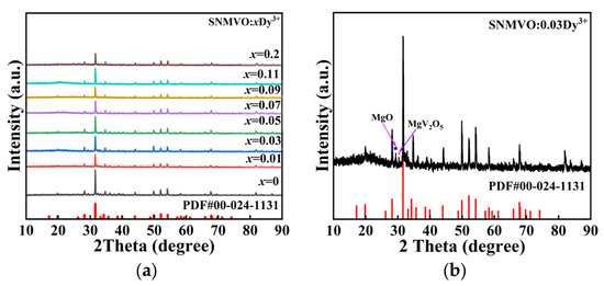
To confirm the phase purity, XRD patterns of SNMVO: xDy3+ (x = 0~0.2 mol) were analyzed, as depicted in Figure 1a. All the doped specimens exhibit diffraction features in the range of 10–90° that conform to the standard SNMVO card (PDF#00-024-1131) [23]. The localized scan from 25° to 30° in Figure 1b reveals secondary phases related to Mg and V oxides, which are identified as MgO (29°) and MgV2O5 (30°). The sharp and distinct nature of these diffraction lines implies a high degree of crystallinity and validates the successful entry of Dy3+ ions into the host.
Figure 1.
XRD patterns of (a) SNMVO: xDy3+ (x = 0~0.2 mol) phosphors, (b) SNMVO: 0.03Dy3+.
3.2. Morphology and Elemental Composition
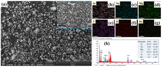
As shown in Figure 2, the SEM images revealed that the sample consisted of irregularly shaped and distinctly aggregated microparticles. This morphology is closely related to the extensive grain growth and particle fusion during the high-temperature solid-state synthesis process. The existence of Sr, Na, Mg, V, O, and Dy components in the as-prepared sample was explicitly demonstrated by the respective EDS elemental mapping results. All the constituent elements were uniformly distributed within selected particle regions, confirming thorough elemental mixing.
Figure 2.
(a) SEM images [inserted figure shows the area for EDS analysis]. (b–g) SEM-EDS mapping. (h) EDS spectrum of SNMVO:0.05 Dy3+ phosphor [inset shows the element percentages].
3.3. X-Ray Photoelectron Spectroscopy Analysis
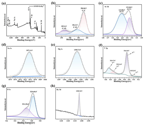
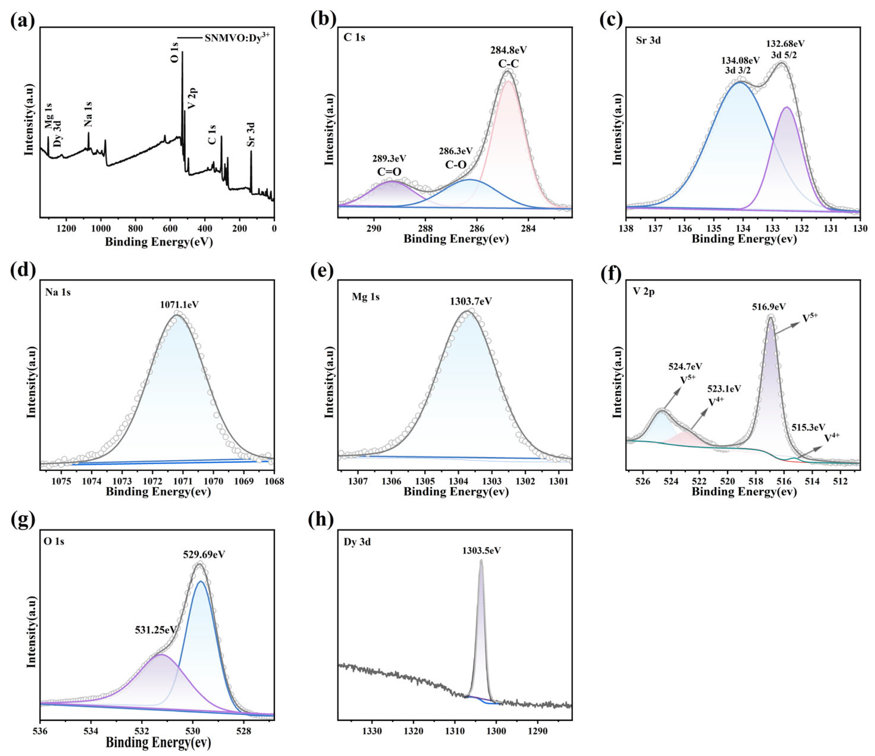
The chemical composition and electronic state of the SNMVO:0.05Dy3+ system were analyzed by XPS. The survey spectrum measured in the binding energy range of 0–1350 eV is shown in Figure 3a. Characteristic peaks corresponding to Sr, Na, Mg, V, O, and Dy are clearly observed, along with a C 1s signal used for charge correction (calibrated to 284.8 eV). High-resolution XPS spectra of C 1s, Sr 3d, Na 1s, Mg 1s, V 2p, O 1s, and Dy 3d are presented in Figure 3b–h, respectively.
Figure 3.
(a) XPS survey spectrum of the SNMVO: 0.05Dy3+ phosphor in the range of 0–1350 eV, (b–h) High-resolution XPS spectra of C 1s, Sr 3d, Na 1s, Mg 1s, V 2p, O 1s, and Dy 3d elements in the sample, respectively.
Figure 3b displays the C 1s spectrum. The binding energy of the C 1s peak is determined to be 284.8 eV, which serves as the reference for charge correction. Figure 3c presents the Sr 3d core-level spectrum. It consists of two peaks located at 132.68 eV and 134.08 eV, corresponding to Sr 3d5/2 and Sr 3d3/2, respectively, indicating the presence of Sr2+ [24]. Figure 3d shows the Na 1s spectrum, which exhibits a single symmetric peak at 1071.1 eV, characteristic of Na+ ions in an oxide matrix [25]. Figure 3e displays the Mg 1s spectrum, where a single symmetric peak is observed at 1303.7 eV, confirming the divalent state of magnesium (Mg2+) [26,27,28]. The V 2p spectrum is shown in Figure 3f. Due to spin–orbit splitting, two characteristic peaks corresponding to V 2p3/2 and V 2p1/2 are observed. After further peak deconvolution, the V 2p3/2 region exhibits two components at 515.3 eV and 516.9 eV, while the V 2p1/2 region shows two components at 523.1 eV and 524.7 eV. This indicates that vanadium predominantly exists in the pentavalent (V5+) oxidation state, accompanied by a minor contribution from a lower oxidation state (V4+) [29,30]. The O 1s spectrum (Figure 3g) displays an asymmetric peak that can be deconvoluted into two components: a dominant peak at 529.69 eV attributed to lattice oxygen (O2−) in the VO43− groups, and a weaker peak at 531.25 eV associated with surface-adsorbed oxygen species (e.g., hydroxyl groups or carbonate) [31]. Figure 3h shows the spectrum of Dy 3d present in the sample, which exhibits a wide peak at 1303.5 eV. This peak can be assigned to the bonded dysprosium–oxygen (Dy–O bond) of Dy 3d5/2 species. This confirms the presence of Dy in the +3 ionic state in the phosphor [25]. No additional impurity elements are detected in the XPS analysis, further confirming the high purity of the synthesized phosphor.
3.4. FTIR Spectroscopy Analysis
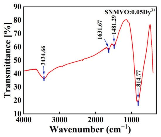
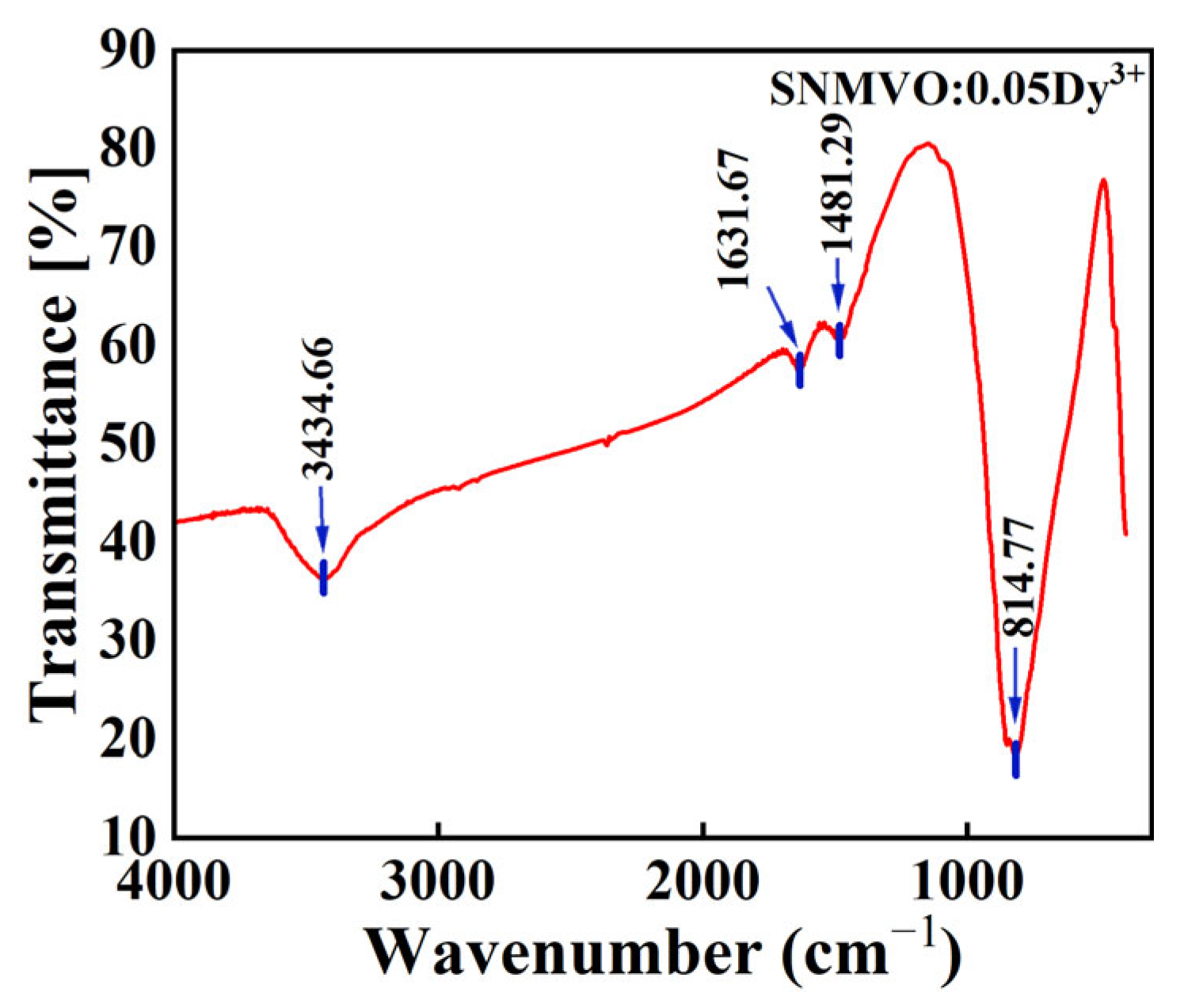
FTIR spectra were collected in the range of 4000–400 cm−1 to characterize the functional groups of SNMVO: 0.05Dy3+ phosphor is shown in Figure 4. The absorption bands at 3434.66, 1631.67 and 814.77 cm−1 are attributed to O–H stretching vibration of adsorbed water [32,33], H–O–H bending vibration, and V–O stretching vibration of [VO4]3− tetrahedra [34], respectively. A weak peak at 1481.29 cm−1 originates from residual carbonate ions [35]. The C 1s peak at 284.8 eV corresponds to adventitious carbon, which was standardly utilized for charge correction. The weak peak at 1481.29 cm−1 is assigned to trace surface carbonate residues, which, due to its low intensity, does not affect the matrix crystal structure.
Figure 4.
Fourier transform infrared spectroscopy (FTIR) of SNMVO: 0.05Dy3+ sample.
3.5. Photoluminescence Analysis
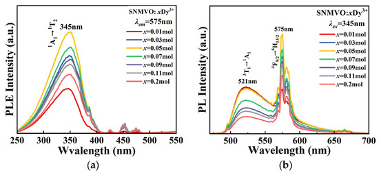
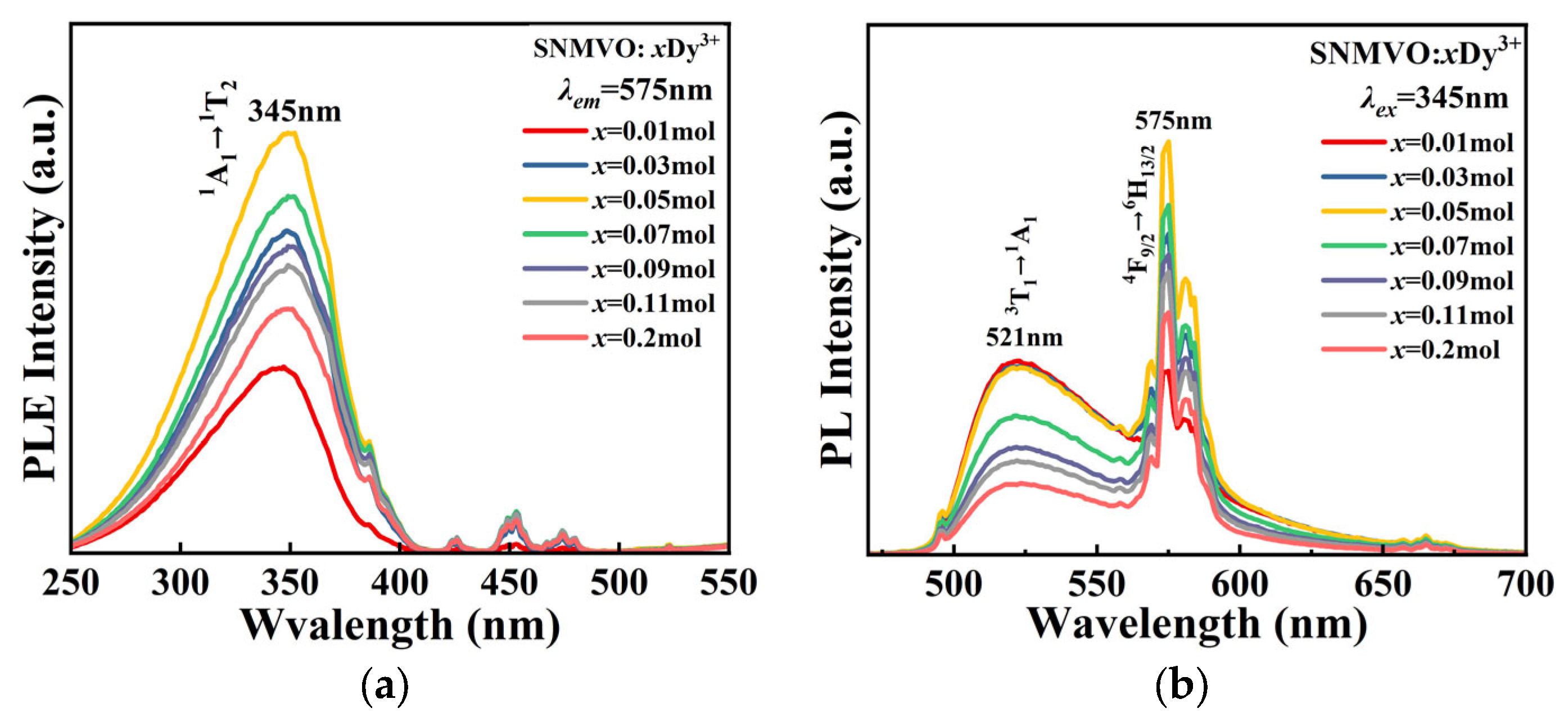
The photoluminescence excitation (PLE) spectrum of the SNMVO: xDy3+ sample is shown in Figure 5a. Characteristic absorption peaks of VO43− groups are detected in the range of 250–500 nm, corresponding to the transition of 1A1→1T2 at 345 nm. The stronger emission at 521 nm and 575 nm corresponds to the transitions from energy level 3T1 to 1A1 and from 4F9/2 to 6H13/2, respectively. The former is attributed to vanadate ions (VO43−), and the latter to Dy3+ ions in Figure 2b.
Figure 5.
(a) PLE and (b) PL of the SNMVO: xDy3+ samples.
3.5.1. Concentration Quenching Analysis
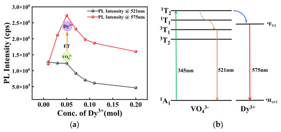
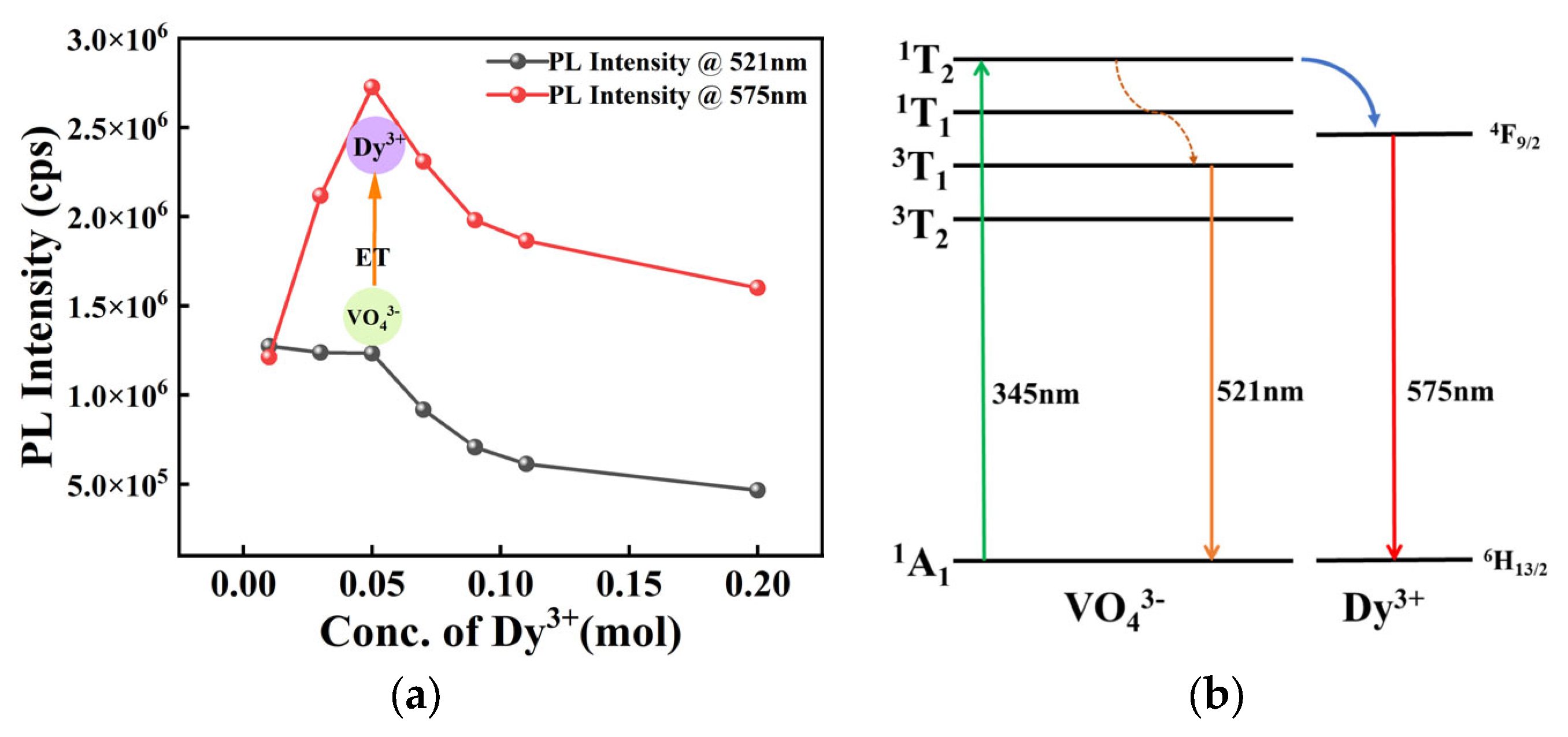
Figure 6a shows the variation in the emission intensity of the strongest emission peak (575 nm) with the Dy3+ ion concentration. With increasing doping concentration, the emission intensity gradually increases, reaching a maximum at a doping concentration of x = 0.05 mol, and then decreases, exhibiting concentration quenching during this process.
Figure 6.
(a) The PL intensity varies with the concentration of Dy3+ and (b) energy level diagram demonstrating the energy transfer process between VO43− and Dy3+.
The decrease in fluorescence intensity due to concentration quenching and the non-radiative energy transfer between Dy3+ ions can be attributed to three factors: exchange interactions, radiative reabsorption, and electric multipole-electric multipole interactions. Dexter theory is typically used to analyze the concentration quenching mechanism.
where C is a coefficient, and = 6, 8, and 10 correspond to dipole–dipole, dipole–quadrupole, and quadrupole–quadrupole interactions, respectively.
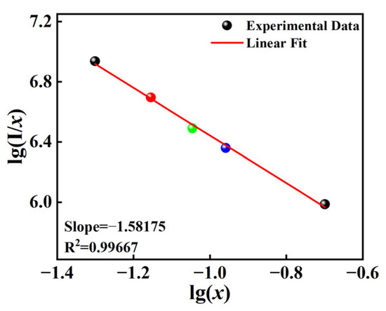
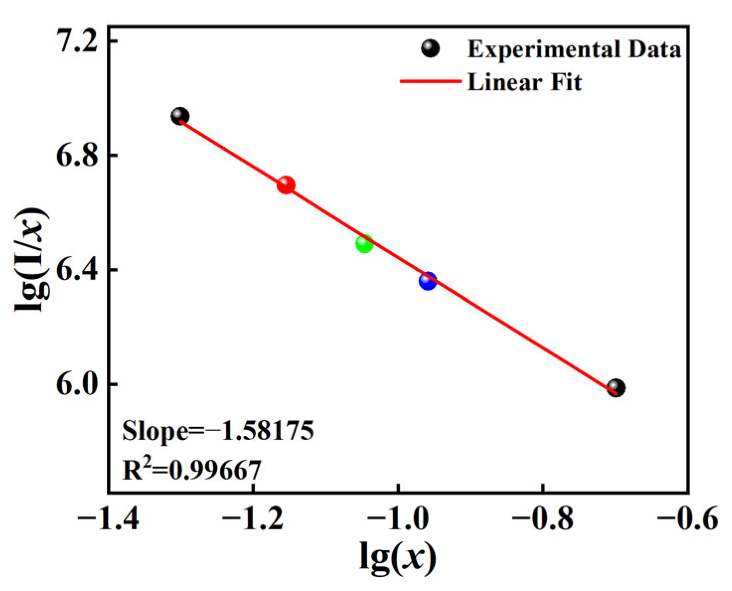
The relationship between lg(I/x) and lg(x) is shown in Figure 7. A linear fit was performed on the data points, yielding a slope of −1.58175, and the calculated value was 4.75, which is closer to 6. Therefore, the concentration quenching mechanism is dipole–dipole (d-d) interaction.
Figure 7.
Relationship of .
3.5.2. Energy Transfer (ET) Mechanism
The PL intensity changes in the two emission peaks (521 nm for VO43− and 575 nm for Dy3+) in Figure 6a suggest that there is energy transfer between VO43− and Dy3+. For the synthesized phosphors SNMVO: Dy3+, the energy transfer efficiency (η) between Dy3+ and VO43− can be quantitatively evaluated using the following equation:
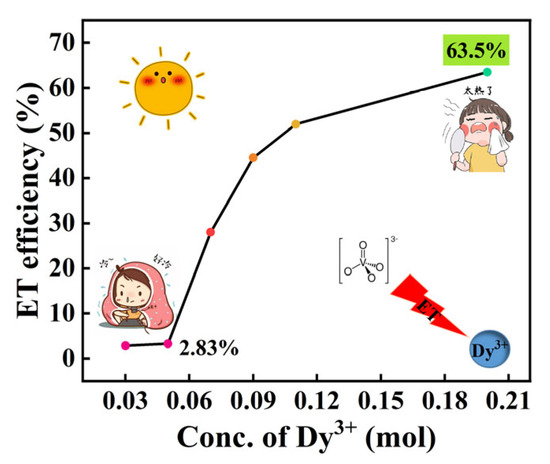
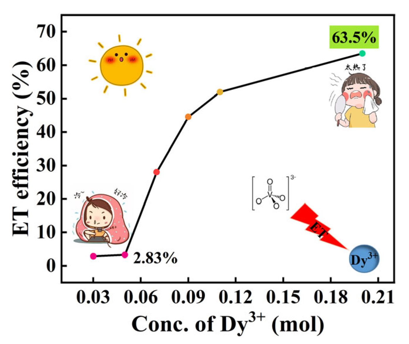
where denotes the photoluminescence (PL) intensity @ 521 nm of the 0.01Dy-doped SNMVO sample, while corresponds to the PL intensity of the 0.03~0.2 mol Dy3+-doped samples. The calculated energy transfer efficiency η is as high as 63.5% in Figure 8.
Figure 8.
The energy transfer efficiency (η) of the phosphor under different Dy3+.
3.6. Thermal Stability
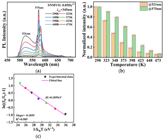
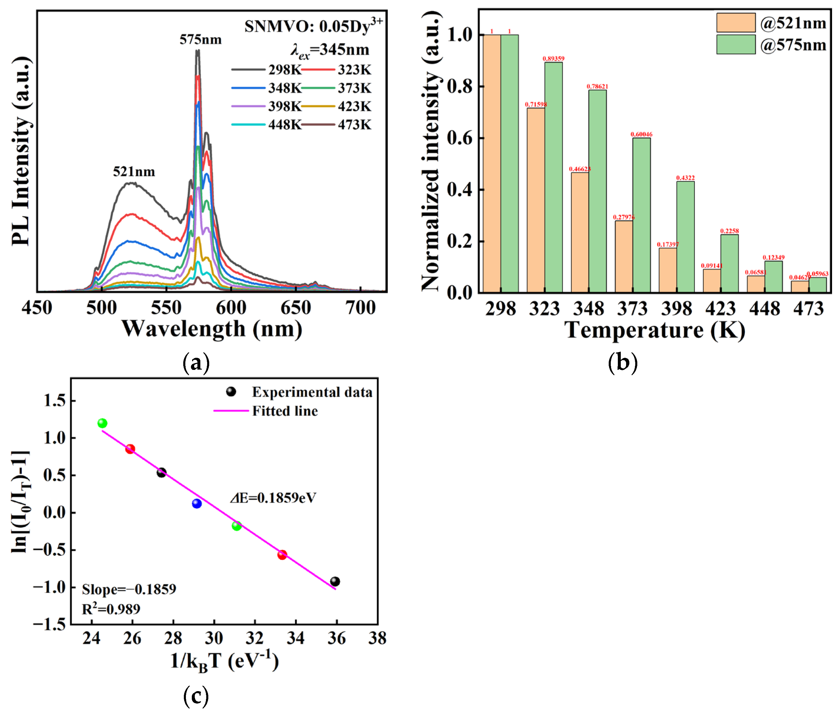
Figure 9a shows the temperature-response photoluminescence (PL) curves of SNMVO:0.05Dy3+ in the temperature range of 298–473 K. Due to the inherent thermal quenching properties of rare-earth phosphors, the emission intensity under 345 nm excitation decreases monotonically with increasing temperature. Figure 9b shows the temperature dependence of normalized emission intensity. Notably, the emission intensity of Dy3+ at 575 nm still retains approximately 78.6% of its value at 348 K at high temperatures, further confirming the good thermal stability of the prepared sample.
Figure 9.
Photoluminescence thermal stability of the SNMVO: 0.05Dy3+ phosphor: (a) PL spectrum changes with temperature. (b) Normalized PL intensity changes with temperature. (c) Active thermal activation energy fitting.
The activation energy () was evaluated to gain deeper insights into the thermal quenching behavior of the synthesized material. According to the Arrhenius formulation [36],
Herein, and correspond to the photoluminescence intensities at the starting temperature and elevated temperature T, respectively, while A and kB stand for the constant and the Boltzmann constant. Plotting ln[(I0/IT) − 1] against 1/kBT yields a straight line, as depicted in Figure 9c. The slope obtained from the linear regression is approximately −0.1859, which directly translates to an activation energy () of 0.1859 eV. Such results verify the superior thermal stability of our specimens, and a comprehensive comparison with other literature data is listed in Table 1.
Table 1.
Comparison of activation energy with other Dy3+-doped phosphors.
3.7. CIE and CCT Analysis
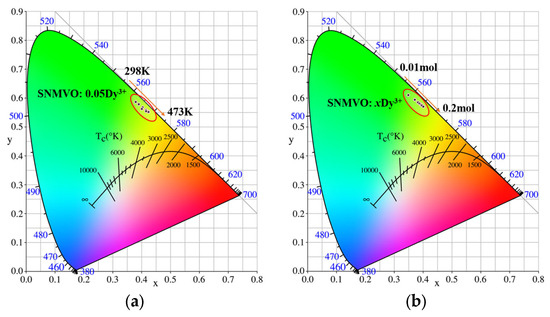
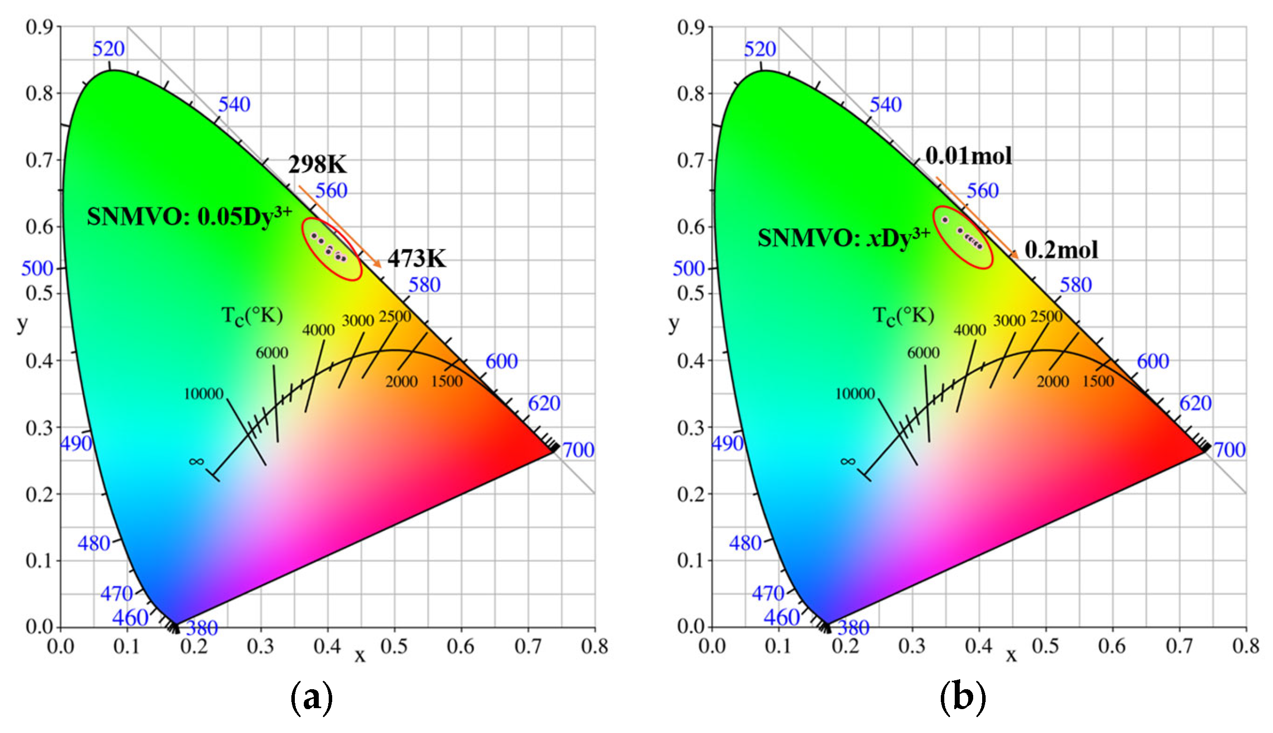
The temperature-dependent CIE chromaticity coordinates of the SNMVO: 0.05Dy3+ phosphor under 345 nm excitation are illustrated in Figure 10a. It can be seen that, as shown in the corresponding chromaticity diagrams, the CIE chromaticity coordinates of the samples change significantly at different temperatures, indicating a temperature dependence of color. Figure 10b shows the chromaticity diagrams and chromaticity coordinates of SNMVO: xDy3+ (x = 0.01, 0.03, 0.05, 0.07, 0.09, 0.11, and 0.2 mol). As shown, with increasing doping concentration, the fluorescence color of the samples gradually changes from yellow-green to yellow.
Figure 10.
Color coordinates of (a) SNMVO: 0.05Dy3+ at different temperatures, and (b) SNMVO: xDy3+ phosphor at different concentrations x = 0.02~0.2 mol.
The correlated color temperature (CCT) is an essential parameter for evaluating the color quality of phosphors. In this study, the CCT values were calculated using the McCamy approximate formula [42],
where n = (x − 0.3320)/(y − 0.1858). Table 2 summarizes the calculated CCT values for the singly doped SNMVO: Dy3+ phosphors. For the singly doped system, as the Dy3+ doping concentration increases, the CCT values gradually decrease from 40,170 K to 5543 K. This suggests that the prepared phosphor can be used as a component of warm white LEDs.
Table 2.
CCT values for SNMVO: xDy3+ and 0.05Dy3+ phosphors.
3.8. LED and Anti-Counterfeiting Application
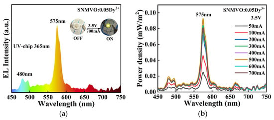
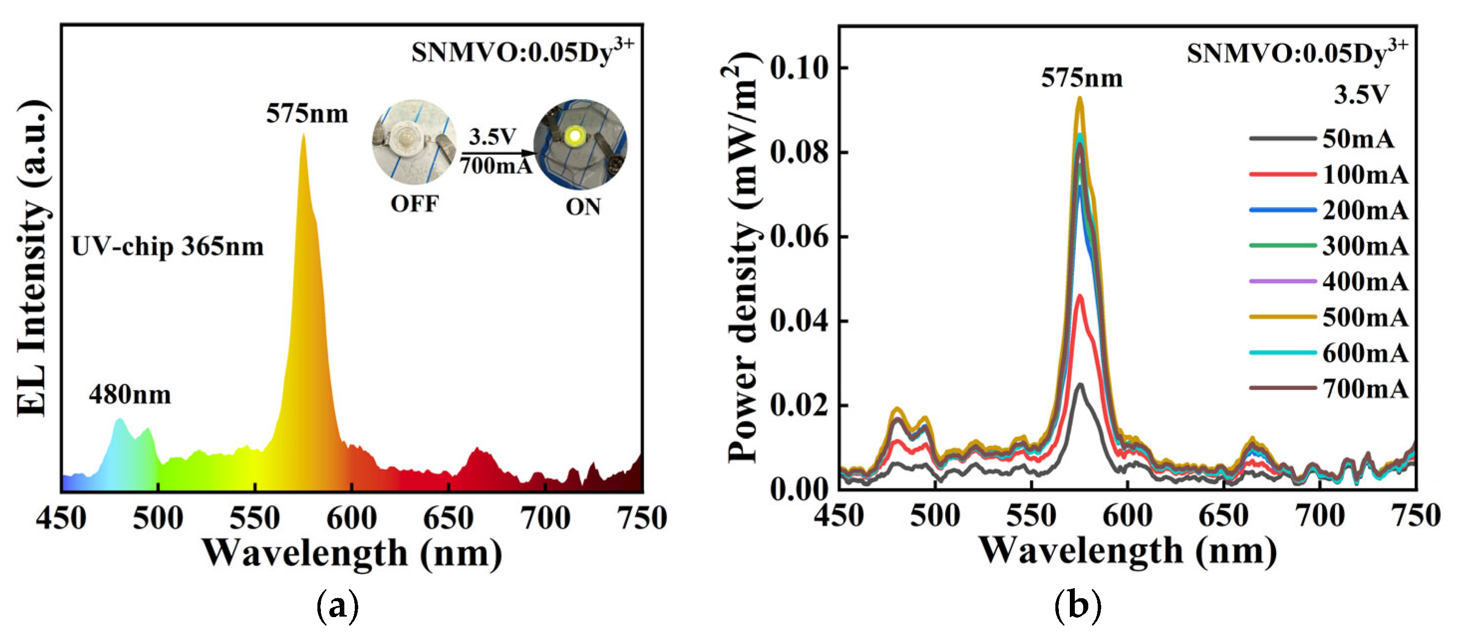
A device was fabricated to evaluate the electroluminescence performance of SNMVO: 0.05Dy3+ phosphor in a practical LED device by encapsulating the phosphor onto a 365 nm excited LED chip using A/B adhesive (Voltage: 3.4–3.6 V, Maximum output current: 700 mA, Shenzhen Xusheng Electronics Technology Co., Ltd., Shenzhen, China). Figure 11a shows the EL spectrum of the LED chip in the wavelength range of 450–750 nm. Figure 11b shows the EL spectra of the LED applied at different currents of 50–700 mA. LEDs achieve optimal energy density at a drive current of 500 mA. The correlated color temperature of LED devices is 3643 K–3820 K, which can be used as a component of yellow light in warm white LEDs.
Figure 11.
(a) EL spectra, (b) power density at 50–700 mA of SNMVO: 0.05Dy3+ LED.
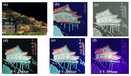
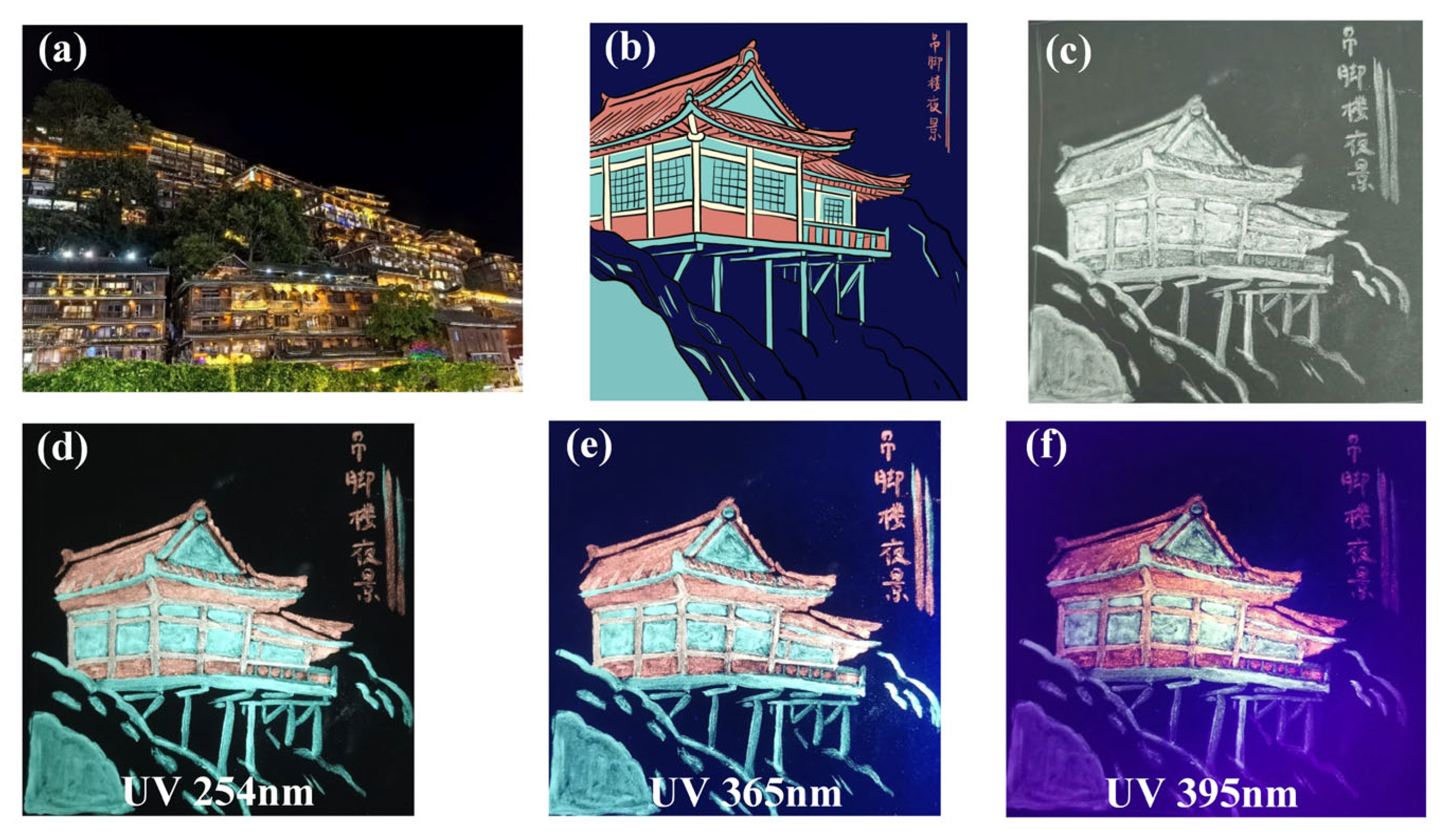
As shown in Figure 12, the authors combined the cultural symbols of the stilt houses in Xijiang Miao Village, Guizhou, with rare-earth luminescent materials; by incorporating self-developed phosphors into transparent AB adhesive, they designed unique patterns and created an anti-counterfeiting coating. Figure 12a shows a night view of the stilt houses, while Figure 12b presents a simplified version of the image; a pattern rendered using the prepared phosphors is displayed in Figure 12c (sunlight). Upon excitation by ultraviolet (UV) light at wavelengths of 254 nm, 365 nm, and 395 nm, the coating exhibits distinct color combinations, as shown in Figure 12d–f, respectively. The yellow-green areas correspond to Dy3+-doped phosphors, whereas the red sections represent the authors’ current work involving Dy3+/Eu3+ co-doped SNMVO phosphors. These images display cyan-green and coral-red hues, demonstrating the material’s multi-wavelength excitation characteristics—specifically, its unique color tunability under different UV light sources.
Figure 12.
Anti-counterfeiting pattern of stilted houses in Xijiang Miao Village: (a) a night view of the stilt houses; (b) a simplified version of the image; (c) a pattern rendered using the prepared phosphors; and the image of the pattern under different excitation conditions: (d) UV 254 nm, (e) UV 365 nm, and (f) UV 395 nm.
4. Conclusions
In this work, a series of Dy3+ -doped SNMVO phosphors were successfully prepared. The prepared samples exhibited strong 521 nm (green) and 575 nm (yellow) light under 345nm excitation. The optimal Dy3+ doping concentration was 0.05 mol, and the energy transfer between VO43− and Dy3+ was revealed, with a maximum energy transfer efficiency of 63.5%. LEDs prepared with SNMVO: 0.05Dy3+ exhibited good brightness and color temperature and can be used in LEDs. Furthermore, the phosphors can be used for drawing anti-counterfeiting patterns.
Author Contributions
Y.T.: writing—original draft, experiments, and data plotting and analysis. J.L.: writing—original draft, methodology, investigation, formal analysis, and data curation. C.D.: writing—review and editing and resources. M.Z.: writing—review and editing, visualization, validation, supervision, and resources. All authors have read and agreed to the published version of the manuscript.
Funding
This work was supported by the National Natural Science Foundation of China (No. 12474422) and Open Fund of Hubei Provincial Key Laboratory of Green Materials for Light Industry, Hubei University of Technology (202509B05).
Data Availability Statement
All data are available from the authors upon request.
Conflicts of Interest
The authors declare no conflicts of interest.
References
- Zhang, Y.; Liu, X.; Yang, J. Digital economy, green dual innovation and carbon emissions. Sustainability 2024, 16, 7291. [Google Scholar] [CrossRef] [Scilit]
- Shrivastava, R.; Kaur, J.; Dubey, V. White light emission by Dy3+ doped phosphor matrices: A short review. J. Fluoresc. 2016, 26, 105–111. [Google Scholar] [PubMed]
- Nair, G.B.; Swart, H.C.; Dhoble, S.J. A review on the advancements in phosphor-converted light emitting diodes (pc-LEDs): Phosphor synthesis, device fabrication and characterization. Prog. Mater. Sci. 2020, 109, 100622. [Google Scholar] [CrossRef] [Scilit]
- Sheu, J.K.; Chang, S.J.; Kuo, C.H.; Su, Y.; Wu, L.; Lin, Y.; Lai, W.; Tsai, J.; Chi, G.; Wu, R. White-light emission from near UV InGaN-GaN LED chip precoated with blue/green/red phosphors. IEEE Photonics Technol. Lett. 2003, 15, 18–20. [Google Scholar] [CrossRef] [Scilit]
- Li, K.; Shen, C. White LED based on nano-YAG: Ce3+/YAG: Ce3+, Gd3+ hybrid phosphors. Optik 2012, 123, 621–623. [Google Scholar] [CrossRef] [Scilit]
- Pereira, P.F.S.; Matos, M.G.; Avila, L.R.; Nassor, E.C.; Cestari, A.; Ciuffi, K.J.; Calefi, P.S.; Nassar, E.J. Red, green and blue (RGB) emission doped Y3Al5O12 (YAG) phosphors prepared by non-hydrolytic sol–gel route. J. Lumin. 2010, 130, 488–493. [Google Scholar] [CrossRef] [Scilit]
- Kolesnikov, I.E.; Mamonova, D.V.; Kurochkin, M.A.; Kolesnikov, E.Y.; Lähderanta, E. Optical thermometry by monitoring dual emissions from YVO4 and Eu3+ in YVO4: Eu3+ nanoparticles. ACS Appl. Nano Mater. 2021, 4, 1959–1966. [Google Scholar] [CrossRef] [Scilit]
- Setlur, A.A.; Comanzo, H.A.; Srivastava, A.M.; Beers, W.W. Spectroscopic evaluation of a white light phosphor for UV-LEDs—Ca2NaMg2V3O12: Eu3+. J. Electrochem. Soc. 2005, 152, H205–H208. [Google Scholar] [CrossRef] [Scilit]
- Dhobale, A.R.; Mohapatra, M.; Natarajan, V.; Godbole, S. Synthesis and photoluminescence investigations of the white light emitting phosphor, vanadate garnet, Ca2NaMg2V3O12 co-doped with Dy and Sm. J. Lumin. 2012, 132, 293–298. [Google Scholar] [CrossRef] [Scilit]
- Liang, Y.; Liu, M.; Yang, F.; Wu, X.; Yang, W.; Li, X. Preparation and characteristics of Ca2NaMg2V3O12: Sm3+ single-phased white-emitting phosphors. J. Inorg. Organomet. Polym. Mater. 2013, 23, 684–689. [Google Scholar] [CrossRef] [Scilit]
- Mehare, C.M.; Mishra, G.; Dhoble, N.S.; Dhoble, S. Improvement of self-activated luminescence properties of Ca2KZn2(VO4)3 down-conversion materials by SSR method based on co-doped Eu3+, Dy3+ rare earth ions concentrations. J. Mol. Struct. 2022, 1264, 133250. [Google Scholar] [CrossRef] [Scilit]
- Jeyakumaran, T.; Bharathi, N.V.; Sriramachandran, P.; Shanmugavel, R.; Ramaswamy, S. Synthesis and luminescence investigation of Eu3+ doped Ca2KZn2V3O12 phosphors: A potential material for WLEDs applications. J. Inorg. Organomet. Polym. Mater. 2021, 31, 674–682. [Google Scholar]
- Tong, Y.; Zhang, W.N.; Wei, R.F.; Chen, L.; Guo, H. Na2YMg2(VO4)3: Er3+, Yb3+ phosphors: Up-conversion and optical thermometry. Ceram. Int. 2021, 47, 2600–2606. [Google Scholar] [CrossRef] [Scilit]
- Li, Y.; Wei, X.; Chen, H.; Pang, G.; Pan, Y.; Gong, L.; Zhu, L.; Zhu, G.; Ji, Y. A new self-activated vanadate phosphor of Na2YMg2(VO4)3 and luminescence properties in Eu3+ doped Na2YMg2(VO4)3. J. Lumin. 2015, 168, 124–129. [Google Scholar] [CrossRef] [Scilit]
- Bindhu, A.; Naseemabeevi, J.I.; Ganesanpotti, S. Vibrationally induced photophysical response of Sr2NaMg2V3O12: Eu3+ for dual-mode temperature sensing and safety signs. Adv. Photonics Res. 2022, 3, 2100159. [Google Scholar]
- Mu, Q.; Li, H.; Pang, R.; Zhang, S.; Li, X.; Jiang, L. Luminescence characteristics and low temperature optical temperature measurement of Pr3+ doped Sr2NaMg2V3O12 phosphors. Dalton Trans. 2026, 55, 2156–2166. [Google Scholar] [CrossRef] [Scilit] [PubMed]
- Tong, Y.; Chen, Y.H.; Chen, S.Y.Z.; Wei, R.; Chen, L.; Guo, H. Luminescent properties of Na2GdMg2(VO4)3: Eu3+ red phosphors for NUV excited pc-WLEDs. Ceram. Int. 2021, 47, 12320–12326. [Google Scholar] [CrossRef] [Scilit]
- Li, L.J.; Tong, Y.; Chen, J.; Chen, Y.; Ashraf, G.A.; Chen, L.; Pang, T.; Guo, H. Up-conversion and temperature sensing properties of Na2GdMg2(VO4)3: Yb3+, Er3+ phosphors. J. Am. Ceram. Soc. 2022, 105, 384–391. [Google Scholar]
- Shang, M.; Geng, D.; Kang, X.; Yang, D.; Zhang, Y.; Lin, J. Hydrothermal derived LaOF: Ln3+ (Ln = Eu, Tb, Sm, Dy, Tm, and/or Ho) nanocrystals with multicolor-tunable emission properties. Inorg. Chem. 2012, 51, 11106–11116. [Google Scholar] [CrossRef] [Scilit] [PubMed]
- Su, Q.; Liang, H.; Li, C.; He, H.; Lu, Y.; Li, J.; Tao, Y. Luminescent materials and spectroscopic properties of Dy3+ ion. J. Lumin. 2007, 122, 927–930. [Google Scholar] [CrossRef] [Scilit]
- Rao, B.V.; Jang, K.; Lee, H.S.; Yi, S.-S.; Jeong, J.-H. Synthesis and photoluminescence characterization of RE3+ (=Eu3+, Dy3+)-activated Ca3La(VO4)3 phosphors for white light-emitting diodes. J. Alloys Compd. 2010, 496, 251–255. [Google Scholar] [CrossRef] [Scilit]
- Rao, B.V.; Buddhudu, S. Emission analysis of RE3+ (Dy3+ or Tb3+): Ca3Ln(Y, Gd)(VO4)3 powder phosphors. Mater. Chem. Phys. 2008, 111, 65–68. [Google Scholar] [CrossRef] [Scilit]
- Bindhu, A.; Naseemabeevi, J.I.; Ganesanpotti, S. Augmenting cyan emission in vanadate garnets via Dy3+ activation for light emitting devices and multi-mode optical thermometry. Dalton Trans. 2023, 52, 11705–11715. [Google Scholar] [CrossRef] [Scilit] [PubMed]
- Ramesh, B.; Devarajulu, G.; Raju, B.D.P.; Kumar, G.B.; Dillip, G.; Banerjee, A.; Joo, S. Determination of strain, site occupancy, photoluminescent, and thermoluminescent-trapping parameters of Sm3+-doped NaSrB5O9 microstructures. Ceram. Int. 2016, 42, 1234–1245. [Google Scholar] [CrossRef] [Scilit]
- Munirathnam, K.; Nagajyothi, P.C.; Prakashbabu, D.; Raju, B.D.P.; Shim, J. X-ray photoelectron spectroscopy and optical analysis of pure white light emitting Dy3+ and Mn2+ codoped Na3Y(PO4)2 phosphors for solid-state lighting. Ceram. Int. 2019, 45, 686–694. [Google Scholar] [CrossRef] [Scilit]
- Haycock, D.E.; Kasrai, M.; Nicholls, C.J.; Urch, D.S. The electronic structure of magnesium hydroxide (brucite) using X-ray emission, X-ray photoelectron, and auger spectroscopy. J. Chem. Soc. Dalton Trans. 1978, 7, 1791–1796. [Google Scholar] [CrossRef] [Scilit]
- Zhou, Y.; Peng, J.; Wang, M.; Mo, J.; Deng, C.; Zhu, M. Tribochemical behavior of pure magnesium during sliding friction. Metals 2019, 9, 311. [Google Scholar] [CrossRef] [Scilit]
- Bouvier, Y.; Mutel, B.; Grimblot, J. Use of an Auger parameter for characterizing the Mg chemical state in different materials. Surf. Coat. Technol. 2004, 180, 169–173. [Google Scholar] [CrossRef] [Scilit]
- Deng, Y.; Zhang, L.; Zheng, J.; Dang, D.; Zhang, J.; Gu, X.; Yang, X.; Tan, W.; Wang, L.; Zeng, L.; et al. VOx Matrix Confinement Approach to Generate Sub-3 nm L10-Pt-Based Intermetallic Catalysts for Fuel Cell Cathode. Small 2024, 20, 2400381. [Google Scholar] [CrossRef] [Scilit]
- Xue, M.; Jian, L.; Wang, F.; Yu, M.; Zhou, P.; Dang, F.; Yi, G.; Zhao, Z. Agar-Assisted Biopolymer-Templated Carbon-Coated Interfaces Facilitate Advanced ZnV2O4 Anode Materials for High-Stability Lithium-Ion Batteries. Energy Technol. 2026, 14, e202600003. [Google Scholar] [CrossRef] [Scilit]
- Lv, Y.; Shi, L.; Shi, J.W.; Zhang, Y.; Ma, D.; Cheng, Y.; Niu, C. In-doped LiCa2. 98MgV3O12 rare-earth-free phosphor with a high photoluminescence quantum yield of 67.4%. J. Am. Ceram. Soc. 2021, 104, 5837–5847. [Google Scholar] [CrossRef] [Scilit]
- Al-Mokhtar, K.M. FTIR and optical absorption studies of [B2O3]-[Na2O]-[Li2O]-[CuO] doped with [MgF2] nanoparticle. J. Nanotechnol. Adv. Mater. 2020, 8, 45–51. [Google Scholar]
- Al-Alawi, A.; Van De Voort, F.R.; Sedman, J. A new Fourier transform infrared method for the determination of moisture in edible oils. Appl. Spectrosc. 2005, 59, 1295–1299. [Google Scholar] [CrossRef] [Scilit] [PubMed]
- Frost, R.L.; Erickson, K.L.; Weier, M.L.; Carmody, O. Raman and infrared spectroscopy of selected vanadates. Spectrochim. Acta Part A Mol. Biomol. Spectrosc. 2005, 61, 829–834. [Google Scholar] [CrossRef] [Scilit]
- Frost, R.L.; Xi, Y. Assessment of the molecular structure of the borate mineral sakhaite Ca12Mg4(BO3)7(CO3)4Cl(OH)2·H2O using vibrational spectroscopy. Spectrochim. Acta Part A Mol. Biomol. Spectrosc. 2012, 96, 611–616. [Google Scholar] [CrossRef] [Scilit]
- Menzinger, M.; Wolfgang, R. The meaning and use of the Arrhenius activation energy. Angew. Chem. Int. Ed. Engl. 1969, 8, 438–444. [Google Scholar] [CrossRef] [Scilit]
- Liu, S.; Gao, D.; Chen, X.; Cui, M.; Zhang, X.; Yin, H.; Zhu, Y.; Wang, L.; Song, W.; Zhang, J. Judd–Ofelt Analysis and Luminescent Properties of Dy3+-Doped SrLaNaTeO6 Phosphors. J. Electron. Mater. 2025, 54, 6671–6684. [Google Scholar] [CrossRef] [Scilit]
- Lei, Y.; Yang, M.; Zhang, A.; Sun, H.; Deng, B. Synthesis and luminescence properties of novel stannate La3Ga5SnO14 doped with Dy3+ ions phosphors for white light-emitting devices. J. Mater. Sci. Mater. Electron. 2025, 36, 606. [Google Scholar] [CrossRef] [Scilit]
- Long, J.; Xu, Y.; Cheng, K.; Liu, X.; Huang, W.; Deng, C. Luminescent and thermal properties of a novel double perovskite La2MgTiO6: Dy3+ phosphors for w-LEDs application. J. Mater. Sci. Mater. Electron. 2023, 34, 368. [Google Scholar] [CrossRef] [Scilit]
- Long, Q.; Wang, C.; Li, Y.; Ding, J.; Wang, X.; Wang, Y. Solid state reaction synthesis and photoluminescence properties of Dy3+ doped Ca3Sc2Si3O12 phosphor. Mater. Res. Bull. 2015, 71, 21–24. [Google Scholar] [CrossRef] [Scilit]
- Kavya, J.V.; Anjana, P.S.; Raji, R.; Prasad, K.D.; Haranath, D.; Gopakumar, N. Judd-Ofelt parametrization and optical temperature sensing characteristics of white emitting Dy3+ doped SrGa2O4 phosphor. Inorg. Chem. Commun. 2025, 185, 116036. [Google Scholar]
- McCamy, R.C. Approximations for a Class of Functional Differential Equations; Carnegie Mellon University: Pittsburgh, PA, USA, 1971. [Google Scholar]

Disclaimer/Publisher’s Note: The statements, opinions and data contained in all publications are solely those of the individual author(s) and contributor(s) and not of MDPI and/or the editor(s). MDPI and/or the editor(s) disclaim responsibility for any injury to people or property resulting from any ideas, methods, instructions or products referred to in the content. |
© 2026 by the authors. Licensee MDPI, Basel, Switzerland. This article is an open access article distributed under the terms and conditions of the Creative Commons Attribution (CC BY) license.