


Abstract
Objective: The purpose of this investigation was to assess and compare the internal adaptation of different distinct CAD (Computer-aided design)/CAM (Computer-aided manufacturing) endocrown materials: feldspathic porcelain, indirect composite, hybrid ceramic, reinforced lithium disilicate, and lithium disilicate, utilizing microcomputed tomography. Methods: Standardized endocrown restorations were fabricated for mandibular first molar models. A total of seventy-five restorations were evenly allocated into five groups (n = 15 each): Group I (Cerec Blocks), Group II (Lava Ultimate), Group III (PICN Vita Enamic), Group IV (Celtra Duo), and Group V (Cerec Tessera). The restorations were bonded using PANAVIA V5 adhesive resin cement. To evaluate internal adaptations within the restorations, three distinct locations were selected for the acquisition of high-resolution micro-CT scans: the margin, the axial wall, and the pulpal floor. Data were analyzed using SPSS. To identify statistically significant differences among groups, a two-way ANOVA was conducted, followed by post hoc Tukey tests. Results: The statistical analysis did not reveal significant differences in internal gap measurements across the various material groups (p = 0.055). However, significant variations were observed within individual material groups (p < 0.001) at distinct locations, with the most pronounced discrepancies in thickness evident at the pulpal floor. Conclusion: While no significant differences were observed in internal adaptations among the various endocrown materials, substantial intra-group variability, particularly in terms of pulpal floor thickness, was evident. Since the study maintained a consistent preparation design across all groups, the observed variations in internal adaptation are likely attributed to differences in material behavior rather than changes in preparation geometry.
1. Introduction
Endocrowns represent a specialized dental restoration designed primarily for endodontically treated teeth, which require restorative solutions that are both durable and conservative [1]. With the emergence of advanced adhesive technologies and a shift toward minimally invasive dentistry, traditional post-and-crown restorations are being reconsidered [2]. Endocrowns merge the functionalities of dental crowns and core build-ups, providing an innovative approach to tooth rehabilitation.
Endocrowns are particularly suitable for molars and premolars with substantial tooth structural loss due to decay or fractures [3]. Distinct from conventional crowns that depend heavily on the remaining coronal tooth structure for support, endocrowns utilize the underlying dentin, thereby minimizing reliance on the compromised coronal structure. Al-Dabbagh’s (2020) systematic review and meta-analysis provided evidence that endocrowns are a practical conservative treatment modality for endodontically treated posterior teeth, demonstrating satisfactory long-term survival rates [4]. Endocrowns offer several advantages, including reduced tooth preparation, enhanced preservation of natural tooth structure compared to conventional crowns, and consistently favorable clinical outcomes, as stated by Fennis et al., 2019 [5].
The success and durability of endocrowns are influenced by several factors, including the anatomical features of the tooth, the adhesive capabilities of contemporary dental materials, and their bond strength to dentin [6], as well as the materials used in the fabrication of endocrown restorations [7]. A diverse array of materials, including dental ceramics, composite resins, and hybrid materials, can be utilized for endocrown fabrication [8,9,10,11,12,13]. Material selection is critical, as it affects the mechanical properties, esthetics, and longevity of the restoration. Ceramic materials such as lithium disilicate and zirconia-based ceramics have been widely used due to their superior esthetics and fracture resistance [14]. However, composite resins and hybrid ceramics have been introduced as alternatives, offering advantages such as improved elasticity modulus and lower brittleness, which may enhance marginal adaptation and stress distribution [15].
Similarly, the choice of cement plays a crucial role in the long-term success of endocrowns. Resin-based adhesives have been favored due to their superior bond strength and ability to reinforce the restoration–tooth interface [16]. Studies suggest that resin-modified glass ionomer cement and self-adhesive resin cement provide effective adhesion with favorable clinical outcomes [17]. The selection of cement should align with the material properties of the endocrown, as variations in bonding efficiency may affect marginal integrity, retention, and overall durability [18].
How do the internal adaptation and marginal gap of endocrowns differ based on materials used in their fabrication, and what implications does this have for their clinical efficacy and suitability? To address these questions, researchers have employed various techniques to assess internal adaptation and marginal gaps in dental restorations. These methods include external or internal replica techniques, external or internal microscopic examination, and digital microscopy with magnification power [19,20]. Micro-computed tomography (micro-CT, µCT) has proven to be a valuable tool for evaluating the internal and marginal fit of crown and inlay restorations produced with various CAD/CAM systems and digital scanners [21,22].
Higher marginal gaps, which indicate poor marginal fitness, can have several negative effects on the survival of the endocrown [23,24]. They might result in the cement being exposed to oral fluids, increasing the risk of secondary caries development, leakage, and plaque build-up. Furthermore, insufficient marginal fitness may lead to periodontal inflammation and, in extreme circumstances, the prosthodontic treatment’s total failure [25,26,27]. These elements emphasize how crucial it is to accomplish precise internal and marginal fitness to guarantee the longevity and success of ceramic restorations.
Clinical randomized trials are widely regarded as the gold standard for gathering evidence; however, their practical limitations, such as logistical challenges and often low recall rates, can restrict their applicability. In contrast, in vitro studies offer valuable insights into the adaptation and clinical performance of various endocrown materials [28,29,30,31,32]. The internal adaptation of ceramic and resin dental restorations has been a subject of debate. Some studies have attributed potentially superior marginal adaptation to resin materials’ lower hardness, elasticity modulus, and flexural strength, facilitating machining [33]. However, other research contradicts this, demonstrating comparable or even superior adaptation with ceramics. Studies by Sağlam [34], Zimmermann et al. [35], and Hajimahmoudi et al. [36], for instance, found no significant differences in marginal fit between ceramic and resin-based CAD/CAM materials. These conflicting results highlight the complex interplay between material properties and fabrication accuracy.
The aim of this in vitro study was to evaluate and compare the internal adaptation of various endocrown materials using micro-CT to help clinicians select the appropriate material for endocrowns. The null hypothesis of this study states that there is no statistically significant difference in the internal adaptation of endocrowns fabricated from different materials, as assessed by micro-CT.
2. Materials and Methods
2.1. Model Selection and Pre-Scanning
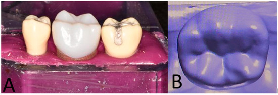
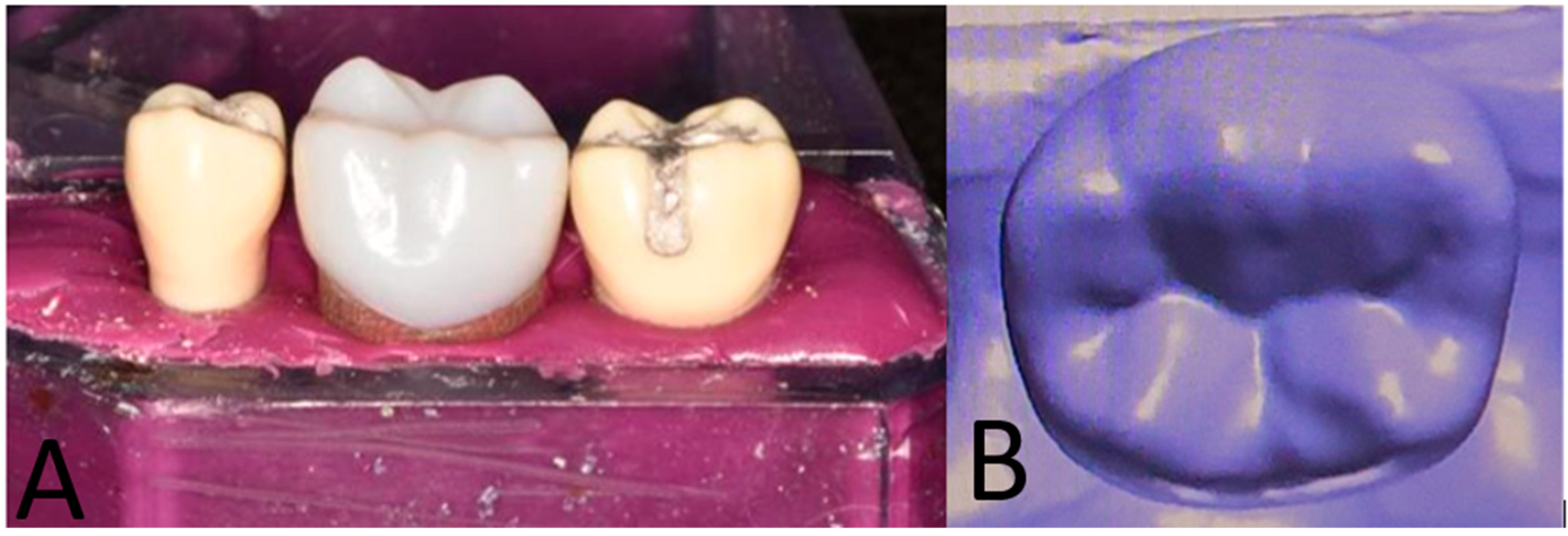
A hard thermo-setting plastic tooth model representing a mandibular right first molar (Model #46, Frasaco, Tettnang, Germany) was selected for the preparation of an endocrown according to established guidelines. The model was embedded in silicone material (Hydrorise Putty, Zhermack SpA, Polesine, Italy) within a hard plastic jaw, flanked by models of teeth #45 and #47. A 3D scanner (Ceramill Map 400+, Amann Girrbach AG, Koblach, Austria) was used to capture a digital impression of the prepared tooth. For subsequent fabrication of the endocrown restoration, the acquired data were saved as full-contour reference models in STL format (Figure 1).
Figure 1.
(A) The plastic tooth with adjacent teeth #47 and #45; (B) the scanned plastic tooth.
2.2. Model Preparation
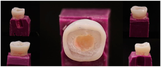
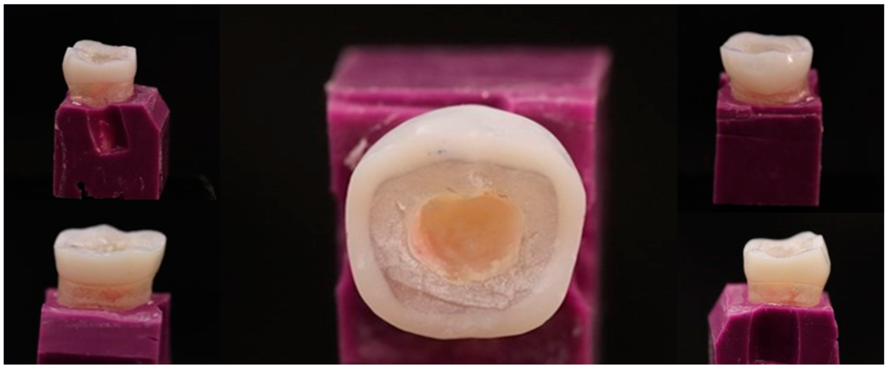
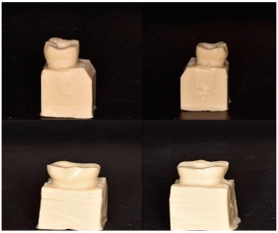
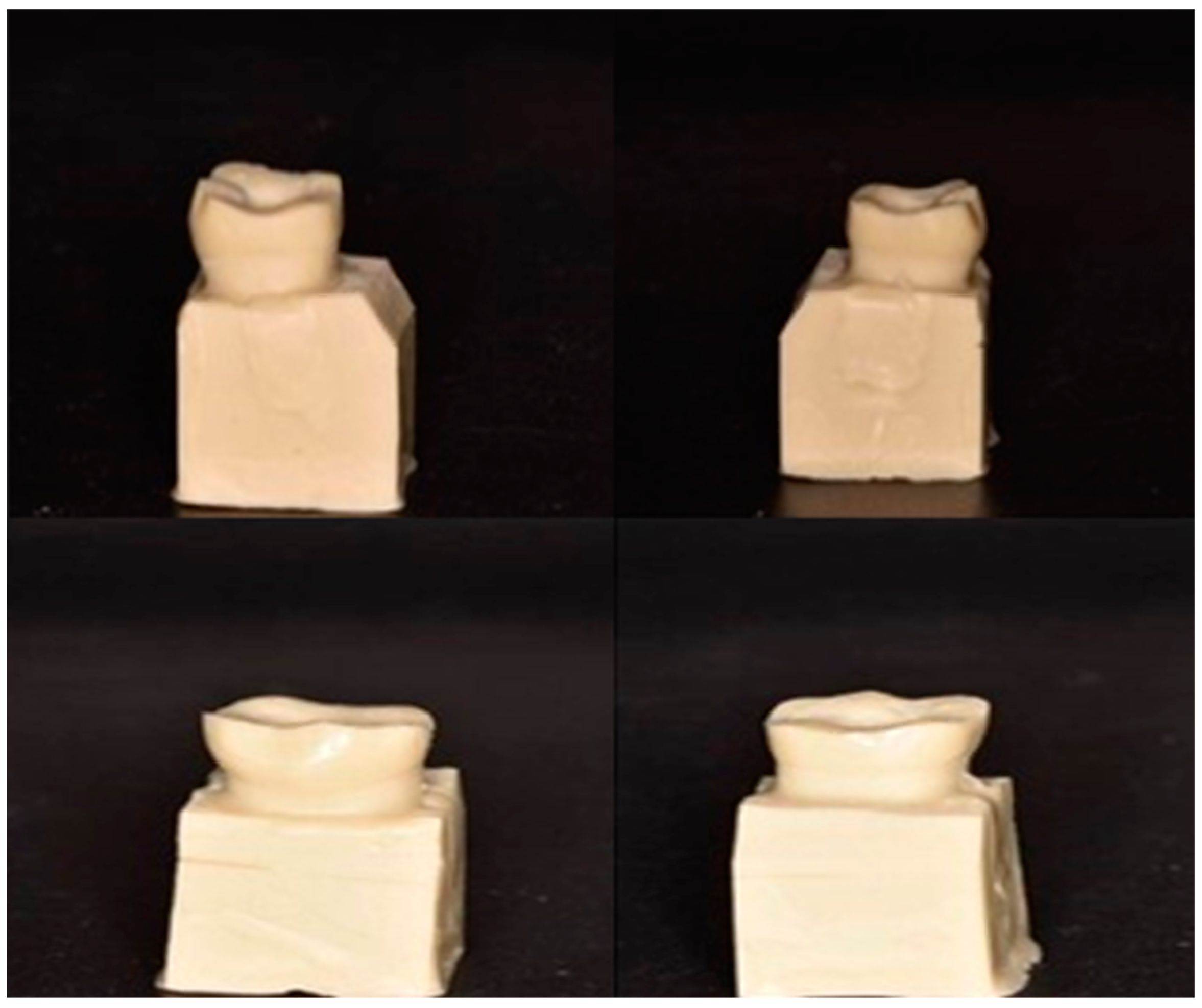
A silicon impression (Elite Double 22 Extra Fast, Zhermack SpA, Polesine, Italy) of the tooth model was fabricated and subsequently used to guide the preparation process. Occlusal reduction was achieved using a diamond bur with a tapered round-end (856-016, medium coarse, Brasseler, Savannah, GA, USA), with reductions of 1.5 mm and 2 mm for non-functional and functional cusps, respectively. A graded periodontal probe was utilized to determine a reduction in depth. A 7–10° occlusal convergence angle and a 4 mm deep flat pulpal floor were created by unifying the coronal pulp chamber and endodontic access cavity with a cylindrical–conical blue diamond bur. Intaglio surfaces were meticulously smoothed to eliminate sharp edges and undercuts, thereby ensuring a suitable path for restoration insertion (Figure 2).
Figure 2.
Different views demonstrating the different sides of the preparation.
2.3. Post-Scanning
After preparation, the model underwent a second scan using the same 3D scanner to obtain a precise dataset for the endocrown restoration fabrication.
2.4. Sample Size Calculation
Sample size was determined using G*Power (version 3.1.9.3, Macintosh). A total sample size of 75, with 15 samples per group, was determined based on a desired power of 90%, a two-sided alpha error of 0.05, and an anticipated large effect size (d = 0.5).
2.5. Die Duplication
A silicone material (Elite Double 22 Extra Fast, Zhermack SpA, Italy) was used to create molds from the prepared tooth and duplicate the plastic tooth. The molds were subsequently filled with alpha die MF ivory, a polyurethane-based model stump material manufactured by Schütz Dental GmbH, Rosbach vor der Höhe, Germany. Each die was made by mixing one scoop of powder with 10 mL of base liquid and 5 mL of hardener. Seventy-five duplicated dies were created as replicas of the prepared plastic tooth and divided into five groups of fifteen dies each (Figure 3).
Figure 3.
The model of the duplicated die with different views.
2.6. Grouping and Material Selection
A total of 75 standardized endocrown restorations were fabricated and subsequently divided into five experimental groups (n = 15) based on the restorative material employed. The mechanical features and composition of each material are summarized in Table 1.
Table 1.
The companies, producers, elastic modulus, and materials’ compositions utilized in the investigation.
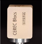
Group I: Endocrowns fabricated from feldspathic ceramic blocks (Cerec Blocks, Sirona Dental Systems, Bensheim, Germany).
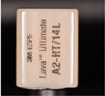
Group II: Endocrowns fabricated from 80 wt% nano-filled resin composite Lava Ultimate (3M ESPE, St. Paul, MN, USA).
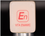
Group III: Endocrowns fabricated from Polymer Infiltrated Ceramic Network (PICN) (VITA, Bad Säckingen, Germany), consisting of a 75% ceramic and 25% polymer composition by volume.
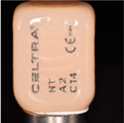
Group IV: Endocrowns in Group IV were fabricated from Celtra Duo (Sirona Dentsply, Milford, DE, USA), a lithium silicate/zirconia composite containing 10% zirconia within a silica-based glass matrix.
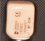
Group V: Endocrowns fabricated from lithium disilicate glass ceramic (Cerec Tessera) (Dentsply Sirona, York, PA, USA).
2.7. Milling Process
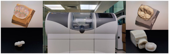
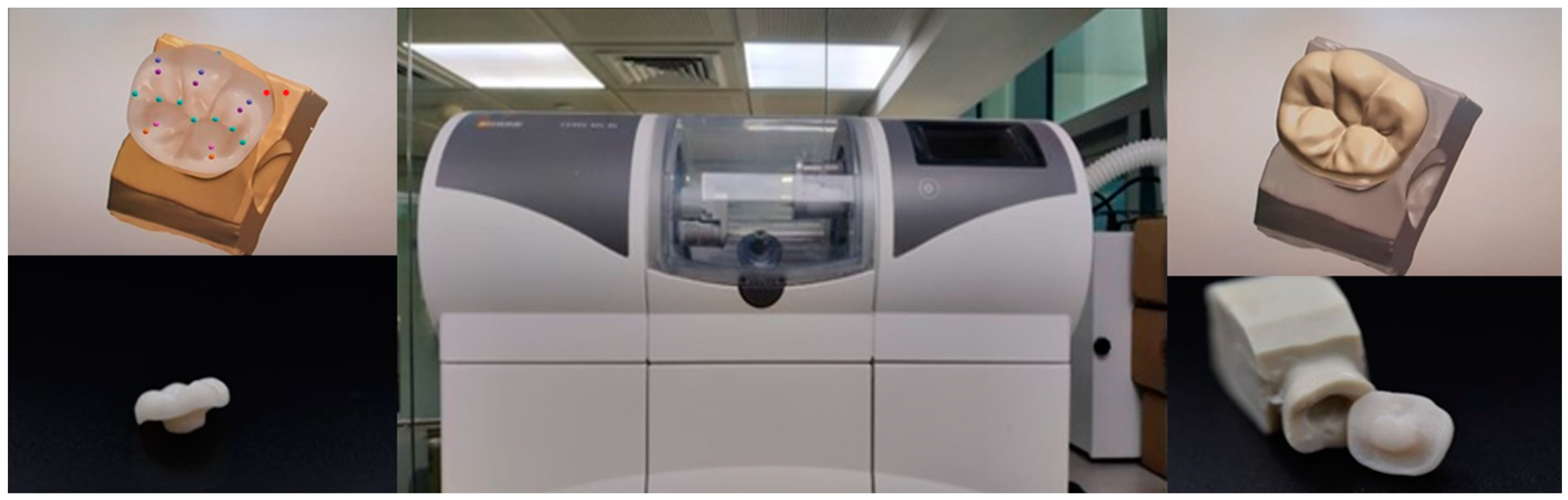
All endocrown restorations were fabricated using a pre-scanned full-contour reference model and standardized design parameters to ensure uniformity. The operator set a spacer thickness of 40 µm to account for the internal discrepancy. All restorations exhibited consistent occlusal anatomy and identical occluso-gingival height. Milling was performed in fine mode using a 5-axis CEREC MC XL milling unit (Dentsply Sirona), with burs being changed every 10 millings (Figure 4).
Figure 4.
Milling with Sirona CEREC MC XL.
2.8. Cementation Process
Each endocrown was bonded to its corresponding die using a PANAVIA V5 (Kuraray Noritake Dental Inc., Okayama, Japan) adhesive system. Following cementation, a standardized static load was applied (0.5 kg), and excess cement was removed per the manufacturer’s protocol.
2.9. Thermocycling
All samples were subjected to 5000 thermocycling cycles, alternating between 5 °C and 55 °C to simulate oral temperature fluctuations.
2.10. µ CT Scanning
All samples underwent high-resolution µCT scanning (µCT 100, Scanco Medical AG, Wangen-Brüttisellen, Switzerland), with parameters set to optimize image quality and detail resolution. The images were reconstructed and analyzed to assess the internal adaptation at predetermined points.
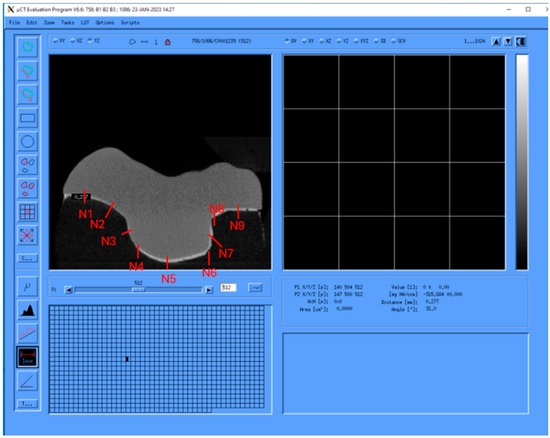
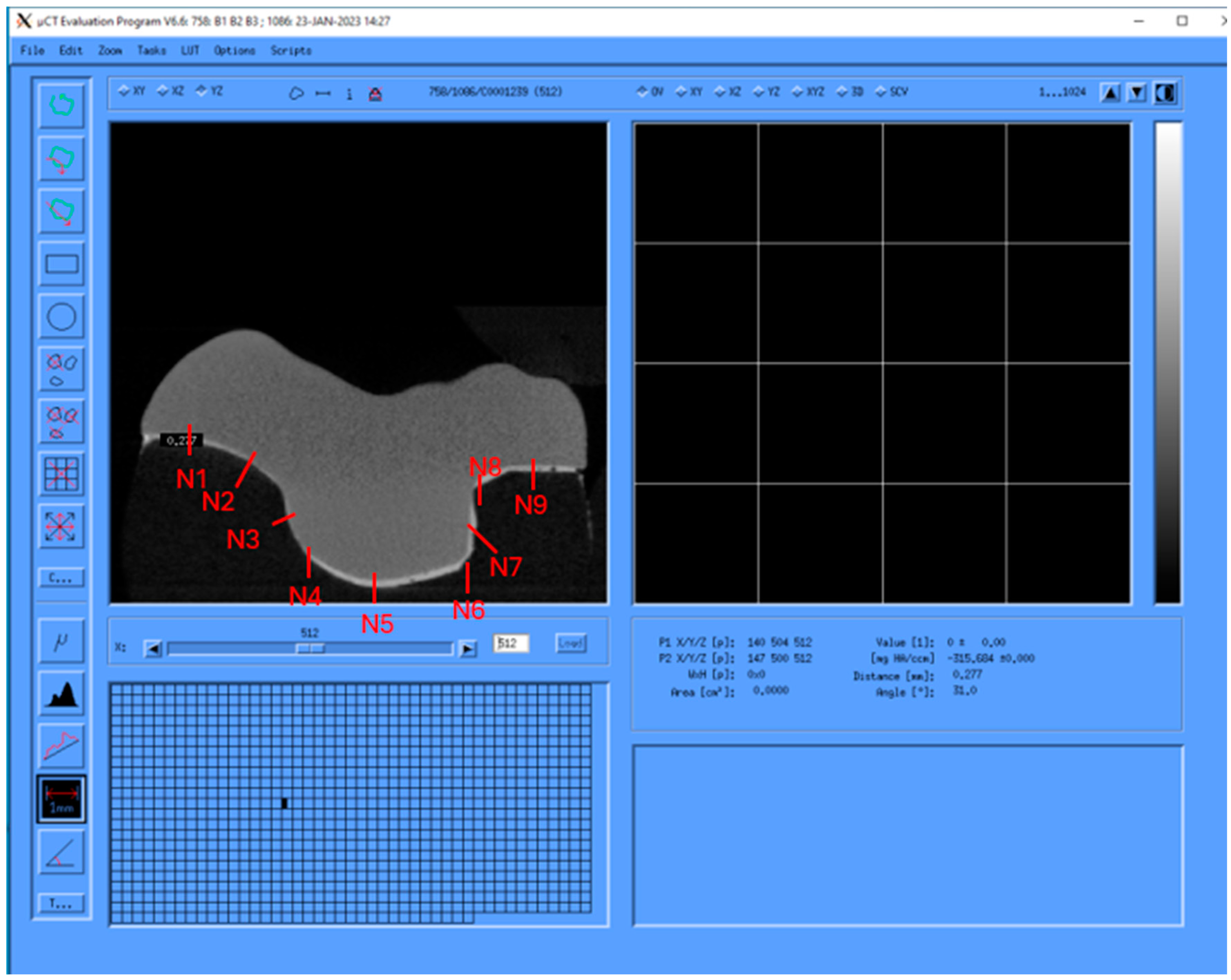
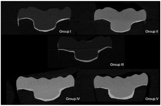
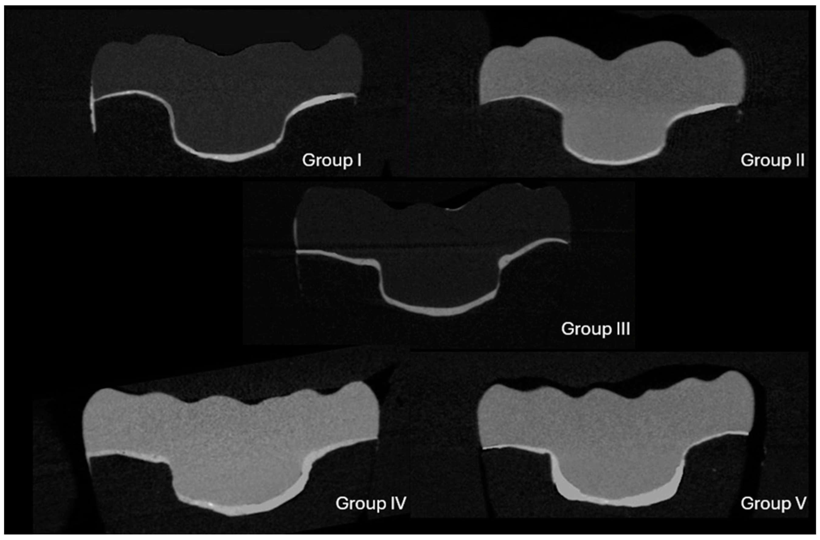
Adaptation was examined using the same sections, focusing on nine designated areas (N1 through N9) to evaluate discrepancies between the die material and endocrown restorations (Figure 5). Three symmetrical sections were selected from the central region of each specimen in the mesiodistal direction. Measurements were acquired at nine predetermined points along the mesiodistal (MD) sections: four points at the marginal seat (N1, N8, N2, N9), two points on the axial walls (N7, N3), and three points on the pulpal floor (N5, N4, N6) (Figure 5). In total, 27 points were selected to measure the internal adaptation of each specimen. Pictures of samples of each group are presented in Figure 6.
Figure 5.
The nine points (N1–N9) chosen to measure the degree of internal adaptation in each specimen.
Figure 6.
Images of representative samples from each group.
2.11. Statistical Analysis
Data were analyzed using SPSS Statistics version 22 (IBM Corp., Armonk, NY, USA). Descriptive statistics were calculated, and normality was assessed using Shapiro–Wilk tests. Significant differences in internal adaptation between materials and groups were evaluated using two-way ANOVA. Post hoc Tukey HSD tests were conducted to identify specific group differences. Statistical significance was defined as p ≤ 0.05.
3. Results
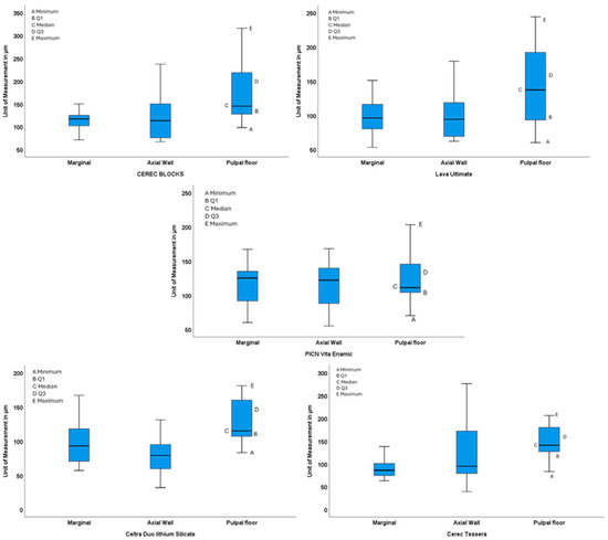
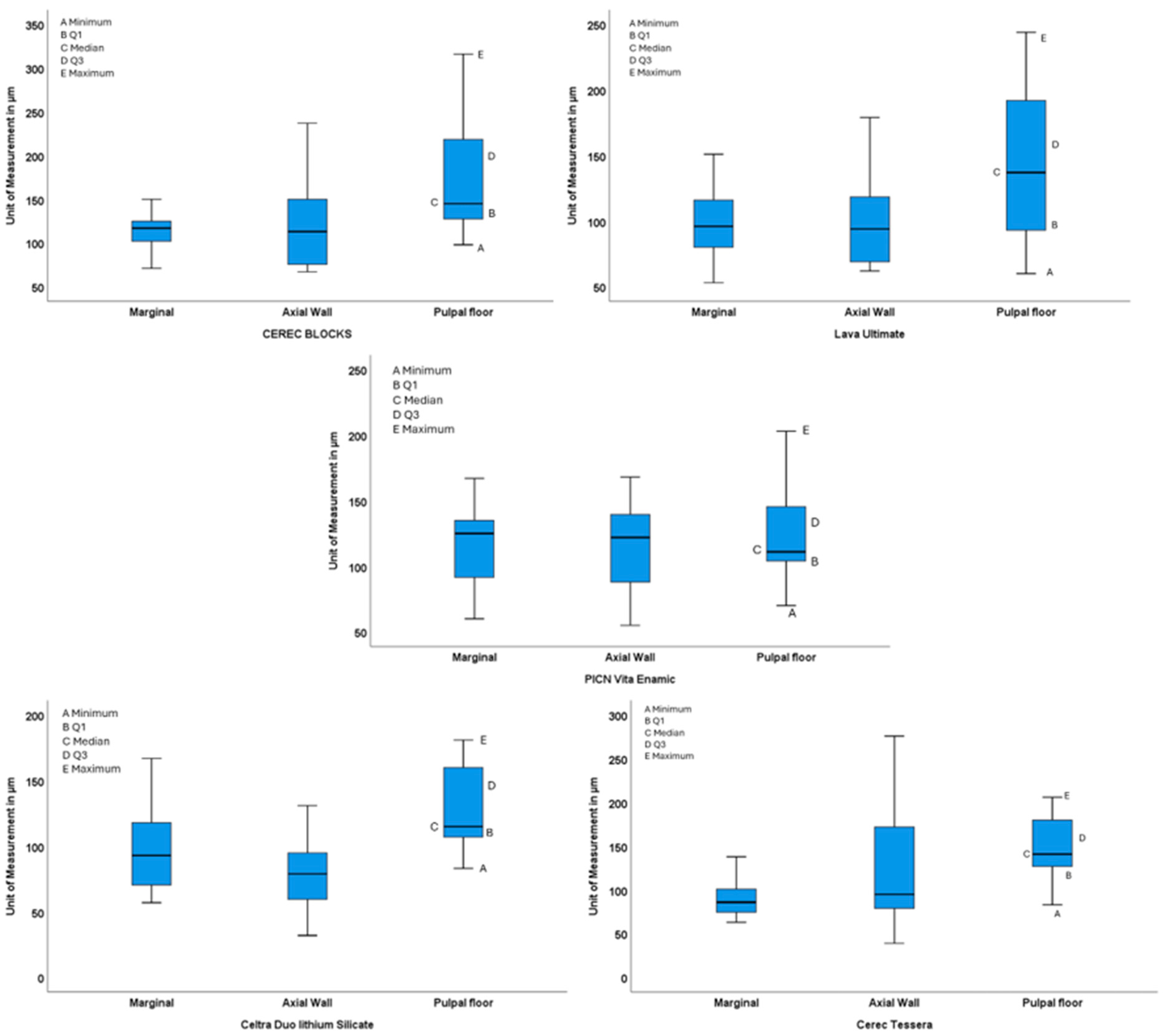
A comprehensive analysis of internal gap measurements was performed within and between different groups at three distinct locations: marginal, axial wall, and pulpal floor. Two-way ANOVA indicated no statistically significant differences among the experimental groups at the specified locations (p = 0.055). Nonetheless, a notable difference was identified among different locations within the same group (p < 0.001) (Table 2). The results reveal significant differences in at least one of the marginal, axial walls, or pulpal floor measurements for the Cerec Blocks, Lava Ultimate, Celtra Duo lithium silicate, and Cerec Tessera groups based on p-values < 0.05. The PICN Vita Enamic group shows no significant differences across measurements (F = 0.44, p = 0.649). The Celtra Duo lithium silicate group demonstrated significant differences between axial walls vs. pulpal floor and marginal vs. pulpal floor, while the Cerec Tessera group exhibited significant differences between marginal vs. pulpal floor measurements. The post hoc test emphasized that the pulpal floor consistently exhibited a larger discrepancy than other sites across all groups. Significant differences were observed at the pulpal floor compared to the axial wall in Group I (p = 0.05), the marginal region in Group V (p = 0.004), and both the axial wall and marginal regions in Groups II (p = 0.05) and IV. No significant difference was observed in relation to other walls in Group III (Table 3) (Figure 7).
Table 2.
A two-way ANOVA analysis was performed to analyze the effect of material types and location on the internal adaptation of endocrown restoration.
Table 3.
Multiple comparisons among the experimental groups (unit of measurement in µm).
Figure 7.
Box plot showing the measurements of marginal, axial walls, or pulpal floor for the Cerec Blocks, Lava Ultimate, Celtra Duo lithium silicate/zirconia, PICN Vita Enamic, and Cerec Tessera groups.
4. Discussion
A wide array of novel hybrid polymer and ceramic CAD/CAM materials have emerged within the dental industry for tooth-borne restorations [25,32], necessitating further research to comprehensively assess their clinical performance.
The findings of this study showed no statistically significant differences in mean internal gap measurements among the experimented materials (p = 0.055). While Group II (indirect composite) and Group III (PICN; hybrid ceramic) exhibited lower internal gap measurements at specific locations compared to other groups, the two-way ANOVA analysis failed to reveal statistically significant differences between the experimental groups. Consequently, the initial null hypothesis, which postulated no significant difference in internal fit among the different CAD/CAM endocrown materials evaluated, was accepted.
The lack of statistically significant differences between the materials investigated in this study may be assigned to numerous variables. The utilization of high-precision scanning and milling techniques, such as the 5-axis CEREC MC XL milling unit, coupled with standardized preparation and fabrication protocols, likely minimized the influence of material-specific biomechanical properties on internal adaptation. Advancements in CAD/CAM technology, particularly with respect to 5-axis milling, have significantly enhanced the control and precision of restoration fabrication, thereby improving internal fit [33].
The findings of this study contradict the results of Akhlaghian et al. [34], who reported variations in marginal fit among endocrowns fabricated from different CAD/CAM materials (polymer-infiltrated hybrid ceramic, zirconia-reinforced lithium silicate glass-ceramic, and lithium disilicate glass-ceramic), all of which were deemed clinically acceptable. These discrepancies may be attributed to several factors, including the use of different methodologies. Akhlaghian et al. [34] utilized naturally extracted teeth and employed a digital camera stereomicroscope for marginal gap measurements, whereas this study utilized a different cement (Panavia V) compared to the dual-cure resin cement (3M ESPE Relyx U200 Self-Adhesive Resin Cement; 3M) used in their study.
El Ghoul et al. observed larger discrepancies in resin-based groups compared to ceramic-based groups, attributing this to the potential difficulty in reproducing fine details with milled ceramics due to their inherent strength and brittleness [7]. However, the present study did not replicate this finding. The high modulus of elasticity and flexural strength of the glass-ceramic materials used in this study likely contributed to their resistance to crack propagation and chipping, potentially mitigating the challenges associated with milling.
Jalalian et al. [35] reported acceptable vertical marginal adaptation for both lithium disilicate and zirconia-reinforced lithium silicate endocrowns, with zirconia-reinforced lithium silicate demonstrating superior marginal adaptation. However, their study did not consider the effects of cementation or thermomechanical cycling. Similarly, Sağlam et al. [29] compared lithium disilicate and zirconia-reinforced lithium silicate endocrowns fabricated for mandibular first molars and observed significantly larger marginal gaps for lithium disilicate. El Ghoul et al. [36], comparing LDS (Lithium disilicate) and ZLS (Zirconia-reinforced Lithium Silicate) endocrowns for mandibular molars, also reported a larger marginal gap in the ZLS group, although the difference did not reach statistical significance.
Direct comparison of these findings with the present study is limited due to variations in methodology. These studies utilized different scanning techniques (e.g., intraoral scanners), measurement methods (e.g., replica technique), and finish line designs, potentially influencing the observed results.
To facilitate a more detailed analysis of internal fit, the measurements of the internal gap were divided into three distinct areas of interest: marginal/cervical, axial, and pulpal floor [27,35].
According to the results of the post hoc analysis, there was no significant difference among different locations in hybrid ceramic Group III (p < 0.05), but there was a significant difference between the pulpal with the axial wall in Cerec Block Group I and with the marginal in reinforced lithium disilicate Group V. Pulpal wall showed significant difference with both the axial wall and the marginal in Groups Lava Ultimate II and lithium disilicate IV. These findings are in correlation with the results of Akhlaghian et al. [34] and El Ghoul et al. [7], who found that the pulpal area demonstrated the most considerable misfit in all groups examined.
For all tested groups, the pulpal floor consistently showed the largest gap (180 ± 78 mm), particularly for the Cerec Block group. These outcomes matched those of the earlier research [27,37]) which may be affected by the scanner’s limited optical depth and the preparation’s small convergence angle, producing a hazy image in the pulpal area [38]. Another explanation is the size of the milling tools. Larger tools may cause uneven milling on flat surfaces, like the pulpal retention areas of endocrowns, leading to overmilling.
Hajimahmoudi et al. [31] found the largest gap at the pulpal floor (p < 0.001) compared to other walls. Shin et al. reported that differences at the pulpal floor were more pronounced compared to other sites and that an endocrown with a 4 mm cavity exhibited larger marginal and internal volumes than one with a 2 mm cavity [27]. Topkara and Keleş identified the pulpal floor as the region with the poorest fit [26].
These findings align with previous research [39,40], which consistently reported larger internal gaps at the pulpal floor compared to other sites.
Even though the measurement was performed after cementation, the marginal discrepancy is deemed acceptable. The clinical acceptability of marginal gaps in dental restorations remains a subject of debate, with studies suggesting different thresholds depending on material type, cementation protocols, and long-term stability. Contrepois et al. (2013) conducted a systematic review of the marginal adaptation of ceramic crowns and found that gaps below 120 µm are generally considered clinically acceptable, as they allow for adequate cement seal and minimize bacterial infiltration [41]. However, discrepancies exceeding 200–300 µm may pose a risk for cement dissolution, plaque accumulation, and secondary caries formation. In the present study, although some measured gaps reached 300 µm, the mean values were within the clinically acceptable range, particularly when considering the compensatory role of adhesive resin cement in sealing the marginal interface. The butt margin design employed in this study likely contributed to improved marginal adaptation by reducing stress concentration at the interface. Furthermore, the marginal discrepancies were reduced at the butt joint tooth/restoration interface of endocrowns due to an enhanced bonding mechanism between the resin cement and the increased bulk of ceramics [40,42]. These findings align with the previous literature suggesting that high-precision CAD/CAM milling and optimized cementation techniques can help maintain clinically acceptable adaptation levels, ultimately influencing the longevity of endocrown restorations.
A plastic replica of the mandibular right first molar #46 was utilized to standardize measurements, simulating a clinical scenario requiring both high mechanical properties and esthetics in restoration. The use of precise CAD/CAM designing the restoration, milling machinery, and diamond rotary burs helped standardize the preparation design and eliminate manual errors. This allowed for comparisons between internal fits among various tested materials.
The virtual cement spacing used in this investigation was 50 μm. Zheng et al. observed that the virtual cement space setting has a considerable impact on the adaptability of CAD/CAM endocrown restorations [24]. A 30 μm spacing setting resulted in an unsatisfactory fit; however, 60 μm or 120 μm were regarded acceptable. Furthermore, considering the remarkable concordance between design and reality, they proposed a spacing setting of 60 μm for ceramic materials and 120 μm for resin composites.
The CAD/CAM system setting allows the adjustment of different parameters, including the virtual cement space, during the virtual three-dimensional design of the restoration. Setting a certain cement space width around the fabricated CAD-CAM restoration is important for proper adaptation between the restoration and the prepared abutments and for a good distribution of the luting agents. Studies have shown that the cement space value significantly affects the marginal and internal fit of CAD-CAM crowns.
The conflict in the measurement between the virtual cement space and the marginal adaptation is due to whether the restorations were adjusted and the film thickness of the cement. Such a conclusion, in considering the discrepancy between design and reality, is confirmed by the study of Zheng et al., 2022 [24].
One limitation of this study is the use of a standardized plastic tooth model, which may not fully replicate the complex anatomical and physical properties of natural teeth. While these models ensure consistency across samples, they do not account for the inherent variability observed in natural tooth structures, including variations in tooth morphology, enamel thickness, and dentin composition, which may influence the internal adaptation of endocrowns. Additionally, the experimental setup, including the use of a single cementation protocol and controlled static load, may not adequately reflect clinical scenarios where variations in operator technique, occlusal forces, and oral environment conditions can significantly impact the adaptation of endocrowns.
A limitation of this study is its in vitro nature, which precludes the evaluation of factors present in the dynamic oral environment. Furthermore, this study did not assess long-term durability or the potential for marginal leakage, which are critical aspects for evaluating the clinical success of endocrown restorations. Future studies involving in vivo assessments and a wider range of clinical settings are necessary to validate the findings and ensure their applicability to real-world dental practice.
In light of the findings, the lack of statistically significant differences between the materials analyzed may, in part, be attributed to the high precision of modern CAD/CAM systems. The use of advanced 5-axis milling technology and standardized preparation protocols likely minimized material-specific biomechanical differences, contributing to the observed uniformity in internal fit. Additionally, the virtual cement space setting of 50 μm, which was chosen based on the literature recommendations [24], could have also impacted the results by ensuring adequate space for cementation, thereby influencing the overall adaptation. However, it is important to consider that other experimental variables, such as variations in cementation protocols, thermomechanical cycling, and occlusal load application, may also contribute to the lack of significant differences observed between materials. Future studies should explore these factors to fully understand their role in the internal adaptation of CAD/CAM materials.
The discrepancy observed at the pulpal floor is indeed a noteworthy finding and warrants further exploration. Although this study did not involve variations in preparation design, previous research suggests that factors such as milling tool size, scanning techniques, and preparation angle could play a crucial role in minimizing the internal gap at the pulpal floor. Studies have shown that using smaller milling tools and ensuring precise scanning may reduce discrepancies in this area [27,38]. Furthermore, adjustments to the preparation design, including optimizing the convergence angle, may help minimize the internal gap at the pulpal floor. While this study does not provide sufficient data to support such claims, it provides a valuable foundation for further investigations that could assess the impact of preparation design on internal adaptation in CAD/CAM restorations.
The novelty of this study lies in its comprehensive evaluation of multiple CAD/CAM endocrown materials using a standardized methodology that closely replicates clinical conditions, including cementation and thermocycling. Unlike previous studies that primarily focused on marginal fit or used non-cemented replica techniques, this research provides insights into the internal adaptation of different materials under conditions that mimic real-world applications. Additionally, this study highlights the influence of cavity geometry on internal adaptation, particularly at the pulpal floor, which remains a critical yet underexplored aspect in restorative dentistry.
Several studies have assessed the internal and marginal adaptation of endocrowns using different CAD/CAM materials and methods, including micro-CT analysis and replica techniques [7,25]. However, this study expands upon previous research by integrating high-precision milling and a controlled experimental setup to analyze the adaptation of various endocrown materials, contributing valuable data to the ongoing discourse on the clinical adaptation of endocrown restorations. Previous systematic reviews have also emphasized the importance of adaptation in endocrowns and their impact on longevity [1,4]. Furthermore, investigations into the effects of cavity design and preparation depth on internal fit have shown that these variables significantly impact restoration performance, reinforcing the clinical relevance of this study [27,37].
By addressing these factors, this research contributes to the body of evidence supporting the optimization of endocrown design and material selection, ensuring improved long-term outcomes for patients.
5. Conclusions
This study demonstrates that all tested CAD/CAM endocrown materials exhibited clinically acceptable internal gaps, reinforcing their reliability and suitability for restorative dentistry. No statistically significant differences in internal adaptation were observed among the materials, suggesting that the choice of material may not be the primary determinant of internal fit. However, the consistently larger internal discrepancies observed at the pulpal floor emphasize the influence of material behavior and milling accuracy on restoration adaptation. While this study utilized a standardized preparation approach, future research should explore how variations in preparation geometry may affect internal adaptation to further optimize restorative outcomes.
Clinical Significance
These findings provide valuable insights into the clinical application of CAD/CAM technology for endocrown fabrication. The demonstrated versatility of materials allows clinicians to make material choices based on factors such as esthetics, cost, and patient-specific requirements without compromising fit quality. Furthermore, the larger discrepancies observed at the pulpal floor emphasize the importance of meticulous preparation in this critical area. Improved preparation techniques that minimize irregularities at the pulpal floor could further enhance the longevity and clinical performance of endocrowns, ensuring better outcomes for patients with structurally compromised teeth.
Author Contributions
Conceptualization: A.R.S.; methodology: W.S.; investigation: W.S.; resources: A.R.S.; data curation: W.S.; writing—original draft: A.R.S. and M.A.; writing—review and editing: A.R.S. and M.A.; visualization: M.A.; supervision: A.R.S. All authors have read and agreed the published version of this manuscript.
Funding
The authors acknowledge Ajman University in the support for the fund of publication of this paper.
Institutional Review Board Statement
Not Applicable.
Informed Consent Statement
Not Applicable.
Data Availability Statement
The original contributions presented in this study are included in this article. Further inquiries can be directed at the corresponding author.
Acknowledgments
The authors would like to express their sincere gratitude to Sriraman Devara-jan (sriraman.devarajan@yahoo.com) for his invaluable assistance with the statistical analysis of this study.
Conflicts of Interest
The authors declare no conflict of interest.
References
- Sedrez-Porto, J.A.; de Oliveira da Rosa, W.L.; da Silva, A.F.; Münchow, E.A.; Pereira-Cenci, T. Endocrown restorations: A systematic review and meta-analysis. J. Dent. 2016, 52, 8–14. [Google Scholar] [CrossRef] [Scilit] [PubMed]
- Magne, P. ‘It should not be about aesthetics but tooth conserving dentistry’. Interview by Ruth Doherty. Br. Dent. J. 2012, 213, 189–191. [Google Scholar] [PubMed]
- Biacchi, G.R.; Mello, B.; Basting, R.T. The endocrown: An alternative approach for restoring extensively damaged molars. J. Esthet. Restor. Dent. 2017, 29, 404–412. [Google Scholar] [CrossRef] [Scilit]
- Al-Dabbagh, R.A. Survival and success of endocrowns: A systematic review and meta-analysis. J. Prosthet. Dent. 2021, 125, 415.e1–415.e9. [Google Scholar] [CrossRef] [Scilit] [PubMed]
- Fennis, W.M.; Kuijs, R.H.; Roeters, F.J.; Creugers, N.H.; Kreulen, C.M. A randomized controlled trial of the composite endocrown: 5-year follow-up. J. Dent. 2019, 86, 40–45. [Google Scholar]
- Fages, M.; Bennasar, B. The endocrown: A different type of all-ceramic reconstruction for molars. J. Can. Dent. Assoc. 2013, 79, d140. [Google Scholar] [PubMed]
- El Ghoul, W.A.; Özcan, M.; Ounsi, H.; Tohme, H.; Salameh, Z. Effect of different CAD-CAM materials on the marginal and internal adaptation of endocrown restorations: An in vitro study. J. Prosthet. Dent. 2020, 123, 128–134. [Google Scholar] [CrossRef] [Scilit] [PubMed]
- Vijayakumar, J.; Varadan, P.; Balaji, L.; Rajan, M.; Kalaiselvam, R.; Saeralaathan, S.; Ganesh, A. Fracture resistance of resin based and lithium disilicate endocrowns. Which is better?—A systematic review of in-vitro studies. Biomater. Investig. Dent. 2021, 8, 104–111. [Google Scholar]
- Awada, A.; Nathanson, D. Mechanical properties of resin-ceramic CAD/CAM restorative materials. J. Prosthet. Dent. 2015, 114, 587–593. [Google Scholar] [CrossRef] [Scilit]
- Zoidis, P.; Bakiri, E.; Polyzois, G. Using modified polyetheretherketone (PEEK) as an alternative material for endocrown restorations: A short-term clinical report. J. Prosthet. Dent. 2017, 117, 335–339. [Google Scholar] [CrossRef] [Scilit]
- Alamoush, R.A.; Silikas, N.; Salim, N.A.; Al-Nasrawi, S.; Satterthwaite, J.D. Effect of the Composition of CAD/CAM Composite Blocks on Mechanical Properties. Biomed. Res. Int. 2018, 2018, 4893143. [Google Scholar] [CrossRef] [Scilit] [PubMed]
- Koller, M.; Arnetzl, G.V.; Holly, L.; Arnetzl, G. Lava ultimate resin nano ceramic for CAD/ CAM: Customization case study. Int. J. Comput. Dent. 2012, 15, 159–164, (In English and German). [Google Scholar] [PubMed]
- Palacios, T.; Tarancón, S.; Abad, C.; Pastor, J.Y. Saliva Influence on the Mechanical Properties of Advanced CAD/CAM Composites for Indirect Dental Restorations. Polymers 2021, 13, 808. [Google Scholar] [CrossRef] [Scilit] [PubMed] [PubMed Central]
- Abduo, J.; Lyons, K.; Swain, M. Fit of zirconia fixed partial denture: A systematic review. J. Oral Rehabil. 2010, 37, 866–876. [Google Scholar] [CrossRef] [Scilit]
- Mansi, Z.M.; Al-Shamma, A.M.; Saleh, A.R. Assessment of marginal adaptation of indirect zirconia crown restorations using self-adhesive resin cements modified with polylysine antibacterial particles. J. Bagh. Coll. Dent. 2024, 36, 50–56. [Google Scholar] [CrossRef] [Scilit]
- Ekici, Z.; Kılıçarslan, M.A.; Bilecenoğlu, B.; Ocak, M. Micro-CT evaluation of the marginal and internal fit of crown and inlay restorations fabricated via different digital scanners belonging to the same CAD-CAM system. Int. J. Prosthodont. 2021, 34, 381–389. [Google Scholar] [CrossRef] [Scilit]
- Al-Hassiny, A.; Végh, D.; Bányai, D.; Végh, Á.; Géczi, Z.; Borbély, J.; Hermann, P.; Hegedüs, T. User Experience of Intraoral Scanners in Dentistry: Transnational Questionnaire Study. Int. Dent. J. 2023, 73, 754–759. [Google Scholar] [CrossRef] [Scilit]
- Hashem, R.M. Marginal fit and fracture resistance of advanced lithium disilicate occlusal veneer with different preparation designs. Egypt Dent. J. 2023, 69, 2115–2125. [Google Scholar] [CrossRef] [Scilit]
- Goujat, A.; Abouelleil, H.; Colon, P.; Jeannin, C.; Pradelle, N.; Seux, D.; Grosgogeat, B. Marginal and internal fit of CAD-CAM inlay/onlay restorations: A systematic review of in vitro studies. J. Prosthet. Dent. 2019, 121, 590–597.e3. [Google Scholar] [CrossRef] [Scilit]
- Felton, D.A.; Kanoy, B.E.; Bayne, S.C.; Wirthman, G.P. Effect of in vivo crown margin discrepancies on periodontal health. J. Prosthet. Dent. 1991, 65, 357–364. [Google Scholar] [CrossRef] [Scilit]
- Jacobs, M.S.; Windeler, A.S. An investigation of dental luting cement solubility as a function of the marginal gap. J. Prosthet. Dent. 1991, 65, 436–442. [Google Scholar] [CrossRef] [Scilit] [PubMed]
- Knoernschild, K.L.; Campbell, S.D. Periodontal tissue responses after insertion of artificial crowns and fixed partial dentures. J. Prosthet. Dent. 2000, 84, 492–498. [Google Scholar] [CrossRef] [Scilit] [PubMed]
- Hassouneh, L.; Jum’ah, A.; Ferrari, M.; Wood, D.J. A Micro-computed Tomography Analysis of Marginal and Internal Fit of Endocrowns Fabricated from Three CAD/CAM Materials. Oper Dent. 2023, 48, 79–89. [Google Scholar] [CrossRef] [Scilit] [PubMed]
- Zheng, Z.; Wang, H.; Mo, J.; Ling, Z.; Zeng, Y.; Zhang, Y.; Wang, J.; Yan, W. Effect of virtual cement space and restorative materials on the adaptation of CAD-CAM endocrowns. BMC Oral Health 2022, 22, 580. [Google Scholar] [CrossRef] [Scilit] [PubMed] [PubMed Central]
- Abduljawad, D.E.; Rayyan, M.R. Marginal and internal fit of lithium disilicate endocrowns fabricated using conventional, digital, and combination techniques. J. Esthet. Restor. Dent. 2022, 34, 707–714. [Google Scholar] [CrossRef] [Scilit] [PubMed]
- Topkara, C.; Keleş, A. Examining the adaptation of modified endocrowns prepared with CAD-CAM in maxillary and mandibular molars: A microcomputed tomography study. J. Prosthet. Dent. 2022, 127, 744–749. [Google Scholar] [CrossRef] [Scilit]
- Shin, Y.; Park, S.; Park, J.W.; Kim, K.M.; Park, Y.B.; Roh, B.D. Evaluation of the marginal and internal discrepancies of CAD-CAM endocrowns with different cavity depths: An in vitro study. J. Prosthet. Dent. 2017, 117, 109–115. [Google Scholar]
- Mörmann, W.H.; Stawarczyk, B.; Ender, A.; Sener, B.; Attin, T.; Mehl, A. Wear characteristics of current aesthetic dental restorative CAD/CAM materials: Two-body wear, gloss retention, roughness and Martens hardness. J. Mech. Behav. Biomed. Mater. 2013, 20, 113–125. [Google Scholar]
- Saglam, G.; Cengiz, S.; Karacaer, O. Marginal adaptation and fracture resistance of feldspathic and polymer-infiltrated ceramic network CAD/CAM endocrowns for maxillary premolars. Niger. J. Clin. Pract. 2020, 23, 1–6. [Google Scholar] [CrossRef] [Scilit] [PubMed]
- Zimmermann, M.; Valcanaia, A.; Neiva, G.; Mehl, A.; Fasbinder, D. Three-Dimensional Digital Evaluation of the Fit of Endocrowns Fabricated from Different CAD/CAM Materials. J. Prosthodont. 2019, 28, e504–e509. [Google Scholar] [CrossRef] [Scilit]
- Hajimahmoudi, M.; Raseipour, S.; Mroue, M.; Ghodsi, S. Evaluation of Marginal and Internal Fit of CAD/CAM Endocrowns with Different Cavity Tapers. Int. J. Prosthodont. 2023, 36, 189–193. [Google Scholar] [CrossRef] [Scilit] [PubMed]
- Hiraba, H.; Nishio, K.; Takeuchi, Y.; Ito, T.; Yamamori, T.; Kamimoto, A. Application of one-piece endodontic crowns fabricated with CAD-CAM system to molars. Jpn. Dent. Sci. Rev. 2024, 60, 81–94. [Google Scholar] [CrossRef] [Scilit] [PubMed] [PubMed Central]
- Yun, M.J.; Jeon, Y.C.; Jeong, C.M.; Huh, J.B. Comparison of the fit of cast gold crowns fabricated from the digital and the conventional impression techniques. J. Adv. Prosthodont. 2017, 9, 1–13. [Google Scholar] [CrossRef] [Scilit] [PubMed]
- Akhlaghian, M.; Khaledi, A.A.; Mosaddad, S.A.; Dabiri, S.; Giti, R.; Kadkhodae, F.; Gholami, S. The internal and marginal adaptation of lithium disilicate endocrowns fabricated using intra and extraoral scanners: An in-vitro study. PLoS ONE 2024, 19, e0301361. [Google Scholar] [CrossRef] [Scilit] [PubMed] [PubMed Central]
- Jalalian, E.; Zarbakhsh, A.; Khorshidi, S.; Golalipour, S.; Mohammadnasl, S.; Sayyari, M. Comparative analysis of endocrown fracture resistance and marginal adaptation: CAD/CAM technology using lithium disilicate vs. zirconia-reinforced lithium silicate ceramics. Saudi Dent. J. 2024, 36, 353–358. [Google Scholar] [CrossRef] [Scilit] [PubMed] [PubMed Central]
- El Ghoul, W.; Özcan, M.; Silwadi, M.; Salameh, Z. Fracture resistance and failure modes of endocrowns manufactured with different CAD/CAM materials under axial and lateral loading. J. Esthet. Restor. Dent. 2019, 31, 378–387. [Google Scholar] [CrossRef] [Scilit] [PubMed]
- Gaintantzopoulou, M.D.; El-Damanhoury, H.M. Effect of preparation depth on the marginal and internal adaptation of computer-aided design/computerassisted manufacture endocrowns. Oper Dent. 2016, 41, 607–616. [Google Scholar] [CrossRef] [Scilit]
- Kokubo, Y.; Nagayama, Y.; Tsumita, M.; Ohkubo, C.; Fukushima, S.; Vult von Steyern, P. Clinical marginal and internal gaps of in-ceram crowns fabricated using the GN-I system. J. Oral Rehabil. 2005, 32, 753–758. [Google Scholar] [CrossRef] [Scilit]
- Grenade, C.; Mainjot, A.; Vanheusden, A. Fit of single tooth zirconia copings: Comparison between various manufacturing processes. J. Prosthet. Dent. 2011, 105, 249–255. [Google Scholar] [CrossRef] [Scilit]
- Nakamura, T.; Dei, N.; Kojima, T.; Wakabayashi, K. Marginal and internal fit of CEREC 3 CAD/CAM all-ceramic crowns. Int. J. Prosthodont. 2003, 16, 244–248. [Google Scholar]
- Contrepois, M.; Soenen, A.; Bartala, M.; Laviole, O. Marginal adaptation of ceramic crowns: A systematic review. J. Prosthet. Dent. 2013, 110, 447–454.e10. [Google Scholar] [CrossRef] [Scilit] [PubMed]
- Cho, L.; Choi, J.; Yi, Y.J.; Park, C.J. Effect of finish line variants on marginal accuracy and fracture strength of ceramic optimized polymer/fiber-reinforced composite crowns. J. Prosthet. Dent. 2004, 91, 554–560. [Google Scholar] [CrossRef] [Scilit]

Disclaimer/Publisher’s Note: The statements, opinions and data contained in all publications are solely those of the individual author(s) and contributor(s) and not of MDPI and/or the editor(s). MDPI and/or the editor(s) disclaim responsibility for any injury to people or property resulting from any ideas, methods, instructions or products referred to in the content. |
© 2025 by the authors. Licensee MDPI, Basel, Switzerland. This article is an open access article distributed under the terms and conditions of the Creative Commons Attribution (CC BY) license (https://creativecommons.org/licenses/by/4.0/).