


Abstract
Paralympic Powerlifting is a maximal strength sport that requires high training intensity and efficient recovery to maintain performance and prevent injury. Intense training can induce oxidative stress, potentially compromising performance and physiological adaptation. Caffeine (CA) is a widely used ergogenic aid with potential to enhance muscle strength and reduce oxidative damage. This study investigated the acute effects of CA supplementation (9 mg/kg) on static strength indicators and oxidative stress markers in 14 male Paralympic powerlifting athletes. The protocol included two conditions (CA and placebo), with athletes performing 5 sets of 5 repetitions at 80% of 1 RM in the adapted bench press. Strength variables (maximal isometric force, 1-s peak force, and force variability) and biomarkers (TBARS/MDA, total antioxidant capacity, sulfhydryl groups, and uric acid) were analyzed. CA supplementation significantly increased maximal isometric strength, improved 1-s peak force, and reduced variability, suggesting enhanced motor unit recruitment. However, CA also led to increased oxidative stress, as evidenced by elevated TBARS levels, reduced antioxidant capacity, decreased sulfhydryl groups, and higher uric acid concentrations post-exercise. In conclusion, while CA acutely improves neuromuscular performance, it does not attenuate—and may even exacerbate—exercise-induced oxidative stress. Therefore, its use in strength sports should be approached with caution.
1. Introduction
Paralympic Powerlifting (PP) is a maximal strength sport characterized by high-intensity training [1,2,3]. Due to the demands placed on physical performance and training intensity, optimal recovery and strength improvement are essential components [4,5]. In this context, the use of ergogenic aids to enhance strength and facilitate recovery has been increasingly explored. Inadequate recovery can compromise subsequent training sessions by reducing training volume and intensity, and may also increase the risk of injury [5,6,7,8]. Therefore, improving both strength and post-training recovery is crucial, as it can support performance enhancement and progression, even under conditions of intense and prolonged training, particularly among elite athletes [9]. Intense physical exercise induces oxidative stress, as high-intensity activity stimulates the production of reactive oxygen species (ROS) [10,11,12]. When ROS levels become unbalanced, they can cause tissue damage and negatively affect cellular structures such as organelles, nucleic acids, lipids, and proteins, posing significant health risks [6,7,10].
In this regard, caffeine (CA) may attenuate exercise-induced oxidative stress by neutralizing reactive oxygen species (ROS) and scavenging free radicals [13,14], thereby contributing to the prevention of or a reduction in muscle damage [15]. CA administration has been shown to reduce malondialdehyde (MDA) levels and improve liver function in a dose-dependent manner [16]. Furthermore, caffeine supplementation can increase neuronal strength and excitability by modulating intramuscular calcium dynamics, stimulating calcium (Ca2+) release from the sarcoplasmic reticulum and increasing motor unit recruitment [17,18]. Physiologically, this occurs via binding to ryanodine receptors, antagonism of adenosine receptors and modulation of voltage-dependent calcium channels (VOCCs) [18].
Additionally, it may promote lipolysis, contributing to the sparing of muscle glycogen [16,19]. However, the effects of caffeine on oxidative stress markers and strength following resistance exercise in athletes remain conflicting [14,20,21,22,23,24]. On one hand, some studies have reported improved performance without affecting oxidative stress levels [20,22,24]. On the other hand, minimal reductions in oxidative stress have been observed, with increases only in glutathione levels, without significant changes in other ROS-related parameters [21]. Other studies have reported a more pronounced reduction in oxidative stress following caffeine supplementation [14,23].
Considering the lack of consensus on the effects of CA in strength training, the aim of this study was to evaluate the acute supplementation of 9 mg/g of CA during Paralympic weightlifting training on indicators of static strength and post-exercise oxidative stress, after training at 80% of one repetition maximum (1 RM). Low doses (3 mg/kg) improve speed at light loads (25–50% of 1 RM), while high loads (such as 90% of 1 RM) and high-intensity activities require higher doses (6–9 mg/kg) to optimize power [25]. The 9 mg/kg protocol stands out as a safe strategy to maximize performance in adapted strength sports [26]. Therefore, our hypothesis was that CA supplementation would increase muscle strength and reduce post-exercise oxidative stress.
2. Materials and Methods
2.1. Study Design
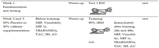
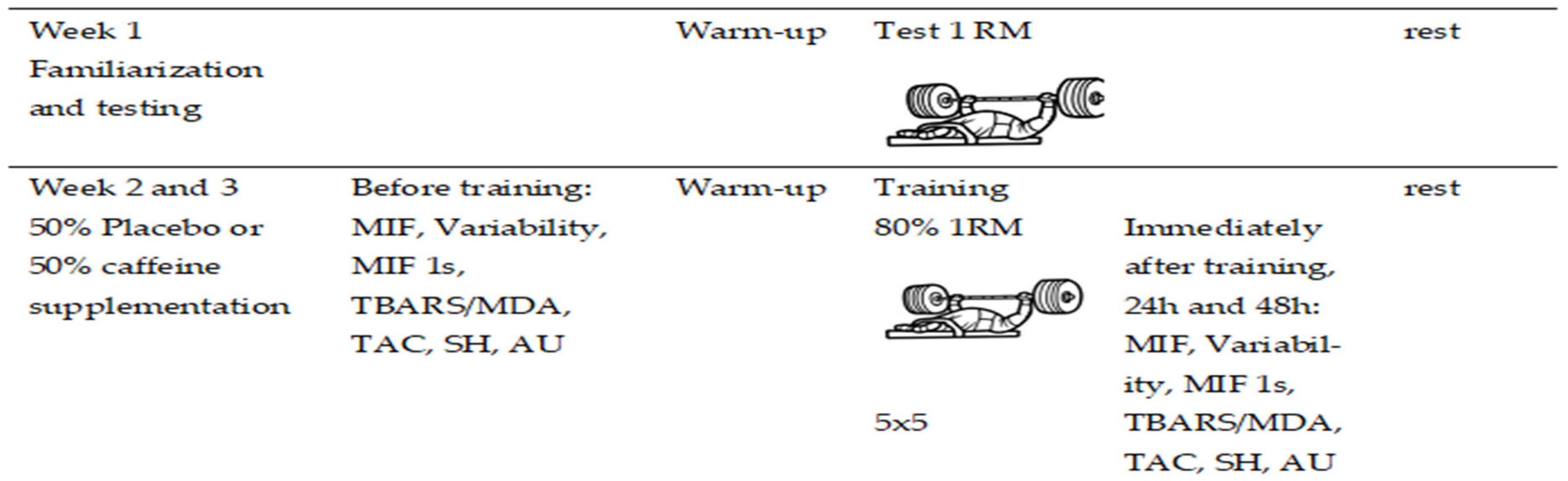
This study, outlined in Figure 1, was conducted over a period of three weeks. In the first week, participants were familiarized with the adapted bench press, followed by the one-repetition maximum (1 RM) test. During the second and third weeks, participants received either anhydrous caffeine (CA) at a dose of 9 mg/kg or a placebo (PLA), administered one hour prior to training [25,26,27,28]. The order of conditions (CA or PLA) was randomized, with 50% of participants allocated to each condition in a crossover design.
Figure 1.
Experimental design of the study. Legend: MIF (Maximum static force), Variability (variability of isometric force), MIF 1 s (maximum force in one second), TBARS (Thiobarbituric Acid Reactive Substances)/MDA (Malondialdehyde), TAC (Total Antioxidant Capacity), SH (Sulfhydryl Groups) and AU (Uric acid).
In the third week, the supplementation was switched: participants who had received CA in the second week were given PLA, and those who had received PLA were given CA. These sessions consisted of five sets of five repetition maximum (5 × 5) at 80% of one repetition maximum (1-RM), with three to five minutes of rest between sets described by Austin and Mann [29]. During this period, data were collected on static strength—Maximum Static Strength (MIF), Isometric Strength Variability (Variability), and Maximum Force in One Second (MIF 1 s)—and markers of oxidative stress, such as Thiobarbituric Acid Reactive Substances (TBARS)/Malondialdehyde (MDA), Total Antioxidant Capacity (TAC), Sulfhydryl Groups (SH), and Uric Acid (UA).
All collections were performed between 9:00 a.m. and 12:00 p.m. Blood collection and all measurements occurred 60 min before training, immediately after, 24 h after, and 48 h after the session. To ensure the impartiality of the results, the athletes underwent a restriction period during the three weeks of testing. During this time, the consumption of alcohol, tobacco, medications, and any supplements or ergogenic substances was prohibited. The control included the strict exclusion of caffeine sources, such as coffee, teas, cola soft drinks, chocolate, and energy drinks, a condition that was individually validated through interviews before the start of the interventions. Before starting the training protocol, the athletes performed an upper-limb warm-up consisting of three exercises: elbow extension on the pulley, dumbbell shoulder rotations, and dumbbell shoulder abduction. Three sets of 10 to 20 reps (1-RM) were performed, totaling approximately 10 min of warm-up [5,6,7]. Next, a specific warm-up was performed with the bench press exercise, using 30% of 1-RM. This warm-up consisted of 10 slow repetitions (3-s eccentric phase and 1-s concentric phase) and 10 fast repetitions (1-s eccentric and concentric phases). During this phase, the athletes were verbally encouraged to perform to their fullest.
2.2. Sample
The sample consisted of 14 male Powerlifting (PP) athletes who occupied the top 10 national rankings in their respective weight classes. The athletes presented the following physical disabilities: two had left leg atrophy, three had left leg amputations, two had right leg amputations, one had a right leg injury resulting in loss of muscle function and tone, and one had a spinal cord injury caused by schistosomiasis. Finally, three athletes had polio sequelae in the lower limbs, and two others were diagnosed with arthrogryposis.
The inclusion criteria were: being clinically stable, having at least 18 months of experience in the sport, actively participating in sports competitions, and being eligible for Paralympic sports.
The exclusion criteria were: individuals with cardiorespiratory diseases, pre-existing cardiac conditions, or using any type of ergogenic aid. Women were also excluded from participation due to the potential interference of hormonal fluctuations and the possibility of interaction with hormonal supplements, variables that could compromise the reliability of the effects attributed to the intervention.
The sample size was determined using the open-source software G*Power® (version 3.0; Berlin, Germany), adopting an “F family statistic (ANOVA)” with a standard α < 0.05, β = 0.80, for the rate of force development (RFD) in Powerlifting athletes. This allowed for an estimated sample power of 0.80, with a minimum of eight individuals per group, suggesting that the sample is adequate. The observed power in blood indicators and static strength indicators was greater than 0.80. Corroborating this, other studies that worked with similar indicators and with a population of Paralympic athletes used similar samples [26,27,30,31]. All participants participated in the study voluntarily. The ethical integrity of the study was ensured by approval from the Research Ethics Committee of the Federal University of Sergipe under CAAE number 67953622.7.0000.5546 and opinion number 6.523.247, dated 22 November 2023. The study was conducted in accordance with the ethical principles described in the Declaration of Helsinki (1964, revised in 2013) of the World Medical Association, as well as Resolution 466/2012 of the National Research Ethics Committee (CONEP) of the National Health Council. The sample characteristics are shown in Table 1.
Table 1.
Characteristics of the study participants (n = 14).
2.3. Instruments
A Michetti brand digital wheelchair-safe electronic scale, MicWelchair (Michetti, São Paulo, SP, Brazil), was used to measure participants’ body mass. The exercise was performed on an official bench (2.10 m long, Eleiko, Halmstad, Sweden) and Olympic barbell (220 cm total length, 20 kg weight) approved by the International Paralympic Committee (IPC) [32,33].
2.3.1. Upper Limb Muscle Strength
Static strength, maximal isometric strength (MIF), variability, and maximal average force in one second (FMI1S) were measured using a Chronojump force sensor (Chronojump®, BoscoSystem, Madrid, Spain). It was attached to a bench press using Spider HMS carabiners (Simond, Chamonix, France), which had a breaking load of 21 kN and are certified for climbing by the Union International des Associations d’Alpinisme (UIAA). A steel chain with a breaking load of 2300 kg was used to attach the load cell to the bench. During the assessment, the volunteers were instructed to perform a single elbow extension movement with maximum force and speed, followed by relaxation.
2.3.2. Anhydrous Caffeine (CA) and Placebo (PLA)
The CA (9.0 mg/kg) and PLA capsules were compounded in a specialized pharmacy to ensure physical uniformity (color, size, and volume). This rigorous standardization guaranteed the effectiveness of the blinding, preventing athletes from identifying the ingested substance through sensory perception.
2.3.3. Biochemical Analysis
Blood samples (10 mL) were collected from the antecubital vein of the participants’ forearm, a procedure performed by a properly qualified nursing technician. Immediately after collection, the blood was transferred to tubes containing the anticoagulant, ethylenediaminetetraacetic acid (EDTA). The samples were initially centrifuged at 3000× g for 10 min. After this process, the plasma and serum were separated and aliquoted, and then kept refrigerated at 4 °C for subsequent analysis. An additional fraction of the blood was processed by centrifugation at 800× g for 15 min at 4 °C. The serum obtained was then stored in an ultrafreezer at −80 °C for preservation for future analysis.
2.3.4. Tissue Reactive Species (TBARS) Marker Analysis
To quantify lipid oxidation, we determined TBARS, following the method of Lapenna et al. [34]. First, 200 µL of each blood sample was mixed with 400 µL of a solution containing 15% trichloroacetic acid (TCA), 0.25 N HCl, 0.375% thiobarbituric acid (TBA), 2.5 mM butylated hydroxytoluene (BHT), and 40 µL of 8.1% sodium dodecyl sulfate (SDS). The mixture was then heated at 95 °C for 30 min in a dry bath incubator. To prevent lipid peroxidation during heating, the pH of the solution was adjusted to 0.9 using concentrated HCl, and BHT was added as an antioxidant. After cooling the solution to room temperature, 4 mL of butanol was added, and the samples were centrifuged at 800× g for 15 min at approximately 4 °C. Finally, the absorbance of the supernatant was measured at 532 nm, using a molar extinction coefficient of 1.54 × 10⁵ M−1·cm−1. TBARS results are expressed as malondialdehyde equivalents (MDA), in nmol MDAeq/mL of plasma.
2.3.5. Total Sulfhydryl (SH) Analysis
Plasma antioxidant activity was assessed by quantifying sulfhydryl groups (SH), following the method described by Faure and Lafond [35]. Briefly, 50 µL aliquots of plasma samples were mixed with 1 mL of Tris-EDTA buffer (pH 8.2). The first absorbance (A1) was measured at 412 nm. Subsequently, the samples were transferred to test tubes containing 20 µL of DTNB (10 mM), diluted in methanol (4 mg/mL), and kept in the dark at room temperature. After 15 min, a second absorbance (A2) was measured at 412 nm. The concentration of sulfhydryl groups was calculated using the following equation—(A2 − A1 − B) × 1.57 mM × 1000—and the results are expressed as nmol/mg.
2.3.6. Analysis of Enzymatic Antioxidant Activity
Total antioxidant capacity (TAC) quantification was performed using a commercial kit (Labtest®, Minas Gerais, Brazil), strictly following the manufacturer’s instructions. To construct the calibration curve, an aqueous standard solution of Trolox (2.0 mmol/L) was used, prepared at different concentrations: 0.1; 0.25; 0.5; 1.0 and 1.5 mmol/L. Aliquots of 20 µL of the standard solutions were added to 800 µL of reagent R1. After a stabilization period of 3 min at room temperature (RT), 80 µL of reagent R2 was added. The mixture was incubated for 5 min, also at RT, and then the absorbance at 620 nm was read. Based on the readings obtained for the different concentrations of Trolox, a standard curve was generated, from which the TAC results are expressed in millimoles equivalent of Trolox per liter (mmol Trolox eq/L).
2.3.7. Uric Acid Analysis
Uric acid (UA) quantification was performed by an enzymatic method (UV, uricase-peroxidase), using a commercial kit (Labtest®, Minas Gerais, Brazil). Aliquots of 20 μL of plasma from each athlete were homogenized with specific reagents, under a controlled temperature of 37 ± 0.2 °C. Absorbances were determined in a spectrophotometer, with reading at 540 nm.
2.4. Statistical Analysis
Descriptive statistics are reported as mean (X−) ± standard deviation (SD), along with 95% confidence intervals (95% CI). The normality of the data was assessed using the Shapiro–Wilk test, considering the sample size.
To compare performance across different four time points (Pre × Post × Post 24 h × Post 48 h) and two conditions (Placebo and Caffeine), the assumptions for applying a two-way ANOVA for repeated measures were applied. When significant main or interaction effects were found, Bonferroni post hoc tests were used to identify specific differences between time points. The effect size for the ANOVA was evaluated using partial eta squared (η2p), with the following thresholds: small effect (≤0.05), medium effect (0.05 to 0.25), large effect (0.25 to 0.50), very large effect (>0.50) [36]. All statistical analyses were conducted using SPSS Statistics for Windows, version 25.0 (IBM Corp., Armonk, NY, USA), adopting a significance level of p < 0.05.
3. Results
Table 2.
Oxidative Stress (Mean ± SD; 95% CI) at different time points with Placebo and Caffeine supplementation in Paralympic Powerlifting.
Table 3.
Assessment of static strength (maximal isometric force (MIF), Variability and 1 s MIF) (Mean ± SD; 95% CI) at different times with Placebo and Caffeine supplementation in Paralympic Powerlifting.
A difference was observed between the PLA and CA groups for TBARS (p = 0.003). For TAC, differences were observed between the time points: PLA before training and PLA 48 h later (p = 0.040), CA before training and CA after training (p = 0.009), CA before training and 48 h after (p < 0.001), and PLA after training and CA after training (p = 0.001).
In the sulfhydryl group analysis, differences were identified in the PLA time points before and after training (p = 0.034). There were differences in the PLA time points after training and 48 h later (p = 0.033), and PLA after and CA after training (p = 0.020). For Uric Acid, differences were observed at the following time points: PLA before training and 48 h later (p = 0.002), before training with CA and after training with CA (p = 0.001), before training with CA and 24 h later (p = 0.011), before training with CA and 48 h later (p = 0.013), and PLA after training and CA after training (p < 0.001).
The results of the static strength evaluation for MIF showed a difference for the PLA condition before and after training (p = 0.015), PLA after training and 48 h after (p = 0.008), PLA 24 h after training and 48 h after training (p = 0.002), and PLA after training and CA after training (p = 0.005). For variability, there was a difference for the conditions: PLA before training and 48 h after training (p = 0.039), CA before training and 24 h after training (p = 0.021), PLA after training and 48 h after training (p = 0.016), CA after training and 24 h after training (p = 0.003), CA after training and 48 h after training (p = 0.004), and PLA after training and CA after training (p = 0.014). For maximum strength in one second, there were differences in the conditions PLA before training and after training (p = 0.025), PLA after training and 48 h after training (p = 0.025), PLA 24 h after training and 48 h after training (p = 0.009), PLA 24 h after training and 48 h after training (p = 0.001), and PLA after training and CA after training (p = 0.011).
4. Discussion
This study aimed to evaluate the effects of acute caffeine supplementation during Paralympic powerlifting training on static strength indicators and oxidative stress following exercise performed at 80% of one-repetition maximum (1 RM). Although the participants were unaware of the substance administered and were not prompted to identify the contents of the supplement, our findings demonstrated significantly higher levels of MDA/TBARS immediately after exercise with CA supplementation compared to PLA. This increase in lipid peroxidation indicates that exercise-induced oxidative stress was elevated under the influence of CA. In this context, the ergogenic action of caffeine on the central nervous system should be considered, which, by improving neuromuscular therapy and muscle contractility, can intensify oxidative stress due to the increased flow of electrons in the mitochondrial chain during exertion [19,37].
These results are consistent with the findings of Olcina et al. [19], who reported an increase in plasma MDA levels following caffeine supplementation during a cycling exercise protocol. However, Mahdavi et al. [24] found no significant effect of CA supplementation on MDA levels after a supramaximal test in basketball players. Oxidative stress is known to represent an imbalance in redox homeostasis, resulting either from an excessive increase in reactive oxygen species ROS production or a reduction in the body’s endogenous antioxidant capacity [5,6,7]. Malondialdehyde, one of the primary byproducts generated during lipid peroxidation induced by ROS, is widely used as a biomarker for assessing oxidative damage to cellular lipid membranes [38].
The elevated MDA levels observed may be associated with CA’s role as an adenosine receptor antagonist, which results in greater activation of the sympathetic nervous system and increased catecholamine release [39]. Additionally, CA can enhance Ca2+ release via ryanodine receptors and the sarcoplasmic reticulum [17,40], while also inhibiting its reuptake [40], and intensifying energy turnover [39]. These physiological mechanisms—particularly under conditions of high metabolic demand—may lead to increased ROS production, thereby contributing to lipid peroxidation [39]. This supports the notion that intense physical exercise induces oxidative stress, primarily due to the elevated generation of ROS in the human body [5,6,7]. The literature suggests that higher doses of CA may have a significant effect on MDA production [24], with some studies reporting a dose-dependent reduction in MDA levels [16]. However, previous studies using a moderate dose of 5 mg/kg of CA have shown conflicting results: while some have reported an increase in MDA levels post-exercise—indicating intensified lipid peroxidation [19]—others found no significant changes, possibly due to the lower dosage used [24].
The increase in cytosolic Ca2+ concentration promotes its uptake by the mitochondrial matrix, stimulating metabolism and ATP synthesis. Although this mechanism optimizes muscle contractility, it also increases the production of reactive oxygen species (ROS). It is important to note that these effects are dose-dependent, with only higher doses of caffeine shown to significantly increase Ca2+ release [17,20,40]. In our study, total antioxidant capacity (TAC) was lower immediately after exercise in the CA condition compared to PLA, indicating a reduction in antioxidant defense following caffeine supplementation. This decrease in TAC was observed at pre-exercise, immediate post-exercise, and 48 h post-exercise time points in the caffeine condition. Caffeine supplementation can enhance the production of ROS during exercise, an effect mediated by its ergogenic properties, tissue mechanical stress, and the high muscle tension generated. This metabolic scenario demands greater efficiency from endogenous antioxidant systems to mitigate excess free radicals and maintain redox homeostasis [19].
When analyzing plasma sulfhydryl group concentrations, a reduction was observed in the CA post-exercise group compared to the PLA post-exercise group, suggesting that high-dose CA supplementation contributed to exercise-induced oxidative stress. Thiols, compounds containing SH groups in their structure, play a crucial role as antioxidant agents, primarily by donating electrons to neutralize ROS, thereby helping to maintain redox homeostasis in the intracellular environment [19]. A decrease in total thiol levels has been associated with increased lipid peroxidation, reflecting a disruption in the cellular antioxidant system [19]. These findings align with the elevated MDA levels observed in our study following exercise with high-dose CA, and support the conclusion that this oxidative stress response was reflected in the lower plasma SH levels.
Uric acid, a non-enzymatic antioxidant, was the only marker that showed a significant increase after exercise with CA supplementation, compared to PLA post-exercise. The effects of caffeine on UA levels remain uncertain. In a review by Ősz et al. [16], studies and meta-analyses were reported with mixed results, ranging from reductions to no impact on UA levels. These discrepancies may be influenced by various factors, including the source of caffeine, genetic variability, sex, and ethnicity. High-intensity exercise tends to promote a greater release of UA compared to lower-intensity exercise [41]. This increase is related to energy catabolism, the degradation of adenosine triphosphate (ATP), and the subsequent production of purines, which are metabolized into UA through a sequential enzymatic pathway [42]. Initially, ATP is hydrolyzed into adenosine diphosphate (ADP) and adenosine monophosphate (AMP) by nucleoside triphosphate diphosphohydrolases. AMP is then converted into adenosine by the action of 5′-nucleotidase, which is subsequently deaminated into inosine by adenosine deaminase. Inosine is transformed into hypoxanthine by purine nucleoside phosphorylase, which is further oxidized to xanthine and, finally, to uric acid by the enzyme xanthine oxidase [42]. Furthermore, exercise-induced muscle microdamage also contributes to the increased production of UA [41]. Caffeine consumption may potentiate this process, as it is associated with enhanced muscular strength [17,20], which can intensify the mechanical and metabolic load during exercise, thereby promoting greater UA release.
The increase in UA concentrations observed in our study may be interpreted not only as a marker of metabolic stress, but also as part of a physiological defense mechanism, acting as an antioxidant to neutralize the excess free radicals generated during high-intensity exercise [41]. Another piece of evidence supporting the elevation of UA in our study lies in the metabolism of caffeine. As an alkaloid belonging to the purine group, caffeine is primarily metabolized in the liver by the cytochrome P450 enzymatic system (CYP1A2), resulting in the formation of three major dimethylxanthines: paraxanthine, theophylline, and theobromine. These metabolites are subsequently converted into uric acid derivatives [42].
Regarding maximal isometric force, our findings demonstrated an increase in the CA group compared to PLA. Consistent with our results, high doses of caffeine have been shown to significantly enhance muscle strength in men, likely due to increased Ca2+ release into the plasma [17]. However, Wilk et al. [43] reported contradictory results. When administering 9 to 11 mg/kg of caffeine, they observed no improvements in strength or muscular endurance during the bench press exercise in trained athletes, and noted adverse side effects from the supplementation. Lower caffeine doses appear effective in enhancing high-speed, low-intensity muscle actions. However, for high-intensity exercise, higher doses (e.g., 9 mg/kg) may be required [25]. In a previous study involving powerlifters, supplementation with 9 mg/kg of caffeine promoted the maintenance of velocity and power output compared to placebo [27], and did not induce adverse effects, supporting its safety in strength-based sports [26].
Caffeine may enhance muscular strength through several mechanisms. At high doses, it increases Ca2+ release from the sarcoplasmic reticulum, thereby stimulating the contraction of skeletal striated muscles [16]. Additionally, CA can stimulate lipolysis by increasing cyclic AMP (cAMP) levels, which raises the availability of free fatty acids that are subsequently oxidized for energy production, thus sparing muscle glycogen [16,19]. An increase in plasma Ca2+ concentrations was observed in studies using 8 mg/kg of caffeine, with elevated levels maintained throughout the entire testing period [17,20]. This elevation was associated with improved strength performance and muscle contractility, attributed to the enhanced availability of Ca2+ [17,20]. When comparing the PLA and CA groups, a reduction in force variability was observed in both PLA post-exercise and CA post-exercise conditions; however, the reduction was more pronounced with CA, suggesting greater force stability following CA ingestion. This indicates that muscle function was better maintained with CA use. Similarly, Filip-Stachnik et al. [44] reported an increase in both mean and peak bar velocity during the bench press throw following ingestion of high doses of caffeine.
Maximum force in one second was higher in the CA group compared to PLA, suggesting an acute improvement in motor unit recruitment and explosive force generation. These findings reinforce the positive influence of nutritional ergogenic strategies in mitigating fatigue and optimizing performance during strength-based exercise. In the context of supplementation, a previous study involving a similar population used ibuprofen (IBU) and reported reduced fatigue in Paralympic powerlifters following IBU administration [6]. These results also highlight that this type of resistance training induces significant muscular fatigue [5,6]. Muscle force can decline as a result of muscle fatigue, which may be either central or peripheral in origin [5]. Muscle fatigue is a key factor in the reduction in force-generating capacity and can impair motor control during movement execution [6]. Caffeine exerts both central and peripheral actions: centrally, by increasing catecholamine release [45], and peripherally, by enhancing Ca2+ release and muscle contraction duration [17,40]. Moreover, caffeine inhibits phosphodiesterase, leading to elevated cAMP levels, which in turn stimulates lipolysis and spares muscle glycogen [46]. Caffeine has been shown to significantly prolong time to exhaustion, enhance movement velocity, and amplify force output during submaximal exercise intensities [46].
Limitations
Despite the relevant findings, this study has some limitations. There was no strict control over external individual behavioral variables, such as diet and sleep quality, which may have influenced the physiological outcomes assessed. These factors are recognized as potential confounding variables that can influence systemic redox balance. However, the randomized, crossover, blinded, and counterbalanced experimental design, in which each participant acted as their own control, allowed the athletes to serve as their own control, minimizing interindividual biological variability. Furthermore, all participants were instructed to maintain their usual routines and avoid strenuous exercise in the 24 h preceding data collection, seeking to mitigate acute interferences in the analyzed markers. Future studies with multidimensional monitoring (food diaries) are recommended to refine the understanding of these interactions.
Moreover, the intervention was acute in nature, which limits the extrapolation of results to long-term contexts, where adaptive responses may differ. Another point relates to TBARS; although it is a well-established method for assessing lipid peroxidation, we recognize its limitations regarding specificity, since other non-lipid substances can react with thiobarbituric acid. However, MDA serves here as a useful indicator of the overall redox state after caffeine supplementation. Future studies should incorporate more specific markers, such as F2-isoprostanes, or evaluate the enzymatic defense system (e.g., SOD and GPx) to confirm the antioxidant potential of caffeine in athletes. Finally, the findings are specific to trained Paralympic powerlifting athletes and may not be generalizable to non-athletic populations, who could exhibit different physiological responses to the same stimuli.
5. Conclusions
Delayed fatigue, increased muscle strength, and efficient post-exercise recovery are critical factors, especially for high-performance athletes. These elements not only contribute to enhanced physical performance, but also support physiological adaptation to training and allow for the continuity of intense and prolonged training programs without compromising performance. In the present study, acute caffeine supplementation at a dose of 9 mg/kg demonstrated positive effects on maximal isometric force, peak force in one second, and reduced force variability during Paralympic powerlifting exercise performed at 80% of 1 RM. However, despite the neuromuscular performance benefits, significant changes were observed in oxidative stress markers, including increased TBARS levels, reduced total antioxidant capacity, decreased sulfhydryl groups, and elevated uric acid concentrations. Thus, it can be concluded that although caffeine promoted an acute improvement in strength performance, its supplementation was not effective in attenuating exercise-induced oxidative stress in Paralympic powerlifting athletes. From a practical point of view, the results suggest that the use of 9 mg/kg of CA may be a strategy to increase strength after training in weightlifters.
Author Contributions
Conceptualization, J.L.M. and F.J.A.; methodology, F.J.A.; material preparation, data collection and analysis, J.L.M., F.J.A., J.U.d.O., J.L.d.S. and A.C.M.; software, J.L.d.S.; investigation, J.L.M. and F.J.A.; data curation, F.J.A.; writing—original draft preparation, J.L.M.; writing—review & editing, all authors; visualization, J.L.d.S.; project administration, F.J.A. All authors have read and agreed to the published version of the manuscript.
Funding
This study was financed in part by the Coordenação de Aperfeiçoamento de Pessoal de Nível Superior–Brasil (CAPS)–Finance Code 001. Nuno Domingos Garrido is supported by National Funds by FCT—Foundation for Science and Technology, under the projects UID/04045/2025 (https://doi.org/10.54499/UID/04045/2025), UID/PRR/04045/2025 (https://doi.org/10.54499/UID/PRR/04045/2025) and UID/PRR2/04045/2025 (https://doi.org/10.54499/UID/PRR2/04045/2025). This research received no external funding.
Institutional Review Board Statement
This study was carried out in accordance with Resolution 466/2012 of the National Commission for Research Ethics (CONEP) of the National Health Council and following the ethical principles of the Declaration of Helsinki (1964, revised in 2013) of the World Medical Association. This study was approved by the Research Ethics Committee of the Federal University of Sergipe (ID-CAAE: 67953622.7.0000.5546) under opinion number 6.523.247, dated 22 November 2023.
Informed Consent Statement
Informed consent was obtained from all subjects involved in the study.
Data Availability Statement
All data are available by the corresponding author upon reasonable request.
Conflicts of Interest
The authors declare no conflicts of interest.
References
- Reddy, V.S.; Shiva, S.; Manikantan, S.; Ramakrishna, S. Pharmacology of Caffeine and Its Effects on the Human Body. Eur. J. Med. Chem. Rep. 2024, 10, 100138. [Google Scholar] [CrossRef] [Scilit]
- Ribeiro Neto, F.; Dorneles, J.R.; Luna, R.M.; Spina, M.A.; Gonçalves, C.W.; Gomes Costa, R.R. Performance Differences Between the Arched and Flat Bench Press in Beginner and Experienced Paralympic Powerlifters. J. Strength Cond. Res. 2022, 36, 1936. [Google Scholar] [CrossRef] [Scilit] [PubMed]
- van den Hoek, D.; Garrett, J.; Howells, R.; Latella, C. Paralympians Are Stronger Than You Know: A Comparison of Para and Nondisabled Powerlifting Bench Press World Records. J. Strength Cond. Res. 2023, 37, 452–456. [Google Scholar] [CrossRef] [Scilit] [PubMed]
- Giacomini, T.d.P.; Rodrigues, F.V.; Lourenço, T.F.; da Silva, S.B.; De Oliveira, V.; Aroni, A.L. Health and Performance in the National Para Powerlifting Team: Associations Between Injuries, Sleep Parameters, Nutritional Factors, Mood States, and Performance. Int. J. Environ. Res. Public Health 2026, 23, 459. [Google Scholar] [CrossRef] [Scilit] [PubMed]
- Aidar, F.J.; Fraga, G.S.; Getirana-Mota, M.; Marçal, A.C.; Santos, J.L.; de Souza, R.F.; Ferreira, A.R.P.; Neves, E.B.; Zanona, A.d.F.; Bulhões-Correia, A.; et al. Effects of Ibuprofen Use on Lymphocyte Count and Oxidative Stress in Elite Paralympic Powerlifting. Biology 2021, 10, 986. [Google Scholar] [CrossRef] [Scilit] [PubMed]
- Aidar, F.J.; Fraga, G.S.; Getirana-Mota, M.; Marçal, A.C.; Santos, J.L.; Souza, R.F.; Vieira-Souza, L.M. Evaluation of Ibuprofen Use on the Immune System Indicators and Force in Disabled Paralympic Powerlifters of Different Sport Levels. Healthcare 2022, 10, 1331. [Google Scholar] [CrossRef] [Scilit] [PubMed]
- Fraga, G.S.; Aidar, F.J.; Matos, D.G.; Marçal, A.C.; Santos, J.L.; Souza, R.F. Effects of Ibuprofen Intake in Muscle Damage, Body Temperature and Muscle Power in Paralympic Powerlifting Athletes. Int. J. Environ. Res. Public Health 2020, 17, 5157. [Google Scholar] [CrossRef] [Scilit] [PubMed]
- Xiao, F.; Kabachkova, A.; Jiao, L.; Zhao, H.; Kapilevich, L. Effects of Cold Water Immersion after Exercise on Fatigue Recovery and Exercise Performance--Meta Analysis. Front. Physiol. 2023, 14, 1006512. [Google Scholar] [CrossRef] [Scilit] [PubMed]
- Puce, L.; Trabelsi, K.; Trompetto, C.; Mori, L.; Marinelli, L.; Currà, A.; Faelli, E.; Ferrando, V.; Okwen, P.; Kong, J.D.; et al. A Bibliometrics-Enhanced, PAGER-Compliant Scoping Review of the Literature on Paralympic Powerlifting: Insights for Practices and Future Research. Healthcare 2022, 10, 2319. [Google Scholar] [CrossRef] [Scilit] [PubMed]
- Nocella, C.; Cammisotto, V.; Pigozzi, F.; Borrione, P.; Fossati, C.; D’Amico, A.; Cangemi, R.; Peruzzi, M.; Gobbi, G.; Ettorre, E.; et al. Impairment between Oxidant and Antioxidant Systems: Short- and Long-Term Implications for Athletes’ Health. Nutrients 2019, 11, 1353. [Google Scholar] [CrossRef] [Scilit] [PubMed]
- Cavarretta, E.; Peruzzi, M.; Frati, G.; Sciarretta, S. When Enough Is More than Enough: The Hidden Side of the Cardiac Effects of Intense Physical Exercise. Int. J. Cardiol. 2018, 258, 224–225. [Google Scholar] [CrossRef] [Scilit] [PubMed]
- Seifi-skishahr, F.; Damirchi, A.; Farjaminezhad, M.; Babaei, P. Physical Training Status Determines Oxidative Stress and Redox Changes in Response to an Acute Aerobic Exercise. Biochem. Res. Int. 2016, 2016, 3757623. [Google Scholar] [CrossRef] [Scilit] [PubMed]
- Lee, C. Antioxidant Ability of Caffeine and Its Metabolites Based on the Study of Oxygen Radical Absorbing Capacity and Inhibition of LDL Peroxidation. Clin. Chim. Acta 2000, 295, 141–154. [Google Scholar] [CrossRef] [Scilit] [PubMed]
- Salicio, V.M.M.; Fett, C.A.; Salicio, M.A.; Brandäo, C.F.C.C.M.; Stoppiglia, L.F.; Fett, W.C.R.; Botelho, C. The effect of caffeine supplementation on trained individuals subjected to maximal treadmill test. Afr. J. Tradit. Complement. Altern. Med. 2016, 14, 16–23. [Google Scholar] [CrossRef] [Scilit] [PubMed]
- Merry, T.L.; Ristow, M. Do Antioxidant Supplements Interfere with Skeletal Muscle Adaptation to Exercise Training? J. Physiol. 2016, 594, 5135–5147. [Google Scholar] [CrossRef] [Scilit] [PubMed]
- Ősz, B.-E.; Jîtcă, G.; Ștefănescu, R.-E.; Pușcaș, A.; Tero-Vescan, A.; Vari, C.-E. Caffeine and Its Antioxidant Properties—It Is All about Dose and Source. Int. J. Mol. Sci. 2022, 23, 13074. [Google Scholar] [CrossRef] [Scilit] [PubMed]
- Ferreira, L.H.B.; Forbes, S.C.; Barros, M.P.; Smolarek, A.C.; Enes, A.; Lancha-Junior, A.H.; Martins, G.L.; Souza-Junior, T.P. High Doses of Caffeine Increase Muscle Strength and Calcium Release in the Plasma of Recreationally Trained Men. Nutrients 2022, 14, 4921. [Google Scholar] [CrossRef] [Scilit] [PubMed]
- Bilondi, H.T.; Valipour, H.; Khoshro, S.; Jamilian, P.; Ostadrahimi, A.; Zarezadeh, M. The Effect of Caffeine Supplementation on Muscular Strength and Endurance: A Meta-Analysis of Meta-Analyses. Heliyon 2024, 10, e35025. [Google Scholar] [CrossRef] [Scilit] [PubMed]
- Olcina, G.J.; Muñoz, D.; Timón, R.; Caballero, M.J.; Maynar, J.I.; Córdova, A.; Maynar, M. Effect of Caffeine on Oxidative Stress During Maximum Incremental Exercise. J. Sports Sci. Med. 2006, 5, 621–628. [Google Scholar] [PubMed]
- Khcharem, A.; Souissi, W.; Masmoudi, L.; Sahnoun, Z. Repeated Low-Dose Caffeine Ingestion during a Night of Total Sleep Deprivation Improves Endurance Performance and Cognitive Function in Young Recreational Runners: A Randomized, Double-Blind, Placebo-Controlled Study. Chronobiol. Int. 2022, 39, 1268–1276. [Google Scholar] [CrossRef] [Scilit] [PubMed]
- Filip-Stachnik, A.; Krzysztofik, M.; Del Coso, J.; Pałka, T.; Sadowska-Krępa, E. The Effect of Acute Caffeine Intake on Resistance Training Volume, Prooxidant-Antioxidant Balance and Muscle Damage Markers Following a Session of Full-Body Resistance Exercise in Resistance-Trained Men Habituated to Caffeine. J. Sports Sci. Med. 2023, 22, 436–446. [Google Scholar] [CrossRef] [Scilit] [PubMed]
- Khcharem, A.; Souissi, M.; Atheymen, R.; Souissi, W.; Sahnoun, Z. Acute Caffeine Ingestion Improves 3-Km Run Performance, Cognitive Function, and Psychological State of Young Recreational Runners. Pharmacol. Biochem. Behav. 2021, 207, 173219. [Google Scholar] [CrossRef] [Scilit] [PubMed]
- Khcharem, A.; Souissi, M.; Atheymen, R.; Souissi, W.; Sahnoun, Z. Effects of Caffeine Ingestion on Psychomotor State and Oxidative Stress Markers after an 8-Km Run Competition in Sleep-Deprived Recreational Runners. Biol. Rhythm Res. 2022, 53, 1334–1346. [Google Scholar] [CrossRef] [Scilit]
- Mahdavi, R.; Daneghian, S.; Homayouni, A.; Jafari, A. Effects of Caffeine Supplementation on Oxidative Stress, Exercise-Induced Muscle Damage and Leukocytosis. Pharm. Sci. 2019, 18, 177–182. [Google Scholar]
- Pallarés, J.G.; Fernández-Elías, V.E.; Ortega, J.F.; Muñoz, G.; Muñoz-Guerra, J.; Mora-Rodríguez, R. Neuromuscular Responses to Incremental Caffeine Doses: Performance and Side Effects. Med. Sci. Sports Exerc. 2013, 45, 2184. [Google Scholar] [CrossRef] [Scilit] [PubMed]
- Menezes, J.L.; Aidar, F.J.; Badicu, G.; Cataldi, S.; Carvutto, R.; Silva, A.F.; Clemente, F.M.; Cerulli, C.; de Jesus, J.B.; Vieira-Souza, L.M.; et al. Does Caffeine Supplementation Associated with Paralympic Powerlifting Training Interfere with Hemodynamic Indicators? Biology 2022, 11, 1843. [Google Scholar] [CrossRef] [Scilit] [PubMed]
- Menezes, J.L.; Aidar, F.J.; Fischetti, F.; Badicu, G.; Getirana-Mota, M.; González-Valero, G.; Clemente, F.M.; Cataldi, S.; Greco, G. Effect of Acute Caffeine Supplementation on Dynamic Force Indicators at Different Training Moments in Paralympic Powerlifting. J. Biol. Regul. Homeost. Agents 2023, 37, 4571–4579. [Google Scholar] [CrossRef] [Scilit]
- Wilk, M.; Filip, A.; Krzysztofik, M.; Maszczyk, A.; Zajac, A. The Acute Effect of Various Doses of Caffeine on Power Output and Velocity during the Bench Press Exercise among Athletes Habitually Using Caffeine. Nutrients 2019, 11, 1465. [Google Scholar] [CrossRef] [Scilit] [PubMed]
- Austin, D.; Mann, B. Powerlifting: The Complete Guide to Technique, Training, and Competition; Human Kinetics: Champaign, IL, USA, 2021. [Google Scholar]
- Souza, R.L.M.; Aidar, F.J.; Santos, L.d.; Santos, J.L.d.; Vieira Souza, L.M.; Carneiro, A.L.G.; de Almeida-Neto, P.F.; de Araújo Tinoco Cabral, B.G.; Marçal, A.C.; Nikolaidis, P.T. Exercise-Induced Acute Physiological Responses of Velocity, Power, and Temperature in Paralympic and Conventional Powerlifting Athletes. Physiologia 2025, 5, 23. [Google Scholar] [CrossRef] [Scilit]
- Prata, P.S.; Aidar, F.J.; Santos, T.P.; de Almeida Paz, Â.; da Silva Paixão, S.L.; Alves, R.C.; Moreira, O.C.; Nikolaidis, P.T. The Effect of Warm-Up on Muscle Strength and Body Temperature in Athletes with Disabilities. Biomechanics 2025, 5, 83. [Google Scholar] [CrossRef] [Scilit]
- dos Santos, M.D.M.; Aidar, F.J.; Alejo, A.A.; de Matos, D.G.; de Souza, R.F.; de Almeida-Neto, P.F.; de Araújo Tinoco Cabral, B.G.; Nikolaidis, P.T.; Knechtle, B.; Clemente, F.M.; et al. Analysis of Grip Amplitude on Velocity in Paralympic Powerlifting. J. Funct. Morphol. Kinesiol. 2021, 6, 86. [Google Scholar] [CrossRef] [Scilit] [PubMed]
- Santos, W.Y.H.d.; Aidar, F.J.; Matos, D.G.d.; Van den Tillaar, R.; Marçal, A.C.; Lobo, L.F.; Marcucci-Barbosa, L.S.; Machado, S.d.C.; Almeida-Neto, P.F.d.; Garrido, N.D.; et al. Physiological and Biochemical Evaluation of Different Types of Recovery in National Level Paralympic Powerlifting. Int. J. Environ. Res. Public Health 2021, 18, 5155. [Google Scholar] [CrossRef] [Scilit] [PubMed]
- Lapenna, D.; Ciofani, G.; Pierdomenico, S.D.; Giamberardino, M.A.; Cuccurullo, F. Reaction Conditions Affecting the Relationship between Thiobarbituric Acid Reactivity and Lipid Peroxidesin Human Plasma. Free Radic. Biol. Med. 2001, 31, 331–335. [Google Scholar] [CrossRef] [Scilit] [PubMed]
- Faure, P.; Lafond, J.-L. Measurement of Plasma Sulfhydryl and Carbonyl Groups as a Possible Indicator of Protein Oxidation. In Analysis of Free Radicals in Biological Systems; Favier, A.E., Cadet, J., Kalyanaraman, B., Fontecave, M., Pierre, J.-L., Eds.; Birkhäuser: Basel, Switzerland, 1995; pp. 237–248. [Google Scholar]
- Cohen, J. A Power Primer. Psychol. Bull. 1992, 112, 155–159. [Google Scholar] [CrossRef] [PubMed]
- Tallis, J.; James, R.S.; Cox, V.M.; Duncan, M.J. The Effect of a Physiological Concentration of Caffeine on the Endurance of Maximally and Submaximally Stimulated Mouse Soleus Muscle. J. Physiol. Sci. 2013, 63, 125–132. [Google Scholar] [CrossRef] [Scilit] [PubMed]
- da Silva, C.S.; de Cássia Gonçalves de Lima, R.; Elekofehinti, O.O.; Ogunbolude, Y.; Duarte, A.E.; Rocha, J.B.T.; Alencar de Menezes, I.R.; Barros, L.M.; Tsopmo, A.; Lukong, K.E.; et al. Caffeine-Supplemented Diet Modulates Oxidative Stress Markers and Improves Locomotor Behavior in the Lobster Cockroach Nauphoeta Cinerea. Chem. Biol. Interact. 2018, 282, 77–84. [Google Scholar] [CrossRef] [Scilit] [PubMed]
- Guest, N.S.; VanDusseldorp, T.A.; Nelson, M.T.; Grgic, J.; Schoenfeld, B.J.; Jenkins, N.D.M.; Arent, S.M.; Antonio, J.; Stout, J.R.; Trexler, E.T.; et al. International Society of Sports Nutrition Position Stand: Caffeine and Exercise Performance. J. Int. Soc. Sports Nutr. 2021, 18, 1. [Google Scholar] [CrossRef] [Scilit] [PubMed]
- Barcelos, R.P.; Lima, F.D.; Carvalho, N.R.; Bresciani, G.; Royes, L.F. Caffeine Effects on Systemic Metabolism, Oxidative-Inflammatory Pathways, and Exercise Performance. Nutr. Res. 2020, 80, 1–17. [Google Scholar] [CrossRef] [Scilit] [PubMed]
- McLeay, Y.; Stannard, S.; Houltham, S.; Starck, C. Dietary Thiols in Exercise: Oxidative Stress Defence, Exercise Performance, and Adaptation. J. Int. Soc. Sports Nutr. 2017, 14, 12. [Google Scholar] [CrossRef] [Scilit] [PubMed]
- Bogdanis, G.C.; Mastorakos, G.; Tsirigkakis, S.; Stavrinou, P.S.; Kabasakalis, A.; Mantzou, A.; Mougios, V. Bout Duration in High-Intensity Interval Exercise Modifies Hematologic, Metabolic and Antioxidant Responses. J. Exerc. Sci. Fit. 2022, 20, 216–223. [Google Scholar] [CrossRef] [Scilit] [PubMed]
- Matsumura, N.; Aoyama, K. Glutathione-Mediated Neuroprotective Effect of Purine Derivatives. Int. J. Mol. Sci. 2023, 24, 13067. [Google Scholar] [CrossRef] [Scilit] [PubMed]
- Wilk, M.; Krzysztofik, M.; Filip, A.; Zajac, A.; Del Coso, J. The Effects of High Doses of Caffeine on Maximal Strength and Muscular Endurance in Athletes Habituated to Caffeine. Nutrients 2019, 11, 1912. [Google Scholar] [CrossRef] [Scilit] [PubMed]
- Annunciato, R.; Mello, R.; Faria, T.V.O.d.; Marcelino, J.B.; Navarro, A.C. Suplementação aguda de cafeína relacionada ao aumento de força. RBNE-Rev. Bras. Nutr. Esportiva 2009, 3, 508–517. [Google Scholar]
- Filip-Stachnik, A.; Krzysztofik, M.; Del Coso, J.; Wilk, M. Acute Effects of High Doses of Caffeine on Bar Velocity during the Bench Press Throw in Athletes Habituated to Caffeine: A Randomized, Double-Blind and Crossover Study. J. Clin. Med. 2021, 10, 4380. [Google Scholar] [CrossRef] [Scilit] [PubMed]

Disclaimer/Publisher’s Note: The statements, opinions and data contained in all publications are solely those of the individual author(s) and contributor(s) and not of MDPI and/or the editor(s). MDPI and/or the editor(s) disclaim responsibility for any injury to people or property resulting from any ideas, methods, instructions or products referred to in the content. |
© 2026 by the authors. Licensee MDPI, Basel, Switzerland. This article is an open access article distributed under the terms and conditions of the Creative Commons Attribution (CC BY) license.