


Abstract
This study aimed to characterize the phytochemical profile of Eleutherine bulbosa bulb ethanolic crude extract, evaluate its anti-tyrosinase activity, and develop liposomal formulations to enhance skin delivery for topical skin-lightening applications. The extract contained total phenolics 89.88 ± 4.36 mg GAE/g crude extract, total flavonoids 7.99 ± 0.05 mg QE/g crude extract, and anthraquinones 114.12 ± 0.85 mg/g crude extract. The UHPLC-QTOF-MS analysis revealed the presence of naphthalenes, naphthoquinones, anthraquinones, coumarins, chalcone, myricetin, quercetin, epicatechin, catechin and their derivatives. Mangiferin, rhapontin and gingerol were also detected in E. bulbosa bulbs. The extract exhibited anti-tyrosinase activity (IC50 = 119.71 ± 3.76 µg/mL), comparable to that of alpha-arbutin (IC50 = 136.66 ± 12.49 µg/mL), although the difference was not statistically significant (p > 0.05). Liposomes loaded with E. bulbosa extract were prepared using the proliposome method and further fabricated by ultrasonication and microfluidics, achieving 86.16 ± 3.04% entrapment efficiency. The optimized liposomes (particle size 114.0 ± 1.23 nm, PDI 0.24 ± 0.04, zeta potential −38.3 ± 0.07 mV) demonstrated enhanced skin permeation versus non-liposomal extract using Strat-M™ membrane in Franz diffusion cells. These results demonstrate that E. bulbosa bulb extract is a promising candidate for further cosmeceutical research.
1. Introduction
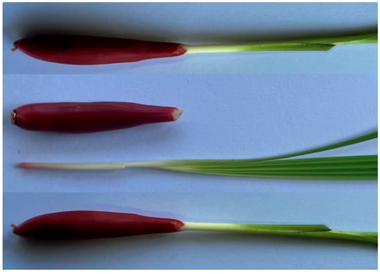
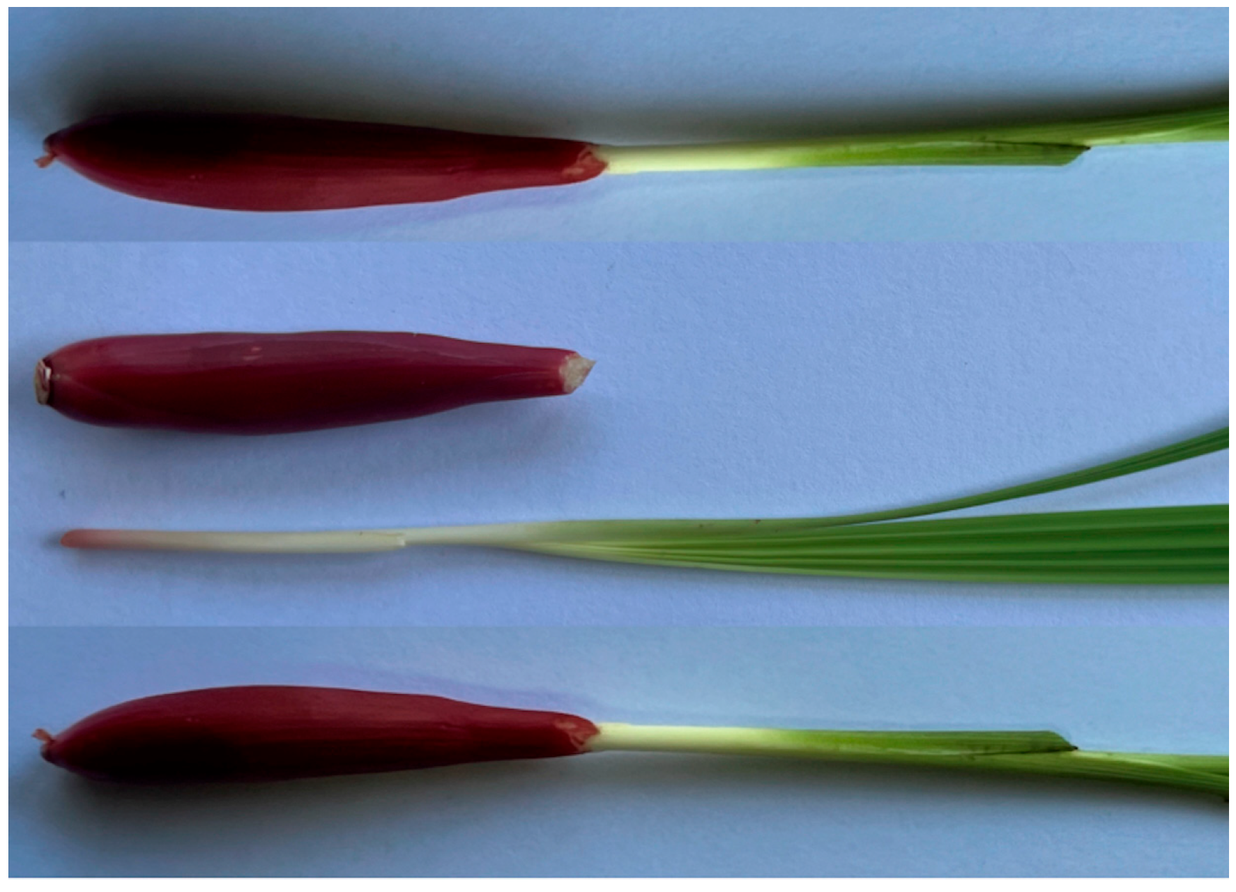
Eleutherine bulbosa (Mill.) Urb. (Figure 1), commonly known as the Dayak onion, is a bulbous plant of the Iridaceae family traditionally used by the Dayak people of Borneo. It is of significant scientific interest due to its bulb phytochemistry rich in naphthalene, anthraquinone, and naphthoquinone derivatives. Previous studies have reported a broad spectrum of biological properties for this species, including antimicrobial, antifungal, antiviral, antiparasitic, and antioxidant functions [1]. In particular, its potent antioxidant effects highlight its strong potential for cosmetic applications [1,2].
Figure 1.
Eleutherine bulbosa.
The utilization of natural extracts as active ingredients for skin-whitening and anti-aging (antioxidant) formulations has garnered considerable attention. As safety concerns regarding synthetic actives persist, consumers increasingly gravitate toward non-cytotoxic, plant-derived alternatives [3]. This search has identified numerous natural agents from diverse biological sources that often match the efficacy of synthetic counterparts while exhibiting fewer adverse effects [4,5]. Prominent among these are phenolic compounds and flavonols. For instance, aloesin (a chromone from Aloe vera) is widely employed to modulate melanogenesis [6,7], while catechins—the principal antioxidants in green tea—have been extensively studied and successfully incorporated into commercial cosmetic lines [8].
To address the challenges associated with the stability, efficacy, and sensory attributes of labile botanical actives, encapsulation technologies are increasingly integrated into cosmetic formulations [9,10]. Liposomes are particularly advantageous carriers due to their amphiphilic structure, biocompatibility, and biodegradability [11]. These systems can markedly enhance transdermal delivery. Notably, liposomal systems containing only 0.1% linoleic acid have demonstrated whitening effects comparable to non-liposomal 10% linoleic acid, illustrating the capacity of encapsulation to reduce required concentrations without compromising efficacy [12].
While synthetic inhibitors such as kojic acid and alpha-arbutin are industry standards, ongoing concerns regarding their long-term safety and limited skin penetration have renewed interest in plant-derived tyrosinase inhibitors [13,14,15]. Anthraquinones and related quinones from plant sources have specifically demonstrated tyrosinase-inhibitory effects that often exceed the potency of kojic acid [4,16,17,18,19]. Despite containing a diverse array of phenolics, flavonoids, and quinone-type constituents—all plausible anti-tyrosinase agents—E. bulbosa has received limited comprehensive chemical profiling or formulation-oriented evaluation. Nanocarrier systems, particularly liposomes, offer a strategic means to enhance the cutaneous bioavailability and controlled release of these phytoconstituents, thereby improving topical performance. Consequently, liposomes were selected as the delivery system for the extract in this study. Liposome preparation strategies have evolved from conventional batch techniques to controlled microfluidic platforms. Traditional methods, including thin-film hydration, solvent injection, reverse-phase evaporation, and detergent removal, rely on organic solvents and frequently require subsequent downsizing via extrusion or sonication [20,21]. Milder alternatives, such as ethanol-based proliposome methods, utilize concentrated liquid-crystalline lipid slurries that spontaneously hydrate into vesicles upon buffer addition [22]. Meanwhile, microfluidic technologies enable continuous, inline lipid self-assembly under ambient temperatures, offering precise control over vesicle size and polydispersity [23].
In this study, E. bulbosa bulbs were extracted with ethanol—a solvent selected for its high efficiency in recovering bioactive constituents [2]—and the resulting extract was characterized via Ultra-High Performance Liquid Chromatography–Quadrupole Time-of-Flight Mass Spectrometry (UHPLC-QTOF-MS). The tyrosinase-inhibitory activity of the extract, a previously unreported bioactivity for this species, was evaluated. Subsequently, the extract was encapsulated into liposomal carriers prepared using both high-energy ultrasonication and microfluidic techniques. The resulting formulations were assessed for entrapment efficiency, physicochemical properties (particle size, polydispersity index, and zeta potential), and bioactivity. This research aims to determine whether liposomal incorporation enhances the delivery of E. bulbosa extract, supporting its potential development as a novel, natural skin-lightening ingredient for topical cosmetic applications.
2. Materials and Methods
2.1. Plant Samples and Chemicals
E. bulbosa was collected from a private garden in Chiang Rai Province, Thailand. The plant was identified by comparison with the medicinal plant database of the Queen Sirikit Botanic Garden Herbarium, Thailand. Folin–Ciocalteu reagent, gallic acid, L-tyrosine, mushroom tyrosinase and quercetin were purchased from Sigma-Aldrich (St. Louis, MO, USA). Anthraquinone (97%) was from Sigma Aldrich®, Czech Republic. Solvent for extraction (95% Ethanol) was of cosmetic grade. The Strat-M™ membrane (Transdermal Diffusion Test Model, 25 mm diameter) was from Merck, Germany. Hydrogenated lecithin (Nikkol Lecinol S-10E) was obtained from Nippon Surfactant Industries Co., Ltd. (Tokyo, Japan).
2.2. Preparation of Eleutherine bulbosa Bulb Crude Extract
The plant material was collected, washed to remove soil, and air-dried for one day to reduce surface moisture. Fresh E. bulbosa bulbs were cut into small pieces and extracted with 95% ethanol at a plant-to-solvent ratio of 1:3 (w/v). The mixture was sonicated using an ultrasonic bath (TUC-100, 40 kHz; Jeken Ultrasonic Cleaner Ltd., Dongguan, China) for 2 h, at ambient temperature, followed by filtration through filter paper. The extraction process was repeated twice, and the filtrates were combined. Ethanol was then evaporated under reduced pressure using a rotary evaporator to obtain the crude extract.
2.3. Total Phenolic Content
The total phenolic content of the sample was determined by a Folin-Ciocalteu total phenolic assay [2]. Gallic acid was used as a standard. Deionized water (1.58 mL) and Folin–Ciocalteu reagent (100 μL) were added to the sample, the standard, or the blank (20 μL). The reaction mixture was incubated at room temperature for 5 min. Sodium carbonate solution (300 μL, 10% w/v) was then mixed with it and incubated for 90 min. The absorbance of the mixture was measured spectrophotometrically at 765 nm (SP-8001; Metertech Inc., Taipei, Taiwan). The total phenolic content was calculated from a standard curve with a coefficient of determination (R2) of 0.999 and expressed as gallic acid equivalent (GAE) per gram of crude extract.
2.4. Total Flavonoid Content
The aluminum chloride colorimetric method was used for the determination of the total flavonoid content of the sample [2]. Quercetin was used to make the standard calibration curve. An amount of 1.2 mL of standard quercetin solution or extract was separately mixed with 1.2 mL of 2% aluminum chloride. After mixing, the solution was incubated for 60 min at room temperature. The absorbance of the reaction mixtures was measured against the blank at 420 nm using a spectrophotometer (Metertech SP-8001 UV/Visible, Taiwan). The concentration of total flavonoid content in the test samples was calculated from the calibration plot with a coefficient of determination (R2) of 0.999 and expressed as the quercetin equivalent (QE) per gram of crude extract.
2.5. Determination of Anthraquinone Content
Quantitative analysis of total anthraquinones in the E. bulbosa extract was conducted via UV-Vis spectrophotometry, following previously established protocols [2]. Anthraquinone was employed as the reference standard for the quantification. A five-point calibration curve was constructed by measuring the absorbance of standard solutions across a concentration range of 0.00625 to 0.1 mg/mL. Measurements were performed at the maximum absorption wavelength of 325 nm using a spectrophotometer (Metertech SP-8001 UV/Visible, Taiwan), with all samples analyzed in triplicate to ensure reproducibility. The relationship between concentration and absorbance was determined using a linear regression analysis with a coefficient of determination (R2) of 0.999. The total anthraquinone content was subsequently calculated and expressed as milligrams of anthraquinone equivalents (AE) per gram of crude extract.
2.6. Determination of Tyrosinase Inhibitory Activity
Tyrosinase inhibitory activity was determined according to the method previously described [24]. A reaction mixture consisting of 0.9 mL of inhibitor and 2 mL L-tyrosine solution (0.244 mM) in aqueous phosphate buffer (pH 6.8; I = 0.01 M) was prepared for the control sample. Oxidation of L-tyrosine was initiated by introducing 0.1 mL of aqueous mushroom tyrosinase solution. The test mixture and control mixture were incubated for 30 min at 37 °C. Dopachrome appearance was monitored spectrophotometrically at 475 nm (Metertech SP-8001 UV/Visible, Taiwan). The commercial grades of kojic acid and alpha-arbutin were used as reference standards. The tyrosinase inhibition (%) was calculated by the following equation.
where Acontrol is the absorbance of the control system, which contains all reagents except the tested sample, and Asample is the absorbance of the sample or standard.
Tyrosinase inhibition (%) = [(Acontrol − Asample)/Acontrol] × 100
Tyrosinase inhibition of the prepared liposomes was also assessed by mixing 5 g liposomes with 5 g ethanol, sonicating for 10 min, and centrifuging at 5000 rpm for 10 min. The supernatant was used for the tyrosinase inhibition assay at a 10% concentration (final liposome concentration 5% w/w).
2.7. UHPLC-QTOF-MS/MS Analysis
An optimized LC-QTOF method using a UHPLC Agilent 1290 Infinity II System coupled to an Agilent 6545 LC-QTOF/MS (Agilent Technologies, CA, USA) was developed for the qualification of active compounds. This method involved optimizing various chromatographic parameters, including the mobile phase composition, column type, flow rate, and solvent ratio. A Waters XBridge C18 (100 mm × 2.1 mm, 2.5 μm) column was used for the separation. The extract was diluted with ethanol at a ratio of 1:25 and filtered using 0.22 µm syringe filters (Labfil; Zhejiang ALWSCI Technologies Co., Ltd., Shaoxing, China) and transferred into HPLC vials. The elution was achieved using a binary gradient system, with 0.1% formic acid in water that was LCMS grade (J.T.Baker, Avantor, Inc., Radnor, PA, USA) as eluent A and 0.1% formic acid in acetonitrile (ACN) as eluent B. The gradient steps were 5–17% B at 0–13 min, followed by 17–100% B at 13–20 min, 100% B at 20–25 min, and 100–5% B at 25–27 min, with a flow rate of 0.2 mL/min. A postrun was set to equilibrate the column for 6 min. Dual AJS (Agilent Jet Stream) ESI was used as the ion source, with a sheath gas flow of 12 L/min, sheath gas temperature at 250 °C, drying gas temperature at 350 °C, drying gas flow at 10 L/min and capillary voltage at 3500 V. A complete mass scan ranging from m/z 50 to 1100 was used. MS/MS analyses were carried out in automatic mode with collision energies of (10, 20 and 40 eV) for fragmentation. Peak identification was performed in both positive and negative modes, while the instrument control, data acquisition and processing were performed using MassHunter workstation software (Qualitative Analysis, version B.8.00) (Agilent Technologies, Inc., Santa Clara, CA, USA).
2.8. Preparation of Liposomes
A proliposome method was employed for liposome preparation according to Perrett et al. (1991) [22], with modifications. The empty liposomes were prepared by dissolving hydrogenated lecithin in ethanol, stirred and heated at 50 °C until clear. The acetate buffer (25 mmol/L, pH 3.8 ± 0.1) was added and stirred on a hotplate stirrer for 15 min. For E. bulbosa-loaded liposomes, the E. bulbosa solution was prepared by dissolving the required amount of crude extract in ethanol. The solution was sonicated in an ultrasonicator (Jeken, TUC-100) for 1 h. The required amount of lecithin was then dissolved in E. bulbosa solution, stirred and heated at 50 °C until clear. The acetate buffer (25 mmol/L, pH 3.8 ± 0.1) was added and stirred in a hotplate stirrer for 15 min. Size reduction was performed by an ultrasonic processor (SONICs, VCX 130, USA) at 40% amplitude for 10 min. Different concentrations of the hydrogenated lecithin and the crude extract on the formation of the liposome were studied. The microscopy imaging of liposomes was recorded by microscopy (Motic, BA300, USA). The particle size and zeta potential values were obtained and collected using a nanoparticle size analyzer (Horiba, SZ-100Z2, Japan). Finally, a microfluidic technique (Microfluidizer LM20, High Shear Fluid Processor, USA) was employed to prepare nanoliposomes at the optimal concentration of hydrogenated lecithin and the crude extract.
2.9. Encapsulation Efficiency
The encapsulation efficiency of E. bulbosa bulb extract-loaded liposomes was determined indirectly by measuring the amount of unencapsulated extract. The liposome was frozen at −2 °C and centrifuged at 8000 rpm, 4 °C for 10 min. Incomplete separation was excluded by inspection of pellet integrity and supernatant clarity (compact pellet, optically clear supernatant); samples exhibiting turbidity were re-centrifuged until a clear supernatant was obtained. The upper supernatant solution, which contains free active (unencapsulated extract), was measured at 245 nm using a spectrophotometer (Metertech SP-8001 UV/Visible, Taiwan). The encapsulation efficiency of the liposomes containing E. bulbosa was calculated according to the following equation.
where Total extract is the E. bulbosa extract being added to the liposomal system and Free extract is the unencapsulated extract in the supernatant.
%Encapsulation efficiency (EE) = [(Total extract − Free extract)/Total extract] × 100
2.10. In Vitro Drug Release and Skin Permeation Experiment
Franz diffusion cells (Model FDC-6; Logan Instruments Corp., Somerset, NJ, USA) were used to evaluate the in vitro release and transdermal permeation of the extract. All experiments were conducted for 22 h under controlled conditions at 37 ± 1 °C with continuous stirring at 600 rpm. The receptor compartments were filled with 15 mL of acetate buffer (0.025 M, pH 3.8), while 1000 μL of the sample was placed in each donor compartment. For the in vitro release study, a cellulose acetate (CA) membrane with a pore size of 0.45 μm was used. For the skin permeation study, synthetic membranes made of polyethersulfone or Strat-M™ (Merck Millipore), both with a diameter of 25 mm, were used as skin models. At predetermined time points (0.5, 1, 2, 3, 4, 5, 22 h), 0.5 mL samples were manually withdrawn from the receptor compartment and immediately replaced with an equal volume of fresh acetate buffer to maintain a constant receptor-phase volume. The extract concentration in the collected samples was determined spectrophotometrically at 245 nm using a UV–visible spectrophotometer (Metertech SP-8001, Taiwan). Skin permeation parameters: steady-state flux (Jss), permeability coefficient (Kp), and enhancement ratio (ER = Jss_liposome/Jss_extract) were estimated over the linear permeation interval (1–5 h).
2.11. Statistical Analysis
All data points were compiled as the mean ± standard deviation. Intra-group longitudinal variations over time were calculated utilizing the paired-samples t-test. Statistical significance thresholds were fixed at p < 0.05 (95% confidence level) using SPSS version 21.0.
3. Results
3.1. E. bulbosa Bulb Crude Extract and Its Bioactive Compounds
The crude extract yield from E. bulbosa bulbs was 6.41 ± 1.36% (mean ±SD), based on three batches of varying sizes. The crude extracts were orange–red solids. The extract’s bioactive profile was quantified by its total phenolic content (TPC) and total flavonoid content (TFC), yielding values of 89.88 ± 4.36 mg GAE/g and 7.99 ± 0.05 mg QE/g of crude extract, respectively. The E. bulbosa bulb extract also displayed an anthraquinone content of 114.12 ± 0.85 mg AE/g crude extract. The anthraquinone content was determined spectrophotometrically using anthraquinone (97% purity, CAS 84-65-1) as the external standard, with measurements performed at the compound’s absorption maximum (λmax = 325 nm). Quantification was carried out independently of the total phenolic assay.
3.2. In Vitro Anti-Tyrosinase Activities
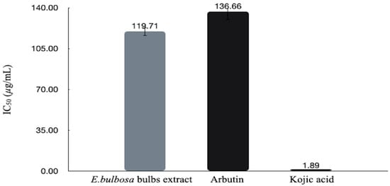
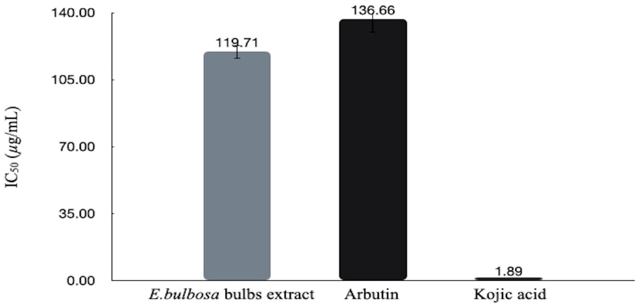
The E. bulbosa bulb extract exhibited anti-tyrosinase activities (Figure 2). The cosmetic grades of kojic acid and alpha arbutin were used as standards. The half-maximal inhibitory concentration (IC50) values were determined, and the E. bulbosa bulb extract exhibited comparable tyrosinase inhibition (IC50 = 119.71 ± 3.76 µg/mL) to the commercial α-arbutin (IC50 = 136.66 ± 12.49 µg/mL), as this difference did not reach statistical significance (p > 0.05). Given its comparable activity to a benchmark depigmenting agent, the crude E. bulbosa bulb extract warrants further investigation as a potential natural candidate for topical skin-lightening formulations. Compared with commercial kojic acid (IC50 = 1.89 ± 0.26 µg/mL), the E. bulbosa bulb extract exhibited markedly lower tyrosinase inhibitory potency.
Figure 2.
The anti-tyrosinase activities of E. bulbosa bulbs crude extract, kojic acid and alpha-arbutin.
3.3. Qualification of Active Compounds by UHPLC-QTOF-MS/MS
The identities of the biological compounds in E. bulbosa bulbs crude ethanolic extract were obtained by matching the molecular m/z values from the UHPLC-ESI-QTOF-MS/MS.
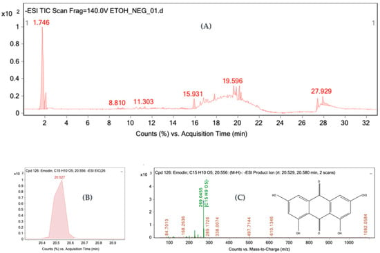
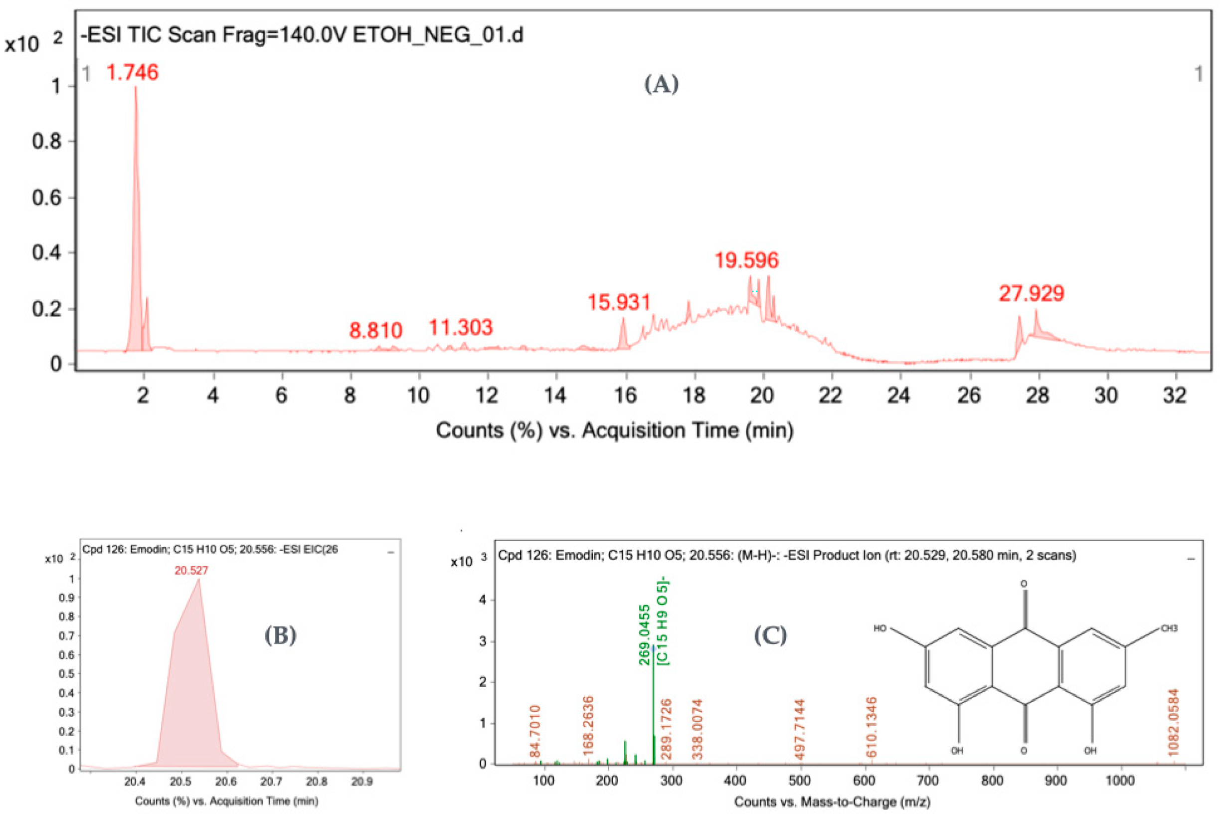
Figure 3 presents the total ion chromatogram alongside the extracted ion chromatogram and m/z spectrum for emodin as a representative example from the analysis. Naphthalenes, naphthoquinones, anthraquinones, catechin, epicatechin, and their derivatives were detected in the extract. Additionally, myricetin, quercetin compounds, emodin, rutaretins, 2-hydroxy-3,4,6-trimethoxydihydrochalcone, ceanothic acid, mangiferin, rhapontin, gingerol, and 2,3,4′-trihydroxy-4-methoxybenzophenone were present (Table 1). Tentative compound annotations in this work were assigned primarily on the basis of high-resolution mass accuracy and library spectral matching. For high-confidence identification, the mass errors between the observed and theoretical (m/z) values were kept below the accepted 5 ppm database threshold, alongside excellent spectral library match scores (≥90%) [25].
Figure 3.
Representation of the total ion chromatogram (A), extracted ion chromatogram of emodin (B), and its corresponding m/z spectrum (C).
Table 1.
Tentatively identified compounds in the E. bulbosa bulbs crude ethanolic extract obtained by UHPLC-QTOF-MS/MS.
3.4. Optimization and Characterization of Liposomes
The liposomal formulations were prepared using a two-stage fabrication process starting with the proliposome method followed by ultrasonication to achieve particle size reduction. During this initial phase, the effects of varying lecithin and extract concentrations on liposome formation were systematically evaluated. The resulting formulations were screened for key physicochemical properties—including physical texture, particle size, and zeta potential—to determine the optimal extract-to-lecithin baseline ratio. This optimized combination was subsequently selected for microfluidic fabrication. Finally, microfluidic processing parameters, specifically the number of passes, as well as higher extract and lecithin loadings, were evaluated and optimized based on physical texture, particle size, zeta potential, and encapsulation efficiency (%EE).
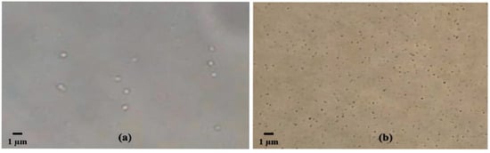
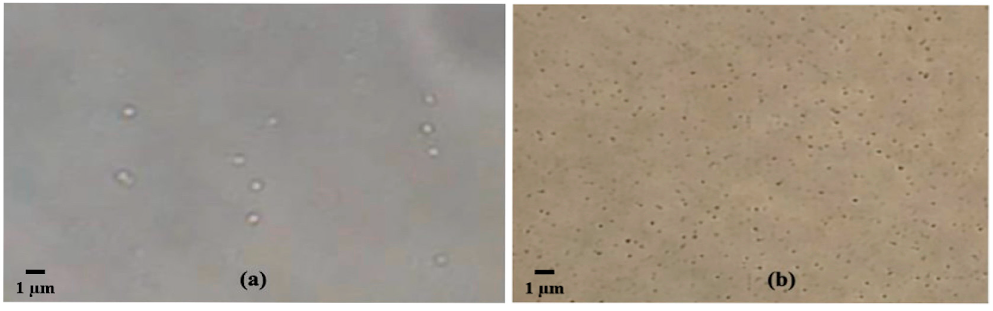
The empty liposomes and E. bulbosa-loaded liposomes were prepared using the proliposome method [22]. The hydrogenated lecithin (0.5 g) was dissolved in an E. bulbosa solution (0.1% w/v, 2 g), stirred and heated at 50 °C until clear. Acetate buffer (q.s. to 50 g total) was added and stirred on a hotplate stirrer for 15 min. The liposomes exhibited a translucent liquid appearance under microscopy (Figure 4a). The particle size of the liposomes was reduced using a high-intensity ultrasonic processor (Figure 4b).
Figure 4.
The microscopy imaging of liposomes (a) before size reduction and (b) after size reduction by high-intensity ultrasonic processor.
The particle sizes of the empty liposomes and E. bulbosa-loaded liposomes before the ultrasonic processor process were 447.0 ± 36.63 nm (PDI 0.49 ± 0.03) and 497.05 ± 74.88 nm (PDI 0.63 ± 0.14), respectively. The particle sizes after the ultrasonic processor were 167.2 ± 4.24 nm (PDI 0.31 ± 0.03) and 208.8 ± 6.08 nm (PDI 0.36 ± 0.07), respectively, and the zeta potential values were −43.0 ± 0.28 mV and −45.9 ± 0.64 mV, respectively. The preparation of E. bulbosa-loaded liposomes was then fabricated through the variation of the hydrogenated lecithin and the crude extract.
3.4.1. Effects of Hydrogenated Lecithin and Extract Concentrations on Liposome Formation
Hydrogenated lecithin at 1.0, 2.0, 3.0, 3.6, and 4% (w/w) was dissolved in E. bulbosa solution (0.004% w/w) after being mixed with buffer solution and homogenized by the ultrasonic processor. At high concentrations of hydrogenated lecithin, the liposomes displayed a viscous texture. The particle sizes of the liposomes ranged from 175.80 ± 6.36 nm to 211.40 ± 3.11 nm with a low polydispersity index (PDI). The zeta potential values decreased at higher lecithin concentrations.
Different E. bulbosa concentrations (0.004, 0.008, 0.016, and 0.04% w/w) were loaded into the liposomes. The particle size of the liposomes loaded with E. bulbosa decreased at higher concentrations of extract, and a similar decreasing trend was obtained for the zeta potential values (Table 2).
Table 2.
Physicochemical characteristics of liposomes with different lecithin and E. bulbosa crude extract concentrations fabricated by ultrasonication.
3.4.2. Preparation of Liposome Using High-Pressure Microfluidization
E. bulbosa crude extract (0.04% w/w) and hydrogenated lecithin (3.6% w/w) were used as a starting condition for fabrication via an LM20 Microfluidizer™ High Shear Fluid Processor at a pressure of 10,000 psi. Table 3 shows the characteristics of the liposomes after each cycle. The size of the liposomes decreased from 2156.3 ± 125.02 nm to 119.8 ± 0.49 nm after passing through four cycles.
Table 3.
The characteristics of E. bulbosa-loaded liposomes produced by microfluidic technique.
Following these initial tests, the preparation of E. bulbosa-loaded liposomes was studied with higher concentrations of E. bulbosa extract and hydrogenated lecithin, homogenized by passing three times through the Microfluidizer at 10,000 psi (Table 4). Increasing the extract and lecithin concentrations led to reduced liposomal zeta potentials. Entrapment efficiency declined to 48.70 ± 2.10% for Formula 2 (0.08% extract with 7.2% lecithin). Formula 1, prepared with 0.06% w/w E. bulbosa bulb extract and 5.4% w/w hydrogenated lecithin, exhibited an encapsulation efficiency of 86.16 ± 3.04% and, at 5% w/w, produced 92.79 ± 0.43% tyrosinase inhibition.
Table 4.
The characteristics of liposomes produced by microfluidic technique using different concentrations of E. bulbosa bulb extract and hydrogenated lecithin.
3.5. In Vitro Release Evaluation and Transdermal Permeation Behavior
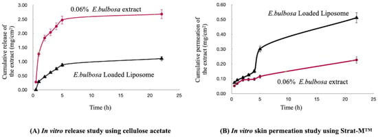
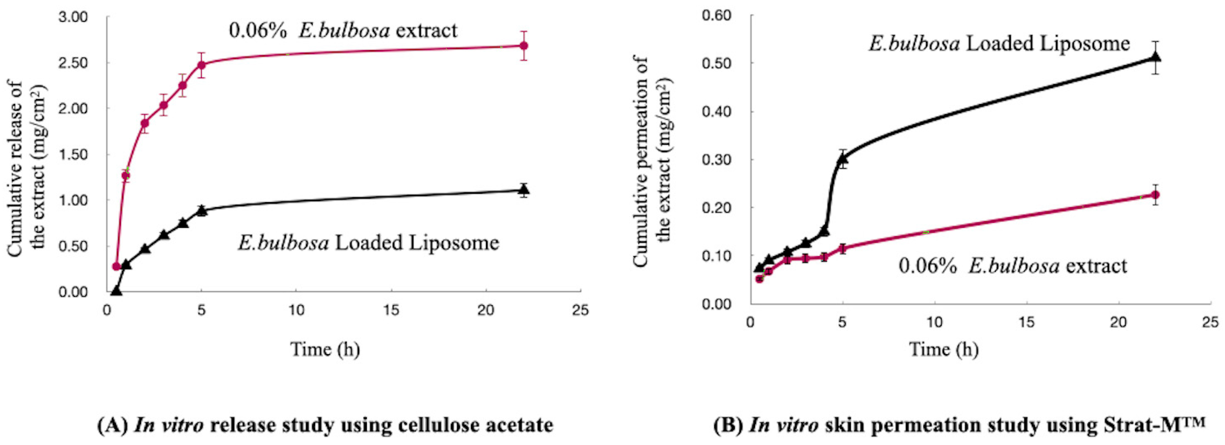
The release and skin permeation profiles of the E. bulbosa bulb extract from the loaded liposomes (Formula (1)) were evaluated using Franz diffusion cells and compared with the diffusion profile of an ethanolic extract control solution.
Across the cellulose acetate (CA) membrane (pore size 0.45 µm), diffusion of the extract from the control solution occurred rapidly, reaching a maximum concentration in the acceptor compartment after approximately 5 h (Figure 5A). The extract released from the liposomes through the CA membrane was slower. Skin permeation parameters [26,27]—steady-state flux (Jss), permeability coefficient (Kp), and enhancement ratio (ER = Jss_liposome/Jss_extract)—were estimated over the linear permeation interval (1–5 h) (Figure 5). Across CA, Jss was 417.87 μg/cm2/h for the extract and 177.51 μg/cm2/h for the extract-loaded liposomes. Corresponding Kp values were 0.6956 cm/h (extract) and 0.2959 cm/h (liposomes), yielding an ER of 0.42. Thus, under CA conditions, the non-encapsulated extract exhibited higher apparent flux and permeability than the liposomal formulation (ER < 1), indicating reduced release/permeation from liposomes through CA. Across Strat-M®, Jss was 12.261 μg/cm2/h for the extract and 41.588 μg/cm2/h for the extract-loaded liposomes. Corresponding Kp values were 0.0204 cm/h (extract) and 0.0693 cm/h (liposomes). The ER of 3.4 indicates that liposomal encapsulation increased steady-state permeation approximately 3.4-fold relative to the non-encapsulated extract under these conditions.
Figure 5.
Cumulative amount of the E. bulbosa bulb extract from the in vitro drug release and skin permeation study.
4. Discussion
4.1. Phytochemical Profile and Extraction Efficiency
The total phenolic content (TPC) obtained in this study (89.88 ± 4.36 mg GAE/g) is slightly higher than the 85.52 ± 1.30 mg GAE/g value reported by Kamarudin et al. (2020), who utilized 90% ethanol via conventional maceration (45 °C, 70 min) [28]. The variations in TPC values between these studies may be attributed to differences in extraction conditions, specifically our utilization of the ultrasonication technique, fresh plant samples, and varying plant-to-solvent ratios. Furthermore, our results align conceptually with Morabandza et al. (2016), who demonstrated that ethanolic E. bulbosa bulb extracts display significantly higher total phenolic (27.12 mg GAE/g DW) and flavonoid content (17.97 mg RE/g DW) compared to aqueous alternatives [29]. Similarly, Agustin et al. (2016) reported 70% methanol as an effective solvent for polyphenol recovery from this matrix (20 mg GAE/g DW and 15.03 QE/g DW) [30]. Collectively, these comparisons emphasize that the selection of organic solvents and advanced processing techniques like ultrasonication critically dictate the final yield and chemical profile of the extract.
Regarding the anthraquinone assay, it must be noted that the spectrophotometric method applied at 325 nm is not completely specific for anthraquinones. Other co-extracted constituents absorbing close to this wavelength—including alternative quinones, phenolic compounds, conjugated aromatics, and minor degradation products—may contribute to the overall absorbance, potentially leading to an overestimation of the total anthraquinone content. Nevertheless, these data provide a valuable, practical approximation of anthraquinone abundance in E. bulbosa, where they are widely recognized as major bioactive constituents [1].
4.2. Anti-Tyrosinase and Skin-Lightening Activity
Tyrosinase plays a crucial role in melanin biosynthesis and serves as a primary target in cosmetic and medical research for managing hyperpigmentation. Plant extracts exhibit a broad spectrum of efficacy characterized by their IC50 values [31]. The moderate anti-tyrosinase activity of the E. bulbosa extract (IC50 = 119.71 ± 3.76 µg/mL) falls well within the ranges reported for other regional botanical sources. For instance, extracts from the Brazilian Cerrado display IC50 values spanning 11.88 to 361.23 µg/mL (relative to a kojic acid benchmark of 13.14 µg/mL) [32], while South African plant extracts range from 2.95 to 231.10 µg/mL (with kojic acid at 6.45 µg/mL) [33]. Furthermore, our extract performs comparably to commercial green tea extracts, which typically feature IC50 values between 50 and 100 µg/mL [34]. According to the classification framework by Ekanayaka and De Silva (2023), the E. bulbosa extract exhibits a clear moderate potency [35]. However, direct benchmarking against the diverse literature remains complex due to baseline variations in anti-tyrosinase assay conditions, extraction methodologies, and solvent selections.
The multi-targeted skin-lightening potential observed in the crude extract is directly supported by its complex phytochemical constituents identified via UHPLC-QTOF-MS/MS. The presence of naphthalenes, naphthoquinones, and anthraquinones matches the previous qualitative literature [1,28,36,37,38,39,40,41].
Catechins, epicatechins, myricetins, quercetins and kaempferols are naturally present in plants and are known for their tyrosinase inhibitory activities [14,42]. Emodin, rutaretins, and 2-hydroxy-3,4,6-trimethoxydihydrochalcone were also found in the extract. Emodin (1,3,8-trihydroxy-6-methyl-anthraquinone) is a natural anthraquinone aglycone that has been reported to possess potential therapeutic applications, such as anticancer, neuroprotective, antidiabetic, antioxidant and anti-inflammatory ones [43]. It has been reported that the anthraquinones from Polygonum cuspidatum showed moderate to strong inhibition of tyrosinase and were recommended as tyrosinase inhibitors for dermal use [18,19,35]. Rutaretin and rutaretin 9-rutinoside are coumarin and coumarin glycoside compounds, which have been studied in areas of anti-inflammatory, anti-depressant and antimicrobial activity [44,45]. Also, coumarin-based compounds have shown potential activity as anti-melanoma agents and tyrosinase inhibitors [46]. 2-Hydroxy-3,4,6-trimethoxydihydrochalcone is a chalcone that belongs to the flavonoid class of phenolic compounds. Chalcones were reported to exhibit potential anticancer, anti-inflammatory, antimicrobial, antioxidant, antiparasitic and anti-tyrosinase properties [47,48]. In addition, ceanothic acid, mangiferin, rhapontin, gingerol and 2,3,4′-trihydroxy-4- methoxybenzophenone were also tentatively identified in the E. bulbosa bulb extract. Among these, mangiferin, rhapontin and gingerol are compounds with reported potent skin-whitening effects. Mangiferin acts as a strong antioxidant capable of directly inhibiting L-DOPA oxidation [49]. Rhapontin is utilized as a commercial skin-whitening raw material [50], and gingerol actively suppresses cellular melanogenesis [51].
As summarized in Table 5, the E. bulbosa bulb extract possesses a diverse array of chemical constituents with potential applications in skin-lightening formulations. Flavonoids (catechin, epicatechin, myricetin, and quercetin), coumarins, and 2-hydroxy-3,4,6-trimethoxydihydrochalcone directly inhibit tyrosinase and disrupt melanin synthesis. Complementing these, mangiferin prevents L-DOPA oxidation, gingerol suppresses melanogenesis, and rhapontin acts as a proven skin-whitening agent, providing a multi-targeted mechanism for cosmetic and therapeutic use. It is noteworthy to address that tentative compound identifications were assigned using high-resolution mass accuracy (≤5 ppm threshold) and spectral library matching (≥90 score threshold). Key actives showed high-confidence annotations with low mass errors (1–2 ppm) and high library match scores (>97% similarity). Nevertheless, definitive identification requires comparison with authentic reference standards or orthogonal structural verification (e.g., NMR or authentic standards), and quantitative profiling is needed to identify the main active drivers.
Table 5.
Summary of the phytochemical compounds identified via UHPLC-QTOF-MS/MS analysis that possess documented skin-lightening effects.
Moreover, because the current in vitro assay focuses solely on the direct inhibition of isolated mushroom tyrosinase, it cannot fully replicate the regulatory complexities of human melanogenesis. To substantiate formal cosmetic depigmentation claims, further research using human-relevant cellular models and controlled clinical trials remains essential [52,53,54].
From a safety perspective, the crude extract previously demonstrated no cytotoxicity in human dermal fibroblasts, indicating a favorable safety profile for topical application [2]. Nevertheless, clinical safety trials on human skin are recommended to fully validate these findings.
4.3. Evaluation of Liposomal Delivery Systems and Transdermal Permeation
Liposomes function as effective spherical phospholipid vesicles that encapsulate active ingredients, improving their stability, bioavailability, and controlled release profiles within the skin [55,56]. The selection of the optimal formulation parameters (3.6% w/w hydrogenated lecithin) was governed by the balance between viscosity and colloidal stability; higher lecithin concentrations yielded high viscosity but caused a concurrent reduction in absolute zeta potential, which can compromise long-term colloidal stabilization. Interestingly, loading higher concentrations of the E. bulbosa extract led to a decrease in liposomal particle size and a reduction in zeta potential (Table 2), indicating a strong structural interaction between the amphiphilic phytochemicals and the lipid bilayer.
When scaling up formulation processing, the microfluidic technique (Microfluidizer™) proved highly efficient compared to standard ultrasonication. The high pressure (10,000 psi) forces samples through fixed microchannels, driving particle breakup via controlled jet impact, cavitation, and turbulent eddies to deliver smaller mean diameters, narrower size distributions (PDI), and superior batch-to-batch reproducibility [57,58,59]. The cycle-dependent shift in liposomal properties (Table 3) reflects high-pressure microfluidization dynamics at 10,000 psi. Initial passes generate intense shear and cavitation, downsizing multilamellar vesicles into small unilamellar vesicles and exposing a higher density of negatively charged phosphate headgroups to increase the negative zeta potential. Conversely, excessive cycling causes shear heating and high-impact collisions, driving amphiphilic E. bulbosa constituents to the bilayer interface. This surface migration sterically shields charged lipid headgroups and disrupts surface hydration, shifting the zeta potential toward less negative values. Optimizing the pass number is therefore critical to achieve size reduction without altering the surface charge or membrane stability. This is clearly evidenced by the stepwise reduction in size down to 119.8 ± 0.49 nm and a highly uniform PDI (0.239 ± 0.03) over four continuous cycles (Table 3), confirming that microfluidics is a reliable method for nanoliposome production [60].
However, overloading the system with excessive extract and lipid materials (Formulas 2 and 3) resulted in a dramatic drop in encapsulation efficiency (47.80%) and a drop in the zeta potential magnitude (−28.9 mV), likely due to bilayer saturation [61]. Hydrogenated lecithin forms rigid, tightly packed membranes due to its saturated acyl chains. Exceeding the maximum loading capacity with the complex, amphiphilic E. bulbosa extract introduces steric hindrance and disrupts inter-lipid chain packing [62]. This thermodynamic constraint leads to payload expulsion into the aqueous phase and surface charge masking. Thus, Formula 1 (0.06% w/w extract, 5.4% w/w lecithin) represented the ideal equilibrium, striking an outstanding encapsulation efficiency (86.16 ± 3.04%) and strong functional tyrosinase inhibition (92.79 ± 0.43%).
The performance differences observed between the two diffusion membranes in the Franz cell experiments highlight distinct transport mechanisms. Synthetic cellulose acetate (CA) serves as a simple, highly porous barrier with no rate-limiting properties, meaning it does not correlate with human skin-barrier physics. Under CA conditions, the unencapsulated control extract solution diffused rapidly and exhibited a higher apparent flux and permeability coefficient than the liposomes (ER = 0.42). This confirmed that the liposomal bilayer successfully retained the active compounds, providing a prolonged, controlled release profile that prevents rapid, unchecked dumping of the active compounds. Conversely, when using the biomimetic Strat-M™ membrane—which closely replicates the multi-layered epidermal/dermal lipid gradient of human skin [63]—the extract-loaded liposomes flipped the performance dynamically, achieving a 3.4-fold increase in steady-state flux (Jss) and permeability (Kp) compared to the control solution. This enhanced transdermal delivery (ER = 3.4) is theorized to be due to the structural nature of nanoliposomes; their small spherical phospholipid bilayers effectively mimic natural cellular membrane constituents, facilitating superior partition and diffusion through the specialized lipid matrix [26,27,60].
Ultimately, these findings demonstrate that delivering E. bulbosa bulb extract via optimized nanoliposomes yields the dual advantage of prolonged, controlled active release and significantly enhanced skin permeation, underscoring its viability for advanced topical skin-lightening applications.
Limitations of the study: This study has several methodological limitations. First, biological evaluation relied on cell-free, non-human tyrosinase assays; follow-up studies in human melanocyte models are required to validate safety, cellular uptake, and depigmenting efficacy. Second, formulation characterization was restricted to baseline, leaving longitudinal stability testing to demonstrate shelf life for a dedicated future publication. Finally, phytochemical profiling via UHPLC-QTOF-MS/MS and UV-Vis spectrophotometry was qualitative and semi-quantitative due to the unavailability of reference standards and instrument constraints; validated HPLC-DAD quantification of key active markers remains necessary to ensure batch reproducibility.
5. Conclusions
The ethanolic crude extract of Eleutherine bulbosa bulbs demonstrated significant in vitro tyrosinase inhibitory activity (IC50 = 119.71 ± 3.76 µg/mL), showing a potency comparable to the commercial standard α-arbutin (IC50 = 136.66 ± 12.49 µg/mL). UHPLC-QTOF-MS/MS profiling revealed a complex phytochemical composition dominated by phenolics, flavonoids, anthraquinones, naphthalenes, naphthoquinones, coumarins, chalcones, and flavonols. Among these constituents, several possess documented skin-lightening effects, including mangiferin, rhapontin, 2-hydroxy-3,4,6-trimethoxydihydrochalcone, and various coumarins, as well as flavonoids such as catechin, epicatechin, myricetin, and quercetin. Microfluidic production yielded optimized extract-loaded liposomes with favorable physicochemical properties (size 114.0 ± 1.23 nm, PDI 0.24 ± 0.04, zeta potential −38.3 ± 0.07 mV), high entrapment efficiency (86.16 ± 3.04%), and enhanced in vitro dermal permeation compared to the unencapsulated extract. Key limitations include reliance on cell-free assays, qualitative profiling, and a lack of long-term stability data. Future work should focus on quantitative marker validation, cell-based melanogenesis testing, long-term stability monitoring, and incorporation into topical formulation for cosmetic translation.
Author Contributions
Conceptualization, A.J.; methodology, A.J. and N.S.; investigation, A.J. and N.S.; resources, A.J.; data curation, A.J.; writing—original draft preparation, A.J.; writing—review and editing, A.J. and N.S.; supervision, A.J.; project administration, A.J.; funding acquisition, A.J. All authors have read and agreed to the published version of the manuscript.
Funding
The research on ‘In Vitro Tyrosinase Inhibitory Activity of Eleutherine bulbosa Bulb Extract and Characterization of Its Liposomal Delivery System’ by Mae Fah Luang University has received funding support from the National Science, Research, and Innovation Fund (NSRF) (grant no. 662A02024).
Institutional Review Board Statement
Not applicable.
Informed Consent Statement
Not applicable.
Data Availability Statement
The original contributions presented in this study are included in the article. Further inquiries can be directed to the corresponding author.
Acknowledgments
Mae Fah Luang University is acknowledged for providing space and facilities.
Conflicts of Interest
The authors declare no conflicts of interest. The funders had no role in the design of the study; in the collection, analyses, or interpretation of data; in the writing of the manuscript; or in the decision to publish the results.
Abbreviations
The following abbreviations are used in this manuscript:
| UHPLC-QTOF-MS | Ultra-High Performance Liquid Chromatography–Quadrupole Time-of-Flight Mass Spectrometry |
| TPC | Total phenolic content |
| TFC | Total flavonoid content |
| AC | Anthraquinone content |
| CA | Cellulose acetate |
| IC50 | The half-maximal inhibitory concentration |
| PDI | Polydispersity index |
| Jss | Steady-state flux |
| Kp | Permeability coefficient |
| ER | Enhancement ratio |
References
- Kamarudin, A.A.; Sayuti, N.H.; Saad, N.; Razak, N.A.A.; Esa, N.M. Eleutherine bulbosa (Mill.) Urb. bulb: Review of the pharmacological activities and its prospects for application. Int. J. Mol. Sci. 2021, 22, 6747. [Google Scholar] [CrossRef] [Scilit]
- Panyachariwat, N.; Jimtaisong, A.; Saewan, N. Antioxidative potentials of Eleutherine bulbosa bulb and its utilization in topical cosmetic emulsion. Cosmetics 2024, 11, 111. [Google Scholar] [CrossRef] [Scilit]
- Kerdudo, A.; Burger, P.; Merck, F.; Dingas, A.; Rolland, Y.; Michel, T.; Fernandez, X. Development of a natural ingredient—Natural preservative: A case study. C. R. Chim. 2016, 19, 1077–1089. [Google Scholar] [CrossRef] [Scilit]
- Chang, T.S. An updated review of tyrosinase inhibitors. Int. J. Mol. Sci. 2009, 10, 2440–2475. [Google Scholar] [CrossRef] [Scilit]
- Ribeiro, A.S.; Estanqueiro, M.; Oliveira, M.B.; Sousa Lobo, J.M. Main benefits and applicability of plant extracts in skin care products. Cosmetics 2015, 2, 48–65. [Google Scholar] [CrossRef] [Scilit]
- Choi, S.; Lee, S.K.; Kim, J.E.; Chung, M.H.; Park, Y.I. Aloesin inhibits hyperpigmentation induced by UV radiation. Clin. Exp. Dermatol. 2002, 27, 513–515. [Google Scholar] [CrossRef] [Scilit]
- Jones, K.; Hughes, J.; Hong, M.; Jia, Q.; Orndorff, S. Modulation of melanogenesis by aloesin: A competitive inhibitor of tyrosinase. Pigm. Cell Res. 2002, 15, 335–340. [Google Scholar] [CrossRef] [Scilit]
- Peres, I.; Rocha, S.; Gomes, J.; Morais, S.; Pereira, C.; Coelho, M. Preservation of catechin antioxidant properties loaded in carbohydrate nanoparticles. Carbohydr. Polym. 2011, 86, 147–153. [Google Scholar] [CrossRef] [Scilit]
- Casanova, F.; Santos, L. Encapsulation of cosmetic active ingredients for topical application—A review. J. Microencapsul. 2016, 33, 1–17. [Google Scholar] [CrossRef] [Scilit]
- Yang, S.; Liu, L.; Han, J.; Tang, Y. Encapsulating plant ingredients for dermocosmetic application: An updated review of delivery systems and characterization techniques. Int. J. Cosmet. Sci. 2020, 42, 16–28. [Google Scholar] [CrossRef] [Scilit]
- Castañeda-Reyes, E.D.; Perea-Flores, M.J.; Davila-Ortiz, G.; Lee, Y.; Gonzalez de Mejia, E. Development, characterization and use of liposomes as amphipathic transporters of bioactive compounds for melanoma treatment and reduction of skin inflammation: A review. Int. J. Nanomed. 2020, 15, 7627–7650. [Google Scholar] [CrossRef] [Scilit]
- Yasutami, S.; Hiromichi, I.; Hideya, A.; Atsuko, R.; Naoto, O.; Naomichi, B.; Taketoshi, M. Skin whitening effect of linoleic acid is enhanced by liposomal formulations. Biol. Pharm. Bull. 2004, 27, 591–594. [Google Scholar] [CrossRef] [Scilit]
- Kim, Y.J.; Uyama, H. Tyrosinase inhibitors from natural and synthetic sources: Structure, inhibition mechanism and perspective for the future. Cell. Mol. Life Sci. 2005, 62, 1707–1723. [Google Scholar] [CrossRef] [Scilit]
- Hassan, M.; Shahzadi, S.; Kloczkowski, A. Tyrosinase inhibitors naturally present in plants and synthetic modifications of these natural products as anti-melanogenic agents: A review. Molecules 2023, 28, 378. [Google Scholar] [CrossRef] [Scilit]
- Tung, X.Y.; Yip, J.Q.; Gew, L.T. Searching for natural plants with antimelanogenesis and antityrosinase properties for cosmeceutical or nutricosmetics applications: A systematic review. ACS Omega 2023, 8, 33115–33201. [Google Scholar] [CrossRef] [Scilit]
- Devkota, K.P.; Khan, M.T.H.; Ranjit, R.; Lannang, A.M.; Samreen; Choudhary, M.I. Tyrosinase inhibitory and antileishmanial constituents from the rhizomes of Paris polyphylla. Nat. Prod. Res. 2007, 21, 321–327. [Google Scholar] [CrossRef] [Scilit]
- Zeng, H.J.; Sun, D.Q.; Chu, S.H.; Zhang, J.J.; Hu, G.Z.; Yang, R. Inhibitory effects of four anthraquinones on tyrosinase activity: Insight from spectroscopic analysis and molecular docking. Int. J. Biol. Macromol. 2020, 160, 153–163. [Google Scholar] [CrossRef] [Scilit]
- Leu, Y.L.; Hwang, T.L.; Hu, J.W.; Fang, J.Y. Anthraquinones from Polygonum cuspidatum as tyrosinase inhibitors for dermal use. Phytother. Res. 2008, 22, 552–556. [Google Scholar] [CrossRef] [Scilit]
- Du, R.; Ye, L.; Chen, X.; Meng, Y.; Zhou, L.; Chen, Q.; Zheng, G.; Hu, J.; Shi, Z. Screening of key components for melanogenesis inhibition of Polygonum cuspidatum extract based on the spectrum–effect relationship and molecular docking. Molecules 2024, 29, 857. [Google Scholar] [CrossRef] [Scilit]
- Shah, S.; Dhawan, V.; Holm, R.; Nagarsenker, M.S.; Perrie, Y. Liposomes: Advancements and innovation in the manufacturing process. Adv. Drug Deliv. Rev. 2020, 154–155, 102–122. [Google Scholar] [CrossRef] [Scilit]
- Lombardo, D.; Kiselev, M.A. Methods of liposomes preparation: Formation and control factors of versatile nanocarriers for biomedical and nanomedicine application. Pharmaceutics 2022, 14, 543. [Google Scholar] [CrossRef] [Scilit]
- Perrett, S.; Golding, M.; Williams, W.P. A simple method for the preparation of liposomes for pharmaceutical applications: Characterization of the liposomes. J. Pharm. Pharmacol. 1991, 43, 154–161. [Google Scholar] [CrossRef] [Scilit]
- Kastner, E.; Verma, V.; Lowry, D.; Perrie, Y. Microfluidic-controlled manufacture of liposomes for the solubilisation of a poorly water soluble drug. Int. J. Pharm. 2015, 485, 122–130. [Google Scholar] [CrossRef] [Scilit]
- Özer, Ö.; Mutlu, B.; Kıvçak, B. Antityrosinase activity of some plant extracts and formulations containing ellagic acid. Pharm. Biol. 2007, 45, 519–524. [Google Scholar] [CrossRef] [Scilit]
- Schymanski, E.L.; Jeon, J.; Gulde, R.; Fenner, K.; Ruff, M.; Singer, H.P.; Hollender, J. Identifying small molecules via high resolution mass spectrometry: Communicating confidence. Environ. Sci. Technol. 2014, 48, 2097–2098. [Google Scholar] [CrossRef] [Scilit]
- Potts, R.O.; Guy, R.H. Predicting skin permeability. Pharm. Res. 1992, 9, 663–669. [Google Scholar] [CrossRef] [Scilit]
- Organisation for Economic Co-operation and Development (OECD). Test No. 428: Skin Absorption: In Vitro Method. In OECD Guidelines for the Testing of Chemicals, Section 4; OECD Publishing: Paris, France, 2004. [Google Scholar]
- Kamarudin, A.A.; Esa, N.M.; Saad, N.; Sayuti, N.H.; Razak, N.A.A. Heat-assisted extraction of phenolic compounds from Eleutherine bulbosa (Mill.) bulb and its bioactive profiles using response surface methodology. Ind. Crops Prod. 2020, 144, 112064. [Google Scholar] [CrossRef] [Scilit]
- Morabandza, C.J.; Okiemy-Akieli, M.G.; Okiemy, E.; Andzi-Barhé, T.; Ongoka, P.R. Total phenols, total flavonoids content; antioxidant and anti-fungal activities of ethanolic and aqueous extracts of Eleutherine bulbosa (Iridaceae). World J. Pharm. Sci. 2016, 4, 252–255. [Google Scholar]
- Agustin, A.R.; Faika, S.; Ju, Y. Influence of extracting solvents on its antioxidant properties of Bawang Dayak (Eleutherine palmifolia L. Merr). Int. J. Chem. Petrochem. Tech. 2016, 6, 1–10. [Google Scholar]
- Lianza, M.; Mandrone, M.; Chiocchio, I.; Tomasi, P.; Marincich, L.; Poli, F. Screening of ninety herbal products of commercial interest as potential ingredients for phytocosmetics. J. Enzyme Inhib. Med. Chem. 2020, 35, 1287–1291. [Google Scholar] [CrossRef] [Scilit]
- Souza, P.M.; Elias, S.T.; Simeoni, L.A.; de Paula, J.E.; Gomes, S.M.; Guerra, E.N.; Fonseca, Y.M.; Silva, E.C.; Silveira, D.; Magalhães, P.O. Plants from Brazilian Cerrado with potent tyrosinase inhibitory activity. PLoS ONE 2012, 7, e48589. [Google Scholar] [CrossRef] [Scilit]
- Stapelberg, J.; Nqephe, M.; Lambrechts, I.; Crampton, B.; Lall, N. Selected South African plants with tyrosinase enzyme inhibition and their effect on gene expression. S. Afr. J. Bot. 2018, 120, 280–285. [Google Scholar] [CrossRef] [Scilit]
- No, J.K.; Soung, D.Y.; Kim, Y.J.; Shim, K.H.; Jun, Y.S.; Rhee, S.H.; Yokozawa, T.; Chung, H.Y. Inhibition of tyrosinase by green tea components. Life Sci. 1999, 65, PL241–PL246. [Google Scholar] [CrossRef] [Scilit]
- Ekanayaka, E.; Silva, W. A review on tyrosinase inhibition potential of plant extracts for skin whitening. Vavuniya J. Sci. 2023, 2, 1–7. [Google Scholar] [CrossRef] [Scilit]
- Komura, H.; Mizukawa, K.; Minakata, H.; Huan, H.; Qin, G.; Xu, R. New anthraquinones from Eleutherine americana. Chem. Pharm. Bull. 1983, 31, 4206–4208. [Google Scholar] [CrossRef] [Scilit]
- Chen, Z.; Huang, H.; Wang, C.; Li, Y.; Ding, J.; Ushio, S.; Hiroshi, N.; Yoichi, I. Hongconin, a new naphthalene derivative from Hong-Cong the rhizome of Eleutherine americana Merr and Heyne (Iridaceae). Chem. Pharm. Bull. 1986, 34, 2743–2746. [Google Scholar] [CrossRef] [Scilit]
- Bianchi, C.; Ceriotti, G. Chemical and pharmacological investigations of constituents of Eleutherine bulbosa (Miller) Urb. (Iridaceae). J. Pharm. Sci. 1975, 64, 1305–1308. [Google Scholar] [CrossRef] [Scilit]
- Xu, J.; Qiu, F.; Qu, G.; Wang, N.; Yao, X. Studies on antifungal constituents isolated from Eleutherine americana. Zhongguo Yaowu Huaxue Zazhi 2005, 15, 157–161. [Google Scholar]
- Hong, J.H.; Yu, E.S.; Han, A.R.; Nam, J.W.; Seo, E.K.; Hwang, E.S. Isoeleutherin and eleutherinol, naturally occurring selective modulators of Th cell-mediated immune responses. Biochem. Biophys. Res. Commun. 2008, 371, 278–282. [Google Scholar] [CrossRef] [Scilit]
- Paramapojn, S.; Ganzera, M.; Gritsanapan, W.; Stuppner, H. Analysis of naphthoquinone derivatives in the Asian medicinal plant Eleutherine americana by RP-HPLC and LC–MS. J. Pharm. Biomed. Anal. 2008, 47, 990–993. [Google Scholar] [CrossRef] [Scilit]
- El-Nashar, H.A.S.; El-Din, M.I.G.; Hritcu, L.; Eldahshan, O.A. Insights on the inhibitory power of flavonoids on tyrosinase activity: A survey from 2016 to 2021. Molecules 2021, 26, 7546. [Google Scholar] [CrossRef] [Scilit]
- Cui, Y.; Chen, L.J.; Huang, T.; Ying, J.Q.; Li, J. The pharmacology, toxicology and therapeutic potential of anthraquinone derivative emodin. Chin. J. Nat. Med. 2020, 18, 425–435. [Google Scholar] [CrossRef] [Scilit]
- Flores-Morales, V.; Villasana-Ruíz, A.P.; Garza-Veloz, I.; González-Delgado, S.; Martinez-Fierro, M.L. Therapeutic effects of coumarins with different substitution patterns. Molecules 2023, 28, 2413. [Google Scholar] [CrossRef] [Scilit]
- Rotelli, A.E.; Guardia, T.; Juárez, A.O.; de la Rocha, N.E.; Pelzer, L.E. Comparative study of flavonoids in experimental models of inflammation. Pharmacol. Res. 2003, 48, 601–606. [Google Scholar] [CrossRef] [Scilit]
- Nunes, J.A.; Araújo, R.S.A.; Silva, F.N.D.; Cytarska, J.; Łączkowski, K.Z.; Cardoso, S.H.; Mendonça-Júnior, F.J.B.; Silva-Júnior, E.F.D. Coumarin-based compounds as inhibitors of tyrosinase/tyrosine hydroxylase: Synthesis, kinetic studies, and in silico approaches. Int. J. Mol. Sci. 2023, 24, 5216. [Google Scholar] [CrossRef] [Scilit]
- Jasim, H.A.; Nahar, L.; Jasim, M.A.; Moore, S.A.; Ritchie, K.J.; Sarker, S.D. Chalcones: Synthetic chemistry follows where nature leads. Biomolecules 2021, 11, 1203. [Google Scholar] [CrossRef] [Scilit]
- Nerya, O.; Musa, R.; Khatib, S.; Tamir, S.; Vaya, J. Chalcones as potent tyrosinase inhibitors: The effect of hydroxyl positions and numbers. Phytochemistry 2004, 65, 1389–1395. [Google Scholar] [CrossRef] [Scilit]
- Hering, A.; Stefanowicz-Hajduk, J.; Dziomba, S.; Halasa, R.; Krzemieniecki, R.; Sappati, S.; Baginski, M.; Ochocka, J.R. Mangiferin affects melanin synthesis by an influence on tyrosinase: Inhibition, mechanism of action and molecular docking studies. Antioxidants 2023, 12, 1016. [Google Scholar] [CrossRef] [Scilit]
- Jeon, M.; Lee, K.M.; Lim, Y.H.; Kim, J.K. Rhapontigenin production by bioconversion and inhibition of melanin synthesis. Microbiol. Biotechnol. Lett. 2009, 37, 49–54. [Google Scholar]
- Huang, H.C.; Chiu, S.H.; Chang, T.M. Inhibitory effect of [6]-gingerol on melanogenesis in B16F10 melanoma cells and a possible mechanism of action. Biosci. Biotechnol. Biochem. 2011, 75, 1067–1072. [Google Scholar] [CrossRef] [Scilit]
- Zolghadri, S.; Bahrami, A.; Hassan Khan, M.T.; Munoz-Munoz, J.; Garcia-Molina, F.; Garcia-Canovas, F.; Saboury, A.A. A comprehensive review on tyrosinase inhibitors. J. Enzyme Inhib. Med. Chem. 2019, 34, 279–309. [Google Scholar] [CrossRef] [Scilit]
- Oyama, T.; Yoshimori, A.; Ogawa, H.; Shirai, Y.; Abe, H.; Kamiya, T.; Tanuma, S. The structural differences between mushroom and human tyrosinase cleared by investigating the inhibitory activities of stilbenes. J. Mol. Struct. 2022, 1272, 134180. [Google Scholar] [CrossRef] [Scilit]
- Fan, Y.F.; Zhu, S.X.; Hou, F.B.; Zhao, D.F.; Pan, Q.S.; Xiang, Y.W.; Qian, X.K.; Ge, G.B.; Wang, P. Spectrophotometric assays for sensing tyrosinase activity and their applications. Biosensors 2021, 11, 290. [Google Scholar] [CrossRef] [Scilit]
- Liu, P.; Chen, G.; Zhang, J. A review of liposomes as a drug delivery system: Current status of approved products, regulatory environments, and future perspectives. Molecules 2022, 27, 1372. [Google Scholar] [CrossRef] [Scilit]
- Cheng, X.; Yan, H.; Pang, S.; Ya, M.; Qiu, F.; Qin, P.; Zeng, C.; Lu, Y. Liposomes as multifunctional nano-carriers for medicinal natural products. Front. Chem. 2022, 10, 963004. [Google Scholar] [CrossRef] [Scilit]
- Microfluidics International Corp. Microfluidizer® Technology. Available online: https://microfluidicscorp.com/technology/ (accessed on 5 April 2026).
- Jafari, S.M.; He, Y.; Bhandari, B. Optimization of nano-emulsions production by microfluidization. Eur. Food Res. Technol. 2007, 224, 49–64. [Google Scholar] [CrossRef] [Scilit]
- Lee, L.; Norton, I.T. Comparing droplet breakup for a high-pressure valve homogeniser and a Microfluidizer for the potential production of food-grade nanoemulsions. J. Food Eng. 2013, 114, 158–163. [Google Scholar] [CrossRef] [Scilit]
- Yu, B.; Lee, R.J.; Lee, L.J. Microfluidic methods for production of liposomes. Methods Enzymol. 2009, 465, 129–141. [Google Scholar] [CrossRef] [Scilit]
- Lasic, D.D. Liposomes: From Physics to Applications; Elsevier Science: Amsterdam, The Netherlands, 1993. [Google Scholar]
- Bonechi, C.; Donati, A.; Tamasi, G.; Leone, G.; Consumi, M.; Rossi, C.; Lamponi, S.; Magnani, A. Protective effect of quercetin and rutin encapsulated liposomes on induced oxidative stress. Biophys. Chem. 2018, 233, 55–63. [Google Scholar] [CrossRef] [Scilit]
- Uchida, T.; Kadhum, W.R.; Kanai, S.; Todo, H.; Oshizaka, T.; Sugibayashi, K. Prediction of skin permeation by chemical compounds using the artificial membrane, Strat-M™. Eur. J. Pharm. Sci. 2015, 67, 113–118. [Google Scholar] [CrossRef] [Scilit]

Disclaimer/Publisher’s Note: The statements, opinions and data contained in all publications are solely those of the individual author(s) and contributor(s) and not of MDPI and/or the editor(s). MDPI and/or the editor(s) disclaim responsibility for any injury to people or property resulting from any ideas, methods, instructions or products referred to in the content. |
© 2026 by the authors. Licensee MDPI, Basel, Switzerland. This article is an open access article distributed under the terms and conditions of the Creative Commons Attribution (CC BY) license.