


Abstract
Rhinogobio nasutus, an endangered fish species endemic to the upper and middle reaches of the Yellow River in China, lacks essential genomic information on gonadal development, hindering research into its reproductive biology. To address this, mRNA-seq and miRNA-seq datasets derived from adult testis (n = 3) and ovary (n = 3) were integrated to characterize sex-biased expression profiles and potential regulatory mechanisms. A total of 34,813 genes and 68,623 transcripts were detected, and 16,105 differentially expressed genes (DEGs) were identified between testis and ovary, including 9365 testis-biased and 6740 ovary-biased genes. Small-RNA profiling identified 51 differentially expressed miRNAs (DEMs: 31 testis-biased; 20 ovary-biased). The sex-biased mRNA profiles highlighted conserved candidate genes associated with germ-cell maintenance, somatic regulation, ovarian differentiation, and oocyte maturation, including vasa, piwi, dmrt1, amh, cyp19a1a, zar1, zar1l, and rbpms2. Integrated miRNA–mRNA analysis further predicted potential interactions involving key sex-related genes, suggesting that DEMs may contribute to post-transcriptional regulation during gonadal differentiation. Functional enrichment (GO and KEGG analyses) highlighted pathways associated with gonadal differentiation, germline maintenance, and signal transduction pathways. qRT-PCR validation of nine mRNAs and nine miRNAs showed expression patterns consistent with the sequencing results. Collectively, these results provide an integrated mRNA and miRNA resource for R. nasutus gonads and identify candidate genes and miRNAs for future studies on sex-biased gonadal development, reproductive regulation, and artificial propagation.
Key Contribution:
This study presents the integrated mRNA and miRNA transcriptomic analysis of gonadal development in Rhinogobio nasutus, an endangered cypriniform fish endemic to the Yellow River. The identification of sex-biased gene expression patterns and miRNA-mRNA regulatory interactions provides essential molecular resources for understanding gonadal differentiation and offers a foundational dataset to support artificial propagation and conservation efforts for this threatened species.
1. Introduction
Rhinogobio nasutus is an endemic benthic fish species distributed in the middle and upper reaches of the Yellow River in China [1,2]. In recent decades, its natural population has undergone a pronounced decline, primarily attributed to habitat loss, overfishing, and water pollution, and other natural and anthropogenic disturbances [3,4,5]. Consequently, the species has been designated as a second-class protected aquatic organism under the National List of Key Protected Wildlife and classified as Near Threatened (NT) in the China Red List of Biodiversity (https://www.gov.cn/) [1,2]. Recent telomere-to-telomere genomic analyses further confirm this threatened status and highlight persistent risks from habitat alteration and anthropogenic stressors [6].
In the field of endangered fish conservation, artificial propagation strategies primarily include traditional hormone-induced spawning and have come to include the emerging technique of germline stem cell transplantation-based surrogacy, the latter of which has been successfully applied to produce Chinese sturgeon (Acipenser sinensis) sperm using Yangtze sturgeon (Acipenser dabryanus) as recipients [7]. Although hormone-induced breeding has been preliminarily achieved in wild-caught R. nasutus, the establishment of proven breeding protocols and the cultivation of mature broodstock under captive conditions remain unachieved. A common challenge in conservation breeding programs for protected fish species is the insufficient understanding of environmental and endocrine cues that coordinate key reproductive processes, including gonadal maturation, vitellogenesis, final oocyte maturation, and spawning. Disruption of these processes may result in incomplete gonadal development and reduced spawning success under captive conditions [8]. Therefore, gonadal transcriptomic information from R. nasutus is needed to provide molecular resources for future studies of reproductive regulation and for the further establishment of artificial propagation protocols in this species.
In teleosts, genes involved in gonadal development that function downstream of sex determination play critical roles not only in gonadal differentiation but also in sex maintenance and gonadal maturation [9]. For example, during testicular differentiation and maturation, genes such as dmrt1 and amh are expressed in Sertoli and granulosa cells, where they contribute to male fate maintenance and testicular maturation [10]. In the ovary, the evolutionarily conserved foxl2-cyp19a1a pathway directly regulates estrogen (E2) synthesis and is involved in ovarian formation, differentiation, and oocyte maturation [11]. Therefore, the identification of gonadal development-related genes in R. nasutus may provide molecular information for future studies advancing artificial propagation. In addition, the surrogacy approach based on germline stem cell transplantation has emerged as a promising strategy for the propagation of both economically valuable and endangered fish species [7,12]. Because the application of this technique requires basic information on germ-cell development in the target species, profiling conserved germ-cell-related genes, such as vasa and piwi, may provide species-specific molecular references for future studies of germ-cell development in R. nasutus.
Gene function is regulated through multiple mechanisms, among which microRNAs represent a key post-transcriptional regulatory layer. miRNAs are critical modulators of reproductive processes, including germline stem cell differentiation [13], gametogenesis [14], and gonadal cell apoptosis [15]. In teleosts, miRNAs influence all stages of germ cell development, from primordial germ cell specification to gamete differentiation. Therefore, integrating miRNA and mRNA expression data is essential for a comprehensive understanding of fish gonadal development and maturation. However, sex determination factors vary among in teleosts, and integrated mRNA and miRNA data from R. nasutus are still needed to characterize the molecular basis of gonadal development in this species.
Although gonadal transcriptomic studies have been reported in several teleost species, species-specific mRNA and miRNA resources related to gonadal development and reproductive regulation remain limited in R. nasutus. In this study, mRNA and miRNA transcriptomic analyses were performed on the testis and ovary of R. nasutus to characterize global expression profiles and identify key regulators involved in gonadal development. Differentially expressed genes (DEGs) and miRNAs (DEMs) were validated by qRT-PCR, and their potential interactions were examined through target prediction and pathway enrichment. The resulting predicted regulatory network provides new insights into the molecular basis of gonad development and germ–somatic communication in R. nasutus, advancing understanding of reproductive regulation and informing work to develop artificial breeding protocols are further investigated and support long-term conservation of this ecologically valuable species.
2. Materials and Methods
2.1. Sample Collection, RNA Extraction, and Library Construction
Sexually mature R. nasutus specimens were collected from the Ningxia Hui Autonomous Region, China. Gonadal tissues were dissected from three males (T1, T2, and T3) and three females (O1, O2 and O3). The development stage of gonad were verified by examination of gonadal morphology after dissection. Individuals from artificial breeding pools with paired white and slender testis were classified as mature males, whereas individuals with yellow ovary containing visible oocytes were classified as mature females. Only individuals with clearly distinguishable gonads were used for subsequent analyses. Each sample was divided into three aliquots for (i) cDNA library construction, (ii) miRNA library construction, and (iii) sex verification. Samples were rinsed with 1 × PBS buffer to remove residual blood, transferred into 2 mL cryotubes, and immediately stored at −80 °C. Total RNA was extracted using RNAiso Plus reagent (Takara, Kyoto, Japan) according to the manufacturer’s instructions. RNA concentration was measured with a NanoDrop 2000 spectrophotometer (Thermo Fisher Scientific, Waltham, MA, USA), and RNA integrity was verified by 1% agarose gel electrophoresis. All experimental procedures were performed in strict accordance with the guidelines approved by the Committee for Laboratory Animal Research of Shanghai Ocean University.
2.2. mRNA and miRNA Sequencing
Raw sequencing read were pre-processed to ensure reliability. Low-quality sequences and adapters were removed from raw FASTQ files using fastp (v0.21.0; parameters: -l 30) to obtain clean reads. Clean reads were aligned to the R. nasutus reference genome using STAR v2.7.9a (https://github.com/alexdobin/STAR, accessed on 1 December 2024).
For miRNA sequencing, small RNA libraries were constructed by sequential ligation of 3′ and 5′ adapters to RNA fragments, followed by first-strand cDNA synthesis using reverse transcription primers. Double-stranded cDNA libraries were then amplified via PCR, purified, and size-selected to enrich for fragments containing 18–40 bp inserts. Qualified libraries were pooled according to effective concentration and sequenced on an Illumina platform using a paired-end strategy.
2.3. Differential Expression Analysis of mRNAs and miRNAs
Gene expression was quantified with RSEM to read counts per transcript or calculate gene read counts aligned to each transcript/gene. Expression values were normalized as FPKM (fragments per kilobase per million mapped fragments).
miRNA expression was normalized to TPM (transcripts per million) as follows: TPM = (read count × 1,000,000)/total library size. For samples with biological replicates, differential expression analysis was performed using DESeq2 (v1.34.0), which employs a negative binomial distribution model. For samples without replicates, read counts were normalized using the TPM method in edgeR 3.24.3 (https://bioconductor.org/packages/edgeR, accessed on 1 December 2024).
2.4. Validation of Sex-Biased mRNAs and miRNAs
Nine mRNAs and nine miRNAs were selected for qRT-PCR validation. β-actin and U6 served as reference genes for mRNAs and miRNAs, respectively. Total RNA was extracted from testis and ovary. First-strand cDNA was synthesized using the PrimeScript™ First Strand cDNA Synthesis Kit (Takara, Kyoto, Japan) for mRNAs and the miRNA First Strand cDNA Synthesis Kit (Sangon Biotech, Shanghai, China) for miRNAs. Primers were designed with Primer Premier 6.0 (Table 1 and Table 2). qRT PCR reactions were performed in triplicate on an ABI7500 Real-Time PCR system using TB Green® Premix Ex Taq™ II (Takara, Japan). Relative expression levels were calculated using the 2−ΔΔCt method.
Table 1.
Primers used for qRT-PCR validation of target and reference genes.
Table 2.
Primers used for qRT-PCR validation of selected miRNAs.
2.5. Target Gene Prediction and Enrichment Analysis
Putative miRNA target genes were predicted using miRanda 3.3a (https://github.com/hacktrackgnulinux/miranda, accessed on 1 December 2024) and RNAhybrid v2.0 (https://bibiserv.cebitec.uni-bielefeld.de/rnahybrid, accessed on 1 December 2024). Overlapping results from both tools were retained as high-confidence targets. Targeting relationships between differentially expressed miRNAs (DEMs) and sex-biased mRNAs were analyzed and visualized using Cytoscape v3.9.0 (https://cytoscape.org/, accessed on 1 December 2024).
3. Results
3.1. Characterization of Gonadal mRNA and miRNAs
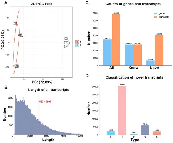
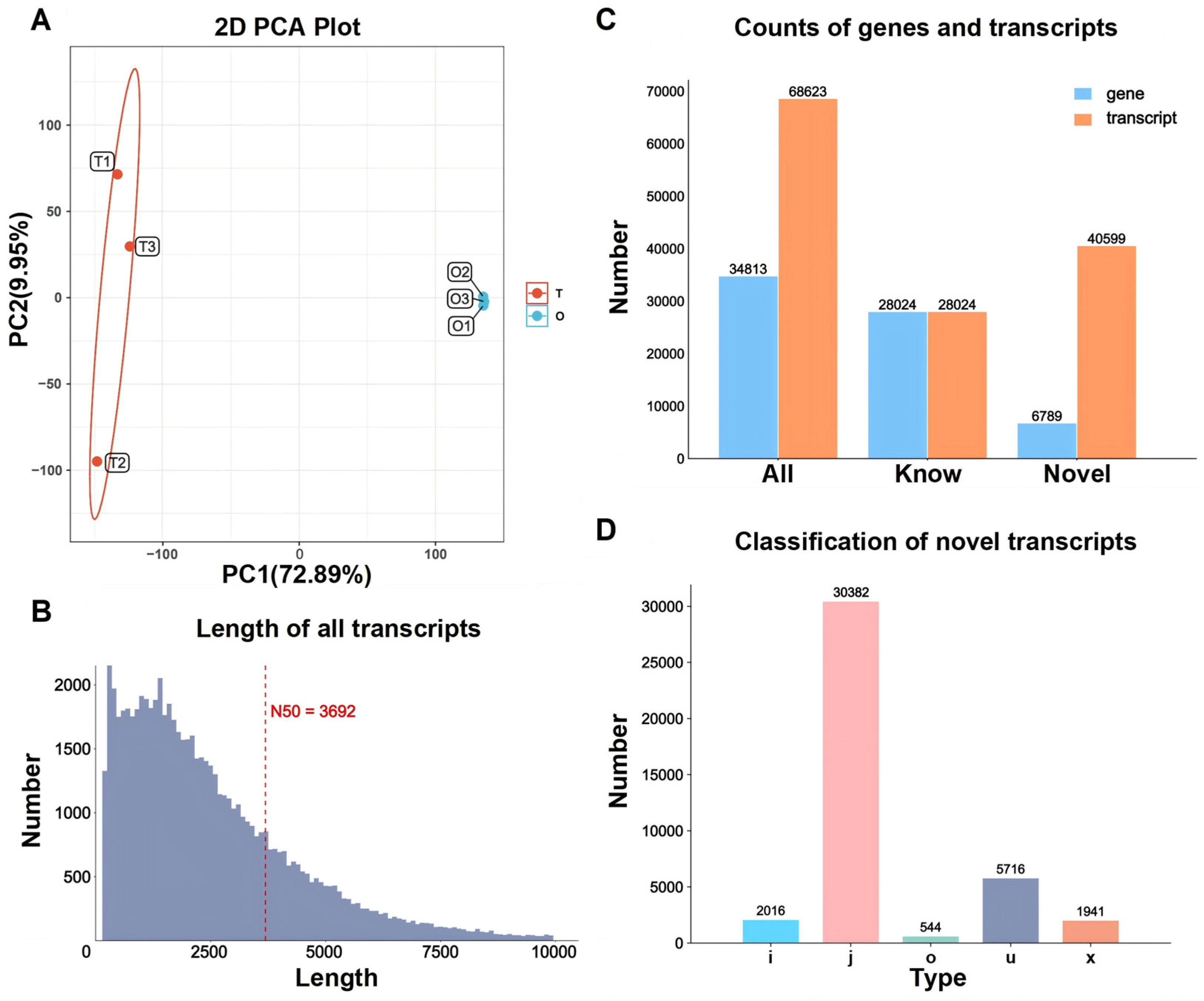
mRNA sequencing of six gonadal samples (three testis and three ovary) from adult R. nasutus yielded 259,263,654 clean reads after quality filtering, with mean Q30 and GC contents of 95.6% and 46.8%, respectively. Principal component analysis (PCA) distinctly separated testis from ovary along PC1 (72.89%), with replicates clustering tightly, demonstrating high data reproducibility (Figure 1A). Alignment to the reference genome detected 34,813 expressed genes and 68,623 assembled transcripts, with an N50 length of 3692 bp (Figure 1B). Among these, 28,024 genes produced a single annotated transcript, while 6789 generated 40,599 transcripts, likely corresponding novel loci or isoforms (Figure 1C). Seven categories of novel transcripts (i, j, o, u, x) were identified, with j-type transcripts being the most abundant (30,382) (Figure 1D).
Figure 1.
Transcriptome quality control and assembly overview of R. nasutus. (A) PCA plot illustrating clear separation between testis (red) and ovary (blue) samples along PC1 (72.89%). (B) Transcript length distribution after redundancy removal, with N50 at 3692 bp. (C) Counts of total, known, and novel genes and transcripts. (D) Classification of novel transcripts based on Gff compare class codes: i (fully intronic), j (potential novel isoforms), o (other exon overlaps on the same strand), u (intergenic), and x (antisense overlap).
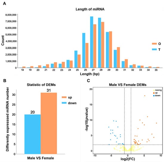
For miRNA sequencing, six small RNA libraries were constructed (three testis and three ovary). Length distribution analysis showed a major peak at 27 nt, followed by 28 and 29 nt in testis, while ovary exhibited a peak at 28 nt, followed by 27 and 29 nt (Figure 3A). Comparison with the miRBase database identified known miRNAs, while novel miRNAs were predicted using miRDeep2. In total, 228 known and 36 novel miRNAs were identified in testis, whereas 246 known and 44 novel miRNAs were detected in ovary.
3.2. Differentially Expressed Genes (DEGs) and Functional Enrichment
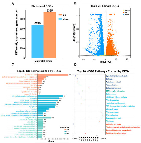
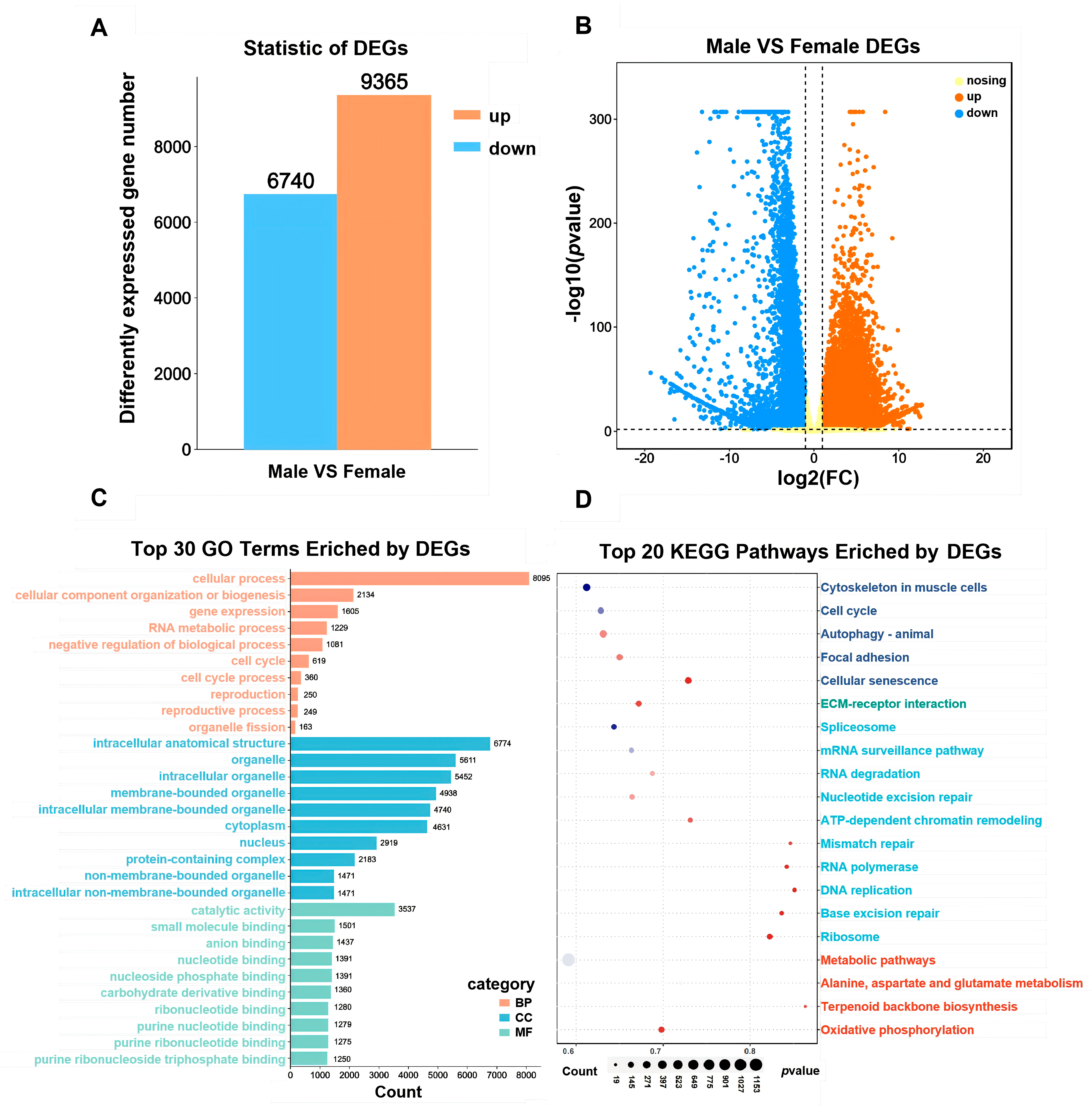
Having established a comprehensive transcriptomic and small RNA profile of R. nasutus gonads, we next compared gene expression patterns between testis and ovary to identify sex-biased transcriptional signatures. A total of 16,105 differentially expressed genes (DEGs) were identified between testis and ovary, including 9365 upregulated and 6740 downregulated genes in the testis (Figure 2A). GO annotation showed significant enrichment across biological process (BP), cellular component (CC), and molecular function (MF) categories (Figure 2C). Within BP, DEGs were predominantly involved in cellular processes, cellular component organization or biogenesis, and gene expression. In CC, DEGs were mainly associated with intracellular anatomical structure, organelles, and intracellular organelle. In MF, they were predominantly linked to catalytic activity, small molecule binding, and anion binding. KEGG pathway enrichment analysis mapped DEGs to 182 pathways, among which metabolic pathways (1255 genes), cytoskeleton in muscle cells (276 genes), and focal adhesion (243 genes) were the most significantly represented (Figure 2D).
Figure 2.
Differential expression and functional enrichment analysis of DEGs in the gonads of R. nasutus. (A) Statistic of differently expressed genes (DEGs) in gonads of R. nasutus. (B) Volcano plot of DEGs in R. nasutus gonads, with orange, blue, and yellow dots representing up-regulated, down-regulated, and non-significant genes, respectively. (C) GO enrichment analysis GO terms of DEGs in female and male R. nasutus gonads. (D) Bubble diagram of KEGG enrichment analysis. The smaller the p-value, the more distinctive the red color. The bigger the point is, the more genes are enriched in the pathway.
3.3. Differentially Expressed miRNAs (DEMs) and Target Prediction
To further elucidate the post-transcriptional mechanisms contributing to these sex-biased expression profiles, we examined the differential expression patterns of miRNAs between testis and ovary. A total of 51 DEMs were identified between testis and ovary, comprising 31 upregulated and 20 downregulated miRNAs in the testis (Figure 3B). A volcano plot highlighted more pronounced expression shifts among upregulated miRNAs, whereas hierarchical clustering based on log10(TPM + 1) values revealed distinct clustering patterns between gonads. These clustering patterns suggest that DEMs within each group may share related regulatory roles or participate in similar biological processes (Figure 3C).
Figure 3.
Identification and differential expression analysis of miRNAs in the gonads of R. nasutus. (A) Length distribution of clean reads from six miRNA libraries of the R. nasutus. (B) Statistic of differently expressed miRNAs (DEMs) in gonads of R. nasutus. (C) Volcano map of genes targeted by DEMs in female and male R. nasutus gonads, with orange, blue, and yellow dots representing up-regulated, down-regulated, and non-significant miRNAs, respectively.
3.4. Functional Annotation and Signaling Pathway Enrichment of DEM Target Genes
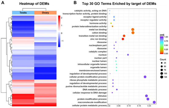
Target gene prediction for the identified DEMs revealed significant functional enrichment across multiple biological categories. In biological processes (BP), the targets were mainly involved in cellular protein metabolic processes, macromolecule modification, and protein modification processes, indicating active post-transcriptional and translational regulation. Within the cellular component (CC) category, the majority of gene products were localized in the nucleus and nucleoplasm, suggesting potential roles in nuclear signaling and gene transcription control. For molecular function (MF), target genes were predominantly enriched in cation binding, metal ion binding, and zinc ion binding activities, highlighting their involvement in metal-dependent catalytic and structural regulation (Figure 4A).
Figure 4.
Differential expression and functional enrichment of miRNAs in gonads of R. nasutus (A) Heatmap of differentially expressed miRNAs (DEMs) between testis and ovary. The hierarchical clustering clearly separates the two gonadal types, with red and blue colors representing up- and down-regulated DEMs, respectively. (B) Bubble plot showing the top 30 enriched Gene Ontology (GO) terms associated with predicted target genes of the identified DEMs. Dot size corresponds to the number of genes in each term, while color intensity indicates adjusted p-value (padj).
Further KEGG pathway analysis demonstrated that these targets participated in critical signaling pathways, including the neuroactive ligand–receptor interaction, MAPK signaling, and calcium signaling pathways, which collectively suggest that DEMs may mediate gonadal differentiation and function through modulation of hormone signaling and intracellular communication (Figure 4B).
3.5. Validation of DEGs and DEMs by qRT-PCR
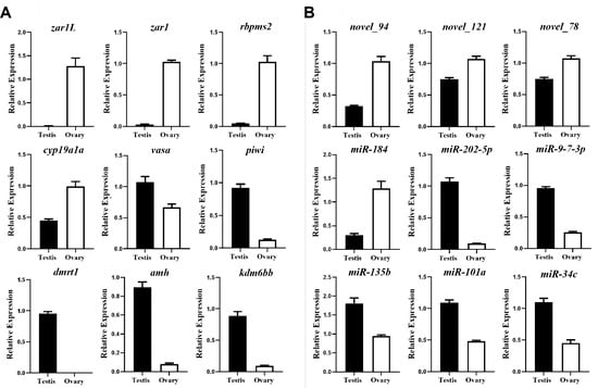
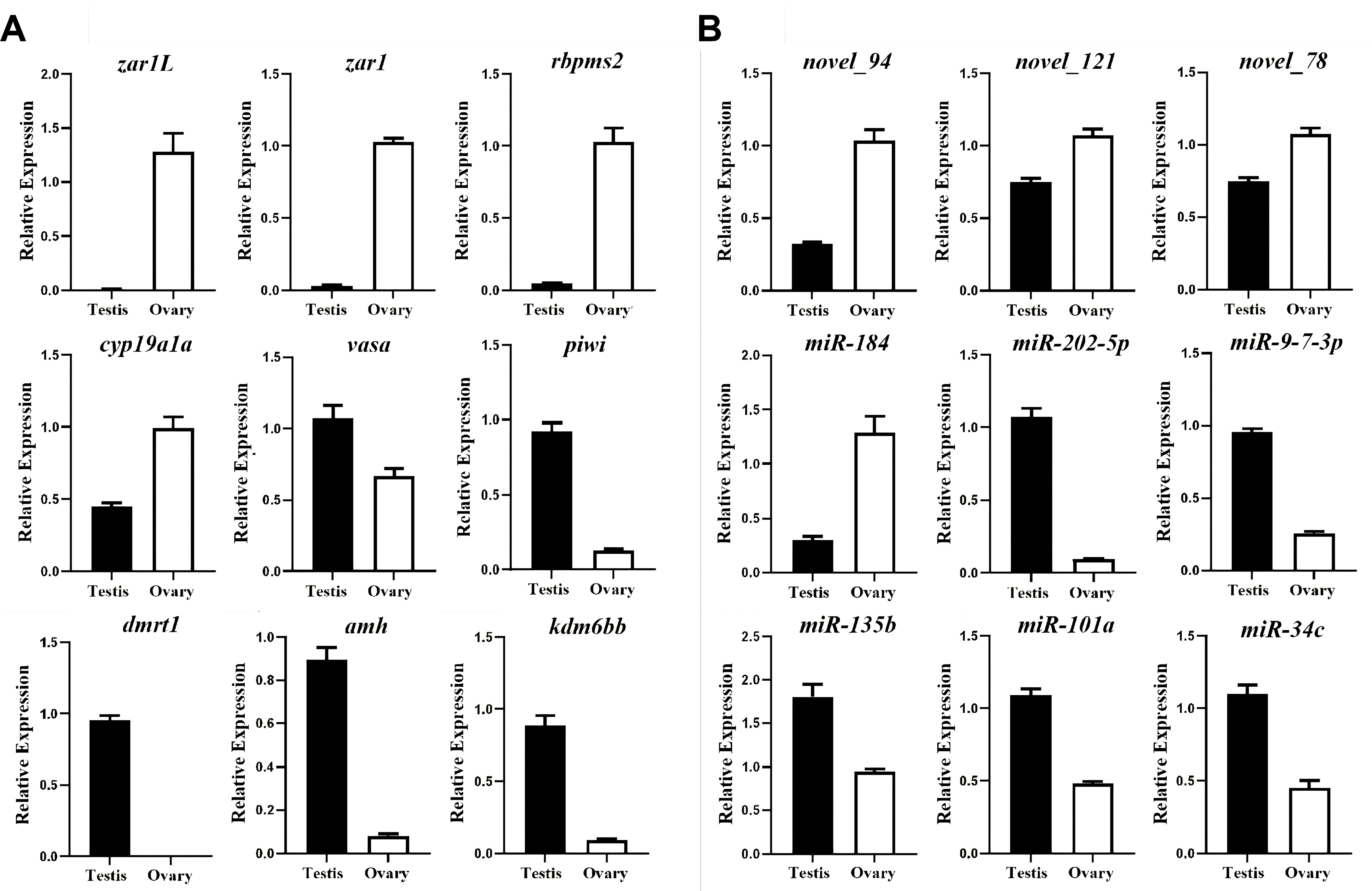
Since miRNAs typically regulate gene expression through their target mRNAs, we next performed target gene prediction and functional enrichment analyses to infer the biological roles of the differentially expressed miRNAs. Nine sex-related genes were analyzed by qRT-PCR. zar1, zar1L, rbpms2, and cyp19a1 exhibited higher expression levels in ovary, whereas vasa, piwi, kdm6bb, amh, and dmrt1 showed significantly higher expression in testis (Figure 5A). These expression patterns were consistent with the RNA-seq data. Similarly, nine miRNAs were randomly selected for validation. Ovary-biased expression was confirmed for novel_94, and novel_121, novel_78, miR-184, and miR-202-5p, miR-9-7-3p, miR-135b, miR-101a, miR-34c were testis-biased (Figure 5B). The qRT-PCR results closely matched the sequencing data, further confirming the reliability of both mRNA and miRNA datasets.
Figure 5.
Validation of mRNA and miRNA expression profiles in the gonads of R. nasutus. (A) Relative expression levels of nine sex-related genes in testis and ovary. Expression values were calculated using the comparative CT (ΔΔCT) method, with β-actin serving as the reference gene. (B) Relative expression levels of nine differentially expressed miRNAs in testis and ovary. Expression values were determined using the same method, normalized against U6 as the internal control. Data are presented as mean ± SE (n = 3), and statistical significance was assessed using t-tests or ANOVA (p < 0.05).
3.6. Predicted miRNA Binding Sites in Key Sex-Determining Genes
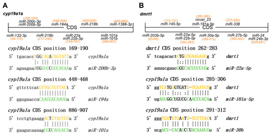
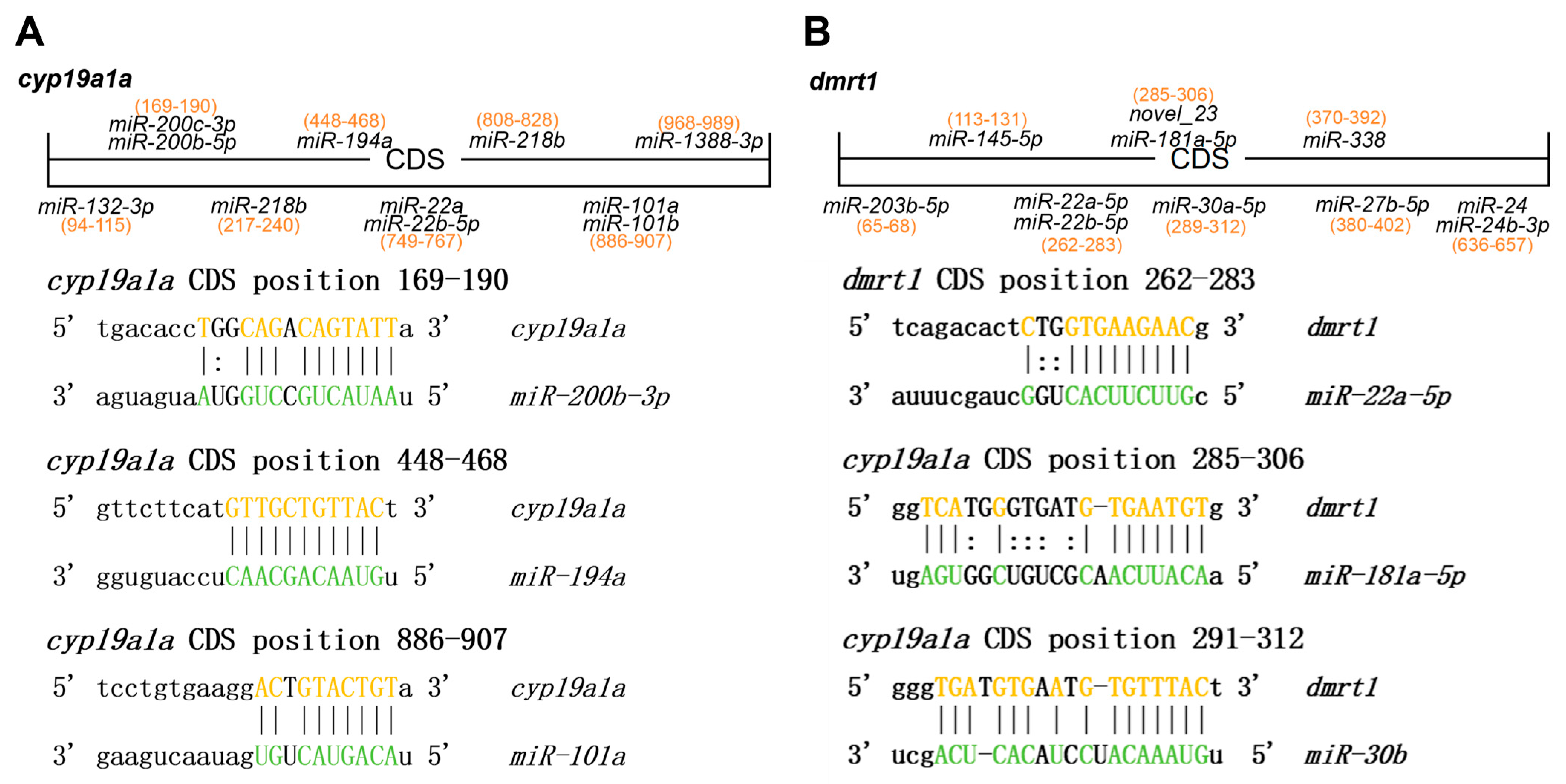
To further investigate the potential post-transcriptional regulation of sex-related genes, we analyzed the predicted miRNA binding sites within the coding regions (CDS) of cyp19a1a and dmrt1 (Figure 6A,B). Several differentially expressed miRNAs (DEMs) were predicted to bind to these two key genes. For cyp19a1a, three major binding sites were detected: miR-101a at positions 169–190 bp, miR-429b at positions 448–468 bp, and miR-194a at positions 886–907 bp (Figure 6A). In addition, multiple binding sites were also predicted on dmrt1, with miR-22a-5p at 262–283 bp, miR-181a-5p at 285–306 bp, and miR-30b at 291–312 bp. The base-pairing diagrams indicated strong complementarity between seed regions of these miRNAs and their target sites within the CDS (Figure 6B). Collectively, these results suggest that cyp19a1a and dmrt1 may be potential targets of multiple DEMs, highlighting a possible role of miRNA-mediated post-transcriptional regulation in the sex differentiation of R. nasutus.
Figure 6.
Predicted miRNA binding sites and differential expression of candidate miRNAs targeting (A) cyp19a1a and (B) dmrt1 in R. nasutus. Highlighted bases indicate the predicted miRNA–mRNA binding regions.
4. Discussion
Despite the availability of genomic resources and the preliminary success of hormone-induced artificial propagation in Rhinogobio nasutus, challenges remain in broodstock cultivation and achieving complete gonadal maturation under captive conditions. Therefore, characterizing gonadal development through transcriptomic and post-transcriptional regulatory dynamics may contribute to a better understanding of reproductive regulation and provide molecular references while artificial breeding protocols are further developed for this species. To address this, the present study employed an integrated transcriptomic approach combining mRNA-seq and miRNA-seq to elucidate the gene regulatory networks and miRNA-mRNA interactions underlying gonadal development in R. nasutus. This approach revealed coordinated expression of germ cell, steroidogenic, and somatic regulatory genes that collectively shape gonadal maturation and function.
Transcriptomic profiling identified 16,105 differentially expressed genes (DEGs) between testis and ovary, including well-known germline markers (vasa, piwi) and somatic regulators (dmrt1, amh, sox9, cyp19a1a). These genes exhibited conserved sex-biased expression patterns, supporting their fundamental roles in vertebrate gonad development. The high expression of vasa and piwi in R. nasutus gonads indicates an active germline maintenance system. Functionally, Vasa encodes an ATP-dependent RNA helicase essential for germ-cell formation and differentiation, whereas Piwi proteins are conserved germline regulators required for maintaining genomic integrity, germ-cell survival, and proper gametogenic progression [16,17,18]. Together, the conserved upregulation of these genes in R. nasutus suggests that this Piwi-Vasa germline regulatory axis is evolutionarily stable across teleosts, underscoring its indispensable role in germline development, stem cell renewal, and gametogenesis.
Building upon the identification of these germline markers, germ cell transplantation (GCT) has emerged as a powerful strategy for germplasm conservation and genetic breeding in teleosts. To date, GCT has been successfully established in multiple fish species, including endangered species such as the Yangtze sturgeon (Acipenser dabryanus) and Chinese sturgeon (A. sinensis) germplasm [7], as well as economically important aquaculture species like grass carp (Ctenopharyngodon idella) [12] and ricefield eel (Monopterus albus) [13]. These advances underscore the broad applicability of GCT across diverse teleost lineages and highlight the importance of foundational knowledge on germline regulatory genes.
Ovarian transcriptomes showed marked upregulation of cyp19a1a and elevated expression of foxl2, which together form a key regulatory axis for estrogen biosynthesis and ovarian differentiation [19,20,21,22]. Aromatase (cyp19a1a) catalyzes the conversion of androgens to estrogens, establishing an estrogenic microenvironment necessary for follicle growth and oocyte maturation [23,24]. Foxl2 acts upstream of cyp19a1a, directly activating its transcription in granulosa cells while repressing testicular genes, thereby stabilizing ovarian fate [25,26]. The concurrent upregulation of zar1, zar1L, and rbpms2 further reflects active oogenic progression. zar1 encodes a maternal-effect factor required for oocyte-to-embryo transition, whereas rbpms2 contributes to Balbiani body assembly and female germ-cell fate determination [27,28,29]. Together, these ovarian-biased transcripts illustrate a tightly coordinated germ–somatic interaction during folliculogenesis, wherein somatic cells provide trophic cues, signaling molecules, and structural support that drive oocyte development and maturation.
At the post-transcriptional level, miRNA-mediated reproductive regulation has been relatively well characterized in mammals; however, in fishes—especially non-model species—the molecular mechanisms by which miRNAs contribute to gonadal development and sex differentiation remain largely unexplored [30,31,32,33,34]. In this study, integrated miRNA–mRNA analysis identified several miRNAs potentially involved in regulating gonad-expressed genes in Rhinogobio nasutus. The predicted miRNA–target interaction pairs are provided in the Supplementary Materials. Among these, miR-200b-3p, miR-194b, and miR-101a exhibited potential binding sites within the coding region of cyp19a1a, suggesting that aromatase expression may be subject to miRNA-mediated post-transcriptional fine-tuning during follicular maturation [33,34,35].
Previous studies have established a close association between the miR-200 family and gonadal differentiation as well as ovarian function in fish. For instance, in Japanese flounder (Paralichthys olivaceus), miR-200 family members (including miR-200a and miR-200b) show ovary-biased expression and can influence gonadal differentiation and development by targeting amh. Similarly, ovary-biased expression of miR-200 family members has been reported in yellowfin seabream (Acanthopagrus latus) and other teleosts [36,37]. In addition, miR-101 has been linked to vertebrate gonadal differentiation and fish reproductive regulation [32]. In contrast, the functional role of miR-194b in fish reproduction remains poorly documented; thus, its potential interaction with cyp19a1a represents a novel regulatory relationship warranting further validation. Moreover, miR-184 exhibited ovary-biased expression in R. nasutus, consistent with its known roles in granulosa cell proliferation, follicular integrity, and oocyte quality in mammals [38,39,40,41].
Collectively, these findings suggest that ovary-biased miRNAs in R. nasutus do not constitute an independent female regulatory pathway, but rather serve as an additional post-transcriptional layer modulating core ovarian axes such as foxl2–cyp19a1a, thereby fine-tuning the balance among steroidogenesis, follicular development, and somatic cell function [30,31,32,33,34]. It should be noted that these miRNA–mRNA interactions were mainly inferred from bioinformatic prediction, and direct experimental evidence is currently lacking [32,37]. Therefore, further in vitro validation is needed to clarify the specific roles of these interactions in fish reproductive development.
In testis, transcriptomic profiling revealed pronounced upregulation of dmrt1, amh, sox9, and kdm6bb, indicating activation of canonical male developmental pathways involved in somatic-cell differentiation and spermatogenesis. Dmrt1 acts as a pivotal transcription factor maintaining Sertoli-cell identity and repressing ovarian genes such as foxl2 and cyp19a1a, thereby preserving testicular structure [42,43,44,45]. Amh, a glycoprotein secreted by Sertoli cells, modulates germ-cell proliferation and local steroidogenesis to facilitate testis cord organization and spermatogonial differentiation [46,47]. The parallel upregulation of sox9 reinforces Sertoli-cell lineage commitment, promoting the cytoskeletal and junctional rearrangements that characterize testis morphogenesis. Elevated kdm6bb expression adds an epigenetic dimension to this process: KDM6B-family demethylases remove repressive H3K27me3 marks at the dmrt1 promoter, sustaining transcriptional activation of the male pathway [48,49,50,51]. Such chromatin-level modulation parallels temperature-induced masculinization reported in other teleosts and reptiles, underscoring a conserved epigenetic mechanism that orchestrates gonadal fate decisions across vertebrates.
microRNAs play indispensable roles in vertebrate testis differentiation and spermatogenesis by modulating transcriptional networks that govern germ-cell proliferation, meiosis, and Sertoli–germ cell communication. Previous studies in teleosts and mammals have demonstrated that miRNAs participate in the regulation of steroidogenic enzymes, meiotic initiation, and sperm maturation, emphasizing their importance in maintaining male fertility [48,49,50,51], the integrative miRNA–mRNA analysis in the present study revealed that dmrt1 transcripts are potentially targeted by miR-22a-5p, miR-181a-5p, and miR-30b, suggesting an additional post-transcriptional regulatory layer that fine-tunes Sertoli-cell differentiation and spermatogenic progression. miRNA-mediated control is a well-established mechanism for refining gene expression during testis maturation, ensuring precise timing and dosage of differentiation-related transcripts.
Among the testis-biased miRNAs identified, miR-202-5p was prominently enriched and has been implicated in germline maintenance and spermatogonial stem-cell regulation across multiple species [15,52,53]. This miRNA localizes within germ plasm and is essential for maintaining reproductive capacity in medaka (Oryzias latipes) [46]. Similarly, miR-34c, another strongly testis-biased miRNA, has been reported in spermatogonia and spermatids of sturgeons and mammals, where it contributes to spermatogenic efficiency and sperm quality [54]. The elevated expression of miR-101a and miR-135b in the present dataset also aligns with their recognized roles in meiotic initiation and Sertoli–germ-cell communication [55,56]. Together, these findings reinforce the conserved function of testis-enriched miRNAs as critical post-transcriptional regulators orchestrating germ-cell differentiation, somatic support, and overall spermatogenic integrity.
5. Conclusions
In summary, integrative mRNA and miRNA profiling of Rhinogobio nasutus gonads provides conserved insights among cypriniform fish into the molecular basis of sex differentiation and germ cell development. Conserved germline markers such as vasa and piwi, together with ovary-biased expression of cyp19a1a, zar1, zar1L, and rbpms2, highlight the conservation of female developmental pathways in cyprinids. Conversely, the testis-specific upregulation of dmrt1, amh, sox9, and kdm6bb underscores a multilayered framework for male differentiation involving transcriptional, post-transcriptional, and epigenetic regulation. Importantly, predicted interactions between sex-biased miRNAs and the coding sequences of cyp19a1a and dmrt1 reveal an additional layer of regulatory complexity, suggesting that miRNAs fine-tune the balance between male and female pathways. Collectively, these findings establish a molecular foundation for understanding reproductive biology in R. nasutus and provide valuable genetic resources to support artificial propagation.
Supplementary Materials
The following supporting information can be downloaded at: https://www.mdpi.com/article/10.3390/fishes11060342/s1, Table S1: Predicted miRNA-Gene Target Relationships.
Author Contributions
J.Y.: Writing—original draft, Validation, Conceptualization. Y.L.: Writing—review and editing, Visualization, Formal analysis, Data curation, Conceptualization. M.J.: Writing—review and editing, Data curation, Conceptualization. H.X.: Writing—review and editing, Validation, Methodology. M.L. (Muyan Li): Writing—review and editing, Methodology. Z.L.: Supervision, Resources, Project administration, Funding. M.L. (Mingyou Li): Supervision, Resources, Project administration, Funding acquisition. All authors have read and agreed to the published version of the manuscript.
Funding
This work was supported by the Key R&D Program of Ningxia Province (2023BCF01013).
Institutional Review Board Statement
This study was conducted in accordance with the guidelines of the Declaration of Helsinki and was approved by the Animal Care and Use Committee of the Shanghai Ocean University under approval number SHOU-2023-031 on 1 September 2023.
Informed Consent Statement
Not applicable.
Data Availability Statement
Data are contained within the article. The raw data of this work was uploaded to NGDC (https://ngdc.cncb.ac.cn) with BioProject number PRJCA060031.
Conflicts of Interest
Author Yanbin Li and Zongqiang Lian were employed by the Ningxia Fisheries Research Institute. The remaining authors declare that the research was conducted in the absence of any commercial or financial relationships that could be construed as a potential conflict of interest.
References
- Wang, J.; Zhang, T.; Chang, S.; Li, J.; Jiang, Y. Complete Mitochondrial Genome and Its Phylogenetic Implications of Rhinogobio nasutus, an Endemic Species from the Yellow River. Mol. Biol. Rep. 2023, 50, 10651–10656. [Google Scholar] [CrossRef] [Scilit]
- Jiang, C.; Du, Y.; Lou, Z.; Zhang, Y.; Wang, T. Telomere-to-Telomere Reference Genome of Rhinogobio nasutus, an Endangered Endemic Fish from the Yellow River. Sci. Data 2025, 12, 462. [Google Scholar] [CrossRef] [Scilit] [PubMed]
- Chen, Z.; An, B.; Wang, Z.; Wu, F.; Sun, Z.; Zhang, L. Fish Diversity and Conservation in the Lanzhou Reach of the Yellow River. Biodivers. Sci. 2021, 29, 1658–1672. [Google Scholar] [CrossRef] [Scilit]
- Wang, J.; Chen, L.; Tang, W.; Heino, J.; Jiang, X. Effects of Dam Construction and Fish Invasion on the Species, Functional and Phylogenetic Diversity of Fish Assemblages in the Yellow River Basin. J. Environ. Manag. 2021, 293, 112863. [Google Scholar] [CrossRef] [Scilit] [PubMed]
- Jia, Y.; Kennard, M.J.; Liu, Y.; Sui, X.; Li, K.; Wang, G.; Chen, Y. Human Disturbance and Long-Term Changes in Fish Taxonomic, Functional and Phylogenetic Diversity in the Yellow River, China. Hydrobiologia 2020, 847, 3711–3725. [Google Scholar] [CrossRef] [Scilit]
- Jiang, M.; Zhao, C.; Ma, F.; Yin, D.; Wang, C.; Jian, J.; Liu, K. The Telomere-to-Telomere Gap-Free Reference Genome and Taxonomic Reassessment of Siniperca roulei. GigaScience 2025, 14, giaf068. [Google Scholar] [CrossRef] [Scilit]
- Ye, H.; Takeuchi, Y.; Du, H.; Yue, H.; Ruan, R.; Li, C.; Wei, Q. Spermatogonia From Cryopreserved Testes of Critically Endangered Chinese Sturgeon Efficiently Colonized and Preferentially Proliferated in the Recipient Gonads of Yangtze Sturgeon. Mar. Biotechnol. 2022, 24, 136–150. [Google Scholar] [CrossRef] [Scilit]
- Yue, H.; Li, C.; Du, H.; Zhang, S.; Wei, Q. Sequencing and de Novo Assembly of the Gonadal Transcriptome of the Endangered Chinese Sturgeon (Acipenser sinensis). PLoS ONE 2015, 10, e0127332. [Google Scholar] [CrossRef] [Scilit]
- Kobayashi, Y.; Nagahama, Y.; Nakamura, M. Diversity and Plasticity of Sex Determination and Differentiation in Fishes. Sex. Dev. 2013, 7, 115–125. [Google Scholar] [CrossRef] [Scilit]
- Lin, Q.; Mei, J.; Li, Z.; Zhang, X.; Zhou, L.; Gui, J.F. Distinct and Cooperative Roles of Amh and Dmrt1 in Self-Renewal and Differentiation of Male Germ Cells in Zebrafish. Genetics 2017, 207, 1007–1022. [Google Scholar] [CrossRef] [Scilit]
- Yang, Y.J.; Wang, Y.; Li, Z.; Zhou, L.; Gui, J.F. Sequential, Divergent, and Cooperative Requirements of Foxl2a and Foxl2b in Ovary Development and Maintenance of Zebrafish. Genetics 2017, 205, 1551–1572. [Google Scholar] [CrossRef] [Scilit] [PubMed]
- Sun, X.; Tao, B.; Wang, Y.; Hu, W.; Sun, Y. Isolation and Characterization of Germline Stem Cells in Protogynous Hermaphroditic Monopterus albus. Int. J. Mol. Sci. 2022, 23, 5861. [Google Scholar] [CrossRef] [Scilit]
- Shcherbata, H.R. MiRNA Functions in Stem Cells and Their Niches: Lessons from the Drosophila Ovary. Curr. Opin. Insect Sci. 2019, 31, 29–36. [Google Scholar] [CrossRef] [Scilit] [PubMed]
- Guo, S.; Zhong, Y.; Zhang, Y.; Zhu, Y.; Guo, J.; Fu, Y.; Li, M. Transcriptome Analysis Provides Insights into Long Noncoding RNAs in Medaka Gonads. Comp. Biochem. Physiol. Part D Genom. Proteom. 2021, 39, 100842. [Google Scholar] [CrossRef] [Scilit]
- Zhang, M.; Zhang, Q.; Hu, Y.; Xu, L.; Jiang, Y.; Zhang, C.; Ding, L.; Jiang, R.; Sun, J.; Sun, H.; et al. MiR-181a Increases FoxO1 Acetylation and Promotes Granulosa Cell Apoptosis via SIRT1 Downregulation. Cell Death Dis. 2017, 8, e3088. [Google Scholar] [CrossRef] [Scilit]
- Begum, S.; Gnanasree, S.M.; Anusha, N.; Senthilkumaran, B. Germ Cell Markers in Fishes—A Review. Aquac. Fish. 2022, 7, 540–552. [Google Scholar] [CrossRef] [Scilit]
- Cuykendall, T.N.; Houston, D.W. Identification of Germ Plasm-associated Transcripts by Microarray Analysis of Xenopus Vegetal Cortex RNA. Dev. Dyn. 2010, 239, 1838–1848. [Google Scholar] [CrossRef] [Scilit]
- Juliano, C.; Wessel, G. Versatile Germline Genes. Science 2010, 329, 640–641. [Google Scholar] [CrossRef] [Scilit]
- Fleming, N.I.; Knower, K.C.; Lazarus, K.A.; Fuller, P.J.; Simpson, E.R.; Clyne, C.D. Aromatase Is a Direct Target of FOXL2: C134W in Granulosa Cell Tumors via a Single Highly Conserved Binding Site in the Ovarian Specific Promoter. PLoS ONE 2010, 5, e14389. [Google Scholar] [CrossRef] [Scilit]
- Pannetier, M.; Fabre, S.; Batista, F.; Kocer, A.; Renault, L.; Jolivet, G.; Mandon-Pépin, B.; Cotinot, C.; Veitia, R.; Pailhoux, E. FOXL2 Activates P450 Aromatase Gene Transcription: Towards a Better Characterization of the Early Steps of Mammalian Ovarian Development. J. Mol. Endocrinol. 2006, 36, 399–413. [Google Scholar] [CrossRef] [Scilit]
- Uhlenhaut, N.H.; Jakob, S.; Anlag, K.; Eisenberger, T.; Sekido, R.; Kress, J.; Treier, A.-C.; Klugmann, C.; Klasen, C.; Holter, N.I.; et al. Somatic Sex Reprogramming of Adult Ovaries to Testes by FOXL2 Ablation. Cell 2009, 139, 1130–1142. [Google Scholar] [CrossRef] [Scilit] [PubMed]
- Yang, B.; Guo, H.; Zhang, Y.; Chen, L.; Ying, D.; Dong, S. Microrna-145 Regulates Chondrogenic Differentiation of Mesenchymal Stem Cells by Targeting SOX9. PLoS ONE 2011, 6, e21679. [Google Scholar] [CrossRef] [Scilit]
- Karube, M.; Fernandino, J.I.; Strobl-Mazzulla, P.; Strüssmann, C.A.; Yoshizaki, G.; Somoza, G.M.; Patiño, R. Characterization and Expression Profile of the Ovarian Cytochrome P-450 Aromatase (Cyp19A1) Gene during Thermolabile Sex Determination in Pejerrey, Odontesthes bonariensis. J. Exp. Zool. A Ecol. Genet. Physiol. 2007, 307A, 625–636. [Google Scholar] [CrossRef] [Scilit] [PubMed]
- Maruo, K.; Suda, M.; Yokoyama, S.; Oshima, Y.; Nakamura, M. Steroidogenic Gene Expression during Sex Determination in the Frog Rana rugosa. Gen. Comp. Endocrinol. 2008, 158, 87–94. [Google Scholar] [CrossRef] [Scilit]
- Fang, D.-A.; Yang, X.; Feng, X.; Zhou, Y.-F.; Xu, D.-P.; Zhang, M.-Y.; Liu, K. FoxL2 Combined with Cyp19a1a Regulate the Spawning Upstream Migration in Coilia nasus. Gene 2019, 710, 307–315. [Google Scholar] [CrossRef] [Scilit]
- Shen, Z.-G.; Eissa, N.; Yao, H.; Xie, Z.-G.; Wang, H.-P. Effects of Temperature on the Expression of Two Ovarian Differentiation-Related Genes Foxl2 and Cyp19a1a. Front. Physiol. 2018, 9, 1208. [Google Scholar] [CrossRef] [Scilit] [PubMed]
- Kaufman, O.H.; Lee, K.; Martin, M.; Rothhämel, S.; Marlow, F.L. Rbpms2 Functions in Balbiani Body Architecture and Ovary Fate. PLoS Genet. 2018, 14, e1007768. [Google Scholar] [CrossRef] [Scilit]
- Wilson, M.L.; Romano, S.N.; Khatri, N.; Aharon, D.; Liu, Y.; Kaufman, O.H.; Draper, B.W.; Marlow, F.L. Rbpms2 Promotes Female Fate Upstream of the Nutrient Sensing Gator2 Complex Component Mios. Nat. Commun. 2024, 15, 5248. [Google Scholar] [CrossRef] [Scilit]
- Wu, X.; Viveiros, M.M.; Eppig, J.J.; Bai, Y.; Fitzpatrick, S.L.; Matzuk, M.M. Zygote Arrest 1 (Zar1) Is a Novel Maternal-Effect Gene Critical for the Oocyte-to-Embryo Transition. Nat. Genet. 2003, 33, 187–191. [Google Scholar] [CrossRef] [Scilit]
- Alvi, S.M.; Zayed, Y.; Malik, R.; Peng, C. The Emerging Role of MicroRNAs in Fish Ovary: A Mini Review. Gen. Comp. Endocrinol. 2021, 311, 113850. [Google Scholar] [CrossRef] [Scilit]
- Gay, S.; Bugeon, J.; Bouchareb, A.; Henry, L.; Delahaye, C.; Legeai, F.; Montfort, J.; Le Cam, A.; Siegel, A.; Bobe, J.; et al. MiR-202 Controls Female Fecundity by Regulating Medaka Oogenesis. PLoS Genet. 2018, 14, e1007593. [Google Scholar] [CrossRef] [Scilit]
- Bhat, R.A.; Priyam, M.; Foysal, M.J.; Gupta, S.K.; Sundaray, J.K. Role of Sex-biased MiRNAs in Teleosts—A Review. Rev. Aquac. 2021, 13, 269–281. [Google Scholar] [CrossRef] [Scilit]
- Wang, F.; Jia, Y.; Wang, P.; Yang, Q.; Du, Q.; Chang, Z. Identification and Profiling of Cyprinus carpio MicroRNAs during Ovary Differentiation by Deep Sequencing. BMC Genom. 2017, 18, 333. [Google Scholar] [CrossRef] [Scilit] [PubMed]
- Tao, W.; Sun, L.; Shi, H.; Cheng, Y.; Jiang, D.; Fu, B.; Conte, M.A.; Gammerdinger, W.J.; Kocher, T.D.; Wang, D. Integrated Analysis of MiRNA and MRNA Expression Profiles in Tilapia Gonads at an Early Stage of Sex Differentiation. BMC Genom. 2016, 17, 328. [Google Scholar] [CrossRef] [Scilit] [PubMed]
- Ma, X.; Xu, H.; Lu, T.; Lin, W.; Sun, L.; Jawad, M.; Li, M. Comparative MiRNA Transcriptome Analysis Reveals MiR-375-3p Targets Cyp19a and Regulates Ovarian Development in Medaka (Oryzias latipes). Comp. Biochem. Physiol. Part D Genom. Proteom. 2025, 56, 101535. [Google Scholar] [CrossRef] [Scilit]
- Zhang, D.; Shi, B.; Shao, P.; Shao, C.; Wang, C.; Li, J.; Liu, X.; Ma, X.; Zhao, X. The Identification of MiRNAs That Regulate Ovarian Maturation in Cynoglossus semilaevis. Aquaculture 2022, 555, 738250. [Google Scholar] [CrossRef] [Scilit]
- Ma, Z.; Yang, J.; Zhang, Q.; Xu, C.; Wei, J.; Sun, L.; Wang, D.; Tao, W. MiR-133b Targets Tagln2 and Functions in Tilapia Oogenesis. Comp. Biochem. Physiol. B Biochem. Mol. Biol. 2021, 256, 110637. [Google Scholar] [CrossRef] [Scilit]
- Cheng, W.-T.; Rosario, R.; Muthukaruppan, A.; Wilson, M.K.; Payne, K.; Fong, P.C.; Shelling, A.N.; Blenkiron, C. MicroRNA Profiling of Ovarian Granulosa Cell Tumours Reveals Novel Diagnostic and Prognostic Markers. Clin. Epigenet. 2017, 9, 72. [Google Scholar] [CrossRef] [Scilit]
- Huang, L.; Yin, Z.J.; Feng, Y.F.; Zhang, X.D.; Wu, T.; Ding, Y.Y.; Ye, P.F.; Fu, K.; Zhang, M.Q. Identification and Differential Expression of MicroRNAs in the Ovaries of Pigs (Sus Scrofa) with High and Low Litter Sizes. Anim. Genet. 2016, 47, 543–551. [Google Scholar] [CrossRef] [Scilit]
- Salilew-Wondim, D.; Ahmad, I.; Gebremedhn, S.; Sahadevan, S.; Hossain, M.M.; Rings, F.; Hoelker, M.; Tholen, E.; Neuhoff, C.; Looft, C.; et al. The Expression Pattern of MicroRNAs in Granulosa Cells of Subordinate and Dominant Follicles during the Early Luteal Phase of the Bovine Estrous Cycle. PLoS ONE 2014, 9, e106795. [Google Scholar] [CrossRef] [Scilit]
- Shan, B.; Huo, Y.; Guo, Z.; Li, Q.; Pan, Z.; Li, Q.; Du, X. MiR-184, a Downregulated Ovary-Elevated MiRNA Transcriptionally Activated by SREBF2, Exerts Anti-Apoptotic Properties in Ovarian Granulosa Cells through Inducing SMAD3 Expression. Cell Death Dis. 2024, 15, 892. [Google Scholar] [CrossRef] [Scilit] [PubMed]
- Lan, K.-C.; Chen, Y.-T.; Chang, C.; Chang, Y.-C.; Lin, H.-J.; Huang, K.-E.; Kang, H.-Y. Up-Regulation of SOX9 in Sertoli Cells from Testiculopathic Patients Accounts for Increasing Anti-Mullerian Hormone Expression via Impaired Androgen Receptor Signaling. PLoS ONE 2013, 8, e76303. [Google Scholar] [CrossRef] [Scilit] [PubMed]
- Vining, B.; Ming, Z.; Bagheri-Fam, S.; Harley, V. Diverse Regulation but Conserved Function: SOX9 in Vertebrate Sex Determination. Genes 2021, 12, 486. [Google Scholar] [CrossRef] [Scilit]
- Wagner, S.; Whiteley, S.L.; Castelli, M.; Patel, H.R.; Deveson, I.W.; Blackburn, J.; Holleley, C.E.; Marshall Graves, J.A.; Georges, A. Gene Expression of Male Pathway Genes Sox9 and Amh during Early Sex Differentiation in a Reptile Departs from the Classical Amniote Model. BMC Genom. 2023, 24, 243. [Google Scholar] [CrossRef] [Scilit] [PubMed]
- Yan, Y.-L.; Batzel, P.; Titus, T.; Sydes, J.; Desvignes, T.; BreMiller, R.; Draper, B.; Postlethwait, J.H. A Hormone That Lost Its Receptor: Anti-Müllerian Hormone (AMH) in Zebrafish Gonad Development and Sex Determination. Genetics 2019, 213, 529–553. [Google Scholar] [CrossRef] [Scilit]
- Pfennig, F.; Standke, A.; Gutzeit, H.O. The Role of Amh Signaling in Teleost Fish—Multiple Functions Not Restricted to the Gonads. Gen. Comp. Endocrinol. 2015, 223, 87–107. [Google Scholar] [CrossRef] [Scilit]
- Wu, K.; Yue, Y.; Zhou, L.; Zhang, Z.; Shan, H.; He, H.; Ge, W. Disrupting Amh and Androgen Signaling Reveals Their Distinct Roles in Zebrafish Gonadal Differentiation and Gametogenesis. Commun. Biol. 2025, 8, 371. [Google Scholar] [CrossRef] [Scilit]
- Ge, C.; Ye, J.; Weber, C.; Sun, W.; Zhang, H.; Zhou, Y.; Cai, C.; Qian, G.; Capel, B. The Histone Demethylase KDM6B Regulates Temperature-Dependent Sex Determination in a Turtle Species. Science 2018, 360, 645–648. [Google Scholar] [CrossRef] [Scilit]
- Piferrer, F. Epigenetics of Sex Determination and Differentiation in Fish. In Sex Control in Aquaculture; Wang, H.-P., Piferrer, F., Chen, S.-L., Shen, Z.-G., Eds.; John Wiley & Sons, Ltd.: Chichester, UK, 2018; pp. 65–83. [Google Scholar] [CrossRef] [Scilit]
- Wang, Q.; Hao, X.; Liu, K.; Feng, B.; Li, S.; Zhang, Z.; Tang, L.; Mahboob, S.; Shao, C. Early Response to Heat Stress in Chinese Tongue Sole (Cynoglossus semilaevis): Performance of Different Sexes, Candidate Genes and Networks. BMC Genom. 2020, 21, 745. [Google Scholar] [CrossRef] [Scilit]
- Weber, C.; Zhou, Y.; Lee, J.G.; Looger, L.L.; Qian, G.; Ge, C.; Capel, B. Temperature-Dependent Sex Determination Is Mediated by PSTAT3 Repression of Kdm6b. Science 2020, 368, 303–306. [Google Scholar] [CrossRef] [Scilit]
- Chen, Q.; Yang, D.; Chen, M.; Xiong, J.; Huang, J.; Ding, W.; Gao, K.; Lai, B.; Zheng, L.; Tang, Z.; et al. Smad4 and FoxH1 Potentially Interact to Regulate Cyp19a1a Promoter in the Ovary of Ricefield Eel (Monopterus albus). Biol. Sex Differ. 2024, 15, 60. [Google Scholar] [CrossRef] [Scilit]
- Shen, F.; Chao, Q.; Cai, Z.; Zhang, H.; Wu, J.; Zhang, J. Expression, Localization, and a Regulated Target Gene (Ccnd1) of MiR-202-5p in the Japanese Flounder Gonads. Aquac. Fish. 2023, 8, 267–273. [Google Scholar] [CrossRef] [Scilit]
- Zhang, S.; Yu, M.; Liu, C.; Wang, L.; Hu, Y.; Bai, Y.; Hua, J. MIR-34c Regulates Mouse Embryonic Stem Cells Differentiation into Male Germ-like Cells through RARg. Cell Biochem. Funct. 2012, 30, 623–632. [Google Scholar] [CrossRef] [Scilit]
- Chadourne, M.; Poumerol, E.; Jouneau, L.; Passet, B.; Castille, J.; Sellem, E.; Pailhoux, E.; Mandon-Pépin, B. Structural and Functional Characterization of a Testicular Long Non-Coding RNA (4930463O16Rik) Identified in the Meiotic Arrest of the Mouse Topaz1−/− Testes. Front. Cell Dev. Biol. 2021, 9, 700290. [Google Scholar] [CrossRef] [Scilit]
- Prause, R.; Eckart, J.; Skrobanek, J.; Thönnes, M.; Pfennig, F. Differentially Expressed MicroRNAs in Testes of Dominant and Subordinate Nile Tilapia Males and Identification of Oni-MiR-499 as Regulator of Amh Gene Expression. Sex. Dev. 2025, 19, 10–31. [Google Scholar] [CrossRef] [Scilit]

Disclaimer/Publisher’s Note: The statements, opinions and data contained in all publications are solely those of the individual author(s) and contributor(s) and not of MDPI and/or the editor(s). MDPI and/or the editor(s) disclaim responsibility for any injury to people or property resulting from any ideas, methods, instructions or products referred to in the content. |
© 2026 by the authors. Licensee MDPI, Basel, Switzerland. This article is an open access article distributed under the terms and conditions of the Creative Commons Attribution (CC BY) license.