


Abstract
This study was conducted to explore the impacts of L-carnitine (CAR) on growth performance, antioxidant capacity, hepatic lipid deposition and fatty acid β-oxidation in farmed fish fed a high-fat diet (HFD). A total of 150 juvenile tiger puffer (15.23 ± 0.01 g) were randomly assigned into six tanks (three tanks each group) and fed with a control diet (HFD, 16% lipid) and a HFD supplemented with 1.5% CAR for eight weeks, respectively. The results indicated that the growth performance and the most proximate compositions were not influenced by dietary CAR. However, compared to the control group, the hepatosomatic index and crude lipid content in the liver were significantly reduced in the CAR group, indicating that CAR performed a lipid-lowering effect in tiger puffer. Serum lipid profiles remained stable, implying that the cholesterol metabolism was not influenced by dietary CAR. Furthermore, dietary CAR activated fatty acid β-oxidation (FAO) in both the mitochondria and peroxisomes and drove lipolysis and fatty acid intracellular transport to supply sufficient substrates for FAO, manifested by the activation of related genes, proteins and enzyme activity. Additionally, CAR lowered hepatic MDA levels, indicating improved antioxidant capacity. In conclusion, CAR shows potential as a lipid-lowering feed additive for marine fish under high-fat dietary conditions.
Key Contribution:
L-carnitine may be a promising lipid-lowering feed additive for marine fish fed with HFD in aquaculture.
1. Introduction
The rising cost of protein sources for feeds, driven by rapid aquaculture development and declining wild fishery resources, has prompted the widespread use of lipids and carbohydrates (non-protein energy) in fish dietary formulations [1]. Lipid, which is a major energy source for fish growth and development, provides more than twice the energy per unit compared to glycogen [2]. Furthermore, it has been demonstrated that for many fish species, fatty acid β-oxidation constitutes the main source of energy [3]. Consequently, high-fat diets are now commonly employed in aquaculture to achieve the “protein sparing effect” [4]. Nevertheless, long-term high lipid intake usually causes excess lipid accumulation in the liver, which induces disorders of lipid metabolism [5]. Therefore, it is crucial for the health and growth of farmed fish to seek a functional additive, which promotes hepatic lipid decomposition, to incorporate into high-fat diets.
L-carnitine (CAR) is a quaternary amine that is synthesized endogenously using lysine and methionine, mainly in the liver and kidney [6]. As the most critical organ for lipid metabolism, the liver is responsible for the majority of lipid synthesis and utilization processes [7]. Due to its function in promoting lipid metabolism, CAR has been utilized in human medicine as an adjunctive therapy for a variety of hepatic disorders, such as non-alcoholic fatty liver disease and cirrhosis [6,8]. In aquatic nutrition, the most well-known function of CAR is to reduce the hepatic fat deposition of fish fed with a high-fat diet [9,10,11]. Facilitated by carnitine palmitoyltransferase 1 (CPT1), it promotes the transport of long-chain fatty acids from the cytoplasm into the mitochondria for β-oxidation, thereby generating energy [12,13]. On the contrary, it has been found that dietary CAR supplementation induced hepatic lipid deposition in yellow catfish [14]. Such inconsistency may be due to interspecies differences, divergent rearing environments, or differing concentrations of the dietary supplement. Furthermore, studies have shown that CAR and its esters help mitigate oxidative stress and inflammation, and are common in the treatment of clinical diseases such as cardiovascular disorders and non-alcoholic steatohepatitis [15,16]. A series of studies have also demonstrated that moderate dietary CAR enhances the antioxidant capacity of aquatic animals, including large yellow croaker [9], golden pompano [10], tilapia [17], juvenile narrow clawed crayfish [18], and amur minnow [19].
Tiger puffer (Takifugu rubripes) is a popular and major commercial fish in East Asia, owing to its excellent taste and abundant nutrients [20]. Furthermore, the fact that lipids are stored predominantly in the liver makes this species an ideal model for researching hepatic lipid metabolism [21]. In this study, CAR was supplemented in the high-fat diet to investigate its effect on growth, antioxidant status, and fatty acid β-oxidation in tiger puffer. It was hypothesized that dietary CAR may improve the growth performance and antioxidant capacity, and activate the hepatic fatty acid β-oxidation. These findings may provide offer potential insights for developing high-fat diet formulations for this species.
2. Materials and Methods
2.1. Experimental Diets
The control group was fed a basal diet containing approximately 46% crude protein and 16% crude lipid. For the CAR group, 1.5% CAR was added into the basal diet. The two diets were isonitrogenous and isolipidic, and prepared according to our standard procedures [22]. The diet formulations and measured composition are detailed in Table 1.
Table 1.
Diet composition and proximate analysis (% dry matter basis).
2.2. Experimental Fish and Rearing Conditions
Tiger puffers obtained from Huanghai Fishery Co. (Yantai, China) were acclimated for 7 days in polyethylene tanks fed with the control diet, using a flowing seawater system provided by the company. To prevent mutual biting, the fish teeth were clipped during this period. Following acclimation, 150 healthy fish (15.23 ± 0.01 g) were distributed to control or CAR groups randomly (three tanks each group, 25 fish each tank), and fed to visual satiety twice daily (7:00 and 18:30) over eight weeks. Residual feed and feces were removed by siphoning two hours after each feeding. To maintain the water quality, 80% of the rearing water was exchanged daily, and the tanks were scrubbed every other day. Throughout the trial, water temperature (20–24 °C); salinity (28–32 g/L); pH (7.5–8.0); and dissolved oxygen (5–7 mg/L) were kept constant.
2.3. Sampling
Prior to sampling, fish underwent fasting for 24 h and were anesthetized with eugenol (99.9% purity; 1:10,000 v/v in water). Fish were counted and weighed in each tank. Three fish per tank were used for whole-body composition. Blood from another six fish was collected from the caudal vein, held at 4 °C for 6 h, centrifuged (836 g, 10 min), and then the serum was stored at −80 °C for biochemical analysis. Liver and muscle samples were collected from the same six fish, immediately frozen in liquid nitrogen, and transferred to −80 °C. Additionally, three randomly selected fish were measured for final body weight and length, hepatopancreas and visceral weight to determine the following indices: weight gain (WG) = [initial body weight (IBW) − final body weight (FBW)]/IBW × 100%; survival rate (SR) = number of final fish/number of initial fish × 100%; specific growth rate (SGR) = [ln FBW − ln IBW]/days of feeding × 100%; feed conversion ratio (FCR) = total feed fed/(FBW − IBW); feed intake (FI) = total feed fed/[experimental days × (IBW + FBW)/2] × 100%; condition factor (CF) = body weight/body length3 × 100%; hepatosomatic index (HSI) = hepatopancreas weight/body weight × 100%; viscerosomatic index (VSI) = viscera weight/body weight × 100%.
2.4. Analysis of Proximate Composition in Diets and Fish
Proximate composition of the diets and fish samples was determined following the methods of the Association of Official Analytical Chemists [23]. In brief, moisture was measured by drying to constant weight at 105 °C. Samples were burned in a muffle furnace at 550 °C for 8 h to analyze the ash content. The Kjeldahl method (N × 6.25) was used for analyzing the crude protein content. Crude lipid in diets, whole body, and liver was extracted with petroleum ether via Soxhlet apparatus, while muscle lipid was analyzed using the chloroform–methanol procedure described by Folch [24].
2.5. Analysis of Serum Biochemical Parameters
Serum levels of triglyceride (TG), total cholesterol (TC), low-density lipoprotein cholesterol (LDL-C) and high-density lipoprotein cholesterol (HDL-C) were quantified using commercial kits (Nanjing Jiancheng Bioengineering Institute, Nanjing, China; A110-2-1, A111-2-1, A113-1-1, and A112-1-1, respectively) following the manufacturer’s protocols.
2.6. Analysis of the Malondialdehyde (MDA) Content in Liver
The content of hepatic MDA was quantified using a commercial kit (Nanjing Jiancheng Bioengineering Institute, Nanjing, China; A003-1-2) in accordance with the manufacturer’s instructions.
2.7. Quantitative Real-Time PCR (qPCR)
The procedures for total RNA isolation, reverse transcription, and qPCR were conducted as described earlier [25]. The 2−ΔΔCT method was employed to quantify the relative gene expression levels, using beta-actin (β-actin) and elongation factor 1 alpha (ef1α) as endogenous controls for normalization. Table 2 lists the sequences of primers used to amplify the target genes: β-actin, ef1α, adipose triglyceride lipase (atgl), fatty acid binding protein 1 (fabp1), hepatocyte nuclear factor 4 alpha (hnf4α), peroxysome proliferator-activated receptor alpha (pparα), peroxlsome proliferator-activated receptor-γ coactivator 1 alpha (pgc1α), carnitine palmitoyltransferase 1Ab (cpt1ab), and acyl-coenzyme A oxidase 1 (acox1).
Table 2.
Primer sequences utilized in qPCR.
2.8. Western Blotting Analysis
Total protein in the liver was isolated by RIPA lysis buffer (Solarbio Technology Co., Ltd., Beijing, China, R0010) and quantified using the BCA assay method (the same supplier, PC0020). After sodium dodecyl sulfate-polyacrylamide gel electrophoresis separation and polyvinylidene difluoride membrane transfer, membranes were blocked with 5% skim milk in tris-buffered saline with Tween-20 (TBST) for 2 h at room temperature, then incubated overnight at 4 °C with primary antibody. Following TBST washes, HRP-conjugated secondary antibody (Abbkine Scientific Co., Ltd., Wuhan, China A21020) was applied. Detection was performed using an ECL chemiluminescence substrate (Beyotime Biotechnology Co., Ltd., Shanghai, China, P0018S), and the quantification was performed with ImageJ 2.0 software, using β-actin as the reference. Primary antibodies included those against Hnf4α (Proteintech Group, Inc., Wuhan, China, 26245-1-AP), Cpt1ab (Abcam Trade Co., Ltd., Cambridge, UK, ab234111), Pgc1α (Abcam Trade Co., Ltd., Cambridge, UK, ab191838), Aocx1 (Abcam Trade Co., Ltd., Cambridge, UK, ab184032), and β-actin (Abcam Trade Co., Ltd., Cambridge, UK, ab8227).
2.9. Cpt1 Enzyme Activity Assay
The hepatic enzyme activity of Cpt1 was assayed based on the manufacturer’s instructions (Nanjing Jiancheng Bioengineering Institute, Nanjing, China, H230-1-2).
2.10. Statistical Analysis
Statistical analyses were conducted using SPSS 25.0 (SPSS, Chicago, IL, USA). Data were expressed as means ± standard error. Normality and homogeneity of variance were verified with Shapiro–Wilk and Levene’s tests, respectively. Intergroup differences were evaluated by Student’s t-test, and p < 0.05 was taken as the threshold for significance.
3. Results
3.1. Effect of Dietary CAR on the Growth Performance and Chemical Compositions in Tiger Puffer
As depicted in Table 3, the different growth parameters were not markedly influenced by dietary CAR, but WG indicated an increasing trend compared to the control group. In addition, dietary CAR markedly decreased HSI compared to the control group (p < 0.05).
Table 3.
Growth performance and biological indices in tiger puffer in response to dietary CAR 1.
As shown in Table 4, dietary CAR did not notably influence the crude protein content in the whole body, muscle and liver. The crude lipid content in whole body and muscle, the content of moisture in muscle and liver, and the ash in whole body were not markedly affected by dietary CAR either. Furthermore, dietary CAR notably increased the moisture content in the whole body compared to the control group (p < 0.05). Compared to the control group, the crude lipid content in liver was markedly reduced in the CAR group (p < 0.05). Collectively, these results indicate that dietary CAR decreased the hepatic lipid content in tiger puffer.
Table 4.
Proximate compositions of whole body, muscle and liver in tiger puffer (% wet basis) 1.
3.2. Effect of Dietary CAR on Serum Biochemical Parameters in Tiger Puffer
As presented in Table 5, dietary CAR had no significant effect on serum TG, TC, LDL-C and HDL-C levels.
Table 5.
Effects of dietary CAR on serum biochemical parameters in tiger puffer 1.
3.3. Dietary CAR Enhanced the Antioxidant Capacity in the Liver of Tiger Puffer
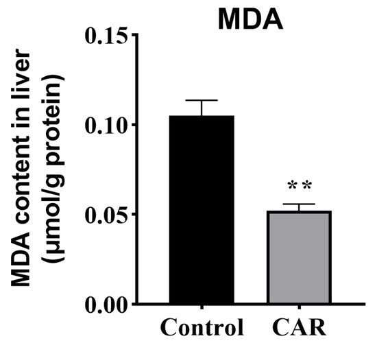
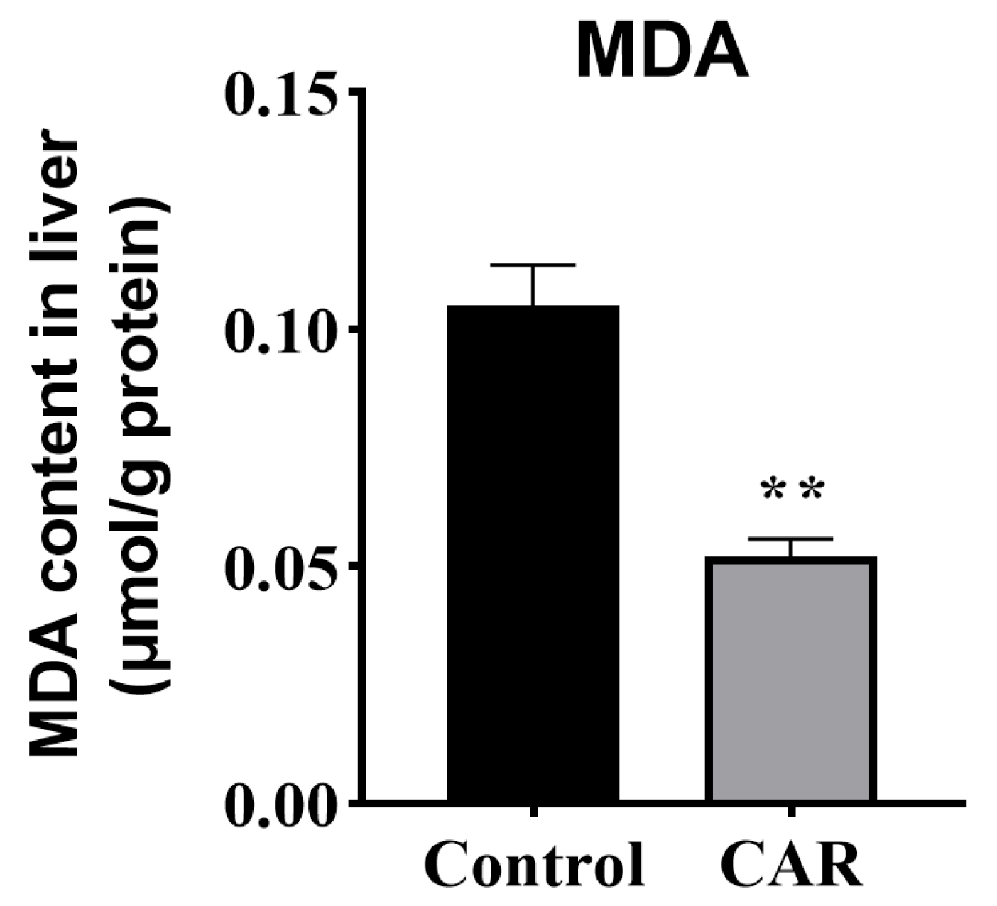
As depicted in Figure 1, the MDA content was significantly reduced by dietary CAR supplementation compared to the control group, which indicates that dietary CAR improved the hepatic antioxidant capacity of tiger puffer fed with a high-fat diet (p < 0.05).
Figure 1.
Dietary CAR decreased the MDA content in tiger puffer liver. Data were represented as mean ± standard error, n = 3. Statistical significance was assessed by Student’s t-test (** p < 0.01).
3.4. Dietary CAR Activated Hepatic Fatty Acid β-Oxidation in Tiger Puffer
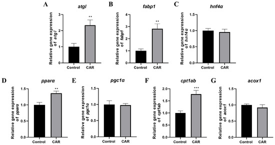
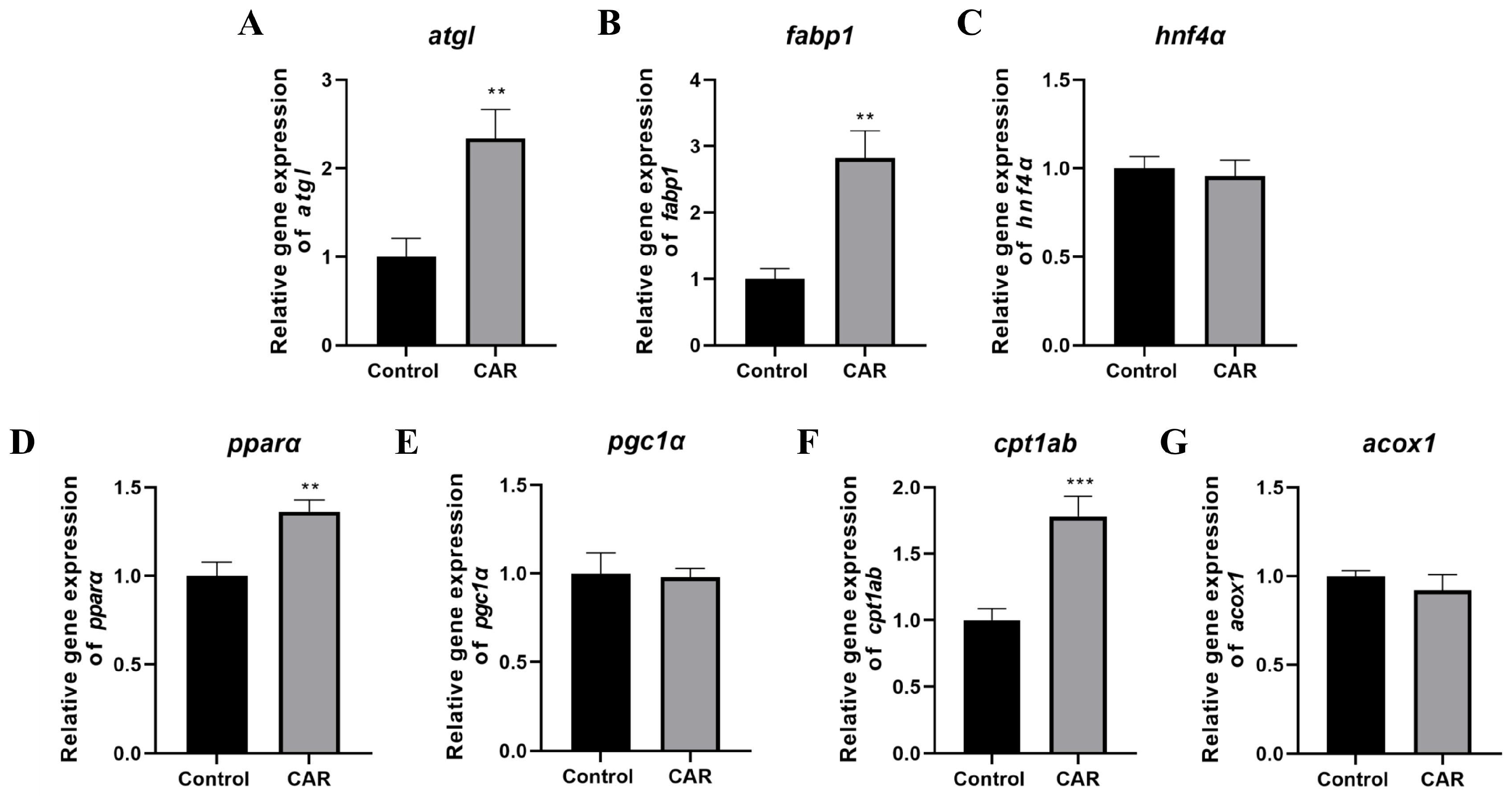
The expression in the liver of genes involved in lipolysis, intracellular fatty acid transport, and fatty acid β-oxidation was determined. Compared to the control group, dietary CAR supplementation significantly up-regulated the levels of atgl and fabp1 (p < 0.05) (Figure 2A,B). Simultaneously, the genes related to mitochondrial fatty acid β-oxidation were also markedly up-regulated by dietary CAR, including pparα and ctp1ab (p < 0.05), but not hnf4α and pgc1α (Figure 2C–G). In addition, the mRNA expression level of acox1, which is a marker gene for peroxisomal fatty acid β-oxidation, was also not influenced by the dietary CAR.
Figure 2.
Effect of dietary CAR on the mRNA expression of lipid metabolism-related genes in tiger puffer liver. (A) The relative mRNA expression level of adipose triglyceride lipase (atgl). (B) The relative mRNA expression level of fatty acid binding protein 1 (fabp1). (C) The relative mRNA expression level of hepatocyte nuclear factor 4 alpha (hnf4α). (D) The relative mRNA expression level of peroxysome proliferator-activated receptor alpha (pparα). (E) The relative mRNA expression level of peroxlsome proliferator-activated receptor-γ coactivator 1 alpha (pgc1α). (F) The relative mRNA expression level of carnitine palmitoyl transferase 1 ab (cpt1ab). (G) The relative mRNA expression level of acyl-coenzyme A oxidase 1 (acox1). Data were represented as mean ± standard error, n = 3. Statistical significance was assessed by Student’s t-test (** p < 0.01, *** p < 0.001).
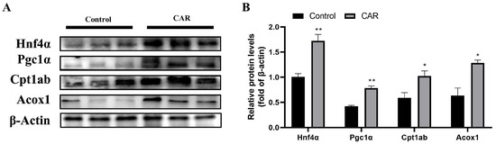
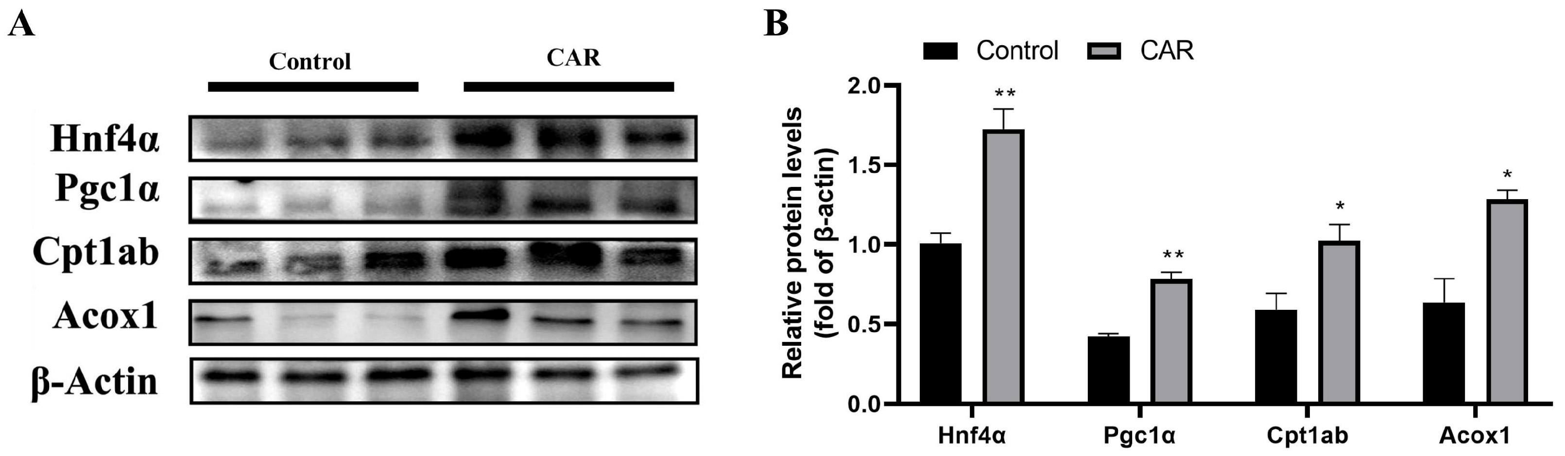
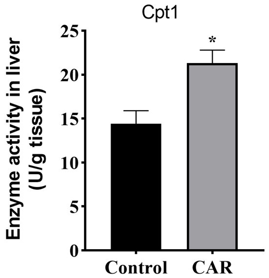
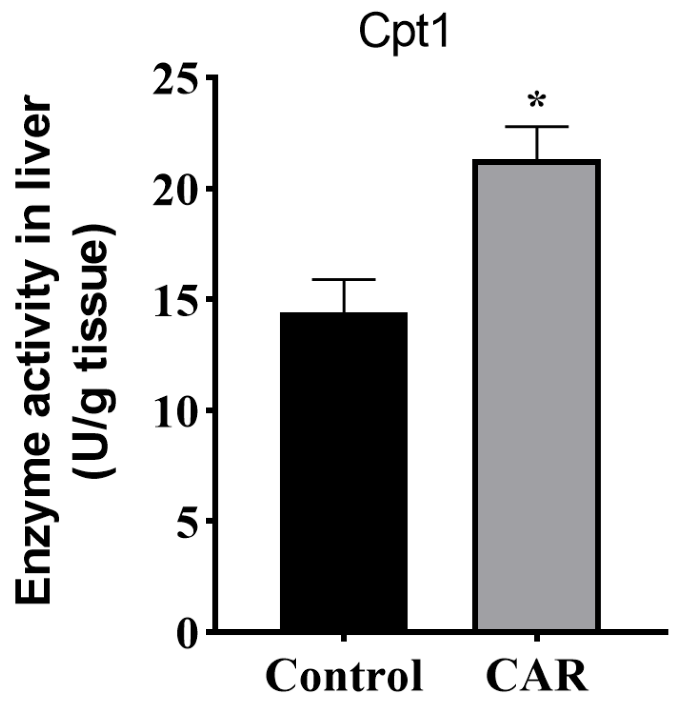
The protein expression levels of Hnf4α, Pgc1α, Ctp1ab and Acox1 were notably up-regulated by dietary CAR (p < 0.05) (Figure 3A,B). Furthermore, compared to the control group, the activity of Ctp1 was also notably increased by CAR supplementation (p < 0.05) (Figure 4). The results indicate that dietary CAR induced hepatic lipolysis, intracellular fatty acid transport, and fatty acid β-oxidation in tiger puffer fed with a high-fat diet.
Figure 3.
Effect of dietary CAR on the expression of fatty acid β-oxidation-related proteins in tiger puffer liver. (A) Western blotting of proteins in the liver; (B) Quantification analysis of the proteins expression. Data were represented as mean ± standard error, n = 3. Statistical significance was assessed by Student’s t-test (* p < 0.05, ** p < 0.01).
Figure 4.
Effect of dietary CAR on the enzyme activity of Cpt1 in tiger puffer liver. Data were represented as mean ± standard error, n = 3. Statistical significance was assessed by Student’s t-test (* p < 0.05).
4. Discussion
In aquaculture, lipids are the second most important nutrient after protein [10]. While appropriately increasing dietary lipid levels improves feed utilization and spare protein, excessively high lipid levels lead to adverse effects in fish, including reduced growth, abnormal hepatic lipid metabolism, and oxidative stress [4,5]. Although dietary CAR supplementation has been shown to effectively mitigate lipid deposition in fish fed with high-fat diets, its efficacy in tiger puffer remains unclear. In this study, we found that while CAR did not affect the growth, it activated fatty acid β-oxidation and inhibited high-fat diet-induced hepatic lipid accumulation.
It has been observed that a moderate lipid content in the diet enhances fish growth performance, whereas excessive lipid levels exert detrimental effects [26]. Previous studies have demonstrated that dietary CAR supplementation enhances growth performance in yellow catfish [14], largemouth bass [27], and silver catfish [28]. This may be because CAR enhances fatty acid β-oxidation for energy production, thereby further strengthening the protein-sparing effect of lipids. In contrast, here we found that dietary CAR does not markedly affect the growth performance in tiger puffer, manifested by the unchanged WG and SGR, while there is an increasing trend in WG. This aligns with the results of previous studies on Nile tilapia [29], juvenile golden pompano [30], and juvenile giant grouper [31]. The inconsistent findings suggest that the efficacy of CAR as a dietary supplement in promoting growth may be influenced by variables including fish species, diet formulation, aquaculture conditions, and developmental stage. Tiger puffer stores most of its lipid in the liver, which typically results in a relatively high HSI. In this study, dietary CAR significantly reduced the HSI of tiger puffer, which counteracted part of the WG. As a result, despite an increasing trend in WG being found in the CAR group, no significant difference was detected.
Studies have shown that excessive reactive oxygen species disrupt the redox balance and induce oxidative stress, during which the content of MDA, a lipid peroxidation product, increases in the liver [32,33]. In this study, dietary 1.5% CAR supplemented to a high-fat diet significantly reduced the hepatic MDA content in tiger puffer. Similar results have been shown in other fish species. For example, as dietary lipid levels increased, hepatic MDA increased significantly in golden pompano, while dietary CAR markedly decreased its content [30]. Another study on large yellow croaker also found that dietary CAR significantly alleviated the elevation of serum and hepatic MDA content induced by a high-fat diet [9]. However, an experiment in mice revealed that a high dose of CAR suppressed the activities of superoxide dismutase and glutathione peroxidase, elevated MDA levels in the liver, and significantly increased serum aspartate aminotransferase and alanine aminotransferase activities, suggesting that excessive CAR intake may weaken the antioxidant system and lead to liver damage [34]. In summary, moderate supplementation of CAR in the diet generally reduces hepatic MDA content, indicating that it inhibits lipid peroxidation, thereby alleviating oxidative damage to hepatocytes. This helps maintain normal physiological liver function. In mammals, poultry, and pigs, its antioxidant effects may be related to scavenging lipid peroxides and enhancing redox signaling through activation of the Nrf2-Keap1 pathway [35]. In fish, the specific antioxidant mechanisms of CAR remain to be further elucidated.
The liver serves as both a critical organ for metabolism and a vital reservoir for nutrient storage in fish. In this study, dietary CAR supplementation did not affect the crude protein content in tiger puffer liver; similar observations were reported in largemouth bass [11]. This may be because dietary CAR primarily exerts its functions by regulating lipid metabolism, rather than participating in protein metabolism. As expected, the CAR group exhibited a significant reduction in both HSI and hepatic crude lipid content, indicating that dietary CAR supplementation effectively alleviates hepatic lipid accumulation in tiger puffer; similar results have also been reported in large yellow croaker [9]. However, studies have shown that dietary CAR only decreased the crude lipid content in the whole body and muscle of tilapia [29], black sea bream [36], and zebrafish [37], not in liver. Such inconsistencies may be attributed to the tissue-specific effects of CAR in different fish species, as well as variations in dietary composition.
The levels of TG, TC, HDL-C, and LDL-C in serum are widely used to assess physiological status in fish. Studies on golden pompano [10] and Nile tilapia [38,39] have found that supplementing CAR to a high-fat diet significantly reduced the TG and TC contents in serum. In contrast, the present study, along with research on black seabream [40], reported stable TG and TC levels, which were not influenced by dietary CAR. HDL-C and LDL-C are closely associated with the efficiency of cholesterol transport [41]. The primary function of LDL-C is to transport cholesterol from the liver to peripheral tissues, while HDL-C clears it via reverse transport from these tissues to the liver for catabolism and elimination [42,43]. It is notable that the HDL-C and LDL-C levels in serum did not show significant changes following dietary CAR supplementation, indicating that the cholesterol metabolism was not affected in the liver of tiger puffer.
The lipid-lowering effect of CAR was mainly attributed to its ability to promote fatty acid β-oxidation to generate energy. As expected, dietary CAR supplementation enhanced the activity of Cpt1, along with its transcriptional and protein expression levels. Concurrently, it significantly increased the transcriptional level of pparα and the protein expression levels of Hnf4α and Pgc1α, all of which are key regulatory factors of mitochondrial fatty acid β-oxidation. These findings collectively demonstrate that CAR activates mitochondrial fatty acid β-oxidation in the liver of tiger puffer, which aligns with the observations in large yellow croaker [9]. Additionally, the protein expression level of Acox1 was also up-regulated by dietary CAR, indicating that CAR promotes peroxisomal fatty acid β-oxidation in the liver of tiger puffer simultaneously, which contrasts the results found in zebrafish fed with a normal level of lipid diet [37]. It is speculated that the synergistic promotion of fatty acid β-oxidation in mitochondria and peroxisomes by CAR is limited to the feeding background of a high-fat diet. However, the synergistic mechanism by which dietary CAR promotes both mitochondrial and peroxisomal fatty acid β-oxidation in tiger puffer liver warrants further investigation. Furthermore, dietary CAR promoted the transcriptional expression of the lipolysis-related gene atgl and the intracellular fatty acid transport-related gene fabp1, suggesting that the enhanced fatty acid β-oxidation drives lipid droplet hydrolysis and intracellular transport to supply sufficient substrates for sustained oxidation.
5. Conclusions
In conclusion, dietary CAR supplementation to a high-fat diet did not affect the growth performance, but enhanced the antioxidant capacity. In addition, dietary CAR alleviated the hepatic lipid deposition in tiger puffer, attributing to the activated fatty acid β-oxidation both in mitochondria and peroxisomes. These findings imply that CAR may be a promising lipid-lowering feed additive for marine fish fed with high-fat diets in aquaculture.
Author Contributions
Conceptualization and funding acquisition were assigned to C.B., H.X. and M.L. Formal analysis, data curation, and methodology were assigned to Z.L. and J.L. Formal analysis and software were assigned to C.B., Z.L., Q.M. and Y.W. Investigation was assigned to W.L. Writing of the original draft was assigned to C.B. Writing, reviewing, and editing of the manuscript were assigned to C.B. and H.X. Supervision was assigned to H.X. All authors have read and agreed to the published version of the manuscript.
Funding
This work was financially supported by the National Natural Science Foundation of China (NSFC, Grant Number: 32503203), Applied Research Program for Post-doctors in Qingdao (QDBSH20250102007), Ningbo “Sci-Tech Innovation Yongjiang 2035” Key R&D Program (2025Z100), Central Public-interest Scientific Institution Basal Research Fund, CAFS (2025XT0804; 2023TD52), and Earmarked Fund for China Agriculture Research System (CARS47-G15).
Institutional Review Board Statement
The animal experiments in this study were conducted in accordance with protocols approved by the Institutional Animal Care and Use Committee of the Yellow Sea Fisheries Research Institute (Approval ID: YSFRI-2025040; Approval date: 1 January 2025).
Informed Consent Statement
Not applicable.
Data Availability Statement
The original contributions presented in this study are included in the article. Further inquiries can be directed to the corresponding author.
Conflicts of Interest
Author Weidong Li was employed by the company Tangshan Haidu Seafood Co., Ltd. The remaining authors declare that the research was conducted in the absence of any commercial or financial relationships that could be construed as a potential conflict of interest.
References
- Zhang, Z.Y.; Limbu, S.M.; Zhao, S.H.; Chen, L.Q.; Luo, Y.; Zhang, M.L.; Du, Z.Y. Dietary l-carnitine supplementation recovers the increased pH and hardness in fillets caused by high-fat diet in Nile tilapia (Oreochromis niloticus). Food Chem. 2022, 382, 132367. [Google Scholar] [CrossRef] [Scilit]
- Halver, J.; Hardy, R.; Goddard, S. Fish Nutrition; Elsevier: Amsterdam, The Netherlands, 2003. [Google Scholar]
- Wang, H.L.; Bruce, T.J.; Su, B.F.; Li, S.; Dunham, R.A.; Wang, X. Environment-dependent heterosis and transgressive gene expression in reciprocal hybrids between the channel catfish Ictalurus punctatus and the blue catfish Ictalurus furcatus. Biology 2022, 11, 117. [Google Scholar] [CrossRef] [Scilit] [PubMed]
- Pang, G.; Li, Y.; Zhang, Z.; Sun, B.; Song, F.; Zheng, S.; Li, X. Evaluation of the effects of diets containing different lipid levels for juvenile yellowfin seabream Acanthopagrus latus. Aquac. Rep. 2024, 34, 101898. [Google Scholar] [CrossRef] [Scilit]
- Cao, X.; Huang, Y.; Chu, X.; Wang, X.; Zheng, S.; Shi, X.; Jiang, G. Differential pathways to hepatic steatosis in fish: Divergent molecular mechanisms underlying high-carbohydrate versus high-lipid diet-induced lipid accumulation. Aquac. Rep. 2025, 45, 103090. [Google Scholar] [CrossRef] [Scilit]
- Panchal, S.K.; Poudyal, H.; Ward, L.C.; Waanders, J.; Brown, L. Modulation of tissue fatty acids by L-carnitine attenuates metabolic syndrome in diet-induced obese rats. Food Funct. 2015, 6, 2496–2506. [Google Scholar] [CrossRef] [Scilit]
- Ke, B.; Kupiec-Weglinski, J.W. Lipid metabolites: The alarm signal to trigger liver ischemia-reperfusion injury. Transplantation 2018, 102, 887–889. [Google Scholar] [CrossRef] [Scilit]
- Zhou, X.; Huang, G.; Wang, L.; Zhao, Y.; Li, J.; Chen, D.; Yang, B. L-carnitine promotes liver regeneration after hepatectomy by enhancing lipid metabolism. J. Transl. Med. 2023, 21, 487. [Google Scholar] [CrossRef] [Scilit]
- Li, J.M.; Zhang, Z.; Kong, A.; Lai, W.; Xu, W.; Cao, X.; Ai, Q. Dietary L-carnitine regulates liver lipid metabolism via simultaneously activating fatty acid β-oxidation and suppressing endoplasmic reticulum stress in large yellow croaker fed with high-fat diets. Br. J. Nutr. 2023, 129, 29–40. [Google Scholar] [CrossRef] [Scilit]
- Chen, X.; Niu, J.; Wang, J.; Zhao, W. Effects of L-carnitine supplementation in high-fat diet on growth, antioxidant capacity and lipid metabolism of golden pompano (Trachinotus ovatus). Front. Mar. Sci. 2022, 9, 831153. [Google Scholar] [CrossRef] [Scilit]
- Qin, D.; Chen, J.; Li, J.; Liu, Z.; Huang, W.; Liu, H.; Dong, X. L-carnitine alleviates hepatic lipid accumulation and inflammatory responses in high-fat-diet fed largemouth bass (M. salmoides): Insights into growth performances, fat metabolism and liver pathology. Aquac. Rep. 2025, 45, 103179. [Google Scholar] [CrossRef] [Scilit]
- Bremer, J. Carnitine--metabolism and functions. Physiol. Rev. 1983, 63, 1420–1480. [Google Scholar] [CrossRef] [Scilit]
- Zammit, V.A.; Ramsay, R.R.; Bonomini, M.; Arduini, A. Carnitine, mitochondrial function and therapy. Adv. Drug Deliv. Rev. 2009, 61, 1353–1362. [Google Scholar] [CrossRef] [Scilit]
- Zheng, J.L.; Luo, Z.; Zhuo, M.Q.; Pan, Y.X.; Song, Y.F.; Hu, W.; Chen, Q.L. Dietary L-carnitine supplementation increases lipid deposition in the liver and muscle of yellow catfish (Pelteobagrus fulvidraco) through changes in lipid metabolism. Br. J. Nutr. 2014, 112, 698–708. [Google Scholar] [CrossRef] [Scilit] [PubMed]
- Tamai, Y.; Chen, Z.; Wu, Y.; Okabe, J.; Kobayashi, Y.; Chiba, H.; Takei, Y. Branched-chain amino acids and l-carnitine attenuate lipotoxic hepatocellular damage in rat cirrhotic liver. Biomed. Pharmacother. 2021, 135, 111181. [Google Scholar] [CrossRef] [Scilit] [PubMed]
- Lyu, J.; Okada, H.; Sunagozaka, H.; Kawaguchi, K.; Shimakami, T.; Nio, K.; Honda, M. Potential utility of l-carnitine for preventing liver tumors derived from metabolic dysfunction-associated steatohepatitis. Hepatol. Commun. 2024, 8, e0425. [Google Scholar] [CrossRef] [Scilit] [PubMed]
- Guzmán-Guillén, R.; Prieto, A.I.; Vázquez, C.M.; Vasconcelos, V.; Cameán, A.M. The protective role of l-carnitine against cylindrospermopsin-induced oxidative stress in tilapia (Oreochromis niloticus). Aquat. Toxicol. 2013, 132, 141–150. [Google Scholar] [CrossRef] [Scilit]
- Safari, O.; Atash, M.M.S.; Paolucci, M. Effects of dietary L-carnitine level on growth performance, immune responses and stress resistance of juvenile narrow clawed crayfish, Astacus leptodactylus leptodactylus Eschscholtz, 1823. Aquaculture 2015, 439, 20–28. [Google Scholar] [CrossRef] [Scilit]
- Wang, Q.; Luo, S.; Ghonimy, A.; Chen, Y.; Guo, Z.; Liu, H.; Zhang, D. Effect of dietary l-carnitine on growth performance and antioxidant response in Amur minnow (Phoxinus lagowskii Dybowskii). Aquac. Nutr. 2019, 25, 749–760. [Google Scholar] [CrossRef] [Scilit]
- Jia, Y.; Jing, Q.; Xing, Z.; Gao, X.; Zhai, J.; Guan, C.; Huang, B. Effects of two different culture systems on the growth performance and physiological metabolism of tiger pufferfish (Takifugu rubripes). Aquaculture 2018, 495, 267–272. [Google Scholar] [CrossRef] [Scilit]
- Xu, H.; Liao, Z.; Zhang, Q.; Wei, Y.; Liang, M. A moderately high level of dietary lipid inhibited the protein secretion function of liver in juvenile tiger puffer Takifugu rubripes. Aquaculture 2019, 498, 17–27. [Google Scholar] [CrossRef] [Scilit]
- Xu, H.; Mu, Y.; Zhang, Y.; Li, J.; Liang, M.; Zheng, K.; Wei, Y. Graded levels of fish protein hydrolysate in high plant diets for turbot (Scophthalmus maximus): Effects on growth performance and lipid accumulation. Aquaculture 2016, 454, 140–147. [Google Scholar] [CrossRef] [Scilit]
- AOAC (Association of Official Analytical Chemists). Official Methods of Analysis of Official Analytical Chemists International, 18th ed.; Association of Official Analytical Chemists: Gaithersburg, MD, USA, 2005. [Google Scholar]
- Folch, J.; Lees, M.; Stanley, G.S. A simple method for the isolation and purification of total lipides from animal tissues. J. Biol. Chem. 1957, 226, 497–509. [Google Scholar] [CrossRef] [Scilit] [PubMed]
- Xu, H.; Bi, Q.; Meng, X.; Duan, M.; Wei, Y.; Liang, M. Response of lipid and fatty acid composition of turbot to starvation under different dietary lipid levels in the previous feeding period. Food Res. Int. 2022, 151, 110905. [Google Scholar] [CrossRef] [Scilit] [PubMed]
- Xun, P.; Lin, H.; Wang, R.; Yu, W.; Zhou, C.; Tan, X.; Yu, W. Effects of dietary lipid levels on growth performance, plasma biochemistry, lipid metabolism and intestinal microbiota of juvenile golden pompano (Trachinotus ovatus). Aquac. Nutr. 2021, 27, 1683–1698. [Google Scholar] [CrossRef] [Scilit]
- Chen, Y.; Sun, Z.; Liang, Z.; Xie, Y.; Su, J.; Luo, Q.; Wang, A. Effects of dietary fish oil replacement by soybean oil and l-carnitine supplementation on growth performance, fatty acid composition, lipid metabolism and liver health of juvenile largemouth bass, Micropterus salmoides. Aquaculture 2020, 516, 734596. [Google Scholar] [CrossRef] [Scilit]
- Goes, M.D.; Feihrmann, A.C.; Carvalho, K.I.F.S.; Baumgartner, L.A.; de Oliveira Pedreira, A.C.; Chagas, T.V.; Bombardelli, R.A. Diets with soybean oil and L-carnitine alter lipid metabolism and improve the growth and reproduction of Rhamdia quelen females. Aquaculture 2024, 579, 740158. [Google Scholar] [CrossRef] [Scilit]
- Li, L.Y.; Lu, D.L.; Jiang, Z.Y.; Limbu, S.M.; Qiao, F.; Chen, L.Q.; Du, Z.Y. Dietary L-carnitine improves glycogen and protein accumulation in Nile tilapia via increasing lipid-sourced energy supply: An isotope-based metabolic tracking. Aquac. Rep. 2020, 17, 100302. [Google Scholar] [CrossRef] [Scilit]
- Yi, Y.; Ma, H.; Tan, B.; Wei, H.; Wu, J.; Mao, M.; Yang, Q. Interaction between dietary L-carnitine and lipid levels on growth, lipid metabolism, and antioxidant capacity in juvenile golden pompano (Trachinotus ovatus). Aquac. Rep. 2025, 41, 102714. [Google Scholar] [CrossRef] [Scilit]
- Lin, Y.H.; Lee, Y.C.; Yeh, C.C. Effects of l-carnitine supplementation in diet on growth, lipid metabolism, and carnitine biosynthesis in juvenile giant grouper (Epinephelus lanceolatus) fed high plant protein diets. Aquaculture 2025, 609, 742821. [Google Scholar] [CrossRef] [Scilit]
- Tocher, D.R.; Mourente, G.; Van der Eecken, A.; Evjemo, J.O.; Diaz, E.; Bell, J.G.; Olsen, Y. Effects of dietary vitamin E on antioxidant defence mechanisms of juvenile turbot (Scophthalmus maximus L.), halibut (Hippoglossus hippoglossus L.) and sea bream (Sparus aurata L.). Aquac. Nutr. 2002, 8, 195–207. [Google Scholar] [CrossRef] [Scilit]
- Lin, L.; Liu, X.; Xu, J.; Weng, L.; Ren, J.; Ge, J.; Zou, Y. Mas receptor mediates cardioprotection of angiotensin-(1-7) against Angiotensin II-induced cardiomyocyte autophagy and cardiac remodeling through inhibition of oxidative stress. J. Cell. Mol. Med. 2016, 20, 48–57. [Google Scholar] [CrossRef] [Scilit] [PubMed]
- Wu, Q.; Zhang, X.; Zhao, Y.; Yang, X. High l-carnitine ingestion impairs liver function by disordering gut bacteria composition in mice. J. Agric. Food Chem. 2020, 68, 5707–5714. [Google Scholar] [CrossRef] [Scilit]
- Surai, P.F. Antioxidant action of carnitine: Molecular mechanisms and practical applications. EC Vet. Sci. 2015, 2, 66–84. [Google Scholar]
- Ma, J.J.; Xu, Z.R.; Shao, Q.J.; Xu, J.Z.; Hung, S.S.; Hu, W.L.; Zhuo, L.Y. Effect of dietary supplemental L-carnitine on growth performance, body composition and antioxidant status in juvenile black sea bream, Sparus macrocephalus. Aquac. Nutr. 2008, 14, 464–471. [Google Scholar] [CrossRef] [Scilit]
- Li, J.M.; Li, L.Y.; Qin, X.; Ning, L.J.; Lu, D.L.; Li, D.L.; Du, Z.Y. Systemic regulation of L-carnitine in nutritional metabolism in zebrafish, Danio rerio. Sci. Rep. 2017, 7, 40815. [Google Scholar] [CrossRef] [Scilit]
- Yang, S.D.; Wen, Y.C.; Liou, C.H.; Liu, F.G. Influence of dietary l-carnitine on growth, biological traits and meat quality in Tilapia. Aquac. Res. 2009, 40, 1374–1382. [Google Scholar] [CrossRef] [Scilit]
- Assar, D.H.; Salah, A.S.; Rashwan, A.G.; Al-Hawary, I.I.; Hendam, B.M.; Elsheshtawy, A.; Elbialy, Z.I. Dietary l-carnitine supplementation recovers the hepatic damage induced by high-fat diet in Nile tilapia (Oreochromis niloticus L.) via activation of Nrf2/Keap pathway and inhibition of pro-inflammatory cytokine. Fish Physiol. Biochem. 2025, 51, 40. [Google Scholar] [CrossRef] [Scilit]
- Jin, M.; Pan, T.; Cheng, X.; Zhu, T.T.; Sun, P.; Zhou, F.; Zhou, Q. Effects of supplemental dietary L-carnitine and bile acids on growth performance, antioxidant and immune ability, histopathological changes and inflammatory response in juvenile black seabream (Acanthopagrus schlegelii) fed high-fat diet. Aquaculture 2019, 504, 199–209. [Google Scholar] [CrossRef] [Scilit]
- Yao, J.; Chen, P.; Apraku, A.; Zhang, G.; Huang, Z.; Hua, X. Hydrolysable tannin supplementation alters digestibility and utilization of dietary protein, lipid, and carbohydrate in grass carp (Ctenopharyngodon idellus). Front. Nutr. 2019, 6, 183. [Google Scholar] [CrossRef] [Scilit]
- Olson, R.E. Discovery of the lipoproteins, their role in fat transport and their significance as risk factors. J. Nutr. 1998, 128, 439S–443S. [Google Scholar] [CrossRef] [Scilit]
- Brown, W.V. High-density lipoprotein and transport of cholesterol and triglyceride in blood. J. Clin. Lipidol. 2007, 1, 7–19. [Google Scholar] [CrossRef] [Scilit]

Disclaimer/Publisher’s Note: The statements, opinions and data contained in all publications are solely those of the individual author(s) and contributor(s) and not of MDPI and/or the editor(s). MDPI and/or the editor(s) disclaim responsibility for any injury to people or property resulting from any ideas, methods, instructions or products referred to in the content. |
© 2026 by the authors. Licensee MDPI, Basel, Switzerland. This article is an open access article distributed under the terms and conditions of the Creative Commons Attribution (CC BY) license.