Next Issue
Volume 8, February
Previous Issue
Volume 7, December


J. Imaging, Volume 8, Issue 1 (January 2022) – 14 articles

Cover Story (view full-size image): 
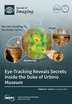
Although our understanding of cognitive disciplines has advanced, we know relatively little about how the human brain perceives art. Thanks to the growing interest in visual perception, eye-tracking technology has increasingly been used to study the interaction between individuals and works of art. In this study, eye-tracking was used to provide information on the visual be-havior of visitors as they moved freely in the famous historical hall of the “Studiolo del Duca” inside the Renaissance Ducal Palace of Urbino. Both behaviors and eye movements of visitors were collected and analyzed. This study revealed which parts of the artifact captured the attention of visitors and also provides interesting information on the main models of use. View this paper
  • Issues are regarded as officially published after their release is announced to the table of contents alert mailing list.
  • You may sign up for e-mail alerts to receive table of contents of newly released issues.
  • PDF is the official format for papers published in both, html and pdf forms. To view the papers in pdf format, click on the "PDF Full-text" link, and use the free Adobe Reader to open them.
Order results
Result details
Section
Select all
Export citation of selected articles as:
37 pages, 1060 KiB  
Article
Retinal Processing: Insights from Mathematical Modelling
by Bruno Cessac
J. Imaging 2022, 8(1), 14; https://doi.org/10.3390/jimaging8010014 - 17 Jan 2022
Cited by 1 | Viewed by 2588
Abstract
The retina is the entrance of the visual system. Although based on common biophysical principles, the dynamics of retinal neurons are quite different from their cortical counterparts, raising interesting problems for modellers. In this paper, I address some mathematically stated questions in this [...] Read more.
The retina is the entrance of the visual system. Although based on common biophysical principles, the dynamics of retinal neurons are quite different from their cortical counterparts, raising interesting problems for modellers. In this paper, I address some mathematically stated questions in this spirit, discussing, in particular: (1) How could lateral amacrine cell connectivity shape the spatio-temporal spike response of retinal ganglion cells? (2) How could spatio-temporal stimuli correlations and retinal network dynamics shape the spike train correlations at the output of the retina? These questions are addressed, first, introducing a mathematically tractable model of the layered retina, integrating amacrine cells’ lateral connectivity and piecewise linear rectification, allowing for computing the retinal ganglion cells receptive field together with the voltage and spike correlations of retinal ganglion cells resulting from the amacrine cells networks. Then, I review some recent results showing how the concept of spatio-temporal Gibbs distributions and linear response theory can be used to characterize the collective spike response to a spatio-temporal stimulus of a set of retinal ganglion cells, coupled via effective interactions corresponding to the amacrine cells network. On these bases, I briefly discuss several potential consequences of these results at the cortical level. Full article
Show Figures

Figure 1

18 pages, 8763 KiB  
Tutorial
Use of Low-Cost Spherical Cameras for the Digitisation of Cultural Heritage Structures into 3D Point Clouds
by Sorin Herban, Domenica Costantino, Vincenzo Saverio Alfio and Massimiliano Pepe
J. Imaging 2022, 8(1), 13; https://doi.org/10.3390/jimaging8010013 - 17 Jan 2022
Cited by 21 | Viewed by 3484
Abstract
The digitization of Cultural Heritage is an important activity for the protection, management, and conservation of structures of particular historical and architectural interest. In this context, the use of low-cost sensors, especially in the photogrammetric field, represents a major research challenge. In this [...] Read more.
The digitization of Cultural Heritage is an important activity for the protection, management, and conservation of structures of particular historical and architectural interest. In this context, the use of low-cost sensors, especially in the photogrammetric field, represents a major research challenge. In this paper, the use of cameras capable of capturing a 360° scene with a single image was assessed. By using spherical photogrammetry and the algorithm based on the structure from motion and multi-view stereo, it is possible to reconstruct the geometry (point cloud) of an object or structure. In particular, for this experiment, the Ricoh theta SC2 camera was used. The analysis was conducted on two sites: one in the laboratory and another directly in the field for the digitization of a large structure (Colonada in Buziaș, Romania). In the case study of the laboratory, several tests were carried out to identify the best strategy for reconstructing the 3D model of the observed environment. In this environment, the approach that provided the best result in terms of both detail and dimensional accuracy was subsequently applied to the case study of Colonada in Buziaș. In this latter case study, a comparison of the point cloud generated by this low-cost sensor and one performed by a high-performance Terrestrial Laser Scanner (TLS), showed a difference of 15 centimeters for 80% of the points. In addition, the 3D point cloud obtained from 360° images is rather noisy and unable to construct complex geometries with small dimensions. However, the photogrammetric dataset can be used for the reconstruction of a virtual tour for the documentation and dissemination of Cultural Heritage. Full article
Show Figures

Figure 1

22 pages, 7321 KiB  
Article
Principles for an Implementation of a Complete CT Reconstruction Tool Chain for Arbitrary Sized Data Sets and Its GPU Optimization
by Jürgen Hofmann, Alexander Flisch and Robert Zboray
J. Imaging 2022, 8(1), 12; https://doi.org/10.3390/jimaging8010012 - 15 Jan 2022
Cited by 2 | Viewed by 2480
Abstract
This article describes the implementation of an efficient and fast in-house computed tomography (CT) reconstruction framework. The implementation principles of this cone-beam CT reconstruction tool chain are described here. The article mainly covers the core part of CT reconstruction, the filtered backprojection and [...] Read more.
This article describes the implementation of an efficient and fast in-house computed tomography (CT) reconstruction framework. The implementation principles of this cone-beam CT reconstruction tool chain are described here. The article mainly covers the core part of CT reconstruction, the filtered backprojection and its speed up on GPU hardware. Methods and implementations of tools for artifact reduction such as ring artifacts, beam hardening, algorithms for the center of rotation determination and tilted rotation axis correction are presented. The framework allows the reconstruction of CT images of arbitrary data size. Strategies on data splitting and GPU kernel optimization techniques applied for the backprojection process are illustrated by a few examples. Full article
(This article belongs to the Special Issue X-ray Digital Radiography and Computed Tomography)
Show Figures

Figure 1

18 pages, 4407 KiB  
Article
Automatic Aortic Valve Cusps Segmentation from CT Images Based on the Cascading Multiple Deep Neural Networks
by Gakuto Aoyama, Longfei Zhao, Shun Zhao, Xiao Xue, Yunxin Zhong, Haruo Yamauchi, Hiroyuki Tsukihara, Eriko Maeda, Kenji Ino, Naoki Tomii, Shu Takagi, Ichiro Sakuma, Minoru Ono and Takuya Sakaguchi
J. Imaging 2022, 8(1), 11; https://doi.org/10.3390/jimaging8010011 - 14 Jan 2022
Cited by 7 | Viewed by 3121
Abstract
Accurate morphological information on aortic valve cusps is critical in treatment planning. Image segmentation is necessary to acquire this information, but manual segmentation is tedious and time consuming. In this paper, we propose a fully automatic aortic valve cusps segmentation method from CT [...] Read more.
Accurate morphological information on aortic valve cusps is critical in treatment planning. Image segmentation is necessary to acquire this information, but manual segmentation is tedious and time consuming. In this paper, we propose a fully automatic aortic valve cusps segmentation method from CT images by combining two deep neural networks, spatial configuration-Net for detecting anatomical landmarks and U-Net for segmentation of aortic valve components. A total of 258 CT volumes of end systolic and end diastolic phases, which include cases with and without severe calcifications, were collected and manually annotated for each aortic valve component. The collected CT volumes were split 6:2:2 for the training, validation and test steps, and our method was evaluated by five-fold cross validation. The segmentation was successful for all CT volumes with 69.26 s as mean processing time. For the segmentation results of the aortic root, the right-coronary cusp, the left-coronary cusp and the non-coronary cusp, mean Dice Coefficient were 0.95, 0.70, 0.69, and 0.67, respectively. There were strong correlations between measurement values automatically calculated based on the annotations and those based on the segmentation results. The results suggest that our method can be used to automatically obtain measurement values for aortic valve morphology. Full article
(This article belongs to the Special Issue Current Methods in Medical Image Segmentation)
Show Figures

Figure 1

18 pages, 4848 KiB  
Article
Historic Timber Roof Structure Reconstruction through Automated Analysis of Point Clouds
by Taşkın Özkan, Norbert Pfeifer, Gudrun Styhler-Aydın, Georg Hochreiner, Ulrike Herbig and Marina Döring-Williams
J. Imaging 2022, 8(1), 10; https://doi.org/10.3390/jimaging8010010 - 13 Jan 2022
Cited by 2 | Viewed by 2873
Abstract
We present a set of methods to improve the automation of the parametric 3D modeling of historic roof structures using terrestrial laser scanning (TLS) point clouds. The final product of the TLS point clouds consist of 3D representation of all objects, which were [...] Read more.
We present a set of methods to improve the automation of the parametric 3D modeling of historic roof structures using terrestrial laser scanning (TLS) point clouds. The final product of the TLS point clouds consist of 3D representation of all objects, which were visible during the scanning, including structural elements, wooden walking ways and rails, roof cover and the ground; thus, a new method was applied to detect and exclude the roof cover points. On the interior roof points, a region-growing segmentation-based beam side face searching approach was extended with an additional method that splits complex segments into linear sub-segments. The presented workflow was conducted on an entire historic roof structure. The main target is to increase the automation of the modeling in the context of completeness. The number of manually counted beams served as reference to define a completeness ratio for results of automatically modeling beams. The analysis shows that this approach could increase the quantitative completeness of the full automatically generated 3D model of the roof structure from 29% to 63%. Full article
Show Figures

Figure 1

16 pages, 9538 KiB  
Article
Fast and Accurate Background Reconstruction Using Background Bootstrapping
by Bruno Sauvalle and Arnaud de La Fortelle
J. Imaging 2022, 8(1), 9; https://doi.org/10.3390/jimaging8010009 - 11 Jan 2022
Cited by 5 | Viewed by 2293
Abstract
The goal of background reconstruction is to recover the background image of a scene from a sequence of frames showing this scene cluttered by various moving objects. This task is fundamental in image analysis, and is generally the first step before more advanced [...] Read more.
The goal of background reconstruction is to recover the background image of a scene from a sequence of frames showing this scene cluttered by various moving objects. This task is fundamental in image analysis, and is generally the first step before more advanced processing, but difficult because there is no formal definition of what should be considered as background or foreground and the results may be severely impacted by various challenges such as illumination changes, intermittent object motions, highly cluttered scenes, etc. We propose in this paper a new iterative algorithm for background reconstruction, where the current estimate of the background is used to guess which image pixels are background pixels and a new background estimation is performed using those pixels only. We then show that the proposed algorithm, which uses stochastic gradient descent for improved regularization, is more accurate than the state of the art on the challenging SBMnet dataset, especially for short videos with low frame rates, and is also fast, reaching an average of 52 fps on this dataset when parameterized for maximal accuracy using acceleration with a graphics processing unit (GPU) and a Python implementation. Full article
(This article belongs to the Section Computer Vision and Pattern Recognition)
Show Figures

Figure 1

15 pages, 7779 KiB  
Article
Exploring Visitors’ Visual Behavior Using Eye-Tracking: The Case of the “Studiolo Del Duca
by Serena Mandolesi, Danilo Gambelli, Simona Naspetti and Raffaele Zanoli
J. Imaging 2022, 8(1), 8; https://doi.org/10.3390/jimaging8010008 - 09 Jan 2022
Cited by 8 | Viewed by 3088
Abstract
Although the understanding of cognitive disciplines has progressed, we know relatively little about how the human brain perceives art. Thanks to the growing interest in visual perception, eye-tracking technology has been increasingly used for studying the interaction between individuals and artworks. In this [...] Read more.
Although the understanding of cognitive disciplines has progressed, we know relatively little about how the human brain perceives art. Thanks to the growing interest in visual perception, eye-tracking technology has been increasingly used for studying the interaction between individuals and artworks. In this study, eye-tracking was used to provide insights into non-expert visitors’ visual behaviour as they move freely in the historical room of the “Studiolo del Duca” of the Ducal Palace in Urbino, Italy. Visitors looked for an average of almost two minutes. This study revealed which parts of the artefact captured visitors’ attention and also gives interesting information about the main patterns of fruition. Full article
(This article belongs to the Special Issue Human Attention and Visual Cognition)
Show Figures

Figure 1

13 pages, 11311 KiB  
Article
Towards a First-Person Perspective Mixed Reality Guidance System for Needle Interventions
by Leah Groves, Natalie Li, Terry M. Peters and Elvis C. S. Chen
J. Imaging 2022, 8(1), 7; https://doi.org/10.3390/jimaging8010007 - 07 Jan 2022
Cited by 9 | Viewed by 2717
Abstract
While ultrasound (US) guidance has been used during central venous catheterization to reduce complications, including the puncturing of arteries, the rate of such problems remains non-negligible. To further reduce complication rates, mixed-reality systems have been proposed as part of the user interface for [...] Read more.
While ultrasound (US) guidance has been used during central venous catheterization to reduce complications, including the puncturing of arteries, the rate of such problems remains non-negligible. To further reduce complication rates, mixed-reality systems have been proposed as part of the user interface for such procedures. We demonstrate the use of a surgical navigation system that renders a calibrated US image, and the needle and its trajectory, in a common frame of reference. We compare the effectiveness of this system, whereby images are rendered on a planar monitor and within a head-mounted display (HMD), to the standard-of-care US-only approach, via a phantom-based user study that recruited 31 expert clinicians and 20 medical students. These users performed needle-insertions into a phantom under the three modes of visualization. The success rates were significantly improved under HMD-guidance as compared to US-guidance, for both expert clinicians (94% vs. 70%) and medical students (70% vs. 25%). Users more consistently positioned their needle closer to the center of the vessel’s lumen under HMD-guidance compared to US-guidance. The performance of the clinicians when interacting with this monitor system was comparable to using US-only guidance, with no significant difference being observed across any metrics. The results suggest that the use of an HMD to align the clinician’s visual and motor fields promotes successful needle guidance, highlighting the importance of continued HMD-guidance research. Full article
Show Figures

Figure 1

14 pages, 9854 KiB  
Article
Metaheuristic Algorithms Applied to Color Image Segmentation on HSV Space
by Donatella Giuliani
J. Imaging 2022, 8(1), 6; https://doi.org/10.3390/jimaging8010006 - 05 Jan 2022
Cited by 10 | Viewed by 2637
Abstract
In this research, we propose an unsupervised method for segmentation and edge extraction of color images on the HSV space. This approach is composed of two different phases in which are applied two metaheuristic algorithms, respectively the Firefly (FA) and the Artificial Bee [...] Read more.
In this research, we propose an unsupervised method for segmentation and edge extraction of color images on the HSV space. This approach is composed of two different phases in which are applied two metaheuristic algorithms, respectively the Firefly (FA) and the Artificial Bee Colony (ABC) algorithms. In the first phase, we performed a pixel-based segmentation on each color channel, applying the FA algorithm and the Gaussian Mixture Model. The FA algorithm automatically detects the number of clusters, given by histogram maxima of each single-band image. The detected maxima define the initial means for the parameter estimation of the GMM. Applying the Bayes’ rule, the posterior probabilities of the GMM can be used for assigning pixels to clusters. After processing each color channel, we recombined the segmented components in the final multichannel image. A further reduction in the resultant cluster colors is obtained using the inner product as a similarity index. In the second phase, once we have assigned all pixels to the corresponding classes of the HSV space, we carry out the second step with a region-based segmentation applied to the corresponding grayscale image. For this purpose, the bioinspired Artificial Bee Colony algorithm is performed for edge extraction. Full article
Show Figures

Figure 1

13 pages, 5112 KiB  
Article
How Do Roots Interact with Layered Soils?
by Nina Kemp, Vasileios Angelidakis, Saimir Luli and Sadegh Nadimi
J. Imaging 2022, 8(1), 5; https://doi.org/10.3390/jimaging8010005 - 05 Jan 2022
Cited by 1 | Viewed by 2367
Abstract
Vegetation alters soil fabric by providing biological reinforcement and enhancing the overall mechanical behaviour of slopes, thereby controlling shallow mass movement. To predict the behaviour of vegetated slopes, parameters representing the root system structure, such as root distribution, length, orientation and diameter, should [...] Read more.
Vegetation alters soil fabric by providing biological reinforcement and enhancing the overall mechanical behaviour of slopes, thereby controlling shallow mass movement. To predict the behaviour of vegetated slopes, parameters representing the root system structure, such as root distribution, length, orientation and diameter, should be considered in slope stability models. This study quantifies the relationship between soil physical characteristics and root growth, giving special emphasis on (1) how roots influence the physical architecture of the surrounding soil structure and (2) how soil structure influences the root growth. A systematic experimental study is carried out using high-resolution X-ray micro-computed tomography (µCT) to observe the root behaviour in layered soil. In total, 2 samples are scanned over 15 days, enabling the acquisition of 10 sets of images. A machine learning algorithm for image segmentation is trained to act at 3 different training percentages, resulting in the processing of 30 sets of images, with the outcomes prompting a discussion on the size of the training data set. An automated in-house image processing algorithm is employed to quantify the void ratio and root volume ratio. This script enables post processing and image analysis of all 30 cases within few hours. This work investigates the effect of stratigraphy on root growth, along with the effect of image-segmentation parameters on soil constitutive properties. Full article
(This article belongs to the Special Issue Recent Advances in Image-Based Geotechnics)
Show Figures

Figure 1

23 pages, 1064 KiB  
Review
An Overview of X-ray Photon Counting Spectral Imaging (x-CSI) with a Focus on Gold Nanoparticle Quantification in Oncology
by Oliver L. P. Pickford Scienti and Dimitra G. Darambara
J. Imaging 2022, 8(1), 4; https://doi.org/10.3390/jimaging8010004 - 31 Dec 2021
Cited by 3 | Viewed by 2428
Abstract
This review article offers an overview of the differences between traditional energy integrating (EI) X-ray imaging and the new technique of X-ray photon counting spectral imaging (x-CSI). The review is motivated by the need to image gold nanoparticles (AuNP) in vivo if they [...] Read more.
This review article offers an overview of the differences between traditional energy integrating (EI) X-ray imaging and the new technique of X-ray photon counting spectral imaging (x-CSI). The review is motivated by the need to image gold nanoparticles (AuNP) in vivo if they are to be used clinically to deliver a radiotherapy dose-enhancing effect (RDEE). The aim of this work is to familiarise the reader with x-CSI as a technique and to draw attention to how this technique will need to develop to be of clinical use for the described oncological applications. This article covers the conceptual differences between x-CSI and EI approaches, the advantages of x-CSI, constraints on x-CSI system design, and the achievements of x-CSI in AuNP quantification. The results of the review show there are still approximately two orders of magnitude between the AuNP concentrations used in RDEE applications and the demonstrated detection limits of x-CSI. Two approaches to overcome this were suggested: changing AuNP design or changing x-CSI system design. Optimal system parameters for AuNP detection and general spectral performance as determined by simulation studies were different to those used in the current x-CSI systems, indicating potential gains that may be made with this approach. Full article
(This article belongs to the Special Issue X-ray Digital Radiography and Computed Tomography)
Show Figures

Figure 1

20 pages, 1971 KiB  
Systematic Review
Diagnostic Accuracy of Imaging Findings in Pleural Empyema: Systematic Review and Meta-Analysis
by Desiree Zettinig, Tugba Akinci D’Antonoli, Adrian Wilder-Smith, Jens Bremerich, Jan A. Roth and Raphael Sexauer
J. Imaging 2022, 8(1), 3; https://doi.org/10.3390/jimaging8010003 - 28 Dec 2021
Cited by 5 | Viewed by 3724
Abstract
Computed tomography (CT) diagnosis of empyema is challenging because current literature features multiple overlapping pleural findings. We aimed to identify informative findings for structured reporting. The screening according to inclusion criteria (P: Pleural empyema, I: CT C: culture/gram-stain/pathology/pus, O: Diagnostic accuracy measures), data [...] Read more.
Computed tomography (CT) diagnosis of empyema is challenging because current literature features multiple overlapping pleural findings. We aimed to identify informative findings for structured reporting. The screening according to inclusion criteria (P: Pleural empyema, I: CT C: culture/gram-stain/pathology/pus, O: Diagnostic accuracy measures), data extraction, and risk of bias assessment of studies published between 01-1980 and 10-2021 on Pubmed, Embase, and Web of Science (WOS) were performed independently by two reviewers. CT findings with pooled diagnostic odds ratios (DOR) with 95% confidence intervals, not including 1, were considered as informative. Summary estimates of diagnostic accuracy for CT findings were calculated by using a bivariate random-effects model and heterogeneity sources were evaluated. Ten studies with a total of 252 patients with and 846 without empyema were included. From 119 overlapping descriptors, five informative CT findings were identified: Pleural enhancement, thickening, loculation, fat thickening, and fat stranding with an AUC of 0.80 (hierarchical summary receiver operating characteristic, HSROC). Potential sources of heterogeneity were different thresholds, empyema prevalence, and study year. Full article
(This article belongs to the Topic Medical Image Analysis)
Show Figures

Figure 1

20 pages, 4195 KiB  
Article
Effectiveness of Learning Systems from Common Image File Types to Detect Osteosarcoma Based on Convolutional Neural Networks (CNNs) Models
by Chanunya Loraksa, Sirima Mongkolsomlit, Nitikarn Nimsuk, Meenut Uscharapong and Piya Kiatisevi
J. Imaging 2022, 8(1), 2; https://doi.org/10.3390/jimaging8010002 - 27 Dec 2021
Cited by 9 | Viewed by 2781
Abstract
Osteosarcoma is a rare bone cancer which is more common in children than in adults and has a high chance of metastasizing to the patient’s lungs. Due to initiated cases, it is difficult to diagnose and hard to detect the nodule in a [...] Read more.
Osteosarcoma is a rare bone cancer which is more common in children than in adults and has a high chance of metastasizing to the patient’s lungs. Due to initiated cases, it is difficult to diagnose and hard to detect the nodule in a lung at the early state. Convolutional Neural Networks (CNNs) are effectively applied for early state detection by considering CT-scanned images. Transferring patients from small hospitals to the cancer specialized hospital, Lerdsin Hospital, poses difficulties in information sharing because of the privacy and safety regulations. CD-ROM media was allowed for transferring patients’ data to Lerdsin Hospital. Digital Imaging and Communications in Medicine (DICOM) files cannot be stored on a CD-ROM. DICOM must be converted into other common image formats, such as BMP, JPG and PNG formats. Quality of images can affect the accuracy of the CNN models. In this research, the effect of different image formats is studied and experimented. Three popular medical CNN models, VGG-16, ResNet-50 and MobileNet-V2, are considered and used for osteosarcoma detection. The positive and negative class images are corrected from Lerdsin Hospital, and 80% of all images are used as a training dataset, while the rest are used to validate the trained models. Limited training images are simulated by reducing images in the training dataset. Each model is trained and validated by three different image formats, resulting in 54 testing cases. F1-Score and accuracy are calculated and compared for the models’ performance. VGG-16 is the most robust of all the formats. PNG format is the most preferred image format, followed by BMP and JPG formats, respectively. Full article
(This article belongs to the Topic Medical Image Analysis)
Show Figures

Figure 1

35 pages, 5964 KiB  
Article
Nearly Exact Discrepancy Principle for Low-Count Poisson Image Restoration
by Francesca Bevilacqua, Alessandro Lanza, Monica Pragliola and Fiorella Sgallari
J. Imaging 2022, 8(1), 1; https://doi.org/10.3390/jimaging8010001 - 23 Dec 2021
Cited by 5 | Viewed by 2321
Abstract
The effectiveness of variational methods for restoring images corrupted by Poisson noise strongly depends on the suitable selection of the regularization parameter balancing the effect of the regulation term(s) and the generalized Kullback–Liebler divergence data term. One of the approaches still commonly used [...] Read more.
The effectiveness of variational methods for restoring images corrupted by Poisson noise strongly depends on the suitable selection of the regularization parameter balancing the effect of the regulation term(s) and the generalized Kullback–Liebler divergence data term. One of the approaches still commonly used today for choosing the parameter is the discrepancy principle proposed by Zanella et al. in a seminal work. It relies on imposing a value of the data term approximately equal to its expected value and works well for mid- and high-count Poisson noise corruptions. However, the series truncation approximation used in the theoretical derivation of the expected value leads to poor performance for low-count Poisson noise. In this paper, we highlight the theoretical limits of the approach and then propose a nearly exact version of it based on Monte Carlo simulation and weighted least-square fitting. Several numerical experiments are presented, proving beyond doubt that in the low-count Poisson regime, the proposed modified, nearly exact discrepancy principle performs far better than the original, approximated one by Zanella et al., whereas it works similarly or slightly better in the mid- and high-count regimes. Full article
(This article belongs to the Special Issue Inverse Problems and Imaging)
Show Figures

Figure 1

Previous Issue
Next Issue
Back to TopTop