


Abstract
The study first demonstrated that burdock fructooligosaccharide (BFO) could inhibit peel browning in green banana, with 0.5% BFO treatment showing the most significant suppression of peel browning during low-temperature storage (7 ± 1 °C). The results revealed that 0.5% BFO treatment effectively restrained the increase in electrolyte leakage and malondialdehyde (MDA) content and maintained cell membrane integrity. Furthermore, BFO treatment enhanced total phenolic content and antioxidant capacity, alleviated oxidative damage, and better preserved the external quality of banana peel. Simultaneously, BFO treatment markedly inhibited both the activities of chlorophyll-degrading enzymes and their relative gene expression levels in banana peel, thereby maintaining higher chlorophyll content. This research provided a new insight into the mechanism of inhibiting peel browning for low-temperature storage preservation of banana.
1. Introduction
The banana, a globally important tropical fruit, ripens and decays rapidly after harvest. Although cold storage extends its shelf life, its chilling sensitivity readily leads to peel discoloration and other chilling injuries, severely compromising its commercial value. Studies have demonstrated that low-temperature storage conditions could increase cell membrane permeability, reduce antioxidant capacity, and disrupt the metabolism of superoxide and chlorophyll degradation in banana, ultimately leading to chilling injury symptoms [1]. Consequently, developing an effective method to reduce peel browning is critical to enhance the storage quality and commercial value of the banana.
Low temperature induces the activity of CLH (chlorophyllase), which catalyzes the removal of the phytol group from chlorophyll a to form de-phytylated chlorophyll a. Subsequently, enzymes including MDC (Mg-dechelatase), RCCR (red chlorophyll catabolite reductase), PAO (pheophorbide a oxygenase), and PPH (pheophytin pheophorbide hydrolase) act sequentially, ultimately converting chlorophyll into colorless non-fluorescent chlorophyll degradation products, thereby promoting chlorophyll degradation [2]. Loss of green color indicates banana ripening, and delaying chlorophyll degradation is essential for prolonging the postharvest shelf life [3]. Exogenous substance treatments or other interventions can effectively inhibit chlorophyll degradation and mitigate chilling injury [4,5]. UV-C treatment suppresses chlorophyllase activity, thereby delaying chlorophyll degradation and yellowing, and ultimately mitigating cold-induced damage in bananas during low-temperature storage [6].
Chilling damage can compromise the integrity of cell membranes and induce the accumulation of ROS (reactive oxygen species), whereas the antioxidant enzyme system functions to scavenge ROS and protect cell membranes from further damage [7,8]. Studies have shown that Sodium hydrosulfide application can effectively alleviate the symptoms of chilling injury by regulating ROS homeostasis and enhancing the antioxidant capacity in plant cells [9]. The combination of hot water (52 °C) and glycine betaine improved their cold tolerance in banana by reducing the accumulation of ROS and enhancing overall antioxidant capacity [10]. Melatonin treatment decreased the accumulation of ROS and the activities of LOX (lipoxygenase), increased the activities of POD (peroxidase) and CAT (catalase) in banana peel, thus eliminating free radicals, mitigating lipid peroxidation, and stabilizing the membrane structure [11]. Tyr-Asp treatment enhanced redox homeostasis that was influenced by upregulating oxidoreductase-associated genes and transcriptional regulators and downregulating lipid metabolism and ATP hydrolysis-related genes, which is beneficial to alleviate chilling injury in banana [12]. Anoxic treatment maintained membrane integrity, reduced lipid peroxidation, and enhanced antioxidant capacity, effectively inhibiting banana ripening and deterioration at low temperatures and preventing subsequent ambient temperature deterioration [13].
BFO(Burdock fructooligosaccharide), a non-toxic and biodegradable natural functional polysaccharide, is extracted from burdock roots, which meets the consumer requirement of food safety [14]. Experimental evidence demonstrates that BFO elicits systemic acquired resistance and activates defense-related physiological mechanisms in plants [15,16]. Experimental evidence demonstrated that 0.5% BFO treatment induced blueberry resistance to Alternaria alternata, which resulted in protecting blueberries from oxidative damage [17]. Rhodotorula mucilaginosa in BFO-amended nutrient yeast dextrose agar (NYDA) exhibited significantly enhanced antagonistic activity against Rhizopus and blue mold in peaches, compared to NYDA without BFO [18]. According to Sun et al. [19], BFO induced fungal resistance through regulating the salicylic acid-dependent pathway in postharvest grapes. Moreover, BFO treatment had a significant contribution to preserving the quality of grape exocarp through inhibition of sucrose biosynthesis and the senescence-associated metabolic pathway [20].
Although increasing attention has been paid to the biological function of BFO, there are few reports on BFO inducing cold resistance of fruits and vegetables. Generally, peel browning is the obvious feature for bananas in low-temperature storage, so the BFO-treated banana stored at 7 °C was selected as the research subject in this study. The objective of this research was to investigate the effect of BFO treatment on the resistance of peel browning by analyzing antioxidant capacity and chlorophyll metabolism, which might provide a theoretical foundation and technical strategy on low temperature storage of green bananas.
2. Materials and Methods
2.1. Fruit and Treatment
Banana fruit (Musa spp. (Guangxi cultivar)) at the mature-green stage was purchased from a local wholesale market without prior ethylene degreening. After separation into individual fingers, bananas of uniform size and color, and free from physical damage or disease symptoms, were selected for the experiment. For BFO (obtained from Shanghai Yuanye Co., Ltd., Shanghai, China) treatments, a total of 240 fingers were randomly allocated into four groups, with 3 repetitions and each repetition consisting of 20 fingers. The fingers were immersed in 0 (double-distilled water as control), 0.25%, 0.5% and 1% BFO for 10 min at 20 °C. After air-drying, fingers were packaged in polyethylene bags and maintained at 7 ± 1 °C for 12 days, followed by the assessment of peel browning (Supplementary S1). Based on the first experiment (four BFO concentrations) and phenotypic observations, we conducted a second experiment to investigate the biochemical and gene expression changes in 0.5% BFO-treated samples, with samples collected for analysis. A total of 240 fingers were randomly allocated into two groups, with 3 repetitions and each repetition consisting of 40 fingers. The fingers were immersed in 0 (double-distilled water as control) and 0.5% BFO for 10 min at 20 °C. After air-drying, fingers were packaged in polyethylene bags and maintained at 7 ± 1 °C for 12 days. Additionally, peel tissues from the two groups of bananas in the second experiment were snap-frozen in liquid nitrogen and subsequently stored at −80 °C for biochemical and gene expression analyses.
2.2. Peel Browning Index
The ratio of peel browning area to the total area of the banana was recorded (P). According to the area of browning on the fruit surface relative to the total fruit area, the degree of peel browning of the banana was categorized into six grades. The development of peel browning was rated on the scale of 0–5 as follows: level 0 (P = 0), level 1 (0 < P ≤ 20%), level 2 (20% < P ≤ 40%), level 3 (40% < P ≤ 60%), level 4 (60% < P ≤ 80%), and level 5 (P > 80%). The peel browning index was calculated as the weighted average of the browning levels using the following formula: Peel browning index = ∑ (peel browning level × number of fruits at that level)/total fruit number.
2.3. Fruit Color
Twelve bananas were randomly selected from each level and were used for testing by the SR-64 colorimeter (Shenzhen 3nh Technology Co., Ltd., Shenzhen, China), and four points around the central area of each banana were measured. The L*, a*, and b* values were measured, and the hue angle was calculated as arctan (b*/a*).
2.4. Electrolyte Leakage (EL) and Malondialdehyde (MDA) Content
Electrolyte leakage (EL) was measured following the procedure described by Lin et al. [21]. The stainless steel puncher was used for cutting small round slices with an 8 mm diameter from the middle of the equator of the banana peel. Fifteen slices from the control or treatment group were placed, respectively, and washed three times with distilled water. After drying the surface water, the collected slices were mixed with 50 mL of distilled water. The slices were incubated for 30 min at 25 °C, and the electrical conductivity of the solution was determined using a conductivity meter (DDS-11A, Shanghai INESA Scientific Instrument Co., Ltd., Shanghai, China). After measurement, the tissue discs were heated in a boiling water bath for 30 min and then cooled to 25 °C to measure the total conductivity. Electrolyte leakage was expressed as relative conductivity, calculated as (conductivity of tissue solution/total conductivity) × 100%.
Following the method of Xu et al., with adaptations, MDA content was assayed [22]. 1.0 g of sample was ground with 5.0 mL of 100 g L−1 TCA solution at 4 °C. After centrifugation, 2.0 mL of the supernatant was mixed with 2.0 mL of 0.67% TBA solution, and the mixture was boiled for 20 min in a boiling water bath. After cooling and centrifugation, a spectrophotometer was employed to measure the absorbance of the supernatant (MAMPDA V-1100D, Shanghai MAMPDA Instrument Co., Ltd., Shanghai, China) at wavelengths of 532 nm, 600 nm and 450 nm. The MDA content was calculated according to the following formula: MDA = 6.45 × (A532 − A600) − 0.56 × A450 and expressed as nmol g−1.
2.5. 1,1-Diphenyl-2-Picrylhydrazyl (DPPH) Scavenging Activity and O2− Content
1,1-Diphenyl-2-Picrylhydrazyl (DPPH) Scavenging Activity and O2− Content were analyzed using established methods [23].
To determine the DPPH scavenging activity, 0.5 g of the sample was homogenized with 5.0 mL of methanol in an ice bath, and the mixture was stored for 12 h at 4 °C. A 100 μL aliquot of the supernatant was combined with 2.9 mL of DPPH solution (0.01 g DPPH dissolved in 250 mL alcohol and incubated at 25 °C for 30 min in darkness. At 517 nm, absorbance was recorded, and DPPH scavenging capacity was calculated as per the formula: DPPH scavenging rate (%) = (1 − A1/A2) × 100 (A1: sample absorbance value, A2: control tube absorbance value).
For the determination of O2− content, 5.0 mL of extraction buffer, containing 1 mmol L−1 ethylenediaminetetraacetic acid solution, 2% polyvinylpyrrolidone, and 0.3% Triton X-100, was added to 3.0 g of the sample for grinding. After ice grinding, the homogenate was centrifuged for 30 min at 4 °C, and 500 μL of the supernatant was combined with 1.0 mL of 50 mmol L−1 phosphate buffer (pH 7.8) and 1.0 mL of 1 mmol L−1 hydroxylamine hydrochloride solution. After the incubation at 25 °C for 1 h, 1.0 mL of 17 mmol L−1 p-aminobenzene sulfonic acid solution and 1.0 mL of 7 mmol L−1 α-naphthylamine solution were added to the previous buffer, and the mixture was incubated at 25 °C for 20 min for color reaction, and the absorbance of the resulting solution was measured at 530 nm. The standard curve (y = 0.002x − 0.0003, R2 = 0.9985) was prepared using a KNO2 solution, and the content of O2− was presented in units of mol kg−1.
2.6. Enzyme Assays
The activities of POD (peroxidase) and PPO (polyphenol oxidase) were assayed according to the method established by Cui et al. [24]. 5.0 mL of extraction buffer, containing 1 mmol L−1 polyethylene glycol, 4% cross-linked polyvinylpyrrolidone and 1% Triton X-100, was mixed with 2.0 g of the sample. The mixture was centrifuged at 12,000× g for 30 min at 4 °C, and the supernatant was collected and stored at 4 °C for enzyme assays.
For PPO activity determination, 100 μL of the enzyme extract was mixed with 4.0 mL of 50 mmol L−1 sodium acetate buffer (pH 5.5) and 1.0 mL of 50 mmol L−1 catechol solution. Absorbance values at 420 nm were recorded every 15 s for 120 s.
For the POD activity test, 300 μL of the enzyme extract was mixed with 3.0 mL of 25 mmol L−1 guaiacol solution, and 200 μL of 0.5 mol L−1 H2O2 solution was added for the reaction. Absorbance values at 470 nm were recorded every 15 s for 120 s.
The PAL (phenylalanine ammonia lyase) activity was measured based on the protocol established by Zhang et al. [25]. 5.0 mL of extraction, containing 2.0 mm polyethylene glycol, 4 mmol L−1 mercaptoethanol, and 40 g L−1 polyvinylpyrrolidone, was mixed with 5.0 g ground sample. The mixture was blended in an ice bath and then centrifuged at 4 °C for 30 min at 12,000× g. The resulting supernatant was collected and stored at a low temperature, and 500 μL of the enzyme extract was mixed with 3.0 mL of 50 mmol L−1 boric acid buffer (pH 8.8) and 0.5 mL of 20 mmol L−1 phenylalanine solution. In the control group, 0.5 mL of inactivated enzyme solution (boiled for 5 min) was added instead of the active enzyme. Both the treatment and control groups were maintained at 37 °C for 60 min. After that, 0.1 mL of 6 mol L−1 hydrochloric acid solution was immediately introduced into the mixture to stop the reaction. Distilled water was used as the reference blank to zero the spectrophotometer, and the absorbance values were recorded at 290 nm every 15 s for 120 s.
APX (Ascorbate peroxidase) activity was measured based on the protocol established by Wang et al. [26]. A total of 5.0 mL of extraction buffer (0.1 mmol L−1 ethylenediaminetetraacetic acid, 1 mmol L−1 ascorbic acid, and 2% (w/v) cross-linked polyvinylpyrrolidone) was added to 5.0 g of the grinding sample. The sample was homogenized by ice bath grinding and then centrifuged at 12,000× g for 30 min at 4 °C, and the supernatant was collected as the enzyme extract. Then, 100 μL of the enzyme extract was combined with 2.6 mL of reaction buffer (containing 2.9 mg of ethylenediaminetetraacetic acid, 8.8 mg of ascorbic acid, and pH 7.5 potassium phosphate-buffered solution), and 0.3 mL of 2 mmol L−1 H2O2 solution was added to initiate the enzymatic reaction. Absorbance values were recorded at 290 nm every 15 s for 120 s.
CAT (Catalase) activity was determined following the method described by Li et al. [27]. 5.0 mL of extraction buffer, including 5 mmol L−1 dithiothreitol and 5% polyvinylpyrrolidone, was added to 1.0 g of the ground sample. The mixture was homogenized in an ice bath and then centrifuged at 4 °C for 30 min at 12,000× g. The system, consisting of 2.9 mL of 20 mmol L−1 H2O2 solution and 100 μL of the enzyme extract, was reacted, and the absorbance values were recorded at 240 nm every 15 s for 120 s.
The SOD (superoxide dismutase) activity of banana peel samples was test according to the manufacturer’s instructions, of SOD detection kit (Solarbio Inc., Beijing, China). Absorbance values were recorded every 15 s at 450 nm for 120 s. The POD, PPO, PAL, APX, CAT and SOD activity unit (U) was expressed as U mg−1 protein.
To evaluate the activities of CLH (chlorophyllase) and MDC (Mg-dechelatase), banana peel samples were ground in liquid nitrogen, and the activities of these enzymes were determined using assay kits (BoxBio Technology Co., Ltd., Beijing, China) in accordance with manufacturer’s instructions.
The absorbance at 450 nm was determined, and standard curves for CLH (y = 0.0234x + 0.1513, R2 = 0.9968) and MDC (y = 0.0016x + 0.0714, R2 = 0.9961) were established, respectively, which were used to calculate the enzyme activities in the samples.
2.7. Chlorophyll Content Measurement
According to the previous method [28], 1.0 g of sample was extracted with 30 mL ice-cold acetone for 5 min, followed by centrifugation at 6000× g and 4 °C for 5 min. Using acetone as the blank control, the absorbance of the supernatant was measured spectrophotometrically at 665 nm and 649 nm, respectively. The chlorophyll content (mg g−1) was calculated using the following equation:
Chlorophyll a content = (12.72A665 − 2.59A649) × V/(1000 m)
Chlorophyll b content = (22.88A649 − 4.67A665) × V/(1000 m)
Total chlorophyll content = Chlorophyll a content + Chlorophyll b content
A649 and A665 are the absorbance values at wavelengths of 649 nm and 665 nm, respectively; V is the total volume of the solutionsolution; m is the sample mass.
2.8. Measurement of Total Phenolic Content
The measurement was performed following a previously reported method [28], 0.5 g of ground sample was extracted with 5.0 mL of cold ethanol and kept in a refrigerator at 4 °C overnight, and then the mixture was centrifuged at 12,000× g and 4 °C for 20 min. The supernatant (0.2 mL) was mixed with 1.8 mL of distilled water, 1.0 mL of Folin–Ciocalteu reagent and 1.0 mL of 7.5% (m/v) anhydrous sodium carbonate solution, and the mixture was reacted at 25 °C for 2.0 h. The absorbance at 765 nm was measured to test the reaction system. A standard curve was prepared using a gallic acid solution (y = 0.0135x − 0.0933, R2 = 0.9678), and the total phenolic content was expressed as μg g−1.
2.9. RNA Extraction and Quantitative Real-Time PCR Analysis
According to the method of Zhao et al. [29], RNA was extracted from the peel using the M5 Plant RNease Complex Mini RNA extraction kit, and 1 µg of RNA was taken for reverse transcription. Primers were designed using NCBI and synthesized by Bioengineering (Shanghai) Co., Ltd., Shanghai, China (Supplementary S2). MaACTIN was employed as the internal reference gene. The relative expression levels of the target genes were quantified by real-time fluorescent quantitative PCR using a CFX96 quantitative PCR instrument (Biole Life Medical Products (Shanghai) Co., Ltd., Shanghai, China). The relative transcript expression levels were calculated according to the 2−ΔΔCt method.
2.10. Statistical Analysis
Each measurement was performed in triplicate at a minimum, and data are presented as the mean ± standard deviation (SD). Data analysis was performed using IBM SPSS Statistics 25 by Two-way analysis of variance (ANOVA). There was a significant difference between the two values when the p < 0.05 (*), p < 0.01 (**) or p < 0.001(***).
3. Results
3.1. Improvement in the Appearance Quality of Banana Fruit After Treatments
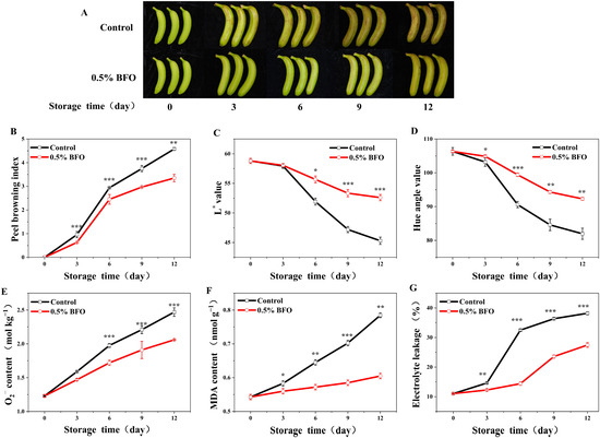
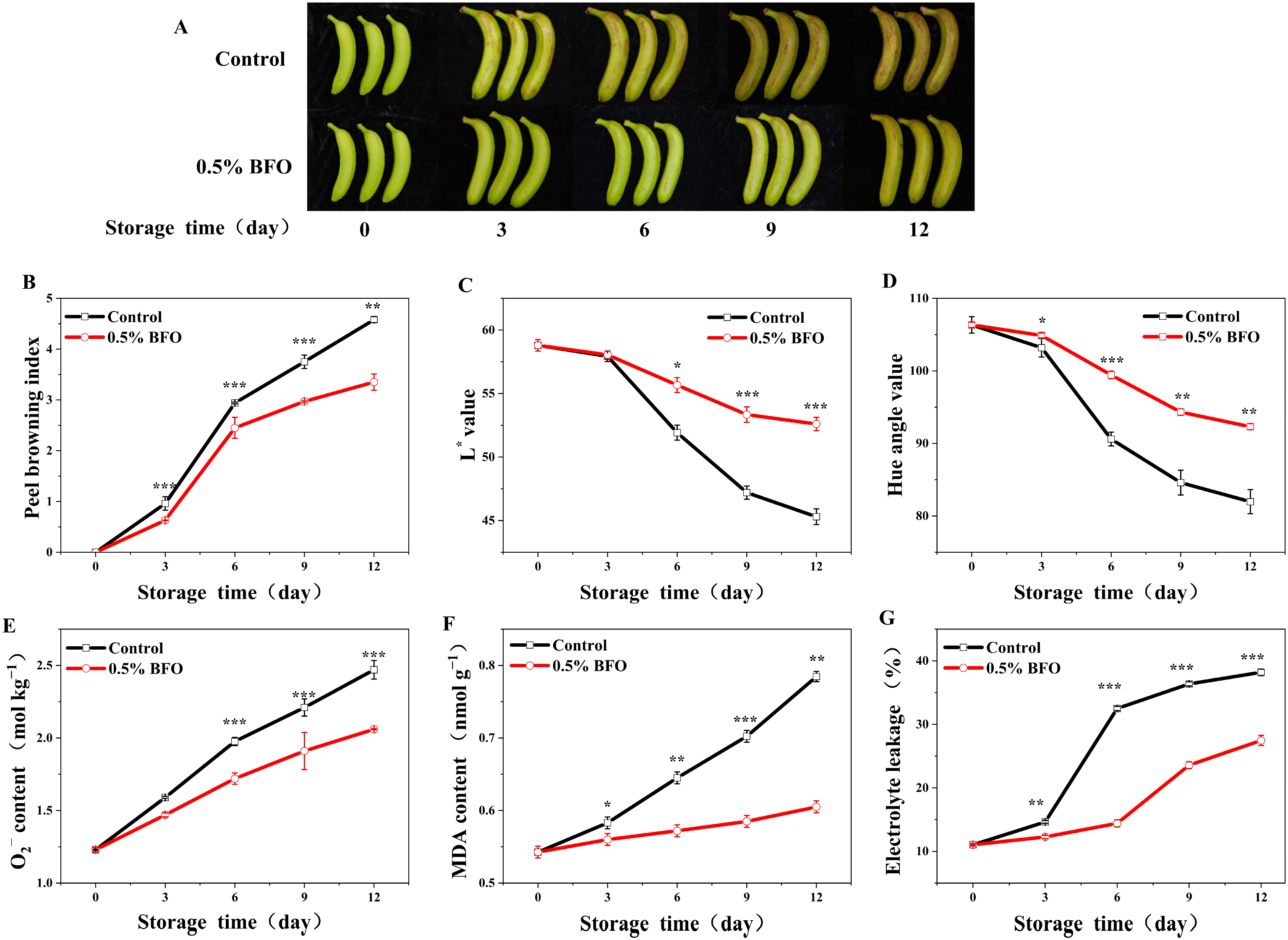
Peel browning and darkening are the most obvious symptoms of green banana chilling injury during cold storage. As Figure 1A shown, the 0.5% BFO could maintain the peel more green and light. Both the control and 0.5% BFO group showed the development of browning during the cold storage; however, compared to the control, the chilling injury index of the 0.5% BFO treatment group was 1.5 times below the level observed in the control group at the end of cold storage, which indicated BFO could alleviate chilling injury in banana (Figure 1A,B).
Figure 1.
Effect of different treatments on visual appearance (A), chilling injury index (B), L* value (C), Hue angle value (D), O2− content (E), MDA content (F), and electrolyte leakage (G) of peel of banana. All statistical analyses were performed using two-way ANOVA. Significance was denoted as p < 0.05 (*), p < 0.01 (**), p < 0.001 (***). Results are shown as mean ± SD (n = 3).
The values of both L* and the hue angle of banana peel color showed a similar downward trend. with the extension of storage time. Moreover, the L* and hue angle of the BFO treatment group were significantly higher than those of the control group during the whole storage process. On day 12, the L* value of the 0.5% BFO treatment group was 52.6, which was 1.16 times higher than that of the control group (Figure 1C). At the same time, the fruit hue angles of the 0.5% BFO treatment group and the control group were 92.3 and 81.9, respectively (Figure 1D).
3.2. Change in Electrolyte Leakage, O2− Content and MDA Contents After Treatments
The production rate of O2− gradually increased with prolonged storage, while BFO treatment significantly reduced its accumulation (Figure 1E). On day 12, the O2− production rate of the control group was 1.19 times higher than that of the treatment group, and the difference between the two groups reached a significant level (p < 0.01).
The MDA content of each group showed a slow upward trend with prolonged storage (Figure 1F). During low-temperature storage, the MDA content of the control group increased more significantly than BFO treated group, and the MDA content of the treated group was 76.92% of the value of the control on day 12 (p < 0.01). Figure 1G showed that changes in electrical conductivity and MDA content followed a similar pattern. BFO treatment delayed the increase in electrical conductivity of the banana peel. On day 12, compared with the treatment group, the control group had a 1.39-fold higher electrical conductivity, and the difference was statistically significant (p < 0.001).
3.3. Effects of BFO Treatment on Antioxidant Enzyme Activities and Related Gene Expression
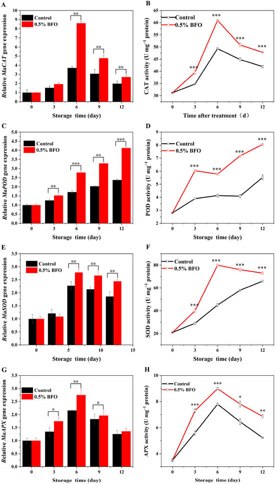
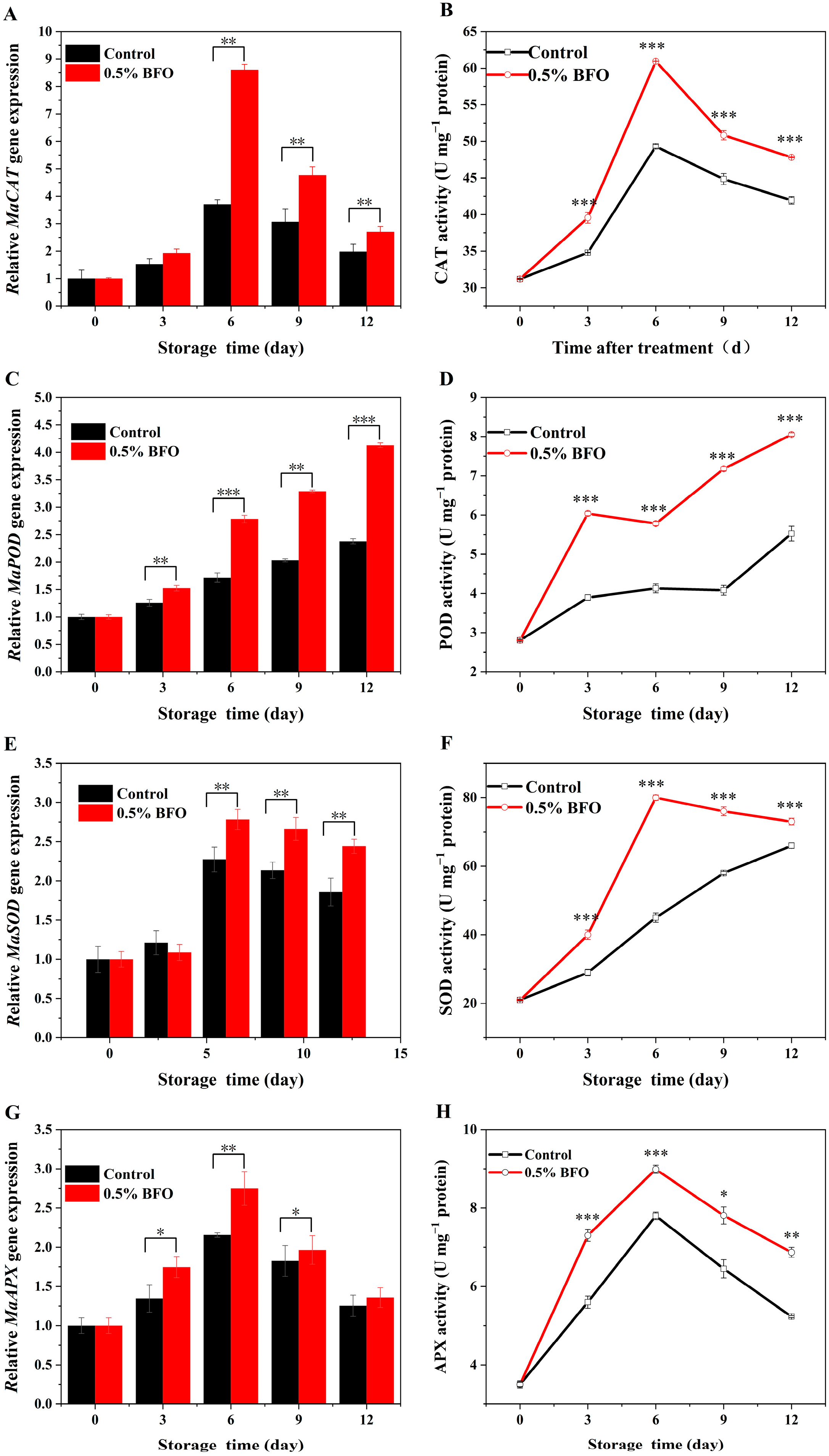
During storage at 7 °C, relative gene expression and antioxidant enzyme activity exhibited an overall increasing trend in both control and BFO-treated fruit, with BFO-treated fruit showing consistently higher levels than control.
Compared to control, the relative MaCAT gene expression and CAT activity of BFO treatment significantly increased, and the trend peaked on day 6, followed by a slight decline. On day 12, the relative MaCAT gene expression and CAT activity in the control group were 76% and 87% of the treatment group, respectively (Figure 2A,B). Moreover, the relative MaPOD gene expression and POD activity in the BFO-treated group gradually increased throughout storage, consistently exceeding those of the control group. On day 12, the relative MaPOD gene expression and POD activity in the control group were 56% and 68% of the treatment group, respectively (Figure 2C,D). BFO treatment also enhanced relative MaSOD gene expression and SOD activity, with both parameters peaking on day 6. On day 12, the relative MaSOD gene expression and SOD activity in the control group were 65% and 76% of those in the treatment group, respectively (Figure 2E,F). Both relative MaAPX gene expression and APX activity of the control and BFO-treated reached maximum values on day 6, and then the trend declined slowly, though the overall trend remained upward. Additionally, the relative MaAPX gene expression and APX activity in the control group were 92% and 90% of the treated group, respectively, on day 12 (Figure 2G,H).
Figure 2.
Effect of different treatments on relative MaCAT gene expression (A), CAT activity (B), relative MaPOD gene expression (C), POD activity (D), relative MaSOD gene expression (E), SOD activity (F), relative MaAPX gene expression (G), and APX activity (H) of peel of banana. Statistical analysis was performed using two-way ANOVA. All statistical analyses were performed using two-way ANOVA. Significance was denoted as p < 0.05 (*), p < 0.01 (**), p < 0.001 (***). Results are shown as mean ± SD (n = 3).
3.4. Effects of BFO Treatment on Total Phenolic Content, DPPH Scavenging Rate and the Enzyme Activities and Related Gene Expression Associated with Phenolic Metabolism
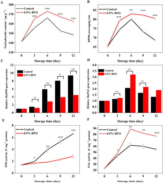
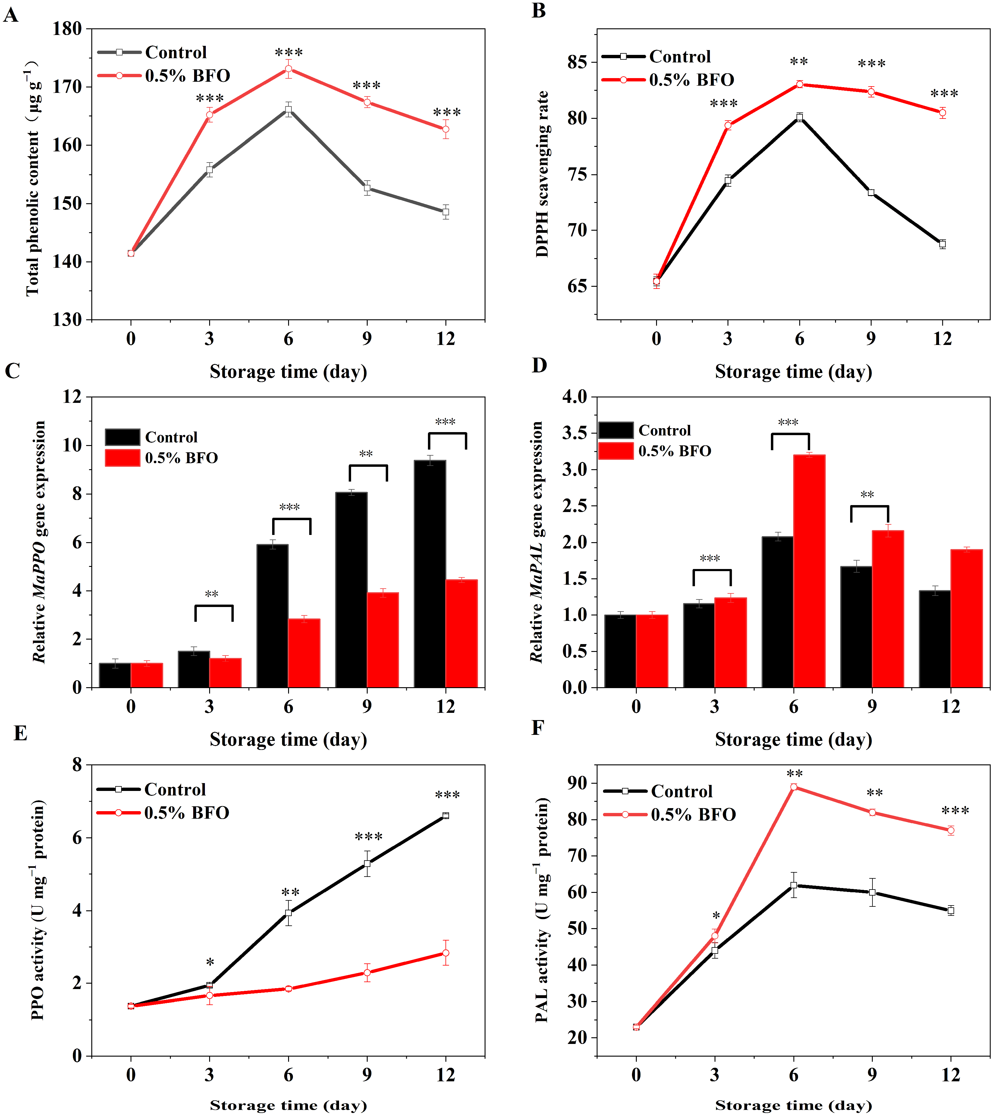
The changes in total phenolic content and DPPH scavenging rate were similar. With the extension of storage time, each group exhibited an initial increase followed by a decrease (Figure 3A,B). After 6 days of storage, with the BFO treatment group effectively increasing these peak values, the total phenolic content and DPPH scavenging rate of each group reached their maximum values. The total phenolic content and DPPH scavenging rate of the BFO treatment group were 1.06 times and 1.04 times those of the control, respectively, and the difference between the two groups was significant (p < 0.01). Additionally, on day 12, compared with the control group, BFO-treated fruit exhibited 1.14 and 1.22 times higher values for total phenolic content and DPPH scavenging rate, respectively.
Figure 3.
Effect of different treatments on total phenolic content (A), DPPH scavenging rate (B), relative MaPPO gene expression (C), relative MaPAL gene expression (D), PPO activity (E), and PAL activity (F) of peel of banana. All statistical analyses were performed using two-way ANOVA. Significance was denoted as p < 0.05 (*), p < 0.01 (**), p < 0.001 (***). Results are shown as mean ± SD (n = 3).
As depicted in Figure 3C,D, the BFO treatment resulted in an increase in the relative expression of MaPAL, while concurrently inhibiting the relative expression of MaPPO. Specifically, the relative expression level of MaPAL in the peel of the treatment group was 1.42 times higher than that in the control on day 12 of storage. Conversely, the relative expression level of MaPPO in the treatment group was merely 0.47 times that of the control group.
As presented in Figure 3E, the PPO activity in the peel increased continuously during the entire storage period. However, compared with the control group, the BFO treatment group demonstrated a significant inhibition of PPO activity in banana peel. Moreover, the PAL enzyme activity initially rose and then gradually declined during the entire storage process. In contrast to the control group, the enzyme activity of PAL in the BFO treatment group not only effectively accelerated the increase in the early stage but also reached its peak on day 6, followed by a slow decline. Moreover, the downward trend was markedly lower than that observed in the control group (Figure 3F).
3.5. Effect of BFO Treatment on Chlorophyll Content
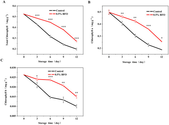
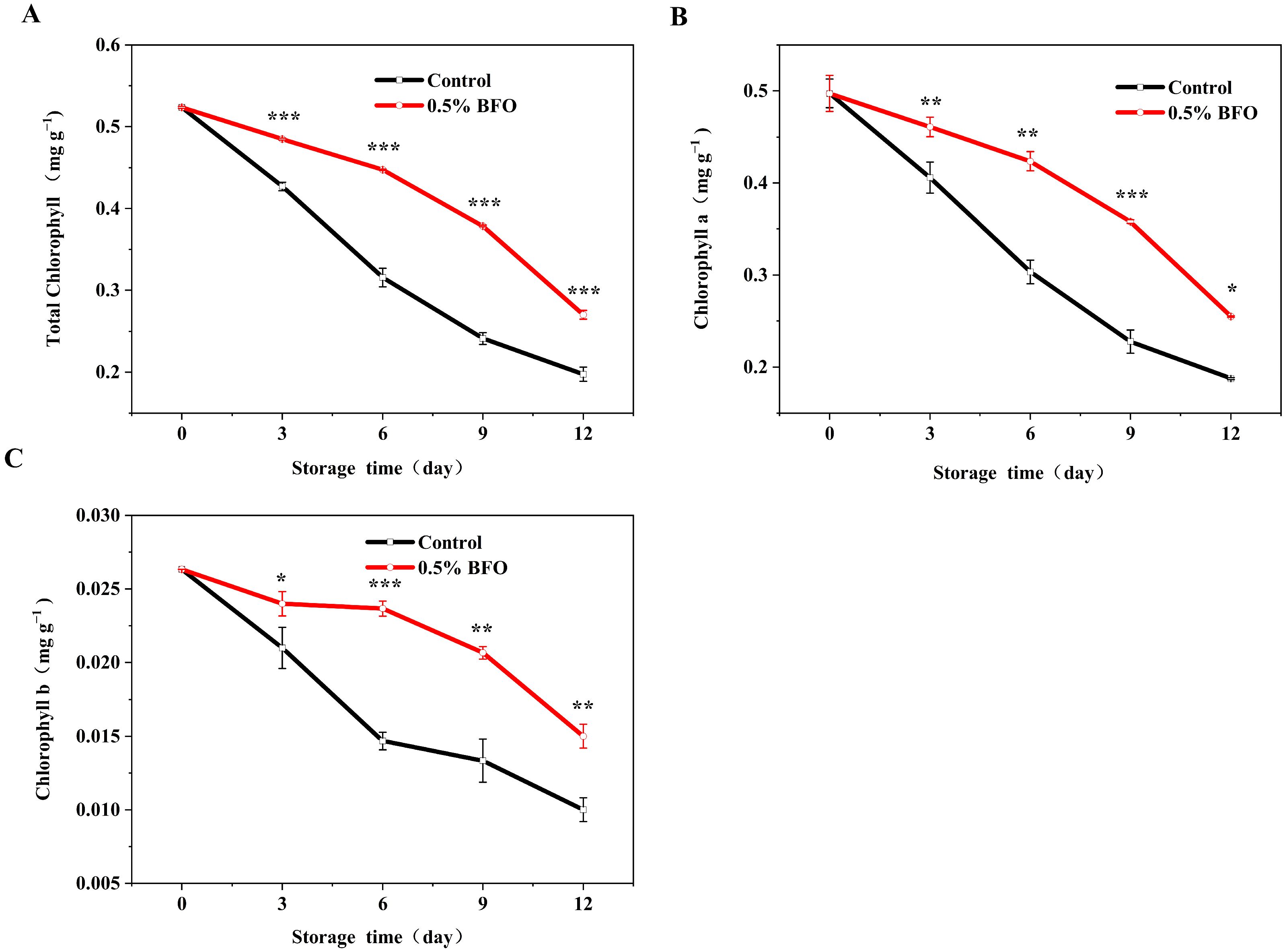
Figure 4 showed the contents of total chlorophyll, chlorophyll a and chlorophyll b exhibited a downward trend during storage, and the chlorophyll content of BFO-treated group was higher than that of the control. On day 12, the contents of total chlorophyll, chlorophyll a, and chlorophyll b in the treatment group were 72.9%, 72.2%, and 66.6% higher compared with the control group, respectively.
Figure 4.
Effect of different treatments on total chlorophyll content (A), chlorophyll a content (B), and chlorophyll b content (C) of peel of banana. All statistical analyses were performed using two-way ANOVA. Significance was denoted as p < 0.05 (*), p < 0.01 (**), p < 0.001 (***). Results are shown as mean ± SD (n = 3).
3.6. Effects of BFO Treatment on Chlorophyll-Degrading Enzyme Activities and Related Gene Expression
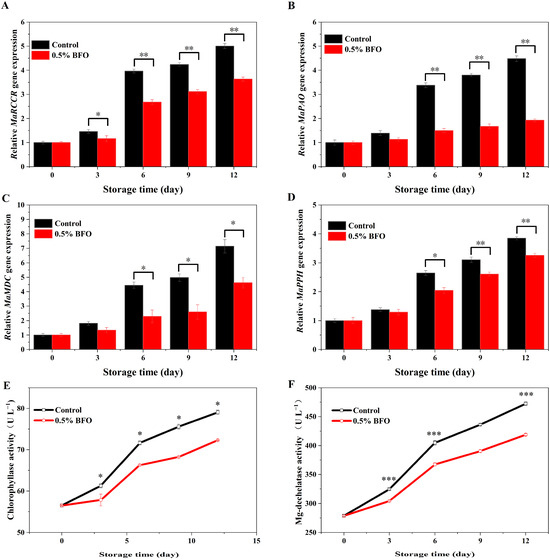
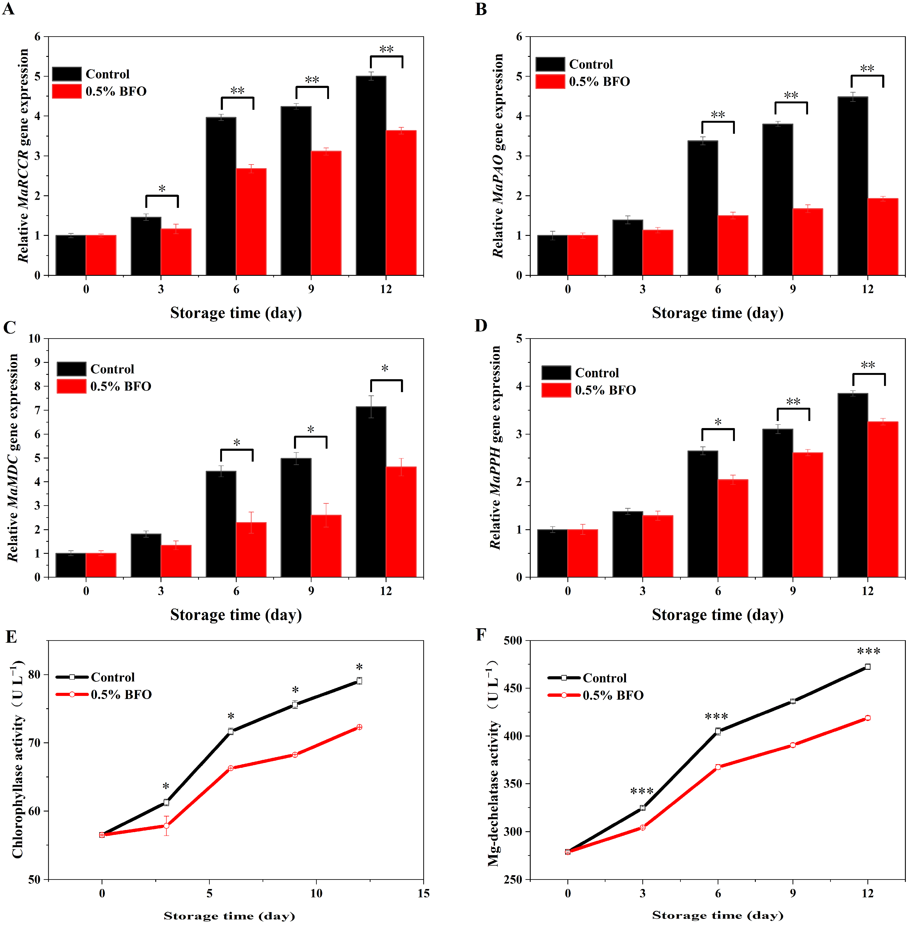
Throughout the entire storage period, the relative gene expression levels of MaRCCR, MaPAO, MaMDC, and MaPPH in both the control and BFO-treated fruit increased continuously. However, BFO treatment inhibited this increase in relative gene expression (Figure 5A–D). After 12 days of storage, the relative gene expression levels of MaRCCR, MaPAO, MaMDC, and MaPPH in the peel of the control group were 1.37, 2.32, 1.54, and 1.18 times those of the treatment group, respectively. During storage at 7 °C, BFO treatment significantly delayed the increase in CLH and MDC activities (Figure 5E,F). On day 12, the CLH activity and MDC activity of the BFO group were 91% and 88% of the control group, respectively.
Figure 5.
Effect of different treatments on relative MaRCCR expression (A), relative MaPAO gene expression (B), relative MaMDC gene expression (C), relative MaPPH gene expression (D), CLH activity (E), and MDC activity (F), of peel of banana. All statistical analyses were performed using two-way ANOVA. Significance was denoted as p < 0.05 (*), p < 0.01 (**), p < 0.001 (***). Results are shown as mean ± SD (n = 3).
4. Discussion
Chilling injury posed a significant challenge to maintaining banana quality over the course of low-temperature storage. The symptom of chilling injury in banana was predominantly manifested in the peel, with the most noticeable symptom being the deterioration of peel color [30]. The changes in the L* value and hue angle value were reliable indicators of color alterations in the banana peel. In this study, the L* and hue angle values of the fruit peel exhibited an overall downward trend, while BFO slowed the decline of these values effectively (Figure 1C,D). Additionally, BFO reduced the chilling injury index significantly by up to 26.8% (Figure 1B). Both of these indicated that BFO could inhibit the chilling injury in banana, which was in accordance with previous reports [31].
Electrolyte leakage and MDA content reflected cell membrane permeability and membrane lipid peroxidation, respectively, which showed the degree of cell membrane damage [32]. Chilling stress was thought to damage membrane integrity, leading to increased cell membrane permeability and MDA content [31]. Throughout the storage period, both control and treated banana samples gradually increased in MDA content and electrical conductivity in peel tissue (Figure 1F,G), but the MDA contents in BFO-treated samples were notably lower than the control group, indicating the BFO treatment could reduce lipid peroxidation and maintain membrane integrity. In previous studies [17], lower MDA content was observed in BFO-treated blueberries, which was consistent with our observation, thereby contributing to the maintenance of fruit quality during storage.
Low-temperature stress triggered reactive oxygen species (ROS) accumulation, which induced oxidative stress and worsened chilling injury symptoms [33]. CAT, POD, APX, and SOD are the key enzymes in plants that could clear accumulated reactive oxygen-free radicals [34]. In this study, the BFO treatment group significantly upregulated the relative expression of their corresponding genes (MaSOD, MaCAT, MaPOD, MaAPX), and concurrently enhanced the activities of key antioxidant enzymes (CAT, POD, APX, and SOD) (Figure 2). This coordinated induction of antioxidant defenses mitigated oxidative damage in the banana effectively. A previous study also obtained that the coordinated action of antioxidant defenses could reduce oxidative damage in bananas significantly, which also supports our conclusion [35].
PAL was a key enzyme in phenol biosynthesis [36], and the PPO catalyzed polyphenol substrates into polyquinones by enzymatic oxidation, resulting in the degradation of total phenols [37]. In this study, BFO increased the accumulation of phenolic compounds by promoting PAL activity and PPO activity, as well as inhibiting the MaPAL and MaPPO expression (Figure 3). These results agreed with those of Khademi [38] research, which reported that low-temperature stress induced the concerted activities of PAL and PPO, resulting in browning, which were highly correlated with CI symptoms in banana peel. The above results indicated that, in addition to regulating antioxidant enzyme activity and relative gene expression, BFO enhanced the antioxidant defense system of bananas by promoting phenol accumulation, thereby effectively inhibiting peel browning.
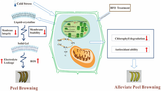
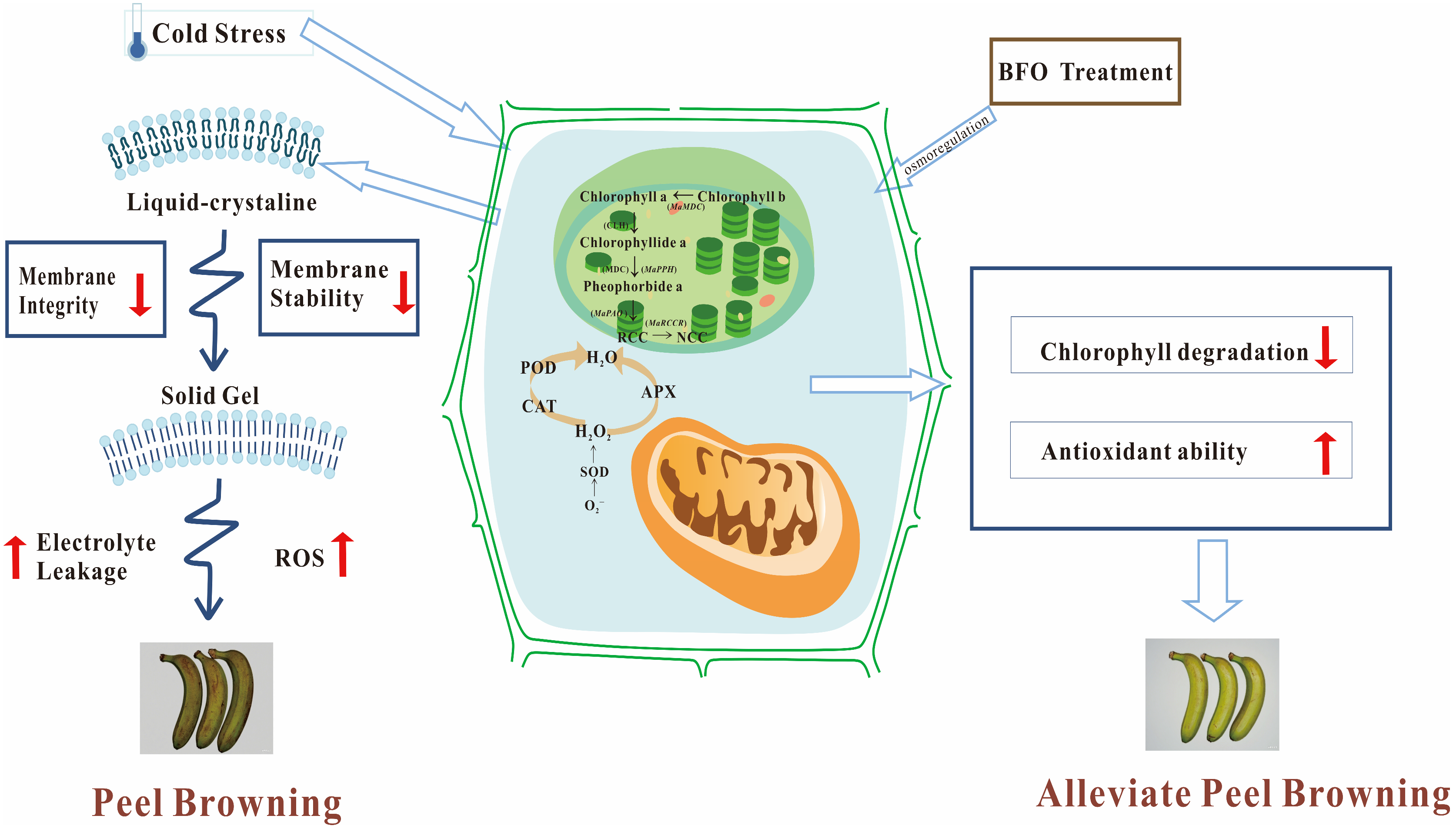
Chlorophyll is an important photosynthetic pigment in plant cells [39], whose degradation probably not only reflected the appearance quality of chilling injury in plants but also destroyed cell structure and function [40]. Chlorophyll degradation is a typical symptom of chilling injury in green bananas, which is primarily shown in browning and yellowing of the peel [41]. This research found that BFO treatment significantly delayed the decline of chlorophyll content, down-regulated chlorophyll degradation-related genes (such as MaMDC, MaPPH, MaPAO, and MaRCCR) and inhibited CLH and MDC activities (Figure 5). Our results indicated that BFO could regulate the chlorophyll metabolism-related relative gene expression and relevant enzyme activities to delay the chlorophyll degradation rate and maintain the peel color, thereby inhibiting the peel browning of banana, which consistented other related studies [42,43]. Therefore, BFO may have delayed chlorophyll degradation by regulating the chlorophyll metabolic pathway, which reduced chilling injury symptoms. This finding provides new experimental evidence for studying the molecular interaction mechanisms between low-temperature stress and pigment metabolism in postharvest physiology (Figure 6).
Figure 6.
The probable mechanism of BFO in reducing peel browning of banana during cold storage. Symbols: Red upward arrows indicate upregulation/enhancement; downward arrows indicate downregulation/inhibition. Cold stress: 7 ± 1 °C. BFO treatment: Banana fruit samples were immersed in 0.5% Burdock Fructooligosaccharide (BFO) solution.
5. Conclusions
This research demonstrated that BFO treatment significantly mitigated peel browning in postharvest banana fruit. The application of 0.5% BFO effectively mitigated cold-induced membrane damage in banana peel during low-temperature storage by preserving cell membrane integrity and inhibiting lipid peroxidation. Moreover, BFO treatment remarkably bolstered the antioxidant defense system through regulating the activation of critical antioxidant enzymes and improving reactive oxygen species scavenging. Additionally, BFO intervention inhibited the chlorophyll decrease by regulating the enzyme activities and the catabolism-related genes of chlorophyll degradation. This experiment provides novel theoretical insights and practical technological strategies for the preservation of postharvest bananas at low temperatures.
Supplementary Materials
The following supporting information can be downloaded at: https://www.mdpi.com/article/10.3390/horticulturae12030316/s1.
Author Contributions
J.Y.: parameter testing, writing. H.Z.: funding acquisition, writing and editing. Y.S.: investigation, methodology establishment. W.G.: editing and validation. Z.X.: reviewing and editing. J.L.: funding acquisition. F.G.: experiment administration and methodology establishment. W.J.: funding acquisition and data checkout. All authors have read and agreed to the published version of the manuscript.
Funding
This work was supported by the project for improving the innovation capacity of small and medium-sized technology-based enterprises of Shandong Province (2023TSGC0619), the project for improving the innovation capacity of small and medium-sized technology-based enterprises of Tai’an (2023TATSGC050), the National Natural Science Foundation of China (32202126), the Cross-Innovation Open Project of Food Flavor and Health, Beijing Technology & Business University (FFHCI-2025016) and the major innovation pilot project of integration of science, education and industry of Qilu University of Technology (Shandong Academy of Science) (2025ZDZX15).
Data Availability Statement
The original contributions presented in this study are included in the article/Supplementary Material. Further inquiries can be directed to the corresponding authors.
Conflicts of Interest
The authors declare no conflicts of interest.
References
- Xiao, X.M.; Li, L.L.; Kuang, J.F.; Chen, J.Y.; Lu, W.J.; Wei, W.; Shan, W. Cold pretreatment promotes chlorophyll degradation of green ripening banana peel by activating MaCBF1 to MaCBR and MaSGR1. Food Chem. 2023, 413, 135575. [Google Scholar] [CrossRef] [Scilit] [PubMed]
- Ali, A.F.; Hatamnia, A.A.; Malekzadeh, P.; Sayyari, M.; Aghdam, M.S. Exogenous dopamine ameliorates chilling injury in banana fruits by enhancing endogenous dopamine and glycine betaine accumulation and promoting ROS scavenging system activity. Postharvest Biol. Technol. 2023, 205, 112521. [Google Scholar] [CrossRef] [Scilit]
- Ali, S.; Ullah, M.A.; Nawaz, A.; Naz, S.; Shah, A.A.; Gohari, G.; Razavi, F.; Khaliq, G.; Razzaq, K. Carboxymethyl cellulose coating regulates cell wall polysaccharides disassembly and delays ripening of harvested banana fruit. Postharvest Biol. Technol. 2022, 191, 111978. [Google Scholar] [CrossRef] [Scilit]
- Ali, S.; Nawaz, A.; Naz, S.; Ejaz, S.; Maqbool, M.; Siddiqui, M.H.; Kalaji, H.M.; Wrobel, J.; Telesinski, A.; Auriga, A. Hydrogen Sulfide Mitigates Chilling Injury of Postharvest Banana Fruits by Regulating gamma-Aminobutyric Acid Shunt Pathway and Ascorbate-Glutathione Cycle. Front. Plant Sci. 2022, 13, 941246. [Google Scholar] [CrossRef] [Scilit]
- Tian, J.; Xie, S.; Zhang, P.; Wang, Q.; Li, J.; Xu, X. Attenuation of postharvest peel browning and chilling injury of banana fruit by Astragalus polysaccharides. Postharvest Biol. Technol. 2022, 184, 111783. [Google Scholar] [CrossRef] [Scilit]
- Pongprasert, N.; Sekozawa, Y.; Sugaya, S.; Gemma, H. A novel postharvest UV-C treatment to reduce chilling injury (membrane damage, browning and chlorophyll degradation) in banana peel. Sci. Hortic. 2011, 130, 73–77. [Google Scholar] [CrossRef] [Scilit]
- Chen, Y.; Xu, Y.; Han, X.; Ma, Q.; Zhou, W.; Guo, H.; Li, D.; Luo, Z. Azacytidine shows potential in controlling the chilling injury of banana peel during cold storage. Food Control 2024, 159, 110283. [Google Scholar] [CrossRef] [Scilit]
- Hao, J.; Li, X.; Xu, G.; Huo, Y.; Yang, H. Exogenous progesterone treatment alleviates chilling injury in postharvest banana fruit associated with induction of alternative oxidase and antioxidant defense. Food Chem. 2019, 286, 329–337. [Google Scholar] [CrossRef] [Scilit]
- Lin, Z.; Wei, W.; Cai, D.; Shan, W.; Chen, J.; Yang, Y.; Si, J.; Lu, W.; Zhu, G.; Wu, C.; et al. Sodium hydrosulfide application induces chilling tolerance in banana fruits by enhancing antioxidant gene expression through the upregulation of the ethylene response factors MaERF53L/121L. Food Chem. 2025, 471, 142770. [Google Scholar] [CrossRef] [Scilit]
- Niaz, N.; Hu, W.; Pan, S.; Ali, K.; Niaz, S. Heat shock combined with chemical treatment partially recovered chilling injury and improved antioxidant capacity in banana. Postharvest Biol. Technol. 2024, 216, 113077. [Google Scholar] [CrossRef] [Scilit]
- Wang, Z.; Zhang, L.; Duan, W.; Li, W.; Wang, Q.; Li, J.; Song, H.; Xu, X. Melatonin maintained higher contents of unsaturated fatty acid and cell membrane structure integrity in banana peel and alleviated postharvest chilling injury. Food Chem. 2022, 397, 133836. [Google Scholar] [CrossRef] [Scilit] [PubMed]
- Zhu, Y.; Zhou, Y.; Li, Z.; Zhong, R.; Jiang, Y.; Jiang, G.; Su, X. Tyr-Asp enhances chilling tolerance in banana fruit by modulating redox homeostasis. Postharvest Biol. Technol. 2025, 222, 113338. [Google Scholar] [CrossRef] [Scilit]
- Sittiprasert, W.; Supapvanich, S.; Sinthusamran, S.; Nimitkeatkai, H.; Techavuthiporn, C. Pre-storage anoxia treatment induces physicochemical characteristics of ‘Sucrier’ bananas (Musa AA group) in response to postharvest cold storage and subsequent ambient temperature. Postharvest Biol. Technol. 2025, 219, 113264. [Google Scholar] [CrossRef] [Scilit]
- Meng, Y.; Ma, Q.; Xu, X.; Feng, L.; Chen, Q.; Chen, Y.; Li, Z.; Liu, C.; Chen, K. Burdock fructooligosaccharide ameliorates the hypercholesterolemia and vascular inflammation in mice by regulating cholesterol homeostasis and anti-inflammatory properties. J. Funct. Foods 2023, 107, 105678. [Google Scholar] [CrossRef] [Scilit]
- Guo, M.; Chen, K.; Zhang, P. Transcriptome profile analysis of resistance induced by burdock fructooligosaccharide in tobacco. J. Plant Physiol. 2012, 169, 1511–1519. [Google Scholar] [CrossRef] [Scilit]
- Wang, F.; Feng, G.; Chen, K. Burdock fructooligosaccharide induces resistance to tobacco mosaic virus in tobacco seedlings. Physiological and Molecular. Plant Pathol. 2009, 74, 34–40. [Google Scholar] [CrossRef] [Scilit]
- Wang, Y.; Wang, J.; Li, Y.; Yang, L.; Sun, B.; Zhang, Y.; Xu, Y.; Yan, X. Controlling effect and mechanism of burdock fructooligosaccharide against Alternaria fruit rot in blueberry during postharvest. Postharvest Biol. Technol. 2023, 196, 112175. [Google Scholar] [CrossRef] [Scilit]
- Zhang, H.; Liu, Z.; Xu, B.; Chen, K.; Yang, Q.; Zhang, Q. Burdock fructooligosaccharide enhances biocontrol of Rhodotorula mucilaginosa to postharvest decay of peaches. Carbohydr. Polym. 2013, 98, 366–371. [Google Scholar] [CrossRef] [Scilit]
- Sun, F.; Zhang, P.; Guo, M.; Yu, W.; Chen, K. Burdock fructooligosaccharide induces fungal resistance in postharvest Kyoho grapes by activating the salicylic acid-dependent pathway and inhibiting browning. Food Chem. 2013, 138, 539–546. [Google Scholar] [CrossRef] [Scilit]
- Sun, F.; Zhu, L.; Wang, X.; Cheng, J.; Cui, B.; Liu, J.; Tan, F.; Fu, M. Sucrose transportation control mediates the fresh-keeping effects of burdock fructooligosaccharide in ‘Crimson Seedless’ grapes. Food Chem. 2020, 332, 127437. [Google Scholar] [CrossRef] [Scilit]
- Lin, Z.; Liu, H.; Yan, R.; Chen, Q.; Fu, M.; Xin, X. The composite edible film of sodium alginate and natamycin maintained color of pedicel and calyx of tomato fruit. Postharvest Biol. Technol. 2025, 222, 113413. [Google Scholar] [CrossRef] [Scilit]
- Xu, M.; Ngolong Ngea, G.L.; Li, T.; Lu, M.; Ren, Y.; Lian, X.; Ya, H.; Zhang, H. Exogenous melatonin treatment can improve the resistance of postharvest Lingbao apples to Penicillium expansum and maintain fruit storage quality. Plant Physiol. Biochem. 2025, 229, 110683. [Google Scholar] [CrossRef] [Scilit] [PubMed]
- Wang, X.; Wang, T.; Su, N.; Liu, W.; Gao, M.; Li, R.; Li, Y.; Bi, Y. L-Phenylalanine preharvest spraying effectively enhances the reactive oxygen metabolism and antioxidant capacity in postharvest broccoli. Plant Physiol. Biochem. 2025, 227, 110185. [Google Scholar] [CrossRef] [Scilit] [PubMed]
- Cui, K.; Ma, J.; Bai, M.; Sun, L.; Duan, Y.; Wu, B. Preharvest VDAL treatment enhances ROS scavenging and activates phenylpropanoid metabolism to improve postharvest storability of prune (Prunus domestica) fruit. Sci. Hortic. 2026, 358, 114699. [Google Scholar] [CrossRef] [Scilit]
- Zhang, H.; Li, T.; Ma, X.; Zhao, P.; Gao, X.; Sun, Y.; Zhang, S.; Zhu, D.; Cheng, F. Cordyceps militaris polysaccharides improved tomato postharvest disease resistance. Postharvest Biol. Technol. 2026, 231, 113881. [Google Scholar] [CrossRef] [Scilit]
- Wang, J.; Wei, Y.; Chen, Y.; Jiang, S.; Ye, J.; Ding, P.; Xu, F.; Shao, X. Alginate oligosaccharide alleviates chilling injury in postharvest peach fruit via regulating antioxidant and membrane lipid metabolisms. Plant Physiol. Biochem. 2025, 228, 110292. [Google Scholar] [CrossRef] [Scilit]
- Li, C.; Wang, R.; Ma, W.; Zhao, Y.; Yao, W.; Shan, S.; Yin, X. Methionine treatment improves postharvest resistance of grapes to Botrytis cinerea: Evidence from integrated transcriptomic and metabolomic analyses. Postharvest Biol. Technol. 2026, 235, 114219. [Google Scholar] [CrossRef] [Scilit]
- Hu, H.; Zhao, H.; Zhang, L.; Zhou, H.; Li, P. The application of 1-methylcyclopropene preserves the postharvest quality of cabbage by inhibiting ethylene production, delaying chlorophyll breakdown and increasing antioxidant capacity. Sci. Hortic 2021, 281, 109986. [Google Scholar] [CrossRef] [Scilit]
- Zhao, X.; Li, F.; Ding, J.; Fu, X.; Shang, J.; Kong, X.; Li, X.; Li, L.; Zhang, X. Comparative RNA-seq analysis of tomato (Solanum lycopersicum L.) provides insights into natural and postharvest ripening. Postharvest Biol. Technol. 2024, 216, 113079. [Google Scholar] [CrossRef] [Scilit]
- Wang, T.; Song, Y.; Lai, L.; Fang, D.; Li, W.; Cao, F.; Su, E. Sustaining freshness: Critical review of physiological and biochemical transformations and storage techniques in postharvest bananas. Food Packag. Shelf Life 2024, 46, 101386. [Google Scholar] [CrossRef] [Scilit]
- Yin, Q.; Qin, W.; Liu, T.; Song, W.; Yang, Y.; Shan, W.; Kuang, J.; Chen, J.; Lu, W. MaMYB4 is involved in the accumulation of naringenin chalcone, phloretin and dihydrokaempferol in the peels of banana fruit under chilling injury. Postharvest Biol. Technol. 2024, 212, 112844. [Google Scholar] [CrossRef] [Scilit]
- Ruan, Z.; Zhu, Y.; Liu, M.; Zhou, Y.; Su, X.; Jiang, Y.; Jiang, G. Tyr-Asp treatment delays senescence in litchi fruit by enhancing redox balance and maintaining energy homeostasis. Postharvest Biol. Technol. 2025, 223, 113420. [Google Scholar] [CrossRef] [Scilit]
- Iakimova, E.T.; Ty, A.J.; Hertog, M.L.; Nicolaï, B.M.; Woltering, E.J. Programmed cell death and postharvest deterioration of fresh horticultural products. Postharvest Biol. Technol. 2024, 214, 113010. [Google Scholar] [CrossRef] [Scilit]
- Tossi, V.E.; Regalado, J.J.; Martínez, J.; Galván, A.; Martinez Tosar, L.J.; Pitta-Alvarez, S.I.; Rebolloso, M.M.; Jamilena, M. UV-B alleviates postharvest chilling injury of zucchini fruit associated with a reduction in oxidative stress. Postharvest Biol. Technol. 2024, 212, 112850. [Google Scholar] [CrossRef] [Scilit]
- Shinga, M.H.; Fawole, O.A.; Pareek, S. Opuntia ficus-indica mucilage coating prolongs the shelf life of bananas (Musa spp.) by enhancing antioxidant activity and modulating antioxidant enzyme systems. Food Biosci. 2025, 65, 106039. [Google Scholar] [CrossRef] [Scilit]
- Saltveit, M.E.; Choi, Y.-J. Aromatic- and di-carboxylates inhibit wound-induced phenolic accumulation in excised lettuce (Lactuca sativa L.) leaf tissue. Postharvest Biol. Technol. 2007, 46, 222–229. [Google Scholar] [CrossRef] [Scilit]
- Ding, X.; Ma, J.; Liu, S.; Dong, X.; Pan, X.; Dong, B. Acid electrolytic water treatment improves the quality of fresh-cut red pitaya fruit by regulating ROS metabolism and phenylpropanoid pathway. Postharvest Biol. Technol. 2024, 207, 112636. [Google Scholar] [CrossRef] [Scilit]
- Khademi, O.; Ashtari, M.; Razavi, F. Effects of salicylic acid and ultrasound treatments on chilling injury control and quality preservation in banana fruit during cold storage. Sci. Hortic. 2019, 249, 334–339. [Google Scholar] [CrossRef] [Scilit]
- Toivonen, P.M.A.; Brummell, D.A. Biochemical bases of appearance and texture changes in fresh-cut fruit and vegetables. Postharvest Biol. Technol. 2008, 48, 1–14. [Google Scholar] [CrossRef] [Scilit]
- Zhu, Q.; Zhang, K.; Chen, W.; Li, X.; Zhu, X. Transcriptomic and metabolomic analyses reveal key factors regulating chilling stress-induced softening disorder in papaya fruit. Postharvest Biol. Technol. 2023, 205, 112534. [Google Scholar] [CrossRef] [Scilit]
- Li, D.; Wu, X.; Li, L.; Wang, Y.; Xu, Y.; Luo, Z. Epibrassinolide enhanced chilling tolerance of postharvest banana fruit by regulating energy status and pyridine nucleotide homeostasis. Food Chem. 2022, 382, 132273. [Google Scholar] [CrossRef] [Scilit]
- Wang, Y.; Luo, Z.; Du, R. Nitric oxide delays chlorophyll degradation and enhances antioxidant activity in banana fruits after cold storage. Acta Physiol. Plant. 2015, 37, 74. [Google Scholar] [CrossRef] [Scilit]
- Zsom, T.; Zsom-Muha, V.; Le Nguyen, L.P.; Nagy, D.; Hitka, G.; Polgári, P.; Baranyai, L. Nondestructive detection of low temperature induced stress on postharvest quality of kápia type sweet pepper. Prog. Agric. Eng. Sci. 2021, 16, 173–186. [Google Scholar] [CrossRef] [Scilit]
Disclaimer/Publisher’s Note: The statements, opinions and data contained in all publications are solely those of the individual author(s) and contributor(s) and not of MDPI and/or the editor(s). MDPI and/or the editor(s) disclaim responsibility for any injury to people or property resulting from any ideas, methods, instructions or products referred to in the content. |
© 2026 by the authors. Licensee MDPI, Basel, Switzerland. This article is an open access article distributed under the terms and conditions of the Creative Commons Attribution (CC BY) license.