


Abstract
Paraprobiotics offer safer alternatives to live probiotics without requiring cell viability. Here, heat-inactivated bacterial preparations (paraprobiotics) were produced from Lacticaseibacillus paracasei subsp. paracasei 431 (L. casei 431) using thermal treatments (65–121 °C). No viable colonies were detected by conventional plating in any treatment, and flow cytometry indicated that more than 96% of cells were membrane-compromised or dead. The most favorable condition (65 °C, 60 min) was identified using a TOPSIS multi-criteria ranking approach integrating loss of culturability with antimicrobial activity against Enterococcus faecalis and Escherichia coli with inactivation efficiency. This paraprobiotic and the corresponding probiotic were subjected to in vitro gastrointestinal digestion and colonic fermentation, followed by short-chain fatty acid (SCFA) and 16S rRNA-based gut microbiota analysis. The fecal inoculum was pooled from five healthy donors and used to run three replicate fermentation vessels per group. Both probiotic and paraprobiotic supplementation significantly increased acetate, butyrate, and total SCFA concentrations compared with the control, and propionate and valerate concentrations were also significantly higher in the paraprobiotic group than in the control. Both interventions increased Chao1 richness and Simpson diversity (p < 0.05), although Shannon diversity did not differ significantly, and induced significant shifts in beta diversity, alongside marked enrichment of butyrate- and SCFA-producing taxa such as Roseburia, Anaerostipes, and Akkermansia muciniphila. However, not all observed microbial changes were unequivocally beneficial; increases in Bilophila wadsworthia and Collinsella aerofaciens warrant cautious interpretation given their context-dependent associations with host health. Spearman correlation analysis identified positive associations between enriched taxa and SCFA concentrations, which should be interpreted as exploratory rather than evidence of causal metabolic cross-feeding. Under the conditions of this in vitro fermentation model, no statistically significant differences were detected between the viable and heat-inactivated preparations for most measured outcomes, indicating that thermal inactivation did not markedly diminish the capacity of L. casei 431 to influence gut microbial composition and short-chain fatty acid production. The multi-criteria selection strategy offers a practical approach for optimizing paraprobiotic production for functional food applications.
1. Introduction
Over the past decades, increasing attention has been directed toward the role of probiotics in promoting human health, particularly through their modulation of gut microbiota and gastrointestinal functions. Numerous studies have demonstrated that specific probiotic strains may exert beneficial effects in a range of gastrointestinal disorders, including irritable bowel syndrome, Helicobacter pylori infections, inflammatory bowel disease, diarrhea, and ulcerative colitis [1,2]. It should be emphasized, however, that these effects are largely strain-specific and outcome-specific, and should not be generalized as inherent properties of probiotics as a functional group. In addition to gastrointestinal health, probiotics have also been associated with positive effects on allergic diseases, metabolic disorders such as obesity and type 2 diabetes, non-alcoholic fatty liver disease, immune regulation, and oral health [3,4,5]. These health-promoting properties have led to the widespread incorporation of probiotics into functional foods and dietary supplements.
Probiotic microorganisms belong to a variety of genera, including Lactobacillus, Bifidobacterium, Bacillus, and Pediococcus, as well as certain yeasts [6]. Among them, Lactobacillus and Bifidobacterium species are the most widely studied and commonly used in food and nutraceutical applications. Their beneficial effects are mainly associated with the regulation of gut microbiota composition, inhibition of pathogenic microorganisms, and stimulation of host immune responses. In particular, probiotics can influence microbial metabolism in the colon and enhance the production of short-chain fatty acids (SCFAs), which are important metabolites involved in maintaining intestinal health.
Despite these beneficial properties, the use of live probiotics in food systems presents several technological and safety challenges. In order to exert their health benefits, probiotic microorganisms must remain viable during food processing, storage, and gastrointestinal transit. However, environmental stresses such as heat treatment, pressure, and oxygen exposure can significantly reduce probiotic viability, limiting their application in many food products [7]. In addition, the use of live microorganisms may pose potential risks for immunocompromised individuals, and there is concern regarding the possible transfer of antibiotic resistance genes to pathogenic bacteria [8,9]; however, such risks are population-, strain-, and context-dependent rather than an inherent property of live probiotics in general. These limitations have stimulated increasing interest in non-viable microbial alternatives that can retain beneficial biological effects without the risks associated with live cells.
In this context, probiotic derivatives such as paraprobiotics and postbiotics have recently emerged as promising alternatives to live probiotics [10]. Paraprobiotics, also known as inactivated probiotics, are defined as non-viable microbial cells or cell components that confer health benefits when administered in adequate amounts [7]. A clear terminological distinction among probiotics, paraprobiotics, postbiotics, purified microbial cell components, and cell-free metabolites is essential, and the present study follows the internationally recognized consensus definitions for these categories. These preparations can be obtained through various inactivation methods including heat treatment, sonication, high pressure, ultraviolet radiation, and gamma irradiation [11,12]. Among these methods, thermal inactivation is considered one of the most practical and scalable approaches for industrial applications. However, the applied inactivation conditions, particularly temperature and treatment duration, may significantly influence the structural integrity and biological activity of the resulting paraprobiotics [13,14]. Despite the growing interest in paraprobiotics, studies investigating their effects on gut microbiota composition and microbial metabolite production remain limited.
The human gastrointestinal tract harbors a highly complex microbial ecosystem composed of approximately 3.8 × 10¹³ microorganisms (a number now considered roughly comparable to that of human cells) that play critical roles in host metabolism, immune regulation, and protection against pathogen colonization [15,16]. One of the most important metabolic functions of gut microbiota is the fermentation of indigestible carbohydrates, resulting in the production of short-chain fatty acids (SCFAs) such as acetate, propionate, and butyrate. These metabolites contribute to intestinal homeostasis through anti-inflammatory, antimicrobial, and immunomodulatory activities [17,18]. While the influence of probiotics on gut microbiota composition and SCFA production has been extensively studied, investigations focusing on the impact of paraprobiotics on gut microbiota dynamics and SCFA formation are still scarce. In particular, the effects of heat-inactivated L. casei 431 on human fecal microbial composition and SCFA production during in vitro colonic fermentation have not previously been investigated, despite the established commercial use of this strain in its viable form.
Therefore, the aim of the present study was to produce paraprobiotics from Lacticaseibacillus paracasei subsp. paracasei (L. casei 431) under different thermal inactivation conditions and to determine the most effective treatment based on antimicrobial activity and cell inactivation levels using a multi-criteria decision-making approach. The selected paraprobiotic was subsequently evaluated for its effects on gut microbiota composition and short-chain fatty acid production using in vitro gastrointestinal digestion and colonic fermentation models. The findings of this study are expected to provide new insights into the potential application of thermally inactivated probiotics as functional ingredients targeting gut microbiota modulation.
2. Materials and Methods
2.1. Materials
Lacticaseibacillus paracasei subsp. paracasei 431 (L. casei 431) was obtained as a lyophilized culture from Chr. Hansen (Copenhagen, Denmark) and stored at −18 °C until use. The identity of the strain was based on the strain designation provided by the manufacturer. The pathogenic strains Escherichia coli O157 CECT 4267 (Gram-negative) and Enterococcus faecalis ATCC 29212 (Gram-positive) were obtained from the Culture Collection of the Department of Dairy Technology, Ege University, İzmir, Türkiye. The identities of these strains were based on their corresponding CECT and ATCC designations and culture collection records.
2.2. Paraprobiotic Production
2.2.1. Activation of Probiotic Culture
Probiotic bacteria were activated by inoculating sterile MRS broth with the lyophilized probiotic preparation at a concentration of 10 mg/L. The cultures were incubated under anaerobic conditions at 37 °C for 24 h. Following incubation, the viable cell density was determined by microbiological enumeration and was approximately 108 CFU/mL. Three independent culture preparations were performed (n = 3). The growth level of the probiotic cultures was monitored using the microbiological enumeration method described below.
2.2.2. Heat Treatment and Paraprobiotic Production
Probiotic bacteria that had been propagated in MRS broth to reach the target concentration of 108 CFU/mL were transferred into 50 mL Falcon tubes and centrifuged at 7000× g for 10 min at 4 °C. Following centrifugation, the supernatant containing metabolites was removed, and the pellet consisting of probiotic cells intended for inactivation was collected. The pellet was subsequently washed three times with phosphate-buffered saline (PBS) [19,20]. The resulting pellet was resuspended in 50 mL of sterile PBS and subjected to heat treatment. Heat treatments were conducted in a water bath at temperatures ranging from 65 to 100 °C and for different durations (15–60 min). The Falcon tubes were fully immersed in the water bath to ensure effective heat transfer. The treatment time started when the temperature of the sample inside the Falcon tube reached the target temperature. Immediately after completion of each heat treatment, the tubes were transferred to an ice-water bath for rapid cooling. The 121 °C treatment was conducted in an autoclave. The specific heat treatment conditions are presented in Table 1. Each heat-treatment experiment was independently repeated using three separate culture preparations (n = 3), while the different temperature–time combinations were applied to the same culture preparation within each experimental repetition.
Table 1.
Heat-treatment design and culturability and flow-cytometric assessment of paraprobiotic preparations.
2.3. Cell Death Verification
2.3.1. Microbiological Analysis
From the heat-treated samples, 1 mL of the undiluted sample was aseptically transferred to sterile Petri dishes, followed by the addition of molten MRS agar using the pour-plate method. No serial dilution was performed because the objective was to determine whether any viable cells remained following heat treatment. The plates were incubated under anaerobic conditions at 37 °C for 48–72 h [21]. One plate was prepared for each independent culture preparation (n = 3) for each treatment condition. Samples showing no colony formation after incubation were considered to have no detectable viable cells under the applied analytical conditions. With 1 mL of undiluted sample plated, the detection limit was 1 CFU/mL. Under conditions where no colony growth was observed, the obtained paraprobiotics were further analyzed to determine dead cell levels using flow cytometry and to evaluate their antimicrobial activities.
2.3.2. Detection of Dead Cells by Flow Cytometry
Flow cytometry analysis of the samples was performed using a Fortessa FACS system (BD Biosciences, Franklin Lakes, NJ, USA) following the method described by Müldür et al. (2025) [22]. To distinguish between live and dead bacterial cells, the Live/Dead™ BacLight™ Kit (Thermo Fisher Scientific, Waltham, MA, USA) was used, which contains Syto9 (stains live cells) and propidium iodide (PI; stains dead cells). To determine the autofluorescence levels of unstained bacterial suspensions, these samples were analyzed in FACS buffer consisting of PBS supplemented with 1% bovine serum albumin and 0.1% sodium azide. An untreated bacterial suspension stained with both Syto9 and PI was used as the live-cell control, whereas a heat-killed control obtained by treatment at 121 °C for 15 min was used as the dead-cell control. In addition, single-stained controls containing either Syto9 or PI alone were included for fluorescence compensation. Prior to staining, bacterial suspensions were adjusted to a 0.5 McFarland standard, washed with phosphate buffer, and subsequently diluted 1:500 in FACS buffer. Each tube was supplemented with 10 µL Syto9 (150 µg/mL) and 10 µL PI (400 µg/mL), followed by incubation in the dark at 4 °C for 20 min. After staining, the cells were fixed with 2% formaldehyde solution, washed with PBS, and resuspended in FACS buffer for analysis. Fixation followed staining, so that the membrane-integrity-dependent distribution of Syto9 and PI was already established before formaldehyde was added, and the same staining, fixation and washing sequence was applied to all samples and controls. All procedures were conducted under dark conditions. The excitation/emission wavelengths were set to 480/520 nm for Syto9 and 500/640 nm for PI. Dot plot analyses were performed using SSC-H and FL1-H channels, and the threshold value for the FL1 channel was set to 50. The unstained control was used to determine background autofluorescence, while the live and dead controls were used to define the corresponding fluorescence regions. Single-stained controls were used for fluorescence compensation and to assess potential spectral spillover between the Syto9 and PI channels. Gating was established using the dot plot of the positive control tube containing only live cells. The live and dead control distributions were used as references for defining the live/membrane-intact and dead/membrane-compromised populations. Using this reference gate, the FL1-H plots of bacterial samples subjected to heat treatments were overlaid for comparative analysis. Both fluorescence channels were recorded, FL1 for Syto9 and the 640 nm channel for PI, and populations were assigned on the combined signal. Because PI displaces Syto9 and quenches its emission by fluorescence resonance energy transfer, cells losing membrane integrity gain red and lose green fluorescence at the same time [23]; FL1-H was therefore used as the display axis, and events outside the Syto9-bright gate were checked against the PI channel before being counted as dead. Events with intermediate staining were not counted as dead, since such states reflect graded membrane damage rather than a binary transition [24]. Syto9 signal is also affected by photobleaching and by dye affinity differences between intact and damaged cells [25], so the cytometric values were treated as relative indicators of membrane integrity, with plate counting as the primary criterion for loss of culturability [26].
2.4. Determination of Antimicrobial Activity
The antimicrobial activity assays were performed using three independently prepared pathogen cultures for each pathogen. Fresh cultures of Escherichia coli and Enterococcus faecalis were prepared by incubating the strains in Tryptic Soy Broth (TSB) at 37 °C for 24 h and subcultured 2–3 times. The turbidity of the cultures was adjusted to the 0.5 McFarland standard (approximately 108 CFU/mL) to obtain standardized inocula [27]. Pathogen suspensions were inoculated into TSB at a rate of 1% (v/v). The paraprobiotic suspension was prepared by resuspending the heat-inactivated probiotic cell pellet obtained from 50 mL of MRS broth in 50 mL of sterile 0.85% (w/v) physiological saline solution. The resulting heat-treated cell suspension was added to the TSB medium at a final concentration of 5% (v/v). Control groups received an equivalent volume of sterile 0.85% (w/v) physiological saline solution. The inoculated tubes were incubated at 37 °C for 24 h. After incubation, appropriate serial dilutions were prepared and plated onto Tryptic Soy Agar (TSA). Plates were incubated at 37 °C for 24 h, and viable cell counts were determined for both the treated samples and the pathogen-only control groups.
Antimicrobial activity (%) was calculated using the following equation:
where Control colony count refers to the number of viable pathogen colonies (CFU/mL) in the pathogen-only control group, and Sample colony count refers to the number of viable pathogen colonies (CFU/mL) in the presence of the heat-inactivated paraprobiotic preparation.
2.5. In Vitro Gastrointestinal Digestion and Colonic Fermentation of Paraprobiotics
2.5.1. In Vitro Gastrointestinal Digestion
In vitro gastrointestinal digestion (GID) was performed based on the standardized INFOGEST 2.0 protocol described by Brodkorb et al. (2019) [28], with modifications according to the experimental design. Probiotic (PRO) and selected paraprobiotic (PARA) cell preparations were obtained from cultures adjusted to 108 CFU/mL prior to cell harvesting. The cells were collected by centrifugation, washed with sterile PBS, and resuspended in 50 mL of sterile PBS. For the PARA preparation, the cell suspension was subjected to the selected heat-treatment condition prior to digestion, whereas the PRO preparation was not heat-treated. Equal volumes of the resulting PRO and PARA suspensions were used for GID. The digestion procedure consisted of sequential oral, gastric, and intestinal phases. For the oral phase, 5 mL of sample was mixed with 5 mL of simulated salivary fluid (SSF) (1:1, v/v), and the pH was adjusted to 7.0. No α-amylase was added. The samples were incubated for 2 min at 37 °C in a shaking water bath. For the gastric phase, 10 mL of simulated gastric fluid (SGF) containing pepsin was added to the 10 mL oral bolus, resulting in a final volume of 20 mL. Pepsin was added to achieve a final activity of 2000 U/mL, and the pH was adjusted to 3.0. The samples were incubated for 2 h at 37 °C in a shaking water bath. Gastric lipase was not used. For the intestinal phase, 20 mL of simulated intestinal fluid (SIF) containing pancreatin and bile salts was added to the gastric chyme, resulting in a final volume of 40 mL. Pancreatin was added to provide a final trypsin activity of 100 U/mL, and bile salts were added at a final concentration of 10 mM. The pH was adjusted to 7.0, and the samples were incubated for 2 h at 37 °C in a shaking water bath. At the end of the digestion process, samples were cooled in an ice bath for 15 min and centrifuged at 10,000× g for 10 min at 4 °C to separate the undigested solid fraction (pellet). The obtained pellet was used for subsequent in vitro colonic fermentation. For the blank digestion control, sterile water was used instead of paraprobiotic samples.
2.5.2. In Vitro Colonic Fermentation
In vitro colonic fermentation was performed according to the method described by Pérez-Burillo et al. (2021) [29]. Fecal samples were obtained from five healthy volunteers aged 18–45 years who had not used antibiotics, probiotics, or prebiotics within the previous three months and were non-smokers. All donors were free of reported gastrointestinal disorders at the time of sample collection. Ethical approval was obtained from the Non-Interventional Clinical Research Ethics Committee of Aydın Adnan Menderes University (E-15189967-050.04-635366, 31 October 2024). Fecal samples from the five donors were pooled to obtain a standardized fecal inoculum. Fecal samples were mixed with an equal amount of 20% (v/v) glycerol solution at a 1:1 (w/v) ratio and stored at −80 °C until use. This resulted in a final glycerol concentration of approximately 10% (v/v). Prior to fermentation, samples were thawed, centrifuged (4000× g, 10 min, 4 °C) to remove glycerol, and the supernatant was discarded. The fecal inoculum was prepared by homogenizing pooled fecal samples in autoclaved phosphate buffer (pH 7.0) to obtain a final concentration of 32% (w/v), followed by centrifugation (550× g, 5 min) to remove large particles. The resulting supernatant was used as the fecal inoculum. For colonic fermentation, 0.5 g of the pellet obtained from each GID sample was recombined with its corresponding digestion supernatant. The resulting GID sample was then supplemented with 2 mL of fecal inoculum and 7.5 mL of sterile fermentation medium under anaerobic conditions. The mixture was flushed with nitrogen gas and incubated at 37 °C for 20 h at 20 rpm in a shaking incubator. After fermentation, samples were placed in an ice bath for 15 min to stop microbial activity and centrifuged at 10,000× g for 2 min. The supernatant was stored for SCFA analysis, while the bacterial pellet was stored at −80 °C for gut microbiota analysis. The control group was prepared using fermentation medium supplemented with ultrapure water instead of probiotic or paraprobiotic samples to evaluate the baseline activity of the colonic microbiota and the associated SCFA production. The blank was subjected to the same complete GID procedure, including all digestion fluids, enzymes, electrolytes, bile, pH adjustments, incubation steps, and centrifugation procedures.
2.6. Determination of SCFAs
SCFAs including acetate, propionate, butyrate, isobutyrate, isovalerate, and valerate were determined using gas chromatography with flame ionization detection (GC-FID) (7697A, Agilent Technologies, Santa Clara, CA, USA). Separation was performed using a DB-WAX capillary column (30 m × 0.25 mm × 0.25 μm; Agilent, Santa Clara, CA, USA). The oven temperature was initially set at 85 °C for 1 min, increased to 160 °C at 5 °C/min and held for 1 min, then raised to 240 °C at 40 °C/min and maintained for 2 min. Nitrogen was used as the carrier gas at a flow rate of 25 mL/min. Samples (1 μL) were injected without split. The injector and detector temperatures were set at 260 °C and 280 °C, respectively. Hydrogen and air were supplied to the FID at flow rates of 40 and 400 mL/min, respectively, while nitrogen was used as the make-up gas at 25 mL/min. Following the 20-h in vitro colonic fermentation, the fermentation samples were centrifuged at 27,670× g for 10 min, and the resulting supernatants were filtered through a 0.45 μm cellulose acetate membrane filter. No acidification step was applied prior to GC-FID analysis. An internal standard (4-methylvaleric acid, 1 mg/mL) was added to each sample prior to analysis. SCFA standards were obtained from WSFA-2 Sigma-Aldrich (Steinheim, Germany).
2.7. Characterization of Fecal Microbial Community Composition After In Vitro Colonic Fermentation by 16S rRNA Gene Sequencing
Samples obtained after in vitro colonic fermentation were stored at −80 °C until DNA extraction. Genomic DNA was extracted from the fermentation pellets using the GeneMATRIX Stool DNA Purification Kit (E3525, EURx Ltd., Gdańsk, Poland) according to the manufacturer’s instructions. DNA concentration and purity were determined using a NanoDrop™ 2000 spectrophotometer (Thermo Fisher Scientific, Waltham, MA, USA). Prior to sequencing, DNA quality was additionally verified by the sequencing provider using fluorometric quantification (Qubit) to ensure samples met the minimum quality thresholds required for library preparation. Negative extraction controls were included during DNA isolation to monitor for potential environmental or kit-derived contamination. The bacterial community composition was analyzed by next-generation sequencing of the 16S rRNA gene targeting the V3–V4 hypervariable region (~460 bp) using the Illumina MiSeq platform (2 × 300 bp paired-end reads). Library preparation and sequencing were performed by a commercial sequencing provider. The V3–V4 region was amplified using the following primers: forward primer 5′-TCGTCGGCAGCGTCAGATGTGTATAAGAGACAGCCTACGGGNGGCWGCAG-3′ and reverse primer 5′-GTCTCGTGGGCTCGGAGATGTGTATAAGAGACAGGACTACHVGGGTATCTAATCC-3′. PCR amplification was performed using KAPA Taq EXtra HotStart ReadyMix (2X) in a 25 µL reaction volume (95 °C for 3 min; 25 cycles of 95 °C for 30 s, 55 °C for 30 s, 72 °C for 30 s; final extension at 72 °C for 5 min). PCR products were purified using the AMPure XP magnetic bead system. Dual indexing was performed using the Nextera XT Index Kit (8 cycles; 95 °C for 3 min; 8 cycles of 95 °C for 30 s, 55 °C for 30 s, 72 °C for 30 s; final extension at 72 °C for 5 min). Libraries were quantified by real-time PCR, normalized using magnetic beads, and pooled prior to sequencing. No-template PCR controls were included in each reaction set to monitor for contamination; any batch showing amplification in negative controls was excluded and repeated. Sequence data were processed using the QIIME2 software package (v. 2023.2) [30]. Quality filtering, truncation, and denoising were performed using the DADA2 pipeline, generating an amplicon sequence variant (ASV) feature table. Taxonomic assignment was performed using a naïve Bayes classifier trained on the SILVA reference database (release 138), with a confidence threshold of 0.7, specific to the targeted 16S rRNA region. Species-level taxonomic assignments derived from V3–V4 16S rRNA sequencing should be regarded as putative identifications throughout this study, as this amplicon region has inherent resolution limitations for species-level classification of many intestinal taxa. Alpha diversity indices (Chao1, Shannon, and Simpson) and beta diversity analyses were performed using the MicrobiomeAnalyst platform (v3.0). Prior to analysis, data were rarefied to 24,379 reads and normalized using total sum scaling (TSS). Beta diversity was evaluated using Bray–Curtis dissimilarity and visualized by principal coordinate analysis (PCoA). Differences in microbial community structure among groups were assessed using permutational multivariate analysis of variance (PERMANOVA) with 999 permutations.
2.8. Statistical Analysis
Three independent culture preparations were used as experimental replicates (n = 3), with two parallel technical measurements per preparation. The Technique for Order Preference by Similarity to the Ideal Solution (TOPSIS) was used to rank the heat-treated paraprobiotic preparations based on dead-cell percentage and antimicrobial activities against E. faecalis and E. coli [31]. These three criteria were considered complementary and assigned equal weights (1/3 each). The decision matrix was normalized using vector normalization, and all criteria were treated as benefit criteria. The positive and negative ideal solutions were determined accordingly, and the relative closeness coefficient was calculated to rank the heat-treatment conditions. Equal weighting was selected because no prior evidence was available to justify assigning greater importance to any individual criterion. Dead-cell percentage was considered an indicator of the extent of heat-induced inactivation, whereas antimicrobial activity represented the residual functional activity of the preparations. To assess the robustness of the TOPSIS ranking to variations in criterion weights, an additional sensitivity analysis was performed. A total of 100,000 alternative weight combinations summing to 1 were generated using a Dirichlet distribution. For each weighting combination, the TOPSIS relative closeness coefficients and rankings were recalculated using the same vector-normalization procedure. The frequency with which each heat-treatment condition achieved the first rank and its mean rank across the weighting scenarios were determined. Statistical analyses were conducted using one-way analysis of variance (ANOVA) with SPSS software (version 27.0, SPSS Inc., Chicago, IL, USA). The assumptions of normality and homogeneity of variances were assessed using the Shapiro–Wilk and Levene’s tests, respectively. Differences among samples were evaluated using Tukey’s honest significant difference (HSD) post hoc test at a significance level of p < 0.05. Differential abundance of microbial taxa across groups was assessed using Kruskal–Wallis test applied to relative abundance data following total sum scaling (TSS) normalization. To account for multiple comparisons, the Benjamini–Hochberg (BH) false discovery rate (FDR) correction was applied across all taxonomic comparisons (phylum, genus, and species levels), diversity endpoints, and microbial–metabolite Spearman correlation matrices. Adjusted p-values (p_adj < 0.05) were considered statistically significant after correction. Spearman’s rank correlation analysis was performed to evaluate the relationship between microbial genera and species and short-chain fatty acid concentrations using the same software. Correlations were calculated across all fermentation vessels encompassing all three treatment groups and should be interpreted as exploratory associations driven by treatment-group separation rather than as independent evidence of direct metabolic interactions, given that all samples were derived from a single pooled fecal inoculum.
3. Results
3.1. Standard Colony Counting
Classical microbiological plating demonstrated complete loss of culturability of L. casei 431 under all heat-treatment conditions tested. No colony formation was detected on MRS agar following incubation, regardless of the applied temperature (65–121 °C) or treatment duration, confirming the absence of detectable viable cells by the culture-based method. These findings confirmed that all thermal treatments effectively eliminated the culturability of L. casei 431 and therefore generated non-culturable preparations.
3.2. Dead Cell Levels Determined by Flow Cytometry
Flow cytometric analysis demonstrated consistently high levels of cell death following all heat treatments, ranging from 96.65% (95 °C for 45 min) to 98.30% (85 °C for 45 min) (Table 1). The remaining fraction of gated events (1.7–3.4%) may represent cells with altered membrane integrity that were not classified as dead by the applied gating criteria and/or background events; such intermediate patterns are common in flow cytometric Live/Dead BacLight staining and reflect graded membrane damage rather than a binary transition [24]. Importantly, this residual fraction did not correspond to detectable culturability, as no colony growth was observed on MRS agar for any of the heat treatments tested.
Representative dot plots of cells treated at 85 °C for 45 min are presented in Figure 1, where Q2, Q3, and Q4 represent dead, viable, and damaged cells, respectively. No significant differences in dead cell percentages were observed among the different temperature–time combinations (p > 0.05). These findings were consistent with the plate count results, which showed complete loss of culturability under all treatment conditions.
Figure 1.
Representative flow cytometry dot plot (SSC-H vs. FL1-H) of L. casei 431 following heat treatment at 85 °C for 45 min, based on SYTO9/propidium iodide (PI) co-staining. Q2, Q3, and Q4 denote the dead (PI-positive; red), viable (SYTO9-positive only; green), and membrane-damaged/intermediate (blue) cell populations, respectively, gated relative to an untreated live-cell positive control; values indicate the percentage of total gated events within each quadrant. Quadrants were assigned from the combined Syto9 (FL1) and PI signals; SSC-H versus FL1-H is shown because PI displaces and quenches Syto9, so membrane-compromised cells lose green fluorescence.
3.3. Antimicrobial Activity of Heat-Treated L. casei 431 Paraprobiotics
The antimicrobial activities of heat-treated L. casei 431 preparations against Enterococcus faecalis and Escherichia coli are presented in Table 2. Antimicrobial efficacy differed significantly according to both the target microorganism and the applied heat treatment (p < 0.05). The heat-treated L. casei 431 preparations resulted in only limited changes in the viable counts of the tested pathogens. Although statistically significant differences were observed among some treatment conditions, the magnitude of the observed changes was small, indicating limited biological relevance under the experimental conditions. It should be noted, however, that the absolute magnitude of inhibition was modest throughout, ranging from 0.34% to a maximum of 4.33%. Therefore, despite statistical differences among some treatments, these effects should be interpreted as limited inhibitory effects with limited biological relevance rather than strong antimicrobial efficacy. The highest inhibition against E. faecalis (4.33%) was observed in samples treated at 121 °C for 15 min. In contrast, inhibition against E. coli was generally lower, and no inhibitory activity was detected following treatment at 121 °C. Among the moderate heat treatments, extending the treatment time at 65 °C from 45 to 60 min resulted in increased antimicrobial activity against both pathogens. Overall, the sample treated at 65 °C for 60 min exhibited relatively higher, though still modest, antimicrobial activity, whereas several higher-temperature treatments showed reduced or no inhibitory effects despite a comparable loss of culturability.
Table 2.
Antimicrobial activity of heat-treated L. casei 431 paraprobiotics against E. coli and E. faecalis under different temperature–time conditions.
3.4. Selection of the Optimal Paraprobiotic by TOPSIS Analysis
The complete TOPSIS ranking and relative closeness coefficients for all heat-treatment conditions are presented in Table 3, while the results of the additional sensitivity analysis are presented in Supplementary Table S1. Under the equal-weighting scheme, the preparation obtained at 65 °C for 60 min achieved the highest relative closeness coefficient (C = 0.840), indicating the most favorable overall performance across the evaluated criteria. Although the differences in dead-cell percentages and absolute antimicrobial inhibition values were relatively small, these criteria were considered jointly in the TOPSIS analysis. Notably, the 65 °C for 60 min treatment remained the highest-ranked alternative across all tested weighting scenarios, indicating that its selection was not dependent on the equal-weighting assumption. Accordingly, the 65 °C for 60 min treatment was selected as the preferred condition for subsequent analyses.
Table 3.
TOPSIS ranking of L. casei 431 paraprobiotic preparations obtained under different heat-treatment conditions.
3.5. SCFA Levels After In Vitro Colonic Fermentation
Following simulated gastrointestinal digestion and in vitro colonic fermentation, the concentrations of individual and total SCFAs were quantified in the PRO, PARA, and control groups (Table 4). Acetate was the predominant SCFA detected in all groups, followed by propionate and butyrate.
Table 4.
SCFA levels detected after in vitro colonic fermentation (µg/mL).
Both the PRO and PARA groups produced significantly higher concentrations of acetate (C vs. PRO: p < 0.001; C vs. PARA: p < 0.001), butyrate (C vs. PRO: p = 0.003; C vs. PARA: p = 0.001), and total SCFAs (C vs. PRO: p = 0.001; C vs. PARA: p = 0.001) than the control group. No statistically significant differences were detected between the PRO and PARA groups for these metabolites (acetate: p = 0.610; butyrate: p = 0.628; total SCFA: p = 0.899) under the conditions tested. It should be noted that the absence of a statistically significant difference between the PRO and PARA groups does not, by itself, demonstrate equivalence between the two preparations, since the study was not statistically powered for formal equivalence testing. Valerate concentrations were significantly higher in the PARA group than in the control group (C vs. PARA: p = 0.005), whereas the PRO group showed intermediate values without differing significantly from either group (C vs. PRO: p = 0.059; PRO vs. PARA: p = 0.150). Propionate concentrations were also significantly higher in the PARA group than in the control group (C vs. PARA: p = 0.007), while the difference between the control and PRO groups did not reach statistical significance (C vs. PRO: p = 0.055; PRO vs. PARA: p = 0.246). In contrast, isovalerate concentrations did not differ significantly among the experimental groups (C vs. PRO: p = 0.902; C vs. PARA: p = 0.617; PRO vs. PARA: p = 0.855).
3.6. Gut Microbiota Composition After In Vitro Colonic Fermentation
Gut microbiota diversity and composition were assessed at the phylum, genus, and species levels following 20 h in vitro colonic fermentation with probiotic and paraprobiotic treatments. All species-level identifications are presented as putative throughout, reflecting the inherent resolution limitations of V3–V4 16S rRNA sequencing for unambiguous species-level classification.
At the phylum level, Bacillota and Bacteroidota predominated in all groups (Figure 2A). Several low-abundance phyla, including Verrucomicrobiota, Actinomycetota, Fusobacteriota, and Pseudomonadota, were detected at low levels in the control group but showed markedly higher relative abundances in both PRO and PARA groups, suggesting treatment-associated expansion rather than de novo emergence. Candidatus Melainabacteria, by contrast, showed reduced relative abundance following both treatments. The Bacillota/Bacteroidota (F/B) ratio differed significantly among groups (PARA: 1.51; PRO: 1.73; C: 1.62; p < 0.05) (Figure 2B).
Figure 2.
Effect of C, PRO and PARA on gut microbiota composition. (A) Relative abundance of gut microbiota at phylum level; (B) the relative abundance ratio of Bacillota/Bacteroidota (Firmicutes/Bacteroidetes, F/B); (C) β-diversity of gut microbiome assessed by Principal Coordinate Analysis (PCoA) based on Bray–Curtis dissimilarity (PERMANOVA: F = 120.26, R2 = 0.976, p = 0.007); α-diversity of gut microbiome based on (D) Chao1 index (p = 0.046), (E) Shannon index (p = 0.06), and (F) Simpson index (p = 0.043). Statistical significance was determined by Kruskal–Wallis test. ns: not statistically significant. * p < 0.05.
Alpha diversity was evaluated using the Chao1, Shannon, and Simpson indices (Figure 2D–F). Significant differences among groups were observed for Chao1 richness (p = 0.046) and Simpson diversity (p = 0.043), whereas Shannon diversity showed no significant difference (p = 0.066).
Principal coordinate analysis (PCoA) based on Bray–Curtis dissimilarity demonstrated distinct clustering of microbial communities according to treatment (Figure 2C). PERMANOVA confirmed significant differences in overall community composition among the three groups (Pseudo-F = 120.26, R2 = 0.976, p = 0.007). PERMDISP analysis showed no significant differences in multivariate dispersion (F = 1.623, p = 0.273), indicating comparable within-group variability across groups.
At the genus level, both treatments significantly increased the relative abundances of Roseburia, Anaerostipes, Coprococcus, Eubacterium, Flintibacter, Dysosmobacter, Butyribacter, Mediterraneibacter, Bifidobacterium, Akkermansia, Collinsella, Dorea, Gemmiger, Faecalibacterium, Odoribacter, and Streptococcus (p < 0.05, FDR-corrected) (Figure 3A,B). At the species level, corresponding increases in relative abundance were observed for Roseburia hominis, Roseburia faecis, Anaerostipes hadrus, Coprococcus catus, Eubacterium ramulus, Flintibacter butyricus, Dysosmobacter welbionis, Bifidobacterium longum, Bifidobacterium pseudocatenulatum, Akkermansia muciniphila, Collinsella aerofaciens, Dorea longicatena, and Streptococcus thermophilus (p < 0.05, FDR-corrected) (Figure 3C). It should be noted that these represent changes in relative abundance and do not necessarily reflect changes in absolute cell numbers, given the compositional nature of 16S rRNA sequencing data. Bacteroides genus showed increased relative abundance in both treatment groups; however, this change did not reach statistical significance after FDR correction (p_adj > 0.05) and should be interpreted with caution. Bilophila at the genus level and Bilophila wadsworthia at the species level also increased significantly in both intervention groups (p < 0.05, FDR-corrected). Conversely, Phocaeicola and Ruminococcus at the genus level—and Phocaeicola vulgatus and Ruminococcus champanellensis at the species level—decreased markedly following both interventions; however, these changes did not reach statistical significance after FDR correction (p_adj > 0.05) and should be interpreted with caution. No statistically significant differences were detected between the PRO and PARA groups for any of the taxa evaluated (p > 0.05). It should be noted, however, that the absence of a statistically significant difference does not demonstrate equivalence between the two treatments, as this study was not designed as a formal equivalence trial.
Figure 3.
Effect of PRO and PARA on gut microbiota composition. (A) Relative abundance of gut microbiota at genus level for each group; (B) heatmap of abundance changes at genus level; (C) relative abundance of gut microbiota at species level.
3.7. Correlation Between Gut Microbiota Composition and SCFA Production
Spearman correlation analysis conducted at both genus and species levels identified associations between microbial taxa and SCFA concentrations (Figure 4A,B). Benjamini–Hochberg FDR correction was applied to the complete correlation matrices. At the species level, the strongest positive associations were observed for Anaerostipes hadrus, Butyribacter intestini, and Dorea longicatena, which correlated significantly with acetate, butyrate, and valerate (r = +0.943, p < 0.01; p_adj = 0.028), as well as with total SCFA (r = +0.886, p < 0.05). Streptococcus thermophilus and Roseburia faecis showed equally strong associations across acetate, propionate, butyrate, valerate, and total SCFA (r = +0.943, p < 0.01; p_adj = 0.028). These associations remained statistically significant after FDR correction. Positive associations with acetate, butyrate, and valerate were also observed for Bifidobacterium longum (r = +0.812, p = 0.050), Bifidobacterium pseudocatenulatum, Roseburia hominis, Flintibacter butyricus, Coprococcus catus, Dysosmobacter welbionis, Eubacterium ramulus, Bacteroides uniformis, and Collinsella aerofaciens (r = +0.829, p < 0.05); however, these associations did not reach statistical significance after FDR correction (p_adj > 0.05) and should be regarded as nominally significant only. Similar association patterns were observed at the genus level, where correlations with r = +0.943 remained significant after FDR correction (p_adj = 0.045) (Figure 4A). Species-level correlations are subject to the same resolution limitations noted above. Bilophila wadsworthia showed positive associations with propionate (r = +0.943, p < 0.01; p_adj = 0.028) and with acetate, butyrate, and valerate (r = +0.829, p < 0.05). In contrast, Ruminococcus champanellensis showed negative associations with acetate, butyrate, and valerate (r = −0.886, p < 0.05) and total SCFA (r = −0.829, p < 0.05); however, these did not reach significance after FDR correction (p_adj > 0.05). No significant associations were detected for Phocaeicola vulgatus, while isovalerate showed no significant associations with any microbial taxon at either taxonomic level. Despite significant changes in abundance, Faecalibacterium prausnitzii and Akkermansia muciniphila were not significantly correlated with any measured SCFA.
Figure 4.
Spearman’s rank correlation between the relative abundance of selected microbial genera (A) and putative species (B) and short-chain fatty acid (SCFA) concentrations detected after in vitro colonic fermentation. Taxa were selected based on their consistent presence across treatment groups and their biological relevance to SCFA production. Correlations were calculated across all fermentation vessels encompassing all three treatment groups (C, PRO, and PARA). The color scale indicates Spearman’s r values from −1 (strong negative correlation, blue) to +1 (strong positive correlation, red). Bold values indicate correlations that remained statistically significant after Benjamini–Hochberg false discovery rate (FDR) correction (p_adj < 0.05). Asterisks indicate nominal significance prior to FDR correction (* p < 0.05; ** p < 0.01). SCFA concentrations are expressed in µg/mL.
4. Discussion
The present study demonstrates that thermal inactivation of L. casei 431 can successfully generate paraprobiotic preparations while largely preserving several of the specific functional attributes assessed in this study, namely antimicrobial activity, SCFA-stimulating capacity, and effects on relative microbial taxonomic profiles, under the applied in vitro conditions. Although all heat treatments resulted in complete loss of culturability, their effects on antimicrobial activity and microbiota modulation differed according to the processing conditions. Notably, the preparation obtained at 65 °C for 60 min was ranked as the most favorable among the conditions tested by the TOPSIS multi-criteria analysis, based on the combination of loss of culturability and antimicrobial activity. These findings highlight that optimization of heat-inactivation processes should consider not only bacterial inactivation but also the preservation of biological activity.
Loss of culturability of L. casei 431 was consistently confirmed by conventional plate counting, and flow cytometry indicated a high proportion of membrane-compromised cells across all treatments. While plate counting demonstrated complete loss of culturability under all treatment conditions, flow cytometry revealed uniformly high dead-cell proportions (96.7–98.3%) with no significant differences among temperature–time combinations (Table 1). Similar observations have been reported for heat-treated lactic acid bacteria, where extensive thermal inactivation was achieved across a broad range of processing conditions [22,32,33]. Compared with conventional culture-based methods, flow cytometry provides complementary information by assessing membrane integrity and therefore offers a more comprehensive evaluation of heat-induced cellular damage. It should be emphasized that membrane-integrity-based flow cytometry assesses cell permeabilization rather than directly confirming irreversible cell death, and a small proportion of events (1.7–3.4%) fell outside the dead-cell gate in every treatment; these are most plausibly damaged but not fully permeabilized cells rather than residual viable, culturable organisms, consistent with the complete absence of colony growth on plating.
Importantly, loss of culturability did not necessarily result in loss of the specific functional attributes measured in this study. Moderate heat treatment, particularly at 65 °C for 60 min, maintained antimicrobial activity against both tested microorganisms while achieving a comparable loss of culturability. In contrast, several higher-temperature treatments exhibited reduced or no antimicrobial activity despite similar dead-cell percentages. These findings suggest that preservation of biological function depends not only on bacterial viability but also on the stability of heat-sensitive cell-associated components. Although this explanation is biologically plausible, the relevant structural components were not directly measured in the present study, and this interpretation should therefore be regarded as speculative pending direct structural or biochemical confirmation. Moderate thermal treatments may better preserve biologically relevant structures, including peptidoglycan, teichoic acids, surface proteins, extracellular polysaccharides, and other bioactive molecules that contribute to antimicrobial and microbiota-modulating activities [14,33]. Conversely, more severe heat treatments are likely to promote structural disruption or protein denaturation, thereby reducing functional activity. Similar observations have been reported for other heat-inactivated lactic acid bacteria, in which moderate thermal processing preserved functional properties more effectively than excessive heat exposure [13,22,34].
The multi-criteria TOPSIS analysis provided a structured ranking framework integrating loss of culturability and antimicrobial activity into a single decision-making score, rather than constituting independent experimental validation of these findings [31]. Among all thermal treatments, heating at 65 °C for 60 min achieved the highest overall performance score under the applied equal-weighting scheme, indicating a favorable balance between microbiological safety and functional efficacy among the conditions tested. Identifying this favorable processing condition may inform future process-optimization efforts; however, translation to industrial-scale production would additionally require evaluation of scale-up heating kinetics, energy consumption, drying and storage stability, and formulation compatibility, none of which were assessed in the present study.
Following simulated gastrointestinal digestion, both the viable probiotic and the heat-inactivated preparation significantly enhanced SCFA production compared with the untreated control. Because the viability of the PRO preparation following simulated gastrointestinal digestion was not directly assessed, it cannot be determined with certainty whether the responses observed in this group originated from surviving viable cells, cells inactivated during digestion, or cell-associated substrates released during processing. Acetate remained the predominant fermentation metabolite, whereas butyrate and total SCFA concentrations increased significantly in both intervention groups. Notably, no significant differences were detected between viable and heat-inactivated L. casei 431, suggesting that bacterial viability may not be essential for promoting microbial fermentative activity under the present in vitro conditions. These observations support the growing concept that non-viable microbial preparations can produce microbiota-modulating effects that are not significantly different from those of their viable counterparts under the specific conditions tested [7,35].
The slightly higher propionate and valerate concentrations observed in the paraprobiotic group may reflect structural alterations induced by thermal processing. Heat treatment may increase the accessibility of bacterial cell-associated components, including peptidoglycan, teichoic acids, and extracellular polysaccharides, thereby facilitating their utilization by resident gut microorganisms during fermentation. Although these mechanisms were not directly evaluated in the present study, similar effects have previously been proposed for heat-inactivated probiotic cells, which can retain—or in some cases enhance—biological activity despite loss of viability [13,14,34]. Nevertheless, SCFA profiles are highly strain-dependent and may vary according to environmental conditions, fermentation substrates, and processing treatments such as heat inactivation [36], which may partly explain the modest differences observed between the PRO and PARA groups for these two metabolites.
The increased SCFA production observed following both interventions is biologically relevant because these metabolites play fundamental roles in maintaining intestinal homeostasis. Acetate serves as an important substrate for microbial cross-feeding, whereas butyrate is recognized in the literature as the principal energy source for colonocytes, contributing to epithelial barrier integrity, immune regulation, and suppression of intestinal inflammation in vivo [37]; these host-level effects were not directly assessed in the present cell-free in vitro fermentation system. Therefore, the comparable SCFA responses observed in the probiotic and paraprobiotic groups suggest that thermal inactivation was not associated with a statistically detectable reduction in the capacity of L. casei 431 to influence microbial metabolism during colonic fermentation.
Both viable and heat-inactivated L. casei 431 supplementation altered the composition of the pooled fecal microbial community during in vitro colonic fermentation. The significant alterations observed in alpha diversity, together with the clear separation of microbial communities in the beta diversity analysis, demonstrate that both preparations produced a comparable community-level shift under the closed batch fermentation conditions used in this study. Importantly, the absence of significant differences between the viable and heat-inactivated groups across diversity indices, community composition, and individual taxa suggests that heat inactivation was not associated with a statistically detectable reduction in this microbiota-altering capacity under the present experimental conditions. These findings are consistent with growing evidence indicating that non-viable microbial preparations can exert microbiota-modulating effects comparable to those of their viable counterparts [7,35,38,39,40,41].
Although the ecological significance of the Bacillota/Bacteroidota ratio has recently been questioned as a universal biomarker of gut health, its modulation in the present study occurred concurrently with increased SCFA production and enrichment of fermentative bacteria. Rather than interpreting this ratio as a primary outcome or health indicator, it is presented here as a descriptive compositional parameter to be considered alongside the broader microbial and metabolic responses [42]. One of the most prominent microbial responses was the coordinated enrichment of several butyrate-producing genera, including Roseburia, Anaerostipes, Butyribacter, Flintibacter, Dysosmobacter, Mediterraneibacter, Coprococcus, and Eubacterium. These microorganisms play central roles in complex carbohydrate fermentation and butyrate biosynthesis [43,44,45,46,47,48,49,50,51]. Their simultaneous enrichment, together with the observed increase in butyrate concentrations, is consistent with enhanced saccharolytic fermentation, although it should be noted that increases in relative abundance do not necessarily reflect increases in absolute cell numbers or directly demonstrate enhanced metabolic activity. Absolute quantification or functional-gene analysis would be required to confirm enhanced saccharolytic activity.
In addition, both treatments increased the relative abundance of Bifidobacterium spp. and Akkermansia muciniphila, two microbial groups frequently associated with intestinal homeostasis [52,53]. Bifidobacterium longum and B. pseudocatenulatum contribute to carbohydrate fermentation through the production of acetate and lactate, which may serve as substrates for secondary butyrate producers [54,55,56,57]. Regarding A. muciniphila, it should be noted that this organism is primarily a mucin-utilizing bacterium [58,59], and the present fermentation medium was not supplemented with mucin. Its apparent enrichment in this system may therefore reflect utilization of alternative substrates present in the fecal inoculum rather than mucin degradation per se. The increase in A. muciniphila relative abundance is reported as an observational finding and should not be interpreted as evidence of mucin-related activity under these in vitro conditions.
Not all microbial alterations can be interpreted as unequivocally beneficial. Bilophila wadsworthia increased significantly following both interventions. Although this species is detected in healthy individuals, elevated abundance has been associated with intestinal inflammation under certain conditions, and its ecological role depends largely on the overall microbial community context [60]. Its increased abundance should therefore be interpreted cautiously. Similarly, the increase in Collinsella aerofaciens should be viewed with caution given its context-dependent associations with host health [61]. Conversely, the reductions in Phocaeicola vulgatus and Ruminococcus champanellensis most likely reflect ecological restructuring during substrate fermentation [41,62]. The correlation analysis identified associations between SCFA concentrations and the bacterial taxa enriched following supplementation. These associations should be interpreted as exploratory findings, potentially influenced by treatment-group separation, rather than as evidence of direct metabolic interactions or an integrated microbial network, given the small sample size and the compositional nature of the relative-abundance data. It should be emphasized that correlation cannot establish causality, and direct demonstration of cross-feeding would require isotope tracing, targeted substrate experiments, metatranscriptomics, or defined microbial consortia—none of which were performed in the present study.
Among the strongest associations were those involving Anaerostipes hadrus, Roseburia faecis, and Butyribacter intestini, all well-characterized butyrate-producing bacteria [43,44,45,46,47,48,49,50,51]. Dorea longicatena also showed positive associations with acetate, butyrate, valerate, and total SCFA concentrations [63]. The concordance between microbial enrichment, SCFA production, and the known metabolic functions of these bacteria provides biological plausibility for the observed associations, though causal interpretation requires confirmation through functional analyses.
The simultaneous enrichment of Bifidobacterium longum, B. pseudocatenulatum, Anaerostipes, and Roseburia is consistent with previously described cross-feeding mechanisms whereby bifidobacteria-derived acetate and lactate may serve as substrates for secondary fermenters promoting butyrate production [64,65]. However, cross-feeding was not directly measured in this study, and these observations should therefore be regarded as consistent with, but not confirmatory of, such mechanisms. Direct validation would require functional approaches such as isotope tracing, targeted substrate-utilization experiments, metatranscriptomics, or defined microbial consortia.
Faecalibacterium prausnitzii and Akkermansia muciniphila did not show significant SCFA correlations despite increased relative abundance, consistent with their beneficial effects extending beyond direct SCFA synthesis to include epithelial barrier maintenance and immune modulation—functions that cannot be directly inferred from taxonomic abundance data in a cell-free in vitro fermentation model [58,59,66,67,68,69,70,71]. Similarly, the absence of significant isovalerate correlations may suggest that branched-chain fatty acid production was more closely related to amino acid metabolism than to the enriched carbohydrate-fermenting taxa [72].
Overall, the microbial–metabolite associations were broadly consistent between the viable and heat-inactivated preparations. Under the conditions of this in vitro pooled-fecal batch fermentation model, heat-inactivated L. casei 431 showed no statistically detectable differences from the viable strain across several measured microbiota and metabolic endpoints. These exploratory findings require confirmation using independent donor-level fermentations, validated microbial and metabolic analyses, and in vivo studies before health or functional equivalence claims can be made.
A notable strength of the present study is the integration of technological optimization with biological characterization within a single experimental framework, combining multi-criteria optimization (TOPSIS), antimicrobial activity, SCFA production, gut microbiota profiling, and microbial–metabolite correlation analysis. This integrated approach provides a broader assessment of paraprobiotic characteristics than evaluations based solely on bacterial viability. However, it should be acknowledged that this strength does not overcome the principal limitation of the study—namely, the lack of independent donor-level biological replication—and the findings should be interpreted accordingly.
Several limitations of the present study should be acknowledged. First and most importantly, fecal samples from five donors were pooled prior to fermentation, and all replicate vessels were derived from this single composite inoculum. This design removes information on inter-individual variability and means that the observed microbial and metabolic responses reflect a single composite community rather than independently replicated biological responses. Donor identity could not therefore be incorporated as a biological replicate or random effect in the statistical analysis, and donor-level replication will be required before broader inferential claims can be supported. Second, the correlation and differential-abundance analyses were based on relative-abundance data, which are compositional in nature, and were performed on a small number of observations; the reported associations should therefore be regarded as exploratory. Third, species-level taxonomic assignments derived from V3–V4 16S rRNA gene sequencing carry inherent resolution limitations for several taxa. Fourth, the findings were obtained using an in vitro gastrointestinal digestion and colonic fermentation model, which cannot fully reproduce the complexity of host–microbiota interactions occurring in vivo. Fifth, microbial function was inferred primarily from 16S rRNA gene-based taxonomic profiling and SCFA measurements rather than being directly assessed. Complementary approaches such as metatranscriptomics and metabolomics would provide deeper mechanistic insight into the functional responses of the gut microbiota. Finally, the biological effects of the heat-inactivated L. casei 431 preparation were not evaluated in animal models or human subjects. Future in vivo studies integrating microbial, metabolic, and host-response analyses are warranted to confirm the physiological relevance of these findings and to elucidate the mechanisms underlying the microbiota-modulating effects of heat-inactivated L. casei 431.
5. Conclusions
This study demonstrates that, under the conditions of this pooled-fecal batch fermentation model, viable and heat-inactivated L. casei 431 significantly increased several short-chain fatty acids and altered the relative microbial community profile of a pooled fecal inoculum compared with the control, including enrichment of butyrate-producing taxa and significant shifts in alpha and beta diversity. It should be noted, however, that not all observed microbial changes can be interpreted as unequivocally beneficial; in particular, the increases in Bilophila wadsworthia and Collinsella aerofaciens warrant cautious interpretation given their context-dependent associations with host health. For most of the measured outcomes, the heat-inactivated preparation was not statistically distinguishable from its viable counterpart, indicating that thermal inactivation did not markedly compromise these specific functional attributes under the tested conditions.
Multi-criteria optimization using TOPSIS ranked heating at 65 °C for 60 min as the most favorable condition among those tested, based on the combination of loss of culturability and antimicrobial activity against the selected indicator strains. These exploratory findings should be regarded as hypothesis-generating and require confirmation through independent donor-level fermentations, validated microbial and metabolic analyses, and in vivo or clinical studies before firm conclusions can be drawn regarding the health or industrial applicability of heat-inactivated L. casei 431 as a functional food ingredient.
Supplementary Materials
The following supporting information can be downloaded at: https://www.mdpi.com/article/10.3390/fermentation12090432/s1, Table S1. TOPSIS analysis and sensitivity assessment for the selection of heat treatment conditions.
Author Contributions
Conceptualization, E.A. and A.Y.; methodology, E.A., A.Y. and E.O.Y.; software, E.A., A.Y. and E.O.Y.; validation, E.A., A.Y. and E.O.Y.; formal analysis, E.A., A.Y., E.O.Y., M.D. and S.G.; investigation, E.A., A.Y., M.D., S.G. and E.O.Y.; resources, E.A., A.Y. and E.O.Y.; data curation, E.A., A.Y. and E.O.Y.; writing—original draft preparation, E.A., A.Y. and E.O.Y.; writing—review and editing, E.A. and E.O.Y.; visualization, E.A., A.Y. and E.O.Y.; supervision, E.A.; project administration, E.A.; funding acquisition, E.A. All authors have read and agreed to the published version of the manuscript.
Funding
This research was funded by the Scientific and Technological Research Council of Türkiye (TÜBİTAK), grant number 124Z689.
Institutional Review Board Statement
The study was conducted in accordance with the Declaration of Helsinki, and approved by the Non-Interventional Clinical Research Ethics Committee of the Faculty of Health Sciences, Aydın Adnan Menderes University (protocol code 2024/57 and date of approval: 30 October 2024).
Informed Consent Statement
Informed consent was obtained from all subjects involved in the study.
Data Availability Statement
The data presented in this study are available on request from the corresponding author.
Acknowledgments
We would like to thank Aydın Adnan Menderes University Agricultural Biotechnology and Food Safety Application and Research Center (TARBIYOMER) for enabling us to conduct the research analyses.
Conflicts of Interest
The authors declare no conflicts of interest.
Abbreviations
The following abbreviations are used in this manuscript:
| ANOVA | Analysis of Variance |
| ASV | Amplicon Sequence Variant |
| CFU | Colony-Forming Unit |
| FACS | Fluorescence-Activated Cell Sorting |
| F/B | Firmicutes/Bacteroidetes (Bacillota/Bacteroidota) ratio |
| GC-FID | Gas Chromatography with Flame Ionization Detection |
| GID | Gastrointestinal Digestion |
| MRS | de Man, Rogosa and Sharpe (medium) |
| PARA | Paraprobiotic (heat-inactivated L. casei 431) |
| PBS | Phosphate-Buffered Saline |
| PCoA | Principal Coordinate Analysis |
| PERMANOVA | Permutational Multivariate Analysis of Variance |
| PERMDISP | Permutational Analysis of Multivariate Dispersions |
| PI | Propidium Iodide |
| PRO | Probiotic (viable L. casei 431) |
| QIIME2 | Quantitative Insights Into Microbial Ecology 2 |
| SCFA | Short-Chain Fatty Acid |
| SPSS | Statistical Package for the Social Sciences |
| TOPSIS | Technique for Order Preference by Similarity to Ideal Solution |
| TSA | Tryptic Soy Agar |
| TSB | Tryptic Soy Broth |
References
- Dahiya, D.K.; Renuka; Puniya, M.; Shandilya, U.K.; Dhewa, T.; Kumar, N.; Kumar, S.; Puniya, A.K.; Shukla, P. Gut microbiota modulation and its relationship with obesity using prebiotic fibers and probiotics: A review. Front. Microbiol. 2017, 8, 563. [Google Scholar] [CrossRef] [Scilit] [PubMed]
- Maftei, N.M.; Raileanu, C.R.; Balta, A.A.; Ambrose, L.; Boev, M.; Marin, D.B.; Lisa, E.L. The potential impact of probiotics on human health: An update on their health-promoting properties. Microorganisms 2024, 12, 234. [Google Scholar] [CrossRef] [Scilit] [PubMed]
- Yang, B.; Yue, Y.; Chen, Y.; Ding, M.; Li, B.; Wang, L.; Wang, Q.; Stanton, C.; Ross, R.P.; Zhao, J.; et al. Lactobacillus plantarum CCFM1143 alleviates chronic diarrhea via inflammation regulation and gut microbiota modulation: A double-blind, randomized, placebo-controlled study. Front. Immunol. 2021, 12, 746585. [Google Scholar] [CrossRef] [Scilit] [PubMed]
- Zhuge, A.; Li, S.; Lou, P.; Wu, W.; Wang, K.; Yuan, Y.; Xia, J.; Li, B.; Li, L. Longitudinal 16S rRNA sequencing reveals relationships among alterations of gut microbiota and nonalcoholic fatty liver disease progression in mice. Microbiol. Spectr. 2022, 10, e00047-22. [Google Scholar] [CrossRef] [Scilit] [PubMed]
- Latif, A.; Shehzad, A.; Niazi, S.; Zahid, A.; Ashraf, W.; Iqbal, M.W.; Rehman, A.; Riaz, T.; Aadil, R.M.; Khan, I.M.; et al. Probiotics: Mechanism of action, health benefits and their application in food industries. Front. Microbiol. 2023, 14, 1216674. [Google Scholar] [CrossRef] [Scilit] [PubMed]
- Soemarie, Y.B.; Milanda, T.; Barliana, M.I. Fermented foods as probiotics: A review. J. Adv. Pharm. Technol. Res. 2021, 12, 335–339. [Google Scholar] [CrossRef] [Scilit] [PubMed]
- Yolmeh, M.; Xavier-Santos, D.; Sant’Ana, A.S. Modulating gut microbiota by paraprobiotics: Mechanisms, advantages, and challenges. Food Biosci. 2024, 60, 104305. [Google Scholar] [CrossRef] [Scilit]
- Bernardeau, M.; Vernoux, J.; Henridubernet, S.; Gueguen, M. Safety assessment of dairy microorganisms: The Lactobacillus genus. Int. J. Food Microbiol. 2008, 126, 278–285. [Google Scholar] [CrossRef] [Scilit] [PubMed]
- van Reenen, C.A.; Dicks, L.M.T. Horizontal gene transfer amongst probiotic lactic acid bacteria and other intestinal microbiota: What are the possibilities? A review. Arch. Microbiol. 2011, 193, 157–168. [Google Scholar] [CrossRef] [Scilit] [PubMed]
- Sabahi, S.; Homayouni Rad, A.; Aghebati-Maleki, L.; Sangtarash, N.; Ozma, M.A.; Karimi, A.; Hosseini, H.; Abbasi, A. Postbiotics as the new frontier in food and pharmaceutical research. Crit. Rev. Food Sci. Nutr. 2023, 63, 8375–8402. [Google Scholar] [CrossRef] [Scilit] [PubMed]
- Ogawa, M.; Saiki, A.; Matsui, Y.; Tsuchimoto, N.; Nakakita, Y.; Takata, Y.; Nakamura, T. Effects of oral intake of heat-killed Lactobacillus brevis SBC8803 (SBL88™) on dry skin conditions: A randomized, double-blind, placebo-controlled study. Exp. Ther. Med. 2016, 12, 3863–3872. [Google Scholar] [CrossRef] [Scilit] [PubMed]
- Almada, C.N.; Almada-Érix, C.N.; Bonatto, M.S.; Pradella, F.; dos Santos, P.; Abud, Y.K.D.; Farias, A.S.; Martínez, J.; Sant’Anna Filho, C.B.; Lollo, P.C.; et al. Obtaining paraprobiotics from Lactobacillus acidophilus, Lacticaseibacillus casei and Bifidobacterium animalis using six inactivation methods: Impacts on the cultivability, integrity, physiology, and morphology. J. Funct. Foods 2021, 87, 104826. [Google Scholar] [CrossRef] [Scilit]
- Karbowiak, M.; Gałek, M.; Szydłowska, A.; Zielińska, D. The influence of the degree of thermal inactivation of probiotic lactic acid bacteria and their postbiotics on aggregation and adhesion inhibition of selected pathogens. Pathogens 2022, 11, 1260. [Google Scholar] [CrossRef] [Scilit] [PubMed]
- Sun, Z.; Zhao, Z.; Fang, B.; Hung, W.; Gao, H.; Zhao, W.; Lan, H.; Liu, M.; Zhao, L.; Zhang, M. Effect of Thermal Inactivation on Antioxidant, Anti-Inflammatory Activities and Chemical Profile of Postbiotics. Foods 2023, 12, 3579. [Google Scholar] [CrossRef] [Scilit] [PubMed]
- Ursell, L.K.; Metcalf, J.L.; Parfrey, L.W.; Knight, R. Defining the human microbiome. Nutr. Rev. 2012, 70, S38–S44. [Google Scholar] [CrossRef] [Scilit] [PubMed]
- Sender, R.; Fuchs, S.; Milo, R. Revised estimates for the number of human and bacteria cells in the body. PLoS Biol. 2016, 14, e1002533. [Google Scholar] [CrossRef] [Scilit] [PubMed]
- Tan, J.; McKenzie, C.; Potamitis, M.; Thorburn, A.N.; Mackay, C.R.; Macia, L. The role of short-chain fatty acids in health and disease. Adv. Immunol. 2014, 121, 91–119. [Google Scholar] [CrossRef] [Scilit] [PubMed]
- Torres-Maravilla, E.; Boucard, A.S.; Mohseni, A.H.; Taghinezhad-S, S.; Cortes-Perez, N.G.; Bermúdez-Humarán, L.G. Role of Gut Microbiota and Probiotics in Colorectal Cancer: Onset and Progression. Microorganisms 2021, 9, 1021. [Google Scholar] [CrossRef] [Scilit] [PubMed]
- Barros, C.P.; Grom, L.C.; Guimarães, J.T.; Balthazar, C.F.; Rocha, R.S.; Silva, R.; Almada, C.N.; Pimentel, T.C.; Venâncio, E.L.; Junior, I.C.; et al. Paraprobiotic obtained by ohmic heating added in whey-grape juice drink is effective to control postprandial glycemia in healthy adults. Food Res. Int. 2021, 140, 109905. [Google Scholar] [CrossRef] [Scilit] [PubMed]
- Stănciuc, N.; Borda, D.; Gurgu-Grigore, L.; Cotârleț, M.; Vasile, A.M.; Nistor, O.V.; Dumitrașcu, L.; Pihurov, M.; Păcularu-Burada, B.; Bahrim, G.E. Lactiplantibacillus plantarum MIUG BL21 paraprobiotics: Evidences on inactivation kinetics and their potential as cytocompatible and antitumor alternatives. Food Chem. X 2024, 21, 101114. [Google Scholar] [CrossRef] [Scilit] [PubMed]
- Terzaghi, B.E.; Sandine, W.E. Improved medium for lactic streptococci and their bacteriophages. Appl. Microbiol. 1975, 29, 807–813. [Google Scholar] [CrossRef] [Scilit]
- Müldür, D.Y.; Erbay, Z.; Yavaş, A.; Akan, E. Comparative analysis of thermal and ultrasound inactivation of Lactobacillus acidophilus LA-5: Viability by flow cytometry and plate count, and postbiotic functional properties. Food Res. Int. 2025, 220, 117073. [Google Scholar] [CrossRef] [Scilit] [PubMed]
- Stocks, S.M. Mechanism and use of the commercially available viability stain, BacLight. Cytom. A 2004, 61, 189–195. [Google Scholar] [CrossRef] [Scilit] [PubMed]
- Berney, M.; Hammes, F.; Bosshard, F.; Weilenmann, H.-U.; Egli, T. Assessment and interpretation of bacterial viability by using the LIVE/DEAD BacLight Kit in combination with flow cytometry. Appl. Environ. Microbiol. 2007, 73, 3283–3290. [Google Scholar] [CrossRef] [Scilit] [PubMed]
- Stiefel, P.; Schmidt-Emrich, S.; Maniura-Weber, K.; Ren, Q. Critical aspects of using bacterial cell viability assays with the fluorophores SYTO9 and propidium iodide. BMC Microbiol. 2015, 15, 36. [Google Scholar] [CrossRef] [Scilit] [PubMed]
- Rosenberg, M.; Azevedo, N.F.; Ivask, A. Propidium iodide staining underestimates viability of adherent bacterial cells. Sci. Rep. 2019, 9, 6483. [Google Scholar] [CrossRef] [Scilit] [PubMed]
- Yerlikaya, O. Probiotic potential and biochemical and technological properties of Lactococcus lactis ssp. lactis strains isolated from raw milk and kefir grains. J. Dairy Sci. 2019, 102, 124–134. [Google Scholar] [CrossRef] [Scilit] [PubMed]
- Brodkorb, A.; Egger, L.; Alminger, M.; Alvito, P.; Assunção, R.; Ballance, S.; Bohn, T.; Bourlieu-Lacanal, C.; Boutrou, R.; Carrière, F.; et al. INFOGEST static in vitro simulation of gastrointestinal food digestion. Nat. Protoc. 2019, 14, 991–1014. [Google Scholar] [CrossRef] [Scilit] [PubMed]
- Pérez-Burillo, S.; Molino, S.; Navajas-Porras, B.; Valverde-Moya, Á.J.; Hinojosa-Nogueira, D.; López-Maldonado, A.; Pastoriza, S.; Rufián-Henares, J.Á. An in vitro batch fermentation protocol for studying the contribution of food to gut microbiota composition and functionality. Nat. Protoc. 2021, 16, 3186–3209. [Google Scholar] [CrossRef] [Scilit] [PubMed]
- Bolyen, E.; Rideout, J.R.; Dillon, M.R.; Bokulich, N.A.; Abnet, C.C.; Al-Ghalith, G.A.; Alexander, H.; Alm, E.J.; Arumugam, M.; Asnicar, F.; et al. Reproducible, interactive, scalable and extensible microbiome data science using QIIME 2. Nat. Biotechnol. 2019, 37, 852–857. [Google Scholar] [CrossRef] [Scilit] [PubMed]
- Himmetagaoglu, A.B.; Erbay, Z.; Cam, M. Production of microencapsulated cream: Impact of wall materials and their ratio. Int. Dairy J. 2018, 83, 20–27. [Google Scholar] [CrossRef] [Scilit]
- Pang, L.; Huang, Y.; Li, R.; Guo, L.; Man, C.; Yang, X.; Jiang, Y. Effects of postbiotics produced by Lactobacillus plantarum JM015 isolated from traditional fermented dairy products on Salmonella-induced intestinal inflammation: A preventive strategy. Food Chem. 2025, 469, 142549. [Google Scholar] [CrossRef] [Scilit] [PubMed]
- Barros, C.P.; Pires, R.P.; Guimarães, J.T.; Abud, Y.K.; Almada, C.N.; Pimentel, T.C.; Sant’ANna, C.; De-Melo, L.D.B.; Duarte, M.C.K.; Silva, M.C.; et al. Ohmic heating as a method of obtaining paraprobiotics: Impacts on cell structure and viability by flow cytometry. Food Res. Int. 2021, 140, 110061. [Google Scholar] [CrossRef] [Scilit] [PubMed]
- Heniedy, A.M.; Mahdy, D.M.; Elenien, W.A.; Mourad, S.; El-Kadi, R.A. Postbiotics as a health-promoting technique: A review article on scientific and commercial interest. Process Biochem. 2024, 144, 6–19. [Google Scholar] [CrossRef] [Scilit]
- Gezgin, S.; Dikme, M.; Akan, E. Exploring paraprobiotics and postbiotics: A new frontier in obesity treatment via gut microbiota modulation. Curr. Nutr. Rep. 2025, 14, 129. [Google Scholar] [CrossRef] [Scilit] [PubMed]
- Pessione, A.; Bianco, G.L.; Mangiapane, E.; Cirrincione, S.; Pessione, E. Characterization of potentially probiotic lactic acid bacteria isolated from olives: Evaluation of short-chain fatty acids production and analysis of the extracellular proteome. Food Res. Int. 2015, 67, 247–254. [Google Scholar] [CrossRef] [Scilit]
- Blaak, E.E.; Canfora, E.E.; Theis, S.; Frost, G.; Groen, A.K.; Mithieux, G.; Nauta, A.; Scott, K.; Stahl, B.; Van Harsselaar, J.; et al. Short-chain fatty acids in human gut and metabolic health. Benef. Microbes 2020, 11, 411–455. [Google Scholar] [CrossRef] [Scilit] [PubMed]
- Duysburgh, C.; Fiore, W.; Marzorati, M. Survival and Impact on Microbial Diversity of Lacticaseibacillus paracasei DG in a Simulation of Human Intestinal Microbial Ecosystem. Nutrients 2025, 17, 2952. [Google Scholar] [CrossRef] [Scilit] [PubMed]
- Miao, H.; Liang, J.; Lan, G.; Wu, Q.; Huang, Z. Heat-Killed Lactobacillus acidophilus Promotes Growth by Modulating the Gut Microbiota Composition and Fecal Metabolites of Piglets. Animals 2024, 14, 2528. [Google Scholar] [CrossRef] [Scilit] [PubMed]
- Lee, H.-J.; Tran, M.T.H.; Le, M.H.; Justine, E.E.; Kim, Y.-J. Paraprobiotic derived from Bacillus velezensis GV1 improves immune response and gut microbiota composition in cyclophosphamide-treated immunosuppressed mice. Front. Immunol. 2024, 15, 1285063. [Google Scholar] [CrossRef] [Scilit] [PubMed]
- Oh, Y.J.; Nam, K.; Kim, Y.; Lee, S.Y.; Kim, H.S.; Kang, J.I.; Lee, S.Y.; Hwang, K.T. Effect of a nutritionally balanced diet comprising whole grains and vegetables alone or in combination with probiotic supplementation on the gut microbiota. Prev. Nutr. Food Sci. 2021, 26, 121–131. [Google Scholar] [CrossRef] [Scilit] [PubMed]
- Johnson, E.L.; Heaver, S.L.; Walters, W.A.; Ley, R.E. Microbiome and metabolic disease: Revisiting the bacterial phylum Bacteroidetes. J. Mol. Med. 2017, 95, 1–8. [Google Scholar] [CrossRef] [Scilit] [PubMed]
- Singh, V.; Lee, G.; Son, H.; Koh, H.; Kim, E.S.; Unno, T.; Shin, J.H. Butyrate producers, “the sentinel of gut”: Their intestinal significance with and beyond butyrate, and prospective use as microbial therapeutics. Front. Microbiol. 2023, 13, 1103836. [Google Scholar] [CrossRef] [Scilit] [PubMed]
- Sheridan, P.O.; Martin, J.C.; Lawley, T.D.; Browne, H.P.; Harris, H.M.B.; Bernalier-Donadille, A.; Duncan, S.H.; O’TOole, P.W.; Scott, K.P.; Flint, H.J. Polysaccharide utilization loci and nutritional specialization in a dominant group of butyrate-producing human colonic Firmicutes. Microb. Genom. 2016, 2, e000043. [Google Scholar] [CrossRef] [Scilit] [PubMed]
- Vital, M.; Karch, A.; Pieper, D.H. Colonic butyrate-producing communities in humans: An overview using omics data. mSystems 2017, 2, e00130-17. [Google Scholar] [CrossRef] [Scilit] [PubMed]
- Le Roy, T.; Moens de Hase, E.; Van Hul, M.; Paquot, A.; Pelicaen, R.; Régnier, M.; Depommier, C.; Druart, C.; Everard, A.; Maiter, D.; et al. Dysosmobacter welbionis is a newly isolated human commensal bacterium preventing diet-induced obesity and metabolic disorders in mice. Gut 2022, 71, 534–543. [Google Scholar] [CrossRef] [Scilit] [PubMed]
- Lee, C.; Bui, T.P.N.; Petitfils, C.; Jian, C.; Wong, G.C.; Puel, A.; Le Roy, T.; Bellais, S.; Ben Abdallah, B.; Nehlich, M.; et al. Novel myo-inositol to butyrate fermentation pathway in the prevalent human gut species Dysosmobacter welbionis, a bacterium associated with improved metabolic and liver health. Gut 2026, 75, 1338–1352. [Google Scholar] [CrossRef] [Scilit] [PubMed]
- Zou, Y.; Xue, W.; Lin, X.; Lv, M.; Luo, G.; Dai, Y.; Sun, H.; Liu, S.W.; Sun, C.H.; Hu, T.; et al. Butyribacter intestini gen. nov., sp. nov., a butyric acid-producing bacterium of the family Lachnospiraceae isolated from human faeces, and reclassification of Acetivibrio ethanolgignens as Acetanaerobacter ethanolgignens gen. nov., comb. nov. Syst. Appl. Microbiol. 2021, 44, 126201. [Google Scholar] [CrossRef] [Scilit] [PubMed]
- Kim, J.S.; Lee, K.C.; Suh, M.K.; Han, K.I.; Eom, M.K.; Lee, J.H.; Park, S.H.; Kang, S.W.; Park, J.E.; Oh, B.S.; et al. Mediterraneibacter butyricigenes sp. nov., a butyrate-producing bacterium isolated from human faeces. J. Microbiol. 2019, 57, 38–44. [Google Scholar] [CrossRef] [Scilit] [PubMed]
- Inokuma, K.; Sasaki, D.; Shintani, T.; Inoue, J.; Oyama, K.; Noda, Y.; Maeda, T.; Yamada, R.; Matsuki, Y.; Kodama, Y.; et al. Combination of probiotics enhancing butyrogenesis in colonic microbiota model of patients with ulcerative colitis. Appl. Microbiol. Biotechnol. 2025, 109, 117. [Google Scholar] [CrossRef] [Scilit] [PubMed]
- Eastwood, J.; van Hemert, S.; Poveda, C.; Elmore, S.; Williams, C.; Lamport, D.; Walton, G. The Effect of Probiotic Bacteria on Composition and Metabolite Production of Faecal Microbiota Using In Vitro Batch Cultures. Nutrients 2023, 15, 2563. [Google Scholar] [CrossRef] [Scilit] [PubMed]
- O’Neill, I.; Schofield, Z.; Hall, L.J. Exploring the role of the microbiota member Bifidobacterium in modulating immune-linked diseases. Emerg. Top. Life Sci. 2017, 1, 333–349. [Google Scholar] [CrossRef] [Scilit] [PubMed]
- Gavzy, S.J.; Kensiski, A.; Lee, Z.L.; Mongodin, E.F.; Ma, B.; Bromberg, J.S. Bifidobacterium mechanisms of immune modulation and tolerance. Gut Microbes 2023, 15, 2291164. [Google Scholar] [CrossRef] [Scilit] [PubMed]
- Salli, K.; Hirvonen, J.; Anglenius, H.; Hibberd, A.A.; Ahonen, I.; Saarinen, M.T.; Maukonen, J.; Ouwehand, A.C. The effect of human milk oligosaccharides and Bifidobacterium longum subspecies infantis Bi-26 on simulated infant gut microbiome and metabolites. Microorganisms 2023, 11, 1553. [Google Scholar] [CrossRef] [Scilit] [PubMed]
- Falony, G.; Vlachou, A.; Verbrugghe, K.; De Vuyst, L. Cross-Feeding between Bifidobacterium longum BB536 and Acetate-Converting, Butyrate-Producing Colon Bacteria during Growth on Oligofructose. Appl. Environ. Microbiol. 2006, 72, 7835–7841. [Google Scholar] [CrossRef] [Scilit] [PubMed]
- De Vuyst, L.; Leroy, F. Cross-feeding between bifidobacteria and butyrate-producing colon bacteria explains bifidobacterial competitiveness, butyrate production, and gas production. Int. J. Food Microbiol. 2011, 149, 73–80. [Google Scholar] [CrossRef] [Scilit] [PubMed]
- Markowiak-Kopeć, P.; Śliżewska, K. The effect of probiotics on the production of short-chain fatty acids by human intestinal microbiome. Nutrients 2020, 12, 1107. [Google Scholar] [CrossRef] [Scilit] [PubMed]
- Dao, M.C.; Everard, A.; Aron-Wisnewsky, J.; Sokolovska, N.; Prifti, E.; Verger, E.O.; Kayser, B.D.; Levenez, F.; Chilloux, J.; Hoyles, L.; et al. Akkermansia muciniphila and improved metabolic health during a dietary intervention in obesity. Gut 2016, 65, 426–436. [Google Scholar] [CrossRef] [Scilit] [PubMed]
- Depommier, C.; Everard, A.; Druart, C.; Plovier, H.; Van Hul, M.; Vieira-Silva, S.; Falony, G.; Raes, J.; Maiter, D.; Delzenne, N.M.; et al. Supplementation with Akkermansia muciniphila in overweight and obese human volunteers: A proof-of-concept exploratory study. Nat. Med. 2019, 25, 1096–1103. [Google Scholar] [CrossRef] [Scilit] [PubMed]
- Sayavedra, L.; Yasir, M.; Goldson, A.; Brion, A.; Le Gall, G.; Moreno-Gonzalez, M.; Altera, A.; Paxhia, M.D.; Warren, M.; Savva, G.M.; et al. Bacterial microcompartments and energy metabolism drive gut colonization by Bilophila wadsworthia. Nat. Commun. 2025, 16, 5049. [Google Scholar] [CrossRef] [Scilit] [PubMed]
- Gargari, G.; Mantegazza, G.; Cremon, C.; Taverniti, V.; Valenza, A.; Barbaro, M.R.; Marasco, G.; Duncan, R.; Fiore, W.; Ferrari, R.; et al. Collinsella aerofaciens as a predictive marker of response to probiotic treatment in non-constipated irritable bowel syndrome. Gut Microbes 2024, 16, 2298246. [Google Scholar] [CrossRef] [Scilit] [PubMed]
- Isenring, J.; Bircher, L.; Geirnaert, A.; Lacroix, C. In vitro human gut microbiota fermentation models: Opportunities, challenges, and pitfalls. Microbiome Res. Rep. 2023, 2, 2. [Google Scholar] [CrossRef] [Scilit] [PubMed]
- Di Renzo, L.; Frank, G.; Pala, B.; Cianci, R.; La Placa, G.; Raffaelli, G.; Palma, R.; Peluso, D.; De Lorenzo, A.; Gualtieri, P.; et al. Effects of Italian Mediterranean Organic Diet on the Gut Microbiota: A Pilot Comparative Study with Conventional Products and Free Diet. Microorganisms 2025, 13, 1694. [Google Scholar] [CrossRef] [Scilit] [PubMed]
- Zhao, S.; Lau, R.; Zhong, Y.; Chen, M. Lactate cross-feeding between Bifidobacterium species and Megasphaera indica contributes to butyrate formation in the human colonic environment. Appl. Environ. Microbiol. 2024, 90, e01019-23. [Google Scholar] [CrossRef] [Scilit] [PubMed]
- Xiao, M.; Zhang, C.; Duan, H.; Narbad, A.; Zhao, J.; Chen, W.; Zhai, Q.; Yu, L.; Tian, F. Cross-feeding of bifidobacteria promotes intestinal homeostasis: A lifelong perspective on the host health. npj Biofilms Microbiomes 2024, 10, 47. [Google Scholar] [CrossRef] [Scilit] [PubMed]
- Plovier, H.; Everard, A.; Druart, C.; Depommier, C.; Van Hul, M.; Geurts, L.; Chilloux, J.; Ottman, N.; Duparc, T.; Lichtenstein, L.; et al. A purified membrane protein from Akkermansia muciniphila or the pasteurized bacterium improves metabolism in obese and diabetic mice. Nat. Med. 2017, 23, 107–113. [Google Scholar] [CrossRef] [Scilit] [PubMed]
- Ashrafian, F.; Keshavarz Azizi Raftar, S.; Shahryari, A.; Behrouzi, A.; Yaghoubfar, R.; Lari, A.; Moradi, H.R.; Khatami, S.; Omrani, M.D.; Vaziri, F.; et al. Comparative effects of alive and pasteurized Akkermansia muciniphila on normal diet-fed mice. Sci. Rep. 2021, 11, 17898. [Google Scholar] [CrossRef] [Scilit] [PubMed]
- Aja, E.; Zeng, A.; Gray, W.; Connelley, K.; Chaganti, A.; Jacobs, J.P. Health effects and therapeutic potential of the gut microbe Akkermansia muciniphila. Nutrients 2025, 17, 562. [Google Scholar] [CrossRef] [Scilit] [PubMed]
- Tingler, A.M.; Engevik, M.A. Breaking down barriers: Is intestinal mucus degradation by Akkermansia muciniphila beneficial or harmful? Infect. Immun. 2025, 93, e0050324. [Google Scholar] [CrossRef] [Scilit] [PubMed]
- Cani, P.D.; Depommier, C.; Derrien, M.; Everard, A.; de Vos, W.M. Akkermansia muciniphila: Paradigm for next-generation beneficial microorganisms. Nat. Rev. Gastroenterol. Hepatol. 2022, 19, 625–637. [Google Scholar] [CrossRef] [Scilit] [PubMed]
- Martín, R.; Rios-Covian, D.; Huillet, E.; Auger, S.; Khazaal, S.; Bermúdez-Humarán, L.G.; Sokol, H.; Chatel, J.-M.; Langella, P. Faecalibacterium: A bacterial genus with promising human health applications. FEMS Microbiol. Rev. 2023, 47, fuad039. [Google Scholar] [CrossRef] [Scilit] [PubMed]
- Rios-Covian, D.; González, S.; Nogacka, A.M.; Arboleya, S.; Salazar, N.; Gueimonde, M.; de los Reyes-Gavilán, C.G. An Overview on Fecal Branched Short-Chain Fatty Acids Along Human Life and as Related with Body Mass Index: Associated Dietary and Anthropometric Factors. Front. Microbiol. 2020, 11, 973. [Google Scholar] [CrossRef] [Scilit] [PubMed]
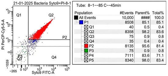
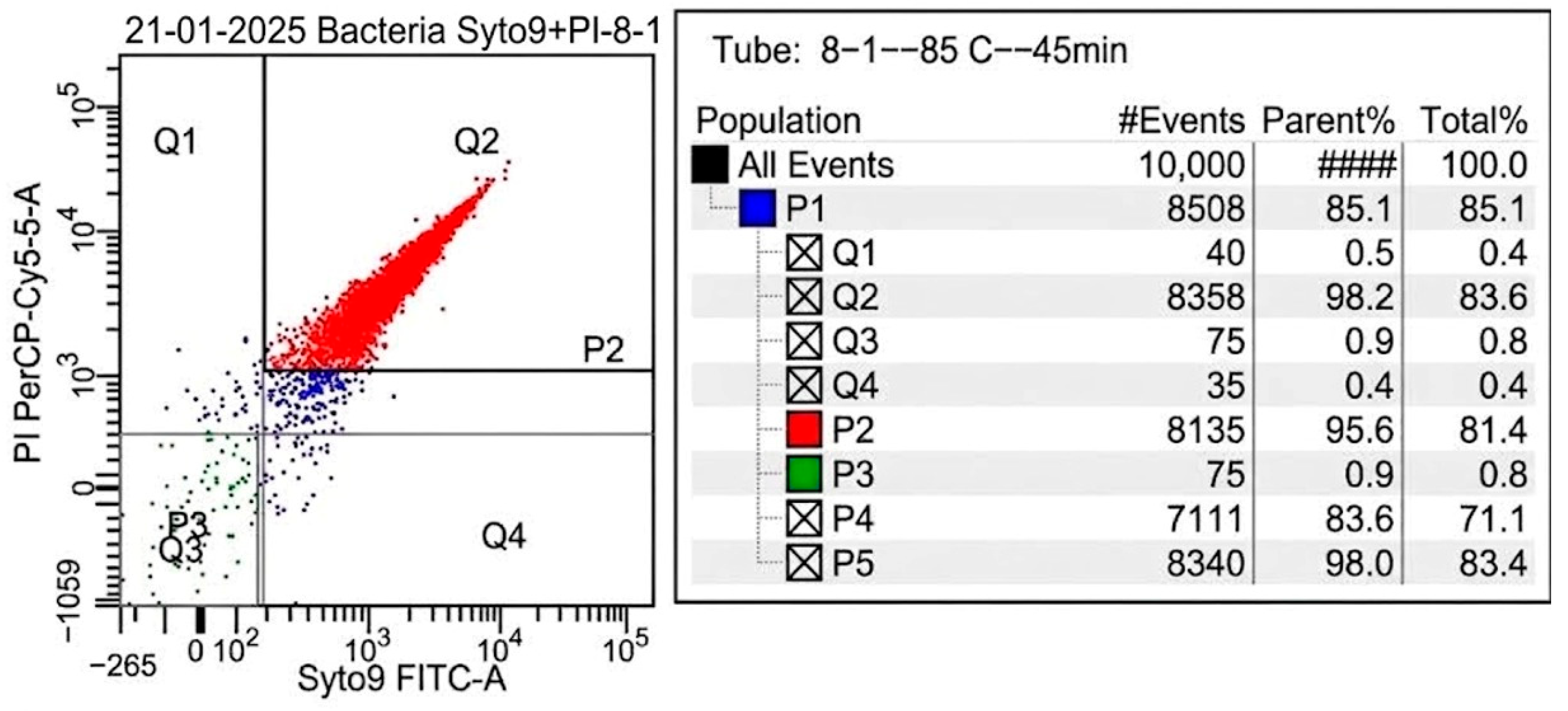
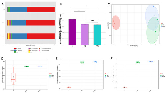
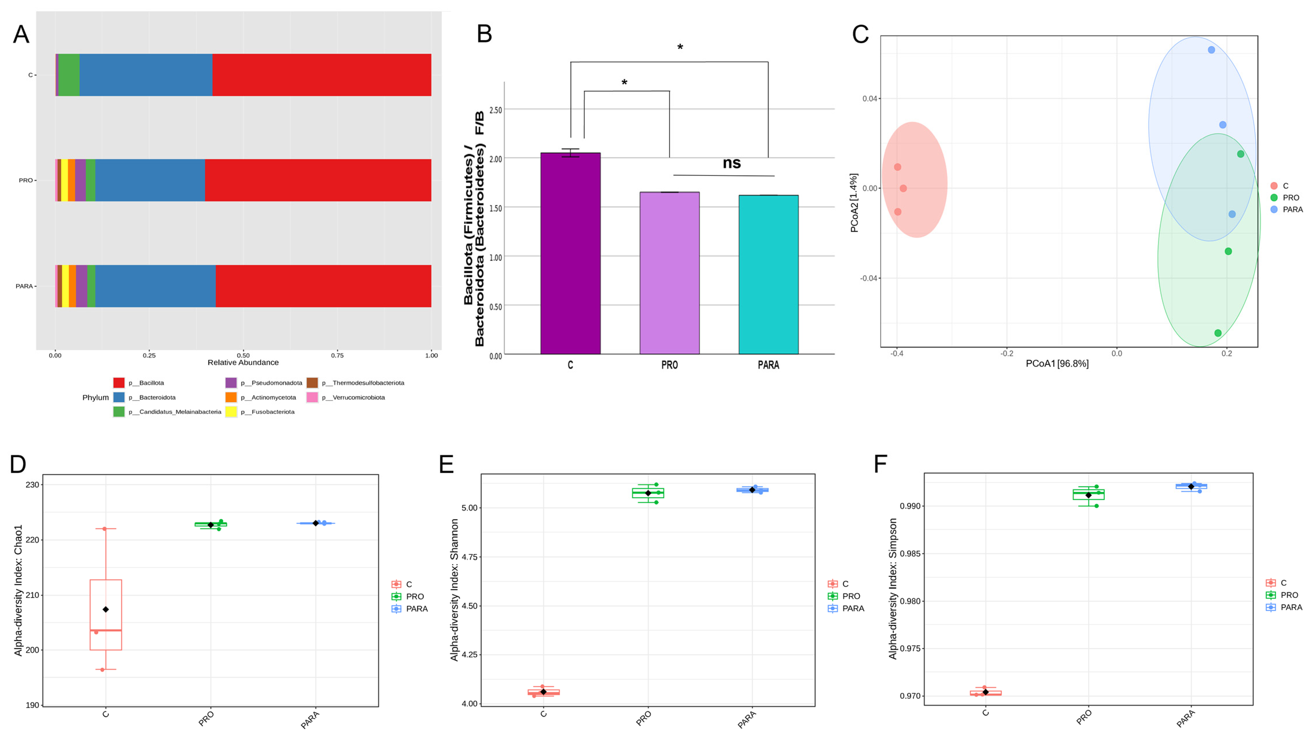
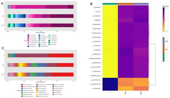
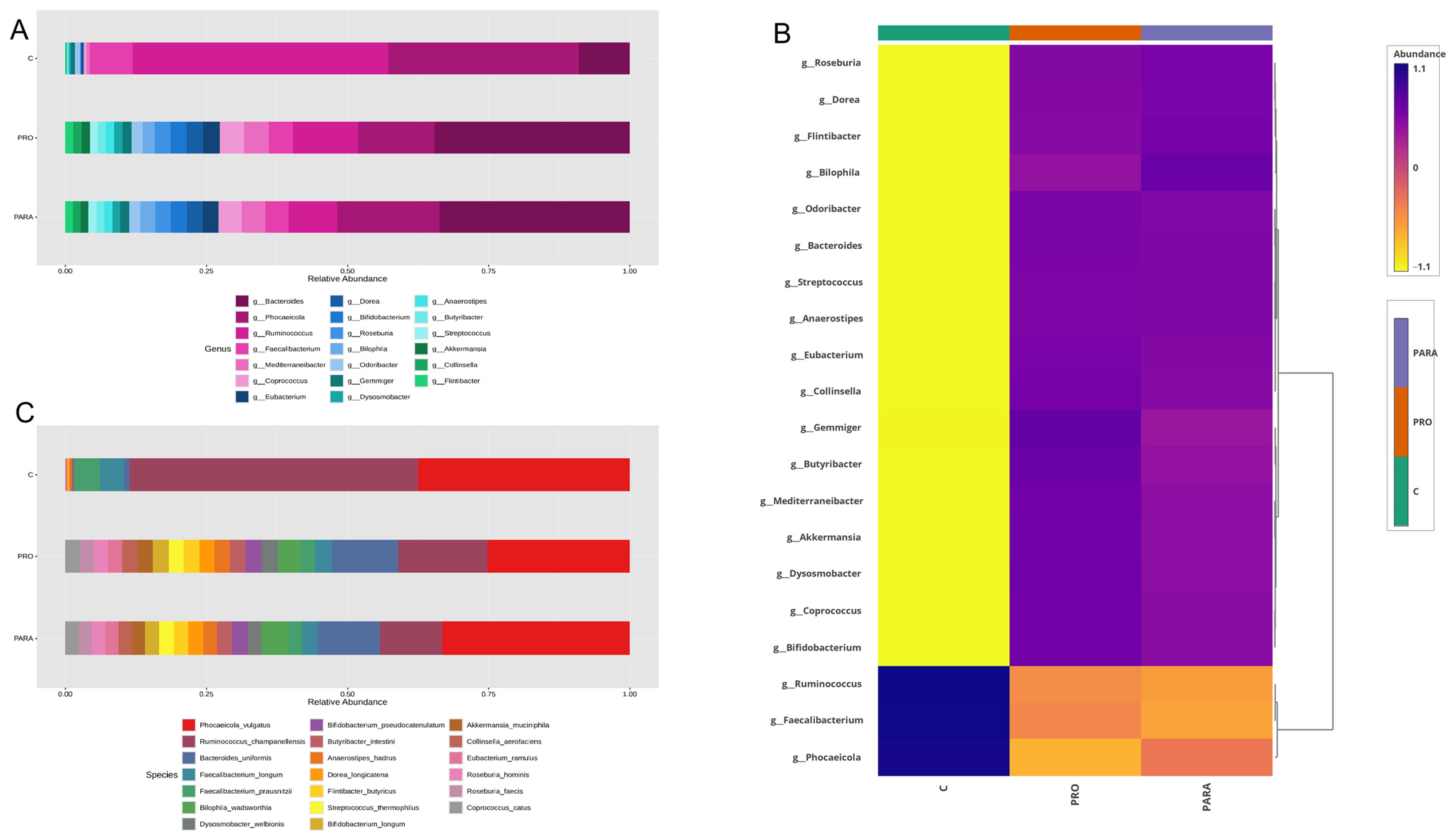
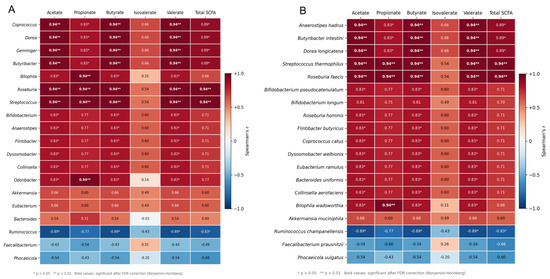
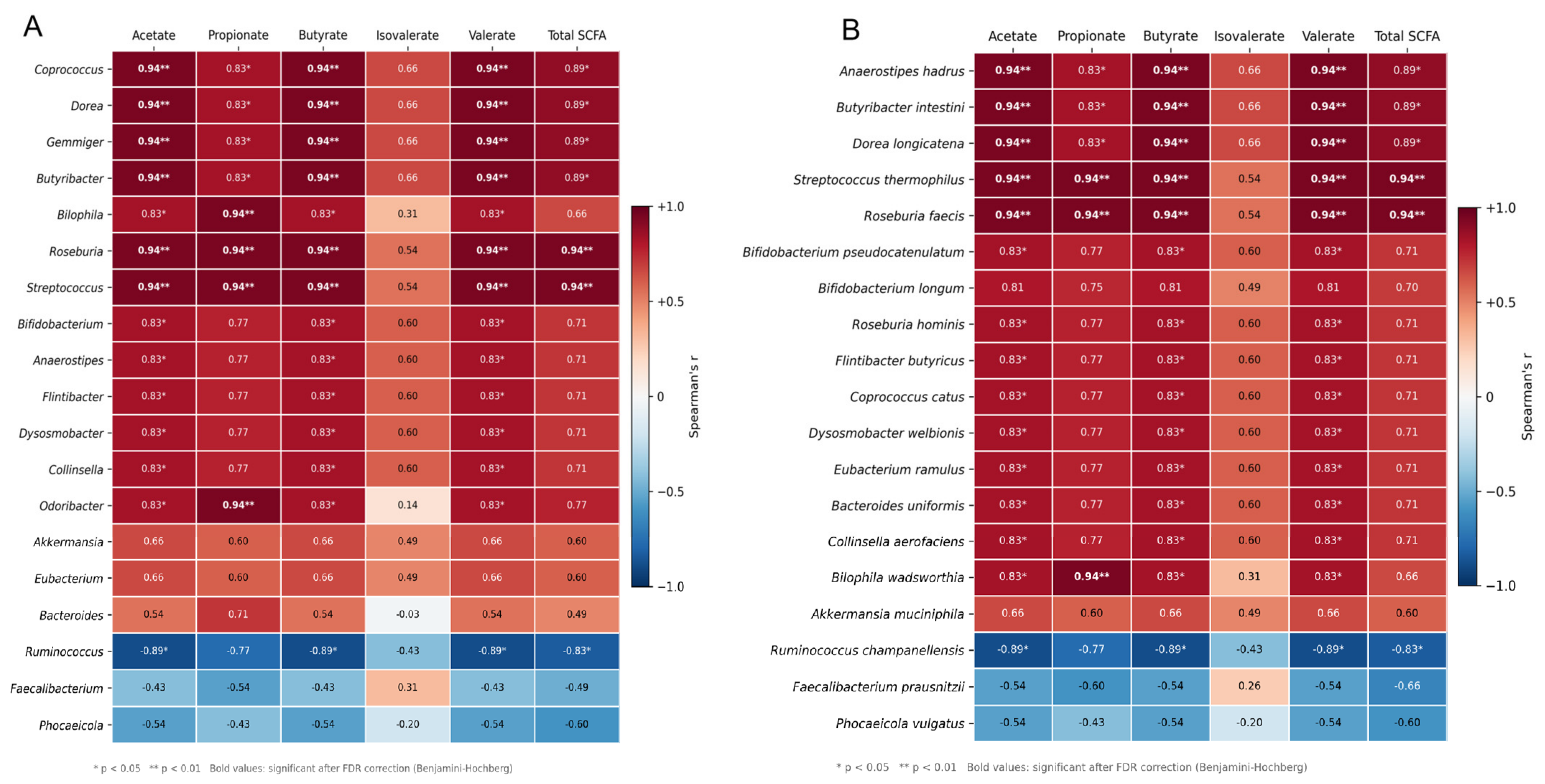

Disclaimer/Publisher’s Note: The statements, opinions and data contained in all publications are solely those of the individual author(s) and contributor(s) and not of MDPI and/or the editor(s). MDPI and/or the editor(s) disclaim responsibility for any injury to people or property resulting from any ideas, methods, instructions or products referred to in the content. |
© 2026 by the authors. Licensee MDPI, Basel, Switzerland. This article is an open access article distributed under the terms and conditions of the Creative Commons Attribution (CC BY) license.