


Abstract
Aflatoxin B1 (AFB1), a potent carcinogenic metabolite produced by Aspergillus flavus and A. parasiticus, constitutes a significant threat to food safety. Conventional decontamination methods are costly or ineffective. Biological detoxification has emerged as a promising alternative. Kefir, a fermented milk beverage, comprises lactic acid bacteria (LAB) and yeasts with documented antifungal properties. This study evaluated whey permeate (WP) as a fermentation substrate for kefir microorganisms and assessed their AFB1-binding capacity. Kefir microorganisms cultivated in WP supported active fermentation, reaching 8.47 log CFU/mL LAB and 7.57 log CFU/mL yeasts, with concomitant lactose consumption and organic acid production. The ability of 13 individual kefir-derived microorganisms and the total kefir microbial community to bind AFB1 was evaluated under diverse conditions. All microorganisms exhibited AFB1 binding ability, although binding depended on strain, growth medium, pH, microbial concentration, and toxin concentration. The highest binding was observed for the total kefir community grown in WP. Binding remained stable over time for the total kefir community and Lactiplantibacillus plantarum CIDCA 83114 and was independent of viability and hydrophobicity. AFB1 binding by kefir microorganisms significantly reduced AFB1-induced cytotoxicity in HepG2 cells. These findings demonstrate that WP can be valorized into a value-added fermented product containing microorganisms with strong AFB1 binding and cytoprotective properties.
1. Introduction
Mycotoxins are secondary metabolites produced by filamentous fungi and represent one of the most critical groups of natural toxins due to their severe health and economic impacts worldwide [1,2]. Approximately 25% of the world’s food crops are estimated to be contaminated with mycotoxins annually, posing a serious threat to food security and global trade [3]. Furthermore, climate change is exacerbating this issue by promoting the spread and increased occurrence of toxin-producing fungi like Aspergillus flavus, as rising temperatures and changing humidity and rainfall patterns create more favorable conditions for their growth [4]. Among mycotoxins, aflatoxin B1 (AFB1), predominantly produced by A. flavus and A. parasiticus, is the most potent hepatocarcinogen identified to date. Chronic dietary exposure to AFB1, which frequently contaminates grains, nuts, spices, and their derivatives, is strongly correlated with the development of hepatocellular carcinoma. AFB1 is classified as a Group 1 human carcinogen by the International Agency for Research on Cancer (IARC) [1,5], making it a major challenge to food safety worldwide.
Given the limitations of conventional physicochemical decontamination methods—which are often costly, ineffective, or may compromise food quality—biological detoxification has emerged as a promising strategy for controlling aflatoxins [6,7]. Numerous studies have reported that lactic acid bacteria (LAB) can inhibit fungal growth and bind mycotoxins both in vitro and in vivo [8,9,10,11]. More recently, it has been demonstrated that LAB strains can reduce AFB1 levels by up to 71%, depending on strain and environmental conditions, with binding rather than biotransformation identified as the main detoxification mechanism. This study provided evidence that the sequestration of AFB1 by LAB is primarily a physical process involving cell wall components, and that the extent of binding is influenced by strain-specific characteristics, pH, and bacterial concentration [12]. Recent evaluations of lactobacilli strains have confirmed their strong capacity for biological detoxification, positioning them as essential tools to reduce dietary exposure to AFB1 [13]. In Latin America, studies have documented aflatoxin contamination throughout the peanut production chain, with concentrations reaching up to 100 µg/kg in some cases [14], underscoring the relevance of developing effective mitigation strategies in the region. Certain yeasts also exhibit notable aflatoxin-binding properties mediated by their cell wall components [15,16]. Recent transcriptomic analyses of Pichia fermentans, a yeast isolated from kefir, have shown that its high-efficiency mycotoxin removal is a multifactorial process involving cell wall adsorption and complex stress-response pathways, including upregulation of genes related to cell wall integrity, oxidative stress response, and membrane transport [17]. Water kefir grains have demonstrated the ability to detoxify AFB1 across broad pH (3–9) and temperature (4–45 °C) ranges through multi-site absorption and partial degradation, reducing AFB1 mutagenicity in various liquid matrices [18].
Kefir is a traditional fermented milk beverage obtained using kefir grains, which consist of a symbiotic consortium of LAB, acetic acid bacteria, and yeasts embedded in a protein–polysaccharide matrix (kefiran) [19]. Both LAB [20] and yeasts [21] isolated from Argentinian kefir have demonstrated potential probiotic properties. Additionally, kefir and its metabolites have been shown to possess antifungal activity [22,23,24] and mycotoxin-binding capacity in vitro [25]. In vivo, dietary kefir has demonstrated hepatoprotective effects against AFB1 exposure in fish models, improving liver function and antioxidant capacity [26].
The AFB1-binding capacity of microorganisms is primarily mediated by cell wall components, including peptidoglycans, teichoic acids, and surface proteins in bacteria, and β-glucans and mannoproteins in yeasts [15,27]. Hernández-Mendoza et al. [28] demonstrated that the integrity of the bacterial cell wall is essential for AFB1 binding, as protoplasts and spheroplasts—which lack partial or complete cell wall structures—showed significantly reduced binding capacity compared to intact cells. Moreover, using bacteria deficient in teichoic acids (grown in phosphate-limited medium) and cells treated with the polycation Polymyxin B, they provided evidence that teichoic acids are key components involved in AFB1 binding. Notably, the binding capacity is not dependent on cell viability. Haskard et al. [10] reported that heat-killed bacteria retain binding abilities comparable to, or even greater than, those of viable cells, with up to 71% of the bound toxin remaining associated after repeated washes. Similarly, Peltonen et al. [29] found that AFB1 binding by dairy strains is rapid, with more than 50% of the toxin remaining bound throughout a 72 h incubation period. Additionally, exopolysaccharides (EPSs) such as kefiran, produced during kefir fermentation, may contribute to mycotoxin sequestration through non-covalent interactions [18,25]. These physical adsorption mechanisms, involving hydrogen bonds, van der Waals forces, and hydrophobic interactions, are generally independent of microbial metabolic activity [10,27]. The binding is partially reversible, as demonstrated by the release of bound AFB1 after successive washes [30], and is influenced by pH [28]. While physical adsorption is the predominant mechanism, some kefir-derived microorganisms have also been reported to possess biotransformation ability. For instance, yeasts isolated from kefir have shown 7–10% biotransformation [31], and the kefir microbial consortium has been assessed for both adsorption and biotransformation of AFB1 [25]. The involvement of multiple cell wall components—including peptidoglycans, teichoic acids, and surface polysaccharides—suggests that AFB1 binding is a multifactorial process mediated by a combination of electrostatic, hydrophobic, and hydrogen-bonding interactions [10,15,27,28].
Whey permeate (WP), the lactose-rich byproduct generated during cheese whey deproteinization, is produced in massive quantities globally and represents an environmental pollutant if not properly utilized [32,33]. The transformation of cheese whey into whey protein generates a large stream of WP that requires further processing due to its high lactose fraction [34]. While physicochemical treatments have been applied to remove the organic load of dairy waste, their effectiveness is limited due to high reagent costs and inadequate removal of soluble organic matter [35]. Fermentation of WP by kefir microorganisms may simultaneously reduce environmental burden and generate functional products with health-promoting properties [22,36].
Although AFB1 binding has been reported for several lactic acid bacteria and isolated kefir microorganisms, it remains unknown whether kefir microbiota cultivated in WP retain these detoxification properties while simultaneously generating a value-added product from this dairy byproduct. We hypothesized that the cultivation of kefir microorganisms in WP could contribute to the valorization of this dairy byproduct and generate microbial biomass capable of mitigating AFB1 toxicity through toxin binding and cytoprotective mechanisms. Accordingly, the objective of this study was to assess the AFB1 binding ability of kefir microorganisms grown in WP under diverse conditions and to evaluate their protective effects against AFB1-induced cytotoxicity.
2. Materials and Methods
2.1. Kefir Microorganisms
Kefir grains CIDCA AGK1 (characterized at the Centro de Investigación y Desarrollo en Ciencia y Tecnología de los Alimentos, CIDCA) [37] were stored in whole milk at −20 °C and activated through two consecutive passages of fermentation in ultra-high-temperature processed (UHT) milk (Sancor, Santa Fe, Argentina). The kefir grains were inoculated into sterile whey permeate (WP) at a concentration of 10% (w/v). Powdered WP (Ilolay, Santa Fe, Argentina) was previously reconstituted at 5% (w/v) in distilled water and sterilized by autoclaving at 121 °C for 15 min. The inoculated WP was then incubated at 30 °C for 24 h. The fermentation products were separated from the grains by passage through a sieve with a 1 mm2 mesh size. The fermented WP, containing the total kefir microbial community, was separated by centrifugation for 15 min at 14,000× g in an Eppendorf 5415D centrifuge (Eppendorf, Hamburg, Germany). The pellets were washed twice with PBS buffer (phosphate-buffered saline solution) and once with sterile purified water. Finally, the pellets were resuspended in PBS and the concentrations of LAB and yeasts were determined by serial dilutions in tryptone (1 g/L) and counted on MRS and YGC agar, respectively. For grains, they were broken up in a mortar and resuspended in tryptone (1 g/L); then, the samples were processed as resuspended pellets.
All microorganisms used in this study were obtained from the CIDCA collection or commercial sources as indicated. The bacterial strains included Lentilactobacillus kefiri strains CIDCA 83111, 8325, and 8348; Lactiplantibacillus plantarum strains CIDCA 83114, 8316, 8318, and 8327; and L. kefiri JCM 5818 (purchased from Japan Collection of Microorganisms). The yeast strains included Saccharomyces cerevisiae strains CIDCA 8112, 8116, and 81103; Kluyveromyces marxianus CIDCA 8154; and S. boulardii (obtained as a commercial lyophilized powder from Floratil Merck, Biocodex, Gentilly, France). LAB and yeast cultures were maintained in MRS broth (for LAB) or YPD broth (for yeasts) at 30 °C. For enumeration, MRS plates were incubated anaerobically at 30 °C for 72 h; YGC plates were incubated aerobically at 30 °C for 48 h. Based on the initial screening and previous studies [20,21], Lpb. plantarum CIDCA 83114 and K. marxianus CIDCA 8154 were selected as representative bacterial and yeast strains. These microorganisms were activated through two consecutive passages of fermentation in MRS broth, inoculated twice in sterile WP, and incubated at 30 °C for 48 h. The washed pellets of microorganisms were obtained as mentioned above. The WP used in this study belonged to the same batch and three fermentation experiments were performed.
2.2. Determination of pH, Ashes, Brix Grades, Nitrogen and Lactose of Kefir-Fermented WP
The pH readings of fermented products were made with a pH meter instrument, Altronix TPX III (Taipei, Taiwan). Water and ash content of the kefir samples were measured according to AOAC Official Methods 934.01 and 942.05, respectively [38]. Total nitrogen was determined by the Kjeldahl method (AOAC 991.20) [38]. The °Brix readings were carried out with a refractometer (Pocket PAL J™; Atago, Tokyo, Japan). Reducing sugars, expressed as lactose equivalents, were determined using the Nelson–Somogyi method [39]. To determine the sample concentration, a lactose standard curve was prepared at the following concentrations: 0, 50, 150, 300, and 400 µg/mL.
2.3. Production of Fermented Cell-Free Supernatants and Determination of Lactic and Acetic Acid Concentration
Once fermentations were complete, the products were centrifuged and filtered through 0.22 µm pore-size membranes (Sartorius®, Göttingen, Germany), designated cell-free supernatants (CFSs), and stored at −80 °C until use. Lactic and acetic acid contents in the CFS were determined by HPLC following the methodology described by Garrote et al. [36] with minor modifications. The system consisted of an Agilent Technologies 1200 series chromatograph (Agilent, Santa Clara, CA, USA) equipped with an Aminex HPX-87H ion-exchange column (Bio-Rad, Hercules, CA, USA), a mobile phase of 0.009 N H2SO4 (Merck, Darmstadt, Germany) at a flow rate of 0.6 mL/min, and an ultraviolet detector set at 214 nm. The injection volume was 10 µL and the column temperature was maintained at 30 °C. Under these conditions, retention times were approximately 10 min for lactic acid and 12 min for acetic acid. Calibration curves were prepared using pure acetic acid (Merck, Darmstadt, Germany) at concentrations of 0.42, 0.83, 1.67, 3.33, 11.70, 25.00, and 66.10 mM and lactic acid (Carlo Erba®, Milan, Italy) at 5.55, 11.10, 44.40, 88.80, 111.00, 155.00, and 209.00 mM. Calibration linearity was confirmed (R2 > 0.999 for both acids); limits of detection (LOD) were 0.1 mM for lactic acid and 0.05 mM for acetic acid; limits of quantification (LOQ) were 0.3 mM and 0.15 mM, respectively.
2.4. Preparation of AFB1 Working Solution
Crystalline AFB1 was obtained from Sigma-Aldrich (St. Louis, MO, USA). A stock solution was prepared in acetonitrile/benzene (98/2, v/v). To prepare methanolic working solutions, the acetonitrile/benzene mixture was evaporated, and the residue was redissolved in methanol. AFB1 concentrations were measured spectrophotometrically at 354 nm using the molar absorption coefficient ε354 = 19,800 L·mol−1·cm−1, and stocks were stored at −20 °C. Aqueous working solutions of various concentrations were prepared in PBS. The final methanol concentration in working solutions was <0.1% (v/v).
2.5. AFB1 Binding Assay
The binding assay used was similar to the procedure reported by Peltonen et al. [29]. A volume of 1 mL of each culture obtained as described in Section 2.1 was centrifuged (4472× g, 10 min) and the microbial pellet was washed twice with 1 mL of sterile double-distilled water, and then suspended in 1 mL of the working solution of AFB1 and incubated at 30 °C for 1 h. Subsequently, the microorganisms were removed by centrifugation. The supernatant fluid containing unbound AFB1 was collected and analyzed by HPLC. For each microorganism, a microbial control (microorganism suspended in PBS) and an AFB1 control (working solution of AFB1 without bacteria) were also incubated. A solvent control (PBS with the same methanol concentration as the working solution) was included to account for any solvent effects.
To evaluate the effect of the viability of the microorganisms on AFB1 binding, total kefir microorganisms, Lpb. plantarum CIDCA 83114, and K. marxianus CIDCA 8154 were killed by pasteurization (72 °C, 30 min) and used as described above.
To study the effect of pH, the pH of PBS was adjusted to 2.1, 5.1, 7.1, and 8.1 using hydrochloric acid or sodium hydroxide. The pH stability after biomass addition was confirmed by re-measurement, and the assays were performed as explained previously.
To evaluate AFB1 binding stability, total kefir microorganisms, Lpb. plantarum CIDCA 83114, and K. marxianus CIDCA 8154 were incubated with AFB1 for 0.5, 24, and 48 h before quantification of unbound toxins, as mentioned below.
To determine the effect of the microorganism and AFB1 concentration on AFB1 binding, different concentrations of AFB1 (150, 500, 1000, and 5000 ng/mL) were challenged against known concentrations of total kefir microorganisms, Lpb. plantarum CIDCA 83114, and K. marxianus CIDCA 8154. Also, different microorganism concentrations (ranging from 105 to 109 CFU/mL) were co-incubated with 150 ng/mL of AFB1.
2.6. Hydrophobicity Assay
These assays were performed using microorganisms previously grown either in MRS or WP. A volume of 2 mL of lactobacilli or yeast suspension in PBS buffer at OD550 nm = 1.0 (OD1) was mixed with 0.5 mL of n-hexadecane for 2 min and after phase separation, the optical density at 550 nm (OD0) was measured. Hydrophobicity was calculated as follows:
where OD0 is the absorbance of the aqueous phase after phase separation and OD1 is the initial absorbance at 550 nm.
H (%) = [(OD0 − OD1)/OD0] × 100
2.7. Quantification of Unbound AFB1
Quantification of unbound AFB1 in supernatants was conducted by a HPLC method with no extraction step, essentially as described by Peltonen et al. [29]. The HPLC system (Waters 717 plus Autosampler, Waters, Milford, MA, USA) with fluorescence detection was used. Chromatographic separations were carried out with a C18 reverse-phase column (150 × 4.6 mm, 5 µm particle size, Waters, Milford, MA, USA). The mobile phase used was a mixture of water/acetonitrile/methanol (70:17:17, v/v/v) at a flow of 1 mL/min. AFB1 was detected by fluorescence at excitation and emission wavelengths of λ 360 nm and λ 420 nm, respectively, and AFB1 quantification was performed by measuring the peak area and extrapolating to the calibration curve previously obtained using Empower software™ (Version 3.0, Waters, Milford, MA, USA). The retention time of AFB1 was approximately 9 min. The method was validated over a calibration range of 1–500 ng/mL, which demonstrated linearity (R2 = 0.99). Recovery ranged from 92% to 105%, and precision (RSD) was <5% for all concentrations tested. The limit of detection (LOD) was estimated at 0.6 ng/mL and the limit of quantification (LOQ) at 2.0 ng/mL, based on a signal-to-noise ratio of 3:1 and 10:1, respectively, as determined from the standard deviation of the blank and the slope of the calibration curve. The bound AFB1 was calculated by subtracting the unbound AFB1 concentration from the initial AFB1 concentration. Additionally, to allow comparison of AFB1 binding efficiency among different microorganisms regardless of their population size, the amount of bound AFB1 was normalized by the number of viable cells. To normalize AFB1 binding by cell number, the parameter AFB1 bound per cell (ng/CFU) was calculated as follows:
This normalization enables the identification of strains with high per-cell binding efficiency, which may not be evident from total binding values alone. However, CFU normalization provides an estimate of per-cell binding but does not account for differences in cell size or surface area.
2.8. Cytotoxicity Effect of AFB1
2.8.1. Cell Culture
The human hepatoma cell line (HepG2) was obtained from the Multidisciplinary Institute of Cell Biology (IMBICE, Buenos Aires, Argentina; ATCC HB-8065, ATCC, Manassas, VA, USA). HepG2 cells (passages 5–15) were seeded at 2 × 10⁵ cells/mL and routinely cultured in tissue culture flasks in Dulbecco’s modified Eagle’s medium (DMEM) supplemented with 10% fetal bovine serum, penicillin (100 units/mL), and streptomycin (100 µg/mL) at 37 °C in a humidified (95%) atmosphere with 5% CO2. The cell medium was replaced twice a week. After reaching confluence, the HepG2 cells were trypsinized and split 1:3. The cell line was authenticated by STR profiling and tested negative for mycoplasma by PCR. The cells were used for various bioassays as follows:
2.8.2. Protective Effect of Kefir Microorganisms Against AFB1 Cytotoxicity on HepG2 Cells
The total kefir microbial community, obtained as described in Section 2.1, was killed by heat treatment (72 °C, 30 min) and used to study their protective activity against AFB1 on HepG2 cells. Commercial AFB1 (Sigma Chemical Co., St. Louis, MO, USA) was prepared at different concentrations in DMEM without fetal serum. To allow toxin binding, heat-killed kefir microorganisms (total kefir microorganisms) were pre-incubated with AFB1 (150 or 300 ng/mL) in DMEM for 1 h at 30 °C, as described for the binding assay in Section 2.5, but using DMEM instead of PBS. HepG2 cells at 70% confluence were then exposed to the entire mixture (microorganisms + AFB1) without prior centrifugation. The mixture was added to the cell monolayer and incubated for 24 h at 37 °C in a humidified (95%) atmosphere with 5% CO2. As controls, cells were treated with DMEM alone (untreated control), with heat-killed microorganisms without AFB1, or with AFB1 alone at the same concentrations. A solvent control (DMEM with the same methanol concentration as the working solution) was included to account for any solvent effects. Cytotoxicity was evaluated by two complementary methods: one measuring cell viability and the other measuring cell damage.
Determination of succinate dehydrogenase mitochondrial activity as a measure of cell viability: cell viability was measured using the cleavage of 3-(4,5-dimethylthiazol-2-yl)-2,5-diphenyl tetrazolium bromide (MTT) [40] by mitochondrial succinate dehydrogenase. MTT was used at a dilution of 5 mg/mL in DMEM without phenol red.
After the 24 h incubation of mixtures on HepG2 cells, the culture medium was removed, and the cells were washed with PBS. MTT solution (5 mg/mL in DMEM without phenol red) was added to each well and the plates were incubated at 37 °C for 3 h. The formazan crystals formed by viable cells were solubilized by adding 200 µL of dimethyl sulfoxide (DMSO) and the absorbance was measured at 550 nm using a spectrophotometer. The percentage of cell viability was calculated as (Absorbance of treated cells/Absorbance of untreated control cells) × 100. The protective assay was performed in quadruplicate for each sample. At least three independent experiments were performed for each condition.
Determination of lactate dehydrogenase release as a measure of cell damage: extracellular lactate dehydrogenase (LDH) activity was evaluated as previously reported [41]. Spontaneous LDH release (untreated cells) and maximum LDH release (cells lysed with 1% Triton X-100) were used as negative and positive controls, respectively, to calculate the percentage of LDH release for each experimental condition. After the 24 h incubation, the cell-free supernatant was carefully collected from each well, avoiding disturbance of the cell monolayer and the microbial pellet.
LDH activity was determined using a commercial kit (Wiener Lab, Rosario, Argentina). The reaction was initiated by adding the substrate (lactate and NAD+) and the rate of NADH formation was measured by the increase in absorbance at 340 nm for 3 min at 37 °C using a spectrophotometer (Beckman DU 650, Palo Alto, CA, USA). The percentage of cytotoxicity was calculated as (1-(Maximum Release-Experimental LDH release)/Maximum release) × 100. The assay was performed in quadruplicate for each sample. At least three independent experiments were performed for each condition.
2.9. Statistical Analysis
All growth parameters were analyzed by SIGMAPLOT 10.0® software (Systat Software, San Jose, CA, USA). The results of three independent assays are presented as mean values ± standard deviation (SD). To evaluate the effects of microbial strain, pH, toxin concentration, and their interactions on AFB1 binding, data were analyzed using one-way and two-way analysis of variance (ANOVA). Significant effects were determined at p < 0.05 using STATGRAPHICS Plus 5.1® software (Statgraphics Technologies, The Plains, VA, USA). When ANOVA indicated significant differences, post hoc comparisons were performed using Tukey’s HSD test to identify which specific groups differed. The assumptions of ANOVA (normality and homogeneity of variances) were verified by Shapiro–Wilk and Levene’s tests, respectively. To examine the relationship between cell surface hydrophobicity and AFB1 binding, Spearman’s rank correlation coefficient was used. All experiments were performed with at least three independent biological replicates, each with three technical replicates.
3. Results
3.1. Kefir Grain Growth and Whey Permeate Fermentation
Table 1 shows the physicochemical changes occurring in WP after fermentation with kefir grains. Fermentation induced significant modifications in the chemical composition of the substrate, reflecting the intense metabolic activity of the kefir microbiota and the efficient conversion of lactose into organic acids. Lactic acid showed the greatest increase, rising from 8.9 mM in unfermented WP to 148.4 mM after fermentation (>16-fold increase). Acetic acid also increased significantly, rising from 6.7 mM to 43.9 mM (approximately a 6.6-fold increase).
Table 1.
Chemical composition of whey permeate (WP) fermented with kefir grains and its unfermented control.
The accumulation of organic acids was associated with a substantial consumption of fermentable carbohydrates. Lactose concentration decreased from 3.33% to 2.23%, indicating that approximately one-third of the initial lactose content was metabolized during fermentation. Similarly, soluble solids measured as °Brix decreased from 5.30% to 4.04%, reflecting the utilization of carbohydrates and other soluble nutrients by the microbial population. In contrast, only minor changes were observed in nitrogen and ash contents.
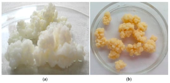
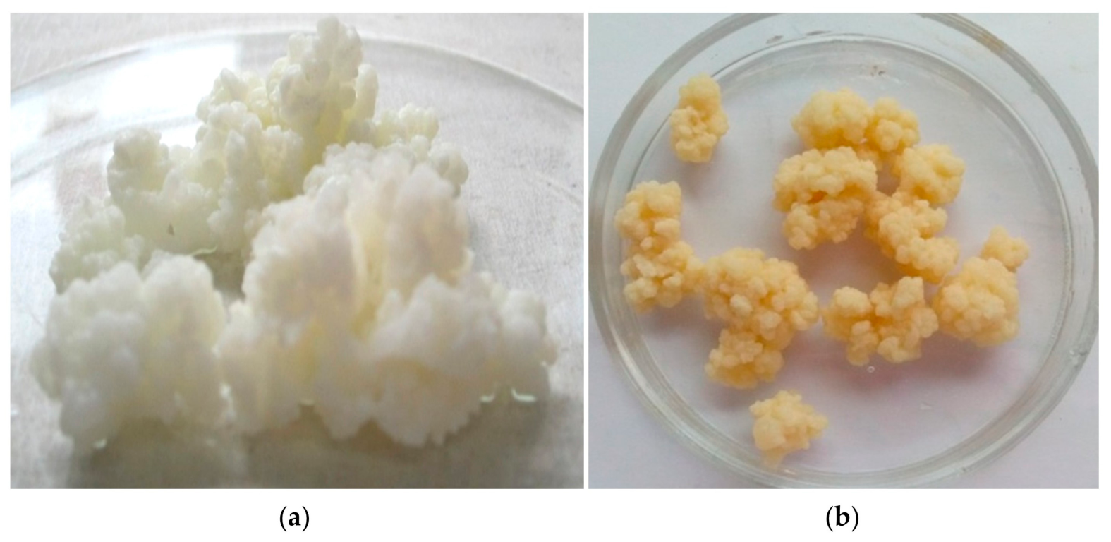
As shown in Figure 1, kefir grains cultured in WP (b) maintained the characteristic irregular cauliflower-like structure observed in milk-grown grains (a). However, grains grown in WP appeared slightly smaller and more translucent than those propagated in milk.
Figure 1.
Appearance of kefir grains cultured in cow milk (a) and whey permeate (b).
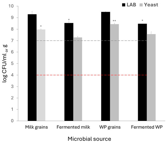
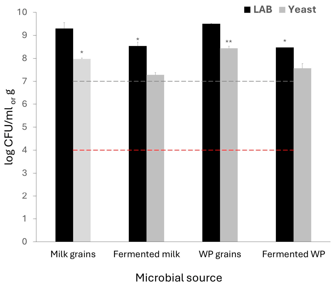
The microbial populations associated with the grains and fermented beverages are shown in Figure 2. After 24 h of fermentation in WP, viable counts reached 8.47 ± 0.08 log CFU/mL for LAB and 7.57 ± 0.20 log CFU/mL for yeasts.
Figure 2.
Microbial counts of kefir grains cultured in cow milk and whey permeate and their beverages. LAB: lactic acid bacteria. WP grains: kefir grains grown in whey permeate. Fermented WP: whey permeate fermented with kefir grains. Gray and red dashed lines represent the minimum LAB and yeasts required by Argentine legislation [42], respectively. Asterisks indicate significant differences between each treatment: * p < 0.05; ** p < 0.01. Treatments without asterisks did not differ significantly from each other (p > 0.05).
3.2. AFB1 Binding by Total Kefir Community and Isolated Microorganisms
Table 2 summarizes the AFB1 binding ability and cell surface hydrophobicity of kefir-derived microorganisms grown either in MRS broth or WP. Overall, all tested microorganisms exhibited some degree of AFB1 binding, although marked strain-dependent differences were observed. The highest overall AFB1 binding was observed for the total kefir community grown in WP, which removed 105.5 ng/mL AFB1, exceeding the binding levels achieved by any individual isolate. The L. kefiri strains evaluated were unable to grow in WP, whereas all Lpb. plantarum strains grew in this medium, although viable counts were generally lower than those obtained in MRS. Lpb. plantarum strains consistently outperformed L. kefiri strains, with Lpb. plantarum CIDCA 83114 showing the highest binding ability (88.9 and 78.1 ng/mL after growth in MRS and WP, respectively). However, slight variations were observed among Lpb. plantarum strains, no significant differences were detected between cells grown in MRS and those grown in WP. Although all Lpb. plantarum strains were able to grow in WP, reaching counts between 9 and 9.3 log CFU/mL, their growth capacity in WP was lower than that observed in MRS, with a decrease in counts between 27.5% and 92.9%.
Table 2.
AFB1 binding and cell surface hydrophobicity of kefir-derived microorganisms grown either in MRS broth or WP.
Yeast growth in WP was strain-dependent: K. marxianus CIDCA 8154 and S. cerevisiae CIDCA 8116 and 8112 grew better in WP than in MRS, whereas S. cerevisiae CIDCA 81103 and S. boulardii did not. Yeasts exhibited greater variability than lactobacilli, and both strain identity and growth medium influenced AFB1 binding. Among the yeasts evaluated, S. cerevisiae CIDCA 8112 exhibited the highest AFB1 binding ability, binding 84.7 ng/mL and 75.7 ng/mL AFB1 after growth in MRS and WP, respectively, with values comparable to those observed for the best-performing Lpb. plantarum strains. In contrast, the probiotic reference strain S. boulardii displayed insignificant binding ability (<3 ng/mL) regardless of the growth medium. Medium-dependent effects were particularly evident for some yeast strains. For example, S. cerevisiae CIDCA 8116 increased AFB1 binding by approximately 59% when grown in WP (62.3 ng/mL) compared with MRS (39.2 ng/mL), whereas K. marxianus CIDCA 8154 exhibited similar binding values in both media (37.5–40.9 ng/mL).
When AFB1 binding was normalized by the number of viable microorganisms, some yeasts exhibited substantially higher binding efficiency on a per-cell basis than lactobacilli. Yeasts exhibited substantially higher per-cell binding efficiency than lactobacilli, with values up to 35.67 × 10⁻⁷ ng/CFU for S. cerevisiae CIDCA 81103 (in WP) and to 32.91 × 10⁻⁷ ng/CFU for K. marxianus CIDCA 8154 (in MRS), compared with ≤ 0.63 × 10⁻⁷ ng/CFU for Lpb. plantarum strains.
No clear relationship was observed between cell surface hydrophobicity and AFB1 binding ability. Spearman correlation analysis showed no significant correlation between hydrophobicity and AFB1 binding (ρ = −0.166, p = 0.46). This lack of association was evident when comparing strains grown under different conditions. For instance, S. cerevisiae CIDCA 8116 exhibited a marked increase in AFB1 binding after growth in WP despite a reduction in hydrophobicity from 48.9% to 27.7%. Conversely, S. cerevisiae CIDCA 81103 showed simultaneous increases in both hydrophobicity (from 51.4% to 95.0%) and AFB1 binding (from 53.5 to 69.2 ng/mL) following growth in WP. Furthermore, K. marxianus CIDCA 8154 maintained nearly identical hydrophobicity values (~95%) in both media while displaying no significant changes in AFB1 binding ability.
3.3. Effect of Viability, Incubation Time, pH and Concentration on AFB1 Binding
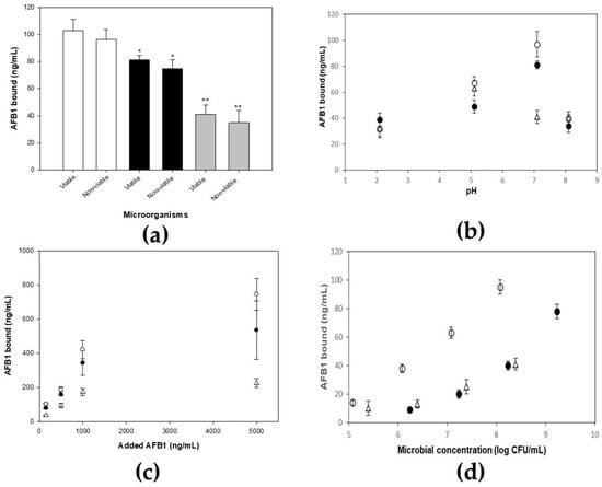
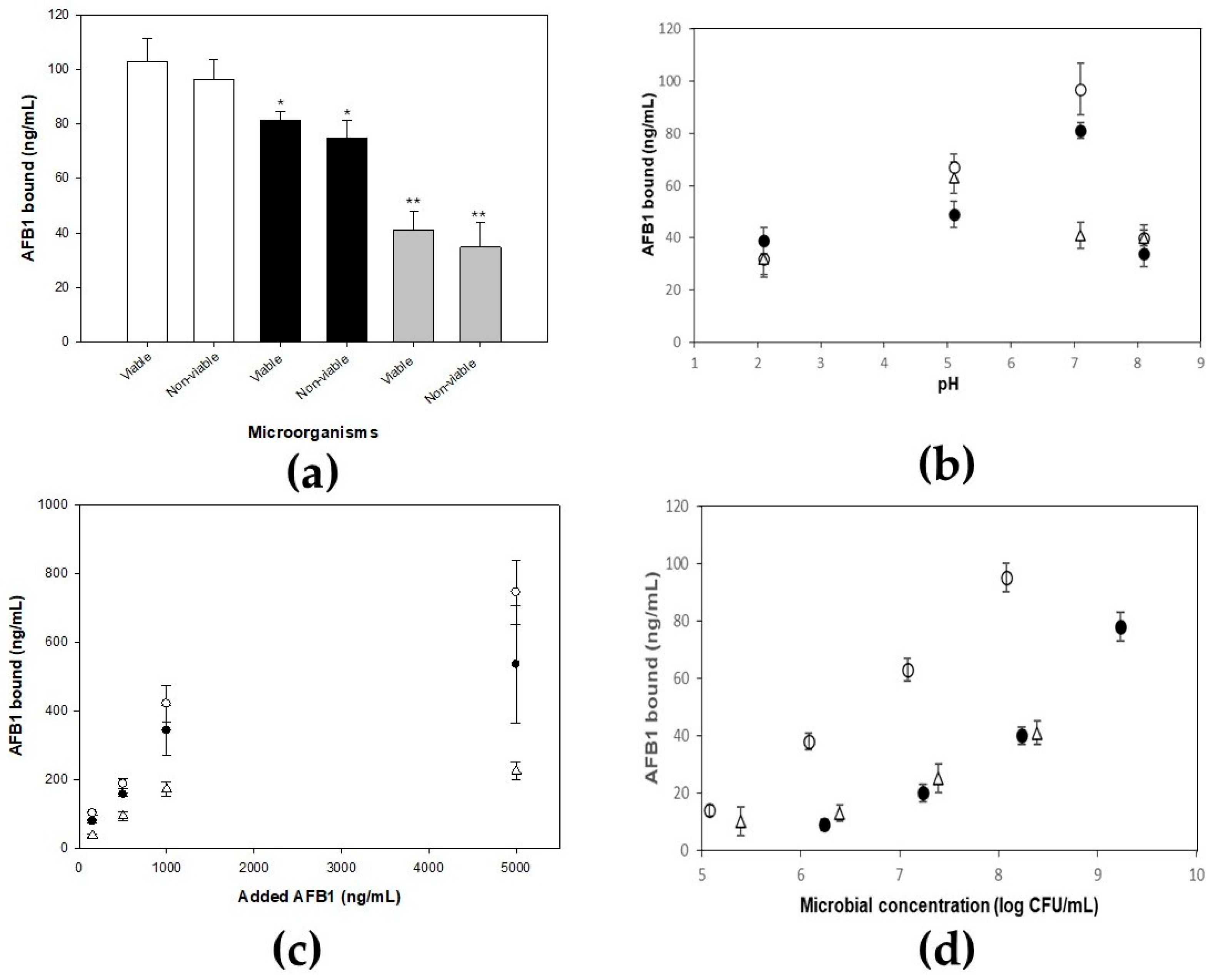
The AFB1 binding capacities of the total kefir community and selected kefir microorganisms (Lpb. plantarum CIDCA 83114 and K. marxianus CIDCA 8154) in viable and non-viable states are presented in Figure 3a. The total kefir community exhibited the highest binding ability, followed by Lpb. plantarum CIDCA 83114 and K. marxianus CIDCA 8154. Heat treatment did not significantly affect AFB1 binding in any of the microorganisms evaluated, as comparable binding values were observed for viable and non-viable cells.
Figure 3.
AFB1 binding by kefir microorganisms. (a) Effect of viability on AFB1 binding by total kefir community (white bars, □), Lpb. plantarum CIDCA 83114 (black bars, ■), and K. marxianus CIDCA 8154 (gray bars, ■). Asterisks indicate significant differences between viable and non-viable cells for each microorganism: * p < 0.05; ** p < 0.01. The absence of asterisks indicates no significant difference (p > 0.05). (b) Effect of pH on AFB1 binding by total kefir community (○), Lpb. plantarum CIDCA 83114 (●), and K. marxianus CIDCA 8154 (△). (c) Effect of AFB1 concentration on AFB1 binding by total kefir community (○), Lpb. plantarum CIDCA 83114 (●), and K. marxianus CIDCA 8154 (△). (d) Effect of microbial concentration on AFB1 binding by total kefir community (○), Lpb. plantarum CIDCA 83114 (●), and K. marxianus CIDCA 8154 (△).
The effect of pH on AFB1 binding is shown in Figure 3b. The total kefir community and Lpb. plantarum CIDCA 83114 showed greater binding capacities at neutral pH (7.1) than under acidic conditions, whereas K. marxianus CIDCA 8154 reached its highest binding value at pH 5.1. Exposure to pH 2.1 significantly reduced microbial viability for Lpb. plantarum CIDCA 83114 (decrease of approximately 3.4 log units; p < 0.05) and for the total kefir community (1.6 and 0.8 log units for LAB and yeasts; p < 0.05), whereas K. marxianus CIDCA 8154 showed only a slight, non-significant decrease (≈ 0.5 log units; p > 0.05) (see Supplementary Materials, Table S1). However, despite this reduction in viability, AFB1 binding ability was maintained.
As shown in Figure 3c, AFB1 binding increased with increasing toxin concentration up to 1000 ng/mL for the total kefir community and selected kefir microorganisms (Lpb. plantarum CIDCA 83114 and K. marxianus CIDCA 8154). At 5000 ng/mL, the total kefir community and Lpb. plantarum CIDCA 83114 displayed additional increases in AFB1 binding, whereas K. marxianus CIDCA 8154 maintained binding values similar to those observed at 1000 ng/mL.
Figure 3d shows a positive relationship between microbial concentration and AFB1 binding. Linear regression analysis confirmed significant positive relationships for all three microbial preparations (R2 = 0.995 for the total kefir community, 0.883 for Lpb. plantarum CIDCA 83114, and 0.864 for K. marxianus CIDCA 8154; p < 0.05). Increasing microbial concentrations resulted in progressively higher amounts of bound AFB1, with the total kefir community consistently showing the highest AFB1 binding.
The stability of AFB1 binding over time was evaluated by incubating the total kefir community, Lpb. plantarum CIDCA 83114, and K. marxianus CIDCA 8154 with the toxin for up to 48 h (Table 3). The total kefir community exhibited consistent binding throughout the incubation period, with no significant differences between 0.5 and 48 h (105.5 ± 3.0 vs. 104.6 ± 5.9 ng/mL). Similarly, Lpb. plantarum CIDCA 83114 maintained binding levels that did not differ significantly over time (83.2 ± 12.3 ng/mL at 0.5 h vs. 75.3 ± 7.1 ng/mL at 48 h). In contrast, K. marxianus CIDCA 8154 exhibited a progressive and significant loss of bound AFB1 over time, declining from 40.9 ± 3.2 ng/mL at 0.5 h to 25.9 ± 4.0 ng/mL at 24 h and further to 13.5 ± 4.5 ng/mL at 48 h. These findings indicate that while the total kefir community and Lpb. plantarum CIDCA 83114 form stable AFB1 complexes, the binding to K. marxianus CIDCA 8154 is weaker and partially reversible.
Table 3.
AFB1 binding as a function of incubation time of total kefir community, Lpb. plantarum CIDCA 83114 and K. marxianus CIDCA 8154.
3.4. Protective Effect Against AFB1 Cytotoxicity
The protective effect of the total kefir community against the cytotoxic effect of AFB1 was evaluated using two complementary cellular biomarkers: mitochondrial dehydrogenase activity (MTT assay), as an indicator of cell viability, and lactate dehydrogenase (LDH) release, as a marker of membrane damage (Table 4). Exposure of HepG2 cells to the total kefir community alone did not significantly affect either parameter compared with untreated control cells, demonstrating the absence of intrinsic cytotoxic effects under the experimental conditions evaluated.
Table 4.
Effect of AFB1 on HepG2 cells with/without total kefir community measured by mitochondrial dehydrogenase and lactate dehydrogenase activity.
In contrast, AFB1 exposure caused a concentration-dependent reduction in cell viability and an increase in cell damage. At 150 ng/mL, AFB1 reduced HepG2 viability from approximately 100% in control cells to 75.2% (a decrease of about 25%), while LDH release increased from 27.6% to 68.4%, indicating substantial membrane injury. Increasing the toxin concentration to 300 ng/mL further exacerbated its cytotoxic effects, reducing cell viability to 52.9% and increasing cell damage to 96.4%, values approaching those observed for the lysed-cell control.
Pre-incubation of AFB1 with a non-viable total kefir community significantly attenuated the toxic effects of the mycotoxin at both concentrations tested. In the presence of the total kefir community, cell viability increased from 75.2% to 97.8% for cultures challenged with 150 ng/mL AFB1 and from 52.9% to 91.4% for cultures exposed to 300 ng/mL AFB1. The total kefir community restored cell viability to levels comparable to those of untreated controls, even at the highest toxin concentration evaluated. Similarly, LDH release was significantly reduced in the presence of the total kefir community, decreasing from 68.4% to 52.4% at 150 ng/mL AFB1 and from 96.4% to 57.2% at 300 ng/mL AFB1.
4. Discussion
The present study demonstrates that kefir-derived microorganisms grown on WP retain a remarkable ability to bind AFB1 and significantly reduce its cytotoxicity. These findings demonstrate that WP can be transformed into a value-added bioproduct with potential applications in mycotoxin mitigation.
The physicochemical changes observed after fermentation confirmed the active metabolism of the kefir consortium in WP. The marked acidification, together with the accumulation of lactic and acetic acids and the reduction in lactose concentration, reflects the efficient conversion of fermentable carbohydrates by LAB and yeasts. The substantial production of organic acids is particularly relevant because these metabolites contribute to preservation and have been associated with antifungal activity and inhibition of mycotoxin production. Indeed, previous studies using the same kefir grains demonstrated that fermented WP inhibits the growth of Aspergillus parasiticus and Fusarium graminearum and reduces aflatoxin and zearalenone biosynthesis [22,36]. Together with the high microbial counts achieved after fermentation, these results confirm that WP constitutes a suitable substrate for kefir growth despite its relatively low nutrient content. However, it is important to acknowledge that a single source of WP was used in this study, and the results may vary with different WP compositions due to variations in lactose, mineral, and nitrogen content across different production batches.
The ability of kefir grains to ferment and develop microbial quantities comparable to those found in milk-fermented kefir further supports the suitability of WP as a fermentation substrate. The LAB and yeast populations obtained exceeded the minimum levels established by Argentine legislation for fermented products [42], demonstrating that this byproduct can sustain the growth and maintenance of the characteristic kefir microbiota. These observations reinforce previous evidence indicating that WP can serve as an alternative culture medium for the production of functional microorganisms while reducing the environmental burden associated with whey disposal.
AFB1 binding was highly strain-dependent, in agreement with previous reports demonstrating that binding ability varies considerably among both LAB and yeasts [30,43,44]. Among the bacterial isolates evaluated, Lpb. plantarum strains consistently exhibited higher binding capacities than L. kefiri strains, with Lpb. plantarum CIDCA 83114 emerging as the most effective bacterial binder. Recent studies have similarly shown that Lactiplantibacillus strains can efficiently remove AFB1, with adsorption rather than biodegradation representing the dominant detoxification mechanism [12].
A particularly relevant finding was that growth in WP did not impair the AFB1 binding ability of Lpb. plantarum strains. Despite generally lower viable counts in WP than in MRS, comparable amounts of toxin were adsorbed, and normalization by cell number revealed increased binding efficiencies on a per-cell basis. These observations align with recent reports showing that non-viable or inactivated bacterial cells often exhibit equal or superior mycotoxin binding compared to viable cells [12,45]. In particular, the inactivation of Lpb. plantarum and Lacticaseibacillus rhamnosus (formerly named Lactobacillus rhamnosus) by thermosonication has been shown to increase surface hydrophobicity and enhance AFB1 binding [46]. Furthermore, the efficiency of toxin binding is known to depend on microbial species, cell wall hydrophobicity, pH, temperature, and mycotoxin type [47]. Together, these findings suggest that growth conditions may modulate cell surface structures involved in toxin binding without compromising the overall detoxification potential of the microorganisms. These observations suggest that WP cultivation may induce modifications in cell wall architecture—a phenomenon documented in bacteria and yeasts [48,49]. β-glucans in yeast cell walls have been directly implicated in mycotoxin complexation [27], supporting this hypothesis.
Among the yeasts, substantial strain-dependent differences were also observed. S. cerevisiae CIDCA 8112 exhibited binding ability comparable to that of the best-performing Lpb. plantarum strains, S. boulardii showed almost no binding activity. Interestingly, some yeast strains displayed enhanced AFB1 binding after growth in WP, indicating that environmental conditions may exert a stronger influence on binding properties in yeasts than in lactobacilli. The considerable variability observed among yeast isolates agrees with previous studies reporting strain-specific detoxification abilities against different mycotoxins [44].
The highest overall AFB1 binding was observed for the total kefir community grown in WP, which outperformed all individual strains. This observation suggests that the complex microbial consortium may provide additive effects that enhance toxin binding. Nevertheless, comparisons of total binding capacity between the consortium and individual strains may be confounded by differences in total microbial biomass. Although normalization by CFU was performed, this does not fully account for differences in cell size or surface area between bacteria and yeasts. Such effects may result from interactions between bacterial and yeast populations, co-aggregation phenomena, and the presence of extracellular polymeric substances such as kefiran. Similarly, Taheur et al. [25] reported highly efficient adsorption of AFB1 and other mycotoxins by the total kefir community, while Ouyang et al. [18] proposed that kefir grains possess multiple adsorption sites and complementary detoxification mechanisms.
The analysis of AFB1 binding normalized by viable cell counts provided additional insight into the binding process. Although lactobacilli exhibited the highest total binding values, several yeast strains showed markedly greater binding efficiencies on a per-cell basis. These findings suggest that differences in cell size, cell wall composition, and surface architecture may influence the density and accessibility of toxin-binding sites. Therefore, both total biomass and intrinsic cellular adsorption efficiency should be considered when selecting microorganisms for mycotoxin mitigation applications.
No clear relationship was observed between cell surface hydrophobicity and AFB1 binding ability (Spearman’s ρ = − 0.166, p = 0.46). Changes in hydrophobicity were not consistently associated with changes in toxin adsorption, indicating that hydrophobic interactions alone cannot explain the observed binding patterns. Similar conclusions were reached by Yiannikouris et al. [27], who proposed that multiple mechanisms, including hydrogen bonding, electrostatic interactions, and the participation of specific polysaccharides and cell wall proteins, contribute to mycotoxin binding. The lack of correlation observed here further supports the complexity of the binding process.
Microbial viability assays demonstrated that AFB1 binding was independent of metabolic activity. Heat-treated microorganisms retained binding abilities comparable to those of viable cells, indicating that binding is primarily mediated by structural cell wall components rather than active cellular processes. This finding agrees with previous studies showing that non-viable microorganisms can effectively bind mycotoxins [12,15,50]. Components such as peptidoglycans, β-glucans, mannoproteins, and other polysaccharides have been proposed as the principal adsorption sites. Haskard et al. [10] further suggested that heat treatment may expose additional binding sites through structural modifications of the cell wall.
The stability of AFB1 binding over 48 h differed markedly among preparations (Table 3). The total kefir community and Lpb. plantarum CIDCA 83114 maintained stable binding levels, whereas K. marxianus CIDCA 8154 released approximately two-thirds of the initially bound toxin, indicating weaker and partially reversible binding. This is consistent with the non-covalent nature of mycotoxin binding described for yeasts [15,27]. The stability of the Lpb. plantarum–AFB1 complex corroborates previous findings from our group on its biopreservative potential in dry pet food [51]. While these findings indicate stability under the conditions tested, complete mass-balance and desorption experiments were not performed; therefore, the stability of the complex under gastrointestinal conditions remains to be determined. Importantly, while those previous studies characterized the chemical nature of the interaction using equilibrium binding assays, the present work provides direct kinetic evidence of gradual toxin release, highlighting that the stability of the yeast–AFB1 complex can vary considerably among different yeast species and strains. These results underscore that AFB1 binding is not a uniform process across microorganisms; while some (like the kefir consortium and Lpb. plantarum) provide stable and durable binding, others may release the toxin under certain conditions.
Environmental conditions significantly influenced AFB1 binding. The total kefir community and Lpb. plantarum CIDCA 83114 exhibited greater binding at neutral pH, whereas K. marxianus CIDCA 8154 reached maximal binding under mildly acidic conditions. These differences likely reflect pH-dependent changes in the ionization state of both AFB1 and microbial surface groups, as previously reported [28]. The viability loss at pH 2.1, particularly for Lpb. plantarum, suggests that extreme acidic conditions may alter cell surface integrity and consequently affect binding efficiency. Furthermore, the positive relationships observed between AFB1 binding and both toxin concentration and microbial concentration indicate the presence of multiple available binding sites and confirm that biomass availability is a key determinant of binding capacity. Similar observations have been reported previously for bacterial mycotoxin binders [52,53]. It should be noted that the binding assays were performed in PBS, which does not fully simulate the complexity of food matrices or gastrointestinal conditions.
Beyond their AFB1 binding ability, the total kefir community significantly reduced the biological effects of AFB1 in HepG2 cells. The marked recovery of cell viability together with the reduction in LDH release demonstrates that toxin binding effectively decreased AFB1 bioavailability. The agreement between MTT and LDH assays strengthens this conclusion and indicates preservation of both cellular metabolic activity and membrane integrity. Interestingly, the protective effect remained evident even at the highest toxin concentration evaluated. Similar reductions in AFB1 toxicity have been reported for other probiotic microorganisms, including Bifidobacterium lactis [54], while in vivo studies have demonstrated that kefir supplementation can alleviate aflatoxin-induced hepatotoxicity through antioxidant and hepatoprotective mechanisms [26]. However, the protective effect in the present study was demonstrated only in HepG2 cells. In vivo validation in animal models or food matrices is still lacking and represents a necessary step to confirm the practical applicability of this approach. Likewise, commercial yeast-derived binders have proven effective in reducing aflatoxin toxicity in aquaculture systems [55].
The present results demonstrate that WP can be efficiently transformed through kefir fermentation into a value-added product containing microorganisms with high AFB1 binding ability and significant cytoprotective activity. This approach simultaneously contributes to mycotoxin mitigation and the valorization of an underutilized dairy byproduct, providing a low-cost alternative with potential applications in food and feed systems.
Because AFB1 binding was independent of cell viability, the inactivated biomass can be developed as a stable biosorbent for food and feed applications. Moreover, previous studies have demonstrated antifungal properties of WP fermented with kefir [36], suggesting that both AFB1 binding and antifungal activity could complement each other in future applications.
Looking forward, the production of kefir through starter cultures, as recently described by Gamba et al. [56] in cow milk, could represent a complementary strategy to further improve scalability and consistency of kefir-fermented WP. Their study demonstrated that sequential fermentation using kefir starter inocula allows better control over microbial dynamics, acidification kinetics, and functional properties compared to traditional kefir beverages. While the present work demonstrates the inherent AFB1 binding ability of the kefir microorganisms grown in WP, the integration of a starter-based approach could facilitate industrial-scale production.
Because AFB1 binding was independent of cell viability, the production of stable dried WP-fermented kefir formulations appears feasible. Previous studies from our group demonstrated that spray drying produces stable kefir powders from WP, maintaining high LAB viability during processing and storage [57], while Lpb. plantarum CIDCA 83114 showed excellent survival under these conditions [58]. Thus, low-cost dried formulations would retain their mycotoxin-binding ability.
Taken together, these findings demonstrate that kefir-fermented WP constitutes a promising platform for producing microbial biomass with AFB1 binding and cytoprotective properties, while simultaneously contributing to dairy byproduct valorization.
5. Conclusions
This study demonstrates that kefir fermentation is an effective strategy for adding value to whey permeate by converting this underutilized dairy byproduct into a functional microbial biomass. The total kefir community grown in WP exhibited the highest AFB1 binding ability, whereas individual kefir-derived strains showed variable binding abilities depending on the experimental conditions. Among them, Lactiplantibacillus plantarum CIDCA 83114 displayed a stable AFB1 binding ability over time. The detoxification potential of both the total kefir community and selected strains was largely preserved after microbial inactivation and under acidic conditions, supporting adsorption as the predominant mechanism under the conditions tested. Furthermore, the reduction in AFB1-induced cytotoxicity in HepG2 cells confirmed the biological relevance of toxin binding. Overall, these findings highlight kefir-fermented WP as a promising approach for dairy byproduct valorization. The resulting low-cost microbial biomass may provide an alternative for the biological mitigation of AFB1 in food and feed systems.
Supplementary Materials
The following supporting information can be downloaded at https://www.mdpi.com/article/10.3390/fermentation12090431/s1. Table S1: Microbial counts of the total kefir community, Lpb. plantarum CIDCA 83114 and K. marxianus CIDCA 8154 after AFB1 binding assays at different pH.
Author Contributions
Conceptualization, R.R.G., A.L.P. and M.A.G.; methodology, R.R.G., D.S., D.A. and A.L.P.; investigation, R.R.G., A.L.P. and M.A.G.; writing—original draft preparation, R.R.G.; writing—review and editing, R.R.G. and M.A.G.; visualization, R.R.G.; supervision, M.A.G. All authors have read and agreed to the published version of the manuscript.
Funding
This research received no external funding.
Institutional Review Board Statement
Not applicable.
Informed Consent Statement
Not applicable.
Data Availability Statement
The original contributions presented in this study are included in the article. Further inquiries can be directed to the corresponding author.
Acknowledgments
The authors are grateful to Graciela Liliana De Antoni for her continuous support. R.R.G., D.S., D.A., and M.A.G. are members of the Research Career of the Consejo Nacional de Investigaciones Científicas y Técnicas (CONICET).
Conflicts of Interest
The authors declare no conflicts of interest.
Abbreviations
The following abbreviations are used in this manuscript:
| AFB1 | Aflatoxin B1 |
| CAA | Código Alimentario Argentino |
| CFS | Cell-Free Supernatant |
| CFU | Colony-Forming Unit |
| DMEM | Dulbecco’s Modified Eagle’s Medium |
| HPLC | High-Performance Liquid Chromatography |
| IARC | International Agency for Research on Cancer |
| LAB | Lactic Acid Bacteria |
| LDH | Lactate Dehydrogenase |
| Lpb. plantarum | Lactiplantibacillus plantarum |
| K. marxianus | Kluyveromyces marxianus |
| MRS | De Man–Rogosa–Sharpe Medium |
| MTT | 3-(4,5-Dimethylthiazol-2-yl)-2,5-diphenyltetrazolium Bromide |
| PBS | Phosphate-Buffered Saline |
| UHT | Ultra-High Temperature |
| WP | Whey Permeate |
| YGC | Yeast Extract Glucose Chloramphenicol Agar |
References
- IARC (International Agency for Research on Cancer). Aflatoxins. In IARC Monographs on the Evaluation of Carcinogenic Risks to Humans; IARC: Lyon, France, 2002; Volume 82, pp. 171–274. [Google Scholar]
- Pitt, J.I.; Hocking, A.D. Fungi and Food Spoilage, 3rd ed.; Springer: Boston, MA, USA, 2009. [Google Scholar] [CrossRef] [Scilit]
- Guo, Z.; Zhang, J.; Wang, H.; Li, S.; Shao, X.; Xia, L.; Darwish, I.A.; Guo, Y.; Sun, X. Advancing detection of fungal and mycotoxins contamination in grains and oilseeds: Hyperspectral imaging for enhanced food safety. Food Chem. 2025, 470, 142689. [Google Scholar] [CrossRef] [Scilit]
- Casu, A.; Camardo Leggieri, M.; Toscano, P.; Battilani, P. Changing climate, shifting mycotoxins: A comprehensive review of climate change impact on mycotoxin contamination. Compr. Rev. Food Sci. Food Saf. 2024, 23, e13323. [Google Scholar] [CrossRef] [Scilit]
- Rotimi, O.A.; Rotimi, S.O.; Goodrich, J.M.; Adelani, I.B.; Agbonihale, E.; Talabi, G. Time course effects of acute aflatoxin B1 exposure on hepatic mitochondrial lipids and oxidative stress in rats. Front. Pharmacol. 2019, 10, 467. [Google Scholar] [CrossRef] [Scilit]
- Mir, S.A.; Dar, B.N.; Shah, M.A.; Sofi, S.A.; Hamdani, A.M.; Oliveira, C.A.F.; Moosavi, M.H.; Khaneghah, A.M.; Sant’Ana, A.S. Application of new technologies in decontamination of mycotoxins in cereal grains: Challenges, and perspectives. Food Chem. Toxicol. 2021, 148, 111976. [Google Scholar] [CrossRef] [Scilit]
- Nahle, S.; El Khoury, A.; Savvaidis, I.; Chokr, A.; Louka, N.; Atoui, A. Detoxification approaches of mycotoxins: By microorganisms, biofilms and enzymes. Int. J. Food Contam. 2022, 9, 3. [Google Scholar] [CrossRef] [Scilit]
- Abbès, S.; Ben Salah Abbès, J.; Jebali, R.; Younes, R.B.; Oueslati, R. Interaction of aflatoxin B1 and fumonisin B1 in mice causes immunotoxicity and oxidative stress: Possible protective role using lactic acid bacteria. J. Immunotoxicol. 2015, 13, 46–54. [Google Scholar] [CrossRef] [Scilit]
- Gerez, C.L.; Torres, M.J.; Font de Valdez, G.; Rollán, G. Control of spoilage fungi by lactic acid bacteria. Biol. Control 2013, 64, 231–237. [Google Scholar] [CrossRef] [Scilit]
- Haskard, C.A.; El Nezami, H.S.; Kankaanpaa, P.E.; Salminen, S.; Ahokas, J.T. Surface binding of aflatoxin B1 by lactic acid bacteria. Appl. Environ. Microbiol. 2001, 67, 3086–3091. [Google Scholar] [CrossRef] [Scilit]
- Schnurer, J.; Magnusson, J. Antifungal lactic acid bacteria as biopreservatives. Trends Food Sci. Technol. 2005, 16, 70–78. [Google Scholar] [CrossRef] [Scilit]
- Lemmetty, J.; Lee, Y.; Laitila, T.; Bredehorst, S.; Coda, R.; Katina, K.; Maina, N.H. Sequestration of aflatoxin B1 by lactic acid bacteria: Role of binding and biotransformation. Food Res. Int. 2025, 199, 115351. [Google Scholar] [CrossRef] [Scilit]
- Escrivá, L.; Calpe, J.; Lafuente, C.; Moreno, A.; Musto, L.; Meca, G.; Luz, C. Aflatoxin B1 and ochratoxin A reduction by Lactobacillus spp. during bread making. J. Sci. Food Agric. 2023, 103, 7095–7103. [Google Scholar] [CrossRef] [Scilit]
- Martins, L.M.; Sant’Ana, A.S.; Fungaro, M.H.P.; Silva, J.J.; Nascimento, M.S.; Frisvad, J.C.; Taniwaki, M.H. The biodiversity of Aspergillus section Flavi and aflatoxins in the Brazilian peanut production chain. Food Res. Int. 2017, 94, 101–107. [Google Scholar] [CrossRef] [Scilit]
- Pizzolitto, R.; Armando, M.; Combina, M.; Cavaglieri, L.; Dalcero, A.; Salvano, M. Evaluation of Saccharomyces cerevisiae strains as probiotic agent with aflatoxin B1 adsorption ability for use in poultry feedstuffs. J. Environ. Sci. Health Part B 2012, 47, 933–941. [Google Scholar] [CrossRef] [Scilit]
- Shetty, P.H.; Jespersen, L. Saccharomyces cerevisiae and lactic acid bacteria as potential mycotoxin decontaminating agents. Trends Food Sci. Technol. 2006, 17, 48–55. [Google Scholar] [CrossRef] [Scilit]
- Gómez Albarrán, C.; Rodríguez Pires, S.; Sáez Matía, A.; Luz, C.; Patiño, B.; Gil Serna, J. Mycotoxin removal and transcriptional response of Pichia fermentans KCB21_L2. Foods 2025, 14, 4181. [Google Scholar] [CrossRef] [Scilit]
- Ouyang, W.; Liao, Z.; Yang, X.; Zhang, X.; Zhu, X.; Zhong, Q.; Wang, J. Microbial composition of water kefir grains and their application for the detoxification of aflatoxin B1. Toxins 2024, 16, 107. [Google Scholar] [CrossRef] [Scilit]
- Abraham, A.G.; De Antoni, G.L. Characterization of kefir grains grown in cows’ milk and in soya milk. J. Dairy Res. 1999, 66, 327–333. [Google Scholar] [CrossRef] [Scilit]
- Golowczyc, M.A.; Gugliada, M.J.; Hollmann, A.; Delfederico, L.; Garrote, G.L.; Abraham, A.G.; De Antoni, G.L. Characterization of homofermentative lactobacilli isolated from kefir grains: Potential use as probiotic. J. Dairy Res. 2008, 75, 211–217. [Google Scholar] [CrossRef] [Scilit]
- Diosma, G.; Romanin, D.E.; Rey Burusco, M.F.; Londero, A.; Garrote, G.L. Yeasts from kefir grains: Isolation, identification, and probiotic characterization. World J. Microbiol. Biotechnol. 2014, 30, 43–53. [Google Scholar] [CrossRef] [Scilit]
- Gamba, R.R.; Ni Colo, C.; Correa Franco, M.; Astoreca, A.L.; Alconada Magliano, T.M.; De Antoni, G.L.; León Peláez, Á.M. Antifungal activity against Aspergillus parasiticus of supernatants from whey permeates fermented with kefir grains. Adv. Microbiol. 2015, 5, 479–492. [Google Scholar] [CrossRef]
- Londero, A.; León Peláez, M.A.; Diosma, G.; De Antoni, G.L.; Abraham, A.G.; Garrote, G.L. Fermented whey as poultry feed additive to prevent fungal contamination. J. Sci. Food Agric. 2014, 94, 3189–3194. [Google Scholar] [CrossRef] [Scilit]
- Miao, J.; Guo, H.; Ou, Y.; Liu, G.; Fang, X.; Liao, Z.; Ke, C.; Chen, Y.; Zhao, L.; Cao, Y. Purification and characterization of bacteriocin F1, a novel bacteriocin produced by Lactobacillus paracasei subsp. tolerans FX-6 from Tibetan kefir, a traditional fermented milk from Tibet, China. Food Control 2014, 42, 48–53. [Google Scholar] [CrossRef] [Scilit]
- Taheur, F.B.; Fedhila, K.; Chaieb, K.; Kouidhi, B.; Bakhrouf, A.; Abrunhosa, L. Adsorption of aflatoxin B1, zearalenone and ochratoxin A by microorganisms isolated from kefir grains. Int. J. Food Microbiol. 2017, 251, 1–7. [Google Scholar] [CrossRef] [Scilit]
- Taheur, F.B.; Mansour, C.; Mechri, S.; Laaouar, H.; Safta Skhiri, S.; Bouricha, M.; Jaouadi, B.; Mzoughi, R.; Zouari, N. Protective effects of dietary kefir against aflatoxin B1 induced hepatotoxicity in Nile tilapia fish, Oreochromis niloticus. Food Sci. Nutr. 2022, 10, 2300–2311. [Google Scholar] [CrossRef] [Scilit]
- Yiannikouris, A.; André, G.; Poughon, L.; François, J.; Dussap, C.G.; Jeminet, G.; Bertin, G.; Jouany, J.P. Chemical and conformational study of the interactions involved in mycotoxin complexation with β-D-glucans. Biomacromolecules 2006, 7, 1147–1155. [Google Scholar] [CrossRef] [Scilit]
- Hernández-Mendoza, A.; Guzman-de-Peña, D.; García, H.S. Key role of teichoic acids on aflatoxin B1 binding by probiotic bacteria. J. Appl. Microbiol. 2009, 107, 395–403. [Google Scholar] [CrossRef] [Scilit]
- Peltonen, K.; El Nezami, H.; Haskard, C.; Ahokas, J.; Salminen, S. Aflatoxin B1 binding by dairy strains of lactic acid bacteria and bifidobacteria. J. Dairy Sci. 2001, 84, 2152–2156. [Google Scholar] [CrossRef] [Scilit]
- Hernández Mendoza, A.; García, H.; Steele, J. Screening of Lactobacillus casei strains for their ability to bind aflatoxin B1. Food Chem. Toxicol. 2009, 47, 1064–1068. [Google Scholar] [CrossRef] [Scilit]
- Eiri, A.; Kaboosi, H.; Niknejad, F.; Ardebili, A.; Joshaghani, H.R. Detoxification of AFB1 by Yeasts Isolates from Kefir and Traditional Kefir-Like Products. Med. Lab. J. 2022, 16, 20–25. [Google Scholar] [CrossRef] [Scilit]
- Bella, K.; Rao, P.V. Anaerobic digestion of dairy wastewater: Effect of different parameters and co-digestion Options—A review. Biomass Convers. Biorefin. 2023, 13, 2527–2552. [Google Scholar] [CrossRef] [Scilit]
- O’Donoghue, L.T.; Murphy, E.G. Nondairy food applications of whey and milk permeates: Direct and indirect uses. Compr. Rev. Food Sci. Food Saf. 2023, 22, 2652–2677. [Google Scholar] [CrossRef] [Scilit]
- Peters, M. A technology and economic feasibility study of whey processing alternatives. J. Dairy Sci. 2005, 88, 1–15. [Google Scholar] [CrossRef] [Scilit]
- Ahmad, T.; Aadil, R.M.; Ahmed, H.; Rahman, U.U.; Soares, B.C.; Souza, S.L.; Pimentel, T.C. Treatment and utilization of dairy industrial waste: A review. Trends Food Sci. Technol. 2019, 88, 361–372. [Google Scholar] [CrossRef] [Scilit]
- Gamba, R.R.; De Antoni, G.L.; León Peláez, Á.M. Whey permeate fermented with kefir grains shows antifungal effect against Fusarium graminearum. J. Dairy Res. 2016, 83, 249–255. [Google Scholar] [CrossRef] [Scilit]
- Garrote, G.L.; Abraham, A.G.; De Antoni, G.L. Chemical and microbiological characterisation of kefir grains. J. Dairy Res. 2000, 67, 609–617. [Google Scholar] [CrossRef] [Scilit]
- AOAC. Official Methods of Analysis, 17th ed.; Association of Official Analytical Chemists: Arlington, VA, USA, 2003. [Google Scholar]
- Collmer, A.; Ried, J.; Mount, M. Assay methods for pectic enzymes. Methods Enzymol. 1988, 161, 333–334. [Google Scholar] [CrossRef] [Scilit]
- Mosmann, T. Rapid colorimetric assay for cellular growth and survival: Application to proliferation and cytotoxicity assays. J. Immunol. Methods 1983, 65, 55–63. [Google Scholar] [CrossRef] [Scilit]
- Kakisu, E.; Bolla, P.; Abraham, A.G.; De Antoni, G.L. Effect of Lactobacillus plantarum and Lactobacillus rhamnosus on the growth of Listeria monocytogenes in raw milk. Rev. Argent. Microbiol. 2011, 43, 183–189. [Google Scholar] [CrossRef] [Scilit]
- Código Alimentario Argentino (CAA). Capítulo VIII: Leche, Productos Lácteos y Derivados Grasos, Artículo 553. Ministerio de Agricultura, Ganadería y Pesca de la Nación, 2023. Available online: https://alimentosargentinos.magyp.gob.ar/contenido/marco/CAA/Capitulo_08.htm (accessed on 1 August 2026).
- El Nezami, H.; Kankaanpaa, P.; Salminen, S.; Ahokas, J. Physicochemical alterations enhance the ability of dairy strains of lactic acid bacteria to remove aflatoxin from contaminated media. J. Food Prot. 1998, 61, 466–468. [Google Scholar] [CrossRef] [Scilit]
- Chlebicz, A.; Śliżewska, K. In vitro detoxification of aflatoxin B1, deoxynivalenol, fumonisins, T 2 toxin and zearalenone by probiotic bacteria from genus Lactobacillus and Saccharomyces cerevisiae yeast. Probiotics Antimicrob. Proteins 2020, 12, 289–301. [Google Scholar] [CrossRef] [Scilit]
- Pires, R.C.; da Costa Calumby, J.; Rosim, R.E.; Pires, R.D.; Borowsky, A.M.; Ali, S.; de Paiva, E.L.; Silva, R.; Pimentel, T.C.; da Cruz, A.G.; et al. Evaluation of ability of inactivated biomasses of Lacticaseibacillus rhamnosus and Saccharomyces cerevisiae to adsorb aflatoxin B1 in vitro. Foods 2024, 13, 3299. [Google Scholar] [CrossRef] [Scilit]
- Abedi, E.; Pourmohammadi, K.; Mousavifard, M.; Sayadi, M. Comparison between surface hydrophobicity of heated and thermosonicated cells to detoxify aflatoxin B1 by co culture Lactobacillus plantarum and Lactobacillus rhamnosus in sourdough: Modeling studies. LWT 2022, 154, 112616. [Google Scholar] [CrossRef] [Scilit]
- Macit, A.; Sevim, S.; Kizil, M. Aflatoxin B1 and M1 detoxification in foodstuffs: Examining the efficacy of probiotics with and without prebiotics-A systematic review. Food Biosci. 2024, 58, 103724. [Google Scholar] [CrossRef] [Scilit]
- Qin, C.; Qi, Y.; Zheng, L.; Zhuang, W.; Liu, D.; Wang, M.; Ying, H. Adaptive laboratory evolution induces cell wall alterations for succinic acid tolerance in Saccharomyces cerevisiae. Bioresour. Technol. 2025, 425, 132302. [Google Scholar] [CrossRef] [Scilit]
- Chapot Chartier, M.P.; Kulakauskas, S. Cell wall structure and function in lactic acid bacteria. Microb. Cell Fact. 2014, 13, S9. [Google Scholar] [CrossRef] [Scilit]
- Moller, C.O.A.; Freire, L.; Rosim, R.E.; Margalho, L.P.; Balthazar, C.F.; Franco, L.T.; Sant’Ana, A.S.; Corassin, C.H.; Rattray, F.P.; Oliveira, C.A.F. Effect of lactic acid bacteria strains on the growth and aflatoxin production potential of Aspergillus parasiticus, and their ability to bind aflatoxin B1, ochratoxin A, and zearalenone in vitro. Front. Microbiol. 2021, 12, 655386. [Google Scholar] [CrossRef] [Scilit]
- Moretti, A.F.; León Peláez, Á.; Golowczyc, M.A. Protection capacity of Lactiplantibacillus plantarum 83114 against contamination of dry pet food with Aspergillus flavus. Anim. Feed. Sci. Technol. 2024, 316, 116085. [Google Scholar] [CrossRef] [Scilit]
- Nasrollahzadeh, A.; Mokhtari, S.; Khomeiri, M.; Sari, P. Mycotoxin detoxification of food by lactic acid bacteria. Int. J. Food Contam. 2022, 9, 1. [Google Scholar] [CrossRef] [Scilit]
- Eiri, A.; Kaboosi, H.; Niknejad, F.; Ardebili, A.; Joshaghani, H.R. In vitro detoxification of aflatoxin B1 by Lactiplantibacillus plantarum isolated from the north of Iran: A pioneering insights into the origin of fermented beverages. Folia Microbiol. 2025, 70, 1075–1086. [Google Scholar] [CrossRef] [Scilit]
- Pakizeh, M.; Nouri, L.; Azizi, M.H. Antimutagenic activity of different forms of Bifidobacterium lactis probiotic against aflatoxin B1 by Ames method. Toxicon 2024, 239, 107608. [Google Scholar] [CrossRef] [Scilit]
- Zaineldin, A.I.; Elsebaey, E.; Habotta, O.A.; Abdo, W.S.; Basuini, M.F.E.; Dawood, M.A.O. Mitigating aflatoxin B1 induced growth impairment and hepatic stress in Nile tilapia (Oreochromis niloticus): Comparative efficacy of Saccharomyces cerevisiae and silicate based detoxifiers. Probiotics Antimicrob. Proteins 2025, 17, 1418–1431. [Google Scholar] [CrossRef] [Scilit]
- Gamba, R.R.; Ibáñez, A.; Sampaolesi, S.; Mobili, P.; Golowczyc, M.A. Fermentation strategies to improve Argentinian kefir quality: Impact of double fermentation on physicochemical, microbial, and functional properties. Fermentation 2025, 11, 584. [Google Scholar] [CrossRef] [Scilit]
- Teijeiro, M.; Pérez, P.F.; De Antoni, G.L.; Golowczyc, M.A. Suitability of kefir powder production using spray drying. Food Res. Int. 2018, 112, 169–174. [Google Scholar] [CrossRef] [Scilit]
- Golowczyc, M.A.; Silva, J.; Abraham, A.G.; De Antoni, G.L.; Teixeira, P. Preservation of probiotic strains isolated from kefir by spray drying. Lett. Appl. Microbiol. 2009, 50, 7–12. [Google Scholar] [CrossRef] [Scilit]

Disclaimer/Publisher’s Note: The statements, opinions and data contained in all publications are solely those of the individual author(s) and contributor(s) and not of MDPI and/or the editor(s). MDPI and/or the editor(s) disclaim responsibility for any injury to people or property resulting from any ideas, methods, instructions or products referred to in the content. |
© 2026 by the authors. Licensee MDPI, Basel, Switzerland. This article is an open access article distributed under the terms and conditions of the Creative Commons Attribution (CC BY) license.