Article

Regulatory Effect of Sea Rice Bio-Fermentation Product on the Melanin Synthesis Pathway

1
Guangdong Marubi Biotechnology Co., Ltd., Guangzhou 510700, China
2
Guangdong Provincial Key Laboratory of Food Quality and Safety, South China Agricultural University, Guangzhou 510642, China
3
BIOST Pharmaceuticals (Guangzhou) Co., Ltd., Guangzhou 510700, China
4
School of Biosciences and Biopharmaceutics, Guangdong Pharmaceutical University, Guangzhou 510006, China
*
Author to whom correspondence should be addressed.
Fermentation 2026, 12(9), 430; https://doi.org/10.3390/fermentation12090430
Submission received: 6 August 2026 / Revised: 31 August 2026 / Accepted: 1 September 2026 / Published: 8 September 2026

Abstract

Sea rice harbors a diverse array of bioactive constituents, among which the efficient liberation of phytic acid is pivotal for unlocking its full functional potential. In this study, a co-fermentation system integrating yeast and lactic acid bacteria was established to generate a sea rice fermentation filtrate (SRF) with enhanced phytic acid (PA) content, aiming to broaden its application as a cosmeceutical ingredient and to facilitate the high-value utilization of sea rice. Response surface methodology was employed to optimize the inoculation ratios of the two microbial strains and the fermentation duration, and the PA levels were quantified using a commercial assay kit. The anti-melanogenic activity of SRF was assessed in murine B16 melanoma cells, and the underlying molecular mechanisms were elucidated with particular emphasis on the PI3K/AKT/GSK3β/MITF signaling cascade. Under the optimal conditions, specifically Saccharomyces cerevisiae (SC) inoculation at 4.5% and Lactobacillus plantarum (LP) at 6.0% with a fermentation time of 18 h, the PA concentration reached 576.61 μg/mL. Mechanistically, SRF treatment enhanced GSK3β phosphorylation, which was associated with reduced MITF phosphorylation and diminished the expression of downstream melanogenic enzymes, thereby effectively curtailing melanin synthesis, suggesting that SRF, as a complex fermentation product containing multiple bioactive constituents including PA, exhibits potent whitening efficacy. Collectively, these results provide novel perspectives for the valorization of sea rice and the development of natural skin-lightening agents, while also contributing to the extension of the sea rice industrial value chain.

1. Introduction

Sea rice (Oryza sativa L.), a member of the grass family (Poaceae), represents a distinctive germplasm resource domesticated from wild rice varieties. In contrast to conventional cultivated rice, sea rice exhibits remarkable adaptability to coastal tidal flats and inland saline–alkaline lands, displaying superior growth vigor and enhanced environmental resilience [1]. Accumulating evidence indicates that beyond its fundamental nutritional profile, sea rice is enriched with a variety of bioactive constituents, notably PA, in addition to glutathione, flavonoids, and β-carotene [2,3,4]. These bioactive compounds have been recognized to exert multiple physiological regulatory functions, including immunoenhancement, anti-aging effects, antioxidant activity, and hypolipidemic properties [5,6,7,8].
Among these bioactive constituents, PA (also known as inositol hexaphosphate, IP6) is particularly abundant, with a content reaching 983.4 mg/kg—approximately 30 times that of ordinary rice [8]. In plant tissues, PA predominantly exists as phytate or in complexed forms [9], which substantially limits its bioavailability. Consequently, the efficient extraction and utilization of PA remain a critical technical challenge. Bidirectional fermentation technology, which harnesses the intracellular enzyme systems of microorganisms to catalyze the transformation of plant substrates, has been shown to promote the release of bioactive molecules, facilitate structural modifications, and even enable the de novo synthesis of novel compounds, thereby augmenting the pharmacological activities of botanical materials or unlocking new functional applications. Nevertheless, despite the recognized antioxidant properties of PA, the specific process by which bidirectional fermentation enables its efficient release from sea rice, along with the underlying mechanism through which the resultant fermented product exerts whitening effects, has yet to be documented.
The solubility of PA is significantly influenced by environmental pH [10]. During fermentation, lactic acid bacteria produce a spectrum of organic acids, which progressively lower the system pH and concomitantly facilitate the release of PA from the cereal matrix. Nevertheless, fermentation relying solely on lactic acid bacteria tends to induce excessive acidification, potentially compromising the fermentation efficiency and product stability. To counteract this drawback, the co-introduction of Saccharomyces cerevisiae (SC) can exert a modulatory effect on the growth and metabolic activity of lactic acid bacteria, thereby alleviating over acidification to a certain extent [11]. Moreover, the combined fermentation employing both yeast and lactic acid bacteria has been reported to substantially enhance overall process efficiency, attributable to the synergistic interactions between the two microbial partners.
In this study, response surface methodology (RSM) was employed to systematically optimize the co-fermentation conditions of sea rice using yeast and lactic acid bacteria, with the objective of maximizing the PA yield. The whitening efficacy of the resulting fermentation filtrate was assessed using a murine B16 melanoma cell model. In parallel, the underlying mechanism of action was elucidated through a comprehensive analysis of its regulatory effects on the phosphatidylinositol 3 kinase (PI3K)/protein kinase B (AKT)/glycogen synthase kinase 3β (GSK3β)/microphthalmia-associated transcription factor (MITF) signaling cascade. The findings of this work are anticipated to furnish both experimental evidence and a mechanistic rationale for the high value utilization of sea rice, as well as to inform the innovation of green cosmetic ingredients. In recent years, microbial fermentation has gained increasing attention as a sustainable approach for the production of cosmetic active ingredients, offering advantages over conventional chemical synthesis and plant extraction methods, including reduced use of organic solvents, lower energy consumption, and improved biodegradability of the final products [12,13,14].

2. Materials and Methods

2.1. Materials

The microbial strains employed in this study, SC GDMCC 2.90 and Lactobacillus plantarum (LP) GDMCC 1.1797, were obtained from the Guangdong Microbial Culture Collection Center (GDMCC, Guangzhou, China). α arbutin was procured from Guangzhou Runpei Chemical Co., Ltd (Guangzhou, China). Sea rice (Oryza sativa L., variety ‘Haidao 86’) grains were harvested from coastal saline–alkali farmlands in Zhanjiang, Guangdong Province, China (2024 harvest season). The grains were dehulled, milled into a fine powder, and passed through an 80-mesh sieve. The resulting sea rice flour was stored in sealed containers at 4 °C prior to use.

2.2. Optimization of Yeast–Lactic Acid Bacterium Co-Fermentation of Sea Rice

SC was streaked onto yeast extract peptone dextrose (YPD) solid medium and incubated statically at 28 °C until well isolated single colonies appeared. A single colony was subsequently transferred to YPD liquid medium and cultivated at 28 °C under orbital shaking at 180 rpm for 24 h to obtain the yeast seed culture. LP was streaked onto De Man–Rogosa–Sharpe (MRS) solid medium and incubated statically at 37 °C until single colonies emerged; it was then transferred to MRS liquid medium and incubated under static conditions at 37 °C for 24 h to prepare the LP seed culture.
A Box–Behnken design, in conjunction with response surface methodology (RSM), was employed to optimize the co-fermentation process. The inoculation volume of the yeast seed culture (A), the inoculation volume of the LP seed culture (B), and the fermentation duration (C) were chosen as the three independent variables. A three factor three level experimental design was conducted, with the specific factors and levels detailed in Table 1. The experimental layout was generated using Design Expert software (version 13.0, Stat Ease, Inc., Minneapolis, MN, USA), and the resulting data were subjected to model fitting and analysis of variance (ANOVA) to identify the optimal fermentation parameters. Subsequently, the optimized conditions were applied to the fermentation of sea rice, and the predictive accuracy as well as the goodness of fit of the model were validated by quantifying the PA yield.

2.3. Physicochemical Analysis of SRF

The pH value was measured using a calibrated pH meter (Mettler Toledo, Zurich, Switzerland). The total phenolic content was determined by the Folin–Ciocalteu method with gallic acid as the standard, and the results were expressed as gallic acid equivalents (mg GAE/mL). The total flavonoid content was measured by the aluminum chloride colorimetric method with rutin as the standard, and the results were expressed as rutin equivalents (mg RE/mL). Organic acids were analyzed by high-performance liquid chromatography (HPLC) equipped with a UV detector at 210 nm and an Aminex HPX-87H column (300 × 7.8 mm), using 5 mM H2SO4 as the mobile phase at a flow rate of 0.6 mL/min.
The fermentation broth was centrifuged at 14,000× g and 4 °C for 15 min, and the resulting supernatant was collected as the test sample. The PA concentration was subsequently determined following the manufacturer’s protocol provided with the PA assay kit (Suzhou Grace Biotechnology Co., Ltd., Suzhou, China).

2.4. In Vitro Antioxidant Activity Assay

2.4.1. DPPH Radical Scavenging Activity Assay

The assay was performed according to the method of Xu et al. [15] with minor modifications. Briefly, a 0.2 mmol/L DPPH ethanolic solution was prepared and stored in the dark prior to use. Vitamin C (Vc) was employed as a positive control at concentration gradients ranging from 15 to 1.25 μg/mL. The test samples were serially diluted from the original concentration (100%) down to 3.13%. Reagents were added to 96 well plates according to the reaction system specified in Table 2, thoroughly mixed, and incubated at room temperature in the dark for 30 min. The absorbance was then recorded at 517 nm. The DPPH radical scavenging activity was calculated using the following equation:
DPPH radical scavenging rate (%) = [1 − (A1 − A2)/A0] × 100
where A0 represents the absorbance of the reagent blank, A1 denotes the absorbance of the sample well, and A2 corresponds to the absorbance of the sample blank.

2.4.2. ABTS Radical Scavenging Activity Assay

The assay was conducted following the method of Jin et al. [16] with appropriate modifications. Briefly, an ABTS solution (7 mmol/L) was mixed with an equal volume of potassium persulfate solution (K2S2O8, 2.45 mmol/L) and incubated in the dark at room temperature for 16 h to generate the ABTS+ radical stock solution. This stock solution was then diluted with absolute ethanol to an ABTS working solution exhibiting an absorbance of 0.70 ± 0.02 at 734 nm. Vitamin C was used as a positive control at concentrations of 25–200 μg/mL. The test samples were serially diluted from the original concentration (100%) down to 6.25%. Reagents were added to the plates according to the system described in Table 3, mixed thoroughly, and incubated at room temperature for 6 min, after which the absorbance was immediately measured at 734 nm. The ABTS radical scavenging rate was calculated using the same formula as described above:
ABTS radical scavenging rate (%) = [1 − (A1 − A2)/A0] × 100
where A0, A1, and A2 are defined as previously indicated.

2.5. Cell Culture and Treatment

Murine B16 melanoma cells were cultured in DMEM complete medium. The cells were seeded into 96 well plates at a density of 4 × 10⁴ cells per well and incubated for 24 h. The medium was then replaced with fresh medium containing varying concentrations (ranging from 0.306% to 50%) of the SRF sample, while the control group received an equal volume of complete medium. Each group consisted of six replicate wells. Following an additional 24 h incubation, cell viability was assessed using the CCK 8 assay, and the relative survival rate was calculated. This broad concentration range was designed to comprehensively evaluate the cytotoxicity profile of SRF and to identify the safe concentration window for subsequent functional assays.
B16 cells were seeded into 6 well plates at a density of 2 × 10⁵ cells per well and incubated for 24 h. The culture medium was then removed and replaced with fresh medium containing either 3 mM α arbutin or varying concentrations of SRF (1%, 0.5%, and 0.2%, which were selected based on the cell viability results as non-cytotoxic concentrations), whereas the control group received an equal volume of complete medium. After an additional 24 h incubation, the cell pellets were harvested for subsequent analyses.

2.6. Melanin Synthesis Inhibition Rate Assay

To each cell pellet, 200 μL of melanin extraction solution (1 mol/L NaOH containing 10% DMSO) was added. The mixture was vortexed and subsequently incubated in a water bath at 80 °C for 1 h. The absorbance was then measured at 405 nm using a microplate reader. The melanin inhibition rate was calculated according to the following formula:
Melanin inhibition rate (%) = [1 − (A1 − A0)/(A2 − A0)] × 100
where A1, A2, and A0 denote the average absorbance values of the sample group, the control group, and the blank group (extraction solution without cells), respectively.

2.7. RNA Isolation and Real-Time Quantitative PCR

Real time quantitative PCR (RT-qPCR) was performed according to the method of Ye et al. [17] to examine the relative mRNA expression levels of key genes involved in the melanin synthesis pathway, including AKT1, GSK3β, MITF, TYR, TRP-1, and TRP-2, in B16 cells. GAPDH was employed as the internal reference gene, and the relative gene expression was calculated using the 2−ΔΔCt method. Data analysis was conducted using ABI QuantStudio™ Design & Analysis Software (version 3.0). The primer sequences used are listed as follows: GAPDH: forward 5′-AGG TCG GTG TGA ACG GAT TTG-3′, reverse 5′-GGG GTC GTT GAT GGC AAC A-3′; TYR: forward 5′-CAT TTT TGA TTT GAG TGT CT-3′, reverse 5′-TGT GGT AGT CGT CTT TGT CC-3′; TRP-1: forward 5′-GCT GCA GGA GCC TTC TTT C-3′, reverse 5′-AAG ACG CTG CAC TGC TGG TCT-3′; TRP-2: forward 5′-GGA TGA CCG TGA GCA ATG GCC-3′, reverse 5′-CGG TTG TGA CCA ATG GGT GCC-3′; MITF: forward 5′-GTA TGA ACA CGC ACT CTC TCG-3′, reverse 5′-CTT CTG CGC TCA TAC TGC TC-3′; AKT1: forward 5′-ATG AAC GAC GTA GCC ATT GTG-3′, reverse 5′-TTG TAG CCA ATA AAG GTG CCA T-3′; GSK3β: forward 5′-ATG GCA GCA AGG TAA CCA CAG-3′, reverse 5′-TCT CGG TTC TTA AAT CGC TTG TC-3′.

2.8. Western Blot

Western blot analysis was conducted according to the method of Ye et al. [18] to evaluate the expression levels of proteins involved in the melanin synthesis pathway, including GSK3β, p-GSK3β (Ser9), MITF, p-MITF (Ser180), TYR, TRP-1, and TRP-2, in B16 cells. All primary and secondary antibodies were obtained from Affinity Biosciences Co., Ltd. (Changzhou, Jiangsu, China). Immunoreactive signals were visualized using an enhanced chemiluminescence (ECL) kit (Lanjieko Technology Co., Ltd., Hangzhou, China) and captured with an Amersham Imager 600 system (GE Healthcare, Wuxi, China). The grayscale intensities of the target bands were quantified using ImageJ software (version 1.52a, National Institutes of Health, Bethesda, MD, USA).

2.9. Statistical Analysis

All experimental data are expressed as the mean ± standard deviation (SD). One way analysis of variance (ANOVA) was performed using SPSS Statistics version 21.0 (IBM Corp., Armonk, NY, USA), and graphical illustrations were generated using OriginPro 2022 (OriginLab Corp., Northampton, MA, USA). A value of p < 0.05 was considered statistically significant.

3. Results

3.1. Response Surface Analysis and Optimization of Sea Rice Fermentation

Based on the Box–Behnken experimental design, a total of 17 experimental runs were performed in this study. A quadratic regression model was established with PA content as the response variable, yielding the following equation:
PA content = 569.50 + 31.67 × A + 23.03 × B + 20.64 × C + 26.66 × AB + 46.41 × AC + 2.08 × BC − 93.29 × A2 − 61.69 × B2 − 5.83 × C2
The coefficient of determination (R2) of the regression model was 0.9790, indicating an excellent fit and satisfactory predictive capability. Analysis of variance (ANOVA) revealed a model F value of 36.21 with a corresponding p value of <0.0001, demonstrating that the model was highly statistically significant. Among the individual terms, the linear coefficients A (SC inoculation volume), B (LP inoculation volume), and C (fermentation time), the interaction terms AB and AC, as well as the quadratic coefficients A2 and B2, all exerted significant effects (p < 0.05). These results suggest that PA content is influenced not only by the individual factors but also by their nonlinear interactive effects. As judged by the F values presented in Table 4, the relative contribution of the three factors to PA yield followed the descending order: SC inoculation volume > LP inoculation volume > fermentation time.

3.2. Analysis of Factor Interactions

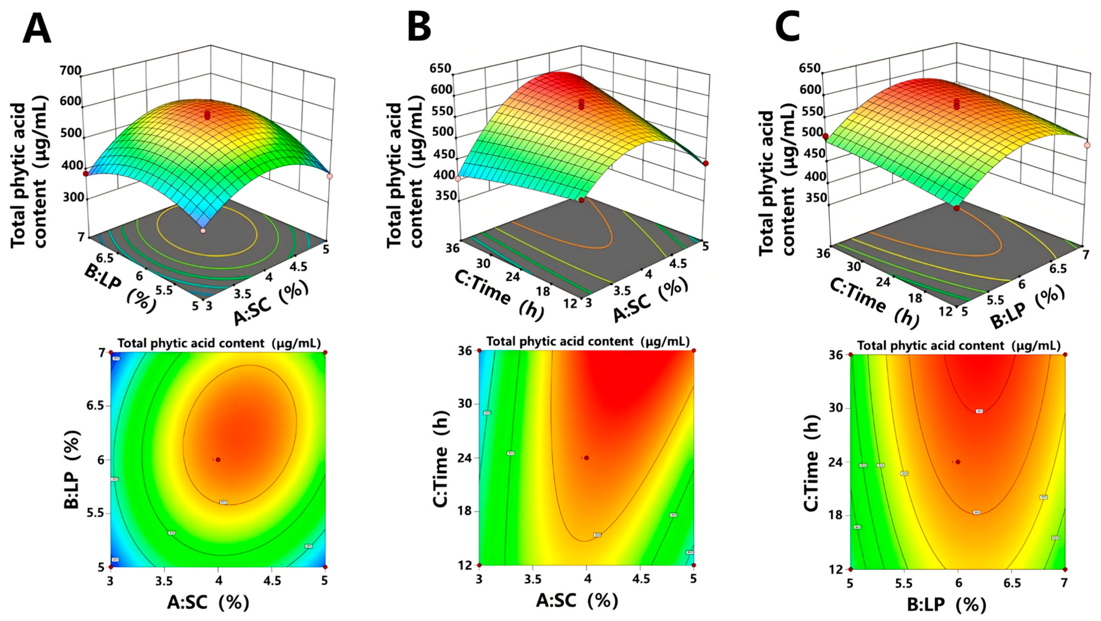
The interactive effects among the yeast inoculation volume (A), lactic acid bacterium inoculation volume (B), and fermentation time (C) were analyzed using Design Expert software, and the corresponding three-dimensional response surface and contour plots are presented in Figure 1. The results revealed that the interactions between A and B (AB) and between A and C (AC) exerted significant effects on the PA content (p < 0.05). In contrast, the BC interaction was not statistically significant (p = 0.8027), indicating negligible synergistic or antagonistic effects between the inoculation volume of LP and fermentation time under the tested conditions.
Based on model optimization, the process conditions predicted to maximize the PA yield were determined as follows: A = 4.51%, B = 6.14%, and C = 18.19 h, with a corresponding predicted PA content of 542.27 μg/mL. Taking practical operational feasibility into account, the optimized parameters were slightly adjusted to A = 4.5%, B = 6.0%, and C = 18 h. Validation experiments conducted under these adjusted conditions yielded a PA content of 576.61 ± 15.21 μg/mL in the fermented sea rice broth, which differed from the model predicted value by 6.3%, indicating excellent agreement between the experimental and predicted results. Collectively, these findings confirm the adequacy and reliability of the established regression model and demonstrate that the optimized co-fermentation conditions effectively promote the enrichment of PA in the sea rice fermentation system. The resulting SRF exhibited a pH of 3.82 ± 0.15, with total phenolic content of 2.47 ± 0.21 mg GAE/mL and total flavonoid content of 1.03 ± 0.09 mg RE/mL. Lactic acid (12.36 ± 1.02 mg/mL) and acetic acid (3.15 ± 0.28 mg/mL) were identified as the major organic acids.

3.3. In Vitro Antioxidant Activity of SRF

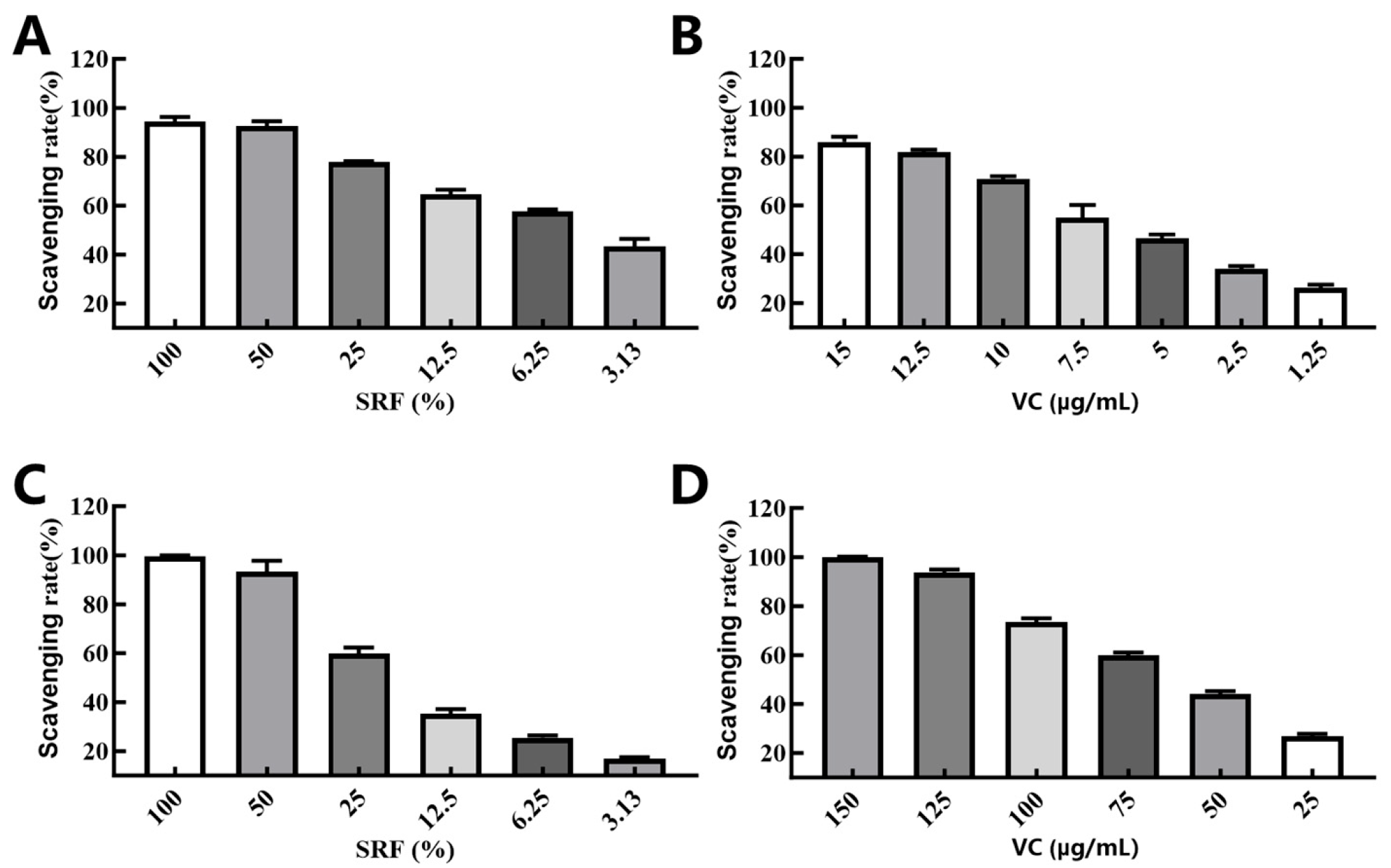
The DPPH and ABTS radical scavenging activities of SRF at various concentrations, alongside those of the positive control Vc, are depicted in Figure 2. DPPH radicals, upon accepting electrons or hydrogen donors, are reduced to stable molecules, accompanied by a decrease in absorbance at 517 nm. Similarly, ABTS is a blue green cationic radical that exhibits characteristic absorbance at 734 nm. For both assays, higher scavenging rates reflect the stronger antioxidant capacity of the test sample.
As shown in Figure 2A, the DPPH radical scavenging activity of SRF displayed a pronounced concentration-dependent pattern. When the SRF concentration increased from 3.13% to 100%, the scavenging rate rose from 43.40% to 94.42%. Notably, at a concentration of 50%, SRF achieved a scavenging rate of 92.56%, which surpassed that of 15 μg/mL Vc (90.89%). The positive control Vc also exhibited a concentration dependent scavenging effect within the range of 1.25–15 μg/mL, with rates increasing from 12.21% to 90.89% (Figure 2B).
Likewise, the ABTS radical scavenging capacity of SRF exhibited a similar upward trend with increasing concentration (Figure 2C). Over the concentration range of 3.13% to 100%, the ABTS scavenging rate of SRF increased from 16.93% to 99.55%, with a corresponding IC50 value of 15.71% (v/v). At 50% concentration, SRF showed a scavenging rate of 93.18%, comparable to that of 150 μg/mL Vc (93.63%). The scavenging rate of Vc increased from 26.80% to 99.96% across the concentration gradient of 25–150 μg/mL (Figure 2D).
Collectively, these results demonstrate that SRF possesses potent concentration dependent radical scavenging activity, with an antioxidant efficacy comparable to that of Vc. This suggests that SRF may contain additional antioxidant constituents that act in synergy with PA to contribute to its overall antioxidant function.

3.4. Effect of SRF on the Proliferation of B16 Melanoma Cells

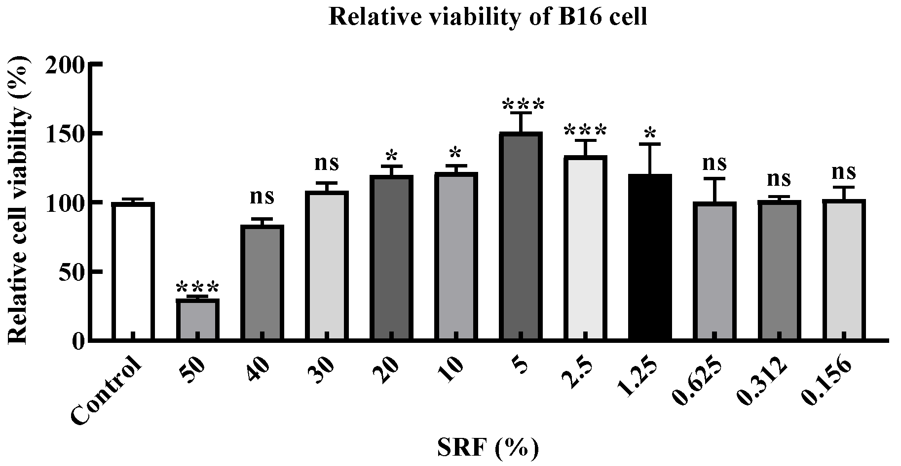
The murine B16 melanoma cell line, a widely utilized model for melanogenesis research, tumor biology, and drug screening, was employed in this study to evaluate the effects of SRF on cell proliferation across a range of concentrations. As illustrated in Figure 3, SRF at concentrations of 1.25–20.0% significantly promoted B16 cell proliferation (p < 0.05). This stimulatory effect is likely attributable to the presence of bioactive components—including PA, polyphenols, and flavonoids—released from sea rice during the fermentation process. However, when the SRF concentration exceeded 40%, the cell viability decreased significantly, ranging from 16% to 69% relative to the control group, suggesting that high concentrations of SRF exerted cytotoxic effects on B16 cells. Based on these findings, three concentrations (1.0%, 0.5%, and 0.2%) that exhibited no obvious cytotoxicity were selected for subsequent investigations into melanin synthesis inhibition and the underlying molecular mechanisms, thereby ensuring that the observed anti-melanogenic effects were not confounded by reduced cell viability.

3.5. Effect of SRF on Melanin Synthesis in B16 Cells

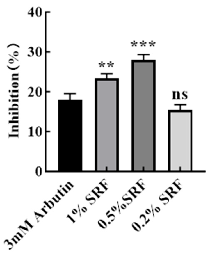
Arbutin is a well-established whitening agent that effectively suppresses melanin synthesis and reduces skin pigmentation [19]. Among its isoforms, α arbutin exhibits superior depigmenting efficacy and enhanced structural stability relative to β arbutin [20]. To evaluate the inhibitory effect of SRF on melanogenesis, three concentrations—high (1.0%), medium (0.5%), and low (0.2%)—were selected, and the results were compared with those of the positive control, α arbutin. As presented in Figure 4, treatment with 3 mM α arbutin significantly inhibited melanin synthesis in B16 cells, which is in agreement with previous findings [21]. Notably, both the high and medium concentrations of SRF exhibited stronger inhibitory effects than did 3 mM α arbutin, with the 0.5% SRF group demonstrating the most pronounced suppression (p < 0.001). In contrast, the low concentration SRF (0.2%) showed a weaker inhibitory effect than α arbutin and did not differ significantly from the control group (p > 0.05).

3.6. Inhibitory Effect of SRF on the PI3K-AKT-GSK3β-MITF Pathway in B16 Cells

The PI3K/AKT signaling pathway is a pivotal regulator of cell survival, proliferation, and metabolism [22]. MITF, a key downstream transcription factor, not only participates in the fundamental physiological processes governed by this pathway but also serves as a master regulator of melanogenesis [23]. To elucidate the molecular mechanism underlying the anti-melanogenic activity of SRF, RT-qPCR and Western blotting were employed to examine the expression of genes and proteins involved in this signaling cascade.
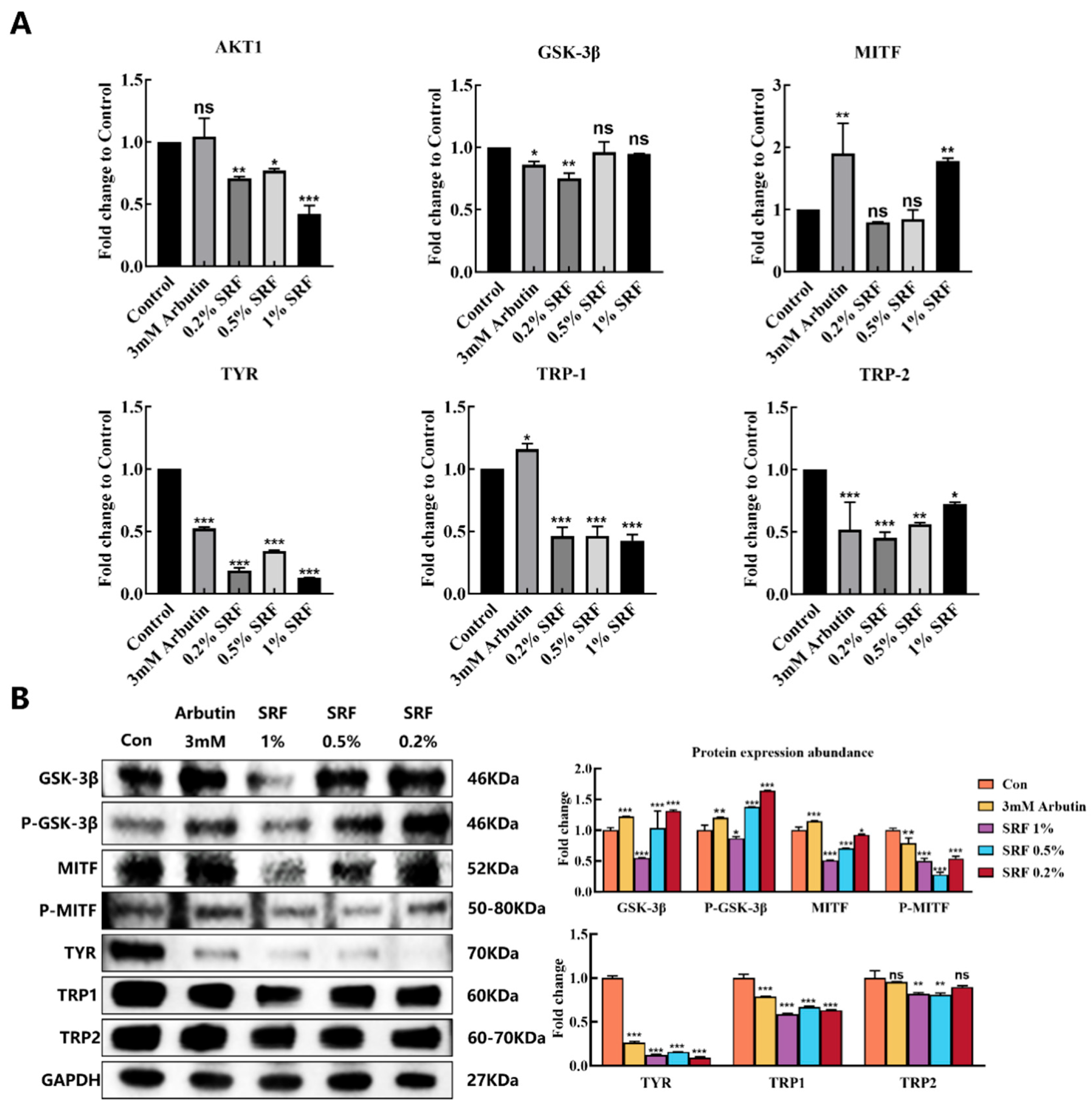
As shown in Figure 5A, treatment with low (0.2%), medium (0.5%), and high (1.0%) concentrations of SRF significantly downregulated the mRNA expression of melanogenesis-related genes, including AKT1, TYR, TRP-1, and TRP-2, in B16 cells compared with the control group. Notably, the transcript levels of TYR and TRP-1 were decreased to 12.76–34.39% and 42.51–46.59% of the control values, respectively (p < 0.001), suggesting that SRF may exert its melanin suppressing effect primarily through the transcriptional repression of these two key enzymes.
Furthermore, the phosphorylation status of key proteins in the PI3K/AKT pathway, namely GSK3β and MITF, was analyzed by Western blotting. As illustrated in Figure 5B, both α arbutin and SRF treatment enhanced the phosphorylation of GSK3β. Specifically, the low and medium concentration SRF groups exhibited particularly pronounced increases in p-GSK3β levels, reaching 117.40–131.50% of the control (p < 0.001). Concurrently, SRF treatment led to a marked reduction in MITF phosphorylation, with p-MITF levels decreasing to 27.79–54.12% of the control, an effect significantly more potent than that of 3 mM α arbutin (p < 0.001). Additionally, in line with the RT-qPCR findings, the protein expression levels of TYR and TRP 1 were also significantly diminished in the SRF treated groups (p < 0.001).

4. Discussion

In this study, a co-fermentation strategy employing yeast and lactic acid bacteria was developed to enrich PA from sea rice. Notably, the PA content achieved via this biological approach was lower than that reported in previous studies using physicochemical extraction methods. This discrepancy can be attributed to two main factors. First, microbial phytase secreted during fermentation partially hydrolyzes PA into lower order inositol phosphates, which exhibit enhanced bioavailability and potential physiological activities. Second, unlike physicochemical procedures that employ strong acids, high temperatures, or vigorous mechanical disruption, the bidirectional fermentation process does not completely liberate tightly bound PA from the cereal matrix. Consequently, although the PA yield was lower, the fermentation derived product is enriched in bioavailable inositol phosphates with superior functional properties.
It should be noted that SRF is a complex fermentation mixture containing multiple bioactive constituents beyond PA, including polyphenols, flavonoids, organic acids, and peptides. The relative contribution of each component to the observed antioxidant and anti-melanogenic activities remains to be systematically delineated. While PA served as the optimization target due to its high content in sea rice and well-documented antioxidant properties, the biological effects of SRF reported herein should be interpreted as the net outcome of its multi-component composition. Therefore, we do not attribute the observed activities exclusively to PA, and future studies are warranted to identify the key active principles through bioassay-guided fractionation.
Currently, industrial preparation of PA from plant sources relies predominantly on acidic chemical extraction (0.5–2.4 mol/L) [24]. Compared with these conventional methods, bidirectional fermentation offers multiple advantages, including the enrichment of bioactive constituents, improved bioavailability, degradation of toxic components, generation of novel compounds, enhanced pharmacological activities, and environmental sustainability [25]. Through RSM optimization, the PA yield from sea rice was substantially improved, underscoring the effectiveness of the synergistic yeast–lactic acid bacterium system in promoting PA release—a process likely associated with microbial metabolic pathways and enzymatic mechanisms [26]. Although previous studies have demonstrated the efficacy of microbial fermentation in extracting bioactive components from plant materials [27,28], to our knowledge, no investigation has specifically addressed the microbial extraction of PA from plant sources. Therefore, the present study establishes the key co-fermentation parameters for sea rice, providing a novel research direction for the valorization of sea rice-derived active ingredients.
Previous studies have demonstrated that fermentation products derived from lactic acid bacteria and yeast possess potent antioxidant activities, contributing to skin barrier repair and retarding skin aging [29,30]. In the present study, SRF exhibited strong antioxidant capacity, which may be attributed to its high PA content, as this class of compounds has been reported to possess significant antioxidant properties [31,32]; however, other bioactive constituents such as polyphenols and flavonoids may also contribute synergistically to the overall antioxidant activity of SRF. We further hypothesized that SRF might inhibit melanin synthesis and exert skin whitening effects by reducing ultraviolet induced reactive oxygen species generation within melanocytes [33]. To test this hypothesis, RT-qPCR and Western blotting were employed to investigate the effects of SRF on the PI3K/AKT signaling pathway and melanogenesis in B16 cells. In our preliminary comparative experiments, unfermented sea rice extract exhibited substantially lower radical scavenging activities and melanin synthesis inhibitory effects than SRF at equivalent concentrations, suggesting that the co-fermentation process significantly enhances the bioactivity of sea rice-derived components, likely through the release of bound bioactive molecules and the generation of new metabolites during microbial fermentation.
Activation of AKT has been shown to phosphorylate and inactivate GSK3β, leading to β-catenin stabilization and the consequent upregulation of MITF transcriptional activity, thereby promoting melanin synthesis [34,35]. Our results suggested that SRF significantly inhibited AKT1 gene expression. Additionally, immunoblotting revealed that SRF increased GSK3β phosphorylation, subsequently decreased MITF phosphorylation, and suppressed melanin production, which aligns with previous reports [22]. Furthermore, the mRNA and protein levels of TYR, TRP-1, and TRP-2 were all significantly reduced (p < 0.05). It is well established that upregulated MITF gene expression activates the transcription of TYR, TRP-1, and TRP-2, thereby stimulating melanin synthesis [36]. Notably, our RT-qPCR results indicated that both α arbutin and the high concentration of SRF (1%) upregulated MITF mRNA expression, which would be expected to activate downstream melanogenic genes. However, the protein level data showed that SRF reduced MITF phosphorylation and downregulated TYR/TRP-1/TRP-2 expression, suggesting a post-translational regulatory mechanism. Several possible explanations may account for this apparent discrepancy. First, MITF activity is governed not only by its transcriptional level but also by multiple phosphorylation events that modulate its transactivation capacity, protein stability, and nuclear translocation. It has been reported that MAPK-mediated phosphorylation of MITF at Ser73 (MITF-M; corresponding to Ser180 in MITF-A) promotes its ubiquitination and subsequent proteasomal degradation [37]. Thus, the enhanced GSK3β phosphorylation (Ser9, inhibitory) induced by SRF may represent one of the upstream events contributing to the observed reduction in MITF phosphorylation and downstream melanogenic enzyme expression. Second, the regulation of MITF occurs at multiple levels—transcriptional, post-transcriptional, and post-translational—and the net functional outcome depends on the integration of these regulatory inputs. The upregulation of MITF mRNA could represent a compensatory feedback response to suppressed MITF protein activity. Third, the observed effects may be concentration-dependent: at higher concentrations, SRF may predominantly act through the post-translational modulation of MITF, whereas at the medium concentration (0.5%), SRF may inhibit melanin synthesis primarily through the direct suppression of tyrosinase activity, independent of MITF transcriptional regulation. The low concentration (0.2%) did not significantly inhibit melanin synthesis relative to the control (p > 0.05). Collectively, these correlative findings suggest that SRF may modulate melanin synthesis through multiple mechanisms depending on the concentration; however, the precise molecular interactions and causal relationships warrant further investigation using gain- and loss-of-function approaches.
Several limitations of this study should be acknowledged. First, SRF is a complex mixture, and the specific bioactive constituents responsible for the observed anti-melanogenic effects remain to be identified. Second, AKT pathway activation was assessed only at the mRNA level for AKT1; protein-level phosphorylation analysis of AKT was not performed in the current study. Although our data suggest modulation of the PI3K/AKT/GSK3β/MITF pathway, the precise molecular interactions and potential off-target effects of SRF components warrant further investigation using protein-based assays and gain- and loss-of-function approaches. Third, the in vivo efficacy and safety of SRF have not been evaluated.
In summary, SRF, as a fermentation filtrate with a combination of potent antioxidant activity and direct inhibition of melanin synthesis, holds promise as a novel whitening ingredient for cosmetic applications. Nevertheless, it is important to recognize that sea rice, being a plant material rich in starch and dietary fiber, contains substantial polysaccharide components that are not fully utilized during fermentation. As a result, appreciable amounts of starch granules and dietary fibers remain in the fermentation filtrate, which may adversely affect the stability and preservability of SRF. Accordingly, future investigations should prioritize improving the storage stability of SRF to facilitate its practical application in the cosmetic industry.

5. Conclusions

In this study, a bidirectional co-fermentation system integrating yeast and lactic acid bacteria was successfully established for the efficient production of PA from sea rice. Through response surface methodology optimization, the optimal fermentation conditions were determined as follows: SC inoculum volume of 4.5%, LP inoculum volume of 6.0%, and fermentation time of 18 h. Under these conditions, the PA content in the fermentation broth reached 576.61 μg/mL. The resulting sea rice fermentation filtrate (SRF) exhibited potent antioxidant activity and significantly promoted the proliferation of B16 cells. Mechanistically, our results suggest that SRF may enhance GSK3β phosphorylation, which in turn may suppress MITF phosphorylation and downregulate its downstream key proteins, including TYR, TRP-1, and TRP-2, ultimately contributing to the inhibition of melanin synthesis and whitening efficacy. However, given the correlative nature of the current data and the compositional complexity of SRF, the specific active constituents and the detailed regulatory network warrant further systematic investigation. Future studies employing bioassay-guided fractionation to identify active compounds, as well as gain- and loss-of-function approaches to validate the mechanistic pathway, are warranted. Collectively, these findings provide a scientific foundation for the high-value utilization of sea rice and establish a theoretical basis for the development of SRF as a natural whitening ingredient in cosmetic formulations.

Author Contributions

Conceptualization, Q.W.; methodology, J.Z.; software, H.Z. and Y.L.; validation, H.Z. and Y.L.; formal analysis, J.Z. and L.Y.; investigation, C.G.; data curation, L.Y.; writing—original draft preparation, Q.W.; writing—review and editing, L.Y.; visualization, J.Z. and Q.W.; supervision, L.Y.; project administration, L.Y.; funding acquisition, L.Y. All authors have read and agreed to the published version of the manuscript.

Funding

This research was funded by the Key-Area Research and Development Program of Guangdong Province, grant number 21202107201900003, 21202107201900005.

Institutional Review Board Statement

Not applicable.

Informed Consent Statement

Not applicable.

Data Availability Statement

The original contributions presented in this study are included in the article. Further inquiries can be directed to the corresponding author.

Conflicts of Interest

Author Qiting Wu, Huirong Zhu, Chaowan Guo and Lin Ye were employed by the company Guangdong Marubi Biotechnology; Jiarui Zhao was employed by the company BIOST Pharmaceuticals (Guangzhou). The remaining authors declare that the research was conducted in the absence of any commercial or financial relationships that could be construed as a potential conflict of interest.

Abbreviations

The following abbreviations are used in this manuscript:
SRFSea rice fermentation filtrate
PI3KPhosphatidylinositol 3 kinase
AKTProtein kinase B
GSK3βGlycogen synthase kinase 3β
MITFMicrophthalmia associated transcription factor
PAPhytic acid
RSMResponse surface methodology
TYRTyrosinase
TRP-1Tyrosinase-related protein 1
TRP-2Tyrosinase-related protein 2
SCSaccharomyces cerevisiae
LPLactobacillus plantarum
RT-qPCRReal time quantitative PCR

References

  1. Lai, S.D.; Long, Y.; Chen, Y.J.; Li, R.; Zhong, S.Y.; Zhou, H.K. Research progress on nutritional functional components and processing utilization of seawater rice. Cereal Oils 2022, 35, 13–15. [Google Scholar] [CrossRef]
  2. Ye, S.Q.; Huang, J.M.; Wu, W.L.; Chen, J.P.; Zhong, S.Y.; Qiu, G.; Zhang, W.; Chen, R.; Liu, Y.J. Optimized extraction technology of glutathione from ‘Haidao 86’ germ rice by response surface methodology. Food Sci. Nutr. 2023, 11, 7255–7263. [Google Scholar] [CrossRef] [Scilit] [PubMed]
  3. Tester, M.; Davenport, R. Na+ tolerance and Na+ transport in higher plants. Ann. Bot. 2003, 91, 503–527. [Google Scholar] [CrossRef] [Scilit] [PubMed]
  4. Liang, W.J.; Ma, X.L.; Wan, P.; Liu, L.Y. Plant salt-tolerance mechanism: A review. Biochem. Biophys. Res. Commun. 2018, 495, 286–291. [Google Scholar] [CrossRef] [Scilit] [PubMed]
  5. Pei, J.; Pan, X.; Wei, G.; Hua, Y. Research progress of glutathione peroxidase family (GPX) in redoxidation. Front. Pharmacol. 2023, 14, 1147414. [Google Scholar] [CrossRef] [Scilit] [PubMed]
  6. Yu, Y.; Wang, D.Q.; Pan, J.C.; Liu, Y. Effects of Germinated Brown Rice on Patients with Metabolic Syndrome. Food Nutr. China 2019, 25, 73–76. [Google Scholar] [CrossRef]
  7. Hou, X.D.; Hu, Q.; Ma, L.J.; Yu, H.N.; Ge, G.B.; Hou, J. Recent advances on pancreatic lipase inhibitors from Chinese herbs. Acta Pharm. Sin. 2020, 55, 1478–1493. [Google Scholar] [CrossRef]
  8. Wang, X.M.; Zhao, X.X.; Chen, J.Y.; Zhou, B.L.; Yang, H.; He, X.L.; Wang, P.P.; Mo, S.; Zhou, H.K. The Precise Development of Saline-Alkali Soil and the Prospect of High-Efficiency Cultivation of Sea Rice Resistant to Saline-Alkali in Zhanjiang. Chin. J. Trop. Agric. 2018, 38, 25–29. [Google Scholar] [CrossRef]
  9. Silva, V.M.; Putti, F.F.; White, P.J.; Reis, A.R.D. Phytic acid accumulation in plants: Biosynthesis pathway regulation and role in human diet. Plant Physiol. Biochem. 2021, 164, 132–146. [Google Scholar] [CrossRef] [Scilit] [PubMed]
  10. Ozkaya, B.; Baumgartner, B.; Ozkaya, H. Effects of concentrated and dephytinized wheat bran and rice bran addition on bread properties. J. Texture Stud. 2018, 49, 84–93. [Google Scholar] [CrossRef] [Scilit] [PubMed]
  11. Alexandre, H.; Costello, P.J.; Remize, F.; Guzzo, J.; Guilloux-Benatier, M. Saccharomyces cerevisiaeOenococcus oeni interactions in wine: Current knowledge and perspectives. Int. J. Food Microbiol. 2004, 93, 141–154. [Google Scholar] [CrossRef] [Scilit] [PubMed]
  12. Di, Z.; Huo, Y.; Wang, G.; Zhuang, Y. Progress in the biosynthesis of cosmetic ingredients through engineering of Saccharomyces cerevisiae. ACS Synth. Biol. 2025, 14, 2955–2971. [Google Scholar] [CrossRef] [Scilit] [PubMed]
  13. Silva, S.; Bautista-Hérnandez, I.; Gomez-García, R.; Costa, E.M.; Machado, M. Precision fermentation as a tool for sustainable cosmetic ingredient production. Appl. Sci. 2025, 15, 9246. [Google Scholar] [CrossRef] [Scilit]
  14. Pérez-Rivero, C.; López-Gómez, J.P. Unlocking the potential of fermentation in cosmetics: A review. Fermentation 2023, 9, 463. [Google Scholar] [CrossRef] [Scilit]
  15. Xu, B.Y.; Yang, X.S.; Zhao, J.; Yu, B.H.; Li, J.X.; Xia, Z. Effects of mixed fermentation on the flavor quality and in vitro antioxidant activity of Zaosu pear-Merlot grape composite alcoholic beverage. Food Chem. X 2024, 25, 102128. [Google Scholar] [CrossRef] [Scilit] [PubMed]
  16. Lee, K.J.; Oh, Y.C.; Cho, W.K.; Ma, J.Y. Antioxidant and Anti-Inflammatory Activity Determination of One Hundred Kinds of Pure Chemical Compounds Using Offline and Online Screening HPLC Assay. Evid.-Based Complement. Altern. Med. 2015, 2015, 165457. [Google Scholar] [CrossRef] [Scilit] [PubMed]
  17. Ye, L.; Chen, H.D.; Wang, J.; Tsim, K.W.K.; Wang, Y.R.; Shen, X.; Lei, H.T.; Liu, Y.L. Aflatoxin B1-induced liver pyroptosis is mediated by disturbing the gut microbial metabolites: The roles of pipecolic acid and norepinephrine. J. Hazard. Mater. 2024, 474, 134822. [Google Scholar] [CrossRef] [Scilit] [PubMed]
  18. Ye, L.; Chen, H.D.; Tsim, K.W.K.; Shen, X.; Li, X.M.; Li, X.L.; Lei, H.T.; Liu, Y.L. Aflatoxin B1 Induces Inflammatory Liver Injury via Gut Microbiota in Mice. J. Agric. Food Chem. 2023, 71, 10787–10797. [Google Scholar] [CrossRef] [Scilit] [PubMed]
  19. Tokiwa, Y.; Kitagawa, M.; Raku, T.; Yanagitani, S.; Yoshino, K. Enzymatic synthesis of arbutin undecylenic acid ester and its inhibitory effect on melanin synthesis. Bioorg. Med. Chem. Lett. 2007, 17, 3105–3108. [Google Scholar] [CrossRef] [Scilit] [PubMed]
  20. Yan, Q.Y.; Han, X.P. Determination and method confirmation of α-arbutin and other 4 raw materials in cosmetics. China Stand. 2024, 23, 215–220. [Google Scholar] [CrossRef]
  21. Peng, L.L.; Liu, C.S.; Cheng, X.; Chen, Y.; Yang, J.J.; Li, Y.L. Effects of the main active ingredients from Baihe (Bulbus lilii) on melanin content and tyrosinase activity in mice B-16 cells. J. Hunan Univ. Chin. Med. 2023, 43, 605–611. [Google Scholar] [CrossRef]
  22. Niu, C.; Yin, L.; Aisa, H.A. Novel Furocoumarin Derivatives Stimulate Melanogenesis in B16 Melanoma Cells by Up-Regulation of MITF and TYR Family via Akt/GSK3β/β-Catenin Signaling Pathways. Int. J. Mol. Sci. 2018, 19, 746. [Google Scholar] [CrossRef] [Scilit] [PubMed]
  23. Vachtenheim, J.; Borovansky, J. “Transcription physiology” of pigment formation in melanocytes: Central role of MITF. Exp. Dermatol. 2010, 19, 617–627. [Google Scholar] [CrossRef] [Scilit] [PubMed]
  24. Bloot, A.P.M.; Kalschne, D.L.; Amaral, J.A.S.; Baraldi, I.J.; Canan, C. A Review of Phytic Acid Sources, Obtention, and Applications. Food Rev. Int. 2021, 39, 73–92. [Google Scholar] [CrossRef] [Scilit]
  25. Wu, Q.T.; Nie, Y.F.; Guo, C.W.; Pei, Y.L.; Zhang, Y.; Guo, Y.Y.; Zhao, J.R.; Ye, L. Application of microbial fermentation technology in the development of cosmetic raw materials. Deterg. Cosmet. 2025, 48, 74–79. [Google Scholar] [CrossRef]
  26. Zhuang, Y.; Pan, Y.; Xie, X.M.; Zhang, L.Y. The Origin, Development and Its Advantage and Potential of “the Bi-directional Solid Fermentation” for Medicinal Fungi. Edible Fungi China 2007, 26, 3–6. [Google Scholar] [CrossRef]
  27. Foti, P.; Caggia, C.; Romeo, F.V. New Insight into Microbial Exploitation to Produce Bioactive Molecules from Agrifood and By-Products’ Fermentation. Foods 2025, 14, 1439. [Google Scholar] [CrossRef] [Scilit] [PubMed]
  28. Nguyen Thai Huynh, N.T.; Van Camp, J.; Smagghe, G.; Raes, K. Improved Release and Metabolism of Flavonoids by Steered Fermentation Processes: A Review. Int. J. Mol. Sci. 2014, 15, 19369–19388. [Google Scholar] [CrossRef] [Scilit] [PubMed]
  29. Komatsu, Y.; Matsunaga, K. Cosmetically Applicable Soluble Agonists for Toll-like Receptor 2 Produced by Fermentation of Asparagus Extract Supplemented with Skimmed Milk Using Lactobacillus delbrueckii subsp. lactis TL24 Consist of Molecules Larger than 100 kDa and Can Be Stabilized by Lyophilization with Dextrin. Molecules 2024, 29, 4557. [Google Scholar] [CrossRef] [Scilit] [PubMed]
  30. Liu, P.P.; Xu, D.N.; Zhao, D.; An, Q.; Wang, C.T.; Li, M. Study on the skin care efficacy and safety of the grape-seed fermentation liquid. China Surfactant Deterg. Cosmet. 2021, 51, 200–207. [Google Scholar] [CrossRef]
  31. Graf, E.; Eaton, J.W. Antioxidant functions of phytic acid. Free Radic. Biol. Med. 1990, 8, 61–69. [Google Scholar] [CrossRef] [Scilit] [PubMed]
  32. Graf, E.; Mahoney, J.R.; Bryant, R.G.; Eaton, J.W. Iron-catalyzed hydroxyl radical formation. Stringent requirement for free iron coordination site. J. Biol. Chem. 1984, 259, 3620–3624. [Google Scholar] [CrossRef] [Scilit]
  33. Feng, D.; Fang, Z.; Zhang, P. The melanin inhibitory effect of plants and phytochemicals: A systematic review. Phytomedicine 2022, 107, 154449. [Google Scholar] [CrossRef] [Scilit] [PubMed]
  34. Choi, H.; Yoon, J.; Youn, K.; Jun, M. Decursin prevents melanogenesis by suppressing MITF expression through the regulation of PKA/CREB, MAPKs, and PI3K/Akt/GSK-3β cascades. Biomed. Pharmacother. 2022, 147, 112651. [Google Scholar] [CrossRef] [Scilit] [PubMed]
  35. Li, C.Y.; Wang, Q.; Shen, S.S.; Wei, X.L.; Li, G.X. Oridonin inhibits migration, invasion, adhesion and TGF-β1-induced epithelial-mesenchymal transition of melanoma cells by inhibiting the activity of PI3K/Akt/GSK-3β signaling pathway. Oncol. Lett. 2017, 15, 1362–1372. [Google Scholar] [CrossRef] [Scilit] [PubMed]
  36. Moriya, M. Placental Extract Inhibits Melanogenesis by Inducing the Proteasome-Dependent Degradation of Tyrosinase and TRP-1. Biol. Pharm. Bull. 2025, 48, 1472–1484. [Google Scholar] [CrossRef] [Scilit] [PubMed]
  37. Wu, M.; Hemesath, T.J.; Takemoto, C.M.; Horstmann, M.A.; Wells, A.G.; Price, E.R.; Fisher, D.Z.; Fisher, D.E. c-Kit triggers dual phosphorylations, which couple activation and degradation of the essential melanocyte factor Mi. Genes Dev. 2000, 14, 301–312. [Google Scholar] [CrossRef] [Scilit]
Figure 1. Response surface and contour plots illustrating the interactive effects of process variables on PA content. (A) Inoculation amount of SC. (B) Inoculation amount of LP. (C) Fermentation time.
Fermentation 12 00430 g001
Figure 2. In vitro antioxidant activity of SRF. (A) DPPH radical scavenging activity of SRF at various concentrations. (B) DPPH radical scavenging activity of Vc at various concentrations. (C) ABTS radical scavenging activity of SRF at various concentrations. (D) ABTS radical scavenging activity of Vc at various concentrations. Values are presented as the mean ± SD (n = 3).
Fermentation 12 00430 g002
Figure 3. Effect of varying concentrations of SRF on the viability of B16 cells. Values are presented as mean ± SD. ns denotes no significant difference compared with the control group (p > 0.05); * and *** indicate statistically significant differences from the control group at p < 0.05 and p < 0.001, respectively.
Fermentation 12 00430 g003
Figure 4. Inhibitory effect of varying concentrations of SRF on melanin synthesis in B16 cells. Values are presented as mean ± SD. ns denotes no significant difference compared with the control group (p > 0.05); ** and *** denote statistically significant differences from the control group at p < 0.01 and p < 0.001, respectively.
Fermentation 12 00430 g004
Figure 5. SRF significantly inhibits melanin synthesis in B16 cells. (A) Relative mRNA expression levels of AKT1, GSK3β, MITF, TYR, TRP-1, and TRP-2 in B16 cells as determined by RT-qPCR. (B) Relative protein abundance of GSK3β, p-GSK3β, MITF, p-MITF, TYR, and TRP-1, as determined by Western blotting. Values are presented as mean ± SD. ns denotes no significant difference compared with the control group (p > 0.05); *, **, and *** denote statistically significant differences from the control group at p < 0.05, p < 0.01, and p < 0.001, respectively.
Fermentation 12 00430 g005
Table 1. Factors and levels employed in the Box–Behnken experimental design. A, inoculum volume of SC seed culture; B, inoculum volume of LP seed culture; C, fermentation time.
FactorsLevels
A: Inoculum volume of SC seed culture (%)345
B: Inoculum volume of LP seed culture (%)567
C: Fermentation time (h)122436
Table 2. Reaction system for the DPPH radical scavenging assay (μL).
Well GroupAbsolute EthanolSampleDPPHTotal Volume
Reagent blank (A0)100-100200
Sample (A1)-100100200
Sample blank (A2)100100-200
Table 3. Reaction system for the ABTS radical scavenging assay (μL).
Well GroupPhosphate-Buffered SalineSampleABTSTotal Volume
Reagent blank (A0)10-190200
Sample (A1)-10190200
Sample blank (A2)19010-200
Table 4. Analysis of variance (ANOVA) of the response surface model for PA content.
SourceSum of SquaresdfMean SquareF-Valuep-Value
Model83,401.7699266.8636.21<0.0001
A8024.1418024.1431.350.0008
B4241.7614241.7616.570.0047
C3406.8813406.8813.310.0082
AB2842.8312842.8311.110.0125
AC8615.2118615.2133.660.0007
BC17.24117.240.06730.8027
A236,645.38136,645.38143.17<0.0001
B216,021.38116,021.3862.6<0.0001
C2143.071143.070.5590.479
Residual1791.677255.95--
Lack of Fit656.953218.980.77190.567
Pure Error1134.714283.68--
Cor Total85,193.4316---
A, inoculum volume of SC seed culture (%); B, inoculum volume of LP seed culture (%); C, fermentation time (h).
Disclaimer/Publisher’s Note: The statements, opinions and data contained in all publications are solely those of the individual author(s) and contributor(s) and not of MDPI and/or the editor(s). MDPI and/or the editor(s) disclaim responsibility for any injury to people or property resulting from any ideas, methods, instructions or products referred to in the content.