


Non-Coding RNA, Volume 11, Issue 3 (June 2025) – 19 articles
Cover Story (view full-size image):
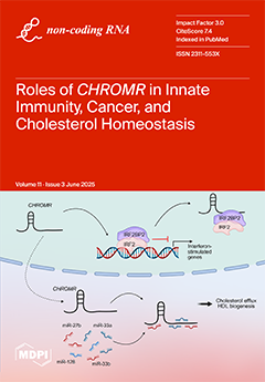
CHROMR is a primate-specific long noncoding RNA with emerging roles in homeostasis and pathophysiology. Like many lncRNAs, CHROMR accumulates in both the nucleus and the cytoplasm, and it assumes distinct functions in each of these cellular compartments. In the nucleus, CHROMR sequesters a transcriptional repressor complex to activate interferon-stimulated gene expression and antiviral immunity. In the cytoplasm, CHROMR competitively inhibits microRNAs involved in cholesterol efflux and cell cycle regulation, thereby impacting gene pathways involved in reverse cholesterol transport, HDL biogenesis, and tumor growth. In this review, we detail the multifaceted functions of CHROMR in cholesterol metabolism, innate immunity, and cancer progression. View this paper
- Issues are regarded as officially published after their release is announced to the table of contents alert mailing list.
- You may sign up for e-mail alerts to receive table of contents of newly released issues.
- PDF is the official format for papers published in both, html and pdf forms. To view the papers in pdf format, click on the "PDF Full-text" link, and use the free Adobe Reader to open them.
Previous Issue
Next Issue