Review

The Multifaceted Roles of CHROMR in Innate Immunity, Cancer, and Cholesterol Homeostasis

by
Emma R. Blaustein
and
Coen van Solingen
*
Cardiovascular Research Center, Department of Medicine (Cardiology), New York University Langone Health, New York, NY 10016, USA
*
Author to whom correspondence should be addressed.
Non-Coding RNA 2025, 11(3), 44; https://doi.org/10.3390/ncrna11030044
Submission received: 17 April 2025 / Revised: 15 May 2025 / Accepted: 4 June 2025 / Published: 10 June 2025
(This article belongs to the Section Long Non-Coding RNA)

Abstract

:
CHROMR is a primate-specific long noncoding RNA with emerging roles in homeostasis and pathophysiology. Elevated blood levels of CHROMR have been observed in patients with cardiovascular disease and several cancers, where it is correlated with poor clinical outcomes. Like many lncRNAs, CHROMR accumulates in both the nucleus and the cytoplasm, and it assumes distinct functions in each of these cellular compartments. In the nucleus, CHROMR sequesters a transcriptional repressor complex to activate interferon-stimulated gene expression and antiviral immunity. In the cytoplasm, CHROMR competitively inhibits microRNAs involved in cholesterol efflux and cell cycle regulation, thereby impacting gene pathways involved in reverse cholesterol transport, HDL biogenesis, and tumor growth. In this review, we detail the multifaceted functions of CHROMR in cholesterol metabolism, innate immunity, and cancer progression. We also explore the potential molecular mechanisms that govern its expression and dynamic subcellular localization, which may be key to its functional versatility. Advancing our understanding of the regulatory networks and cellular environments that shape CHROMR activity will be critical for assessing its promise as a therapeutic target and diagnostic biomarker.

1. Introduction

The discovery of noncoding RNAs (ncRNAs) has reshaped our understanding of how genetic elements influence cellular function. Shedding light on this “dark matter” of the genome has revealed novel RNA classes with critical roles in regulating protein-coding genes and genome architecture. Among the various types of ncRNAs identified over the past decades, long noncoding RNAs (lncRNAs) have greatly expanded our understanding of RNA’s pivotal role in gene regulation [1]. This class of heterogenous transcripts, defined as being >200 nucleotides in length, exhibit poor conservation among species—a factor that has slowed the understanding of their functions [2,3]. LncRNAs can localize to the nucleus or cytoplasm and can take on distinct roles in these subcellular domains [4,5]. In the nucleus, lncRNAs have diverse functions, including forming ribonucleoprotein complexes that can act as guides or decoys, shapers of nuclear organization and higher-order chromosomal architecture [6,7], and scaffolding of effector proteins to attenuate or enhance gene regulatory activities [8]. LncRNAs can function in cis, close to their site of transcription to regulate expression of neighboring genes, or in trans, at distal sites in the genome [9,10]. In the cytoplasm, lncRNAs can act as competitive inhibitors of another class of noncoding RNAs, microRNAs (miRNA), which bind to the 3′ untranslated region of target messenger RNAs (mRNAs) and inhibit mRNA translation. In addition to sponging miRNAs, cytoplasmic lncRNAs can also post-transcriptionally regulate gene expression by altering mRNA stability and translation.
Despite recent advances in studying lncRNAs, our understanding of their roles in health and disease remains limited. The majority of human lncRNAs are not conserved, which has limited the use of preclinical animal models for mechanistic characterization and slowed progress in understanding their physiological functions. The challenge in studying human lncRNAs has likely underestimated their roles in disease progression and slowed efforts to harness their therapeutic potential. Notably, the majority of risk variants from genome-wide association studies (GWAS) reside at or near ncRNA loci [11], and thus their investigation may unveil key mechanisms regulating human health and disease [12]. For example, Antisense Noncoding RNA in the INK4 Locus (ANRIL), which resides in the cardiovascular disease susceptibility locus 9p21, encodes a lncRNA that regulates Cyclin-Dependent Kinase Inhibitor (CDKN)2A and B expression or circularizes to mediate atherogenic functions [13,14,15]. Other prominent lncRNA examples in the cardiovascular field include Myocardial Infarction-Associated Transcript (MIAT) and the imprinted maternally expressed transcript H19, which are associated with increased risk of myocardial infarction (MI) [16] and coronary artery disease [17], as well as Liver-Expressed liver-X receptor (LXR)-Induced Sequence (LeXiS) and Macrophage-Expressed LXR-Induced Sequence (MeXiS), which are involved in cholesterol metabolism [18,19]. This review focuses on the lncRNA CHolesterol-induced Regulator Of Metabolism RNA (CHROMR), a primate-specific lncRNA that was initially shown to coordinate cholesterol homeostasis [20,21] but has now been linked to additional functions in innate immunity [22,23,24,25,26,27] and cancer [28,29,30,31,32].

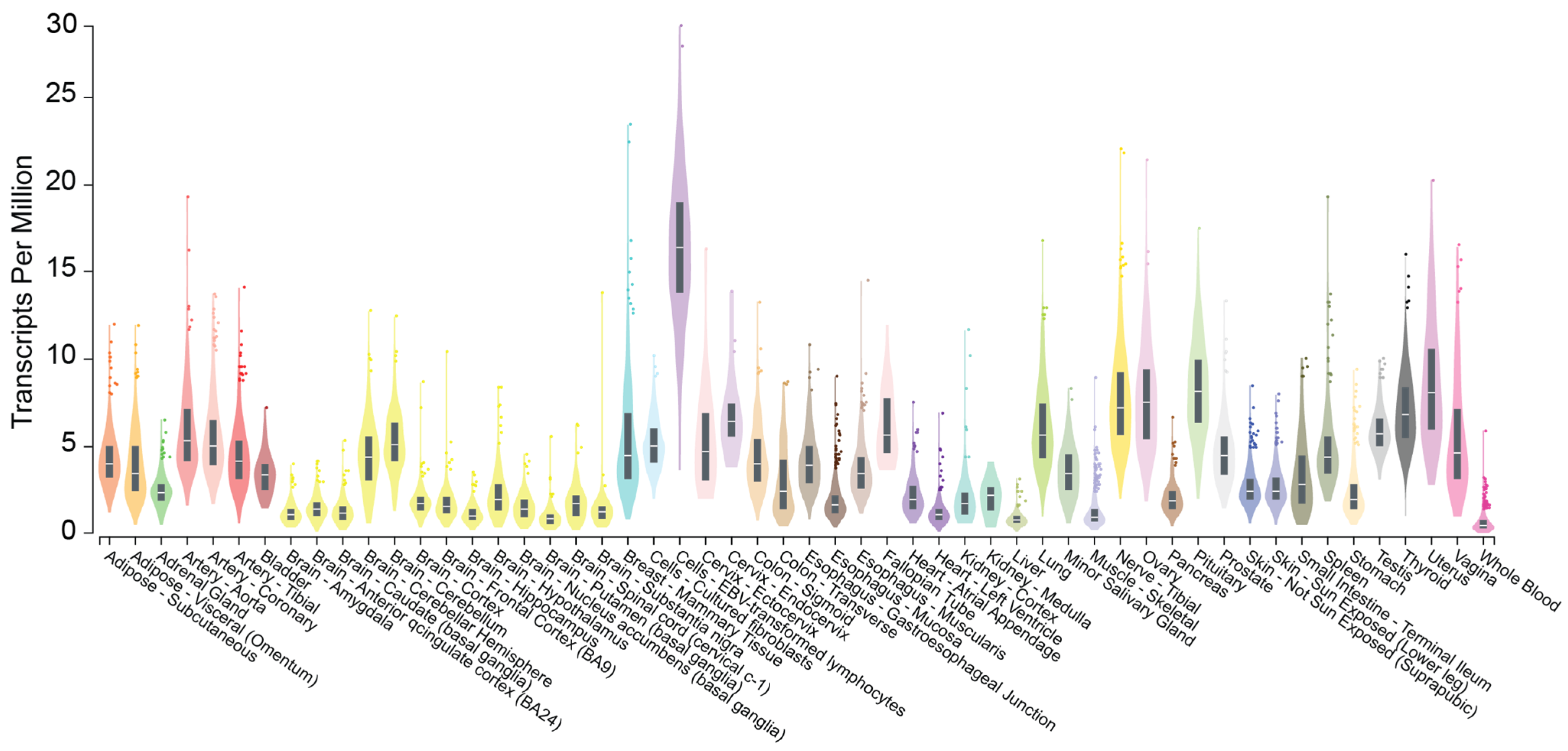
2. CHROMR: Nomenclature, Genomic Location, and Subcellular Localization

Despite efforts by the GENCODE consortium and others to standardize lncRNA nomenclature based on features such as proximity to protein-coding genes, genomic context, and predicted structure or function [33], many lncRNAs remain inconsistently named, resulting in multiple aliases across different databases. CHROMR is no exception; before its functional role was defined, this lncRNA was annotated under various identifiers across genomic databases, including AC009948.5 in GENCODE, ENSG00000223960 in Ensembl (v113), LOC101927027 in NCBI, and it was listed with GenBank accessions NR_110204, NR_110205, and NR_110206. Following its initial characterization, the transcript was named Cholesterol Homeostasis Regulator Of MiRNA Expression (CHROME), sometimes referred to as Protein Activator of Interferon Induced Protein Kinase EIF2AK2 Antisense RNA 1 (PRKRA-AS1), before ultimately being designated as Cholesterol-induced Regulator Of Metabolism RNA (CHROMR) by the Human Genome Organisation (HUGO) Gene Nomenclature Committee (HGNC) [20,22,34].
CHROMR is located on human chromosome 2 between the gene OxySterol Binding Protein-Like 6 (OSBPL6) and Protein Activator of Interferon Induced Protein Kinase EIF2AK2 (PRKRA). The CHROMR locus harbors primate-specific Alu transposable elements, and its exons are conserved across primate genomes but are largely absent from the genomes of other placental mammals and vertebrates (detailed in [20]). CHROMR expression is limited to humans, great apes (chimpanzees, gorillas, orangutans), lesser apes (gibbons), and Old World monkeys (rhesus and crab-eating macaques, baboons, and African green monkeys). CHROMR is broadly expressed in human tissues and cell types [20] as shown in publicly available data provided by the National Institutes of Health Adult Genotype Tissue Expression Project (GTex [35], Gene Expression in 54 tissues from GTEx RNA-seq of 17382 samples, 948 donors [V8, Aug 2019]) (Figure 1). Like many lncRNAs, CHROMR exists in multiple isoforms, most likely generated through alternative splicing of its precursor transcript. To date, no single isoform has been designated as the primary transcript, and databases report varying numbers of splice variants—GENCODE lists 5, NCBI lists 3, while Ensembl reports as many as 75. Notably, the main CHROMR transcripts listed by either database contain a ~75 nucleotide region in the first exon that is common to all splice variants, a sequence feature used to effectively target CHROMR expression using antisense oligonucleotides. Although there is limited evidence for distinct functional roles of individual lncRNA isoforms, this does not preclude the possibility that alternative and aberrant splicing contributes to the regulation of gene expression and may play a role in disease pathogenesis.
The pattern of subcellular localization of lncRNA transcripts can be used to classify lncRNAs into five distinct groups: lncRNAs exhibiting (I) one or two large foci in the nucleus; (II) large nuclear foci and single molecules scattered through the nucleus; (III) predominantly nuclear, without foci; (IV) cytoplasmic and nuclear; and (V) predominantly cytoplasmic, with ~55% being predominantly expressed in the nucleus (class I–III) [36]. CHROMR displays a roughly equal distribution between the nucleus and cytoplasm, with approximately 50% of the transcripts localized to each compartment [22], and as such belongs to class IV lncRNAs. Subcellular localization experiments—such as RNA-FISH and cell fractionation followed by quantitative PCR—have so far been limited to resting, unstimulated cells and only a few cell types, including macrophages, hepatocytes, and human embryonic kidney cells. Interestingly, CHROMR has been reported to localize in the cytoplasm to Processing (P)-bodies, a cellular granule involved in mRNA turnover by miRNAs [20,37]. Given the dynamic nature of RNA, especially lncRNAs, it remains to be determined whether CHROMR’s nuclear and cytoplasmic localization shifts depending on cell type, cellular activation, or cell cycle stage.
RNA modifications like N6-methyladenosine (m6A) methylation are emerging as regulators of protein-binding motif accessibility, cytoplasmic export, or ribosome-targeting [38]. Interestingly, CHROMR expression has been linked to the expression of m6A-related genes in myeloid leukemia cell lines [39]. Thus, RNA methylation may introduce yet another layer of regulation affecting CHROMR’s subcellular distribution (further discussed in Section 6, Perspective). In addition, CHROMR is detectable in whole blood and plasma samples in human subjects, raising the possibility that CHROMR may be packaged in extracellular vesicles or exosomes, bound to RNA-binding protein complexes or lipoprotein molecules, or may have been released from apoptotic/necrotic cells. To date, there is no evidence for the presence of CHROMR in other human biological fluids that contain lncRNAs, such as urine, saliva, breast milk, or amniotic fluid, among others [40].

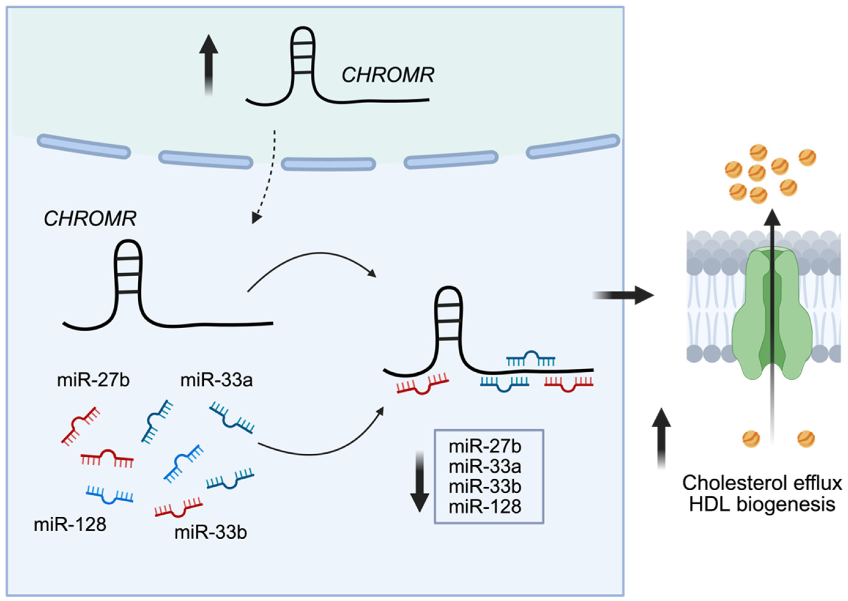
3. CHROMR in Cholesterol Homeostasis

Cardiovascular diseases (CVD) remain the leading cause of death worldwide. Dysregulation of cholesterol homeostasis represents a significant risk factor for the development of CVD. Removing excess cholesterol from cells and delivering it to the liver for clearance is a key process to prevent the pathological deposition of cholesterol in the tissues and artery wall [41,42]. Numerous lncRNAs have been reported to regulate lipoproteins, triglyceride metabolism, as well as cholesterol efflux (reviewed in [43,44]). Certain lncRNAs (e.g., Metastasis Associated Lung Adenocarcinoma Transcript 1 [MALAT1 [45]], H19 [46], lncRNA Hepatitis C Virus Regulated 1 [lncHR1 [47]]) have been shown to act directly on the expression and function of transcription factors, such as sterol regulatory element-binding proteins (SREBP), LXRs, and retinoid X receptors (RXRs), thereby controlling transcription of genes involved in cholesterol synthesis. Other lncRNAs (e.g., MeXis [18], LeXis [19], lncRNA Liver-Specific Triglyceride Regulator [lncLSTR] [48]) can interact with transcriptional regulators to modulate their ability to interact with promoter regions of genes involved in cholesterol metabolism (e.g., ABCA1, HMGCR, CYP8B1) thereby controlling their expression. LncRNAs can also interact with chromatin-modifying complexes to alter the expression of genes involved in lipid metabolism, such as APOlipoprotein A1 antisense RNA (APOA1-AS) on the APOA1 mRNA [49] and APOA4-AS on the APOA4 mRNA [50]. Interestingly, APOA1-AS is negatively correlated with circulating levels of APOA1, while conversely, APOA4-AS1 enhances hepatic expression of APOA4 [49,50]. These findings point to important roles for lncRNAs in regulating lipid metabolism and atherosclerosis susceptibility.
Circulating CHROMR levels are elevated in individuals with coronary artery disease compared to healthy controls and are also increased in atherosclerotic plaques relative to normal arterial samples. Furthermore, CHROMR’s genomic location is near a locus associated with plasma high-density lipoprotein-associated cholesterol (HDL-C) variation in women and linked to premature coronary artery disease [51,52], suggesting a potential regulatory role in CVD. Supporting this, a positive correlation was observed between CHROMR expression in the liver and circulating HDL-C and APOA1 levels in a cohort of 200 individuals [20]. In another study, hypercholesterolemic patients treated with atorvastatin for four weeks exhibited increased CHROMR levels in leukocytes, aligning with an improved lipid profile [21]. Notably, three binding sites for LXR transcription factors were identified near the CHROMR locus, with two of them being occupied by LXR following treatment with the LXR agonist T0901307 [20]. Further supporting its role in cholesterol regulation through LXR, CHROMR expression was elevated in the livers of African green monkeys fed a cholesterol-enriched diet for eight weeks and in response to in vivo treatment with the LXR agonist GW2965 [20]. In vitro studies also demonstrated increased CHROMR levels in human hepatocytes and macrophages following cholesterol loading with acetylated low-density lipoprotein (LDL), cyclodextrin-cholesterol, and the LXR agonist T0901307. Conversely, increased CHROMR expression was dampened upon silencing of LXRα/β using small interfering RNA [20].
Mechanistic studies in hepatocytes and macrophages revealed a role for CHROMR in regulating expression levels of numerous metabolic genes, such as ANGPTL3, GPAM, ABCA1, HNF4A, OSBPL6, CROT, CPT1A, ABCB11, and ATP8B1. This was shown to be due to CHROMR’s ability to function as a competing endogenous RNA (ceRNA) that sequesters multiple miRNAs involved in cholesterol, triglyceride, and fatty acid metabolism [20]. The function of CHROMR in the regulation of cholesterol homeostasis was determined by a series of loss-of-function and gain-of-function assays. Loss of CHROMR using either short hairpin RNAs or GapmeR antisense oligonucleotides led to reduced capacity of macrophages and hepatocytes to efflux cholesterol to exogenous APOA1, without affecting genes and processes involved in LDL uptake and/or cholesterol synthesis. In agreement with these observations, the upregulation of CHROMR using overexpression vectors leads to an increase in cholesterol efflux to APOA1. Moreover, lipid droplet accumulation in macrophages was markedly reduced upon overexpression of CHROMR, further evidence of enhanced cholesterol efflux due to elevated levels of CHROMR [20]. Mechanistically, it was shown that CHROMR in the cytoplasm colocalized with Argonaute 2, a key component of the RNA-Induced Silencing Complex, as well as enhancer of mRNA-decapping protein 4 (EDC4), a marker for P-bodies that mediate miRNA sequestration and mRNA decay. Indeed, CHROMR was found to sequester a group of miRNAs (miR-33a/b, miR-27b, miR-128) that were previously shown to repress a group of common target genes (ANGPTL3, GPAM, ABCA1, HNF4A, OSBPL6, CROT, CPT1A, ABCB11, and ATP8B1) that play key regulatory roles in cholesterol homeostasis and fatty acid oxidation (Figure 2) [53,54,55,56,57]. In agreement with these findings, it was found that in the liver, CHROMR expression was inversely correlated with levels of these miRNAs and positively correlated with their target mRNAs [20]. The discovery that elevated CHROMR levels, triggered by cholesterol overload in macrophages and hepatocytes, can inhibit the activity of a group of metabolic miRNAs further highlights the complex regulatory interplay between ncRNAs in maintaining cholesterol homeostasis in both human health and disease.

4. CHROMR in Cancer

4.1. Diffuse Large B-Cell Lymphoma

Elevated levels of CHROMR have been reported in a variety of cancers and are directly correlated with adverse patient outcomes [28,29,30,31,32]. Mechanistic studies suggest that CHROMR’s impact on cancer prognosis is primarily mediated through its function as a ceRNA (Figure 3), mirroring its role in modulating cholesterol homeostasis in macrophages and hepatocytes. Notably, CHROMR is highly expressed in diffuse large B-cell lymphoma (DLBCL) cells resistant to the anticancer drug rituximab, and its overexpression promotes cancer cell proliferation [58]. Functional studies revealed that CHROMR overexpression leads to increased expression of the cell cycle-associated gene Cyclin and CBS Domain Divalent Metal Cation Transport Mediator 1 (CNNM1) and reduced levels of miR-1299, a miRNA that normally represses CNNM1. CNNM1 is known to regulate cell proliferation and has been implicated in the progression of prostate cancer and hepatocellular carcinoma [58]. By sequestering miR-1299, CHROMR promotes CNNM1 expression, suppressing the expression of apoptosis-related proteins and impairing cell cycle arrest at the G2/M checkpoint. Ultimately, this mechanism contributes to decreased apoptosis and enhanced resistance to rituximab in DLBCL cells [32].
In a subsequent study of DLBCL, CHROMR was further shown to accelerate cancer progression and promote chemoresistance [29]. Liu et al. demonstrated that CHROMR knockdown enhances the transcription of CD20, a key B-cell marker and cell surface marker that is therapeutically targeted by rituximab. Given that CD20 expression is upregulated by histone deacetylase (HDAC) inhibitors, CHROMR was hypothesized to influence HDAC activity. Gain-of-function and loss-of-function experiments revealed that CHROMR indeed reduces the phosphorylation of HDAC3, suggesting a potential mechanism through which CHROMR indirectly regulates CD20 expression. Next, using lentiviral vectors expressing a short hairpin RNA targeting CHROMR, it was demonstrated that CHROMR downregulation could reverse rituximab resistance of DLBCL cells, through upregulation of CD20 levels [29]. Second to its effect on HDAC3 phosphorylation, targeting of CHROMR promoted apoptotic cell death and reduced mitogen-activated protein kinase (MAPK) and protein kinase B (AKT) signaling, pathways essential for DLBCL growth. These effects were linked to miR-27b-3p, a target of CHROMR that is significantly reduced in DLBCL and controls expression of pro-tumorigenic mesenchymal-epithelial transition factor (MET). MiR-27b-3p has been shown to act as a tumor suppressor of DLBCL, and its overexpression reduced the phosphorylation of AKT and extracellular signal-regulated kinases through inhibition of MET [29]. Together, these studies position CHROMR as a central regulator of DLBCL proliferation, where elevated CHROMR levels enhance the expression of proliferation-associated genes such as CNNM1 and MET by sequestering their regulatory miRNAs.

4.2. Lung Adenocarcinoma

Elevated levels of CHROMR were also found in lung adenocarcinoma cells and tissues [28]. Here, CHROMR expression was directly correlated with tumor growth as well as metastasis and poor patient prognosis [28]. Lentiviral knockdown of CHROMR in lung-derived epithelial-like carcinoma cells (i.e., A549 and H1299) attenuated cell proliferation and induced apoptosis. Furthermore, the migration and invasion abilities of these cell lines in vitro decreased significantly after CHROMR knockdown. This was further supported by reduced rates of metastasis in an in vivo xenograph model, where virally transduced cells in which CHROMR was silenced were injected subcutaneously into the back of nude mice. This study further indicated a role for CHROMR in the metastasis of lung adenocarcinoma. The protumorigenic actions of CHROMR were found to be a result of sponging of miR186-5p [28], a miRNA that inhibits Pituitary Tumor-Transforming Gene 1 Protein (PTTG1) and Non-SMC Condensin II Complex Subunit G2 (NCAPG2) [28,59]. High NCAPG2 expression is associated with advanced clinical tumor stage and reduced survival time in patients with lung adenocarcinoma [60], and PTTG1 has been correlated with non-small cell lung cancer progression and is an independent poor prognostic factor in patients with non-small cell lung cancer [61]. In this study, the overexpression of miR186-5p was found to downregulate NCAPG2 expression and inhibit the progression of epithelial–mesenchymal transition in lung adenocarcinoma cells. In turn, CHROMR was shown to promote epithelial–mesenchymal transition by sponging miR-186-5p, thereby increasing invasion and metastasis. suggesting poor patient prognosis when CHROMR levels are elevated [28].

4.3. Stomach Adenocarcinoma and Glioma

CHROMR has also been applied in various correlation analyses seeking to improve the potential diagnosis of certain cancer types. In a study of stomach adenocarcinoma, a signature comprised of twelve necroptosis-related lncRNAs, including CHROMR, was developed to putatively enhance the prediction of patient prognoses. In a data set of 368 patients, CHROMR was associated with higher rates of recurrence and patient death [30]. CHROMR expression is also elevated in glioma, prompting investigation into its association with patient survival. The study by Sirvinskas et al. centered on the potential regulatory relationship between CHROMR and PRKRA, a gene that partially overlaps CHROMR on chromosome 2. While no direct evidence was found for CHROMR controlling PRKRA expression, a lower PRKRA:CHROMR ratio was linked to improved patient survival, consistent with previous reports connecting high CHROMR levels to poorer outcomes [31]. Collectively, these findings highlight CHROMR’s emerging role in cancer biology. Deeper insight into how CHROMR contributes to tumor progression, metastasis, and resistance to therapy may pave the way for novel treatment strategies.

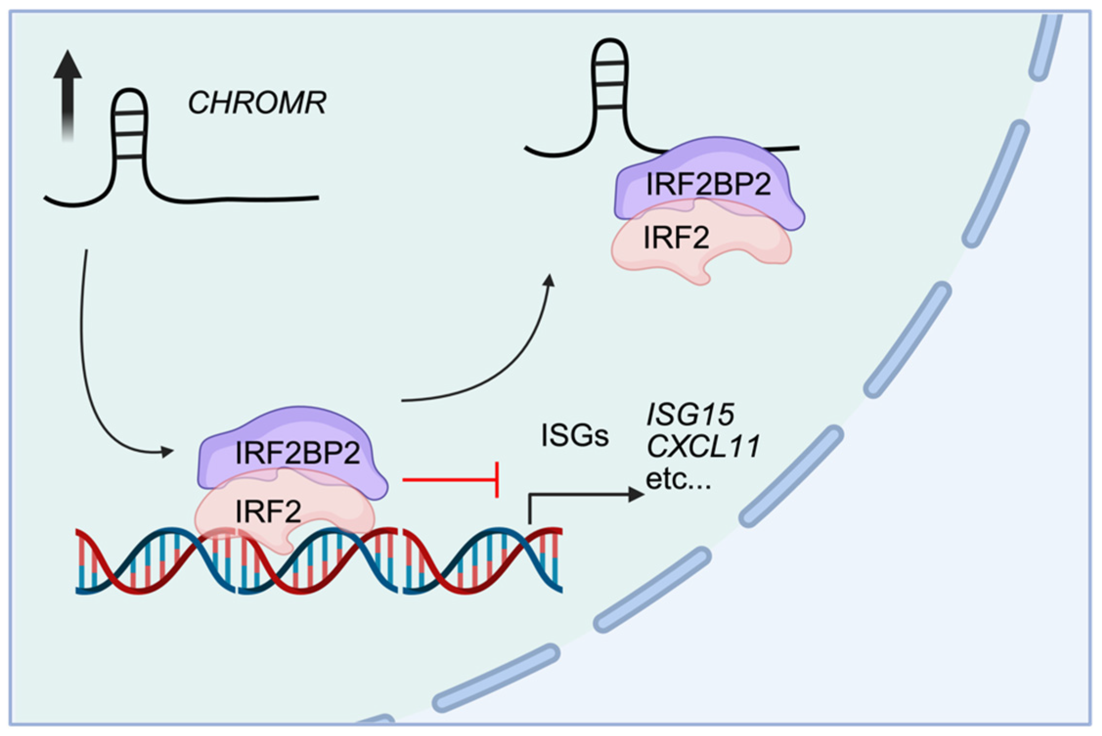
5. CHROMR in Innate Immunity

A genome-wide linkage scan for premature atherosclerotic coronary artery disease, conducted on 4175 affected individuals from 1933 families, identified a locus on human chromosome 2 that encompassed CHROMR [51,62]. Additionally, other genome-wide analyses have linked CHROMR to human immunological diseases. A systems biology study by Teimuri et al. aimed to identify lncRNAs involved in autoimmune and immune-related diseases by integrating manual data mining, investigative analysis of data retrieved from the Gene Expression Omnibus, single nucleotide polymorphism (SNPs) associated with auto-immune and immune-related diseases, and miRNA interactions and applied LncDisease, a sequence-based bioinformatics tool for predicting lncRNA-disease associations [23,63]. This in silico approach identified 8 (out of 26 preselected Th17 cell-lineage specific) lncRNAs associated with auto-immune and immune-related diseases, including CHROMR. CHROMR was found to be associated with an SNP (rs9283487) with genetic risk for multiple sclerosis [64] and Sjögren’s syndrome [23]. Subsequently, expression levels of CHROMR in peripheral blood mononuclear cells of multiple sclerosis patients (n = 38) were tested by quantitative PCR and revealed a significant downregulation of CHROMR compared to a control group of healthy volunteers (n = 17). Interestingly, when patients with multiple sclerosis were split into a relapsing-phase group (n = 19) and a remitting-phase group (n = 19), the remitting-phase group had increased CHROMR expression levels, indicating a beneficial effect for CHROMR during disease progression [23], and potentially identifying CHROMR to be a biomarker for multiple sclerosis.
A larger patient study involving over 19,000 individuals (12,333 controls) with atopic dermatitis (2262) and psoriasis (4489)—the two most common chronic inflammatory skin conditions—revealed that the CHROMR locus contains a single nucleotide variant (SNV, rs62176107, G < A) with opposing effects on these diseases. This SNV, located in an intronic region of CHROMR and the sixth exon of PRKRA, is associated with a lower estimated odds ratio of 0.55 (0.46–0.66) for atopic dermatitis and a higher odds ratio of 1.20 (1.15–1.27) for psoriasis [26]. An exome-wide sequencing analysis of individuals from the UK Biobank examined the role of copy number variation (CNV) in susceptibility to asthma, a chronic inflammatory lung condition. Using a first-stage analysis of 49,953 individuals (7098 patients with asthma and 36,578 controls), followed by a second-stage analysis of an additional 17,280 asthma cases and 115,562 controls, the research identified an asthma-associated CNV on chromosome 2 leading to the duplication of exons 4–8 of PRKRA and the 3′ end of CHROMR [24]. While CHROMR and PRKRA have been shown to play regulatory roles in antiviral immunity [22,25], no direct associations with inflammatory skin or lung conditions have been reported. Further validation of these SNPs, SNVs, and CNVs will be necessary to determine CHROMR’s potential involvement in atopic dermatitis, psoriasis, and/or asthma.
Although the potential regulatory role for CHROMR in autoimmune disease will need to be further investigated, a study analyzing published blood sample datasets from patients with sarcoidosis, a multisystem inflammatory disease of unknown origin, identified CHROMR midst 172 differentially upregulated genes. Moreover, these genes were enriched in biological pathways associated with, among other pathways, Immune Response, Chemokine-mediated Signaling, and Viral Defense [27], highlighting the potential role of CHROMR in innate immune activity. Notably, CHROMR levels are elevated in whole blood of patients infected with respiratory viruses, such as the influenza A virus (IAV), metapneumovirus (MPV), and severe acute respiratory syndrome coronavirus 2 (SARS-CoV-2) [22,65]. CHROMR levels were strongly correlated to 226 differentially expressed interferon (IFN)-stimulated genes (ISGs). Interestingly, the correlation coefficients between CHROMR and these ISGs were comparable to or greater than those of other lncRNAs known to regulate the antiviral response (e.g., BST2 IFN-Stimulated Positive Regulator [BISPR [66]], Negative Regulator of the IFN Response [NRIR [67]], CCR5 Antisense RNA [CCR5AS [68]], and Lung Cancer-Associated Transcript 1 [LUCAT1 [69]]). CHROMR expression was similarly upregulated by IAV and SARS-CoV-2, infection, while other lncRNAs that were tested were preferably enhanced by either IAV (BISPR, NRIR, CCR5AS) or SARS-CoV-2 (LUCAT1) infection.
Transcriptomic analysis of differentially expressed genes upon knockdown of CHROMR in a human monocytic cell line (i.e., THP1) revealed a distinct downregulation of genes that are in pathways related to Interferon Signaling, PPAR Signaling, and Cell Cycle Control of Replication. Moreover, mass spectrometry revealed that CHROMR interacts with IFN regulatory factor 2 binding protein 2 (IRF2BP2), a known co-repressor that associates with the C-terminal repression domain of IFN regulatory factor 2 (IRF2) [70]. Mechanistically, a G-quadruplex structure within the CHROMR sequence was shown to mediate this interaction, sequestering the IRF2/IRF2BP2 repressor complex away from IFN-stimulated response elements throughout the genome. This sequestration facilitates increased transcription of ISGs (Figure 4). Supporting this, chromatin immunoprecipitation sequencing targeting H3K27 acetylation demonstrated that loss of CHROMR reduced histone acetylation at ISG promoters, consistent with transcriptional repression of these genes. Functionally, CHROMR knockdown in macrophages resulted in increased viral replication, further highlighting its critical role as a regulator of antiviral innate immune responses in humans [22]. Individual roles of IRF2BP2, as well as its interaction with IRF2 in the IRF2/IRF2BP2 repression complex, have been described in autoimmune diseases [71,72,73], including asthma [74], multiple sclerosis [75], atopic dermatitis [76], and psoriasis [77], where CHROMR has been implicated [23,24,26]. However, the functional contribution of CHROMR to these conditions remains unexplored, and the potential for therapeutic strategies based on deeper understanding of this has yet to be investigated.

6. Perspective

While CHROMR’s research footprint continues to expand, its roles in cholesterol metabolism and innate immune regulation are well defined. Emerging evidence also implicates CHROMR in cancer biology, including its potential involvement in cell viability and gene regulation in various tumor contexts. Together, these studies provide a growing understanding of CHROMR’s functional relevance across multiple pathways and cell types (Table 1). However, the mechanisms governing its subcellular localization—and how this distribution shapes its biological activity—remain poorly understood. Cell-specific functions of lncRNAs are well documented [78], suggesting that the cellular environment largely shapes mechanisms of action. In macrophages, CHROMR contributes to the transcriptional activation of ISGs in the nucleus, while also functioning as a competing endogenous RNA (ceRNA) for metabolic miRNAs in the cytoplasm. This dual function is biologically relevant, as many viruses manipulate host lipid synthesis and metabolism to support their replication [79,80]. As such, elevated CHROMR expression in virus-infected cells may help counteract lipid accumulation while simultaneously enhancing ISG transcription to strengthen the antiviral immune response. Still, the impact of various environmental stimuli (i.e., viral infection versus cholesterol overload) on the subcellular distribution of CHROMR—and thus its function—remains largely uninvestigated.
Among the various cellular processes governing CHROMR’s spatiotemporal localization, m6A methylation is a potential mediator. Epigenetic RNA modifications including m6A have emerged as key regulators of RNA stability and modulation as well as innate immunity, lipid metabolism, and host defense against viruses [81,82,83]. M6A modification of lncRNAs has been reported to alter their stability, export to the cytoplasm, localization to P-bodies and ribosomes, and function [38,84]. Installation of m6A is a reversible process that occurs at DRACH consensus sequences (GGACU) via the actions of methyltransferase ‘writers’ (e.g., METTL3/14 [85]) and demethylase ‘erasers’ (e.g., FTO, ALKBH5 [86,87]). Notably, m6A modification is dynamically regulated by various cell stressors in a cell type- and tissue-specific manner [88], and is recognized by RNA-binding ‘reader’ proteins [89] that alter RNA splicing (e.g., HNRNPC), nuclear export (e.g., YTHDC1), translation (e.g., YTHDF1/3), stability (e.g., YTHDF2), maturation (e.g., LRPPRC), secondary structure (e.g., YTHDC2), and delivery to ribonucleoprotein granules (e.g., YTHDF3) [90,91,92]. Prominent examples of the impact of m6A modifications on lncRNA function include regulation of Xist in gene silencing [93] and the role of HOTAIR in breast cancer [94]. Understanding how methylation of CHROMR and m6A-reader interactions regulate its stability, spatiotemporal localization, structure, and function is essential to dissecting its discrete functions in subcellular microenvironments and paves the way for further investigation into the guidance of CHROMR’s function by the cell’s milieu.
Numerous lncRNA–mRNA gene pairs located within the same topologically associating domain (TAD) have been shown to undergo coordinated transcriptional regulation, suggesting that the function of the lncRNA can be inferred from the known role of its neighboring protein-coding gene [95]. Interestingly, no such associations have been established for CHROMR to date [22,31]. However, these studies have focused only on a narrow set of genes and cell types—primarily macrophages and glioma tissue—highlighting the need for a broader investigation of genes within the same TAD as CHROMR across diverse cellular contexts to uncover possible novel mechanistic insights. For example, in hepatocytes, CHROMR’s TAD (as determined by high-throughput chromosome conformation capture [96]) consists of ~30 genes and is bordered by RNA5SP112, a ribosomal pseudogene, on the 5′ end, and FKBP7, coding for a peptidyl-prolyl cis-trans isomerase, on the 3′ end (Figure 5).
Among these 30 genes are three miRNA genes (miR-3128, miR-4444-1, and miR-6512) that have not been linked to CHROMR’s ceRNA function described in this review. Aside from OSBPL6, which is regulated by CHROMR through the sequestration of OSBPL6-targeting miRNAs [20], a direct mechanistic link between CHROMR and other genes within the same TAD has not yet been established. Nonetheless, CHROMR’s nuclear localization points to a possible in cis regulatory function that may yet be revealed by further studies of its functions within its TAD.
In the cytoplasm, CHROMR acts as a ceRNA that sponges miRNAs that repress genes involved in cholesterol efflux and HDL biogenesis in macrophages and hepatocytes. Similar cytoplasmic functions were found for CHROMR in certain cancers where it targets miR-27b-3p, miR-186-5p, and miR-1299, repressing cell cycle-regulator genes CNNM1, MET, and NCAPG2, leading to accelerated cancer growth and progression [28,29,32]. Whether CHROMR sequesters additional miRNAs remains to be determined. For example, Hennessy et al. [20] used the prediction algorithm miR-Target2 (miRDB [97]) to predict interactions based on features common to miRNA binding and target suppression. This analysis identified miR-27b, miR-33a/b, miR-128, and miR-1299 as potential targets, but did not predict miR-186-5p. Such discrepancies highlight the importance of combining comprehensive in silico approaches (e.g., RNAcofold, RNAhybrid [98]) with in vitro validation methods—including luciferase reporter assays, site-directed mutagenesis, and functional perturbation studies—to robustly confirm lncRNA–miRNA interactions and their effects on downstream targets. There is evidence to suggest that CHROMR may sequester its miRNA targets in P-bodies [20,37], but the underlying mechanisms regulating spatiotemporal localization of CHROMR within cytoplasmic microenvironments such as P-bodies or stress granules remain unknown. Presence in P-bodies may drive degradation and/or inactivation of miRNA, but further studies are needed to confirm how and when this happens and on what level this is regulated (i.e., extracellular stimuli, RNA modifications, strength of lncRNA–miRNA interaction).
Recent advances in high-throughput methods for assessing the translational capacity of small open reading frames (smORF) have revealed that biologically relevant micropeptides can be produced from genomic regions previously classified as non-coding [99,100]. Initial findings in THP-1 macrophages showed that CHROMR does not associate with polysomes, and both Kozak sequence analysis and other in silico coding potential algorithms suggest it does not encode a micropeptide [20]. However, a CRISPR/Cas9-based study employing dense single-guide RNAs to introduce indels in over 500 preselected ‘high-priority’ open reading frames (ORFs) within lncRNAs found that targeting a smORF within CHROMR led to reduced cell viability over a 21-day period across eight different cancer cell lines [101]. This intriguing discovery raises the possibility that the CHROMR sequence may contain one or more smORFs capable of producing biologically functional peptides, further broadening the potential significance of CHROMR in cellular biology.
The main challenge in studying CHROMR lies in its primate specificity, which precludes the use of conventional pre-clinical models such as mice, rabbits, or pigs. Since CHROMR does not have a clear ortholog in these organisms, in vivo genetic manipulation or disease modeling is challenging. This presents a significant obstacle to investigating CHROMR’s physiological roles and therapeutic potential using traditional approaches. To address this, future studies may need to rely on human-derived ex vivo systems, such as organoids or CRISPR-modified cell lines, or consider the development of humanized mouse models, though each of these comes with its own technical and interpretative challenges.

7. Conclusions

In this review, we have highlighted CHROMR, a primate-specific long noncoding RNA that is upregulated in patients with coronary artery disease and plays a key regulatory role in cholesterol homeostasis. In the cytoplasm of hepatocytes and macrophages, CHROMR acts as a ceRNA, binding and sequestering miR-27b, miR-33a/b, and miR-128—miRNAs known to suppress cholesterol efflux and HDL biogenesis. By modulating cholesterol metabolism at the post-transcriptional level, CHROMR has emerged as a central player in a ncRNA network with promising therapeutic potential for cardiovascular disease. In cancer, CHROMR binds additional miRNAs (e.g., miR-27b-3p, miR-186-5p, miR-1299) that repress cell cycle regulators such as CNNM1, MET, and NCAPG2, thereby promoting tumor proliferation and metastasis. CHROMR has also emerged as a candidate biomarker in cancer diagnostics, with correlation studies linking its expression to poor patient outcomes. CHROMR expression is strongly induced in response to respiratory viral infections including SARS-CoV-2, IAV, and MPV. In macrophages, nuclear CHROMR binds the co-repressor IRF2BP2, enhancing transcription of ISGs and facilitating antiviral responses. As highlighted in this review, CHROMR is an lncRNA with multifaceted and context-dependent roles across a range of heterogeneous diseases. Its specific impact on cellular biology and cell fate is influenced by several factors, including subcellular localization, genetic background, and environmental cues. Additionally, the signaling pathways regulated by CHROMR are shaped by the cell’s activation status, RNA methylation, and the availability of its direct molecular targets. Like many lncRNAs, CHROMR is associated with diverse pathological contexts; however, a potential unifying feature may be its capacity to fine-tune transcriptional and translational programs in a cell type- and condition-specific manner. Future studies focused on elucidating these regulatory networks in relevant cellular systems may uncover common mechanisms underlying their roles.
Since its first functional characterization in 2019, research on CHROMR has steadily uncovered its diverse roles across biological systems. Continued exploration of the molecular mechanisms and cellular contexts that regulate CHROMR function will be essential for evaluating its therapeutic and diagnostic potential.

Author Contributions

E.R.B. and C.v.S. drafted the manuscript and figures. All authors have read and agreed to the published version of the manuscript.

Funding

C.v.S. is supported by a grant from the American Heart Association (23SCEFIA1153739).

Conflicts of Interest

C.v.S. holds a patent on the use of inhibitors targeting CHROMR.

References

  1. Rinn, J.L.; Chang, H.Y. Genome Regulation by Long Noncoding Rnas. Annu. Rev. Biochem. 2012, 81, 145–166. [Google Scholar] [CrossRef] [PubMed]
  2. Palazzo, A.F.; Lee, E.S. Non-Coding Rna: What Is Functional and What Is Junk? Front. Genet. 2015, 6, 2. [Google Scholar] [CrossRef]
  3. Schmitz, S.U.; Grote, P.; Herrmann, B.G. Mechanisms of Long Noncoding Rna Function in Development and Disease. Cell. Mol. Life Sci. 2016, 73, 2491–2509. [Google Scholar] [CrossRef]
  4. Bridges, M.C.; Daulagala, A.C.; Kourtidis, A. Lnccation: Lncrna Localization and Function. J. Cell Biol. 2021, 220, e202009045. [Google Scholar] [CrossRef] [PubMed]
  5. Krause, H.M. New and Prospective Roles for Lncrnas in Organelle Formation and Function. Trends Genet. 2018, 34, 736–745. [Google Scholar] [CrossRef]
  6. Goff, L.A.; Rinn, J.L. Linking Rna Biology to Lncrnas. Genome Res. 2015, 25, 1456–1465. [Google Scholar] [CrossRef]
  7. Satpathy, A.T.; Chang, H.Y. Long Noncoding Rna in Hematopoiesis and Immunity. Immunity 2015, 42, 792–804. [Google Scholar] [CrossRef]
  8. Wang, K.C.; Chang, H.Y. Molecular Mechanisms of Long Noncoding Rnas. Mol. Cell 2011, 43, 904–914. [Google Scholar] [CrossRef] [PubMed]
  9. Guil, S.; Esteller, M. Cis-Acting Noncoding Rnas: Friends and Foes. Nat. Struct. Mol. Biol. 2012, 19, 1068–1075. [Google Scholar] [CrossRef]
  10. Joung, J.; Engreitz, J.M.; Konermann, S.; Abudayyeh, O.O.; Verdine, V.K.; Aguet, F.; Gootenberg, J.S.; Sanjana, N.E.; Wright, J.B.; Fulco, C.P.; et al. Genome-Scale Activation Screen Identifies a Lncrna Locus Regulating a Gene Neighbourhood. Nature 2017, 548, 343–346. [Google Scholar] [CrossRef]
  11. Kumar, V.; Westra, H.J.; Karjalainen, J.; Zhernakova, D.V.; Esko, T.; Hrdlickova, B.; Almeida, R.; Zhernakova, A.; Reinmaa, E.; Vosa, U.; et al. Human Disease-Associated Genetic Variation Impacts Large Intergenic Non-Coding Rna Expression. PLoS Genet. 2013, 9, e1003201. [Google Scholar] [CrossRef] [PubMed]
  12. Kopp, F.; Mendell, J.T. Functional Classification and Experimental Dissection of Long Noncoding Rnas. Cell 2018, 172, 393–407. [Google Scholar] [CrossRef]
  13. Congrains, A.; Kamide, K.; Oguro, R.; Yasuda, O.; Miyata, K.; Yamamoto, E.; Kawai, T.; Kusunoki, H.; Yamamoto, H.; Takeya, Y.; et al. Genetic Variants at the 9p21 Locus Contribute to Atherosclerosis through Modulation of Anril and Cdkn2a/B. Atherosclerosis 2012, 220, 449–455. [Google Scholar] [CrossRef] [PubMed]
  14. Holdt, L.M.; Beutner, F.; Scholz, M.; Gielen, S.; Gabel, G.; Bergert, H.; Schuler, G.; Thiery, J.; Teupser, D. Anril Expression Is Associated with Atherosclerosis Risk at Chromosome 9p21. Arter. Thromb. Vasc. Biol. 2010, 30, 620–627. [Google Scholar] [CrossRef]
  15. Holdt, L.M.; Stahringer, A.; Sass, K.; Pichler, G.; Kulak, N.A.; Wilfert, W.; Kohlmaier, A.; Herbst, A.; Northoff, B.H.; Nicolaou, A.; et al. Circular Non-Coding Rna Anril Modulates Ribosomal Rna Maturation and Atherosclerosis in Humans. Nat. Commun. 2016, 7, 12429. [Google Scholar] [CrossRef] [PubMed]
  16. Ishii, N.; Ozaki, K.; Sato, H.; Mizuno, H.; Saito, S.; Takahashi, A.; Miyamoto, Y.; Ikegawa, S.; Kamatani, N.; Hori, M.; et al. Identification of a Novel Non-Coding Rna, Miat, That Confers Risk of Myocardial Infarction. J. Human Genet. 2006, 51, 1087–1099. [Google Scholar] [CrossRef]
  17. Gao, W.; Zhu, M.; Wang, H.; Zhao, S.; Zhao, D.; Yang, Y.; Wang, Z.M.; Wang, F.; Yang, Z.J.; Lu, X.; et al. Association of Polymorphisms in Long Non-Coding Rna H19 with Coronary Artery Disease Risk in a Chinese Population. Mutat. Res. 2015, 772, 15–22. [Google Scholar] [CrossRef]
  18. Sallam, T.; Jones, M.; Thomas, B.J.; Wu, X.; Gilliland, T.; Qian, K.; Eskin, A.; Casero, D.; Zhang, Z.; Sandhu, J.; et al. Transcriptional Regulation of Macrophage Cholesterol Efflux and Atherogenesis by a Long Noncoding Rna. Nat. Med. 2018, 24, 304–312. [Google Scholar] [CrossRef]
  19. Sallam, T.; Jones, M.C.; Gilliland, T.; Zhang, L.; Wu, X.; Eskin, A.; Sandhu, J.; Casero, D.; Vallim, T.Q.; Hong, C.; et al. Feedback Modulation of Cholesterol Metabolism by the Lipid-Responsive Non-Coding Rna Lexis. Nature 2016, 534, 124–128. [Google Scholar] [CrossRef]
  20. Hennessy, E.J.; van Solingen, C.; Scacalossi, K.R.; Ouimet, M.; Afonso, M.S.; Prins, J.; Koelwyn, G.J.; Sharma, M.; Ramkhelawon, B.; Carpenter, S.; et al. The Long Noncoding Rna Chrome Regulates Cholesterol Homeostasis in Primate. Nat. Metab. 2019, 1, 98–110. [Google Scholar] [CrossRef]
  21. Paez, I.; Prado, Y.; Ubilla, C.G.; Zambrano, T.; Salazar, L.A. Atorvastatin Increases the Expression of Long Non-Coding Rnas Arsr and Chrome in Hypercholesterolemic Patients: A Pilot Study. Pharmaceuticals 2020, 13, 382. [Google Scholar] [CrossRef] [PubMed]
  22. van Solingen, C.; Cyr, Y.; Scacalossi, K.R.; de Vries, M.; Barrett, T.J.; de Jong, A.; Gourvest, M.; Zhang, T.; Peled, D.; Kher, R.; et al. Long Noncoding Rna Chromr Regulates Antiviral Immunity in Humans. Proc. Natl. Acad. Sci. USA 2022, 119, e2210321119. [Google Scholar] [CrossRef]
  23. Teimuri, S.; Hosseini, A.; Rezaenasab, A.; Ghaedi, K.; Ghoveud, E.; Etemadifar, M.; Nasr-Esfahani, M.H.; Megraw, T.L. Integrative Analysis of Lncrnas in Th17 Cell Lineage to Discover New Potential Biomarkers and Therapeutic Targets in Autoimmune Diseases. Mol. Ther. Nucleic Acids 2018, 12, 393–404. [Google Scholar] [CrossRef] [PubMed]
  24. Fawcett, K.A.; Demidov, G.; Shrine, N.; Paynton, M.L.; Ossowski, S.; Sayers, I.; Wain, L.V.; Hollox, E.J. Exome-Wide Analysis of Copy Number Variation Shows Association of the Human Leukocyte Antigen Region with Asthma in Uk Biobank. BMC Med. Genom. 2022, 15, 119. [Google Scholar] [CrossRef]
  25. Kok, K.H.; Lui, P.Y.; Ng, M.H.; Siu, K.L.; Au, S.W.; Jin, D.Y. The Double-Stranded Rna-Binding Protein Pact Functions as a Cellular Activator of Rig-I to Facilitate Innate Antiviral Response. Cell Host Microbe 2011, 9, 299–309. [Google Scholar] [CrossRef] [PubMed]
  26. Baurecht, H.; Hotze, M.; Brand, S.; Buning, C.; Cormican, P.; Corvin, A.; Ellinghaus, D.; Ellinghaus, E.; Esparza-Gordillo, J.; Folster-Holst, R.; et al. Genome-Wide Comparative Analysis of Atopic Dermatitis and Psoriasis Gives Insight into Opposing Genetic Mechanisms. Am. J. Human Genet. 2015, 96, 104–120. [Google Scholar] [CrossRef]
  27. Zhao, M.; Di, X.; Jin, X.; Tian, C.; Cong, S.; Liu, J.; Wang, K. Identification of Biomarkers for Sarcoidosis and Tuberculosis of the Lung Using Systematic and Integrated Analysis. Med. Sci. Monit. 2020, 26, e925438. [Google Scholar] [CrossRef]
  28. Bai, J.; Li, H.; Chen, X.; Chen, L.; Hu, Y.; Liu, L.; Zhao, Y.; Zuo, W.; Zhang, B.; Yin, C. Lncrna-Ac009948.5 Promotes Invasion and Metastasis of Lung Adenocarcinoma by Binding to Mir-186-5p. Front. Oncol. 2022, 12, 949951. [Google Scholar] [CrossRef]
  29. Liu, C.; Zhao, X.; Wang, Z.; Zhang, C.; Zheng, W.; Zhu, X.; Zhang, D.; Gong, T.; Zhao, H.; Li, F.; et al. Lncrna Chromr/Mir-27b-3p/Met Axis Promotes the Proliferation, Invasion, and Contributes to Rituximab Resistance in Diffuse Large B-Cell Lymphoma. J. Biol. Chem. 2024, 300, 105762. [Google Scholar] [CrossRef]
  30. Luo, L.; Li, L.; Liu, L.; Feng, Z.; Zeng, Q.; Shu, X.; Cao, Y.; Li, Z. A Necroptosis-Related Lncrna-Based Signature to Predict Prognosis and Probe Molecular Characteristics of Stomach Adenocarcinoma. Front. Genet. 2022, 13, 833928. [Google Scholar] [CrossRef]
  31. Sirvinskas, D.; Steponaitis, G.; Stakaitis, R.; Tamasauskas, A.; Vaitkiene, P.; Skiriute, D. Antisense Lncrna Chromr Is Linked to Glioma Patient Survival. Front. Mol. Biosci. 2023, 10, 1101953. [Google Scholar] [CrossRef]
  32. Wang, M.; Miao, Z.; Cen, H.; He, J.; Wei, C. Long Non-Coding Rna (Lncrna) Chromr Promotes the Expression of the Cnnm1 Gene by Adsorbing Hsa-Mir-1299 to Obtain Drug Resistance in Diffuse Large B Lymphoma Cells. Transl. Cancer Res. 2022, 11, 1362–1371. [Google Scholar] [CrossRef] [PubMed]
  33. Seal, R.L.; Tweedie, S.; Bruford, E.A. A Standardised Nomenclature for Long Non-Coding Rnas. IUBMB Life 2023, 75, 380–389. [Google Scholar] [CrossRef]
  34. Wright, M.W. A Short Guide to Long Non-Coding Rna Gene Nomenclature. Human Genom. 2014, 8, 7. [Google Scholar] [CrossRef]
  35. Consortium, G.T. The Genotype-Tissue Expression (Gtex) Project. Nat. Genet. 2013, 45, 580–585. [Google Scholar]
  36. Cabili, M.N.; Dunagin, M.C.; McClanahan, P.D.; Biaesch, A.; Padovan-Merhar, O.; Regev, A.; Rinn, J.L.; Raj, A. Localization and Abundance Analysis of Human Lncrnas at Single-Cell and Single-Molecule Resolution. Genome Biol. 2015, 16, 20. [Google Scholar] [CrossRef]
  37. Hubstenberger, A.; Courel, M.; Benard, M.; Souquere, S.; Ernoult-Lange, M.; Chouaib, R.; Yi, Z.; Morlot, J.B.; Munier, A.; Fradet, M.; et al. P-Body Purification Reveals the Condensation of Repressed Mrna Regulons. Mol. Cell 2017, 68, 144–157.e5. [Google Scholar] [CrossRef]
  38. Yang, Y.; Hsu, P.J.; Chen, Y.-S.; Yang, Y.-G. Dynamic Transcriptomic m6A Decoration: Writers, Erasers, Readers and Functions in Rna Metabolism. Cell Res. 2018, 28, 616–624. [Google Scholar] [CrossRef]
  39. Yang, L.-R.; Lin, Z.-Y.; Hao, Q.-G.; Li, T.-T.; Zhu, Y.; Teng, Z.-W.; Zhang, J. The Prognosis Biomarkers Based on M6a-Related Lncrnas for Myeloid Leukemia Patients. Cancer Cell Int. 2022, 22, 10. [Google Scholar] [CrossRef]
  40. Loganathan, T.; Doss, C.G. Non-Coding Rnas in Human Health and Disease: Potential Function as Biomarkers and Therapeutic Targets. Funct. Integr. Genom. 2023, 23, 33. [Google Scholar] [CrossRef]
  41. Goedeke, L.; Fernandez-Hernando, C. Regulation of Cholesterol Homeostasis. Cell. Mol. Life Sci. 2012, 69, 915–930. [Google Scholar] [CrossRef]
  42. Siddiqi, H.K.; Kiss, D.; Rader, D. Hdl-Cholesterol and Cardiovascular Disease: Rethinking Our Approach. Curr. Opin. Cardiol. 2015, 30, 536–542. [Google Scholar] [CrossRef]
  43. van Solingen, C.; Scacalossi, K.R.; Moore, K.J. Long Noncoding Rnas in Lipid Metabolism. Curr. Opin. Lipidol. 2018, 29, 224–232. [Google Scholar] [CrossRef] [PubMed]
  44. Gluba-Sagr, A.; Franczyk, B.; Rysz-Gorzynska, A.; Olszewski, R.; Rysz, J. The Role of Selected Lncrnas in Lipid Metabolism and Cardiovascular Disease Risk. Int. J. Mol. Sci. 2024, 25, 9244. [Google Scholar] [CrossRef]
  45. Yan, C.; Chen, J.; Chen, N. Long Noncoding Rna Malat1 Promotes Hepatic Steatosis and Insulin Resistance by Increasing Nuclear Srebp-1c Protein Stability. Sci. Rep. 2016, 6, 22640. [Google Scholar] [CrossRef]
  46. Liu, C.; Yang, Z.; Wu, J.; Zhang, L.; Lee, S.; Shin, D.J.; Tran, M.; Wang, L. Long Noncoding Rna H19 Interacts with Polypyrimidine Tract-Binding Protein 1 to Reprogram Hepatic Lipid Homeostasis. Hepatology 2018, 67, 1768–1783. [Google Scholar] [CrossRef] [PubMed]
  47. Li, D.; Cheng, M.; Niu, Y.; Chi, X.; Liu, X.; Fan, J.; Fan, H.; Chang, Y.; Yang, W. Identification of a Novel Human Long Non-Coding Rna That Regulates Hepatic Lipid Metabolism by Inhibiting Srebp-1c. Int. J. Biol. Sci. 2017, 13, 349–357. [Google Scholar] [CrossRef]
  48. Li, P.; Ruan, X.; Yang, L.; Kiesewetter, K.; Zhao, Y.; Luo, H.; Chen, Y.; Gucek, M.; Zhu, J.; Cao, H. A Liver-Enriched Long Non-Coding Rna, Lnclstr, Regulates Systemic Lipid Metabolism in Mice. Cell Metab. 2015, 21, 455–467. [Google Scholar] [CrossRef]
  49. Halley, P.; Kadakkuzha, B.M.; Faghihi, M.A.; Magistri, M.; Zeier, Z.; Khorkova, O.; Coito, C.; Hsiao, J.; Lawrence, M.; Wahlestedt, C. Regulation of the Apolipoprotein Gene Cluster by a Long Noncoding Rna. Cell Rep. 2014, 6, 222–230. [Google Scholar] [CrossRef]
  50. Qin, W.; Li, X.; Xie, L.; Li, S.; Liu, J.; Jia, L.; Dong, X.; Ren, X.; Xiao, J.; Yang, C.; et al. A Long Non-Coding Rna, Apoa4-as, Regulates Apoa4 Expression Depending on Hur in Mice. Nucleic Acids Res. 2016, 44, 6423–6433. [Google Scholar] [CrossRef]
  51. Nsengimana, J.; Samani, N.J.; Hall, A.S.; Balmforth, A.J.; Mangino, M.; Yuldasheva, N.; Maqbool, A.; Braund, P.; Burton, P.; Bishop, D.T.; et al. Enhanced Linkage of a Locus on Chromosome 2 to Premature Coronary Artery Disease in the Absence of Hypercholesterolemia. Eur. J. Human Genet. 2007, 15, 313–319. [Google Scholar] [CrossRef]
  52. North, K.E.; Martin, L.J.; Dyer, T.; Comuzzie, A.G.; Williams, J.T.; Framingham Heart, S. Hdl Cholesterol in Females in the Framingham Heart Study Is Linked to a Region of Chromosome 2q. BMC Genet. 2003, 4 (Suppl. S1), S98. [Google Scholar] [CrossRef] [PubMed]
  53. Rayner, K.J.; Suarez, Y.; Davalos, A.; Parathath, S.; Fitzgerald, M.L.; Tamehiro, N.; Fisher, E.A.; Moore, K.J.; Fernandez-Hernando, C. Mir-33 Contributes to the Regulation of Cholesterol Homeostasis. Science 2010, 328, 1570–1573. [Google Scholar] [CrossRef]
  54. Vickers, K.C.; Shoucri, B.M.; Levin, M.G.; Wu, H.; Pearson, D.S.; Osei-Hwedieh, D.; Collins, F.S.; Remaley, A.T.; Sethupathy, P. Microrna-27b Is a Regulatory Hub in Lipid Metabolism and Is Altered in Dyslipidemia. Hepatology 2013, 57, 533–542. [Google Scholar] [CrossRef]
  55. Wagschal, A.; Najafi-Shoushtari, S.H.; Wang, L.; Goedeke, L.; Sinha, S.; deLemos, A.S.; Black, J.C.; Ramirez, C.M.; Li, Y.; Tewhey, R.; et al. Genome-Wide Identification of Micrornas Regulating Cholesterol and Triglyceride Homeostasis. Nat. Med. 2015, 21, 1290–1297. [Google Scholar] [CrossRef] [PubMed]
  56. Najafi-Shoushtari, S.H.; Kristo, F.; Li, Y.; Shioda, T.; Cohen, D.E.; Gerszten, R.E.; Naar, A.M. Microrna-33 and the Srebp Host Genes Cooperate to Control Cholesterol Homeostasis. Science 2010, 328, 1566–1569. [Google Scholar] [CrossRef]
  57. Goedeke, L.; Rotllan, N.; Ramirez, C.M.; Aranda, J.F.; Canfran-Duque, A.; Araldi, E.; Fernandez-Hernando, A.; Langhi, C.; de Cabo, R.; Baldan, A.; et al. Mir-27b Inhibits Ldlr and Abca1 Expression but Does Not Influence Plasma and Hepatic Lipid Levels in Mice. Atherosclerosis 2015, 243, 499–509. [Google Scholar] [CrossRef]
  58. Xie, Y.; Wang, Y.; Gong, R.; Lin, J.; Li, X.; Ma, J.; Huo, L. Snhg7 Facilitates Hepatocellular Carcinoma Occurrence by Sequestering Mir-9-5p to Upregulate Cnnm1 Expression. Cancer Biother. Radiopharm. 2020, 35, 731–740. [Google Scholar] [CrossRef]
  59. Li, H.; Yin, C.; Zhang, B.; Sun, Y.; Shi, L.; Liu, N.; Liang, S.; Lu, S.; Liu, Y.; Zhang, J.; et al. Pttg1 Promotes Migration and Invasion of Human Non-Small Cell Lung Cancer Cells and Is Modulated by Mir-186. Carcinogenesis 2013, 34, 2145–2155. [Google Scholar] [CrossRef]
  60. Zhan, P.; Xi, G.M.; Zhang, B.; Wu, Y.; Liu, H.B.; Liu, Y.F.; Xu, W.J.; Zhu, Q.; Cai, F.; Zhou, Z.J.; et al. Ncapg2 Promotes Tumour Proliferation by Regulating G2/M Phase and Associates with Poor Prognosis in Lung Adenocarcinoma. J. Cell. Mol. Med. 2017, 21, 665–676. [Google Scholar] [CrossRef]
  61. Wang, F.; Liu, Y.; Chen, Y. Pituitary Tumor Transforming Gene-1 in Non-Small Cell Lung Cancer: Clinicopathological and Immunohistochemical Analysis. Biomed. Pharmacother. 2016, 84, 1595–1600. [Google Scholar] [CrossRef] [PubMed]
  62. Samani, N.J.; Burton, P.; Mangino, M.; Ball, S.G.; Balmforth, A.J.; Barrett, J.; Bishop, T.; Hall, A.; Group, B.H.F.F.H.S.R. A Genomewide Linkage Study of 1,933 Families Affected by Premature Coronary Artery Disease: The British Heart Foundation (Bhf) Family Heart Study. Am. J. Human Genet. 2005, 77, 1011–1020. [Google Scholar]
  63. Wang, J.; Ma, R.; Ma, W.; Chen, J.; Yang, J.; Xi, Y.; Cui, Q. Lncdisease: A Sequence Based Bioinformatics Tool for Predicting Lncrna-Disease Associations. Nucleic Acids Res. 2016, 44, e90. [Google Scholar] [CrossRef] [PubMed]
  64. Mero, I.L.; Gustavsen, M.W.; Saether, H.S.; Flam, S.T.; Berg-Hansen, P.; Sondergaard, H.B.; Jensen, P.E.; Berge, T.; Bjolgerud, A.; Muggerud, A.; et al. Oligoclonal Band Status in Scandinavian Multiple Sclerosis Patients Is Associated with Specific Genetic Risk Alleles. PLoS ONE 2013, 8, e58352. [Google Scholar] [CrossRef]
  65. Cyr, Y.; Gourvest, M.; Ciabattoni, G.O.; Zhang, T.; Newman, A.A.; Zahr, T.; Delbare, S.; Schlamp, F.; Dittmann, M.; Moore, K.J.; et al. Lncrna Carinh Regulates Expression and Function of Innate Immune Transcription Factor Irf1 in Macrophages. Life Sci. Alliance 2025, 8, e202403021. [Google Scholar] [CrossRef]
  66. Barriocanal, M.; Carnero, E.; Segura, V.; Fortes, P. Long Non-Coding Rna Bst2/Bispr Is Induced by Ifn and Regulates the Expression of the Antiviral Factor Tetherin. Front. Immunol. 2014, 5, 655. [Google Scholar] [CrossRef]
  67. Mariotti, B.; Servaas, N.H.; Rossato, M.; Tamassia, N.; Cassatella, M.A.; Cossu, M.; Beretta, L.; van der Kroef, M.; Radstake, T.; Bazzoni, F. The Long Non-Coding Rna Nrir Drives Ifn-Response in Monocytes: Implication for Systemic Sclerosis. Front. Immunol. 2019, 10, 100. [Google Scholar] [CrossRef]
  68. Kulkarni, S.; Lied, A.; Kulkarni, V.; Rucevic, M.; Martin, M.P.; Walker-Sperling, V.; Anderson, S.K.; Ewy, R.; Singh, S.; Nguyen, H.; et al. Ccr5as Lncrna Variation Differentially Regulates Ccr5, Influencing Hiv Disease Outcome. Nat. Immunol. 2019, 20, 824–834. [Google Scholar] [CrossRef]
  69. Agarwal, S.; Vierbuchen, T.; Ghosh, S.; Chan, J.; Jiang, Z.; Kandasamy, R.K.; Ricci, E.; Fitzgerald, K.A. The Long Non-Coding Rna Lucat1 Is a Negative Feedback Regulator of Interferon Responses in Humans. Nat. Commun. 2020, 11, 6348. [Google Scholar] [CrossRef]
  70. Childs, K.S.; Goodbourn, S. Identification of Novel Co-Repressor Molecules for Interferon Regulatory Factor-2. Nucleic Acids Res. 2003, 31, 3016–3026. [Google Scholar] [CrossRef]
  71. Keller, M.D.; Pandey, R.; Li, D.; Glessner, J.; Tian, L.; Henrickson, S.E.; Chinn, I.K.; Monaco-Shawver, L.; Heimall, J.; Hou, C.; et al. Mutation in Irf2bp2 Is Responsible for a Familial Form of Common Variable Immunodeficiency Disorder. J. Allergy Clin. Immunol. 2016, 138, 544–550.e4. [Google Scholar] [CrossRef] [PubMed]
  72. Udemgba, C.; Pillay, B.; Shafer, S.; Alberstadt, A.; Abers, M.; Gilliaux, O.; Chen, K.; Rae, W.; Hanitsch, L.; Von Bernuth, H.; et al. Irf2bp2 Deficiency: An Important Form of Common Variable Immunodeficiency with Inflammation. J. Allergy Clin. Immunol. 2025, in press. [Google Scholar] [CrossRef]
  73. Anim, M.; Sogkas, G.; Camacho-Ordonez, N.; Schmidt, G.; Elsayed, A.; Proietti, M.; Witte, T.; Grimbacher, B.; Atschekzei, F. Novel Hypermorphic Variants in Irf2bp2 Identified in Patients with Common Variable Immunodeficiency and Autoimmunity. Clin. Immunol. 2024, 266, 110326. [Google Scholar] [CrossRef]
  74. Kumar, A.; Das, S.; Agrawal, A.; Mukhopadhyay, I.; Ghosh, B. Genetic Association of Key Th1/Th2 Pathway Candidate Genes, Irf2, Il6, Ifngr2, Stat4 and Il4ra, with Atopic Asthma in the Indian Population. J. Human Genet. 2015, 60, 443–448. [Google Scholar] [CrossRef]
  75. Arruda, L.C.; Lorenzi, J.C.; Sousa, A.P.; Zanette, D.L.; Palma, P.V.; Panepucci, R.A.; Brum, D.S.; Barreira, A.A.; Covas, D.T.; Simoes, B.P.; et al. Autologous Hematopoietic Sct Normalizes Mir-16, -155 and -142-3p Expression in Multiple Sclerosis Patients. Bone Marrow Transplant. 2015, 50, 380–389. [Google Scholar] [CrossRef]
  76. Gao, P.S.; Leung, D.Y.; Rafaels, N.M.; Boguniewicz, M.; Hand, T.; Gao, L.; Hata, T.R.; Schneider, L.C.; Hanifin, J.M.; Beaty, T.H.; et al. Genetic Variants in Interferon Regulatory Factor 2 (Irf2) Are Associated with Atopic Dermatitis and Eczema Herpeticum. J. Investig. Dermatol. 2012, 132, 650–657. [Google Scholar] [CrossRef]
  77. Chen, L.; Liu, C.; Xiang, X.; Qiu, W.; Guo, K. Mir-155 Promotes an Inflammatory Response in Hacat Cells Via the Irf2bp2/Klf2/Nf-Kappab Pathway in Psoriasis. Int. J. Mol. Med. 2024, 54, 91. [Google Scholar] [CrossRef] [PubMed]
  78. Mattick, J.S.; Amaral, P.P.; Carninci, P.; Carpenter, S.; Chang, H.Y.; Chen, L.L.; Chen, R.; Dean, C.; Dinger, M.E.; Fitzgerald, K.A.; et al. Long Non-Coding Rnas: Definitions, Functions, Challenges and Recommendations. Nat. Rev. Mol. Cell Biol. 2023, 24, 430–447. [Google Scholar] [CrossRef]
  79. Bajimaya, S.; Frankl, T.; Hayashi, T.; Takimoto, T. Cholesterol Is Required for Stability and Infectivity of Influenza a and Respiratory Syncytial Viruses. Virology 2017, 510, 234–241. [Google Scholar] [CrossRef]
  80. Sanders, D.W.; Jumper, C.C.; Ackerman, P.J.; Bracha, D.; Donlic, A.; Kim, H.; Kenney, D.; Castello-Serrano, I.; Suzuki, S.; Tamura, T.; et al. SARS-Cov-2 Requires Cholesterol for Viral Entry and Pathological Syncytia Formation. eLife 2021, 10, e65962. [Google Scholar] [CrossRef]
  81. Winkler, R.; Gillis, E.; Lasman, L.; Safra, M.; Geula, S.; Soyris, C.; Nachshon, A.; Tai-Schmiedel, J.; Friedman, N.; Le-Trilling, V.T.K.; et al. M6a Modification Controls the Innate Immune Response to Infection by Targeting Type I Interferons. Nat. Immunol. 2019, 20, 173–182. [Google Scholar] [CrossRef] [PubMed]
  82. McFadden, M.J.; Horner, S.M. N6-Methyladenosine Regulates Host Responses to Viral Infection. Trends Biochem. Sci. 2021, 46, 366–377. [Google Scholar] [CrossRef]
  83. Wang, Y.; Wang, Y.; Gu, J.; Su, T.; Gu, X.; Feng, Y. The Role of Rna M6a Methylation in Lipid Metabolism. Front. Endocrinol. 2022, 13, 866116. [Google Scholar] [CrossRef]
  84. He, R.-Z.; Jiang, J.; Luo, D.-X. The Functions of N6-Methyladenosine Modification in Lncrnas. Genes Dis. 2020, 7, 598–605. [Google Scholar] [CrossRef]
  85. Liu, J.; Yue, Y.; Han, D.; Wang, X.; Fu, Y.; Zhang, L.; Jia, G.; Yu, M.; Lu, Z.; Deng, X.; et al. A Mettl3-Mettl14 Complex Mediates Mammalian Nuclear Rna N6-Adenosine Methylation. Nat. Chem. Biol. 2014, 10, 93–95. [Google Scholar] [CrossRef]
  86. Jia, G.; Fu, Y.; Zhao, X.; Dai, Q.; Zheng, G.; Yang, Y.; Yi, C.; Lindahl, T.; Pan, T.; Yang, Y.G.; et al. N6-Methyladenosine in Nuclear Rna Is a Major Substrate of the Obesity-Associated Fto. Nat. Chem. Biol. 2011, 7, 885–887. [Google Scholar] [CrossRef] [PubMed]
  87. Zheng, G.; Dahl, J.A.; Niu, Y.; Fedorcsak, P.; Huang, C.-M.; Li, C.J.; Vagbo, C.B.; Shi, Y.; Wang, W.-L.; Song, S.-H.; et al. Alkbh5 Is a Mammalian Rna Demethylase That Impacts Rna Metabolism and Mouse Fertility. Mol. Cell 2013, 49, 18–29. [Google Scholar] [CrossRef]
  88. Kumari, R.; Ranjan, P.; Suleiman, Z.G.; Goswami, S.K.; Li, J.; Prasad, R.; Verma, S.K. Mrna Modifications in Cardiovascular Biology and Disease: With a Focus on M6a Modification. Cardiovasc. Res. 2022, 118, 1680–1692. [Google Scholar] [CrossRef]
  89. Zaccara, S.; Ries, R.J.; Jaffrey, S.R. Reading, Writing and Erasing Mrna Methylation. Nat. Rev. Mol. Cell Biol. 2019, 20, 608–624. [Google Scholar] [CrossRef]
  90. Yang, D.; Qiao, J.; Wang, G.; Lan, Y.; Li, G.; Guo, X.; Xi, J.; Ye, D.; Zhu, S.; Chen, W.; et al. N6-Methyladenosine Modification of Lincrna 1281 Is Critically Required for Mesc Differentiation Potential. Nucleic Acids Res. 2018, 46, 3906–3920. [Google Scholar] [CrossRef]
  91. Fu, Y.; Zhuang, X. m6A-Binding Ythdf Proteins Promote Stress Granule Formation. Nat. Chem. Biol. 2020, 16, 955–963. [Google Scholar] [CrossRef] [PubMed]
  92. Petri, B.J.; Klinge, C.M. M6a Readers, Writers, Erasers, and the M6a Epitranscriptome in Breast Cancer. J. Mol. Endocrinol. 2023, 70, e220110. [Google Scholar] [CrossRef]
  93. Patil, D.P.; Chen, C.K.; Pickering, B.F.; Chow, A.; Jackson, C.; Guttman, M.; Jaffrey, S.R. m6A Rna Methylation Promotes Xist-Mediated Transcriptional Repression. Nature 2016, 537, 369–373. [Google Scholar] [CrossRef]
  94. Porman, A.M.; Roberts, J.T.; Duncan, E.D.; Chrupcala, M.L.; Levine, A.A.; Kennedy, M.A.; Williams, M.M.; Richer, J.K.; Johnson, A.M. A Single N6-Methyladenosine Site Regulates Lncrna Hotair Function in Breast Cancer Cells. PLoS Biol. 2022, 20, e3001885. [Google Scholar] [CrossRef] [PubMed]
  95. Khyzha, N.; Khor, M.; DiStefano, P.V.; Wang, L.; Matic, L.; Hedin, U.; Wilson, M.D.; Maegdefessel, L.; Fish, J.E. Regulation of Ccl2 Expression in Human Vascular Endothelial Cells by a Neighboring Divergently Transcribed Long Noncoding RNA. Proc. Natl. Acad. Sci. USA 2019, 116, 16410–16419. [Google Scholar] [CrossRef]
  96. Wang, Y.; Song, F.; Zhang, B.; Zhang, L.; Xu, J.; Kuang, D.; Li, D.; Choudhary, M.N.K.; Li, Y.; Hu, M.; et al. The 3d Genome Browser: A Web-Based Browser for Visualizing 3d Genome Organization and Long-Range Chromatin Interactions. Genome Biol. 2018, 19, 151. [Google Scholar] [CrossRef] [PubMed]
  97. Liu, W.; Wang, X. Prediction of Functional Microrna Targets by Integrative Modeling of Microrna Binding and Target Expression Data. Genome Biol. 2019, 20, 18. [Google Scholar] [CrossRef]
  98. Lorenz, R.; Bernhart, S.H.; Honer Zu Siederdissen, C.; Tafer, H.; Flamm, C.; Stadler, P.F.; Hofacker, I.L. Viennarna Package 2.0. Algorithms Mol. Biol. 2011, 6, 26. [Google Scholar] [CrossRef]
  99. van Heesch, S.; Witte, F.; Schneider-Lunitz, V.; Schulz, J.F.; Adami, E.; Faber, A.B.; Kirchner, M.; Maatz, H.; Blachut, S.; Sandmann, C.L.; et al. The Translational Landscape of the Human Heart. Cell 2019, 178, 242–260.e29. [Google Scholar] [CrossRef]
  100. Prensner, J.R.; Abelin, J.G.; Kok, L.W.; Clauser, K.R.; Mudge, J.M.; Ruiz-Orera, J.; Bassani-Sternberg, M.; Moritz, R.L.; Deutsch, E.W.; van Heesch, S. What Can Ribo-Seq, Immunopeptidomics, and Proteomics Tell Us About the Noncanonical Proteome? Mol. Cell. Proteom. 2023, 22, 100631. [Google Scholar] [CrossRef]
  101. Prensner, J.R.; Enache, O.M.; Luria, V.; Krug, K.; Clauser, K.R.; Dempster, J.M.; Karger, A.; Wang, L.; Stumbraite, K.; Wang, V.M.; et al. Noncanonical Open Reading Frames Encode Functional Proteins Essential for Cancer Cell Survival. Nat. Biotechnol. 2021, 39, 697–704. [Google Scholar] [CrossRef] [PubMed]
Figure 1. Bulk tissue gene expression of CHROMR. Violin plot detailing the expression in transcripts per million (TPM) of CHROMR in 54 different human tissues. Data are provided by the National Institutes of Health Adult Genotype Tissue Expression Project. Data are derived from RNA-seq of 17,382 samples, 948 donors (V8, August 2019).
Ncrna 11 00044 g001
Figure 2. CHROMR acts as a microRNA sponge and facilitates cholesterol efflux in macrophages and hepatocytes. Cytoplasmically expressed CHROMR sequesters a set of metabolic miRNAs (e.g., miR-27b, miR-33a, miR-33b, and miR-128) that in turn inhibit genes driving pathways involved in cholesterol homeostasis, including cholesterol efflux and HDL biogenesis.
Ncrna 11 00044 g002
Figure 3. CHROMR promotes cancer cell proliferation, metastasis, and resistance to chemotherapy. Elevated levels of CHROMR are found in stomach adenocarcinoma, diffuse large B-cell lymphoma, lung adenocarcinoma, and glioma. CHROMR can halt phosphorylation of histone deacetylases, such as HDAC3, to allow for diminished transcription of CD20, a marker of B-cell lymphomas and target for anticancer drugs (e.g., rituximab), enhancing chemotherapy resistance. In B-cell lymphoma, CHROMR acts as a competing endogenous RNA for miR-27b-3p and miR-1299, microRNAs responsible for repressing genes (CNNM1, MET) driving tumor cell proliferation and metastasis. In lung adenocarcinoma, CHROMR binds miR-186-5p to inhibit NCAPG2 to similar effects.
Ncrna 11 00044 g003
Figure 4. CHROMR mediates transcriptional activation of interferon-stimulated genes in macrophages. Nuclear CHROMR can bind to the IRF2/IRF2BP2 repression complex to scaffold this complex away from DNA-binding sites, allowing for the transcription of interferon-stimulated genes (ISGs) (e.g., ISG15, CXCL11).
Ncrna 11 00044 g004
Figure 5. In silico Hi-C profiling of the CHROMR locus in hepatic chromatin architecture. High-throughput chromosome conformation capture (Hi-C) heatmap of chromatin interactions at the genomic location (human chr2: 177,000,000–180,000,000) of CHROMR in human hepatocytes, derived from the 3D genome browser. Blue and yellow bars indicate 2 separate topologically associated domains (TADs). Black writing indicates genes on the forward strand, and blue writing indicates genes on the reverse strand.
Ncrna 11 00044 g005
Table 1. List of targets of CHROMR according to disease.
Cardiovascular disease
PathwayDirect target(s)Effects onRef.
Cholesterol homeostasismiR-27b, miR-33a,
miR-33b, miR-128
ABCA1, ABCB11, ANGPTL3, ATP8B1, CPT1A, CROT, GPAM, HNF4A, OSBPL6[20]
Cancer
TypeDirect target(s)Effects onRef.
B-cell lymphomamiR-27b-3p, miR-1299CNNM1, MET[29,32]
HDAC3CD20[29]
Lung adenocarcinomamiR-186-5pNCAPG2[28]
Innate immunity
PathwayDirect target(s)Effects onRef.
Viral responseIRF2BP2Interferon-stimulated genes[22]
Disclaimer/Publisher’s Note: The statements, opinions and data contained in all publications are solely those of the individual author(s) and contributor(s) and not of MDPI and/or the editor(s). MDPI and/or the editor(s) disclaim responsibility for any injury to people or property resulting from any ideas, methods, instructions or products referred to in the content.

