


Abstract
Litchi (Litchi chinensis) is an evergreen fruit tree grown in subtropical and tropical countries. China accounts for 71.5% of the total litchi cultivated area in the world. Anthracnose disease caused by Colletotrichum species is one of the most important diseases of litchi in China. In this study, the causal pathogens of litchi anthracnose in Hainan, China, were determined using phylogenetic and morphological analyses. The results identified eight Colletotrichum species from four species complexes, including a proposed new species. These were C. karsti from the C. boninense species complex; C. gigasporum and the proposed new species C. danzhouense from the C. gigasporum species complex; C. arecicola, C. fructicola species complex; C. arecicola, C. fructicola and C. siamense from the C. gloeosporioides species complex; and C. musicola and C. plurivorum from the C. orchidearum species complex. Pathogenicity tests showed that all eight species could infect litchi leaves using a wound inoculation method, although the pathogenicity was different in different species. To the best of our knowledge, the present study is the first report that identifies C. arecicola, C. danzhouense, C. gigasporum and C. musicola as etiological agents of litchi anthracnose.
1. Introduction
Litchi (Litchi chinensis), originating in southern China and possibly northern Vietnam, is an evergreen fruit tree that is now grown in subtropical and tropical countries like South Africa, Madagascar, Thailand, India and Australia. Litchi cultivation in China goes back over 2000 years and China is the largest litchi cultivation and production country, which accounts for 71.5% of the cultivated area and 62.7% of the yield in the world [1]. However, litchi quality and yield are greatly limited by plant diseases. Anthracnose, caused by Colletotrichum species, is one of the most important diseases of litchi in China. The disease can occur on leaves, stems, flowers and fruits [2]. The pathogens cause black to dark-brown lesions on infected tissues.
Colletotrichum is one of the most important genera of plant pathogenic fungi causing anthracnose on a range of economically important plant hosts [3]. Plant pathogenic Colletotrichum species are often described as causing typical symptoms of anthracnose disease including spots and sunken necrotic lesions on leaves, stems, flowers and fruits. Pathogen identification is the basis for plant disease monitoring and control. Traditionally, the identification of Colletotrichum sp. mainly relied on host range and morphological characteristics. However, these characteristics are not suitable for species identification since they are easily affected by environmental conditions [4]. Multilocus phylogenetic analyses combined with morphological data have widely been used and accepted as the basis for Colletotrichum species identification and many new Colletotrichum species have been reported [4,5,6]. In a recent study, 16 species complexes as well as 15 singleton species were classified into the genus Colletotrichum, and a total of 280 species are accepted in this genus [6].
Some Colletotrichum species from four species complexes have been reported on litchi in different countries. For example, C. tropicale from the C. gloeosporioides species complex was reported in Japan [7]; C. queenslandicum and C. siamense from the C. gloeosporioides species complex, C. simmondsii and C. sloanei from the C. acutatum species complex were reported in Australia [8]; and C. fioriniae, C. guajavae and C. nymphaeae from the C. acutatum species complex, C. karsti from the C. boninense species complex, C. fructicola and C. siamense from the C. gloeosporioides species complex, and C. plurivorum from the C. orchidearum species complex were reported in China [9,10,11]. These reports indicate that the Colletotrichum species causing diseases in litchi vary among regions.
Hainan is one of the main litchi cultivation regions in China [12]. However, only a few strains from this region were used for Colletotrichum species identification [9]. Therefore, more strains were obtained in this study to determine Colletotrichum species associated with litchi anthracnose in Hainan, China, based on phylogenetic, morphological and pathogenicity analyses.
2. Material and Methods
2.1. Sample Collection and Fungal Isolation
In 2023, litchi leaves with anthracnose symptoms were sampled from Haikou, Chengmai and Danzhou in Hainan, China. Small pieces (5 × 5 mm) of leaf tissues consisting of healthy and diseased margins were surface-sterilized with 70% ethanol for 30 s, 1% NaClO for 1 min, washed three times in sterile distilled water and dried on sterile paper. Then, the sterilized samples were placed on potato dextrose agar (PDA, 20% potato infusion, 2% dextrose, 1.5% agar and distilled water) plates and incubated at 25 °C until mycelium grew from the samples. The mycelium from the margin of the emerging mycelium was then subcultured onto new PDA plates and purified by the single-spore or single-hyphal-tip method.
Type specimens of a proposed new species herein were deposited in the Mycological Herbarium, Institute of Microbiology, Chinese Academy of Sciences, Beijing, China (HMAS). Ex-type living cultures were deposited in the China General Microbiological Culture Collection Centre (CGMCC), Beijing, China.
2.2. DNA Extraction, PCR Amplification and Sequencing
Fresh mycelium grown on PDA for 5 to 7 days at 25 °C was collected, and fungal genomic DNA was extracted using the Tiangen Plant Genomic DNA Kit (Tiangen Biotech, Beijing, China) with reference to the manufacturers’ protocol. Isolates were identified at the species complex level based on cultural characteristics on PDA, growth rate and partial sequences of glyceraldehyde-3-phosphate dehydrogenase (GAPDH). Six loci including the 5.8S nuclear ribosomal gene with the two flanking internal transcribed spacers (ITS), partial sequences of GAPDH, actin (ACT), chitin synthase 1 (CHS-1), beta-tubulin (TUB2) and the mating type locus MAT1-2 (ApMat), were amplified using the primer pairs ITS-1 [13]/ITS-4 [14], GDF1/GDR1 [15], ACT-512F/ACT-783R [16], CHS-79F/CHS-354R [16], T1 [17]/Bt2b [13] and AMF1/AMR1 [18], respectively.
PCR amplification was conducted in a thermal cycler (C1000; BioRad, Hercules, CA, USA). A total of 25 μL of reaction mixture including 12.5 μL of Taq-Plus PCR Forest Mix (NOVA, Lianyungang, China), 1 μL of DNA template, 1 μL of each primer (5 μM) and 9.5 μL of ddH2O was used. PCR reactions for GAPDH were performed using the following conditions: initial denaturation at 95 °C for 4 min, followed by 35 cycles each consisting of 30 s at 95 °C, 30 s at 60 °C plus an extension for 45 s at 72 °C, with a final extension step at 72 °C for 7 min. PCR conditions for the other five loci were the same as for GAPDH except the annealing temperatures: ITS at 52 °C, ACT at 58 °C, TUB2 at 55 °C, CHS-1 at 58 °C and ApMat at 62 °C.
PCR products were examined by electrophoresis in 1.0% agarose gels stained with GoodView Nucleic Acid Stain (Beijing SBS Genetech, Beijing, China) and photographed under UV light. The PCR products were sent to the Sangon Biotech Company, Ltd. (Shanghai, China) for DNA purifying and sequencing. Consensus sequences were obtained by assembling the forward and reverse sequences with DNAMAN (v. 9.0; Lynnon Bio soft). Sequences generated in the current study were submitted to GenBank and the accession numbers are listed in Table 1.
Table 1.
Strains of the Colletotrichum species with details of host, location and GenBank accessions of the sequences.
2.3. Phylogenetic Analyses
Isolates were divided into two groups for multilocus phylogenetic analyses, and type isolates of each species were selected and included in the analyses (Table 1). Multiple sequence alignments of each locus were prepared using ClustalW (implemented in MEGA 6.0) and manually edited if necessary. Bayesian inference (BI) was used to construct phylogenies using MrBayes v. 3.2.6 [19]. The optimal nucleotide substitution model for each locus was determined using MrModeltest v. 2.3 [20] based on the corrected Akaike information criterion (AIC). For the C. gloeosporioides species complex, the following nucleotide substitution models were used: SYM + I + G for ITS, HKY + I + G for GAPDH, K80 + G for CHS-1, GTR + G for ACT and TUB2, and HKY + G for ApMat, and they were all incorporated in the analysis. For the isolates from the other three species complexes, the following models were used: SYM + I + G for ITS, HKY + I + G for GAPDH, CHS-1 and TUB2 and GTR + I + G for ACT, and they were all incorporated in the analysis. Two analyses of four Markov chain Monte Carlo (MCMC) chains were run from random trees with 4 × 10⁶ generations for the C. gloeosporioides species complex and 2 × 10⁶ for other three Colletotrichum species complexes. The analyses were sampled every 1000 generations and stopped when standard deviation of split frequencies fell below 0.01. The first 25% of trees were discarded as the burn-in phase of each analysis and posterior probability values were calculated. Clades were regarded as significantly supported if they had a posterior probability ≥0.95 [19]. Furthermore, maximum likelihood (ML) analyses of the multilocus alignments were conducted using RaxmlGUI v. 1.3.1 [21] using a GTRGAMMAI substitution model with 1000 bootstrap replicates. The phylogenetic trees constructed in this study were submitted to TreeBASE (accession number: S30748).
New species and their most closely related neighbors were analyzed using the Genealogical Concordance Phylogenetic Species Recognition (GCPSR) model by performing a pairwise homoplasy index (PHI) test [22]. The PHI test was performed in SplitsTree 4.14.5 [23,24] using concatenated sequences (ITS, GAPDH, ACT, CHS-1, and TUB2) to determine the recombination level within phylogenetically closely related species. The relationship between closely related species was visualized by constructing a split graph.
2.4. Morphological Analysis
The species identified by phylogenetic analysis were selected for morphological characterization. Fresh mycelial discs (5 mm diameter), cut from the edge of 5-day-old colonies, were transferred to new PDA and cultivated at 25 °C in the dark. After 7 day, the colony characteristics were recorded, and colony diameters were measured to calculate fungal growth rate. The conidia shape and size were observed using a light microscope (Eclipse 80i, Nikon, Tokyo, Japan) (30 conidia were selected randomly for each strain). For the new proposed species, morphological and cultural features on oatmeal agar (OM) and synthetic nutrient-poor agar medium (SNA) were also studied.
2.5. Pathogenicity Tests
Young healthy leaves of litchi (cv. Feizixiao), the most widely planted litchi species in China [25], were collected for pathogenicity tests using both wound and nonwound inoculation methods. The tested leaves were washed three times in sterile water and then air-dried on sterilized papers. The left side of the midrib of each leaf was wounded with a sterilized needle (insect pin, 0.5 mm diameter) and then 6 μL of conidial suspension (106 conidia per mL) was dropped on the wound of the left side of the leaf. Similarly, conidial suspension was dropped on the right side of the same leaf without wounding. Three replicates were used for each isolate and each replicate consisted of two leaves. Leaves inoculated with sterile water onto the wound or nonwound was considered as the controls. Treated leaves were put on moist tissue paper in plastic trays, maintained in a moist chamber at 25 °C with a 12 h day/night regime and monitored daily for lesion development. The lesion diameter was measured 4 days after inoculation. The experiment was performed twice. The fungus was reisolated from the resulting lesions and identified as described above, thus fulfilling Koch’s postulates.
3. Results
3.1. Colletotrichum Isolates Associated with Litchi Anthracnose
A total of 61 Colletotrichum isolates were obtained based on morphology and GAPDH sequence data. Based on the BLAST results of the GAPDH sequences, the 61 Colletotrichum isolates were from four species complexes, including the C. boninense species complex (one isolates), C. gigasporum species complex (six isolates), C. gloeosporioides species complex (forty-eight isolates) and C. orchidearum species complex (six isolates). A total of thirty-eight representative isolates (one, five, twenty-eight and four isolates from the C. boninense, C. gigasporum, C. gloeosporioides and C. orchidearum species complex, respectively) were chosen for further species identification based on their morphology (colony characters), GAPDH sequence data and origin (Table 1).
3.2. Multilocus Phylogenetic Analyses
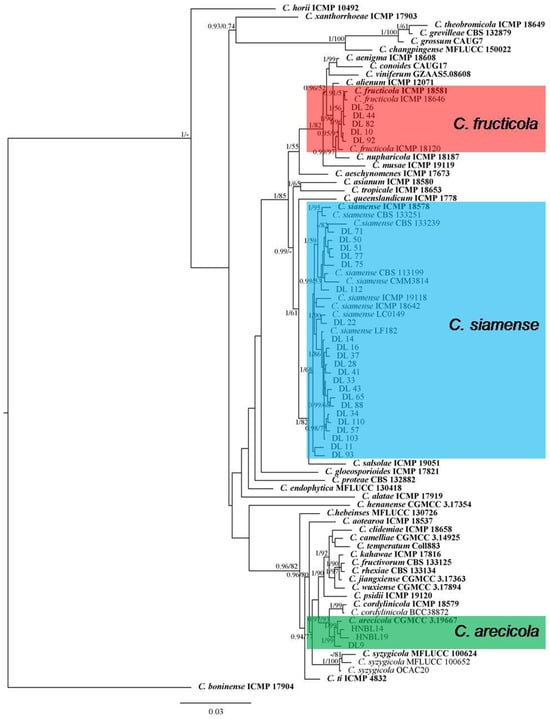
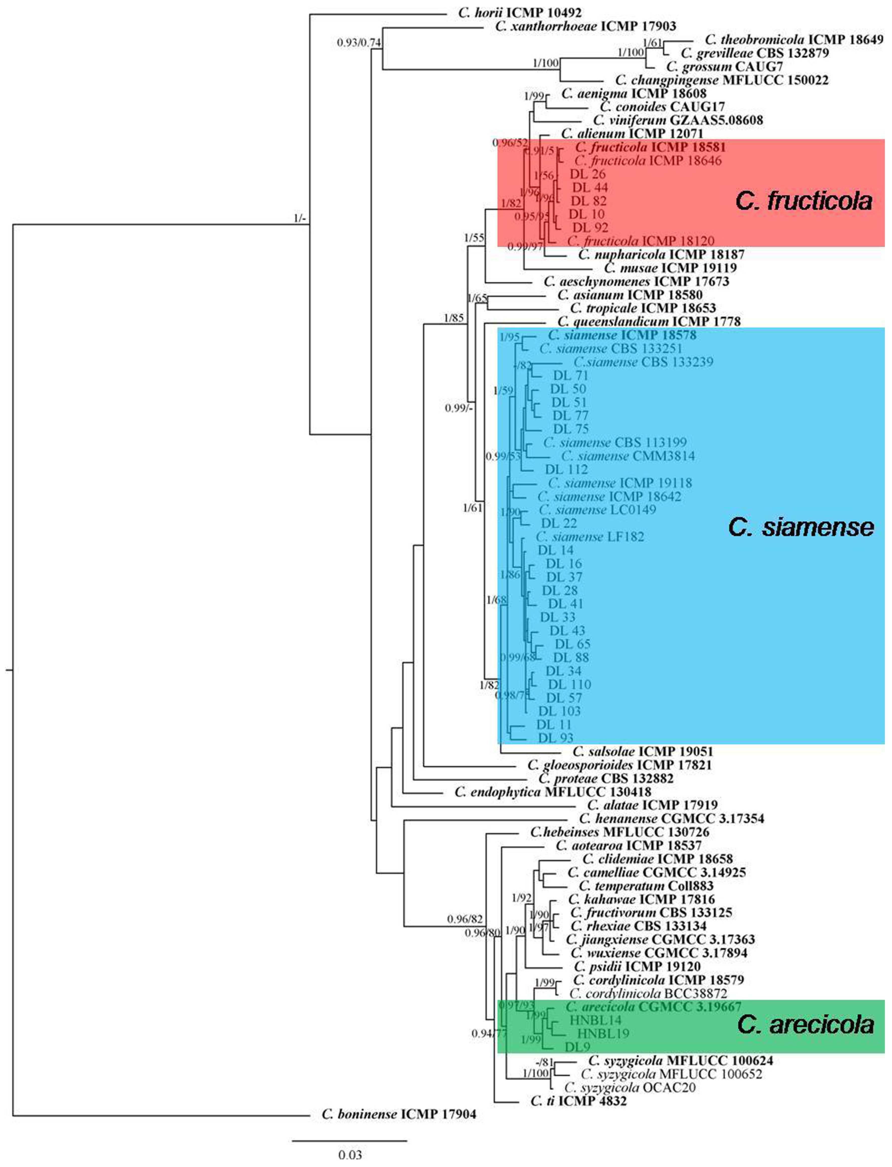
A multilocus phylogenetic analysis with the concatenated ITS, GAPDH, CHS-1, ACT, TUB2 and ApMat sequences was carried out for the isolates from the C. gloeosporioides species complex including 28 isolates from litchi in this study, 51 reference isolates from the C. gloeosporioides species complex and the outgroup C. boninense (ICMP 17904) (Figure 1). The combined gene alignment contained 3200 characters including gaps (gene/locus boundaries of ITS: 1–617, GAPDH: 618–927, CHS-1: 928–1226, ACT: 1227–1534, TUB2: 1535–2261, ApMat: 2262–3200) and the Bayesian analysis was performed based on 1445 unique site patterns (ITS: 142, GAPDH: 224, CHS-1: 85, ACT: 140, TUB2: 291, ApMat: 563). The maximum likelihood tree confirmed the tree topology from the Bayesian analysis. As the phylogenetic tree shows in Figure 1, for the 28 isolates in the C. gloeosporioides species complex, 1 isolate was clustered with C. arecicola (Bayesian posterior probabilities value 1/RAxML bootstrap support value 99), 5 with C. fructicola (0.95/95) and 22 with C. siamense (1/68) (Figure 1).
Figure 1.
A Bayesian inference phylogenetic tree built using concatenated sequences of ITS, ACT, CHS-1, GAPDH, TUB2 and ApMat for the Colletotrichum spp. isolates from the C. gloeosporioides species complex. The species C. boninense (ICMP 17904) was used as an outgroup. Bayesian posterior probability values (PP ≥ 0.90) and RAxML bootstrap support values (ML ≥ 50%) are shown at the nodes. Ex-type isolates are shown in bold. Colored blocks indicate clades including isolates obtained in this study.
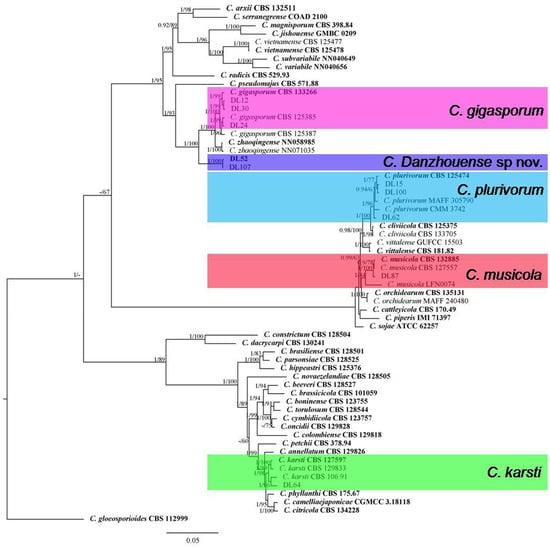
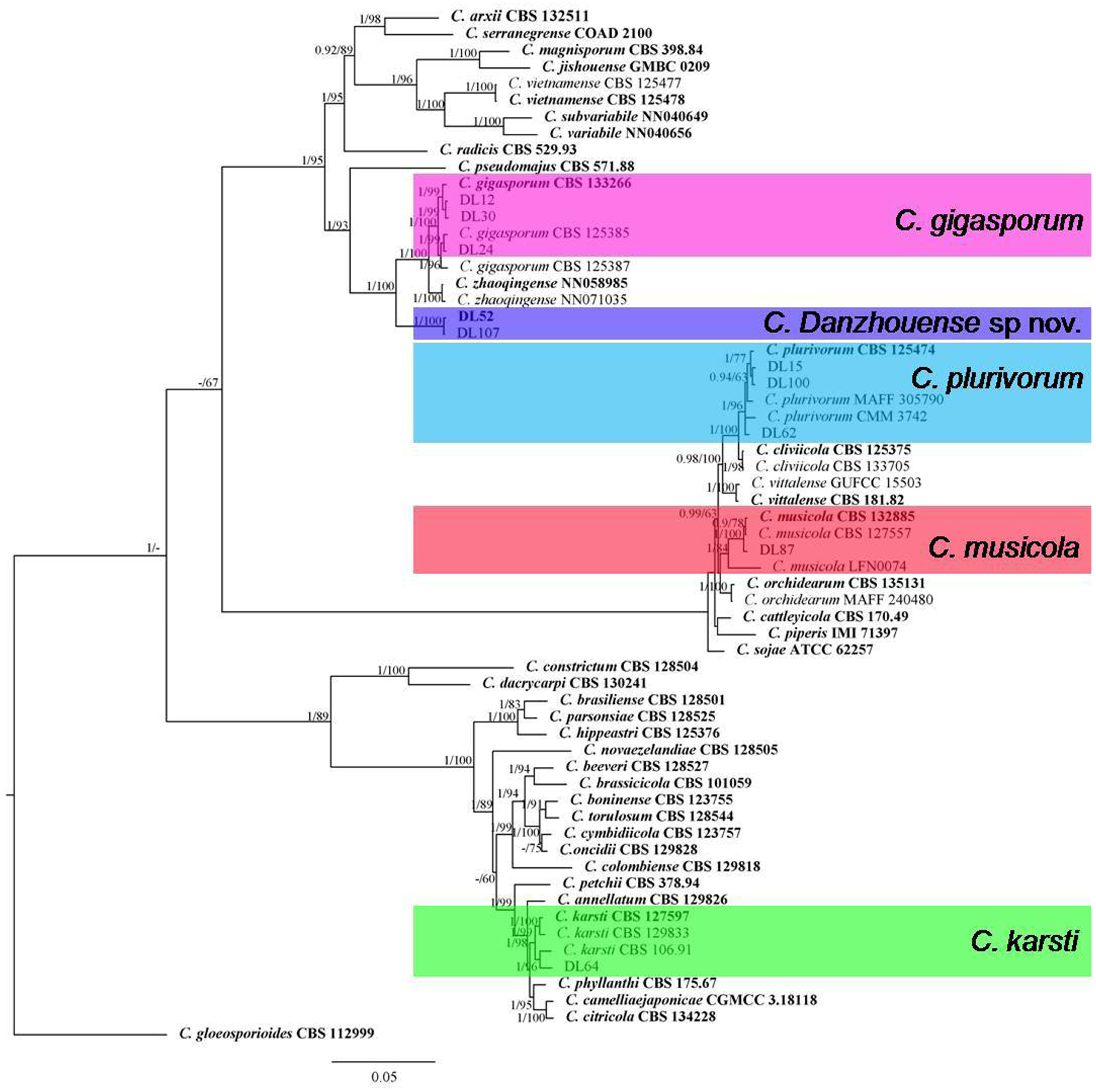
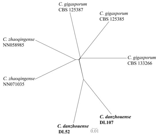
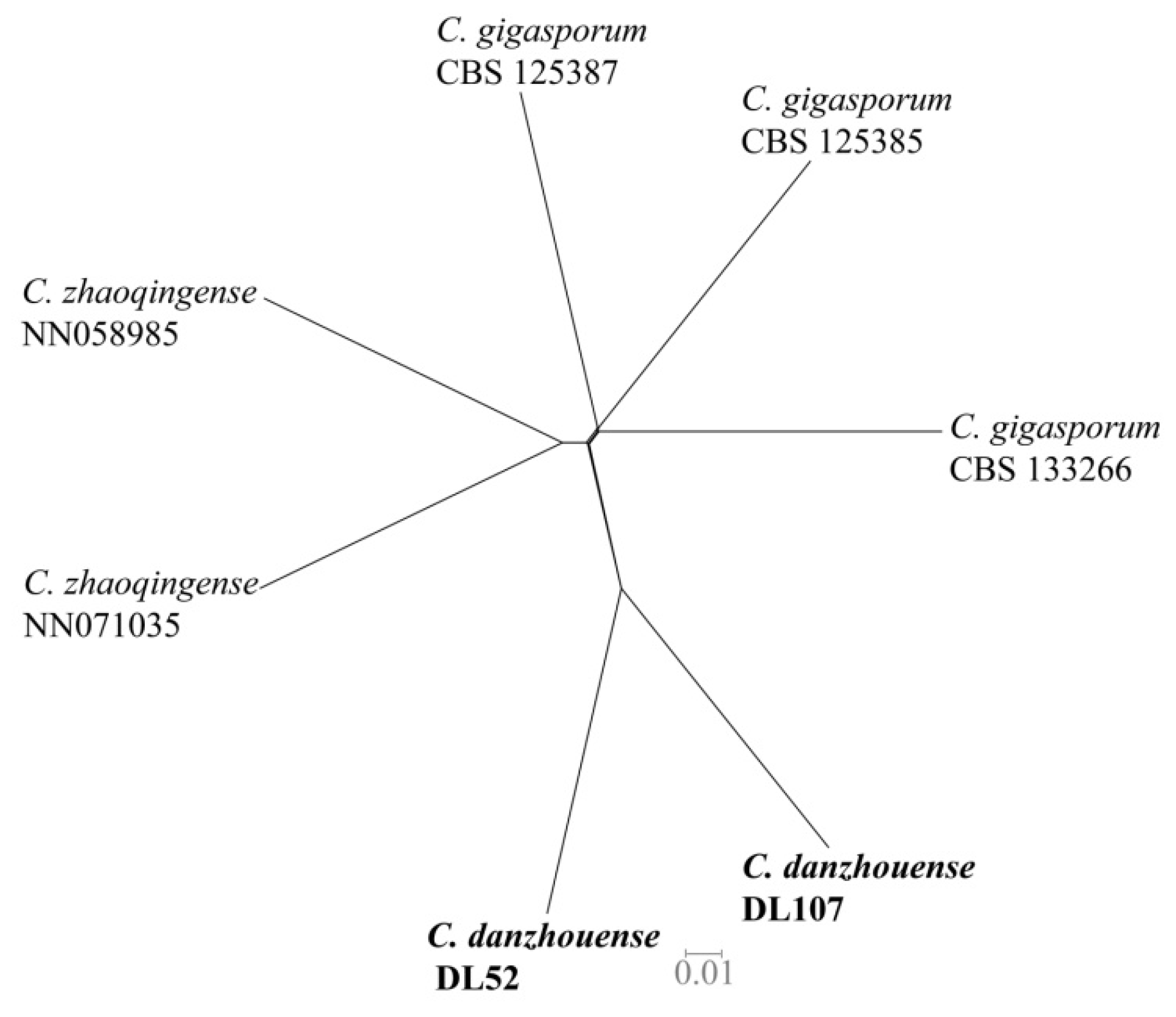
For isolates belonging to the other species complexes, the alignment of combined DNA sequences was obtained from 63 taxa, including 10 isolates from litchi in this study, 52 reference isolates of Colletotrichum species, and 1 outgroup strain C. gloeosporioides (ICMP 17821) (Figure 2). The gene/locus boundaries of the aligned 2299 characters (with gaps) were ITS: 1–618, GAPDH: 619–946, CHS-1: 947–1245, ACT: 1246–1539 and TUB2: 1540–2299), and the Bayesian analysis was performed based on 1169 unique site patterns (ITS: 197, GAPDH: 283, CHS-1: 117, ACT: 170, TUB2: 402). The maximum likelihood tree confirmed the tree topology from the Bayesian analysis. For the four isolates in the C. orchidearum species complex, one isolate was grouped with C. musicola (1/84) and three with C. plurivorum (1/96). One isolate from the C. boninense species complex was identified as C. karsti (1/99). For the five isolates in the C. gigasporum species complex, three of them were grouped with C. gigasporum (1/100), while the other two formed a clade distantly from any reported species in this complex, which was described as a new species, C. danzhouense, in this study (Figure 2). The application of the PHI test to the concatenated five-locus sequences (ITS, GAPDH, ACT, CHS-1 and TUB2) revealed that no significant recombination event (p = 0.14) occurred between C. danzhouense and phylogenetically related species C. gigasporum and C. zhaoqingense (Figure 3). This is further evidence that C. danzhouense is a new species.
Figure 2.
A Bayesian inference phylogenetic tree built using concatenated sequences of ITS, ACT, CHS-1, GAPDH and TUB2 for the Colletotrichum spp. isolates from the C. gigasporum, C. orchidearum and C. boninense species complex with C. gloeosporioides (ICMP 17821) as an outgroup. Bayesian posterior probability values (PP ≥ 0.90) and RAxML bootstrap support values (ML ≥ 50%) are shown at the nodes. Ex-type isolates are shown in bold. Colored blocks indicate clades including isolates obtained in this study.
Figure 3.
The result of the pairwise homoplasy index (PHI) of Colletotrichum danzhouense and its phylogenetically related species using both a LogDet transformation and splits decomposition. No significant recombination event (p = 0.14) was observed within the datasets. Isolates obtained in this study are shown in bold.
- Taxonomy
Colletotrichum danzhouense Fungal Names Number: FN 571654; Figure 4.
Figure 4.
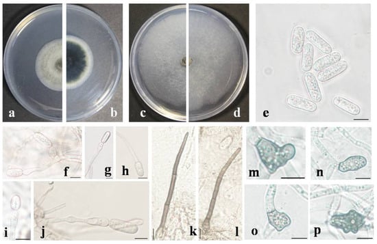
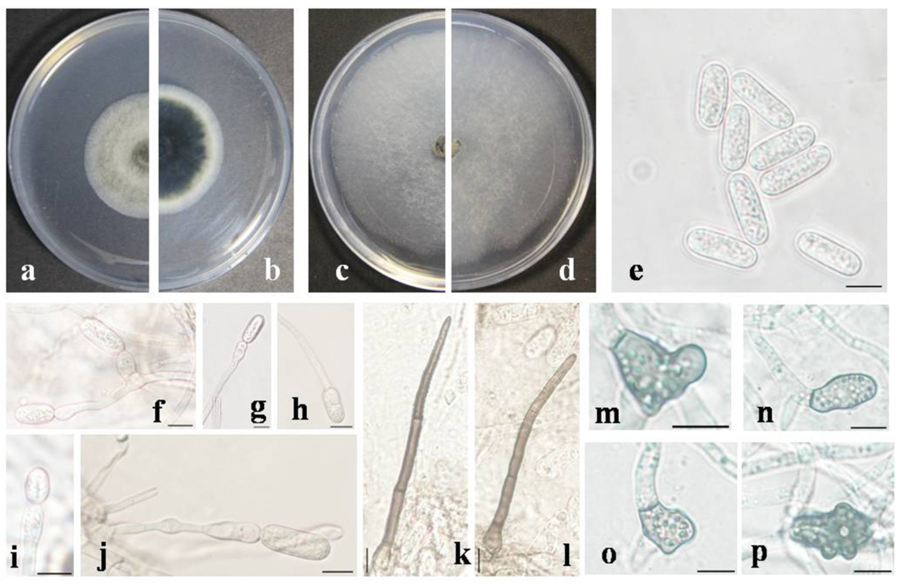
Morphological characteristics of Colletotrichum danzhouense. (a,b) Front and reverse colony on PDA (7 day); (c,d) front and reverse colony on SNA (7 day); (e) conidia; (f–j) conidiophores; (k,l) setae; (m–p) appressoria; (e–p) produced on SNA. Scale bars = 10 μm.
Etymology: Named after the location where the fungus was sampled, Danzhou city.
Type: China, Hainan province, Danzhou City, from diseased leaves of Litchi chinensis, 15 May 2023, X. R. Cao, holotype (HMAS 352507), ex-type living culture CGMCC 3.25375 = DL 52.
Description: Sexual morph not observed. Vegetative hyphae septate, hyaline, smooth-walled, branched. Conidia and setae not observed on PDA or OA. On SNA, conidiomata acervular, scattered, in which conidiophores are hardly observed. Setae 1–4 septate, 70.8–113.4 μm long, basal cells cylindrical, smooth-walled, 4.1–6.8 μm diameter, tip acute. Conidiophores, formed directly on hyphae, usually reduced to conidiogenous cells. Conidiogenous cells hyaline, cylindrical, formed terminally or laterally on hyphae, variable in size. Conidia hyaline, cylindrical with obtuse ends, smooth-walled, granular, 14.4–21.6 × 5.6–7.2 μm, mean ± SD = 17.6 ± 1.7 × 6.5 ± 0.4 μm, L/W ratio = 2.7. Appressoria variable in shape, pale brown, 9.7–19.2 × 8.5–14.3 μm, mean ± SD = 13.2 ± 2.1 × 10.5 ± 1.6 μm, L/W ratio = 1.3.
Culture characteristics: Colonies on PDA flat with entire edge, gray to pale green with a white margin, aerial mycelium floccose, reverse dark green in the center with a white margin. Colonies’ diameters of 52–54 mm, 80–85 mm and 40–44 mm in 7 day incubated at 25 °C on PDA, SNA and OA, respectively. Conidia and setae not observed on PDA or OA.
Additional specimens examined: China, Hainan province, Danzhou City, from diseased leaves of Litchi chinensis, 15 May 2023, X. R. Cao, living culture DL 107.
Notes: Colletotrichum danzhouense is phylogenetically closely related to C. gigasporum and C. zhaoqingense in the C. gigasporum species complex (Figure 2); it was isolated from infected litchi leaves collected from Danzhou in Hainan, China. It shares a low sequence similarity with C. gigasporum at GAPDH (89.5%), CHS-1 (96.0%) and TUB2 (96.4%). Also, a low sequence similarity was observed between the new species and C. zhaoqingense at GAPDH (89.8%), CHS-1 (96.3%) and TUB2 (96.1%). In morphology, C. danzhouense differs from C. gigasporum and C. zhaoqingense by producing shorter conidia (14.4–21.6 × 5.6–7.2 μm vs. 22–32 × 6–9 μm, 14.4–21.6 × 5.6–7.2 μm vs. 20–24 × 5.5–7 μm, respectively).
3.3. Morphological and Cultural Characterization
All species produced dense mycelium except C. karstii (Table 2). C. gigasporum produced larger conidia compared with other species identified in the present study. The three species, C. arecicola, C. fructicola and C. siamense, from the C. gloeosporioides species complex had similar conidia size, while the conidia size was different between the two species, C. danzhouense and C. gigasporum, from the C. gigasporum species complex. Additionally, the width of the conidia from these three species was smaller than that of the other five species obtained in this study. The L/W ratio of the conidia of C. karstii was smaller, while C. gigasporum had a larger L/W ratio. The growth rates of C. danzhouense, C. karstii and C. musicola were relatively slow at <9 mm/d while the growth rate was higher than 11 mm/d for the other five Colletotrichum species obtained in this study (Table 2).
Table 2.
Colony characteristics, sizes of conidia and growth rate of Colletotrichum species associated with anthracnose of litchi in this study.
3.4. Pathogenicity Tests
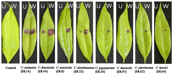
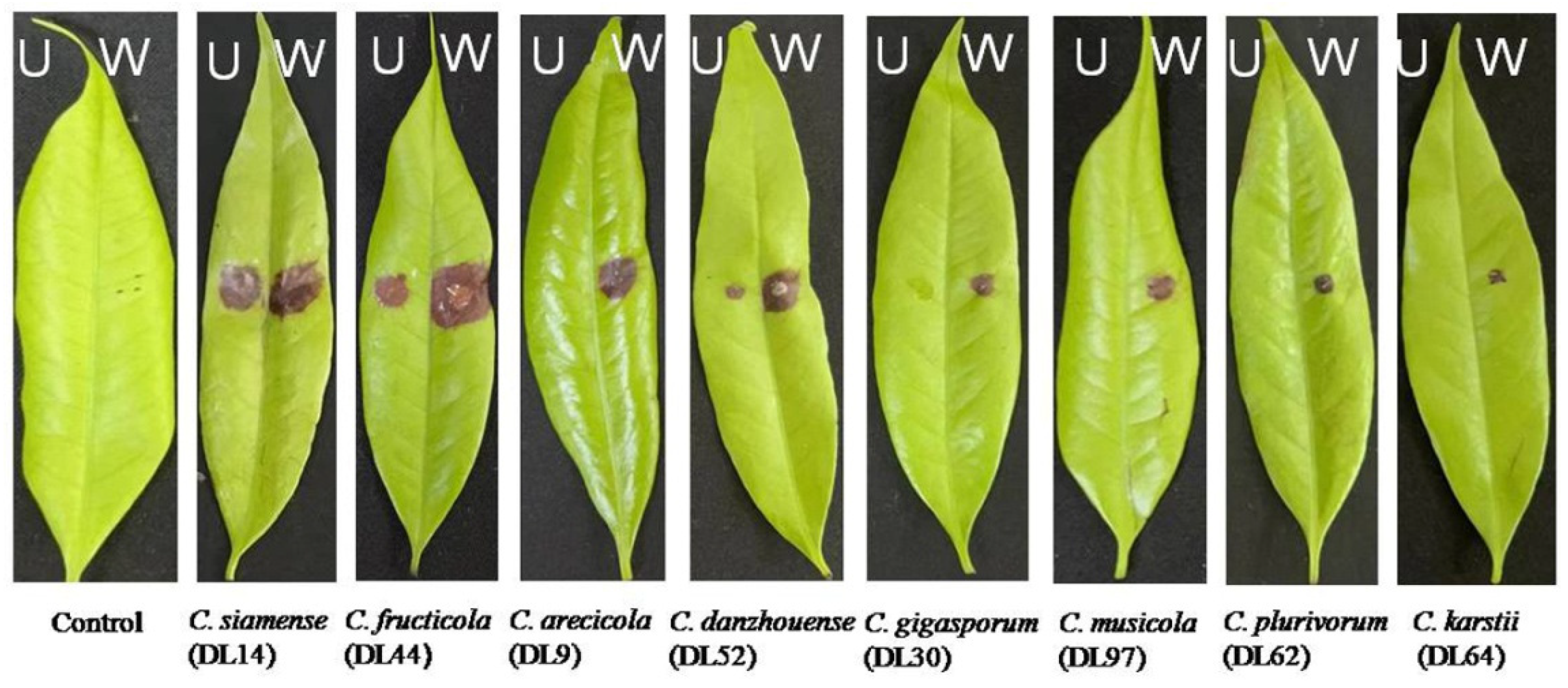
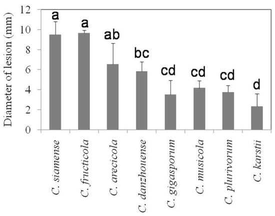
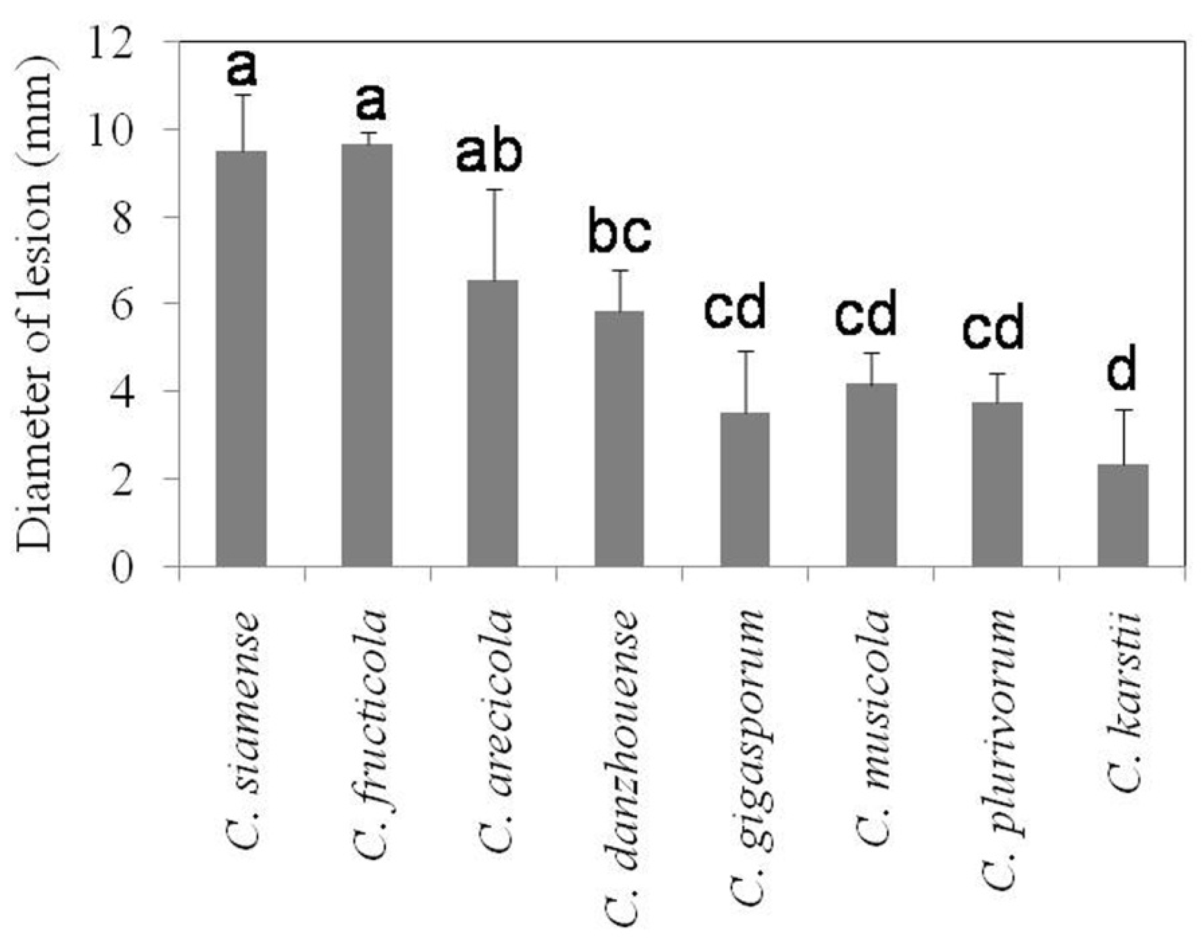
Eight Colletotrichum species were able to infect litchi leaves (cv. Feizixiao) and cause typical symptoms of anthracnose when inoculated onto wounded leaves (Figure 5) with an average lesion diameter ranging from 2.3 to 9.7 mm 4 days after inoculation (Figure 6). The diameters of lesions for C. fructicola and C. siamense (>9 mm) from the C. gloeosporioides species complex were significantly larger than those produced by other species except C. arecicola. The proposed new species, C. danzhouense, produced significantly larger lesion (>5.5 mm) than C. musicola, C. plurivorum and C. karstii, while the diameter of C. karstii was the smallest. However, five of the eight species did not produce visible symptoms on litchi leaves when nonwounded sites were inoculated, whereas C. danzhouense, C. fructicola and C. siamense did produce lesions on nonwounded, inoculated leaves (Figure 5).
Figure 5.
Symptoms of litchi leaves (cv. Feizixiao) induced by inoculation of spore suspensions of eight Colletotrichum spp. after four days at 25 °C under unwounded (U) and wounded (W) conditions.
Figure 6.
Lesion diameters of Colletotrichum species on litchi leaves (cv. Feizixiao) using wound inoculation methods. Letters over the error bars indicate a significant difference at the p = 0.05 level.
4. Discussion
In this study, pathogens from four Colletotrichum species complexes were found to cause litchi anthracnose in Hainan, China, and C. gigasporum species complex was first reported to cause anthracnose on litchi based on morphological and multilocus sequences. Nearly 80% of the isolates obtained in the present study belonged to the C. gloeosporioides species complex, which was consistent with previous reports that C. gloeosporioides was the main pathogen of litchi anthracnose [26,27].
A total of eight Colletotrichum species were found to be responsible for anthracnose of litchi in Hainan, China. Three of them (C. arecicola, C. fructicola and C. siamense) were from the C. gloeosporioides species complex. The former two species were reported on litchi [8,9]. C. siamense was the most common species to cause anthracnose of litchi in Hainan in this study. Also, this species was the dominant species associated with anthracnose of rubber tree, coffee and areca palm in Hainan [28,29,30]. Both rubber tree and areca palm are widely cultivated in Hainan, which is a likely factor contributing to the pathogen cross-infecting other hosts. C. fructicola is a plant pathogen with a broad host range [6]. Also, this species was reported on rubber tree, coffee and areca palm in Hainan. Furthermore, it was proved to be the most predominant species causing tea-oil camellia anthracnose in Hainan [31]. In this study, C. fructicola was isolated from litchi. C. arecicola, which had previously been reported only on areca palm in Hainan [30], was found on litchi for the first time in the present study.
Colletotrichum karsti from the C. boninense species complex is another species commonly detected in China with a broad host range [6]. This species was also reported on litchi in Guangxi, China [11]. In this study, C. karsti was also obtained on litchi in Hainan although only one isolate was obtained.
Two species from the C. orchidearum species complex were isolated in this study. One of them, C. plurivorum, has a broad host range and has been reported on litchi before [9]. The other species was C. musicola, which was first reported on Musa sp. [32]. Then, this species was reported on Colocasia esculenta [33], Glycine max [34] and Manihot esculenta [35]. This study is the first to demonstrate that this species can also occur on litchi, although it was found with a low frequency.
Colletotrichum gigasporum from the C. gigasporum species complex was reported as a causal agent of anthracnose disease on coffee and mango in Hainan [29,36]. This is the first report of this species on litchi. Furthermore, C. danzhouense, which clustered with C. gigasporum and C. zhaoqingense, was proposed as a new species in the C. gigasporum species complex because it had a low sequence similarity to the other two species at GAPDH, CHS-1 and TUB2. The BLAST results of the GAPDH and ITS sequences indicated that this species was most similar to Colletotrichum sp. Also, no significant recombination event (p = 0.14) occurred among these three species. Furthermore, C. danzhouense produced shorter conidia compared with C. gigasporum and C. zhaoqingense.
Colletotrichum species from the C. acutatum species complex was also reported previously as the pathogen causing litchi anthracnose in Australia and China [8,9]. However, no isolates from this complex were obtained in this study. The main reason may be the geographic distribution of the pathogen and the different sample sites studied. Also, C. acutatum was only occasionally obtained from litchi in a previous study [26], but it was rarely found.
Wounding is known to enhance Colletotrichum infection and disease development. Furthermore, for grape leaf [37] and mango fruit [38], wounding is necessary for Colletotrichum to infect. Only 2 of 12 Colletotrichum species from cultivated pear were pathogenic to pear leaves inoculated without wounding [39]. This was also observed in this study, as the pathogenicity tests indicated that all eight species isolated were able to infect litchi leaves when inoculated onto wounded leaves, while only three of the eight species induced visible symptoms on litchi leaves using a nonwound inoculation method. One reason could be that the cuticle and epidermis may act as a barrier for the infection by Colletotrichum spp. [40]. Alternatively, the quiescent infection, which means that the infection of healthy intact leaves may produce visual symptoms only at a later stage when the leaf physiological state changes significantly, is an important feature of Colletotrichum spp. [41]. In field conditions, wounds on litchi leaves can be common in nature due to wind, insect damage and abrasions caused by leaves rubbing. Generally, isolates from C. fructicola and C. siamense were found to cause larger lesions than those caused by other species. These two species were also the most common species obtained in this study.
In conclusion, eight Colletotrichum species from four species complexes were demonstrated as pathogens causing litchi anthracnose in Hainan, China; one species complex and four species were reported on litchi for the first time. The results of this study can be valuable for developing sustainable management strategies for anthracnose of litchi. The precise identification of fungal pathogens is important for disease control measures. Currently, the main strategy for litchi anthracnose management is fungicide application [2]. It was reported that Colletotrichum species displayed differential sensitivity to fungicides [29,42]. Therefore, it is essential to determine the species in a given plantation before fungicide applications.
Author Contributions
Conceptualization, J.W. and X.C.; methodology, X.C., F.L. and H.X.; validation, F.L. and H.X.; formal analysis, S.W.; investigation, F.L., H.X., H.L. and G.W.; writing—original draft preparation, X.C.; writing—review and editing, J.S.W.; supervision, J.W.; funding acquisition, F.L. and J.W. All authors have read and agreed to the published version of the manuscript.
Funding
This study was supported by the earmarked fund for CARS (grant No. CARS-32-01) and the Science and Technology Special Fund of Hainan Province (grant No. ZDYF2021XDNY159).
Institutional Review Board Statement
Not applicable.
Informed Consent Statement
Not applicable.
Data Availability Statement
Alignments generated during the current study are available from TreeBASE (http://treebase.org/treebase-web/home.html; study 30748, accessed on 6 September 2023). All sequence data are available in the NCBI GenBank, following the accession numbers in the manuscript.
Conflicts of Interest
The authors declare no conflict of interest. The funders had no role in the design of the study; in the collection, analyses, or interpretation of data; in the writing of the manuscript; or in the decision to publish the results.
References
- Chen, H.B. The major problem and countermeasure for the developmentof litchi, longan and mango in China. China Fruit News 2017, 34, 11–13. [Google Scholar]
- Li, T.; Li, J.; Zhao, X.; Xu, Y.Q.; Nie, G.; Wu, C.X. Analysis of the main pests occurred and pesticides registered on litchi in China. Pestic. Sci. Adm. 2022, 43, 7–12. [Google Scholar]
- Dean, R.; Kan, J.A.L.V.; Pretorius, Z.A.; Hammond-Kosack, K.E.; Pietro, A.D.; Spanu, P.D.; Rudd, J.J.; Dichman, M.; Kahmann, R.; Ellis, J.; et al. The top 10 fungal pathogens in molecular plant pathology. Mol. Plant Pathol. 2012, 13, 414–430. [Google Scholar] [CrossRef] [Scilit] [PubMed]
- Cai, L.; Hyde, K.D.; Taylor, P.W.J.; Weir, B.; Waller, J.M.; Abang, M.M.; Zang, J.C.; Yang, Y.L.; Phouliyong, S.; Prihastuti, Z.Y.; et al. A polyphasic approach for studying Colletotrichum. Fungal Divers. 2009, 39, 183–204. [Google Scholar]
- Marin-Felix, Y.; Groenewald, J.Z.; Cai, L.; Chen, Q.; Marincowitz, S.; Barnes, I.; Bensch, K.; Braun, U.; Camporesi, E.; Damm, U.; et al. Genera of phytopathogenic fungi: GOPHY 1. Stud. Mycol. 2017, 86, 99–216. [Google Scholar] [CrossRef] [Scilit]
- Liu, F.; Ma, Z.Y.; Hou, L.W.; Diao, Y.Z.; Wu, W.P.; Damm, U.; Song, S.; Cai, L. Updating species diversity of Colletotrichum, with a phylogenomic overview. Stud. Mycol. 2022, 101, 1–56. [Google Scholar] [CrossRef] [Scilit]
- Weir, B.S.; Johnston, P.R.; Damm, U. The Colletotrichum gloeosporioides species complex. Stud. Mycol. 2012, 73, 115–180. [Google Scholar] [CrossRef] [Scilit]
- Shivas, R.G.; Tan, Y.P.; Edwards, J.; Dinh, Q.; Maxwell, A.; Andjic, V.; Liberato, J.R.; Anderson, C.; Beasley, D.R.; Bransgrove, K.; et al. Colletotrichum species in Australia. Australas. Plant Pathol. 2016, 45, 447–464. [Google Scholar]
- Ling, J.F. Identification and Phylogenetic Analysis of Four Fungal Genera Isolates Associated with Diseased Litchi Fruits in China. Ph.D. Thesis, Huanan Agriculture University, Guangzhou, China, 2019. [Google Scholar]
- Ling, J.F.; Song, S.B.; Xi, P.G.; Cheng, B.P.; Cui, Y.P.; Chen, P.; Peng, A.T.; Jiang, Z.D.; Zhang, L.H. Identification of Colletotrichum siamense causing litchi pepper spot disease in mainland China. Plant Pathol. 2019, 68, 1533–1542. [Google Scholar] [CrossRef] [Scilit]
- Zhao, J.; Yu, Z.; Wang, Y.; Li, Q.; Tang, L.; Guo, T.; Huang, S.; Mo, J.; Hsiang, T. Litchi anthracnose caused by Colletotrichum karstii in Guangxi, China. Plant Dis. 2021, 105, 3295. [Google Scholar] [CrossRef] [Scilit]
- Chen, H.B.; Su, Z.X. Analysis on the litchi production situation in 2022. Chin. Trop. Agric. 2022, 3, 5–14. [Google Scholar]
- Gardes, M.; Bruns, T.D. ITS primers with enhanced specificity for basidiomycetes—Application to the identification of mycorrhizae and rusts. Mol. Ecol. 1993, 2, 113–118. [Google Scholar] [CrossRef] [Scilit] [PubMed]
- White, T.J.; Bruns, T.; Lee, S.; Taylor, J. Amplification and direct sequencing of fungal ribosomal RNA genes for phylogenetics. In PCR Protocols: A Guide to Methods and Applications; Innis, M.A., Gelfand, D.H., Sninsky, J.J., White, T.J., Eds.; Academic Press Inc.: New York, NY, USA, 1990; pp. 315–322. [Google Scholar]
- Templeton, M.D.; Rikkerink, E.H.; Solon, S.L.; Crowhurst, R.N. Cloning and molecular characterization of the glyceraldehyde-3-phosphate dehydrogenase-encoding gene and cDNA from the plant pathogenic fungus Glomerella cingulata. Gene 1992, 122, 225–230. [Google Scholar] [CrossRef] [Scilit] [PubMed]
- Glass, N.L.; Donaldson, G.C. Development of primer sets designed for use with the PCR to amplify conserved genes from filamentous ascomycetes. Appl. Environ. Microbiol. 1995, 61, 1323–1330. [Google Scholar] [CrossRef] [Scilit] [PubMed]
- O’Donnell, K.; Cigelnik, E. Two divergent intragenomic rDNA ITS2 types within a monophyletic lineage of the fungus Fusarium are nonorthologous. Mol. Phylogenetics Evol. 1997, 7, 103–116. [Google Scholar] [CrossRef] [Scilit]
- Silva, D.N.; Talhinhas, P.; Várzea, V.; Cai, L.; Paulo, O.S.; Batista, D. Application of the Apn2/MAT locus to improve the systematics of the Colletotrichum gloeosporioides complex: An example from coffee (Coffea spp.) hosts. Mycologia 2012, 104, 396–409. [Google Scholar] [CrossRef] [Scilit]
- Ronquist, F.; Teslenko, M.; van der Mark, P.; Ayres, D.L.; Darling, A.; Höhna, S.; Larget, B.; Liu, L.; Suchard, M.A.; Huelsenbeck, J. MrBayes 3.2: Efficient Bayesian phylogenetic inference and model choice across a large model space. Syst. Biol. 2012, 61, 539–542. [Google Scholar] [CrossRef] [Scilit]
- Nylander, J.A.A. MrModelTest v. 2; Evolutionary Biology Centre, Uppsala University: Uppsala, Sweden, 2004. [Google Scholar]
- Silvestro, D.; Michalak, I. raxmlGUI: A graphical front-end for RAxML. Org. Divers. Evol. 2012, 12, 335–337. [Google Scholar] [CrossRef] [Scilit]
- Quaedvlieg, W.; Binder, M.; Groenewald, J.Z.; Summerell, B.A.; Carnegie, A.J.; Burgess, T.I.; Crous, P.W. Introducing the consolidated species concept to resolve species in the Teratosphaeriaceae. Persoonia 2014, 33, 1–40. [Google Scholar] [CrossRef] [Scilit]
- Huson, D.H. SplitsTree: Analyzing and visualizing evolutionary data. Bioinformatics 1998, 14, 68–73. [Google Scholar] [CrossRef] [Scilit]
- Huson, D.; Bryant, D. Application of phylogenetic networks in evolutionary studies. Mol. Biol. Evol. 2006, 23, 254–267. [Google Scholar] [CrossRef] [Scilit] [PubMed]
- Luo, D.L.; Wang, W.; Bai, C.H.; Zhu, L.W.; Li, H.; Zhou, C.M.; Qiu, Q.M.; Yao, L.X. Foliar nutrient diagnosis norms for litchi (Litchi chinensis Sonn. cv. Feizixiao) in South China. J. Plant Nutr. 2021, 45, 3174–3187. [Google Scholar] [CrossRef] [Scilit]
- Anderson, J.M.; Aitken, E.A.B.; Dann, E.K.; Coates, L.M. Morphological and molecular diversity of Colletotrichum spp. causing pepper spot and anthracnose of lychee (Litchi chinensis) in Australia. Plant Pathol. 2013, 62, 279–288. [Google Scholar] [CrossRef] [Scilit]
- Zhang, X.C.; Xiao, Q.; Gao, Z.Y.; Wang, J.B. The detection of pathogenicity for Colletotricum from different litchi cultivars and regions. J. Fruit Sci. 2014, 31, 296–301. [Google Scholar]
- Cao, X.R.; Xu, X.M.; Che, H.Y.; West, J.S.; Luo, D.Q. Three Colletotrichum species, including a new species, are associated to leaf anthracnose of rubber tree in Hainan, China. Plant Dis. 2019, 103, 117–124. [Google Scholar] [CrossRef] [Scilit] [PubMed]
- Cao, X.R.; Xu, X.M.; Che, H.Y.; West, J.S.; Luo, D.Q. Characteristics and distribution of Colletotrichum species in coffee plantations in Hainan, China. Plant Pathol. 2019, 68, 1146–1156. [Google Scholar] [CrossRef] [Scilit]
- Cao, X.R.; Xu, X.M.; Che, H.Y.; West, J.S.; Luo, D.Q. Eight Colletotrichum species, including a novel species, are associated with areca palm anthracnose in Hainan, China. Plant Dis. 2020, 104, 1369–1377. [Google Scholar] [CrossRef] [Scilit]
- Zhu, H.; He, C. Identification and characterization of Colletotrichum species causing tea-oil camellia (Camellia oleifera C. Abel) anthracnose in Hainan, China. Forests 2023, 14, 1030. [Google Scholar] [CrossRef] [Scilit]
- Damm, U.; Sato, T.; Alizadeh, A.; Groenewald, J.Z.; Crous, P.W. The Colletotrichum dracaenophilum, C. magnum and C. orchidearum species complexes. Stud. Mycol. 2019, 92, 1–46. [Google Scholar] [CrossRef] [Scilit]
- Vásquez-López, A.; Palacios-Torres, R.E.; Camacho-Tapia, M.; Granados-Echegoyen, C.; Lima, N.B.; Vera-Reyes, I.; Tovar-Pedraza, J.M.; Leyva-Mir, S.G. Colletotrichum brevisporum and C. musicola causing leaf anthracnose of Taro (Colocasia esculenta) in Mexico. Plant Dis. 2019, 103, 2963. [Google Scholar] [CrossRef] [Scilit]
- Boufleur, T.R.; Castro, R.R.L.; Rogério, F.; Ciampi-Guillardi, M.; Baroncelli, R.; Massola, J.N.S. First report of Colletotrichum musicola causing soybean anthracnose in Brazil. Plant Dis. 2020, 104, 1858. [Google Scholar] [CrossRef] [Scilit]
- Machado, S.D.; Veloso, J.S.; Camara, M.P.; Campos, F.S.; Damascena, J.F.; de Souza Ferreira, T.P.; Dos Santos, M.M.; Santos, G.R. Occurrence of Colletotrichum musicola causing anthracnose on Manihot esculenta. Plant Dis. 2022, 106, 2758. [Google Scholar] [CrossRef] [Scilit]
- Li, Q.; Bu, J.; Shu, J.; Yu, Z.; Tang, L.; Huang, S.; Guo, T.; Mo, J.; Luo, S.; Solangi, G.S.; et al. Colletotrichum species associated with mango in southern China. Sci. Rep. 2019, 9, 18891. [Google Scholar] [CrossRef] [Scilit] [PubMed]
- Hong, J.K.; Hwang, B.K. Influence of inoculum density, wetness duration, plant age, inoculation method, and cultivar resistance on infection of pepper plants by Colletotrichum coccodes. Plant Dis. 1998, 82, 1079–1083. [Google Scholar] [CrossRef] [Scilit] [PubMed]
- Lima, N.B.; Batista, M.V.D.A.; De Morais, M.A.; Barbosa, M.A.; Michereff, S.J.; Hyde, K.D.; Câmara, M.P. Five Colletotrichum species are responsible for mango anthracnose in northeastern Brazil. Fungal Divers. 2013, 61, 75–88. [Google Scholar] [CrossRef] [Scilit]
- Fu, M.; Crous, P.W.; Bai, Q.; Zhang, P.F.; Xiang, J.; Guo, Y.S.; Zhao, F.F.; Yang, M.M.; Hong, N.; Xu, W.X.; et al. Colletotrichum species associated with anthracnose of Pyrus spp. in China. Persoonia 2019, 42, 1–35. [Google Scholar] [CrossRef] [Scilit]
- Auyong, A.S.M.; Ford, R.; Taylor, P.W.J. The role of cutinase and its impact on pathogenicity of Colletotrichum truncatum. J. Plant Pathol. Microbiol. 2015, 6, 259–269. [Google Scholar] [CrossRef]
- Peres, N.A.; Timmer, L.W.; Adaskaveg, J.E.; Correll, J.C. Life styles of Colletotrichum acutatum. Plant Dis. 2005, 89, 784–796. [Google Scholar] [CrossRef] [Scilit]
- Chen, S.N.; Luo, C.X.; Hu, M.J.; Schnabel, G. Sensitivity of Colletotrichum species, including C. fioriniae and C. nymphaeae, from peachto demethylation inhibitor fungicides. Plant Dis. 2016, 100, 2434–2441. [Google Scholar] [CrossRef] [Scilit]

Disclaimer/Publisher’s Note: The statements, opinions and data contained in all publications are solely those of the individual author(s) and contributor(s) and not of MDPI and/or the editor(s). MDPI and/or the editor(s) disclaim responsibility for any injury to people or property resulting from any ideas, methods, instructions or products referred to in the content. |
© 2023 by the authors. Licensee MDPI, Basel, Switzerland. This article is an open access article distributed under the terms and conditions of the Creative Commons Attribution (CC BY) license (https://creativecommons.org/licenses/by/4.0/).