


Abstract
Gene inactivation in model organisms has identified numerous genes and signaling pathways involved in mammalian cardiac outflow tract (OFT) development. Human genetics data have implicated the VEGFR3 gene in OFT development, but when and where it is required is unknown. In this study we determined the sensitivity of the developing murine heart to reduced Vegfr3 gene dosage, and we tested whether its requirement is dependent upon TBX1, a known regulator of Vegfr3 expression in cardiac and lymphatic endothelial cells. We found that in the mouse, a single copy of the Vegfr3 gene was sufficient for normal heart development in most cases. Mutation of a single copy of the Tbx1 gene greatly enhanced the sensitivity of heart development to Vegfr3 dosage reduction and led to the formation of cardiac defects. In addition, deletion of Vegfr3 in the Tbx1 expression domain also led to severe cardiac OFT abnormalities. We used RNAscope to reveal the location of Vegfr3 and Tbx1 transcripts in midterm mouse embryos. This revealed co-localization of these transcripts in the endothelium of the aortic sac, caudal pharyngeal arch arteries and proximal OFT, suggesting that these are potential sites of genetic interaction between Vegfr3 and Tbx1 that are critical for murine heart development.
1. Introduction
Mutations of the VEGFR3 and TBX1 genes are associated with Milroy syndrome and DiGeorge syndrome (DGS), respectively. Both diseases affect the vascular system. Milroy syndrome is the most frequent primary hereditary lymphedema [1]. Studies in animal models have shown that Vegfr3 is a major lymphangiogenesis gene, being required for the formation of the jugular lymph sacs and systemic lymphatic vessels [2,3,4]. Prior to the development of the lymphatic system, Vegfr3 is required in blood vessels, and its loss causes early embryonic lethality due to the failure of remodeling of the primitive vascular plexus, which requires angiogenic sprouting [5,6]. DiGeorge syndrome, on the other hand, is primarily a TBX1 haploinsufficiency disorder, and is not commonly associated with lymphatic anomalies, although rare cases have been reported [7,8]. In humans, DGS is mainly caused by a heterozygous chromosomal microdeletion on chromosome 22 that encompasses over 40 genes, including TBX1. Heterozygous TBX1 point mutations recapitulate most of the physical defects associated with the 22q11.2 chromosomal microdeletion, including congenital heart defects, skeletal defects, facial dysmorphism and immune defects, thus providing evidence of the pleotropic functions of TBX1 during embryonic and fetal development [9].
Studies in mice have shown that TBX1 plays a critical role in cardio-pharyngeal mesoderm (CPM), a population of progenitors that differentiates into different cell types that contribute to the formation of the heart, facies and neck glands [10], thereby accounting for the aforementioned clinical signs and symptoms of DGS. Furthermore, homozygous inactivation of Tbx1 causes generalized lymphatic vessel hypoplasia or aplasia, which is lethal perinatally [11]. TBX1 activates Vegfr3 expression in murine lymphatic endothelial cells by binding a putative enhancer in the endogenous Vegfr3 gene [11], and the two molecules interact strongly during cardiac lymphangiogenesis [12]. TBX1 also regulates Vegfr3 in brain microvessels during murine embryogenesis [13].
In humans, rare variants of the VEGFR3 gene are associated with cardiac outflow tract (OFT) abnormalities, including Tetralogy of Fallot (TOF) [14,15,16,17], suggesting a previously unknown role for the gene in cardiac development. Interestingly, TOF is the most common congenital heart defect found in patients with DGS [18,19], and at least some of the hallmarks of TOF occur in mouse models of DGS [20]. We hypothesize that interaction between TBX1 and Vegfr3 is required in cardiac progenitors in the CPM, or in their descendants, for development of the cardiac OFT. Studies conducted in mice, including mouse models of DGS, show that TBX1 is required for the deployment of cardiac progenitors from the second heart field (part of the CPM) into the developing OFT [21,22]. This progenitor population gives rise to cardiomyocytes, endothelial cells (EC) and branchiomeric myocytes [23,24,25]. Fate mapping experiments have revealed that ECs in the aortic sac (AS) and OFT are of second heart field (SHF) origin [26,27], but they have also revealed the existence of different populations of SHF progenitors that are spatially and temporally separate and give rise to different structures and cell types. For example, ECs of the anterior pharyngeal arch arteries (PAAs) and aortic sac arise earlier than those of the posterior PAAs and from a different population of SHF progenitors [23,28]. Thus, while SHF-derived ECs populate the OFT, to our knowledge, it is not known whether they play a critical role in cardiac morphogenesis, or whether Tbx1-fated ECs populate the portion of the OFT involved in septation. We investigated this here using mouse genetics approaches.
2. Materials and Methods
Mouse lines and genotyping: Mouse studies were performed in accordance with the animal protocol no. 28/2022-PR (approval date: 18 January 2022), reviewed by the local IACUC committee and by the Italian Istituto Superiore di Sanità and approved by the Italian Ministero della Salute, according to Italian law and European guidelines. Tbx1Cre/+ [29], obtained from the European Mouse Mutant Archive (EMMA, Monterotondo, Italy; # EM11399), Vegfr3flox/+ [30], obtained from the EMMA (Monterotondo, Italy; #EM09463), Vegfr3+/− [12], generated by our laboratory and RosamTmG [31], obtained from Jackson Lab (Bar Harbor, ME, USA; # JAX037456) mice were maintained on a C57BL/6N background. Genotyping of mice was performed as in the original reports.
Immunofluorescence: Embryos were fixed in 4% paraformaldehyde, embedded in paraffin, and sectioned at thickness of 10 µm. The sections were rehydrated, and antigen retrieval was performed in 10 mM sodium citrate pH 6.0. Blocking was carried out using 0.5% milk, 10% fetal bovine serum, and 1% bovine serum albumin H2O (blocking solution). Incubation with the primary antibody in blocking solution was carried out overnight at room temperature, and signal detection was performed using secondary antibodies conjugated with different fluorochromes (1:500). We used the following primary antibodies: anti-GFP (ab13970, Abcam Cambridge, UK; 1:1000) and anti-Vegfr3 (14-5988-82, ebioscence/Thermo Fisher Scientific, Waltham, MA, USA; 1:400). The secondary antibodies were: goat anti-Chicken IgY Alexa Fluor™ 488 (A11039 Invitrogen/Thermo Fisher Scientific, Waltham, MA, USA; 1:500) and goat anti-Rat IgG (H + L) Alexa Fluor™ 594 (A11007, Invitrogen; 1:500). Imaging was carried out using using a Leica THUNDER Imaging System (Leica Microsystems, Wetzlar, Germany) equipped with a Leica DFC9000 GTC camera, a Lumencor fluorescence LED light source (Lumencor, Beaverton, OR, USA), and Leica Application Suite X software (version 3.7.4.23463).
RNAscope in situ hybridization: Whole embryos were fixed in 4% paraformaldehyde, dehydrated with increasing concentrations of methanol, and stored at 20 °C. Embryos were hybridized with mRNA probes following the manufacturer’s instructions (RNAscope™, Advanced Cell Diagnostics (ACD), Newark, CA, USA); briefly, whole embryos were rehydrated in decreasing concentrations of methanol and permeabilized for 20 min with Protease III, followed by overnight incubation at 40 °C with the probe solution (see list below). The signal was developed with TSA FITC (Akoya Bioscience, Marlborough, MA, USA, NEL741001KT; 1:500) and TSA-CY3 (Akoya Bioscience NEL745001KT; 1:2000). The embryos were cryoprotected with increasing concentration of sucrose (10%, 20% and 30% sucrose in PBS 1×) at 4 °C, OCT/sucrose 30% (50:50), embedded in OCT, cryosectioned (sagittal and transverse) at a thickness of 10 µm and imaged using the Leica Thunder Imaging System described above. We used the following commercial probes provided by RNAscope™: Probe-Mm-Tbx1-C2 (481911-C2); Probe-Mm-Flt4 (481371). Imaging was carried out as for immunofluorescence experiments.
3. Results
3.1. Vegfr3 Heterozygosity Is Not a Significant Cause of Cardiac Defects
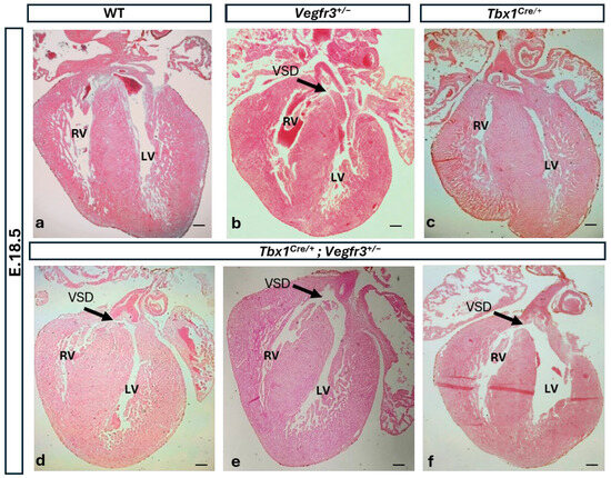
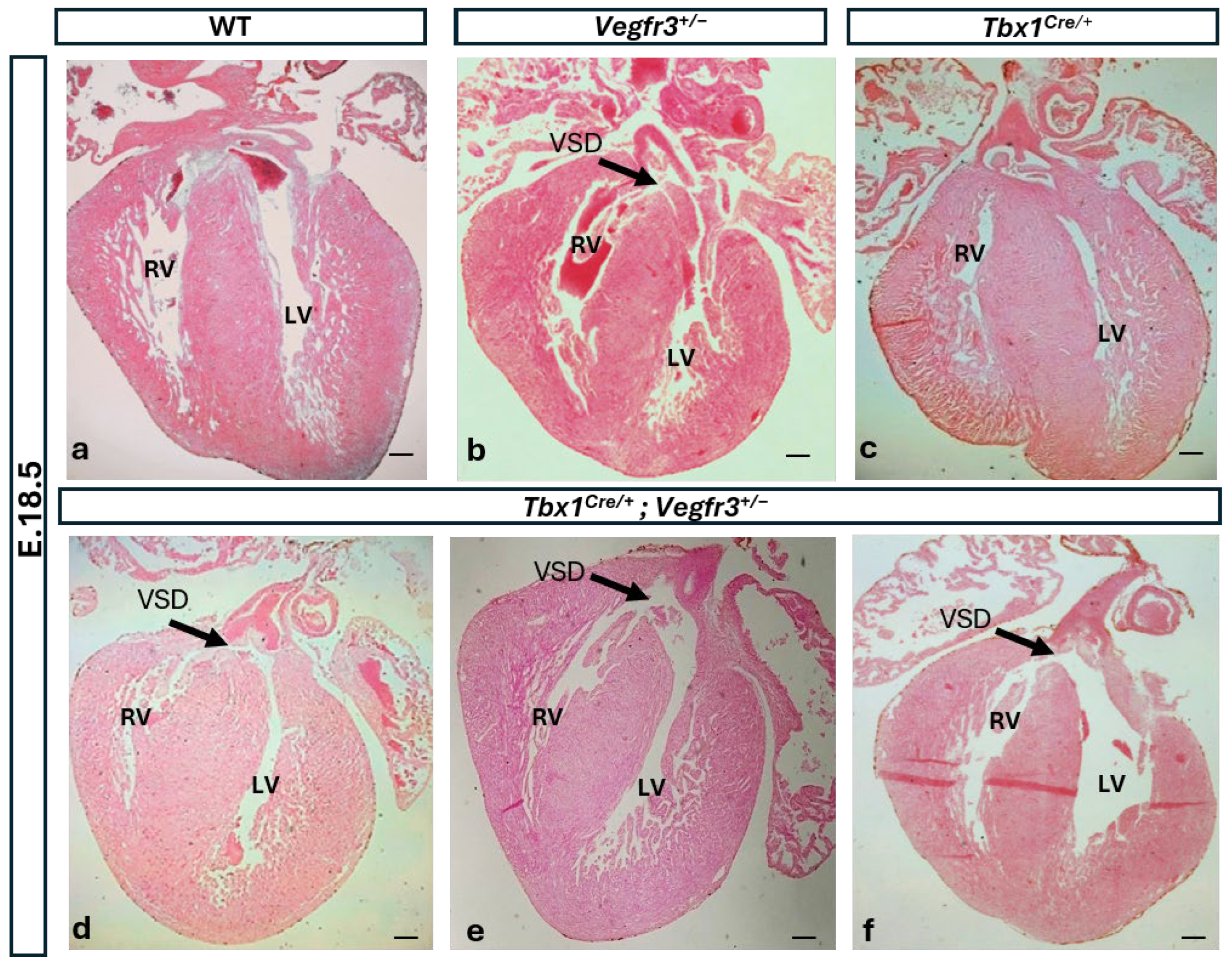
To understand whether Vegfr3 has a role in murine cardiac development, we first evaluated the effect of heterozygous inactivation in the mouse. We analyzed the cardiac phenotype (Table 1, Figure 1) in coronal sections of isolated embryonic hearts at the fetal stage (E)18.5. The results revealed a low penetrance (10%, 1/11) of intraventricular septal defects (VSD) in Vegfr3+/− embryos compared to WT (0/5) (Figure 1b). The VSD was of the central perimembranous VSD (pmVSD) subtype, i.e., located beneath the aortic valve and with the aorta entirely committed to the left ventricle [32,33]. No other intracardiac or great vessel defects were identified, and the cardiac valves appeared to be normal. We have reported mild cardiac lymphatic vessel hypertrophy in Vegfr3+/− embryos [12], indicating that in the mouse, loss of one copy of Vegfr3 affects cardiac lymphatic development but not cardiac morphogenesis.
Table 1.
Cardiac phenotype in embryos (E18.5) with single and combined genotypes.
Figure 1.
Tbx1 and Vegfr3 interact genetically in cardiac development. Coronal sections of isolated hearts, each obtained from a different embryo (E18.5) WT (a), Vegfr3+/− (b), Tbx1Cre/+ (c) and Tbx1Cre/+; Vegfr3+/− (d–f) stained with eosin. Black arrows in panels (b,d–f) indicate the position of a ventricular septal defect (central perimembranous VSD subtype). RV, right ventricle; LV, left ventricle; VSD, ventricular septal defect. Scale bar 100 µm.
3.2. Tbx1 and Vegfr3 Interact Genetically in Cardiac Development
We have previously shown that in the mouse Tbx1 and Vegfr3 interact strongly in brain vascularization and during systemic and cardiac lymphangiogenesis [11,12,13]. To test whether they interact in cardiac development, we intercrossed Tbx1Cre/+ and Vegfr3+/− mice and analyzed the cardiac phenotype in coronal sections of isolated embryonic hearts at E18.5 (Tbx1Cre is a null allele) [29]. We found that seven out of eight compound heterozygous embryos analyzed had a perimembranous VSD compared to only one out of eleven Vegfr3+/− embryos and none out of eight Tbx1Cre/+ embryos analyzed (Table 1 and Figure 1e,f). All the VSDs observed in Tbx1Cre/+; Vegfr3+/− embryos were of the central pmVSD subtype. Thus, Tbx1 mutation greatly enhanced the penetrance of VSD in Vegfr3+/− mutants, suggesting the existence of a genetic interaction.
3.3. Vegfr3 and Tbx1 Are Co-Expressed in the Aortic Sac
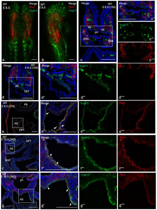
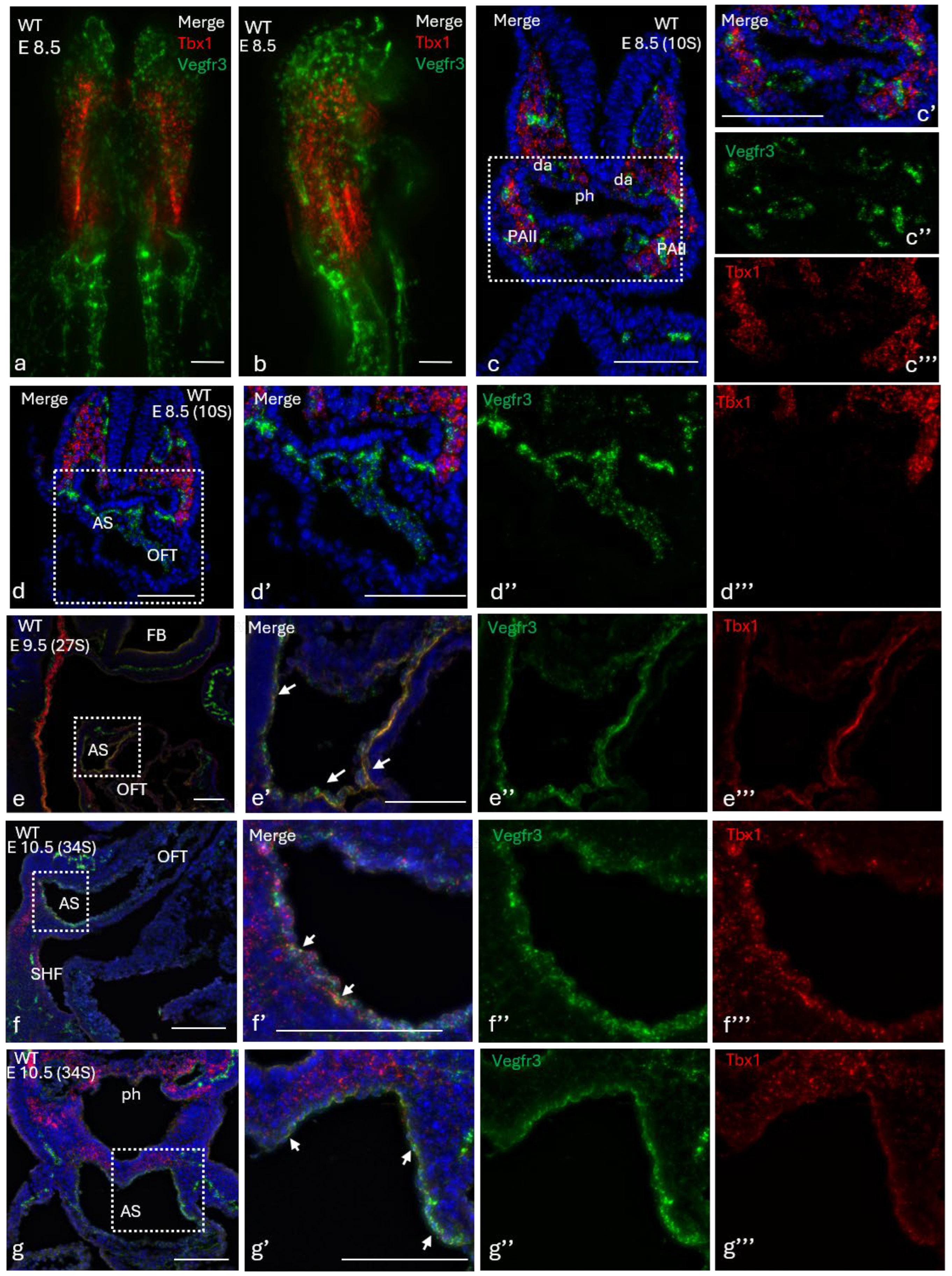
In order to gain insights into where and when a genetic interaction might occur, we analyzed expressions of Tbx1 and Vegfr3 by RNA in situ hybridization using the RNAscope.technology on whole wild-type embryos at E8.5 and E9.5. We then prepared histological sections of the double-stained embryos in order to obtain a detailed view of the distribution of stained cells. Representative embryos are shown in Figure 2. At E8.5, Vegfr3 was expressed throughout the developing vascular plexus as expected [34], in fully formed blood vessels, including the PAAs (Figure 2a,b) aortic sac (AS) and outflow tract (Figure 2d–d″). At this developmental stage, Tbx1 was expressed bilaterally in the head mesenchyme (Figure 2c,d), in the core of the pharyngeal arches (PA) I and II (Figure 2c,c‴), and as previously reported in the pharyngeal endoderm and in the second heart field [35]. No double-stained cells (yellow) were detected in any part of the embryo. At E9.5, in the rostral half of the embryo, expressions of Vegfr3 and Tbx1 were detected in all of the aforementioned tissues. In addition, Tbx1 was expressed in endothelial cells lining the AS, but not in the OFT, in embryos with >27 somites (Figure 2e–g). These results indicate that there is no expression overlap between Tbx1 and Vegfr3 in the rostral half of the embryo prior to E9.5, but this occurs at the level of the AS between E9.5 and E10.5.
Figure 2.
Tbx1 and Vegfr3 co-expression in the aortic sac: (a,b) Tbx1 (red) and Vegfr3 (green) expression revealed by RNAscope in wholemount WT embryos at E8.5 with a rostro-caudal orientation (ventral (a) and dorsal (b) views), and in transverse sections (c–c‴,d–d‴) revealed Vegfr3 but not Tbx1 expression in the aortic sac. Sagittal sections of WT embryos at E9.5 (27 somites) (e–e‴), E10.5 (34 somites, (f–f‴)) and in transverse sections (34 somites, (g–g‴)) showed that Tbx1 and Vegfr3 are co-expressed in the aortic sac. White dashed boxes in (c–g) indicate the regions shown at higher magnification in the adjacent panels. PAA II, pharyngeal arch artery II; da, dorsal aorta; ph, pharynx; AS, aortic sac; OFT, outflow tract. Scale bar 100 µm.
3.4. Vegfr3 Is Required in Tbx- Expressing Cells for Normal Cardiac Development
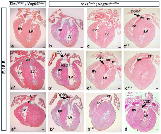
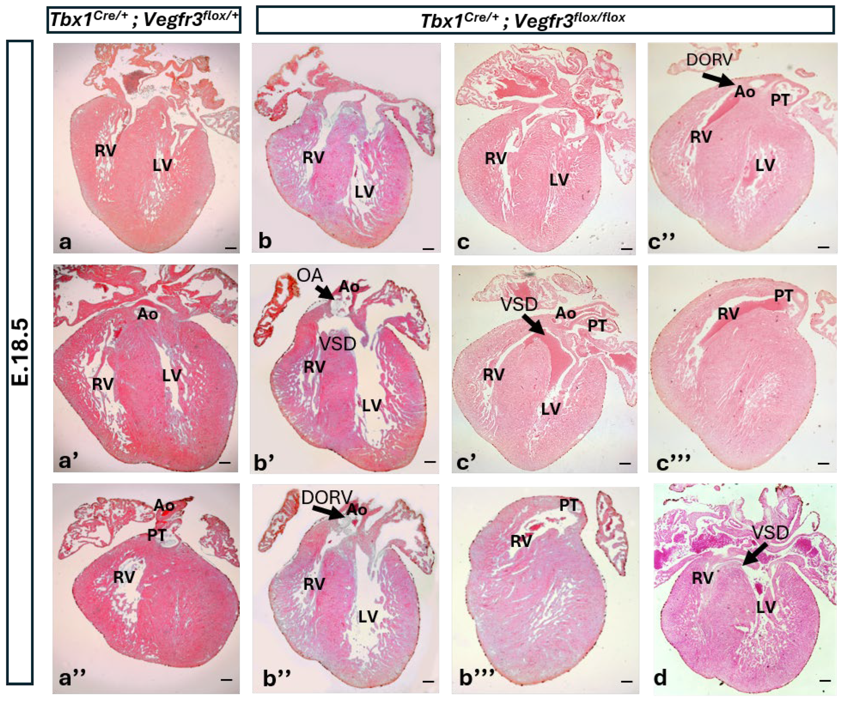
We next tested whether conditional mutation of Vegfr3 in the Tbx1 expression domain alone was sufficient to disrupt cardiac development. For this, we intercrossed Tbx1Cre/+; Vegfr3flox/+ and Vegfr3flox/flox mice and analyzed the cardiac phenotype in E18.5 embryos (Table 2, Figure 3). Results showed that Tbx1Cre-driven heterozygous inactivation of Vegfr3 (thus double heterozygosity in Tbx1-expressing cells and their descendants) had no effect on cardiac development (Figure 3a–a″). In contrast, the hearts of Tbx1Cre-driven conditional Vegfr3 homozygous embryos (Tbx1Cre/+; Vegfr3flox/flox) presented with one or more cardiac defects simultaneously (Figure 3b–d). Cardiac phenotyping of these embryos revealed that VSD was the most prevalent abnormality, being present in six out of seven hearts (85.7%), of which two were classified as isolated central perimembranous VSDs, while the remaining four cases were outlet VSDs with septal malalignment with the great arteries, and they occurred in embryos with double outlet right ventricle (DORV), which was observed in four out of seven hearts (57.1%). Among the DORV cases, two hearts (28.6%) also exhibited an overriding aorta (OA). Finally, we noted that the atrial septum was open in five out of seven hearts (patent foramen ovale, PFO). The FO normally closes after birth with initiation of the pulmonary circulation. Thus, the Tbx1Cre/+; Vegfr3flox/flox genotype was associated with variable expressivity of the cardiac phenotype. This result indicates that Vegfr3 expression in the Tbx1 expression domain is necessary for normal cardiac development, and that a single active copy of the Vegfr3 gene in Tbx1-expressing cells, or in their progeny, is sufficient for normal development.
Table 2.
Cardiac phenotypes in E18.5 embryos with conditional deletion of Vegfr3 in the Tbx1 expression domain.
Figure 3.
Tbx1 conditional Vegfr3 homozygous embryos (Tbx1Cre/+; Vegfr3flox/flox) have diverse cardiovascular defects. Coronal sections of isolated hearts of Tbx1Cre/+; Vegfr3flox/+ (a–a″) and Tbx1Cre/+; Vegfr3flox/flox (b–b‴,c–c‴,d) embryos (E18.5) stained with eosin. Black arrows in panels (b′,b″,c′,c″,d) indicate the position of cardiovascular defects. VSD (ventricular septal defect); DORV (double outlet right ventricle); OA (overriding aorta); RV, right ventricle; LV, left ventricle; Ao, aorta; PT, pulmonary trunk. Sections indicated with the same letter are from the same heart. Scale bar 100 µm.
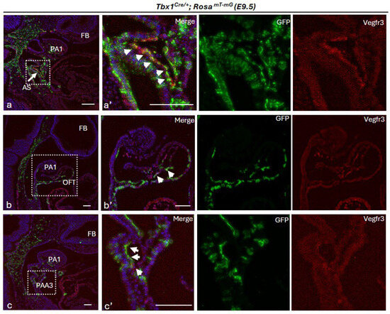
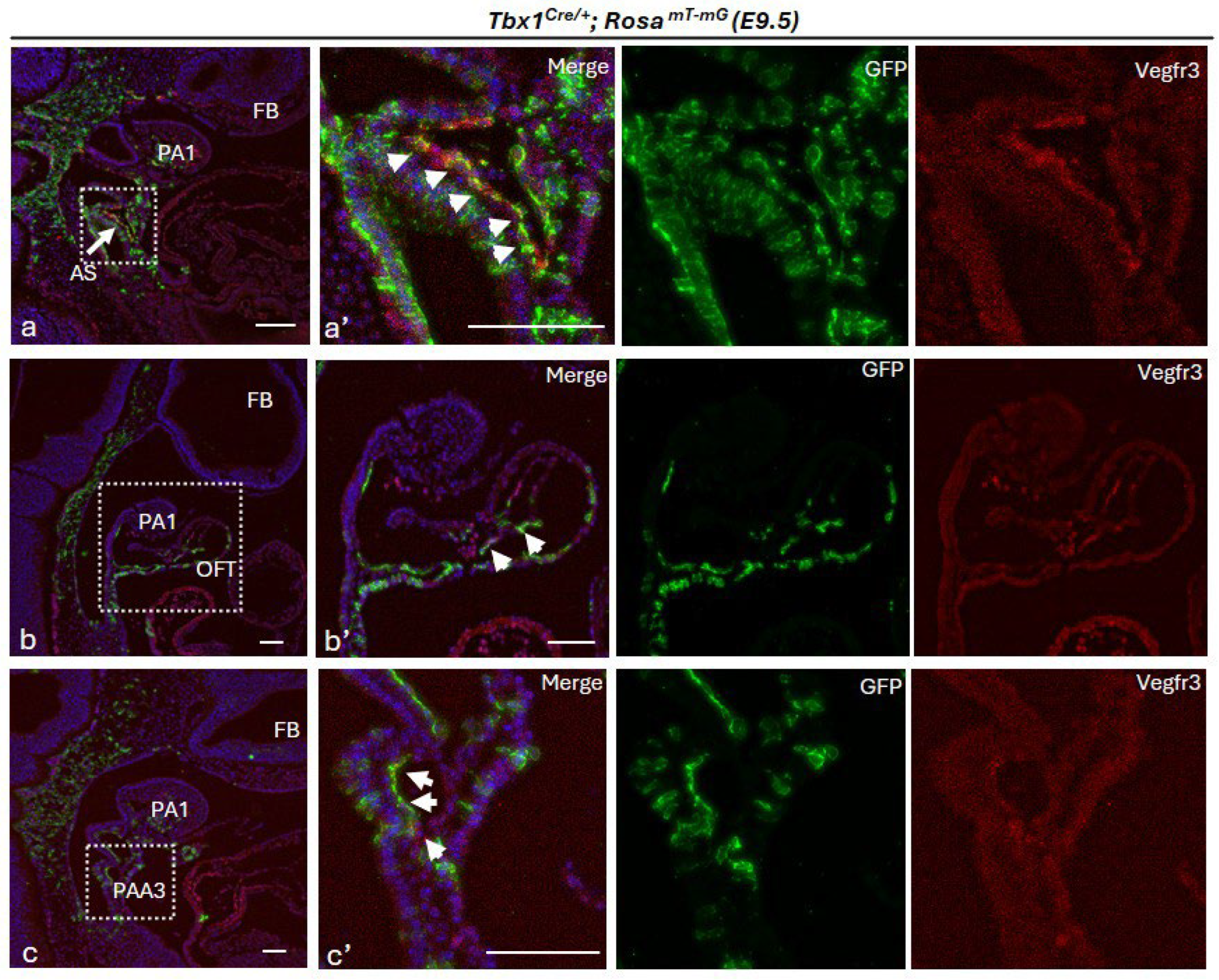
As the endothelium is the only cell type that expresses both Tbx1 and Vegfr3, one would expect that Tbx1Cre-induced recombination of the Vegfr3flox allele would recapitulate the phenotype observed in compound heterozygotes (Table 1). However, this was not the case; in fact, Tbx1Cre/+; Vegfr3flox/+ mutants survived to adulthood, were fertile and had normal hearts (Table 2). In order to better visualize the extent of Cre recombination in endothelial cells we performed double immunofluorescence on sagittal sections of E9.5 Tbx1Cre/+; RosamTmG embryos using anti-GFP and anti-VEGFR3 antibodies. Results showed that in the proximal OFT, the aortic sac and third pharyngeal arch artery, many but not all endothelial cells were double-stained (yellow) (Figure 4a–c′). Thus, there is partial overlap between Cre recombination and VEGFR3 expression in endothelial cells, but evidently, removal of a single copy of the Vegfr3 gene from this region of overlap is not sufficient to generate cardiac defects.
Figure 4.
Tbx1-activated Cre recombination in Vegfr3-expressing endothelial cells: Anti-Vegfr3 (red) and anti-TBX1-GFP (green) immunostaining of sagittal sections of Tbx1Cre/+; RosamT-mG (E.9.5) reveal Tbx1-activated Cre recombination in Vegfr3-expressing endothelial cells of the aortic sac (a,a′), OFT (b,b′) and third pharyngeal arch artery (PAA3) (c,c′) (white arrows). White dashed boxes in (a–c) indicate the regions shown at higher magnification in the adjacent panels. Scale bar 100 µm.
4. Discussion
The results of our work indicate that in the mouse, a single copy of the Vegfr3 gene is sufficient to support normal heart development in most cases, as only a single case of VSD was observed out of eleven Vegfr3+/− pre-term embryos. This low frequency might be due to variable expressivity or to stochastic events during pregnancy. A larger study would be necessary to determine whether 10% is the true frequency of VSD in Vegfr3 heterozygotes. In a recent review by Monaghan and colleagues, the authors proposed that TOF in individuals (human) with VEGFR3 variants or mutations is not due to VEGFR3 haploinsufficiency, but rather to the presence of the truncated VEGFR3 protein during fetal development [36]. This deduction was based on published data, showing that: (i) mice carrying VEGFR3 heterozygous mutations or hypomorphic alleles have normal cardiovascular development [37]; (ii) in humans, truncating mutations are very rare, and thus presumed to be mostly lethal [38,39]; and (iii) in TOF cases with truncating mutations, the mutant protein retained the N-terminal domain but had a shorter C-terminal domain [16]. The authors speculated that truncated VEGFR3 proteins disrupt molecular interactions and cellular functions required for normal cardiogenesis [36]. In our study, the Vegfr3 mutation is inactivating; therefore, the presence of truncated proteins can be excluded. Thus, the CHDs observed in Vegfr3+/− and Tbx1Cre/+; Vegfr3flox/flox embryos in this study are attributable to gene haploinsufficiency or loss of function. Noteworthy is the observation of Vegfr3 haploinsufficiency in cardiac lymphatic development [12], suggesting that haploinsufficiency is context-dependent, where different endothelial populations and subtypes have different sensitivities to Vegfr3 gene dosage.
Our study showed that heterozygous loss of Tbx1 greatly enhanced the penetrance of VSDs in Vegfr3+/− mutants from 10% to >85% (Table 1). This might be because TBX1 positively regulates Vegfr3 in some endothelial cells [11,12,13]; therefore, its mutation might reduce Vegfr3 expression below a critical threshold required for normal cardiac OFT development, or it might do so indirectly, by disrupting the expression of other Vegfr3 regulators or by affecting VEGFR3 functions downstream.
Given the strong effect of the Tbx1 mutation in compound germline Vegfr3 heterozygotes, we were surprised to find that conditional heterozygous inactivation of Vegfr3 in the Tbx1 lineage had no effect on cardiac morphogenesis. We expected that Tbx1Cre/+; Vegfr3flox/+ and Tbx1Cre/+; Vegfr3+/− embryos would have the same, or similar, phenotype, because the Tbx1Cre allele, which is inactivating [28], is present in both compound genotypes. The difference is likely to be due to incomplete overlap between Cre-induced recombination and Vegfr3 expression in the critical cell population, which leaves sufficient VEGFR3 to support normal development in Tbx1Cre/+; Vegfr3flox/+ embryos. Further reduction in VEGFR3 dosage in the homozygous genotype (Tbx1Cre/+; Vegfr3flox/flox) is evidently sufficient to trigger disruption of OFT development even in the absence of complete overlap. It is interesting to note that the latter mutants displayed some of the cardinal features of TOF (perimembranous VSD, DORV, OA).
Where might the TBX1-Vegfr3 interaction occur? The RNAscope experiments showed that Vegfr3 was restricted to ECs, as expected, and that Tbx1 was expressed in a modest number of ECs in the AS between E8.5 and E9.5. In contrast, when we traced Tbx1-expressing cells and their descendants over time (E8.5–E9.5), we observed extensive but incomplete co-localization of the two proteins (GFP of the Cre-reporter and VEGFR3) at E9.5 in the AS and in other EC populations of the OFT and pharyngeal arch arteries. Together, these results suggest that Tbx1 is activated in EC progenitors, consistent with previous in vivo studies [40], destined to contribute to the AS and other structures of the developing cardiovascular system.
In conclusion, this study provides evidence of a regulatory mechanism involving Tbx1 and Vegfr3 that is critical for heart morphogenesis. The results of the study point to a two-hit mechanism, whereby reduced dosage of Vegfr3 in the entire expression domain (not only in the Tbx1-Vegfr3 overlap domain) has a higher impact on OFT development when combined with Tbx1 heterozygosity. The interaction between the two genes may be in part direct, in the region of expression overlap, and in part indirect, between the role of Tbx1 in OFT morphogenesis, weakened by heterozygosity, and the endothelial-dependent role of Vegfr3, also weakened by heterozygosity.
At the molecular level, the vascular functions of TBX1 have been attributed to interactions with molecules in the VEGF-C and Notch signaling pathways [11,12,13]. Previous studies have demonstrated that NOTCH1 signaling in the endocardium is essential for cardiac OFT development [41,42], together with reports identifying dynamic VEGFR3-dependent endothelial populations during cardiovascular morphogenesis [43]. Thus, it is reasonable to think that the regulatory function of TBX1 directly or indirectly affects a NOTCH1-VEGFR3 axis. However, further studies will be required to test directly this hypothesis by dissecting the connections between these molecules.
Author Contributions
S.M.: data curation, formal analysis, validation, investigation, visualization, methodology and writing—original draft. M.B.: data curation, formal analysis, investigation, and methodology. S.C.: data curation, formal analysis, validation, investigation and methodology. I.A.: data curation, formal analysis, validation, and visualization. M.C.: data curation, formal analysis, investigation, and methodology. A.B.: conceptualization, supervision, and funding acquisition. E.I.: conceptualization, supervision, funding acquisition and writing—original draft. All authors have read and agreed to the published version of the manuscript.
Funding
The study was supported by grants from the Fondation Leducq Transatlantic Network of Excellence in Cardiovascular Research, 15CVD01, to E.I. and A.B., and from the Italian Ministry of Health #20179J2P9J to E.I. and A.B. and #P2022ARZ5J to E.I.
Institutional Review Board Statement
The animal study protocol was approved by the local IACUC committee and by the Italian Istituto Superiore di Sanità and approved by the Italian Ministero della Salute (protocol code 28/2022-PR and date of approval: 18 January 2022).
Informed Consent Statement
Not applicable.
Data Availability Statement
All data are contained within the manuscript.
Acknowledgments
We are grateful for the invaluable support provided by the Integrated Microscopy Core and the Animal Facility at the Institute of Genetic and Biophysics “ABT”/CNR, Naples. We thank Giuseppina Divisato at the Microscopy Core of the Department of Molecular Medicine and Medical Biotechnology, University of Naples Federico II; Lucia Mele for technical help with mouse handling; and Rosa Ferrentino for technical and administrative support and the microscopy core of the IGB-CNR. Vegfr3flox/+ mice were generously provided by Kari Alitalo.
Conflicts of Interest
The authors declare no conflict of interest.
Abbreviations
| OFT | outflow tract |
| Vegfr3 | vascular endothelial growth factor receptor 3 |
| DGS | DiGeorge syndrome |
| CPM | cardio-pharyngeal mesoderm |
| AS | aortic sac |
| SHF | second heart field |
| PAAs | pharyngeal arch arteries |
| RV | right ventricle |
| LV | left ventricle |
| R26R | Rosa26 reporter |
| VSD | ventricular septal defect |
| WT | wild type |
| PA | pharyngeal arches |
| DORV | double outlet right ventricle |
| OA | overriding aorta |
| PFO | patent foramen ovale |
| PT | pulmonary trunk |
| CHD | congenital heart disease |
| TOF | Tetralogy of Fallot |
References
- Connell, F.C.; Gordon, K.; Brice, G.; Keeley, V.; Jeffery, S.; Mortimer, P.S.; Mansour, S.; Ostergaard, P. The classification and diagnostic algorithm for primary lymphatic dysplasia: An update from 2010 to include molecular findings. Clin. Genet. 2013, 84, 303–314. [Google Scholar] [CrossRef] [Scilit] [PubMed]
- Karkkainen, M.J.; Ferrell, R.E.; Lawrence, E.C.; Kimak, M.A.; Levinson, K.L.; McTigue, M.A.; Alitalo, K.; Finegold, D.N. Missense mutations interfere with VEGFR-3 signalling in primary lymphoedema. Nat. Genet. 2000, 25, 153–159. [Google Scholar] [CrossRef] [Scilit] [PubMed]
- Karkkainen, M.J.; Haiko, P.; Sainio, K.; Partanen, J.; Taipale, J.; Petrova, T.V.; Jeltsch, M.; Jackson, D.G.; Talikka, M.; Rauvala, H.; et al. Vascular endothelial growth factor C is required for sprouting of the first lymphatic vessels from embryonic veins. Nat. Immunol. 2004, 5, 74–80. [Google Scholar] [CrossRef] [Scilit] [PubMed]
- Mäkinen, T.; Jussila, L.; Veikkola, T.; Karpanen, T.; Kettunen, M.I.; Pulkkanen, K.J.; Kauppinen, R.; Jackson, D.G.; Kubo, H.; Nishikawa, S.; et al. Inhibition of lymphangiogenesis with resulting lymphedema in transgenic mice expressing soluble VEGF receptor-3. Nat. Med. 2001, 7, 199–205. [Google Scholar] [CrossRef] [Scilit] [PubMed]
- Adams, R.H.; Alitalo, K. Molecular regulation of angiogenesis and lymphangiogenesis. Nat. Rev. Mol. Cell Biol. 2007, 8, 464–478. [Google Scholar] [CrossRef] [Scilit] [PubMed]
- Dumont, D.J.; Jussila, L.; Taipale, J.; Lymboussaki, A.; Mustonen, T.; Pajusola, K.; Breitman, M.; Alitalo, K. Cardiovascular failure in mouse embryos deficient in VEGF receptor-3. Science 1998, 282, 946–949. [Google Scholar] [CrossRef] [Scilit] [PubMed]
- Mansir, T.; Lacombe, D.; Lamireau, T.; Taine, L.; Chateil, J.F.; Le Bail, B.; Demarquez, J.L.; Fayon, M. Abdominal lymphatic dysplasia and 22q11 microdeletion. Genet. Couns. 1999, 10, 67–70. [Google Scholar] [PubMed]
- Unolt, M.; Barry, J.; Digilio, M.C.; Marino, B.; Bassett, A.; Oechslin, E.; Low, D.W.; Belasco, J.B.; Kallish, S.; Sullivan, K.; et al. Primary lymphedema and other lymphatic anomalies are associated with 22q11.2 deletion syndrome. Eur. J. Med. Genet. 2018, 61, 411–415. [Google Scholar] [CrossRef] [Scilit] [PubMed]
- McDonald-McGinn, D.M.; Sullivan, K.E.; Marino, B.; Philip, N.; Swillen, A.; Vorstman, J.A.S.; Zackai, E.H.; Emanuel, B.S.; Vermeesch, J.R.; Morrow, B.E.; et al. 22q11.2 deletion syndrome. Nat. Rev. Dis. Primers 2015, 1, 15071. [Google Scholar] [CrossRef] [Scilit] [PubMed]
- Diogo, R.; Kelly, R.G.; Christiaen, L.; Levine, M.; Ziermann, J.M.; Molnar, J.L.; Noden, D.M.; Tzahor, E. A new heart for a new head in vertebrate cardiopharyngeal evolution. Nature 2015, 520, 466–473. [Google Scholar] [CrossRef] [Scilit] [PubMed]
- Chen, L.; Mupo, A.; Huynh, T.; Cioffi, S.; Woods, M.; Jin, C.; McKeehan, W.; Thompson-Snipes, L.; Baldini, A.; Illingworth, E. Tbx1 regulates Vegfr3 and is required for lymphatic vessel development. J. Cell Biol. 2010, 189, 417–424. [Google Scholar] [CrossRef] [Scilit] [PubMed]
- Martucciello, S.; Turturo, M.G.; Bilio, M.; Cioffi, S.; Chen, L.; Baldini, A.; Illingworth, E. A dual role for Tbx1 in cardiac lymphangiogenesis through genetic interaction with Vegfr3. FASEB J. 2020, 34, 15062–15079. [Google Scholar] [CrossRef] [Scilit] [PubMed]
- Cioffi, S.; Flore, G.; Martucciello, S.; Bilio, M.; Turturo, M.G.; Illingworth, E. VEGFR3 modulates brain microvessel branching in a mouse model of 22q11.2 deletion syndrome. Life Sci. Alliance 2022, 5, e202101308. [Google Scholar] [CrossRef] [Scilit] [PubMed]
- Jin, S.C.; Homsy, J.; Zaidi, S.; Lu, Q.; Morton, S.; DePalma, S.R.; Zeng, X.; Qi, H.; Chang, W.; Sierant, M.C.; et al. Contribution of rare inherited and de novo variants in 2,871 congenital heart disease probands. Nat. Genet. 2017, 49, 1593–1601. [Google Scholar] [CrossRef] [Scilit] [PubMed]
- Page, D.J.; Miossec, M.J.; Williams, S.G.; Monaghan, R.M.; Fotiou, E.; Cordell, H.J.; Sutcliffe, L.; Topf, A.; Bourgey, M.; Bourque, G.; et al. Whole Exome Sequencing Reveals the Major Genetic Contributors to Nonsyndromic Tetralogy of Fallot. Circ. Res. 2019, 124, 553–563. [Google Scholar] [CrossRef] [Scilit] [PubMed]
- Reuter, M.S.; Jobling, R.; Chaturvedi, R.R.; Manshaei, R.; Costain, G.; Heung, T.; Curtis, M.; Hosseini, S.M.; Liston, E.; Lowther, C.; et al. Haploinsufficiency of vascular endothelial growth factor related signaling genes is associated with tetralogy of Fallot. Genet. Med. 2019, 21, 1001–1007. [Google Scholar] [CrossRef] [Scilit] [PubMed]
- Tabib, A.; Talebi, T.; Ghasemi, S.; Pourirahim, M.; Naderi, N.; Maleki, M.; Kalayinia, S. A novel stop-gain pathogenic variant in FLT4 and a nonsynonymous pathogenic variant in PTPN11 associated with congenital heart defects. Eur. J. Med. Res. 2022, 27, 286. [Google Scholar] [CrossRef] [Scilit] [PubMed]
- Momma, K. Cardiovascular anomalies associated with chromosome 22q11.2 deletion syndrome. Am. J. Cardiol. 2010, 105, 1617–1624. [Google Scholar] [CrossRef] [Scilit] [PubMed]
- Sauter, C.; Hofbeck, M.; Franz, P.; Kettenstock, L.; Steger, K.; Linhardt, M.; Reiter, A.; Störk, S.; Romanos, M.; Radtke, F. Congenital heart disease in 22q11.2 deletion syndrome: A meta-analysis and systematic review of the literature. J. Med. Genet. 2025, 62, 700–708. [Google Scholar] [CrossRef] [Scilit] [PubMed]
- Zhang, Z.; Baldini, A. In vivo response to high-resolution variation of Tbx1 mRNA dosage. Hum. Mol. Genet. 2008, 17, 150–157. [Google Scholar] [CrossRef] [Scilit] [PubMed]
- Rana, M.S.; Théveniau-Ruissy, M.; De Bono, C.; Mesbah, K.; Francou, A.; Rammah, M.; Domínguez, J.N.; Roux, M.; Laforest, B.; Anderson, R.H.; et al. Tbx1 coordinates addition of posterior second heart field progenitor cells to the arterial and venous poles of the heart. Circ. Res. 2014, 115, 790–799. [Google Scholar] [CrossRef] [Scilit] [PubMed]
- Xu, H.; Morishima, M.; Wylie, J.N.; Schwartz, R.J.; Bruneau, B.G.; Lindsay, E.A.; Baldini, A. Tbx1 has a dual role in the morphogenesis of the cardiac outflow tract. Development 2004, 131, 3217–3227. [Google Scholar] [CrossRef] [Scilit] [PubMed]
- Devine, W.P.; Wythe, J.D.; George, M.; Koshiba-Takeuchi, K.; Bruneau, B.G. Early patterning and specification of cardiac progenitors in gastrulating mesoderm. eLife 2014, 3, e03848. [Google Scholar] [CrossRef] [Scilit] [PubMed]
- Lescroart, F.; Chabab, S.; Lin, X.; Rulands, S.; Paulissen, C.; Rodolosse, A.; Auer, H.; Achouri, Y.; Dubois, C.; Bondue, A.; et al. Early lineage restriction in temporally distinct populations of Mesp1 progenitors during mammalian heart development. Nat. Cell Biol. 2014, 16, 829–840. [Google Scholar] [CrossRef] [Scilit] [PubMed]
- Milgrom-Hoffman, M.; Harrelson, Z.; Ferrara, N.; Zelzer, E.; Evans, S.M.; Tzahor, E. The heart endocardium is derived from vascular endothelial progenitors. Development 2011, 138, 4777–4787. [Google Scholar] [CrossRef] [Scilit] [PubMed]
- Cai, C.-L.; Liang, X.; Shi, Y.; Chu, P.-H.; Pfaff, S.L.; Chen, J.; Evans, S. Isl1 identifies a cardiac progenitor population that proliferates prior to differentiation and contributes a majority of cells to the heart. Dev. Cell 2003, 5, 877–889. [Google Scholar] [CrossRef] [Scilit] [PubMed]
- Verzi, M.P.; McCulley, D.J.; De Val, S.; Dodou, E.; Black, B.L. The right ventricle, outflow tract, and ventricular septum comprise a restricted expression domain within the secondary/anterior heart field. Dev. Biol. 2005, 287, 134–145. [Google Scholar] [CrossRef] [Scilit] [PubMed]
- Wang, X.; Chen, D.; Chen, K.; Jubran, A.; Ramirez, A.; Astrof, S. Endothelium in the pharyngeal arches 3, 4 and 6 is derived from the second heart field. Dev. Biol. 2017, 421, 108–117. [Google Scholar] [CrossRef] [Scilit] [PubMed]
- Huynh, T.; Chen, L.; Terrell, P.; Baldini, A. A fate map of Tbx1 expressing cells reveals heterogeneity in the second cardiac field. Genesis 2007, 45, 470–475. [Google Scholar] [CrossRef] [Scilit] [PubMed]
- Zarkada, G.; Heinolainen, K.; Makinen, T.; Kubota, Y.; Alitalo, K. VEGFR3 does not sustain retinal angiogenesis without VEGFR2. Proc. Natl. Acad. Sci. USA 2015, 112, 761–766. [Google Scholar] [CrossRef] [Scilit] [PubMed]
- Muzumdar, M.D.; Tasic, B.; Miyamichi, K.; Li, L.; Luo, L. A global double-fluorescent Cre reporter mouse. Genesis 2007, 45, 593–605. [Google Scholar] [CrossRef] [Scilit] [PubMed]
- Gittenberger-de Groot, A.C.; Hoppenbrouwers, T.; Miquerol, L.; Kosaka, Y.; Poelmann, R.E.; Wisse, L.J.; Yost, H.J.; Jongbloed, M.R.M.; Deruiter, M.C.; Brunelli, L. 14-3-3epsilon controls multiple developmental processes in the mouse heart. Dev. Dyn. 2016, 245, 1107–1123. [Google Scholar] [CrossRef] [Scilit] [PubMed]
- Anderson, R.H.; Tretter, J.T.; Spicer, D.E.; Mori, S. The Fate of the Outflow Tract Septal Complex in Relation to the Classification of Ventricular Septal Defects. J. Cardiovasc. Dev. Dis. 2019, 6, 9. [Google Scholar] [CrossRef] [Scilit] [PubMed]
- Watanabe, C.; Matsushita, J.; Azami, T.; Tsukiyama-Fujii, S.; Tsukiyama, T.; Mizuno, S.; Takahashi, S.; Ema, M. Generating Vegfr3 reporter transgenic mouse expressing membrane-tagged Venus for visualization of VEGFR3 expression in vascular and lymphatic endothelial cells. PLoS ONE 2019, 14, e0210060. [Google Scholar] [CrossRef] [Scilit] [PubMed]
- Calmont, A.; Ivins, S.; Van Bueren, K.L.; Papangeli, I.; Kyriakopoulou, V.; Andrews, W.D.; Martin, J.F.; Moon, A.M.; Illingworth, E.A.; Basson, M.A.; et al. Tbx1 controls cardiac neural crest cell migration during arch artery development by regulating Gbx2 expression in the pharyngeal ectoderm. Development 2009, 136, 3173–3183. [Google Scholar] [CrossRef] [Scilit] [PubMed]
- Monaghan, R.M.; Naylor, R.W.; Flatman, D.; Kasher, P.R.; Williams, S.G.; Keavney, B.D. FLT4 causes developmental disorders of the cardiovascular and lymphovascular systems via pleiotropic molecular mechanisms. Cardiovasc. Res. 2024, 120, 1164–1176. [Google Scholar] [CrossRef] [Scilit] [PubMed]
- Haiko, P.; Makinen, T.; Keskitalo, S.; Taipale, J.; Karkkainen, M.J.; Baldwin, M.E.; Stacker, S.A.; Achen, M.G.; Alitalo, K. Deletion of vascular endothelial growth factor C (VEGF-C) and VEGF-D is not equivalent to VEGF receptor 3 deletion in mouse embryos. Mol. Cell Biol. 2008, 28, 4843–4850. [Google Scholar] [CrossRef] [Scilit] [PubMed]
- Gordon, K.; Schulte, D.; Brice, G.; Simpson, M.A.; Roukens, M.G.; van Impel, A.; Connell, F.; Kalidas, K.; Jeffery, S.; Mortimer, P.S.; et al. Mutation in vascular endothelial growth factor-C, a ligand for vascular endothelial growth factor receptor-3, is associated with autosomal dominant milroy-like primary lymphedema. Circ. Res. 2013, 112, 956–960. [Google Scholar] [CrossRef] [Scilit] [PubMed]
- Monaghan, R.M.; Page, D.J.; Ostergaard, P.; Keavney, B.D. The physiological and pathological functions of VEGFR3 in cardiac and lymphatic development and related diseases. Cardiovasc. Res. 2021, 117, 1877–1890. [Google Scholar] [CrossRef] [Scilit] [PubMed]
- Xu, H.; Cerrato, F.; Baldini, A. Timed mutation and cell-fate mapping reveal reiterated roles of Tbx1 during embryogenesis, and a crucial function during segmentation of the pharyngeal system via regulation of endoderm expansion. Development 2005, 132, 4387–4395. [Google Scholar] [CrossRef] [Scilit] [PubMed]
- de la Pompa, J.L.; Epstein, J.A. Coordinating tissue interactions: Notch signaling in cardiac development and disease. Dev. Cell 2012, 22, 244–254. [Google Scholar] [CrossRef] [Scilit] [PubMed]
- High, F.A.; Jain, R.; Stoller, J.Z.; Antonucci, N.B.; Lu, M.M.; Loomes, K.M.; Kaestner, K.H.; Pear, W.S.; Epstein, J.A. Murine Jagged1/Notch signaling in the second heart field orchestrates Fgf8 expression and tissue-tissue interactions during outflow tract development. J. Clin. Investig. 2009, 119, 1986–1996. [Google Scholar] [CrossRef] [Scilit] [PubMed]
- Chen, H.I.; Sharma, B.; Akerberg, B.N.; Numi, H.J.; Kivelä, R.; Saharinen, P.; Aghajanian, H.; McKay, A.S.; Bogard, P.E.; Chang, A.H.; et al. The sinus venosus contributes to coronary vasculature through VEGFC-stimulated angiogenesis. Development 2014, 141, 4500–4512. [Google Scholar] [CrossRef] [Scilit] [PubMed]

Disclaimer/Publisher’s Note: The statements, opinions and data contained in all publications are solely those of the individual author(s) and contributor(s) and not of MDPI and/or the editor(s). MDPI and/or the editor(s) disclaim responsibility for any injury to people or property resulting from any ideas, methods, instructions or products referred to in the content. |
© 2026 by the authors. Licensee MDPI, Basel, Switzerland. This article is an open access article distributed under the terms and conditions of the Creative Commons Attribution (CC BY) license.