


Abstract
Heart failure (HF) is a clinical syndrome whose severity cannot be inferred from left ventricular ejection fraction (LVEF) alone. Loading conditions, rhythm, comorbidities, congestion, and measurement variability can produce discordance between EF categories and clinical status. This narrative review proposes a decision-centered framework integrating clinical assessment and biomarkers with relatively stable traits, dynamic physiological states, and longitudinal treatment responses. A new focused PubMed search through 16 October 2026 retrieved 543 unique records; 91 were prioritized for an evidence map, supplemented by the original bibliography and targeted source checking. Application-specific judgments distinguish technical validity, diagnostic or prognostic information, clinical actionability, and demonstrated outcome benefit. Representative pathways address unexplained dyspnea, cardiomyopathic substrate, amyloidosis, right-sided dysfunction, and worsening HF. Conventional echocardiography, natriuretic peptides, clinical risk factors, and congestion assessment anchor these pathways; advanced testing is selected when results could change management. Illustrative scenarios show how positive, negative, and indeterminate findings affect the next step. The proposed framework has not been prospectively validated; its incremental value over guideline-based multimodality assessment, including effects on outcomes, efficiency, and cost, remains hypothetical. Its contribution is an explicit structure for choosing tests and planning reassessment, with evidence boundaries made visible.
1. Introduction
Heart failure is a clinical syndrome with symptoms and signs arising from structural or functional cardiac abnormalities. LVEF has established prognostic value, broad availability, and a central role in the evidence supporting many therapies and devices [1,2,3]. It does not, however, measure overall HF severity, functional limitation, congestion, or the burden of extracardiac disease. Patients with similar LVEF may have very different symptoms, filling pressures, renal function, rhythm, and prognosis; severe HF can coexist with a preserved LVEF. Differences in loading, geometry, valvular function, tissue substrate, and myocardial mechanics further limit a single resting measurement [4,5,6]. A clinically useful phenotype must therefore combine imaging with symptoms, examination, biomarkers, comorbidities, and trajectory.
Existing reviews describe echocardiography, cardiovascular magnetic resonance (CMR), cardiac computed tomography (CT), nuclear imaging, and artificial intelligence (AI) in HF [7,8,9]. The remaining practical question is which test should answer which clinical uncertainty and whether its result changes care. Prognostic association does not establish a treatment threshold, and retrospective discrimination does not prove benefit from deployment.
We organize this review around three temporal targets. A trait phenotype describes a relatively stable etiologic or structural substrate, such as infarct scar, a cardiomyopathic pattern, or amyloid deposition. A state phenotype describes current physiology, including filling pressure, congestion, reserve, ventricular–vascular coupling, and active inflammatory activity. A response phenotype describes change following an intervention, such as reverse remodeling, decongestion, or deterioration during follow-up. These categories depend on the question and time scale: inflammation is a changing state, while the resulting scar may become a persistent trait. The proposed trait–state–response model is an organizational tool whose incremental clinical utility requires prospective evaluation.
The practical starting point is conventional echocardiography with LVEF, diastolic and right-heart assessment, combined with clinical findings and natriuretic peptides. Lung ultrasound can supplement bedside congestion assessment; CMR with late gadolinium enhancement (LGE) can clarify an uncertain cardiomyopathic substrate; exercise testing can investigate unexplained exertional symptoms; and bone scintigraphy can resolve a specific suspicion of transthyretin cardiac amyloidosis within a validated diagnostic pathway. These selective uses should be distinguished from myocardial-work-guided treatment, CT-derived extracellular volume (ECV), autonomous AI phenotyping, and wearable cardiac imaging, which have less established clinical roles [5,8,10,11,12].
2. Literature Search, Selection, and Evidence Appraisal
A new focused literature search was undertaken during revision on 16 October 2026. Seven reproducible PubMed queries covered HF definitions, unexplained dyspnea and HFpEF diagnosis, strain and tissue markers, pharmacological reverse remodeling, remote monitoring, AI, and targeted imaging recommendations. Searches were restricted to English-language publications dated 1 January 2015–16 October 2026; older pivotal studies were considered through the original bibliography and targeted reference checking. All records returned by the seven queries were retrieved. There were 544 query-level records and 543 unique PMIDs; 368 were not selected at title assessment, 175 underwent abstract or metadata assessment, and 91 were prioritized for the narrative evidence map. Complete queries, eligibility rules, counts, selection reasons, source-access information, and the screening register are provided in Supplementary Material S1 and its accompanying CSV files. These counts describe the new focused search and are distinct from the number of manuscript references.
The scope was adult HF assessment and management decisions, rather than comprehensive coverage of every imaging technology or cardiomyopathy. Current guidelines and consensus documents anchored established pathways; randomized trials informed intervention effects; invasive, prospective, multicenter, and external-validation studies informed diagnostic or prognostic validity. Smaller studies were retained when they demonstrated feasibility or an important limitation. Animal and pediatric studies, protocols without results, unrelated applications, and redundant reports without a distinct contribution were not prioritized. Selection was purposive within this narrative scope; it was not restricted to statistically positive findings. Examples include neutral remodeling findings, the overall neutral GUIDE-HF result, and limited sensitivity in external AI validation. Related trial substudies and co-publications were identified as such and were not treated as independent replications.
3. From LVEF Categories to Actionable Phenotypes
3.1. What LVEF Measures and How Its Classification Is Changing
LVEF is stroke volume divided by end-diastolic volume and describes chamber emptying under the conditions present at acquisition. It is not a direct measure of intrinsic contractility or total cardiovascular reserve. Preload, afterload, blood pressure, heart rate, valvular regurgitation, image quality, and tracing methods affect interpretation. In atrial fibrillation, variable cycle lengths and beat-to-beat filling can alter measured EF and strain; representative beat selection or averaging should follow the laboratory protocol, with rhythm and heart rate reported. A small, thick-walled ventricle may preserve EF despite impaired longitudinal shortening and limited forward-flow reserve. An apparent EF change during decongestion or rate control should therefore be interpreted with the accompanying physiology, and a borderline value that would change device eligibility may require confirmation under stable conditions [4,6,13].
Terminology has evolved. The 2021 ESC and 2022 AHA/ACC/HFSA documents used HFrEF at LVEF ≤ 40%, HF with mildly reduced EF at 41–49%, and HFpEF at ≥50% [1,2]. The 2025 trilateral consensus questioned rigid EF categories and emphasized trajectories [6]. The 2026 ESC guideline now defines HFrEF as LVEF < 50% and HFpEF as ≥50%, removin g HFmrEF as a separate category [14]. The 2026 Second Universal Definition also simplifies the terminology toward reduced, preserved, and improved EF while avoiding rigid cutoffs [15]. These developments reinforce a clinical and longitudinal interpretation of EF. They do not retroactively change trial inclusion criteria or device thresholds. Throughout this review, historical EF labels are retained when reporting the population actually studied, and the corresponding numerical cutoff is specified where necessary.
The minimum clinical phenotype should document symptom burden and functional capacity, congestion and perfusion, blood pressure, rhythm, age, body size, diabetes, chronic kidney disease, lung disease, ischemic history, frailty, and treatment exposure. Natriuretic peptides support HF assessment but vary with obesity, atrial fibrillation, and renal function; renal indices, hemoglobin, and iron status provide complementary management information. MAGGIC, developed from 39,372 patients in 30 studies, illustrates how clinical variables add prognostic information beyond EF [16]. It is a risk model, not an etiologic classifier, and local calibration and the intended outcome matter. An imaging-based proposal should be judged against this multicomponent baseline.
3.2. Physiological and Technical Sources of LVEF Variability
Beyond the acquisition conditions described above, several physiological states can change LVEF in stable HF without a change in disease stage. Because LVEF depends on preload, afterload, and LV volume, changes in volume status or blood pressure can shift LVEF [4]; in severe aortic stenosis, afterload mismatch can depress an LVEF that recovers after valve replacement; and in significant mitral regurgitation, a normal LVEF may conceal myocardial dysfunction, with LVEF ≤ 60% already indicating LV dysfunction in chronic severe primary regurgitation [17]. Atrial fibrillation or persistent tachycardia can also depress systolic function itself. In the multicenter CAMERA-MRI trial of patients with persistent atrial fibrillation and otherwise unexplained systolic dysfunction, catheter ablation increased LVEF by an absolute 18 percentage points, compared with 4.4 points with medical rate control, and normalized it in 58% versus 9% of patients; improvement was greater when CMR showed no late gadolinium enhancement [18]. A low LVEF recorded during atrial fibrillation may therefore partly reflect a reversible, rhythm-dependent state rather than a fixed trait.
Measurement adds further variance. In 56 women with stable LV function during breast cancer chemotherapy, the temporal variability of LVEF was approximately 6 percentage points with non-contrast three-dimensional echocardiography but could exceed 10 percentage points with two-dimensional methods [19], and in normal subjects and patients with HF or LV hypertrophy, CMR had substantially better interstudy reproducibility than two-dimensional echocardiography [20]. LVEF should therefore be reported with rhythm, heart rate, blood pressure, volume status, and modality. These variability estimates are study- and protocol-specific and should not be treated as universal thresholds for HF reclassification or treatment adjustment. When a small LVEF change would alter management, interpretation should incorporate image quality, laboratory-specific repeatability, and physiological conditions; repeat assessment under comparable conditions or confirmation with an appropriate alternative modality may be warranted.
3.3. Trait, State, and Response Are Different Targets
A relatively stable trait is useful when the question concerns etiology or persistent risk, such as infarct-pattern or mid-wall scar, amyloid deposition, or a familial cardiomyopathic substrate. Dynamic state measurements are useful when the question concerns current symptoms, exercise-induced filling-pressure elevation, congestion, active inflammation, or right-sided reserve. Response measurements address trajectory after an intervention. A change can be informative without being a validated trigger for changing treatment; this distinction is particularly important for advanced quantitative markers.
These targets overlap and should not be treated as mutually exclusive disease classes. T1 or ECV may reflect a persistent substrate but can also change with edema, loading, or treatment. GLS can characterize a stable cardiomyopathy, vary with afterload, or serve as a longitudinal measurement. Acquisition context, time scale, and the intended decision determine how a measurement is used. Figure 1 places the proposed categories above a common clinical and biomarker foundation.
Figure 1.
Proposed trait–state–response framework. Clinical findings, comorbidities, biomarkers, and conventional echocardiography are the shared foundation. The same measurement may contribute to more than one temporal target. A test result is linked to a defined decision, with technical validity, clinical validity, actionability, and outcome evidence assessed separately. The framework itself has not been prospectively validated.
3.4. A Practical Evidence-Maturity Scale
The A/B/C labels are qualitative judgments proposed for this review, not GRADE certainty ratings, guideline recommendation classes, or formal society consensus. They have not undergone formal inter-rater validation and therefore do not constitute a validated evidence-grading system; their purpose is to make the authors’ reasoning transparent, not to replace GRADE-based assessment or guideline recommendations. Each rating applies to a defined clinical application and population. Four domains are assessed separately: technical validity (acquisition, reproducibility, and interpretability), clinical validity (diagnostic or prognostic performance against an appropriate comparator), actionability (an accepted consequence of the result), and outcome benefit (prospective evidence that using the test or strategy improves patient outcomes). Prognostic association alone cannot establish outcome benefit.
Level A requires a standardized, clinically validated measurement and an explicit action supported by current guidance or an outcome-tested care pathway. Level B indicates credible diagnostic or prognostic information with an incompletely established action threshold. Level C applies to feasibility, insufficient external validation, or an untested treatment-selection rule (Table 1). Accepted diagnostic pathways may qualify as A without randomized outcome evidence; this distinction remains explicit for each application in Table 2. Separate uses receive separate ratings, without A−, C+, or mixed ranges. The Supplementary Material S1 worksheet records supporting and limiting evidence. Borderline judgments default to the lower supported level; independent-rater agreement is not claimed.
Table 1.
Rules for assigning evidence maturity to a specific clinical application.
Table 2.
Evidence maturity of measurements used beyond a static LVEF.
3.5. What Does the Proposed Framework Add to Current Clinical Practice?
Existing multimodality recommendations already support etiologic investigation, complementary imaging, and longitudinal assessment [5,6,8]. We do not claim that combining modalities or following EF over time is new. The proposed contribution is a common decision record: define the uncertainty, specify whether the target is substrate, current physiology, or change, select a test, state what each result would alter, and schedule reassessment only when it could affect care. Separating these steps can expose unsupported transitions from a prognostic association to a treatment rule. The framework also makes negative and indeterminate findings, resource constraints, and the strength of action evidence explicit. Whether this organization improves diagnostic efficiency, treatment selection, or outcomes must be tested against existing care pathways. Until such comparisons are available, its incremental clinical value over guideline-based multimodality assessment remains hypothetical, and it should be used as an organizing aid alongside, not in place of, current guideline recommendations [1,2,3].
4. Stable Trait Phenotyping: Etiology, Tissue, and Structural Substrate
4.1. Echocardiography: Geometry and Mechanics as the First Phenotypic Layer
Transthoracic echocardiography is the first-line imaging test for chamber size, LVEF, wall thickness, diastolic indices, RV function, valves, pulmonary pressure, and pericardium. Three-dimensional imaging reduces geometric assumptions and can improve volume reproducibility when complete, high-quality data are available [13,19,58]. Acoustic windows, resolution, stitching, and analysis remain limiting; three-dimensional reference ranges are required, and CMR remains the reference standard for biventricular volumes.
LV GLS reveals longitudinal systolic dysfunction that may be present despite a preserved LVEF. Standardized acquisition and reporting have improved, and GLS has shown prognostic value across the EF spectrum, including acute HF and HFpEF [10,22,23]. In 4172 patients hospitalized with acute HF, GLS was associated with mortality across EF categories and added information beyond LVEF [22]. In PARAGON-HF, impaired GLS was common among participants with preserved EF and associated with adverse outcomes, yet it did not identify a differential treatment effect of sacubitril/valsartan [23]. That distinction captures the broader evidence gap: a marker may identify higher risk without being a validated treatment-selection biomarker.
GLS is load-sensitive and depends on image quality, acquisition, vendor, software, and tracking decisions [10]. A single cutoff should not be used to stage HF across laboratories, rhythms, and diseases. The incremental value is most useful when it resolves discordance, characterizes a suspected cardiomyopathy, or supports a defined surveillance indication. It adds acquisition and analysis requirements and may be unavailable when tracking is poor. Routine measurement can enrich characterization in an experienced laboratory, but an isolated abnormal GLS does not specify an HF drug, dose, or device indication.
Non-invasive myocardial work combines strain with a cuff-pressure-derived estimate of LV pressure. It can contextualize GLS when afterload differs, and observational studies suggest prognostic information in HFrEF and altered efficiency in HFpEF [25,26,27]. It is neither load-independent nor a direct measure of oxygen consumption. Universal cutoffs, cross-platform harmonization, and outcome evidence for work-guided management are lacking.
4.2. CMR: A Reference Standard for Volumes and a Tissue-Level Phenotype
CMR provides accurate and reproducible biventricular volumes and systolic function, while LGE and quantitative mapping add tissue information that echocardiography cannot directly supply. Standardized cine, LGE, and mapping protocols and site-specific reference ranges are essential [11,28,59]. LGE identifies focal expansion of extracellular space. Depending on the disease context, its distribution may be consistent with infarction, replacement scar, inflammation, or infiltration; LGE should not be equated automatically with histological fibrosis.
In dilated cardiomyopathy, LGE adds prognostic information beyond EF [29,31]. A 2023 meta-analysis of 46 studies involving 10,548 patients with nonischemic cardiomyopathy associated LGE with mortality, ventricular arrhythmia or sudden death, and HF hospitalization, but explicitly identified the need for randomized trials of LGE-guided defibrillator selection [32]. Pattern matters: isolated right-ventricular insertion-point LGE did not independently predict adverse outcomes in one multicenter retrospective cohort [60]. LGE is therefore not a uniform risk label. CMR findings should inform an integrated discussion of genotype, arrhythmia, family history, symptoms, EF, and trajectory within cardiomyopathy guidance [30]. Absence of LGE alone should not be used to withhold otherwise indicated therapy or an implantable cardioverter-defibrillator.
Native T1, T2, and ECV extend tissue characterization to diffuse abnormalities. Native T1 is sensitive to sequence, field strength, and tissue composition; ECV additionally requires appropriate pre- and post-contrast acquisition and contemporaneous hematocrit. T2 can support assessment of edema or active injury [11]. In HFpEF, ECV has associations with histological collagen, clinical severity, and adverse events, but its independent value may diminish after clinical and invasive hemodynamic variables are considered [33,34,35]. ECV does not distinguish fibrosis from every other cause of extracellular expansion, and an isolated value cannot define global HF severity. Cost, scanner access, analysis expertise, contrast suitability, and the absence of a universal action threshold favor selective use. PIROUETTE showed a reduction in CMR-measured fibrosis with pirfenidone in selected HFpEF, but did not establish a routine antifibrotic treatment strategy or a validated surrogate for survival [36].
4.3. Targeted Nuclear Imaging: High Value When the Question Is Specific
Bone-avid tracer scintigraphy has a specific diagnostic role in suspected transthyretin cardiac amyloidosis (ATTR-CM). In a compatible clinical phenotype, grade 2 or 3 myocardial uptake, confirmed as myocardial rather than blood-pool activity with tomographic imaging, can establish a non-biopsy diagnosis when a monoclonal protein process has been excluded by serum and urine immunofixation and serum free-light-chain testing [12,37]. A monoclonal abnormality changes the pathway and may require tissue typing; uptake alone must not be labeled ATTR-CM. Conversely, a negative or equivocal scan does not exclude all forms of cardiac amyloidosis. The value lies in the complete diagnostic pathway and its link to disease-specific care.
For suspected cardiac sarcoidosis, 18F-FDG PET can support detection and monitoring of active inflammation when patient preparation adequately suppresses physiological myocardial glucose uptake [38]. Focal uptake is not etiologically specific and must be integrated with clinical information, perfusion imaging, CMR, extracardiac findings, and—when feasible—histology. Quantitative perfusion PET can measure myocardial blood flow and flow reserve; impaired coronary flow reserve has been associated with later HFpEF events in patients without obstructive coronary disease or reduced EF [61]. However, flow reserve integrates epicardial, diffuse atherosclerotic, and microvascular influences and should not be treated as a specific diagnostic signature of HFpEF.
4.4. CT: An Established Anatomic Test with Emerging Tissue Applications
Coronary CT angiography has an established role in defining coronary anatomy and evaluating an ischemic contribution to HF in appropriately selected patients [39]. CT also supports structural-intervention planning and can provide chamber and valvular information when other imaging is incomplete. Delayed iodine enhancement and dual-energy CT-derived ECV are promising alternatives when CMR is contraindicated or unavailable, but current HF evidence is dominated by small and often retrospective cohorts. A 2024 study of 125 hospitalized patients with HFpEF linked CT-derived ECV with adverse events, but the modest event count and short follow-up make the result hypothesis-generating [40]. Radiation, iodinated contrast, protocol dependence, and limited standardization preclude claims of equivalence with CMR.
5. Dynamic State Phenotyping: Filling Pressure, Reserve, Congestion, and Right-Sided Vulnerability
5.1. Resting Diastolic Assessment Is Probabilistic
Diastolic assessment should combine mitral inflow, annular velocities, E/e′, left atrial size and function, pulmonary pressure estimates, and the clinical context. No single index is sufficiently reliable across all rhythms, loading conditions, and valve diseases. The 2016 ASE/EACVI recommendations provide historical context [62], while the 2025 ASE update incorporates newer evidence, including left atrial strain and separate approaches for special populations [21]. Atrial fibrillation, significant valve disease, and technically incomplete studies require adapted interpretation rather than uncritical application of a general sinus-rhythm algorithm. Estimated filling pressure should be reported with uncertainty when findings disagree.
Left atrial reservoir strain adds information about atrial function and chronic exposure to LV filling pressure. In invasive exercise studies, it improved discrimination of HFpEF from non-cardiac dyspnea beyond some conventional variables [41]. Yet atrial strain reflects LV longitudinal function, atrial compliance, rhythm, mitral regurgitation, and loading as well as pressure. Multicenter invasive data and decongestion studies show that it cannot be interpreted as a stand-alone direct measure of LV filling pressure [42,63]. Its present role is complementary, especially when conventional parameters are discordant.
5.2. Exercise Can Unmask a Concealed State
The H2FPEF score and HFA-PEFF algorithm answer a probability question in patients with suspected HFpEF; they are not severity scales or interchangeable diagnostic labels [64,65]. H2FPEF combines obesity, use of at least two antihypertensive drugs, atrial fibrillation, age > 60 years, estimated pulmonary pressure, and E/e′. Scores of 0–1 suggest low probability, 2–5 intermediate probability, and 6–9 high probability [64]. HFA-PEFF combines functional, morphological, and natriuretic-peptide domains within a stepwise clinical assessment: a score ≤ 1 makes HFpEF less likely, 2–4 requires functional testing, and ≥5 supports the diagnosis [65]. Clinical suspicion and alternative causes must still be considered.
In a six-center invasive exercise study of 736 patients with unexplained dyspnea, H2FPEF discriminated HFpEF better than HFA-PEFF, but the selected referral population and high disease prevalence limit transfer to primary-care screening [66]. Obesity is associated with lower natriuretic-peptide concentrations, and normal peptides or apparently reassuring resting echocardiography cannot reliably exclude HFpEF when suspicion persists [65,67,68]. Intermediate or discordant cases can proceed to exercise echocardiography where expertise and adequate images are available, with invasive exercise hemodynamics considered when noninvasive testing is indeterminate and the diagnosis would change care [68,69]. Position, respiratory averaging, exercise protocol, and measurement quality affect invasive interpretation [70]. Recent multicenter work supports combining exercise indices with left atrial mechanics for triage, but does not establish that noninvasive testing can replace invasive assessment in every uncertain case [71].
5.3. RV Function and RV–PA Coupling
RV dysfunction is a powerful marker of vulnerability across HF phenotypes, but conventional parameters sample different components of a complex chamber. TAPSE and tissue Doppler S′ emphasize longitudinal basal motion, fractional area change integrates two-dimensional area change, and three-dimensional RVEF provides a more global volumetric measurement. RV free-wall longitudinal strain can reveal dysfunction and has shown prognostic value, with some HFrEF data suggesting greater independence from LV septal mechanics than septum-inclusive RV global strain [10,45,46].
TAPSE divided by estimated pulmonary artery systolic pressure is a practical surrogate of RV–PA coupling. It is associated with outcomes in HF, but it should not be equated with the invasive ratio of end-systolic elastance to arterial elastance. Pressure–volume-loop validation confirms a relationship while also revealing limitations [43]. A systematic review found heterogeneous cut-offs, approximately 0.27–0.58 mm/mmHg across studies, so a single universal threshold is not justified [44]. Measurement error in tricuspid regurgitant velocity, right atrial pressure estimation, TAPSE angle, and changing load must be considered. The clinical value is best expressed as a pattern of right-sided vulnerability that prompts etiologic evaluation, hemodynamic clarification, treatment optimization, or advanced HF assessment—not as an isolated binary diagnosis.
5.4. Congestion Is Longitudinal and Multisystem
Congestion is a clinical and physiological state assessed through symptoms, weight trajectory, jugular venous pressure, edema, natriuretic peptides, renal function, and imaging. Lung ultrasound B-lines, venous congestion findings, estimated filling pressure, and RV assessment can add information, but each requires clinical interpretation; pulmonary B-lines are not specific to cardiogenic edema. In the single-center LUS-HF trial of 123 patients discharged after HF hospitalization, lung ultrasound-guided diuretic adjustment reduced the composite of urgent visits, HF hospitalization, or death (hazard ratio 0.518, 95% CI 0.268–0.998; p = 0.049), mainly through fewer urgent visits [72]; this small trial requires confirmation in larger studies. Treatment response should include symptoms, perfusion, blood pressure, renal function, and electrolytes rather than an isolated image count. Figure 2 outlines how the proposed workflow moves from the clinical question to an interpretable test and a reassessment plan.
Figure 2.
Proposed decision workflow. The first-line clinical, biomarker, and echocardiographic assessment defines the unresolved question. The least burdensome suitable test is chosen only after specifying what positive, negative, or indeterminate results would change. Reassessment timing depends on the intervention and expected physiology. This workflow is a proposal for evaluation, not a prospectively validated clinical pathway.
6. Longitudinal Response Phenotyping: From Serial Measurement to Treatment Decisions
6.1. Response Must Exceed Measurement Noise and Alter an Action
A longitudinal difference contains several sources of variation. Intraobserver variability concerns repeated interpretation by the same reader; interobserver variability concerns different readers; test–retest reproducibility includes reacquisition, positioning, image selection, and analysis; biological variability includes changing loading, rhythm, and day-to-day physiology. Excellent repeat tracing of one stored image therefore does not establish reliable serial change in a patient. Use comparable acquisition, the same method and software where possible, and laboratory-specific repeatability data; document blood pressure, rhythm, heart rate, weight, and intervening treatment. A small change near a clinical threshold should be confirmed when it would alter a consequential decision [6,10,13].
Thresholds and timing are indication-specific. For example, a relative GLS decrease > 15% from baseline contributes to definitions of cancer therapy-related cardiac dysfunction in cardio-oncology, interpreted with EF and biomarkers; it is not a universal HF drug-titration threshold [24]. Congestion and treatment safety may require reassessment within days to weeks, whereas structural remodeling is usually evaluated over months. Repeat EF after an adequate period of optimized therapy is relevant to selected device decisions, following the applicable guideline rather than a generic imaging schedule [1,14]. New symptoms, an arrhythmia, or a suspected complication can justify earlier reassessment. Changes in ECV, myocardial work, or AI-derived phenogroup membership remain investigational response markers when no validated action rule exists.
When a clinically meaningful threshold is uncertain, a longitudinal imaging change should be translated into treatment through a stepwise approach rather than a fixed cutoff. First, the change should exceed the known variability of the method and be confirmed under comparable conditions, preferably with the same modality, vendor, and analysis method, or with a more reproducible technique (Section 3.2). Second, it should be concordant with the clinical course, including symptoms, congestion, natriuretic peptides, rhythm, and blood pressure; an isolated imaging change in a clinically stable patient should usually prompt closer follow-up rather than a change in therapy. Third, the resulting action should already be supported for the clinical situation. A confirmed deterioration should prompt a search for reversible causes, such as non-adherence, ischemia, arrhythmia, uncontrolled hypertension, or valve disease, and optimization of guideline-directed therapy; an improvement in LVEF should not by itself justify withdrawal of therapy (Section 6.2); and a persistently low LVEF after an adequate period of optimized therapy informs device eligibility only according to the applicable guideline criteria [1,6,14]. Changes in markers without validated action thresholds, such as ECV, myocardial work, or GLS outside cardio-oncology, may justify closer surveillance, further diagnostic evaluation, or trial enrollment, but should not on their own lead to escalation or de-escalation of therapy. Table 3 summarizes these boundaries for common clinical settings.
6.2. Foundational Pharmacotherapy and Reverse Remodeling
Reverse remodeling in historically defined HFrEF (usually LVEF ≤ 40%) reflects the combined effects of evidence-based beta-blockers, renin–angiotensin system inhibition or angiotensin receptor–neprilysin inhibition, mineralocorticoid receptor antagonists (MRAs), and SGLT2 inhibitors [1,2]. Earlier trials remain informative despite predating the search window. The SOLVD echocardiographic substudy showed that enalapril attenuated ventricular dilatation and hypertrophy [73]. In REVERT, metoprolol succinate improved EF and reduced end-systolic volume in asymptomatic LV systolic dysfunction; this population should not be conflated with symptomatic HFrEF [74]. In symptomatic chronic HF, a meta-analysis of 18 double-blind, placebo-controlled trials including 3023 patients found that beta-blockade increased LVEF by a relative 29% and reduced the combined risk of death or HF hospitalization by 37% [75]. A small randomized spironolactone study in nonischemic HF reported favorable changes in ventricular structure and function [76]. These findings support the biological plausibility of remodeling with multiple foundational classes.
For sacubitril/valsartan, PROVE-HF associated falling NT-proBNP with improved EF and smaller chamber volumes over 12 months, but its single-arm design does not isolate a causal treatment effect [77]. Imaging effects also differ by phenotype and endpoint: the TOPCAT echocardiographic substudy showed no change in cardiac structure or function with spironolactone, whereas a pooled analysis of three trials reported modest improvements in selected measures in pre-HF/HFpEF populations [78,79]. These results should not be used to rank drug classes by comparing changes across unrelated studies.
SGLT2-inhibitor outcome benefits are supported by DAPA-HF, EMPEROR-Reduced, EMPEROR-Preserved, and DELIVER [80,81,82,83], but clinical benefit is not synonymous with a large EF increase. In SUGAR-DM-HF, empagliflozin reduced indexed LV volumes in patients with LVEF ≤ 40% and diabetes or prediabetes, without a significant between-group GLS difference; REFORM was neutral for its remodeling endpoints [84,85]. Differences in population, sample size, background therapy, and follow-up prevent a reliable hierarchy of reverse-remodeling efficacy from these separate trials. Treatment should follow established clinical indications rather than an unvalidated GLS, ECV, or myocardial-work response rule. Improvement in EF also does not prove cure: TRED-HF demonstrated frequent relapse after treatment withdrawal in recovered dilated cardiomyopathy [86]. For obesity-related HFpEF, STEP-HFpEF showed symptom and functional benefits from semaglutide without requiring an advanced imaging response phenotype [87].
6.3. Phenotype-Directed Therapy Is Strongest When the Phenotype Is Etiologic
The most defensible phenotype-to-therapy links are often etiologic. A validated ATTR-CM diagnostic pathway can lead to disease-specific therapy such as tafamidis in eligible patients [12,88]. Evidence of active cardiac sarcoid may support immunosuppressive management in an integrated specialist pathway, although imaging activity and clinical outcomes are not interchangeable [38]. Coronary anatomy and viability questions can direct revascularization evaluation, while CMR-defined cardiomyopathy and scar can refine inherited-disease and arrhythmic-risk assessment [30].
GLS, ECV, atrial strain, and TAPSE/PASP may identify higher risk, but no universal trial-tested algorithm selects an HF drug or dose from these values. Their prognostic value should not be presented as demonstrated treatment-selection utility.
6.4. Device and Structural Interventions
CRT eligibility continues to depend on the applicable guideline criteria, including EF, QRS morphology and duration, rhythm, symptoms, and medical therapy. Mechanical dyssynchrony or scar can help explain non-response in selected cases, but neither an isolated strain abnormality nor a revised general HF definition should replace validated device criteria [1,14,30]. In secondary mitral regurgitation, multiparametric assessment defines severity, ventricular geometry, pulmonary pressure, and procedural suitability. COAPT demonstrated benefit within a selected population receiving optimized therapy [89]. In its baseline GLS substudy, GLS was associated with outcomes only after 10 months, and the benefit of transcatheter repair was consistent across GLS tertiles, illustrating that a marker linked to prognosis is not automatically a treatment-selection marker [90].
After device or structural intervention, response assessment should include symptoms, function, congestion, remodeling, and safety. Imaging change is not necessarily a surrogate for hospitalization or survival. Abrupt changes in loading, as after valve repair, can cause GLS and LVEF to diverge and require physiological interpretation.
6.5. Remote Monitoring: Four Distinct Interventions
Noninvasive physiological or multisensor monitoring measures trends such as activity, respiration, heart rate, and temperature. In LINK-HF, a 100-patient observational study, a chest patch anticipated HF rehospitalization with 76–88% sensitivity, 85% specificity, and a median warning of 6.5 days [91]. The cohort was predominantly male, and the study evaluated prediction rather than whether responding to alerts improved outcomes. An alert should prompt clinical assessment; it is not a diagnosis or an automatic prescription.
Structured telemedical management is a care program rather than a sensor category. TIM-HF2 combined transmitted measurements with organized clinical review and treatment response, reducing days lost to unplanned cardiovascular hospitalization and all-cause death in its selected population [92]. Its results apply to the complete service, including staff and clinical escalation, and should not be attributed to passive data collection alone. The infrastructure and response capacity are part of the intervention.
Implantable PA-pressure monitoring is an invasive hemodynamic strategy linked to a treatment-response protocol. CHAMPION reduced HF hospitalization [49], whereas GUIDE-HF was neutral for its overall primary endpoint; its pre-pandemic analysis should not displace that overall result [47]. MONITOR-HF improved health status and reduced HF hospitalizations in a selected European population [48]. These systems measure pressure rather than continuous EF. Their use depends on implantation suitability, adherence, staffing, alert review, treatment protocols, and local availability; hospitalization benefit in selected settings does not establish a uniform mortality benefit for every HF population.
Wearable cardiac imaging is a separate, investigational category. A wearable ultrasound patch acquired cardiac images during a short rest–exercise–recovery protocol and estimated volumes and EF using deep learning [57]. The reported 24 h observation concerned skin tolerance; continuous operation was demonstrated for approximately one hour. This engineering demonstration does not establish long-term home monitoring accuracy, HF treatment guidance, or outcome benefit. Table 3 distinguishes established longitudinal interpretations from investigational response markers.
Table 3.
Longitudinal response phenotypes and defensible clinical interpretations.
7. Decision Nodes Across the HF Care Pathway
7.1. Suspected HF or Unexplained Dyspnea
Clinical assessment, ECG, natriuretic peptides, laboratory tests, and comprehensive echocardiography establish the probability of HF [1,2]. With preserved EF and equivocal resting findings, exercise testing addresses latent filling-pressure abnormalities, CMR addresses uncertain myocardial substrate, and pulmonary testing addresses competing respiratory mechanisms. The unresolved question determines the next test.
7.2. Newly Recognized LV Systolic Dysfunction
New systolic dysfunction prompts etiologic assessment. Coronary testing follows pretest probability and presentation; CMR characterizes ischemic or nonischemic injury, inflammation, infiltration, volumes, and thrombus. Family history, red flags, and CMR patterns can trigger genetic evaluation [30]. These findings supplement established EF-based therapy and device criteria.
7.3. Increased Wall Thickness or Restrictive Physiology
Increased wall thickness should trigger a structured differential rather than the label “hypertensive heart disease” by default. Echocardiographic geometry, longitudinal strain pattern, valve and RV findings, CMR tissue characterization, ECG voltage, biomarkers, extracardiac features, and monoclonal protein testing guide the pathway. When ATTR-CM remains plausible and light-chain amyloidosis has been excluded, bone scintigraphy may establish a non-biopsy diagnosis [12,37]. AI applied to ECG or echocardiography may support case finding but cannot replace confirmatory testing.
7.4. Right-Sided Failure or Disproportionate Pulmonary Pressure
Disproportionate RV dysfunction or pulmonary pressure requires assessment of left-heart filling pressure, lung disease, chronic thromboembolism, pulmonary vascular disease, valve disease, shunt, and intrinsic RV pathology. RV strain and TAPSE/PASP identify vulnerability; right-heart catheterization may be needed when the mechanism or advanced-treatment decision remains uncertain.
7.5. Worsening HF Despite Apparently Stable Clinic Measurements
Worsening symptoms despite stable office measurements require reassessment of adherence, rhythm, ischemia, valves, renal function, anemia, infection, and non-cardiac causes. Structured monitoring can detect trends in congestion or physiology. Repeat imaging should answer a new structural question or inform treatment, device timing, or intervention follow-up. Table 4 links each decision node to the next informative test.
Table 4.
Clinical decision nodes and the next test most likely to resolve them.
7.6. Illustrative Clinical Scenarios
Table 5 applies the workflow to two fictional educational scenarios. Neither represents patient data or validation of the framework. Each begins with a question that LVEF alone cannot resolve and links positive, negative, or indeterminate findings to management and reassessment.
Table 5.
Illustrative scenarios linking clinical uncertainty to testing, management, and follow-up.
8. AI as an Integration Layer
8.1. Automated Quantification Can Reduce, but Not Eliminate, Variability
EchoNet-Dynamic estimated LVEF with mean absolute errors of 4.1 percentage points internally and 6.0 externally [51]. In a single-center randomized trial of 3495 echocardiograms, cardiologists revised LVEF by more than five points in 16.8% of AI-initialized versus 27.2% of sonographer-initialized studies [50]. Poor-quality studies were excluded. These findings support an assistive first-reader role under clinician oversight.
PanEcho supported 39 interpretation tasks, with a median AUC of 0.91 across 18 classification tasks [52]. EchoPrime was trained on more than 12 million videos and evaluated retrospectively at external centers [93]. Such performance is model-specific and does not establish improved patient outcomes; acquisition, reference labels, calibration, software version, and missing views affect applicability.
8.2. Screening Signals Require Confirmatory Pathways
An AI-ECG model detected LVEF ≤ 35% with an AUC of 0.93 in 52,870 test patients [94]. In a cluster-randomized trial of 22,641 adults without prior HF, displaying AI results increased new low-EF diagnoses (≤50%) within 90 days from 1.6% to 2.1% [53]; a single-center inpatient trial also increased detection [54]. These diagnostic-workflow effects do not establish reduced mortality or hospitalization. Algorithmic EF thresholds, HF diagnosis, and current terminology remain distinct, and positive screening requires clinical assessment and confirmatory imaging.
The same rule applies to etiology. Multicenter ECG and single-view echocardiographic models have shown discrimination for cardiac amyloidosis, and EchoNet-LVH distinguished amyloidosis from other causes of LV hypertrophy with internal and external AUCs of 0.83 and 0.79 [95,96]. At low disease prevalence, positive predictive value can remain modest despite a high AUC. AI should trigger an established amyloidosis work-up, not autonomously label disease.
Negative external results also matter. In a small retrospective invasive validation of an AI echo tool for HFpEF, sensitivity was 30% and specificity 88% among 28 patients with exercise catheterization [97]. The selected, high-prevalence cohort and limited sample constrain generalization, but the finding demonstrates that a negative AI output cannot safely exclude HFpEF in all settings. Model-specific external validation and a defined indeterminate-result pathway are essential before clinical deployment.
8.3. Phenogroups Are Hypotheses Until They Change Care Prospectively
The foundational HFpEF phenomapping study used 67 variables and assessed clusters in a separate prospective cohort [55]. A later retrospective study of 2147 hospitalized patients associated SGLT2-inhibitor exposure with fewer rehospitalizations in a metabolically burdened group [56]. Missing data, residual confounding, multiple comparisons, and tertiary-center selection limit treatment inference. Phenogroups warrant prospective interaction testing and should not determine access to otherwise indicated therapy.
A Scottish feasibility study combined electronic records and automated echo analysis for retrospective case finding; manual audit estimated positive predictive values of 86% for HFrEF and 80% for HFpEF [98]. This supports cohort identification, not autonomous diagnosis or drug selection. Figure 3 separates these uses of AI.
Figure 3.
Evidence requirements for AI-assisted HF assessment. Inputs retain their clinical context. Technical accuracy, diagnostic validity, changes in clinical decisions, and patient outcomes are separate evaluation targets. Human review, confirmatory testing, subgroup assessment, and monitoring after deployment are required. Predictive performance alone does not establish a treatment rule.
8.4. Deployment Requires More than Discrimination
Deployment can fail when prevalence, hardware, acquisition, referral patterns, patient characteristics, or reporting language differ from development data. Calibration, external and subgroup validation, missingness, workflow integration, clinician interaction, and drift monitoring therefore matter alongside discrimination. DECIDE-AI emphasizes human factors and actual decision impact [99].
Evaluation should specify the comparator and intended decision: further review for automated LVEF, confirmatory testing for amyloidosis case finding, or treatment choice for a response model. Treatment-selection applications require external and prospective validation, appropriate model governance, and evidence of benefit without unacceptable harm or inequity.
9. Evidence Maturity, Implementation Barriers, and Research Priorities
9.1. Standardization and Reproducibility
Reports should document vendor and software, image quality, rhythm, blood pressure, contrast, and protocol deviations. Serial studies should use comparable methods. Strain, myocardial work, mapping, CT-ECV, and AI biomarkers require environment-specific reference and reproducibility data [10,11]; referral-cohort thresholds may not transfer to screening.
9.2. Access, Cost, and Diagnostic Stewardship
Routine services should start with history, examination, ECG, standard laboratory tests and natriuretic peptides, and conventional echocardiography. Local expertise may support lung ultrasound or selected strain measurements. Refer advanced tests with a defined question and return plan; shared reporting can support services without local specialist imaging. Established treatment should not be delayed while awaiting advanced testing.
Table 6 compares practical access, resource requirements, and limitations of second-layer tests. Costs are qualitative and depend on the health system. Device safety, contrast suitability, image quality, and patient tolerance require individual assessment; an implanted cardiac device is not an automatic CMR prohibition when specialist protocols permit scanning [100].
Table 6.
Feasibility and limitations of targeted testing across care settings.
Cost-effectiveness cannot be inferred from better discrimination or a lower acquisition price. The German TIM-HF2 economic analysis supported the value of its telemedical program in that health system, but reimbursement, staffing, and infrastructure limit transferability [101]. For the proposed framework, no cost-effectiveness evaluation has been performed. Future evaluation should include confirmatory procedures, false positives, incidental findings, patient travel and time, alert workload, delayed diagnoses, and treatment consequences.
9.3. Data Governance, Privacy, and Fairness
Longitudinal records require consent, data minimization, cybersecurity, model-update governance, and responsibility for missed or false alerts. Selection for testing, image adequacy, labels, and outcome capture can introduce bias. Prospective multicenter studies should predefine subgroup analyses, calibration targets, and monitoring for distribution shift.
9.4. Research Priorities
Trials should compare clinical strategies using time to diagnosis, health status, hospitalization, resource use, and equity. Treatment-interaction studies should distinguish prognostic from predictive markers; longitudinal studies should establish meaningful within-person changes rather than rely on cross-sectional discrimination. For the framework itself, a pragmatic cluster-randomized or stepped-wedge comparison with guideline-based usual care, using prespecified endpoints such as time to etiologic diagnosis, appropriate test use, optimization of guideline-directed therapy, and HF hospitalization, would directly test its incremental value.
The comparator should include symptoms, congestion, blood pressure, rhythm, comorbidities, renal function, natriuretic peptides, conventional echo, and treatment exposure. Advanced measurements must add value beyond this baseline. Remote-monitoring trials should specify alert ownership, response times, medication protocols, missed events, false alerts, and workload.
9.5. Limitations of This Review and Proposed Framework
This is a focused narrative synthesis with a documented new search, not an exhaustive systematic review. English-language and single-database restrictions, title-focused queries, purposive prioritization, and selective full-text access may omit relevant evidence. Selection and appraisal involved qualitative judgment, and no formal certainty grading was performed. The accompanying records make these judgments auditable but do not eliminate selection bias. Some quantitative examples come from small or selected cohorts, and evidence from related publications must not be counted repeatedly. The A/B/C framework has not undergone inter-rater reliability testing, external validation, or prospective clinical evaluation; readers should therefore treat the ratings as transparent author judgments rather than as a validated evidence-grading system. Its feasibility and benefit may differ substantially between tertiary centers and routine services.
10. A Tiered Clinical Workflow
The proposed workflow begins with a complete clinical, biomarker, and conventional echocardiographic assessment. A second tier addresses a defined uncertainty: exercise evaluation for unexplained dyspnea, CMR for tissue substrate or uncertain volumes, coronary CT for an appropriate anatomic question, or targeted nuclear imaging for suspected amyloidosis or inflammatory activity. A third tier follows selected variables when their change could alter a guideline-supported action, referral, or monitoring plan. These tiers are flexible and may overlap; urgent etiologic questions should not be delayed by a rigid sequence.
For each added test, record the unresolved question, the expected consequence of positive and negative results, how an indeterminate study will be handled, the evidence level of the specific application, and when reassessment would matter. Patient preference, competing diagnoses, treatment burden, and local capability should influence selection. The purpose is to make reasoning auditable; improved efficiency or cost-effectiveness remains a hypothesis to be tested.
AI can assist acquisition, quality control, quantification, and integration at each tier. Clinicians remain responsible for the clinical question, representative inputs, confirmatory testing, and decisions that reflect the patient’s preferences and competing risks.
11. Conclusions
LVEF remains important for ventricular characterization and for interpreting therapeutic and device evidence, but HF severity and phenotype require integrated clinical assessment. Symptoms, functional status, congestion, rhythm, renal function, natriuretic peptides, and comorbid risk factors should anchor interpretation. Large-cohort risk models illustrate the additional information contained in clinical variables; neither a risk score nor an isolated imaging biomarker substitutes for individualized diagnosis. Selected strain, tissue characterization, exercise assessment, and monitoring can resolve specific questions when their limitations and management consequences are explicit.
The proposed trait–state–response framework organizes these decisions and their timing. Its novelty is operational clarity, not a new validated biological classification, and no prospective study has established that the framework itself improves outcomes, resource use, or equity. Application-specific evidence should distinguish reliable measurement, diagnostic or prognostic information, an accepted action, and demonstrated patient benefit. Future trials should test the incremental value of phenotype-guided strategies against comprehensive conventional care; until then, the incremental value of the framework over guideline-based multimodality assessment remains hypothetical.
Supplementary Materials
The following supporting information can be downloaded at: https://www.mdpi.com/article/10.3390/jcdd13110564/s1, Supplementary Material S1: Focused revision search and qualitative evidence appraisal, including Figure S1: Actual flow of the new focused search.
Author Contributions
Conceptualization, X.X., L.H., R.J. and C.Z.; literature search and evidence verification, X.X., L.H. and Y.X.; writing—original draft preparation, X.X. and L.H.; writing—review and editing, Y.X., R.J. and C.Z.; visualization, X.X. and L.H.; supervision, R.J. and C.Z. All authors have read and agreed to the published version of the manuscript.
Funding
This research received no external funding.
Institutional Review Board Statement
Not applicable.
Informed Consent Statement
Not applicable.
Data Availability Statement
No individual-level patient data were generated or analyzed. The new literature-search queries, screening register, qualitative evidence map, and bibliographic verification records are provided with Supplementary Material S1. Original clinical data remain available from the cited publications or their authors.
Acknowledgments
GPT-6 (OpenAI) and Claude Opus 5.5 (Anthropic) were used for drafting support, language editing, restructuring, reference formatting, schematic design, and document preparation, and an AI-assisted pass was used to cross-check the results of the authors’ independent literature search and screening. AI outputs were not treated as scientific evidence. The authors reviewed and edited all content and take full responsibility for the search decisions, source interpretations, references, ratings, and final text.
Conflicts of Interest
The authors declare no conflicts of interest.
References
- Heidenreich, P.A.; Bozkurt, B.; Aguilar, D.; Allen, L.A.; Byun, J.J.; Colvin, M.M.; Deswal, A.; Drazner, M.H.; Dunlay, S.M.; Evers, L.R.; et al. 2022 AHA/ACC/HFSA Guideline for the Management of Heart Failure: A Report of the American College of Cardiology/American Heart Association Joint Committee on Clinical Practice Guidelines. Circulation 2022, 145, e895–e1032. [Google Scholar] [CrossRef] [Scilit] [PubMed]
- McDonagh, T.A.; Metra, M.; Adamo, M.; Gardner, R.S.; Baumbach, A.; Böhm, M.; Burri, H.; Butler, J.; Čelutkienė, J.; Chioncel, O.; et al. 2021 ESC Guidelines for the diagnosis and treatment of acute and chronic heart failure. Eur. Heart J. 2021, 42, 3599–3726. [Google Scholar] [CrossRef] [Scilit] [PubMed]
- McDonagh, T.A.; Metra, M.; Adamo, M.; Gardner, R.S.; Baumbach, A.; Böhm, M.; Burri, H.; Butler, J.; Čelutkienė, J.; Chioncel, O.; et al. 2023 Focused Update of the 2021 ESC Guidelines for the diagnosis and treatment of acute and chronic heart failure. Eur. Heart J. 2023, 44, 3627–3639. [Google Scholar] [CrossRef] [Scilit] [PubMed]
- Konstam, M.A.; Abboud, F.M. Ejection Fraction: Misunderstood and Overrated (Changing the Paradigm in Categorizing Heart Failure). Circulation 2017, 135, 717–719. [Google Scholar] [CrossRef] [Scilit] [PubMed]
- Smiseth, O.A.; Donal, E.; Boe, E.; Ha, J.W.; Fernandes, J.F.; Lamata, P. Phenotyping heart failure by echocardiography: Imaging of ventricular function and haemodynamics at rest and exercise. Eur. Heart J. Cardiovasc. Imaging 2023, 24, 1329–1342. [Google Scholar] [CrossRef] [Scilit] [PubMed]
- Rosano, G.M.C.; Teerlink, J.R.; Kinugawa, K.; Bayes-Genis, A.; Chioncel, O.; Fang, J.; Greenberg, B.; Ibrahim, N.E.; Imamura, T.; Inomata, T.; et al. The use of left ventricular ejection fraction in the diagnosis and management of heart failure. A clinical consensus statement of the Heart Failure Association (HFA) of the ESC, the Heart Failure Society of America (HFSA), and the Japanese Heart Failure Society (JHFS). Eur. J. Heart Fail. 2025, 27, 1174–1187. [Google Scholar] [CrossRef] [Scilit] [PubMed]
- Pergola, V.; Cameli, M.; Mattesi, G.; Mushtaq, S.; D’Andrea, A.; Guaricci, A.I.; Pastore, M.C.; Amato, F.; Dellino, C.M.; Motta, R.; et al. Multimodality Imaging in Advanced Heart Failure for Diagnosis, Management and Follow-Up: A Comprehensive Review. J. Clin. Med. 2023, 12, 7641. [Google Scholar] [CrossRef] [Scilit] [PubMed]
- Aimo, A.; Tops, L.F.; Adamopoulos, S.; Bonios, M.; Çelik, A.; Chioncel, O.; Cikes, M.; Crespo-Leiro, M.G.; Damman, K.; Di Nora, C.; et al. Multimodality imaging in advanced heart failure. A clinical consensus statement of the Heart Failure Association of the ESC. Part 1: Multimodality imaging for the evaluation of patients with advanced heart failure. Eur. J. Heart Fail. 2025, 27, 2544–2556. [Google Scholar] [CrossRef] [Scilit] [PubMed]
- Hundertmark, M.J. The Future of Imaging in Heart Failure: Toward Precision Phenotyping, Integration, and Intelligence. Curr. Heart Fail. Rep. 2026, 23, 38. [Google Scholar] [CrossRef] [Scilit] [PubMed]
- Thomas, J.D.; Edvardsen, T.; Abraham, T.; Appadurai, V.; Badano, L.; Banchs, J.; Cho, G.Y.; Cosyns, B.; Delgado, V.; Donal, E.; et al. Clinical Applications of Strain Echocardiography: A Clinical Consensus Statement From the American Society of Echocardiography Developed in Collaboration With the European Association of Cardiovascular Imaging of the European Society of Cardiology. Eur. Heart J. Cardiovasc. Imaging 2026, 27, 335–368. [Google Scholar] [CrossRef] [Scilit] [PubMed]
- Messroghli, D.R.; Moon, J.C.; Ferreira, V.M.; Grosse-Wortmann, L.; He, T.; Kellman, P.; Mascherbauer, J.; Nezafat, R.; Salerno, M.; Schelbert, E.B.; et al. Clinical recommendations for cardiovascular magnetic resonance mapping of T1, T2, T2* and extracellular volume: A consensus statement by the Society for Cardiovascular Magnetic Resonance (SCMR) endorsed by the European Association for Cardiovascular Imaging (EACVI). J. Cardiovasc. Magn. Reson. 2016, 19, 75. [Google Scholar] [CrossRef] [Scilit] [PubMed]
- Gillmore, J.D.; Maurer, M.S.; Falk, R.H.; Merlini, G.; Damy, T.; Dispenzieri, A.; Wechalekar, A.D.; Berk, J.L.; Quarta, C.C.; Grogan, M.; et al. Nonbiopsy Diagnosis of Cardiac Transthyretin Amyloidosis. Circulation 2016, 133, 2404–2412. [Google Scholar] [CrossRef] [Scilit] [PubMed]
- Lang, R.M.; Badano, L.P.; Mor-Avi, V.; Afilalo, J.; Armstrong, A.; Ernande, L.; Flachskampf, F.A.; Foster, E.; Goldstein, S.A.; Kuznetsova, T.; et al. Recommendations for cardiac chamber quantification by echocardiography in adults: An update from the American Society of Echocardiography and the European Association of Cardiovascular Imaging. Eur. Heart J. Cardiovasc. Imaging 2015, 16, 233–271. [Google Scholar] [CrossRef] [Scilit] [PubMed]
- Køber, L.; Adamo, M.; Ruwald, A.C.; Tomasoni, D.; Anderson, L.J.; Andersson, C.; Brugts, J.J.; Chioncel, O.; Donal, E.; Ferdinandy, P.; et al. 2026 ESC Guidelines for the management of heart failure. Eur. Heart J. 2026, 47, 5346–5457. [Google Scholar] [CrossRef] [Scilit] [PubMed]
- Walsh, M.N.; Kober, L.; Sliwa, K.; Adamo, M.; Agarwal, A.; Banerjee, A.; Bozkurt, B.; Cikes, M.; Damasceno, A.; Desai, A.S.; et al. AHA/ACC/ESC/WHF Expert Consensus Document: Second Universal Definition of Heart Failure (2026). Circulation 2026, 154, e279–e293. [Google Scholar] [CrossRef] [Scilit] [PubMed]
- Pocock, S.J.; Ariti, C.A.; McMurray, J.J.; Maggioni, A.; Køber, L.; Squire, I.B.; Swedberg, K.; Dobson, J.; Poppe, K.K.; Whalley, G.A.; et al. Predicting survival in heart failure: A risk score based on 39 372 patients from 30 studies. Eur. Heart J. 2013, 34, 1404–1413. [Google Scholar] [CrossRef] [Scilit] [PubMed]
- Otto, C.M.; Nishimura, R.A.; Bonow, R.O.; Carabello, B.A.; Erwin, J.P., 3rd; Gentile, F.; Jneid, H.; Krieger, E.V.; Mack, M.; McLeod, C.; et al. 2020 ACC/AHA Guideline for the Management of Patients With Valvular Heart Disease: A Report of the American College of Cardiology/American Heart Association Joint Committee on Clinical Practice Guidelines. Circulation 2021, 143, e72–e227. [Google Scholar] [CrossRef] [Scilit] [PubMed]
- Prabhu, S.; Taylor, A.J.; Costello, B.T.; Kaye, D.M.; McLellan, A.J.A.; Voskoboinik, A.; Sugumar, H.; Lockwood, S.M.; Stokes, M.B.; Pathik, B.; et al. Catheter Ablation Versus Medical Rate Control in Atrial Fibrillation and Systolic Dysfunction: The CAMERA-MRI Study. J. Am. Coll. Cardiol. 2017, 70, 1949–1961. [Google Scholar] [CrossRef] [Scilit] [PubMed]
- Thavendiranathan, P.; Grant, A.D.; Negishi, T.; Plana, J.C.; Popović, Z.B.; Marwick, T.H. Reproducibility of echocardiographic techniques for sequential assessment of left ventricular ejection fraction and volumes: Application to patients undergoing cancer chemotherapy. J. Am. Coll. Cardiol. 2013, 61, 77–84. [Google Scholar] [CrossRef] [Scilit] [PubMed]
- Grothues, F.; Smith, G.C.; Moon, J.C.; Bellenger, N.G.; Collins, P.; Klein, H.U.; Pennell, D.J. Comparison of interstudy reproducibility of cardiovascular magnetic resonance with two-dimensional echocardiography in normal subjects and in patients with heart failure or left ventricular hypertrophy. Am. J. Cardiol. 2002, 90, 29–34. [Google Scholar] [CrossRef] [Scilit] [PubMed]
- Nagueh, S.F.; Sanborn, D.Y.; Oh, J.K.; Anderson, B.; Billick, K.; Derumeaux, G.; Klein, A.; Koulogiannis, K.; Mitchell, C.; Shah, A.; et al. Recommendations for the Evaluation of Left Ventricular Diastolic Function by Echocardiography and for Heart Failure With Preserved Ejection Fraction Diagnosis: An Update From the American Society of Echocardiography. J. Am. Soc. Echocardiogr. 2025, 38, 537–569. [Google Scholar] [CrossRef] [Scilit] [PubMed]
- Park, J.J.; Park, J.B.; Park, J.H.; Cho, G.Y. Global Longitudinal Strain to Predict Mortality in Patients With Acute Heart Failure. J. Am. Coll. Cardiol. 2018, 71, 1947–1957. [Google Scholar] [CrossRef] [Scilit] [PubMed]
- Minamisawa, M.; Inciardi, R.M.; Claggett, B.; Cikes, M.; Liu, L.; Prasad, N.; Biering-Sørensen, T.; Lam, C.S.P.; Shah, S.J.; Zile, M.R.; et al. Clinical implications of subclinical left ventricular dysfunction in heart failure with preserved ejection fraction: The PARAGON-HF study. Eur. J. Heart Fail. 2024, 26, 871–881. [Google Scholar] [CrossRef] [Scilit] [PubMed]
- Lyon, A.R.; López-Fernández, T.; Couch, L.S.; Asteggiano, R.; Aznar, M.C.; Bergler-Klein, J.; Boriani, G.; Cardinale, D.; Cordoba, R.; Cosyns, B.; et al. 2022 ESC Guidelines on cardio-oncology developed in collaboration with the European Hematology Association (EHA), the European Society for Therapeutic Radiology and Oncology (ESTRO) and the International Cardio-Oncology Society (IC-OS). Eur. Heart J. 2022, 43, 4229–4361. [Google Scholar] [CrossRef] [Scilit] [PubMed]
- Chan, J.; Edwards, N.F.A.; Khandheria, B.K.; Shiino, K.; Sabapathy, S.; Anderson, B.; Chamberlain, R.; Scalia, G.M. A new approach to assess myocardial work by non-invasive left ventricular pressure-strain relations in hypertension and dilated cardiomyopathy. Eur. Heart J. Cardiovasc. Imaging 2019, 20, 31–39. [Google Scholar] [CrossRef] [Scilit] [PubMed]
- Wang, C.L.; Chan, Y.H.; Wu, V.C.; Lee, H.F.; Hsiao, F.C.; Chu, P.H. Incremental prognostic value of global myocardial work over ejection fraction and global longitudinal strain in patients with heart failure and reduced ejection fraction. Eur. Heart J. Cardiovasc. Imaging 2021, 22, 348–356. [Google Scholar] [CrossRef] [Scilit] [PubMed]
- D’Andrea, A.; Ilardi, F.; D’Ascenzi, F.; Bandera, F.; Benfari, G.; Esposito, R.; Malagoli, A.; Mandoli, G.E.; Santoro, C.; Russo, V.; et al. Impaired myocardial work efficiency in heart failure with preserved ejection fraction. Eur. Heart J. Cardiovasc. Imaging 2021, 22, 1312–1320. [Google Scholar] [CrossRef] [Scilit] [PubMed]
- Kramer, C.M.; Barkhausen, J.; Bucciarelli-Ducci, C.; Flamm, S.D.; Kim, R.J.; Nagel, E. Standardized cardiovascular magnetic resonance imaging (CMR) protocols: 2020 update. J. Cardiovasc. Magn. Reson. 2020, 22, 17. [Google Scholar] [CrossRef] [Scilit] [PubMed]
- Halliday, B.P.; Baksi, A.J.; Gulati, A.; Ali, A.; Newsome, S.; Izgi, C.; Arzanauskaite, M.; Lota, A.; Tayal, U.; Vassiliou, V.S.; et al. Outcome in Dilated Cardiomyopathy Related to the Extent, Location, and Pattern of Late Gadolinium Enhancement. JACC Cardiovasc. Imaging 2019, 12, 1645–1655. [Google Scholar] [CrossRef] [Scilit] [PubMed]
- Arbelo, E.; Protonotarios, A.; Gimeno, J.R.; Arbustini, E.; Barriales-Villa, R.; Basso, C.; Bezzina, C.R.; Biagini, E.; Blom, N.A.; de Boer, R.A.; et al. 2023 ESC Guidelines for the management of cardiomyopathies. Eur. Heart J. 2023, 44, 3503–3626. [Google Scholar] [CrossRef] [Scilit] [PubMed]
- Di Marco, A.; Anguera, I.; Schmitt, M.; Klem, I.; Neilan, T.G.; White, J.A.; Sramko, M.; Masci, P.G.; Barison, A.; Mckenna, P.; et al. Late Gadolinium Enhancement and the Risk for Ventricular Arrhythmias or Sudden Death in Dilated Cardiomyopathy: Systematic Review and Meta-Analysis. JACC Heart Fail. 2017, 5, 28–38. [Google Scholar] [CrossRef] [Scilit] [PubMed]
- Al-Sadawi, M.; Aslam, F.; Tao, M.; Fan, R.; Singh, A.; Rashba, E. Association of late gadolinium enhancement in cardiac magnetic resonance with mortality, ventricular arrhythmias, and heart failure in patients with nonischemic cardiomyopathy: A systematic review and meta-analysis. Heart Rhythm O2 2023, 4, 241–250. [Google Scholar] [CrossRef] [Scilit] [PubMed]
- Duca, F.; Kammerlander, A.A.; Zotter-Tufaro, C.; Aschauer, S.; Schwaiger, M.L.; Marzluf, B.A.; Bonderman, D.; Mascherbauer, J. Interstitial Fibrosis, Functional Status, and Outcomes in Heart Failure With Preserved Ejection Fraction: Insights From a Prospective Cardiac Magnetic Resonance Imaging Study. Circ. Cardiovasc. Imaging 2016, 9, e005277. [Google Scholar] [CrossRef] [Scilit] [PubMed]
- Schelbert, E.B.; Piehler, K.M.; Zareba, K.M.; Moon, J.C.; Ugander, M.; Messroghli, D.R.; Valeti, U.S.; Chang, C.C.; Shroff, S.G.; Diez, J.; et al. Myocardial Fibrosis Quantified by Extracellular Volume Is Associated With Subsequent Hospitalization for Heart Failure, Death, or Both Across the Spectrum of Ejection Fraction and Heart Failure Stage. J. Am. Heart Assoc. 2015, 4, e002613. [Google Scholar] [CrossRef] [Scilit] [PubMed]
- Zhang, X.; Yang, S.; Hao, S.; Li, J.; Qiu, M.; Chen, H.; Huang, Y. Myocardial fibrosis and prognosis in heart failure with preserved ejection fraction: A pooled analysis of 12 cohort studies. Eur. Radiol. 2024, 34, 1854–1862. [Google Scholar] [CrossRef] [Scilit] [PubMed]
- Lewis, G.A.; Dodd, S.; Clayton, D.; Bedson, E.; Eccleson, H.; Schelbert, E.B.; Naish, J.H.; Jimenez, B.D.; Williams, S.G.; Cunnington, C.; et al. Pirfenidone in heart failure with preserved ejection fraction: A randomized phase 2 trial. Nat. Med. 2021, 27, 1477–1482. [Google Scholar] [CrossRef] [Scilit] [PubMed]
- Dorbala, S.; Ando, Y.; Bokhari, S.; Dispenzieri, A.; Falk, R.H.; Ferrari, V.A.; Fontana, M.; Gheysens, O.; Gillmore, J.D.; Glaudemans, A.W.J.M.; et al. Addendum to ASNC/AHA/ASE/EANM/HFSA/ISA/SCMR/SNMMI expert consensus recommendations for multimodality imaging in cardiac amyloidosis: Part 1 of 2-evidence base and standardized methods of imaging. J. Nucl. Cardiol. 2021, 28, 1769–1774. [Google Scholar] [CrossRef] [Scilit] [PubMed]
- Chareonthaitawee, P.; Beanlands, R.S.; Chen, W.; Dorbala, S.; Miller, E.J.; Murthy, V.L.; Birnie, D.H.; Chen, E.S.; Cooper, L.T.; Tung, R.H.; et al. Joint SNMMI-ASNC Expert Consensus Document on the Role of 18F-FDG PET/CT in Cardiac Sarcoid Detection and Therapy Monitoring. J. Nucl. Med. 2017, 58, 1341–1353. [Google Scholar] [CrossRef] [Scilit] [PubMed]
- Narula, J.; Chandrashekhar, Y.; Ahmadi, A.; Abbara, S.; Berman, D.S.; Blankstein, R.; Leipsic, J.; Newby, D.; Nicol, E.D.; Nieman, K.; et al. SCCT 2021 Expert Consensus Document on Coronary Computed Tomographic Angiography: A Report of the Society of Cardiovascular Computed Tomography. J. Cardiovasc. Comput. Tomogr. 2021, 15, 192–217. [Google Scholar] [CrossRef] [Scilit] [PubMed]
- Jiang, Y.; Ye, J.; Yang, Y.; Zhang, Y.; Yan, X.; Qiang, W.; Chen, H.; Xu, S.; Zhou, L.; Qi, R.; et al. Prognostic value of measurement of myocardial extracellular volume using dual-energy CT in heart failure with preserved ejection fraction. Sci. Rep. 2024, 14, 7504. [Google Scholar] [CrossRef] [Scilit] [PubMed]
- Reddy, Y.N.V.; Obokata, M.; Egbe, A.; Yang, J.H.; Pislaru, S.; Lin, G.; Carter, R.; Borlaug, B.A. Left atrial strain and compliance in the diagnostic evaluation of heart failure with preserved ejection fraction. Eur. J. Heart Fail. 2019, 21, 891–900. [Google Scholar] [CrossRef] [Scilit] [PubMed]
- Inoue, K.; Khan, F.H.; Remme, E.W.; Ohte, N.; García-Izquierdo, E.; Chetrit, M.; Moñivas-Palomero, V.; Mingo-Santos, S.; Andersen, Ø.S.; Gude, E.; et al. Determinants of left atrial reservoir and pump strain and use of atrial strain for evaluation of left ventricular filling pressure. Eur. Heart J. Cardiovasc. Imaging 2021, 23, 61–70. [Google Scholar] [CrossRef] [Scilit] [PubMed]
- Schmeisser, A.; Rauwolf, T.; Groscheck, T.; Kropf, S.; Luani, B.; Tanev, I.; Hansen, M.; Meißler, S.; Steendijk, P.; Braun-Dullaeus, R.C. Pressure-volume loop validation of TAPSE/PASP for right ventricular arterial coupling in heart failure with pulmonary hypertension. Eur. Heart J. Cardiovasc. Imaging 2021, 22, 168–176. [Google Scholar] [CrossRef] [Scilit] [PubMed]
- Anastasiou, V.; Papazoglou, A.S.; Moysidis, D.V.; Daios, S.; Barmpagiannos, K.; Gossios, T.; Efthimiadis, G.K.; Karamitsos, T.; Ziakas, A.; Kamperidis, V. The prognostic impact of right ventricular-pulmonary arterial coupling in heart failure: A systematic review and meta-analysis. Heart Fail. Rev. 2024, 29, 13–26. [Google Scholar] [CrossRef] [Scilit] [PubMed]
- Badano, L.P.; Kolias, T.J.; Muraru, D.; Abraham, T.P.; Aurigemma, G.; Edvardsen, T.; D’Hooge, J.; Donal, E.; Fraser, A.G.; Marwick, T.; et al. Standardization of left atrial, right ventricular, and right atrial deformation imaging using two-dimensional speckle tracking echocardiography: A consensus document of the EACVI/ASE/Industry Task Force to standardize deformation imaging. Eur. Heart J. Cardiovasc. Imaging 2018, 19, 591–600. [Google Scholar] [CrossRef] [Scilit] [PubMed]
- Carluccio, E.; Biagioli, P.; Lauciello, R.; Zuchi, C.; Mengoni, A.; Bardelli, G.; Alunni, G.; Gronda, E.G.; Ambrosio, G. Superior Prognostic Value of Right Ventricular Free Wall Compared to Global Longitudinal Strain in Patients With Heart Failure. J. Am. Soc. Echocardiogr. 2019, 32, 836–844.e1. [Google Scholar] [CrossRef] [Scilit] [PubMed]
- Lindenfeld, J.; Zile, M.R.; Desai, A.S.; Bhatt, K.; Ducharme, A.; Horstmanshof, D.; Krim, S.R.; Maisel, A.; Mehra, M.R.; Paul, S.; et al. Haemodynamic-guided management of heart failure (GUIDE-HF): A randomised controlled trial. Lancet 2021, 398, 991–1001. [Google Scholar] [CrossRef] [Scilit] [PubMed]
- Brugts, J.J.; Radhoe, S.P.; Clephas, P.R.D.; Aydin, D.; van Gent, M.W.F.; Szymanski, M.K.; Rienstra, M.; van den Heuvel, M.H.; da Fonseca, C.A.; Linssen, G.C.M.; et al. Remote haemodynamic monitoring of pulmonary artery pressures in patients with chronic heart failure (MONITOR-HF): A randomised clinical trial. Lancet 2023, 401, 2113–2123. [Google Scholar] [CrossRef] [Scilit] [PubMed]
- Abraham, W.T.; Adamson, P.B.; Bourge, R.C.; Aaron, M.F.; Costanzo, M.R.; Stevenson, L.W.; Strickland, W.; Neelagaru, S.; Raval, N.; Krueger, S.; et al. Wireless pulmonary artery haemodynamic monitoring in chronic heart failure: A randomised controlled trial. Lancet 2011, 377, 658–666. [Google Scholar] [CrossRef] [Scilit] [PubMed]
- He, B.; Kwan, A.C.; Cho, J.H.; Yuan, N.; Pollick, C.; Shiota, T.; Ebinger, J.; Bello, N.A.; Wei, J.; Josan, K.; et al. Blinded, randomized trial of sonographer versus AI cardiac function assessment. Nature 2023, 616, 520–524. [Google Scholar] [CrossRef] [Scilit] [PubMed]
- Ouyang, D.; He, B.; Ghorbani, A.; Yuan, N.; Ebinger, J.; Langlotz, C.P.; Heidenreich, P.A.; Harrington, R.A.; Liang, D.H.; Ashley, E.A.; et al. Video-based AI for beat-to-beat assessment of cardiac function. Nature 2020, 580, 252–256. [Google Scholar] [CrossRef] [Scilit] [PubMed]
- Holste, G.; Oikonomou, E.K.; Tokodi, M.; Kovács, A.; Wang, Z.; Khera, R. Complete AI-Enabled Echocardiography Interpretation With Multitask Deep Learning. JAMA 2025, 334, 306. [Google Scholar] [CrossRef] [Scilit] [PubMed]
- Yao, X.; Rushlow, D.R.; Inselman, J.W.; McCoy, R.G.; Thacher, T.D.; Behnken, E.M.; Bernard, M.E.; Rosas, S.L.; Akfaly, A.; Misra, A.; et al. Artificial intelligence-enabled electrocardiograms for identification of patients with low ejection fraction: A pragmatic, randomized clinical trial. Nat. Med. 2021, 27, 815–819. [Google Scholar] [CrossRef] [Scilit] [PubMed]
- Tsai, D.J.; Lin, C.; Liu, W.T.; Lee, C.C.; Chang, C.H.; Lin, W.Y.; Liu, Y.L.; Chang, D.W.; Hsieh, P.H.; Tsai, C.S.; et al. Artificial intelligence-assisted diagnosis and prognostication in low ejection fraction using electrocardiograms in inpatient department: A pragmatic randomized controlled trial. BMC Med. 2025, 23, 342. [Google Scholar] [CrossRef] [Scilit] [PubMed]
- Shah, S.J.; Katz, D.H.; Selvaraj, S.; Burke, M.A.; Yancy, C.W.; Gheorghiade, M.; Bonow, R.O.; Huang, C.C.; Deo, R.C. Phenomapping for novel classification of heart failure with preserved ejection fraction. Circulation 2015, 131, 269–279. [Google Scholar] [CrossRef] [Scilit] [PubMed]
- Li, R.; Liu, Y.; Zhao, Z.; Zhang, C.; Dong, W.; Qi, Y.; Gao, J.; Gu, F.; Carlson, B.E.; Beard, D.A.; et al. Machine learning-based phenotyping and assessment of treatment responses in heart failure with preserved ejection fraction. eClinicalMedicine 2025, 88, 103462. [Google Scholar] [CrossRef] [Scilit] [PubMed]
- Hu, H.; Huang, H.; Li, M.; Gao, X.; Yin, L.; Qi, R.; Wu, R.S.; Chen, X.; Ma, Y.; Shi, K.; et al. A wearable cardiac ultrasound imager. Nature 2023, 613, 667–675. [Google Scholar] [CrossRef] [Scilit] [PubMed]
- Muraru, D.; Baldea, S.M.; Genovese, D.; Tomaselli, M.; Heilbron, F.; Gavazzoni, M.; Radu, N.; Sergio, C.; Baratto, C.; Perelli, F.; et al. Association of outcome with left ventricular volumes and ejection fraction measured with two- and three-dimensional echocardiography in patients referred for routine, clinically indicated studies. Front. Cardiovasc. Med. 2022, 9, 1065131. [Google Scholar] [CrossRef] [Scilit] [PubMed]
- Schulz-Menger, J.; Bluemke, D.A.; Bremerich, J.; Flamm, S.D.; Fogel, M.A.; Friedrich, M.G.; Kim, R.J.; von Knobelsdorff-Brenkenhoff, F.; Kramer, C.M.; Pennell, D.J.; et al. Standardized image interpretation and post-processing in cardiovascular magnetic resonance—2020 update: Society for Cardiovascular Magnetic Resonance (SCMR): Board of Trustees Task Force on Standardized Post-Processing. J. Cardiovasc. Magn. Reson. 2020, 22, 19. [Google Scholar] [CrossRef] [Scilit] [PubMed]
- Yi, J.E.; Park, J.; Lee, H.J.; Shin, D.G.; Kim, Y.; Kim, M.; Kwon, K.; Pyun, W.B.; Kim, Y.J.; Joung, B. Prognostic implications of late gadolinium enhancement at the right ventricular insertion point in patients with non-ischemic dilated cardiomyopathy: A multicenter retrospective cohort study. PLoS ONE 2018, 13, e0208100. [Google Scholar] [CrossRef] [Scilit] [PubMed]
- Taqueti, V.R.; Solomon, S.D.; Shah, A.M.; Desai, A.S.; Groarke, J.D.; Osborne, M.T.; Hainer, J.; Bibbo, C.F.; Dorbala, S.; Blankstein, R.; et al. Coronary microvascular dysfunction and future risk of heart failure with preserved ejection fraction. Eur. Heart J. 2018, 39, 840–849. [Google Scholar] [CrossRef] [Scilit] [PubMed]
- Nagueh, S.F.; Smiseth, O.A.; Appleton, C.P.; Byrd, B.F.; Dokainish, H.; Edvardsen, T.; Flachskampf, F.A.; Gillebert, T.C.; Klein, A.L.; Lancellotti, P.; et al. Recommendations for the Evaluation of Left Ventricular Diastolic Function by Echocardiography: An Update from the American Society of Echocardiography and the European Association of Cardiovascular Imaging. J. Am. Soc. Echocardiogr. 2016, 29, 277–314. [Google Scholar] [CrossRef] [Scilit] [PubMed]
- Deferm, S.; Martens, P.; Verbrugge, F.H.; Bertrand, P.B.; Dauw, J.; Verhaert, D.; Dupont, M.; Vandervoort, P.M.; Mullens, W. LA Mechanics in Decompensated Heart Failure: Insights From Strain Echocardiography With Invasive Hemodynamics. JACC Cardiovasc. Imaging 2020, 13, 1107–1115. [Google Scholar] [CrossRef] [Scilit] [PubMed]
- Reddy, Y.N.V.; Carter, R.E.; Obokata, M.; Redfield, M.M.; Borlaug, B.A. A Simple, Evidence-Based Approach to Help Guide Diagnosis of Heart Failure With Preserved Ejection Fraction. Circulation 2018, 138, 861–870. [Google Scholar] [CrossRef] [Scilit] [PubMed]
- Pieske, B.; Tschöpe, C.; de Boer, R.A.; Fraser, A.G.; Anker, S.D.; Donal, E.; Edelmann, F.; Fu, M.; Guazzi, M.; Lam, C.S.P.; et al. How to diagnose heart failure with preserved ejection fraction: The HFA-PEFF diagnostic algorithm: A consensus recommendation from the Heart Failure Association (HFA) of the European Society of Cardiology (ESC). Eur. Heart J. 2019, 40, 3297–3317. [Google Scholar] [CrossRef] [Scilit] [PubMed]
- Reddy, Y.N.V.; Kaye, D.M.; Handoko, M.L.; van de Bovenkamp, A.A.; Tedford, R.J.; Keck, C.; Andersen, M.J.; Sharma, K.; Trivedi, R.K.; Carter, R.E.; et al. Diagnosis of Heart Failure With Preserved Ejection Fraction Among Patients With Unexplained Dyspnea. JAMA Cardiol. 2022, 7, 891. [Google Scholar] [CrossRef] [Scilit] [PubMed]
- Verbrugge, F.H.; Omote, K.; Reddy, Y.N.V.; Sorimachi, H.; Obokata, M.; Borlaug, B.A. Heart failure with preserved ejection fraction in patients with normal natriuretic peptide levels is associated with increased morbidity and mortality. Eur. Heart J. 2022, 43, 1941–1951. [Google Scholar] [CrossRef] [Scilit] [PubMed]
- Obokata, M.; Kane, G.C.; Reddy, Y.N.; Olson, T.P.; Melenovsky, V.; Borlaug, B.A. Role of Diastolic Stress Testing in the Evaluation for Heart Failure With Preserved Ejection Fraction: A Simultaneous Invasive-Echocardiographic Study. Circulation 2017, 135, 825–838. [Google Scholar] [CrossRef] [Scilit] [PubMed]
- Harada, T.; Kagami, K.; Kato, T.; Ishii, H.; Obokata, M. Exercise Stress Echocardiography in the Diagnostic Evaluation of Heart Failure with Preserved Ejection Fraction. J. Cardiovasc. Dev. Dis. 2022, 9, 87. [Google Scholar] [CrossRef] [Scilit] [PubMed]
- Fudim, M.; Kittipibul, V.; Swavely, A.; Gray, A.; Mikitka, J.; Young, E.; Dobbin, O.; Radzom, M.; Fee, J.; Molinger, J.; et al. Discrepancy in the Diagnosis of Heart Failure With Preserved Ejection Fraction Between Supine Versus Upright Exercise Hemodynamic Testing. Circ. Heart Fail. 2024, 17, e012020. [Google Scholar] [CrossRef] [Scilit] [PubMed]
- Harada, T.; Reddy, Y.N.V.; Sorimachi, H.; Naser, J.A.; Kazui, S.; Tada, A.; Doi, S.; Ibe, T.; Carter, R.E.; Demmer, R.T.; et al. Exercise stress echocardiography for diagnosis of heart failure with preserved ejection fraction: A multicentre study. Eur. Heart J. 2026, ehag716. [Google Scholar] [CrossRef] [Scilit] [PubMed]
- Rivas-Lasarte, M.; Álvarez-García, J.; Fernández-Martínez, J.; Maestro, A.; López-López, L.; Solé-González, E.; Pirla, M.J.; Mesado, N.; Mirabet, S.; Fluvià, P.; et al. Lung ultrasound-guided treatment in ambulatory patients with heart failure: A randomized controlled clinical trial (LUS-HF study). Eur. J. Heart Fail. 2019, 21, 1605–1613. [Google Scholar] [CrossRef] [Scilit] [PubMed]
- Greenberg, B.; Quinones, M.A.; Koilpillai, C.; Limacher, M.; Shindler, D.; Benedict, C.; Shelton, B. Effects of long-term enalapril therapy on cardiac structure and function in patients with left ventricular dysfunction. Results of the SOLVD echocardiography substudy. Circulation 1995, 91, 2573–2581. [Google Scholar] [CrossRef] [Scilit] [PubMed]
- Colucci, W.S.; Kolias, T.J.; Adams, K.F.; Armstrong, W.F.; Ghali, J.K.; Gottlieb, S.S.; Greenberg, B.; Klibaner, M.I.; Kukin, M.L.; Sugg, J.E.; et al. Metoprolol reverses left ventricular remodeling in patients with asymptomatic systolic dysfunction: The REversal of VEntricular Remodeling with Toprol-XL (REVERT) trial. Circulation 2007, 116, 49–56. [Google Scholar] [CrossRef] [Scilit] [PubMed]
- Lechat, P.; Packer, M.; Chalon, S.; Cucherat, M.; Arab, T.; Boissel, J.P. Clinical effects of beta-adrenergic blockade in chronic heart failure: A meta-analysis of double-blind, placebo-controlled, randomized trials. Circulation 1998, 98, 1184–1191. [Google Scholar] [CrossRef] [Scilit] [PubMed]
- Tsutamoto, T.; Wada, A.; Maeda, K.; Mabuchi, N.; Hayashi, M.; Tsutsui, T.; Ohnishi, M.; Sawaki, M.; Fujii, M.; Matsumoto, T.; et al. Effect of spironolactone on plasma brain natriuretic peptide and left ventricular remodeling in patients with congestive heart failure. J. Am. Coll. Cardiol. 2001, 37, 1228–1233. [Google Scholar] [CrossRef] [Scilit] [PubMed]
- Januzzi, J.L.; Prescott, M.F.; Butler, J.; Felker, G.M.; Maisel, A.S.; McCague, K.; Camacho, A.; Piña, I.L.; Rocha, R.A.; Shah, A.M.; et al. Association of Change in N-Terminal Pro-B-Type Natriuretic Peptide Following Initiation of Sacubitril-Valsartan Treatment With Cardiac Structure and Function in Patients With Heart Failure With Reduced Ejection Fraction. JAMA 2019, 322, 1085. [Google Scholar] [CrossRef] [Scilit] [PubMed]
- Shah, A.M.; Claggett, B.; Sweitzer, N.K.; Shah, S.J.; Deswal, A.; Anand, I.S.; Fleg, J.L.; Pitt, B.; Pfeffer, M.A.; Solomon, S.D. Prognostic Importance of Changes in Cardiac Structure and Function in Heart Failure With Preserved Ejection Fraction and the Impact of Spironolactone. Circ. Heart Fail. 2015, 8, 1052–1058. [Google Scholar] [CrossRef] [Scilit] [PubMed]
- Ferreira, J.P.; Cleland, J.G.; Girerd, N.; Bozec, E.; Rossignol, P.; Pellicori, P.; Cosmi, F.; Mariottoni, B.; Solomon, S.D.; Pitt, B.; et al. Spironolactone effect on cardiac structure and function of patients with heart failure and preserved ejection fraction: A pooled analysis of three randomized trials. Eur. J. Heart Fail. 2023, 25, 108–113. [Google Scholar] [CrossRef] [Scilit] [PubMed]
- McMurray, J.J.V.; Solomon, S.D.; Inzucchi, S.E.; Køber, L.; Kosiborod, M.N.; Martinez, F.A.; Ponikowski, P.; Sabatine, M.S.; Anand, I.S.; Bělohlávek, J.; et al. Dapagliflozin in Patients with Heart Failure and Reduced Ejection Fraction. N. Engl. J. Med. 2019, 381, 1995–2008. [Google Scholar] [CrossRef] [Scilit] [PubMed]
- Packer, M.; Anker, S.D.; Butler, J.; Filippatos, G.; Pocock, S.J.; Carson, P.; Januzzi, J.; Verma, S.; Tsutsui, H.; Brueckmann, M.; et al. Cardiovascular and Renal Outcomes with Empagliflozin in Heart Failure. N. Engl. J. Med. 2020, 383, 1413–1424. [Google Scholar] [CrossRef] [Scilit] [PubMed]
- Anker, S.D.; Butler, J.; Filippatos, G.; Ferreira, J.P.; Bocchi, E.; Böhm, M.; Brunner-La Rocca, H.P.; Choi, D.J.; Chopra, V.; Chuquiure-Valenzuela, E.; et al. Empagliflozin in Heart Failure with a Preserved Ejection Fraction. N. Engl. J. Med. 2021, 385, 1451–1461. [Google Scholar] [CrossRef] [Scilit] [PubMed]
- Solomon, S.D.; McMurray, J.J.V.; Claggett, B.; de Boer, R.A.; DeMets, D.; Hernandez, A.F.; Inzucchi, S.E.; Kosiborod, M.N.; Lam, C.S.P.; Martinez, F.; et al. Dapagliflozin in Heart Failure with Mildly Reduced or Preserved Ejection Fraction. N. Engl. J. Med. 2022, 387, 1089–1098. [Google Scholar] [CrossRef] [Scilit] [PubMed]
- Lee, M.M.Y.; Brooksbank, K.J.M.; Wetherall, K.; Mangion, K.; Roditi, G.; Campbell, R.T.; Berry, C.; Chong, V.; Coyle, L.; Docherty, K.F.; et al. Effect of Empagliflozin on Left Ventricular Volumes in Patients With Type 2 Diabetes, or Prediabetes, and Heart Failure With Reduced Ejection Fraction (SUGAR-DM-HF). Circulation 2021, 143, 516–525. [Google Scholar] [CrossRef] [Scilit] [PubMed]
- Singh, J.S.S.; Mordi, I.R.; Vickneson, K.; Fathi, A.; Donnan, P.T.; Mohan, M.; Choy, A.M.J.; Gandy, S.; George, J.; Khan, F.; et al. Dapagliflozin Versus Placebo on Left Ventricular Remodeling in Patients With Diabetes and Heart Failure: The REFORM Trial. Diabetes Care 2020, 43, 1356–1359. [Google Scholar] [CrossRef] [Scilit] [PubMed]
- Halliday, B.P.; Wassall, R.; Lota, A.S.; Khalique, Z.; Gregson, J.; Newsome, S.; Jackson, R.; Rahneva, T.; Wage, R.; Smith, G.; et al. Withdrawal of pharmacological treatment for heart failure in patients with recovered dilated cardiomyopathy (TRED-HF): An open-label, pilot, randomised trial. Lancet 2019, 393, 61–73. [Google Scholar] [CrossRef] [Scilit] [PubMed]
- Kosiborod, M.N.; Abildstrøm, S.Z.; Borlaug, B.A.; Butler, J.; Rasmussen, S.; Davies, M.; Hovingh, G.K.; Kitzman, D.W.; Lindegaard, M.L.; Møller, D.V.; et al. Semaglutide in Patients with Heart Failure with Preserved Ejection Fraction and Obesity. N. Engl. J. Med. 2023, 389, 1069–1084. [Google Scholar] [CrossRef] [Scilit] [PubMed]
- Maurer, M.S.; Schwartz, J.H.; Gundapaneni, B.; Elliott, P.M.; Merlini, G.; Waddington-Cruz, M.; Kristen, A.V.; Grogan, M.; Witteles, R.; Damy, T.; et al. Tafamidis Treatment for Patients with Transthyretin Amyloid Cardiomyopathy. N. Engl. J. Med. 2018, 379, 1007–1016. [Google Scholar] [CrossRef] [Scilit] [PubMed]
- Stone, G.W.; Lindenfeld, J.; Abraham, W.T.; Kar, S.; Lim, D.S.; Mishell, J.M.; Whisenant, B.; Grayburn, P.A.; Rinaldi, M.; Kapadia, S.R.; et al. Transcatheter Mitral-Valve Repair in Patients with Heart Failure. N. Engl. J. Med. 2018, 379, 2307–2318. [Google Scholar] [CrossRef] [Scilit] [PubMed]
- Medvedofsky, D.; Milhorini Pio, S.; Weissman, N.J.; Namazi, F.; Delgado, V.; Grayburn, P.A.; Kar, S.; Lim, D.S.; Lerakis, S.; Zhou, Z.; et al. Left Ventricular Global Longitudinal Strain as a Predictor of Outcomes in Patients with Heart Failure with Secondary Mitral Regurgitation: The COAPT Trial. J. Am. Soc. Echocardiogr. 2021, 34, 955–965. [Google Scholar] [CrossRef] [Scilit] [PubMed]
- Stehlik, J.; Schmalfuss, C.; Bozkurt, B.; Nativi-Nicolau, J.; Wohlfahrt, P.; Wegerich, S.; Rose, K.; Ray, R.; Schofield, R.; Deswal, A.; et al. Continuous Wearable Monitoring Analytics Predict Heart Failure Hospitalization: The LINK-HF Multicenter Study. Circ. Heart Fail. 2020, 13, e006513. [Google Scholar] [CrossRef] [Scilit] [PubMed]
- Koehler, F.; Koehler, K.; Deckwart, O.; Prescher, S.; Wegscheider, K.; Kirwan, B.A.; Winkler, S.; Vettorazzi, E.; Bruch, L.; Oeff, M.; et al. Efficacy of telemedical interventional management in patients with heart failure (TIM-HF2): A randomised, controlled, parallel-group, unmasked trial. Lancet 2018, 392, 1047–1057. [Google Scholar] [CrossRef] [Scilit] [PubMed]
- Vukadinovic, M.; Chiu, I.M.; Tang, X.; Yuan, N.; Chen, T.Y.; Cheng, P.; Li, D.; Cheng, S.; He, B.; Ouyang, D. Comprehensive echocardiogram evaluation with view primed vision language AI. Nature 2026, 650, 970–977. [Google Scholar] [CrossRef] [Scilit] [PubMed]
- Attia, Z.I.; Kapa, S.; Lopez-Jimenez, F.; McKie, P.M.; Ladewig, D.J.; Satam, G.; Pellikka, P.A.; Enriquez-Sarano, M.; Noseworthy, P.A.; Munger, T.M.; et al. Screening for cardiac contractile dysfunction using an artificial intelligence-enabled electrocardiogram. Nat. Med. 2019, 25, 70–74. [Google Scholar] [CrossRef] [Scilit] [PubMed]
- Goto, S.; Mahara, K.; Beussink-Nelson, L.; Ikura, H.; Katsumata, Y.; Endo, J.; Gaggin, H.K.; Shah, S.J.; Itabashi, Y.; MacRae, C.A.; et al. Artificial intelligence-enabled fully automated detection of cardiac amyloidosis using electrocardiograms and echocardiograms. Nat. Commun. 2021, 12, 2726. [Google Scholar] [CrossRef] [Scilit] [PubMed]
- Duffy, G.; Cheng, P.P.; Yuan, N.; He, B.; Kwan, A.C.; Shun-Shin, M.J.; Alexander, K.M.; Ebinger, J.; Lungren, M.P.; Rader, F.; et al. High-Throughput Precision Phenotyping of Left Ventricular Hypertrophy With Cardiovascular Deep Learning. JAMA Cardiol. 2022, 7, 386. [Google Scholar] [CrossRef] [Scilit] [PubMed]
- Sachar, R.; Latz, M.; Allan, T.; Blair, J.E.; Kim, G.; Grinstein, J.; Woodward, G.; Lang, R.; Belkin, M.N. Invasive haemodynamic validation of an artificial intelligence echocardiographic tool for detecting heart failure with preserved ejection fraction. ESC Heart Fail. 2026, 13, xvag052. [Google Scholar] [CrossRef] [Scilit] [PubMed]
- Oo, M.M.; Gao, C.; Cole, C.; Hummel, Y.; Guignard-Duff, M.; Jefferson, E.; Hare, J.; Voors, A.A.; de Boer, R.A.; Lam, C.S.P.; et al. Artificial intelligence-assisted automated heart failure detection and classification from electronic health records. ESC Heart Fail. 2024, 11, 2769–2777. [Google Scholar] [CrossRef] [Scilit] [PubMed]
- Vasey, B.; Nagendran, M.; Campbell, B.; Clifton, D.A.; Collins, G.S.; Denaxas, S.; Denniston, A.K.; Faes, L.; Geerts, B.; Ibrahim, M.; et al. Reporting guideline for the early-stage clinical evaluation of decision support systems driven by artificial intelligence: DECIDE-AI. Nat. Med. 2022, 28, 924–933. [Google Scholar] [CrossRef] [PubMed]
- Kim, D.; Collins, J.D.; White, J.A.; Hanneman, K.; Lee, D.C.; Patel, A.R.; Hu, P.; Litt, H.; Weinsaft, J.W.; Davids, R.; et al. SCMR expert consensus statement for cardiovascular magnetic resonance of patients with a cardiac implantable electronic device. J. Cardiovasc. Magn. Reson. 2024, 26, 100995. [Google Scholar] [CrossRef] [Scilit] [PubMed]
- Sydow, H.; Prescher, S.; Koehler, F.; Koehler, K.; Dorenkamp, M.; Spethmann, S.; Westerhoff, B.; Wagner, C.J.; Liersch, S.; Rebscher, H.; et al. Cost-effectiveness of noninvasive telemedical interventional management in patients with heart failure: Health economic analysis of the TIM-HF2 trial. Clin. Res. Cardiol. 2022, 111, 1231–1244. [Google Scholar] [CrossRef] [Scilit] [PubMed]
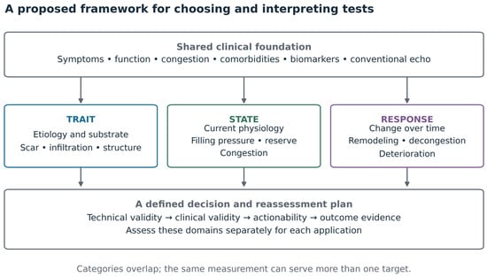
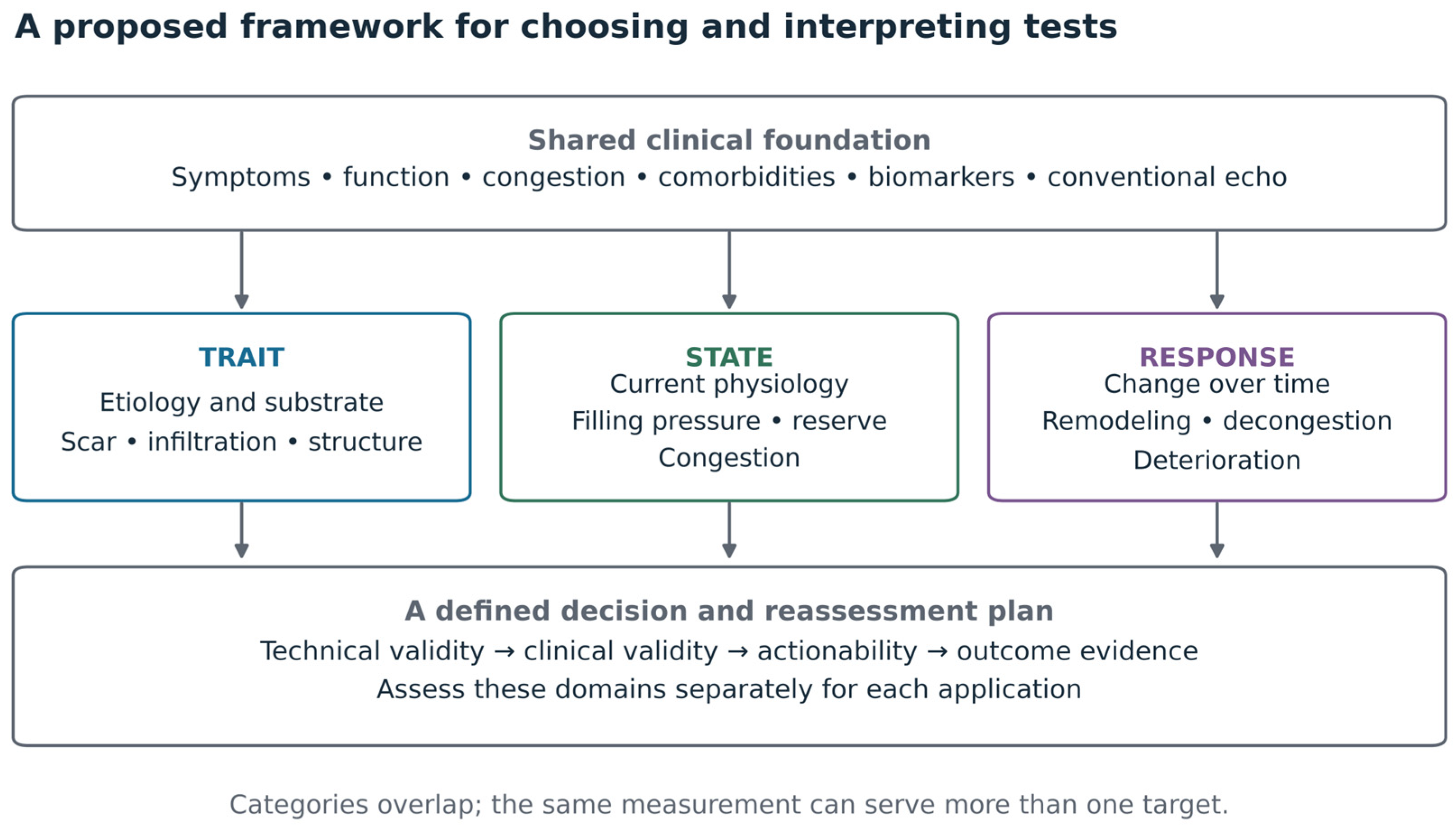
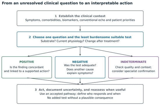
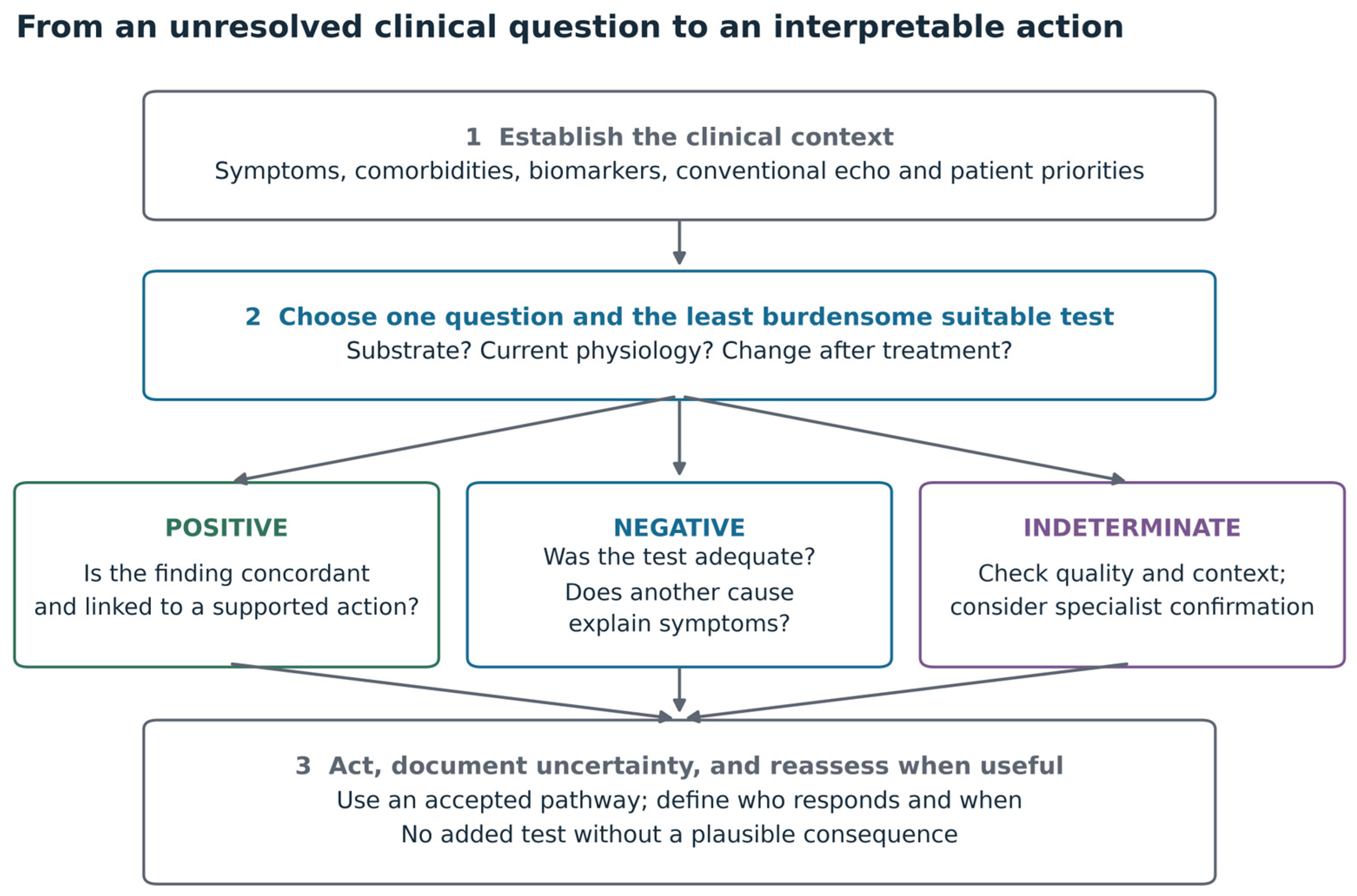
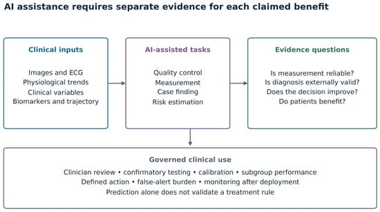

Disclaimer/Publisher’s Note: The statements, opinions and data contained in all publications are solely those of the individual author(s) and contributor(s) and not of MDPI and/or the editor(s). MDPI and/or the editor(s) disclaim responsibility for any injury to people or property resulting from any ideas, methods, instructions or products referred to in the content. |
© 2026 by the authors. Licensee MDPI, Basel, Switzerland. This article is an open access article distributed under the terms and conditions of the Creative Commons Attribution (CC BY) license.