


Simple Summary
Malondialdehyde (MDA) generated from oxidized feed threatens the health and product quality of farmed fish, while its specific harms to channel catfish (Ictalurus punctatus) remain unclear. This study investigated the effects of dietary exogenous MDA on the antioxidant capacity, immunity and body color of channel catfish via a 30-day feeding trial. The results demonstrated that exogenous MDA induced oxidative stress, decreased antioxidant enzyme activities and altered immune gene expression in skin and muscle, which impaired fish antioxidant and immune functions. It also inhibited tyrosinase activity, suppressed melanin synthesis and increased uranidin accumulation in the two tissues, resulting in abnormal body yellowing. These findings revealed MDA’s toxic mechanism and provided effective guidance for aquaculture feed quality control and improvement of catfish commercial value.
Abstract
Malondialdehyde (MDA) is a common oxidation product in deteriorated aquatic feed, which easily induces oxidative damage and quality deterioration in farmed fish, yet its systemic effects on the physiological function and body color of channel catfish (Ictalurus punctatus) remain poorly understood. This study aimed to investigate the influences of dietary exogenous MDA on antioxidant capacity, immune function and body coloration of channel catfish. A 30-day feeding trial was carried out with four dietary MDA levels of 0, 22.3, 44.6 and 66.8 mg/kg. Relevant physiological and pigment indices of skin and muscle were determined. Exogenous MDA significantly increased plasma transaminase levels and tissue MDA content, decreased antioxidant enzyme activities, and altered the transcription of antioxidant and immune-related genes in skin and muscle, causing oxidative stress and immune dysfunction. It also suppressed tyrosinase activity, downregulated melanin-synthesis-related genes, reduced melanin deposition and promoted uranidin accumulation in the two tissues. Collectively, dietary exogenous MDA impairs antioxidant and immune performance, disrupts pigment metabolism, and ultimately leads to body yellowing in channel catfish.
1. Introduction
In intensive aquaculture systems, oxidative stress is a critical determinant affecting fish health and production performance. During fish farming, multiple factors such as high-density rearing, environmental fluctuations, and dietary nutritional imbalance can induce excessive accumulation of ROS (reactive oxygen species) in fish organisms [1]. The imbalance between ROS generation and antioxidant neutralization engenders oxidative stress in fish [2]. It not only inflicts oxidative damage on key biomolecules, including lipids, proteins and DNA (deoxyribonucleic acid), but also activates inflammatory responses and disrupts normal physiological metabolism, ultimately affecting fish growth, immune function and product quality [3]. As the primary source of food and energy for farmed fish, feed runs the entire aquaculture process, and its quality is directly related to the redox homeostasis of fish. However, during transportation and storage, especially under high-temperature and high-humidity conditions, lipids in feed are prone to oxidative rancidity, generating aldehydes, ketones, alcohols and other potentially toxic oxidation derivatives [4,5]. Besides reducing the nutritive value of feed, these oxidation products may also induce or aggravate oxidative stress after being ingested by fish, thereby impairing growth performance, weakening immune competence and even deteriorating fish quality [6]. The implications of feed-derived lipid oxidation for fish growth and immune function have been extensively documented. For example, oxidized feed constituents have been demonstrated to compromise the antioxidant capacity of rainbow trout (Oncorhynchus mykiss) and to suppress growth performance and feed efficiency in Rhynchocypris lagowskii [7,8]. Here, the term antioxidant capacity refers to the levels of enzymatic antioxidants (SOD, CAT, GPx) and non-enzymatic antioxidant substances in fish tissues, rather than the lipid antioxidant capacity of fish muscle.
Earlier research has documented that oxidative stress experienced by fish during aquaculture can affect body coloration [9], and pigment deposition is directly correlated with oxidative stress levels [10]. Malondialdehyde (MDA) is widely recognized as both a major product of lipid peroxidation and a robust biomarker of oxidative injury in living organisms [11]. As a highly reactive aldehyde, MDA can induce cross-linking and structural alterations in key biomolecules, including proteins and nucleic acids, disrupting cellular structure and function, while concurrently amplifying oxidative stress and inflammatory cascades [12]. Consequently, establishing an oxidative stress model by supplementing exogenous MDA into diets has become a common method for simulating in vivo lipid peroxidation damage and exploring the pathogenic mechanism of oxidative stress under laboratory conditions [13].
Dietary malondialdehyde (MDA) is a common oxidation product in deteriorated aquatic feed that readily induces oxidative damage and quality deterioration in farmed fish. However, its systematic effects on the physiological function and body color of channel catfish (Ictalurus punctatus) remain poorly understood. Previous transcriptomic analysis has revealed that oxidative stress may be involved in body color abnormality by inducing inflammation and disrupting pigment metabolism [14]. On this basis, this study was designed to examine whether dietary exogenous MDA affects the antioxidant capacity, immune function, and body coloration of channel catfish. The findings are expected to provide a theoretical reference for feed quality control and offer practical guidance for improving the commercial value of farmed channel catfish.
2. Materials and Methods
2.1. Diet Preparation
MDA stock solution was prepared according to the current valid Chinese national mandatory food safety standard GB 5009.181-2016 [15] (National Food Safety Standard: Determination of malondialdehyde in food), which supersedes the obsolete GB/T 5009.181-2003. The specific preparation procedure was as follows: Accurately measure 31.5 mL of 1,1,3,3-tetraethoxypropane (Sigma-Aldrich, St. Louis, MO, USA, purity ≥ 99%), dissolve it in 95% ethanol and dilute to a final volume of 100 mL. The solution was stirred for 15 min to achieve full homogenization, and the concentration of the prepared MDA stock solution was determined as 1546.7 μmol MDA/mL via the TBA method with assay kits purchased from Jiancheng Bioengineering Institute (Nanjing, China) (kit number A003-1-2). All MDA solutions were prepared fresh for immediate use.
A total of four experimental groups were established, comprising one control group (Con) and three treatment groups (MDA1, MDA2, MDA3). The basal diet used in this experiment was commercial feed (control diet, Con). Volumes of 0.2, 0.4 and 0.6 mL of the prepared MDA solutions were taken and diluted to 100 mL, then sprayed evenly onto the surface of 1 kg of basal diet in each of the three groups, which were named MDA1, MDA2 and MDA3 groups in sequence. The MDA content in the diets was 22.3, 44.6 and 66.8 mg/kg, respectively. All MDA-supplemented diets were freshly prepared immediately before use, sprayed onto the basal formulation rapidly and uniformly, and the fish received the prepared diets right after compounding. The basal diet’s key ingredients and nutritional composition are outlined in Table 1.
Table 1.
Composition of ingredients and nutrients in the basal diet (% dry basis).
2.2. Laboratory Fish and Husbandry
This 30-day trial was designed as a short-term subacute toxicological assay focusing on early biochemical, histological and transcriptomic toxic responses induced by MDA; therefore, routine sampling of growth performance indicators (weight gain, SGR, FCR, feed intake) was not arranged in the experimental protocol. Healthy channel catfish of uniform size and color were selected as experimental fish. A two-week acclimation period on a control diet was used to help the fish adapt to the rearing environment. The feeding experiment was conducted in net cages (2 m × 2 m × 2 m) at the Nanquan Experimental Base (Freshwater Fisheries Research Centre, Chinese Academy of Fishery Sciences). In this study, 180 channel catfish weighing 830 ± 5 g were randomly assigned to four experimental groups. Each group included three replicate net cages with 15 individuals stocked per cage, for a total of 12 cages. Over a 30-day feeding trial, four test diets were administered separately to each group. Fish were fed twice daily at 7:00 and 17:00, with equal rations provided at each feeding. The initial feeding rate was set at 3% of body weight and adjusted to weather conditions. Fish activity and health status were observed daily throughout the experiment, and feed consumption was documented regularly. During the entire experimental duration, aquaculture conditions were standardized as follows: dissolved oxygen concentration maintained above 6 mg/L, water temperature 24–29 °C, ammonia nitrogen concentration capped at 0.02 mg/L, and natural photoperiodic cycles. These parameters were monitored at regular intervals.
2.3. Sample Collection and Chroma Value Analysis
Sampling was conducted upon completion of the rearing trial, following a 24 h fasting period prior to sampling. From each cage, three healthy fish were chosen at random for tissue sampling and the determination of biochemical parameters. Anesthesia was induced in the selected test fish using 200 mg/L MS-222. Once the fish had become sedated, the color parameters of the dorsal skin and muscle tissue were measured. As described by Yu et al. [16], chromatic parameters including L* (lightness), a* (redness), and b* (yellowness) were recorded from the skin and muscle surfaces with a CR400 color difference meter. Blood was collected using sterile syringes, and plasma was harvested by centrifuging at 4000 rpm for 10 min under refrigerated conditions (4 °C). The fish were then dissected, and the dorsal skin as well as the muscle tissues were collected and preserved properly. All specimens were stored in a −80 °C ultra-low temperature freezer pending further determination and analysis.
2.4. Experimental Determination Method
2.4.1. Analysis of Plasma Transaminase Activities and Tissue Antioxidant Indices
Commercially available assay kits from Jiancheng Bioengineering Institute (Nanjing, China) were used to determine plasma levels of aspartate aminotransferase (AST) and alanine aminotransferase (ALT). The microplate method was employed, and the respective kit numbers were (C010-2-1) and (C009-2-1). For antioxidant characterization, skin and muscle tissues were subjected to biochemical analysis using commercial assay kits from the same manufacturer. Total antioxidant capacity (T-AOC) was assessed by the ABTS method (A015-2-1). Enzymatic antioxidant indices, including total superoxide dismutase (T-SOD, WST-1 method, A001-3-2), glutathione peroxidase (GPx, colorimetric method, A005-1-2), and catalase (CAT, ammonium molybdate method, A007-1-1), were determined accordingly. Oxidative damage was evaluated by measuring malondialdehyde (MDA) levels through the TBA method (A003-1-2). All procedures followed the manufacturer’s instructions for each kit.
2.4.2. ELISA Analysis of Pigment-Related Indices in Skin and Muscle
Skin and muscle tissues were analyzed for uranidin, melanin, and tyrosinase using an ELISA kit (Shanghai Enzyme-Linked Biotechnology Co., Ltd., Shanghai, China). The method is as follows: (1) Uranidin was quantified via a competitive assay (YJ270190). The method is briefly described as follows: purified uranidin antibodies are coated onto a microplate to form a solid-phase antibody. Uranidin samples are added to compete for binding with horseradish peroxidase (HRP)-labelled uranidin antigens. After washing, the substrate tetramethyl benzamine (TMB) is added to produce a color. The intensity of the color produced by the sample is inversely proportional to the uranidin content. Uranidin concentration was quantified spectrophotometrically at 450 nm using a microplate reader, with calibration against a constructed standard curve. (2) The competitive method (YJ234125) was utilized for melanin quantification in the tested sample. A brief description follows: The sample, standard, and HRP-labelled detection antibody are added in sequence to a melanin antibody-immobilized microplate. Following incubation and a wash step, TMB substrate is added and develops a blue color under peroxidase catalysis, which turns yellow after acid termination. The color intensity (measured at 450 nm) is positively correlated with melanin concentration and used to calculate its content. (3) Tyrosinase was quantified by competitive assay (H310-1-2). First, biotin-labelled antigen was added to a microplate pre-coated with antibody, and after a 30 min incubation at 37 °C, immune complexes were formed. Subsequently, unbound biotinylated antigen was eliminated by extensively washing the plate with PBST (phosphate-buffered saline containing Tween 20). Streptavidin–HRP was subsequently introduced and allowed to react at 37 °C for a further 30 min. Following washing, the bound HRP initiated TMB coloration, which changes from blue to yellow after acid termination. The OD was measured at 450 nm, and a negative association existed between this value and sample antigen levels. All analytical procedures and calculations were conducted in accordance with the respective kit manuals.
2.4.3. RT-qPCR Analysis
Skin and muscle tissues were processed for total RNA extraction using the RNAiso Plus Kit (Vazyme, Nanjing, China). RNA concentration and quality were evaluated on a NanoDrop 2000 spectrophotometer (Thermo Fisher Scientific, Wilmington, DE, USA), with the quality standard set at an A260/280 ratio of between 1.8 and 2.0. Quantitative RT-PCR was conducted with the One Step SYBR Green Kit (Vazyme, Nanjing, China) on a CFX96 Touch instrument (Bio-Rad, Hercules, CA, USA). In this one-pot system, reverse transcription and quantitative amplification reactions were completed simultaneously within a single reaction tube using total RNA as the direct template, without separate cDNA synthesis. Given the well-documented stable expression of the glyceraldehyde-3-phosphate dehydrogenase (gapdh) gene across multiple tissues in relevant channel catfish studies, it was selected as the internal control [10,17]. Primer sequences, primer amplification efficiencies and corresponding R2 values for all target and reference genes are summarized in Table 2. Standard curves were generated via serial gradient dilutions of pooled total RNA extracted with the Vazyme RNA kit (seven concentration gradients, three technical replicates per gradient) to calculate amplification efficiency in accordance with MIQE guidelines. Primers were synthesized by Shenggong Bioengineering (Shanghai) Co., Ltd. (Shanghai, China). The fold differences in transcript levels between experimental cohorts were mathematically determined according to the 2−ΔΔCt outlined by Schmittgen et al. [18].
Table 2.
Primer sequences and gene abbreviations for RT-qPCR.
2.5. Data Analysis
All statistical analyses were performed using SPSS 26.0. Prior to statistical analysis, the Shapiro–Wilk test was applied to assess data normality, and Levene’s test was used to verify homogeneity of variance. One-way ANOVA with Tukey’s post hoc test was subsequently applied to datasets meeting both assumptions for group comparisons. Results are reported in the format of means ± SD, and differences with p < 0.05 were considered statistically significant. Graphical visualization was conducted with GraphPad Prism 8.0.
3. Results
3.1. Effects of Exogenous MDA on Plasma Transaminase Activities of Channel Catfish
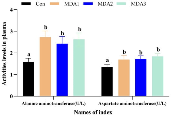
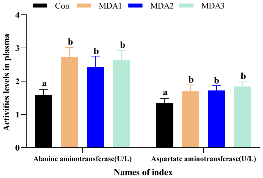
Figure 1 showed that plasma samples from MDA-treated channel catfish exhibited significant upregulation of both transaminase indices, specifically AST and ALT, when compared with the untreated cohort (p < 0.05). AST measurements peaked at the highest values in the MDA3 group, whereas ALT activity attained its peak within the MDA1 treatment arm (p < 0.05).
Figure 1.
The levels of AST and ALT in plasma. Results were expressed in the format of means ± SD. Significant disparities between treatments were denoted by differing superscripts (p < 0.05).
3.2. Effects of Exogenous MDA on Antioxidant Capacity in Skin and Muscle of Channel Catfish
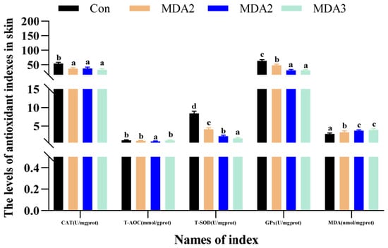
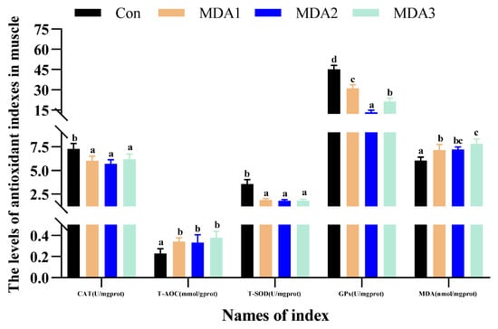
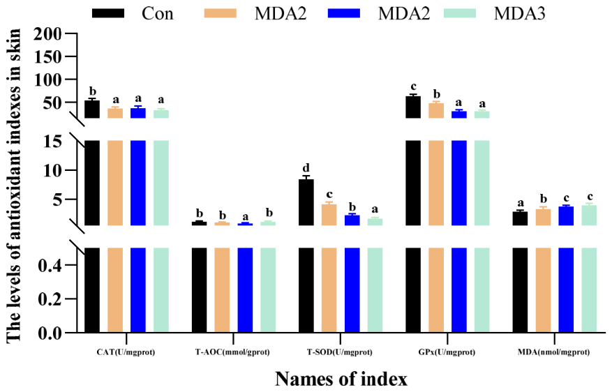
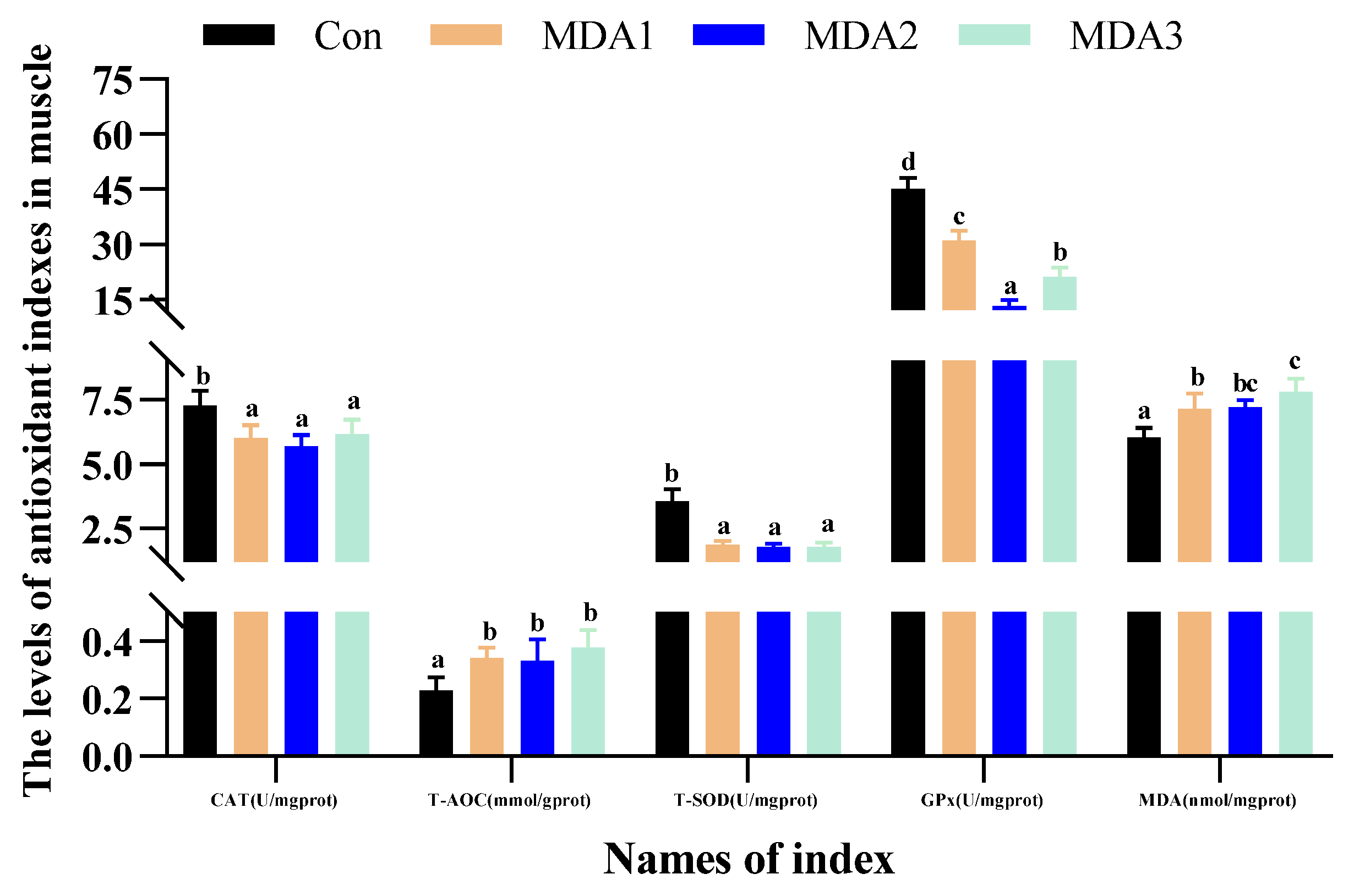
Figure 2 shows that, relative to the control, skin from MDA-treated fish exhibited significantly compromised CAT, T-SOD and GPx profiles alongside elevated MDA burdens (p < 0.05), with T-AOC additionally suppressed in the MDA2 group (p < 0.05). In muscle, the MDA1-MDA3 groups showed equivalent enzymatic reductions, whereas both MDA contents and T-AOC levels were markedly elevated (p < 0.05).
Figure 2.
Antioxidant capacity indices in skin and muscle. Results were expressed in the format of means ± SD. Significant disparities between treatments were denoted by differing superscripts (p < 0.05).
3.3. Effects of Exogenous MDA on Antioxidant and Immune-Related Gene Expression in Skin and Muscle of Channel Catfish
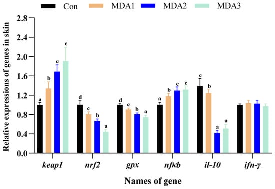
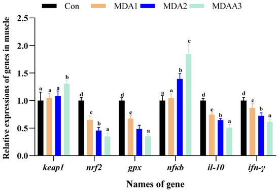
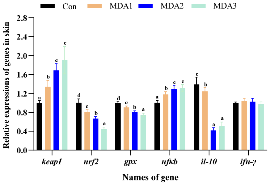
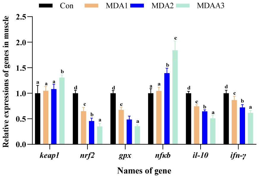
Figure 3 shows that, relative to the control, MDA-treated fish skin exhibited significant downregulation of nrf2, gpx and il-10 transcripts, whereas keap1 and nfκb were markedly upregulated (p < 0.05). The mRNA levels of ifn-γ were unaffected by MDA treatment (p > 0.05). In muscle, treatment with the MDA1-MDA3 groups significantly reduced the mRNA levels of ifn-γ, nrf2, gpx and il-10 (p < 0.05). MDA3 group markedly increased keap1 gene expression, while the MDA2 and MDA3 groups significantly increased nfκb gene expression (p < 0.05).
Figure 3.
The expression levels of genes governing oxidative defense and immune function in skin and muscle. Results were expressed in the format of means ± SD. Significant disparities between treatments were denoted by differing superscripts (p < 0.05); bars lacking superscripts indicate no significant intergroup differences (p > 0.05).
3.4. Effects of Exogenous MDA on Skin and Muscle Coloration and Pigment Deposition of Channel Catfish
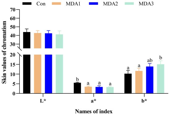
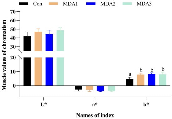
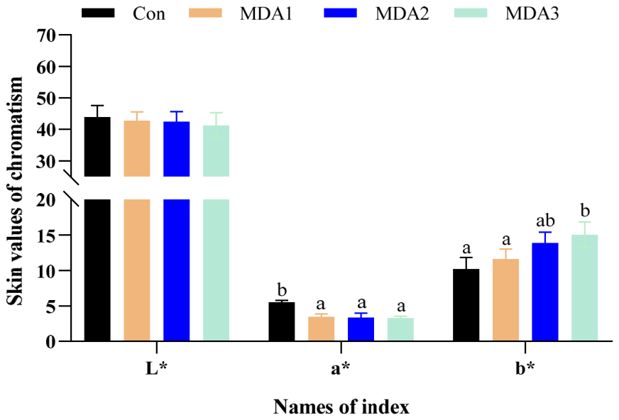
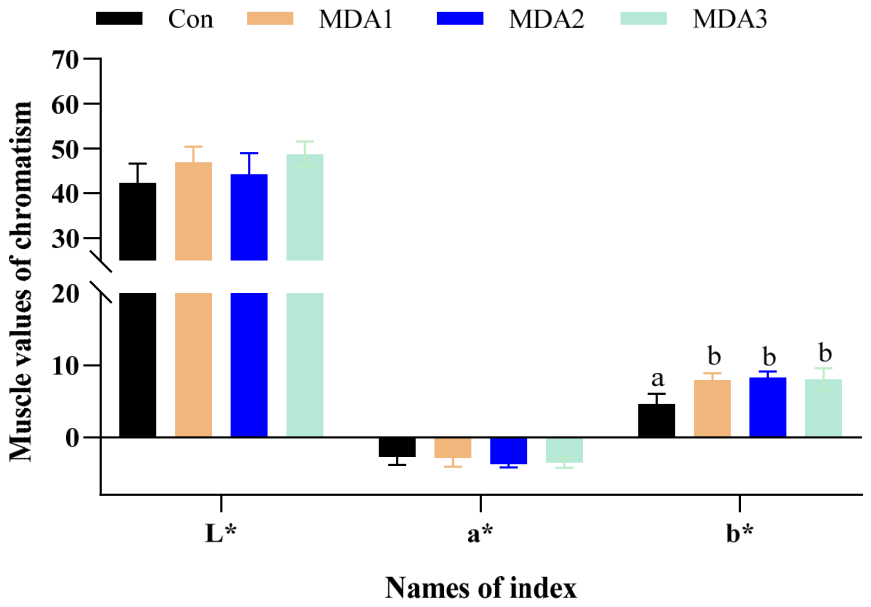
Figure 4 shows that, relative to the control, the L* value of the skin in the MDA1, MDA2 and MDA3 groups showed a gradual decrease as MDA levels increased, whilst the a* value decreased significantly (p < 0.05) and the b* value gradually increased; in the MDA3 group, the b* value increased significantly (p < 0.05). In muscle, the value of L* and a* showed no significant changes among groups (p > 0.05), whereas the value of b* was markedly increased in the MDA1-MDA3 groups (p < 0.05).
Figure 4.
The chromaticity values of skin and muscle. Results were expressed in the format of means ± SD. Significant disparities between treatments were denoted by differing superscripts (p < 0.05); bars lacking superscripts indicate no significant intergroup differences (p > 0.05).
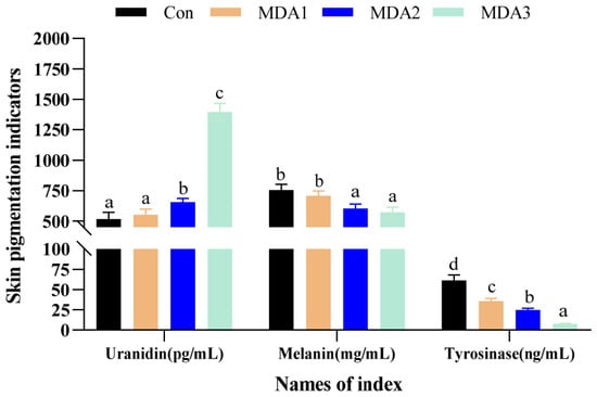
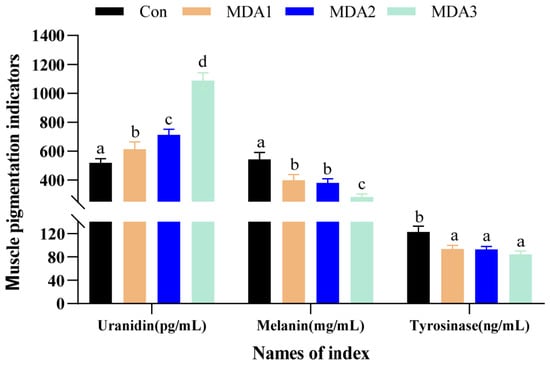
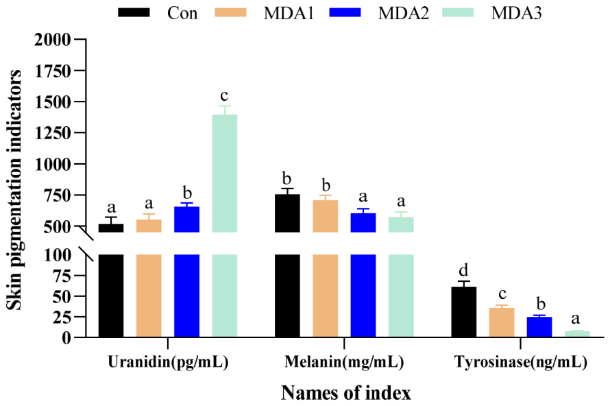
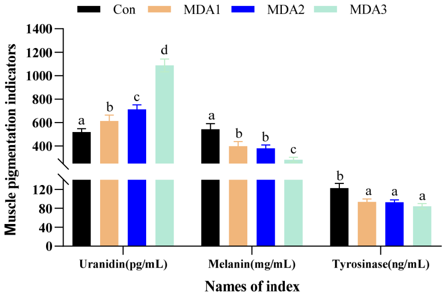
As shown in Figure 5, uranidin content in skin and muscle increased with increased MDA level. Compared to controls, significant elevations occurred in skin (MDA2 and MDA3) and in muscle (all MDA groups) (p < 0.05). An opposing pattern characterized melanin dynamics. The skin melanin content was significantly reduced under MDA2 and MDA3 groups (p < 0.05), whereas muscle melanin showed significant reduction across all MDA-treated groups (p < 0.05). All groups treated with MDA exhibited significantly lower tyrosinase activity in skin and muscle (p < 0.05), and the degree of inhibition increased with higher MDA concentrations, exhibiting a dose-dependent relationship.
Figure 5.
The levels of tyrosinase abundance and pigmentation in skin and muscle of channel catfish. Results were expressed in the format of means ± SD. Significant disparities between treatments were denoted by differing superscripts (p < 0.05).
3.5. Effects of Exogenous MDA on Pigment-Related Genes in Skin and Muscle of Channel Catfish
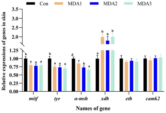
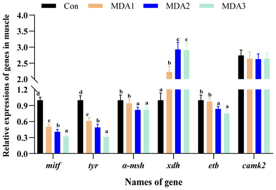
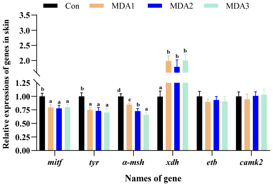
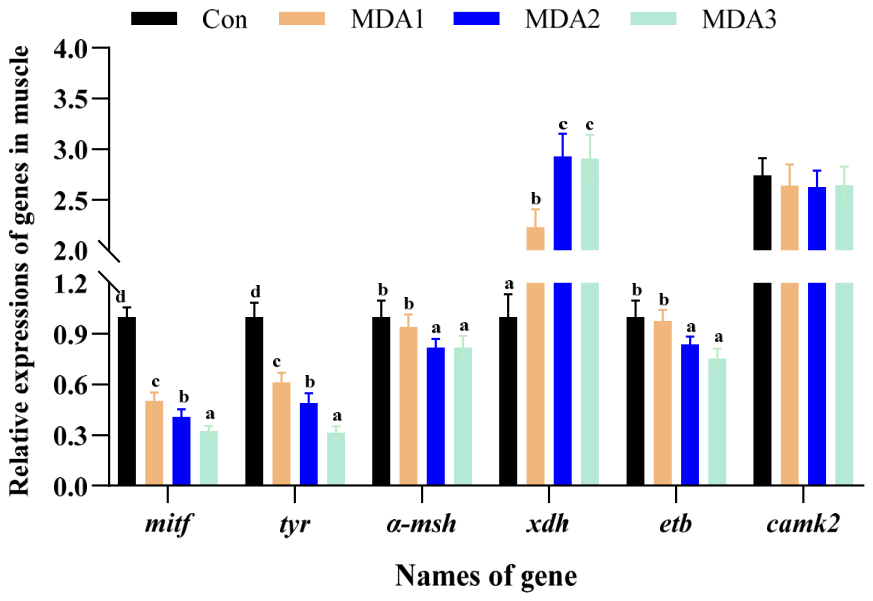
Figure 6 shows that, in reference to the untreated cohort, the mRNA levels of mitf, tyr and α-msh in skin were significantly attenuated under MDA1-MDA3 groups, whereas the mRNA levels of xdh were markedly upregulated (p < 0.05). The mRNA levels of etb and camk2 displayed no notable differences between groups (p > 0.05). In muscle, treatment with the MDA1-MDA3 groups resulted in reduced mRNA levels of the mitf and tyr, alongside a distinct upregulation of the xdh gene expression (p < 0.05). MDA2 and MDA3 groups significantly downregulated the expression of α-msh and etb (p < 0.05). However, no obvious statistical disparities in the mRNA levels of camk2 were found across different treatment groups (p > 0.05).
Figure 6.
The mRNA levels of pigment synthesis-associated genes in skin and muscle. Results were expressed in the format of means ± SD. Significant disparities between treatments were denoted by differing superscripts (p < 0.05); bars lacking superscripts indicate no significant intergroup differences (p > 0.05).
4. Discussion
Our findings suggested that adding exogenous MDA to the diet induced oxidative stress in channel catfish. Plasma biochemical parameters reflect an overall physiological state of the organism, particularly liver health, which was the central organ for metabolic processes in the body, and plasma ALT and AST activities are important indicators for assessing the extent of hepatocyte damage [19]. When hepatocytes are damaged, large amounts of ALT and AST are released from the cells into the plasma, leading to significantly increased levels in plasma [20]. The present results showed that plasma activities of AST and ALT in MDA-treated groups were elevated relative to the control group, corroborating previous observations by Zhang et al. [21] and Shi et al. [17]. Based on these results, exogenous MDA induced oxidative stress and tissue damage in fish. Furthermore, at the tissue level, escalating exogenous MDA exposure elicited pronounced suppression of endogenous antioxidant enzyme activities, encompassing T-SOD, CAT, and GPx, concomitant with a marked surge in MDA accumulation as the terminal product of lipid peroxidation. This indicated that MDA induced oxidative stress in the organism, leading to impairment of the antioxidant defense system and lipid peroxidation damage. These findings aligned with an independent study demonstrating that exogenous MDA supplementation in the diet could inhibit the Keap1/Nrf2/ARE signaling pathway in hybrid groupers (Epinephelus fuscoguttatus ♀ × E. lanceolatus ♂), provoke oxidative stress, and consequently impair antioxidant defense [22]. Collectively, the findings from this section indicated that exogenous MDA disrupted the redox homeostasis and triggered marked oxidative stress in channel catfish [23]. To eliminate excessive intracellular ROS, fish have evolved an innate antioxidant defense system, encompassing antioxidant enzymes such as T-SOD, CAT, and GPx, alongside non-enzymatic antioxidants exemplified by GSH. Changes in the activities or contents of these indicators can reflect the physiological status and antioxidative capacity [24,25]. MDA constitutes a hallmark biomarker of lipid peroxidation, and its levels directly reflect the extent of this process and the associated oxidative damage in vivo [26]. Significant reductions in T-SOD, CAT, GPx and T-AOC indicate that under sustained ROS attack, the synthesis of antioxidant enzymes is inhibited, compensatory regulation is compromised, and overall antioxidant function is markedly impaired [27,28]. In Nile tilapia (Oreochromis niloticus), intraperitoneal injection of H2O2 to induce oxidative stress has been shown to significantly elevate plasma and hepatic MDA levels, while levels of CAT, T-SOD and GSH decrease [29]. Another investigation demonstrated that a diet enriched with oxidized fish oil significantly increased hepatic MDA content in channel catfish, while markedly reducing CAT, T-SOD, GPx activities and T-AOC levels [17]. Comparable outcomes, studies on Wuchang bream (Megalobrama amblycephala) [30] and tilapia (Oreochromis niloticus) [31] have also found that oxidized fish oil in feed significantly reduces antioxidant enzyme activity in these aquatic species and increases their plasma transaminase activity. Our findings showed that, in comparison to the control group, tissues from MDA-treated individuals presented significantly reduced activity of CAT, T-SOD and GPx in skin as well as muscle, alongside a sharp increase in endogenous MDA accumulation. Notably, the level of T-AOC in skin decreased significantly in MDA2 groups, whereas the level of T-AOC in muscle exhibited a compensatory elevation. As the primary physical barrier of the body against exogenous stressors, the skin contains antioxidant substances that are rapidly depleted, rendering the total antioxidant capacity unable to maintain homeostasis via compensatory regulation and consequently leading to MDA accumulation [32]. Consistent with both our findings and earlier reports, it can be concluded that channel catfish exposed to oxidative stress during aquaculture exhibit a compromised antioxidant system, reduced ROS scavenging capacity, and intensified lipid peroxidation. These alterations ultimately result in tissue oxidative damage and disruption of physiological functions in the organism.
In terms of antioxidant regulation, the Keap1/Nrf2/ARE axis represents the core pathway mediating the oxidative stress response in organisms [33]. In this pathway, nrf2 serves as a key transcription factor that activates the transcription of downstream antioxidant genes, while keap1 negatively regulates the antioxidant defense system by blocking nrf2’s nuclear import and promoting its degradation [34]. In this study, oxidative stress induced by MDA markedly upregulated keap1 expression in the skin and muscle tissues, whereas it significantly suppressed nrf2 and its downstream antioxidant gene, gpx. Moreover, such inhibitory effects were enhanced with the increase in MDA concentration, which corroborated previous reports. Shi et al. demonstrated that in the liver of channel catfish [17]. In addition, Yang et al. also confirmed that intake of oxidized fish oil remarkably increased intestinal ROS level and impaired the repair capacity of the antioxidant system in Wuchang bream [35]. These findings suggested that exogenous MDA induced oxidative stress, inhibited the Keap1/Nrf2/ARE signaling pathway, which in turn downregulated the transcription of antioxidant genes and weakened the organism’s ability to eliminate ROS. This further confirmed that the antioxidant defense system of fish was markedly inhibited under oxidative stress conditions. Organism health relies not only on a sound antioxidant defense system but is also critical for immune function. At the level of immune modulation, the NF-κB signaling pathway acts as a core modulator of fish inflammatory responses and the transcriptional activity of immune-associated genes [36]. Altered expression of nfκb, a key regulatory gene within this pathway, can affect the transcription of downstream cytokines with opposing inflammatory roles, including ifn-γ (pro-inflammatory) and il-10 (anti-inflammatory). In this study, adding exogenous MDA markedly enhanced the transcription of nfκb in the skin and muscle tissues, with transcript levels increasing in parallel with adding levels of exogenous MDA, while also leading to a marked downregulation of the anti-inflammatory cytokine il-10. In the skin, ifn-γ expression first increased and then decreased, whereas in the muscle, ifn-γ expression was significantly suppressed. These findings revealed a close correlation between MDA-induced oxidative stress and inflammatory responses. As a central regulator of inflammatory responses, upregulated nfκb can activate the transcription of downstream pro-inflammatory cytokines and exacerbate systemic inflammatory reactions [37]. Playing a central role in resolving inflammation, il-10 acts to restrain the release of pro-inflammatory factors, thereby preserving immunological homeostasis [38]. The expression imbalance between these two genes serves as an important molecular hallmark of inflammatory damage in fish. Earlier research has confirmed that oxidative stress promotes inflammation in fish via activation of the NF-κB pathway, whilst simultaneously suppressing the expression of the anti-inflammatory cytokine il-10. For instance, Zhang et al. confirmed that feeding Rhynchocypris lagowskii with oxidized fish oil led to high expression of pro-inflammatory cytokines accompanied by low expression of anti-inflammatory cytokines [39]. In addition, Zhang et al. also found that prolonged consumption of oxidized fish oil aggravated inflammation in juvenile swamp eel (Monopterus albus) via the upregulation of pro-inflammatory cytokines, which in turn led to compromised immune function [40]. This study revealed that the imbalance between nfκb and il-10 induced by MDA treatment indicated that oxidative stress not only damages the antioxidant system of fish, but also triggers disorder of systemic inflammatory responses, further exacerbating tissue damage.
Oxidative stress could affect the body color and flesh color of fish, mainly manifested as dull and faded body color, as well as whitening, yellowing or loss of vividness in flesh color. Studies have shown that microplastic-induced oxidative stress triggers degradation of the blue structural color and reduced glossiness in the caudal fin of peacock fish (Poecilia reticulata), indicating oxidative stress disrupts iridocyte integrities [41]. Under oxidative stress, carotenoid levels in yellow catfish (Pelteobagrus fulvidraco) muscle decline, causing the flesh color to change from golden yellow to pale white [42]. Environmental factors such as hypoxia and low temperature trigger oxidative stress, which reduces the flesh b* and C* values of Nile tilapia, resulting in pale flesh color and loss of vividness [43]. This study found that as the MDA concentration increased in the treatment groups, the a* values of both skin and muscle in channel catfish decreased, while the overall skin b* values increased, deviating from the normal body color. Collectively, these results indicated that oxidative stress driven by MDA negatively affects the skin coloration of channel catfish under conditions of high ROS levels. Surplus ROS produced under oxidative conditions progressively build up and disrupt the signal transduction routes associated with melanin synthesis [44]. For instance, oxidized lipids affect the differentiation and development of fish melanocytes, reduce the population of mature melanocytes, thereby leading to inadequate melanin synthesis and indirectly elevating skin yellowness value [45]. Melanin is a biological pigment synthesized from tyrosine via a cascade of biochemical reactions, which is classified into eumelanin (black/brown) and pheomelanin (red/yellow). The content and distribution of melanin determine body skin coloration [46,47]. Tyrosinase acts as the key regulatory enzyme in the melanin biosynthetic route, and its catalytic action directly modulates the rate of melanin production [48,49]. Xanthophores are essential for fish body coloration and are capable of transdifferentiating with melanocytes [50]. Our findings indicated that increasing MDA supplementation resulted in a dose-dependent suppression of tyrosinase activity and melanin content in skin and muscle tissues. This correlation was consistent with the well-established conclusion that inhibiting tyrosinase activity can effectively reduce melanin production [51,52,53], which strongly demonstrated that the inhibition of tyrosinase activity and blockade of normal melanin synthesis represent one of the core mechanisms by which oxidative stress induces changes in body color and flesh color [54]. Meanwhile, oxidative stress promoted the manifestation of yellowness in the skin. The main uranidin in channel catfish’s skin and muscle are carotenoids such as lutein, zeaxanthin and isoflavone. The conjugated double bonds in their molecular structures endow them with antioxidant functions of scavenging free radicals and maintaining ROS homeostasis [55]. This study demonstrated that skin and muscle uranidin content increased markedly in parallel with increasing MDA concentration. It was speculated that this change might be attributed to the shunting of metabolic substrates to the uranidin deposition pathway following the inhibition of melanin synthesis. Alternatively, it might serve as an active protective response of the organism against oxidative damage through carotenoid accumulation [56]. The underlying mechanisms remain to be further investigated. Further combined with the analysis of gene expression levels, the molecular mechanism by which oxidative stress regulates abnormal body coloration in channel catfish can be further elucidated. The genes including mitf, tyr, and α-msh are involved in regulating melanin synthesis at three distinct levels: transcriptional, enzymatic, and hormonal [57]. To be specific, microphthalmia-associated transcription factor is encoded by mitf, tyrosinase by tyr, and α-melanocyte-stimulating hormone by α-msh. Both etb and camk2 represent key regulatory components of the melanin synthesis signaling pathway. Abnormal expression levels or activities of the two genes can directly lead to abnormal body color formation in fish by affecting melanocyte differentiation, migration, and the transcriptional regulation of core genes responsible for melanin synthesis [58,59]. Our results indicated that MDA treatment markedly downregulated the transcript levels of mitf, tyr and α-msh in skin and muscle tissues, while etb and camk2 levels also presented a decreasing trend. These results aligned with an earlier study conducted by Ye et al. [60], who found that administering oxidized fish oil to yellow catfish (Pelteobagrus fulvidraco) induced oxidative stress and significant downregulation of key melanin synthesis genes such as tyr, mitf and etb, with the effect being dose-dependent. In addition, a study of zebrafish (Danio rerio) has demonstrated that the loss of etb expression directly causes disorders in melanocyte differentiation and migration, eventually resulting in body color albinism and stripe disappearance [61]. Collectively, these results indicated that oxidative stress could block melanin production at the source by transcriptionally downregulating key synthesis genes. The homolog of the enzyme encoded by the Xdh gene is capable of synthesizing yellow-red pteridine pigments [62,63]. In this study, the transcript levels of the yellow pigment synthesis-related gene xdh increased significantly with escalating MDA doses, mirroring the dose-dependent rise in tissue uranidin content. In conclusion, exogenous MDA-induced oxidative stress mainly inhibits tyrosinase activity, disrupts the deposition balance between melanin and uranidin, and ultimately leads to skin yellowing in channel catfish.
This study has certain limitations. The 30-day short-term subacute MDA exposure trial only focused on early biochemical, histological and transcriptomic toxic responses, and growth-related indicators were not included in the experimental design, as obvious growth inhibition cannot be observed within a short exposure period. Long-term chronic feeding trials integrating growth and toxic injury detection will be conducted in subsequent research to systematically assess MDA toxicity, and multi-reference-gene normalization will be adopted in future qPCR analysis. Nevertheless, the core conclusions on early toxic mechanisms derived from the present data remain credible.
5. Conclusions
Exogenous MDA supplementation in the diet induced oxidative stress and impaired antioxidant capacity, as well as immune function, and caused oxidative tissue damage. By interfering with the pigment metabolic pathway, it inhibited melanin synthesis and promoted uranidin accumulation, ultimately leading to body color yellowing in channel catfish.
Supplementary Materials
The following are available online at https://www.mdpi.com/article/10.3390/vetsci13070632/s1, Figure S1: Representative photographs of normal body skin color and dorsal muscle color in healthy channel catfish (Ictalurus punctatus), Figure S2: Representative photographs of body skin color and dorsal muscle color of yellowed channel catfish (Ictalurus punctatus).
Author Contributions
Conceptualization, C.X. and H.L.; methodology, L.L. and C.X.; formal analysis, L.L. and C.X.; investigation, L.Z. (Lu Zhang) and C.X.; data curation, L.Z. (Leimin Zhang); writing—original draft preparation, L.L. and C.X.; writing—review and editing, H.L. and H.M.; visualization, L.Z. (Leimin Zhang); supervision, M.R.; project administration, D.H. All authors have read and agreed to the published version of the manuscript.
Funding
This study was financially supported by the earmarked fund for CARS (NO. CARS-46), the Central Public-interest Scientific Institution Basal Research Fund, CAFS (NO. 2026XT0702).
Institutional Review Board Statement
The study was approved by the Laboratory Animal Ethics Committee of the Freshwater Fisheries Research Center (LAECFFRC20240309).
Informed Consent Statement
Not applicable.
Data Availability Statement
The original contributions presented in this study are included in the article/Supplementary Materials. Further inquiries can be directed to the corresponding authors.
Conflicts of Interest
Authors Lu Zhang and Haifeng Mi are employed by Tongwei Agricultural Development Co., Ltd. The remaining authors declare that the research was conducted in the absence of any commercial or financial relationships that could be construed as potential conflicts of interest.
References
- Skrzynska, A.K.; Maiorano, E.; Bastaroli, M.; Naderi, F.; Míguez, J.M.; Martínez-Rodríguez, G.; Mancera, J.M.; Martos-Sitcha, J.A. Impact of Air Exposure on Vasotocinergic and Isotocinergic Systems in Gilthead Sea Bream (Sparus aurata): New Insights on Fish Stress Response. Front. Physiol. 2018, 9, 96. [Google Scholar] [CrossRef] [Scilit] [PubMed]
- Lushchak, V.I.; Storey, K.B. Oxidative stress concept updated: Definitions, classifications, and regulatory pathways implicated. EXCLI J. 2021, 20, 956–967. [Google Scholar] [CrossRef] [Scilit] [PubMed]
- Grootveld, M.; Percival, B.C.; Leenders, J.; Wilson, P.B. Potential adverse public health effects afforded by the ingestion of dietary lipid oxidation product toxins: Significance of fried food sources. Nutrients 2020, 12, 974. [Google Scholar] [CrossRef] [Scilit] [PubMed]
- Liu, K.; Liu, Y.; Chen, F. Effect of storage temperature on lipid oxidation and changes in nutrient contents in peanuts. Food Sci. Nutr. 2019, 7, 2280–2290. [Google Scholar] [CrossRef] [Scilit] [PubMed]
- Long, S.; Dong, X.; Tan, B.; Zhang, S.; Chi, S.; Yang, Q.; Liu, H.; Xie, S.; Deng, J.; Yang, Y. The antioxidant ability, histology, proximate, amino acid and fatty acid compositions, and transcriptome analysis of muscle in juvenile hybrid grouper (♀ Epinephelus fuscoguttatus × ♂ Epinephelus lanceolatus) fed with oxidized fish oil. Aquaculture 2022, 547, 737510. [Google Scholar] [CrossRef] [Scilit]
- Desbruslais, A.; Wealleans, A.L. Oxidation in Poultry Feed: Impact on the Bird and the Efficacy of Dietary Antioxidant Mitigation Strategies. Poultry 2022, 1, 246–277. [Google Scholar] [CrossRef] [Scilit]
- Fontagné-Dicharry, S.; Lataillade, E.; Surget, A.; Larroquet, L.; Cluzeaud, M.; Kaushik, S. Antioxidant defense system is altered by dietary oxidized lipid in first-feeding rainbow trout (Oncorhynchus mykiss). Aquaculture 2014, 424, 220–227. [Google Scholar] [CrossRef] [Scilit]
- Chen, X.; Wang, Q.; Guo, Z.; Zhao, Y.; Gao, Y.; Yu, T.; Chen, Y.; Zhang, D.; Wang, G. Effects of dietary oxidized fish oil on growth performance and antioxidant defense mechanism of juvenile Rhynchocypris lagowski Dybowski. Aquaculture 2019, 512, 734368. [Google Scholar] [CrossRef] [Scilit]
- Ninwichian, P.; Phuwan, N.; Jakpim, K.; Sae-Lim, P. Effects of tank color on the growth, stress responses, and skin color of snakeskin gourami (Trichogaster pectoralis). Aquac. Int. 2018, 26, 659–672. [Google Scholar] [CrossRef] [Scilit]
- Xue, C.; Zhang, L.; Mi, H.; Huang, D.; Liang, H.; Ren, M.; Zhang, L. Effects of dietary oxidized rice bran on the coloration, antioxidant capacity, and immunity in channel catfish (Ictalurus punctatus). Aquac. Rep. 2026, 46, 103287. [Google Scholar] [CrossRef] [Scilit]
- Cao, Y.; Yao, W.; Lu, R.; Zhao, H.; Wei, W.; Lei, X.; Zhang, Z.; Liu, B. Reveal the correlation between hub hypoxia/immune-related genes and immunity and diagnosis, and the effect of SAP30 on cell apoptosis, ROS and MDA production in cerebral ischemic stroke. Aging 2023, 15, 15161. [Google Scholar] [CrossRef] [Scilit] [PubMed]
- Zhang, Y.; Liu, P.; Fu, H.; Wang, D.; Zhao, D.; Zhang, J.; Wang, C.; Li, M. Effects of Lactobacillus kefiri fermentation supernatant on skin aging caused by oxidative stress. J. Funct. Foods 2022, 96, 105222. [Google Scholar] [CrossRef] [Scilit]
- Del Rio, D.; Stewart, A.J.; Pellegrini, N. A review of recent studies on malondialdehyde as toxic molecule and biological marker of oxidative stress. Nutr. Metab. Cardiovasc. Dis. 2005, 15, 316–328. [Google Scholar] [CrossRef] [Scilit] [PubMed]
- Xue, C.; Mi, H.; Huang, D.; Liang, H.; Aboseif, A.M.; Ren, M.; Zhang, L. Transcriptome-based studies on the candidate genes related to body color yellowing in channel catfish (Ictalurus punctatus). Aquac. Rep. 2024, 39, 102472. [Google Scholar] [CrossRef] [Scilit]
- GB 5009.181-2016; National Standard for Food Safety—Determination of Malondialdehyde in Food. National Health and Family Planning Commission of the People’s Republic of China. China Quality Inspection Press: Beijing, China, 2016.
- Yu, H.; Liang, H.; Ge, X.; Zhu, J.; Wang, Y.; Ren, M.; Chen, X. Dietary chlorella (Chlorella vulgaris) supplementation effectively improves body color, alleviates muscle inflammation and inhibits apoptosis in largemouth bass (Micropterus salmoides). Fish Shellfish Immunol. 2022, 127, 140–147. [Google Scholar] [CrossRef] [Scilit] [PubMed]
- Shi, Y.; Hu, Y.; Wang, Z.; Zhou, J.; Zhang, J.; Zhong, H.; Fu, G.; Zhong, L. The protective effect of taurine on oxidized fish-oil-induced liver oxidative stress and intestinal barrier-function impairment in juvenile Ictalurus punctatus. Antioxidants 2021, 10, 1690. [Google Scholar] [CrossRef] [Scilit] [PubMed]
- Schmittgen, T.D.; Livak, K.J. Analyzing real-time PCR data by the comparative CT method. Nat. Protoc. 2008, 3, 1101–1108. [Google Scholar] [CrossRef] [Scilit] [PubMed]
- Sahin, K.; Yazlak, H.; Orhan, C.; Tuzcu, M.; Akdemir, F.; Sahin, N. The effect of lycopene on antioxidant status in rainbow trout (Oncorhynchus mykiss) reared under high stocking density. Aquaculture 2014, 418, 132–138. [Google Scholar] [CrossRef] [Scilit]
- Knudsen, A.R.; Andersen, K.J.; Hamilton-Dutoit, S.; Nyengaard, J.R.; Mortensen, F.V. Correlation between liver cell necrosis and circulating alanine aminotransferase after ischaemia/reperfusion injuries in the rat liver. Int. J. Exp. Pathol. 2016, 97, 133–138. [Google Scholar] [CrossRef] [Scilit] [PubMed]
- Zhang, J.; Wang, Z.; Shi, Y.; Xia, L.; Hu, Y.; Zhong, L. Protective effects of chlorogenic acid on growth, intestinal inflammation, hepatic antioxidant capacity, muscle development and skin color in channel catfish Ictalurus punctatus fed an oxidized fish oil diet. Fish Shellfish Immunol. 2023, 134, 108511. [Google Scholar] [CrossRef] [Scilit] [PubMed]
- Fan, J.; Zhang, Y.; Zhou, H.; Liu, Y.; Cao, Y.; Dou, X.; Fu, X.; Deng, J.; Tan, B. Dietary malondialdehyde damage to the growth performance and digestive function of hybrid grouper (Epinephelus fuscoguttatus ♀ × E. lanceolatus ♂). Animals 2023, 13, 3145. [Google Scholar] [CrossRef] [Scilit] [PubMed]
- Dong, G.; Zhu, X.; Ren, H.; Nie, B.; Chen, L.; Li, H.; Yan, B. Effects of oxidized fish oil intake on tissue lipid metabolism and fatty acid composition of channel catfish (Ictalurus punctatus). Aquac. Res. 2014, 45, 1867–1880. [Google Scholar] [CrossRef] [Scilit]
- Sandamalika, W.G.; Kwon, H.; Lim, C.; Yang, H.; Lee, J. The possible role of catalase in innate immunity and diminution of cellular oxidative stress: Insights into its molecular characteristics, antioxidant activity, DNA protection, and transcriptional regulation in response to immune stimuli in yellowtail clownfish (Amphiprion clarkii). Fish Shellfish Immunol. 2021, 113, 106–117. [Google Scholar] [CrossRef] [Scilit] [PubMed]
- Yang, X.; He, Y.; Lin, S.; Dong, X.; Yang, Q.; Liu, H.; Zhang, S.; Chi, S.; Tan, B. Saccharomyces cerevisiae extracts improved the effects of a low fishmeal, complex plant protein diet in the orange-spotted grouper, Epinephelus coioides. Aquac. Rep. 2021, 19, 100574. [Google Scholar] [CrossRef] [Scilit]
- Abdel-Latif, H.M.; Shukry, M.; Noreldin, A.E.; Ahmed, H.A.; El-Bahrawy, A.; Ghetas, H.A.; Khalifa, E. Milk thistle (Silybum marianum) extract improves growth, immunity, serum biochemical indices, antioxidant state, hepatic histoarchitecture, and intestinal histomorphometry of striped catfish, Pangasianodon hypophthalmus. Aquaculture 2023, 562, 738761. [Google Scholar] [CrossRef] [Scilit]
- Ahmed-Farid, O.A.; Rizk, H.A.; Shehata, A.M. Hydrogen peroxide modulates redox status, energy metabolism, and gene expression in a dose-and time-dependent manner in rat liver. J. Biochem. Mol. Toxicol. 2018, 32, e22199. [Google Scholar] [CrossRef] [Scilit] [PubMed]
- Becerra, S.; Arriagada-Solimano, M.; Escobar-Aguirre, S.; Palomino, J.; Aedo, J.; Estrada, J.M.; Barra-Valdebenito, V.; Zuloaga, R.; Valdes, J.A.; Dettleff, P. High temperature induces oxidative damage, immune modulation, and atrophy in the gills and skeletal muscle of the teleost fish black cusk-eel (Genypterus maculatus). Dev. Comp. Immunol. 2025, 164, 105332. [Google Scholar] [CrossRef] [Scilit] [PubMed]
- Jia, R.; Du, J.; Cao, L.; Li, Y.; Johnson, O.; Gu, Z.; Jeney, G.; Xu, P.; Yin, G. Antioxidative, inflammatory and immune responses in hydrogen peroxide-induced liver injury of tilapia (GIFT, Oreochromis niloticus). Fish Shellfish Immunol. 2019, 84, 894–905. [Google Scholar] [CrossRef] [Scilit] [PubMed]
- Song, C.; Liu, B.; Xu, P.; Ge, X.; Zhang, H. Emodin ameliorates metabolic and antioxidant capacity inhibited by dietary oxidized fish oil through PPARs and Nrf2-Keap1 signaling in Wuchang bream (Megalobrama amblycephala). Fish Shellfish Immunol. 2019, 94, 842–851. [Google Scholar] [CrossRef] [Scilit] [PubMed]
- Yu, L.; Wen, H.; Jiang, M.; Wu, F.; Tian, J.; Lu, X.; Xiao, J.; Liu, W. Effects of ferulic acid on growth performance, immunity and antioxidant status in genetically improved farmed tilapia (Oreochromis niloticus) fed oxidized fish oil. Aquac. Nutr. 2020, 26, 1431–1442. [Google Scholar] [CrossRef] [Scilit]
- Chen, X.; Yang, C.; Jiang, G. Research progress on skin photoaging and oxidative stress. Adv. Dermatol. Allergol. 2021, 38, 931–936. [Google Scholar] [CrossRef] [Scilit] [PubMed]
- Shi, Y.; Zhong, L.; Liu, Y.; Zhang, J.; Lv, Z.; Li, Y.; Hu, Y. Effects of Dietary Andrographolide Levels on Growth Performance, Antioxidant Capacity, Intestinal Immune Function and Microbioma of Rice Field Eel (Monopterus albus). Animals 2020, 10, 1744. [Google Scholar] [CrossRef] [Scilit] [PubMed]
- Zhang, B.; Gao, Y.; Shao, Y.; Li, Y.; Ma, J.; Xie, S.; Li, J.; Ma, T.; Wang, Z. Riboflavin improves meat quality, antioxidant capacity, muscle development, and lipids composition of breast muscle in pigeon. Poult. Sci. 2025, 104, 104856. [Google Scholar] [CrossRef] [Scilit] [PubMed]
- Yang, J.; Zheng, X.; Liu, X.; Zhou, Q.; Sun, C.; Song, C.; Wang, A.; Liu, B. miR-144 targets Nrf2 affecting the intestinal oxidative damage induced by oxidized fish oil in Megalobrama amblycephala with emphasis on autophagy and apoptosis. Water Biol. Secur. 2025, 4, 100340. [Google Scholar] [CrossRef] [Scilit]
- Ding, C.; Xiao, T.; Deng, Y.; Yang, H.; Xu, B.; Li, J.; Lv, Z. The Teleost CXCL13-CXCR5 Axis Induces Inflammatory Cytokine Expression through the Akt-NF-κB, p38-AP-1, and p38-NF-κB Pathways. J. Immunol. 2024, 212, 317–334. [Google Scholar] [CrossRef] [Scilit] [PubMed]
- Anilkumar, S.; Wright-Jin, E. NF-κB as an Inducible Regulator of Inflammation in the Central Nervous System. Cells 2024, 13, 485. [Google Scholar] [CrossRef] [Scilit] [PubMed]
- Liu, C.; Li, Z.; Liu, X.; Lv, S.; Yin, X. Efficacy of neonatal mouse muscle extracellular vesicles in skeletal muscle repair and regeneration. Cell Regen. 2026, 15, 6. [Google Scholar] [CrossRef] [Scilit] [PubMed]
- Zhang, D.-M.; Guo, Z.-X.; Zhao, Y.-L.; Wang, Q.-J.; Gao, Y.-S.; Yu, T.; Chen, Y.-K.; Chen, X.-M.; Wang, G.-Q. L-carnitine regulated Nrf2/Keap1 activation in vitro and in vivo and protected oxidized fish oil-induced inflammation response by inhibiting the NF-κB signaling pathway in Rhynchocypris lagowski Dybowski. Fish Shellfish Immunol. 2019, 93, 1100–1110. [Google Scholar] [CrossRef] [Scilit] [PubMed]
- Zhang, J.; Che, C.; Cai, M.; Hu, Y. Taurine improves health of juvenile rice field eel (Monopterus albus) fed with oxidized fish oil: Involvement of lipid metabolism, antioxidant capacity, inflammatory response. Aquac. Rep. 2022, 27, 101388. [Google Scholar] [CrossRef] [Scilit]
- Ren, H.-Y.; Ma, H.-C.; He, R.-P.; Gao, C.-C.; Wen, B.; Gao, J.-Z.; Chen, Z.-Z. Microplastics Induce Structural Color Deterioration in Fish Poecilia reticulata Mediated by Oxidative Stress. Fishes 2025, 10, 382. [Google Scholar] [CrossRef] [Scilit]
- Liu, C.; Liu, H.; Zhu, X.; Han, D.; Jin, J.; Yang, Y.; Xie, S. The effects of dietary Arthrospira platensis on oxidative stress response and pigmentation in yellow catfish Pelteobagrus fulvidraco. Antioxidants 2022, 11, 1100. [Google Scholar] [CrossRef] [Scilit] [PubMed]
- Youssuf, H.; Soror, E.I.; Shehab, A.; El-daim, A.M.; Abo-Gamil, Z.H.; Ahmed-Farid, O.; Hamad, A.; Edris, S.; Matter, A.F. Amelioration of hypoxia and cold stress in Nile tilapia: Comparative effect of Chlorella vulgaris and its nanoparticle dietary supplementation on performance, antioxidant, hepatic functions, and meat quality. Aquac. Int. 2025, 33, 66. [Google Scholar] [CrossRef] [Scilit]
- Asaba, K.; Iwasaki, Y.; Asai, M.; Yoshida, M.; Nigawara, T.; Kambayashi, M.; Hashimoto, K. High glucose activates pituitary proopiomelanocortin gene expression: Possible role of free radical-sensitive transcription factors. Diabetes Metab. Res. Rev. 2007, 23, 317–323. [Google Scholar] [CrossRef] [Scilit] [PubMed]
- Liang, G.; Li, X.; Yang, H.; Huan, D.; Xu, Z.; Leng, X.; Gao, Q. Dietary oxidized oils decreased growth, antioxidative capacity, and negatively affected skin color of channel catfish, Ictalurus punctatus. J. World Aquac. Soc. 2019, 50, 692–706. [Google Scholar] [CrossRef] [Scilit]
- Pillaiyar, T.; Manickam, M.; Namasivayam, V. Skin whitening agents: Medicinal chemistry perspective of tyrosinase inhibitors. J. Enzyme Inhib. Med. Chem. 2017, 32, 403–425. [Google Scholar] [CrossRef] [Scilit] [PubMed]
- Shim, J.-H. Inhibitory effects of cycloheterophyllin on melanin synthesis. Molecules 2021, 26, 2526. [Google Scholar] [CrossRef] [Scilit] [PubMed]
- Baber, M.A.; Crist, C.M.; Devolve, N.L.; Patrone, J.D. Tyrosinase inhibitors: A perspective. Molecules 2023, 28, 5762. [Google Scholar] [CrossRef] [Scilit] [PubMed]
- Roulier, B.; Rush, I.; Lazinski, L.M.; Pérès, B.; Olleik, H.; Royal, G.; Fishman, A.; Maresca, M.; Haudecoeur, R. Resorcinol-based hemiindigoid derivatives as human tyrosinase inhibitors and melanogenesis suppressors in human melanoma cells. Eur. J. Med. Chem. 2023, 246, 114972. [Google Scholar] [CrossRef] [Scilit] [PubMed]
- Lacosta, A.M.; Canudas, J.; Gonzalez, C.; Muniesa, P.; Sarasa, M.; Dominguez, L. Pax7 identifies neural crest, chromatophore lineages and pigment stem cells during zebrafish development. Int. J. Dev. Biol. 2007, 51, 327–331. [Google Scholar] [CrossRef] [Scilit] [PubMed]
- Teng, H.; Fan, X.; Lv, Q.; Zhang, Q.; Xiao, J.; Qian, Y.; Zheng, B.; Gao, H.; Gao, S.; Chen, L. Folium nelumbinis (Lotus leaf) volatile-rich fraction and its mechanisms of action against melanogenesis in B16 cells. Food Chem. 2020, 330, 127030. [Google Scholar] [CrossRef] [Scilit] [PubMed]
- Peng, Z.; Wang, G.; Zeng, Q.-H.; Li, Y.; Wu, Y.; Liu, H.; Wang, J.J.; Zhao, Y. Synthesis, antioxidant and anti-tyrosinase activity of 1, 2, 4-triazole hydrazones as antibrowning agents. Food Chem. 2021, 341, 128265. [Google Scholar] [CrossRef] [Scilit] [PubMed]
- Kubglomsong, S.; Theerakulkait, C.; Reed, R.L.; Yang, L.; Maier, C.S.; Stevens, J.F. Isolation and identification of tyrosinase-inhibitory and copper-chelating peptides from hydrolyzed rice-bran-derived albumin. J. Agric. Food Chem. 2018, 66, 8346–8354. [Google Scholar] [CrossRef] [Scilit] [PubMed]
- Satooka, H.; Cerda, P.; Kim, H.-J.; Wood, W.F.; Kubo, I. Effects of matsutake mushroom scent compounds on tyrosinase and murine B16-F10 melanoma cells. Biochem. Biophys. Res. Commun. 2017, 487, 840–846. [Google Scholar] [CrossRef] [Scilit] [PubMed]
- Peng, J.; Yuan, J.-P.; Wang, J.-H. Effect of diets supplemented with different sources of astaxanthin on the gonad of the sea urchin Anthocidaris crassispina. Nutrients 2012, 4, 922–934. [Google Scholar] [CrossRef] [Scilit] [PubMed]
- Wu, J.; Dong, W.H.; Zheng, F.; Chen, K.; Ke, Y.; Niu, W. Dietary carotenoids and acute respiratory infection in the general US population: NHANES 2003 - 2018. BMC Pediatr. 2025, 25, 823. [Google Scholar] [CrossRef] [Scilit] [PubMed]
- Yin, L.; Coelho, S.G.; Valencia, J.C.; Ebsen, D.; Mahns, A.; Smuda, C.; Miller, S.A.; Beer, J.Z.; Kolbe, L.; Hearing, V.J. Identification of genes expressed in hyperpigmented skin using meta-analysis of microarray data sets. J. Invest. Dermatol. 2015, 135, 2455–2463. [Google Scholar] [CrossRef] [Scilit] [PubMed]
- Sun, J.H.; Bao, B.B. Identification of mutation sites related to body color albinism in Cynoglossus semilaevis. J. Shanghai Ocean Univ. 2022, 31, 849–857. [Google Scholar] [CrossRef]
- Berry, E.G.; Bezecny, J.; Acton, M.; Sulmonetti, T.P.; Anderson, D.M.; Beckham, H.W.; Durr, R.A.; Chiba, T.; Beem, J.; Brash, D.E. Slip versus slop: A head-to-head comparison of UV-protective clothing to sunscreen. Cancers 2022, 14, 542. [Google Scholar] [CrossRef] [Scilit] [PubMed]
- Ye, Y.T.; Wu, P.; Cai, C.F.; Wu, D.W.; Luo, Q.G.; He, J.; Gao, M.M.; Zhou, L.Y.; Wang, M.Y.; Wang, B.; et al. Transcriptome analysis of melanocyte differentiation and the melanin synthesis metabolic pathway in the gastrointestinal mucosa of yellow catfish (Pelteobagrus fulvidraco) fed oxidized fish oil. Acta Hydrobiol. Sin. 2018, 42, 57–67. [Google Scholar] [CrossRef] [PubMed]
- Parichy, D.M.; Mellgren, E.M.; Rawls, J.F.; Lopes, S.S.; Kelsh, R.N.; Johnson, S.L. Mutational analysis of endothelin receptor b1 (rose) during neural crest and pigment pattern development in the zebrafish Danio rerio. Dev. Biol. 2000, 227, 294–306. [Google Scholar] [CrossRef] [Scilit] [PubMed]
- Rubio, A.O.; Stuckert, A.M.; Geralds, B.; Nielsen, R.; MacManes, M.D.; Summers, K. What makes a mimic? Orange, red, and black color production in the mimic poison frog (Ranitomeya imitator). Genome Biol. Evol. 2024, 16, evae123. [Google Scholar] [CrossRef] [Scilit] [PubMed]
- Kwon, Y.M.; Vranken, N.; Hoge, C.; Lichak, M.R.; Norovich, A.L.; Francis, K.X.; Camacho-Garcia, J.; Bista, I.; Wood, J.; McCarthy, S. Genomic consequences of domestication of the Siamese fighting fish. Sci. Adv. 2022, 8, eabm4950. [Google Scholar] [CrossRef] [Scilit] [PubMed]

Disclaimer/Publisher’s Note: The statements, opinions and data contained in all publications are solely those of the individual author(s) and contributor(s) and not of MDPI and/or the editor(s). MDPI and/or the editor(s) disclaim responsibility for any injury to people or property resulting from any ideas, methods, instructions or products referred to in the content. |
© 2026 by the authors. Licensee MDPI, Basel, Switzerland. This article is an open access article distributed under the terms and conditions of the Creative Commons Attribution (CC BY) license.