


Abstract
Heart inflammation and oxidative stress are pivotal pathological drivers in the pathophysiology of various cardiovascular diseases. The present study aims to investigate the beneficial effects induced by extracts derived from edible plants, such as Olea europaea, and sugar cane on heart health. In particular, we investigated the effects of a novel combination constituting Olea europaea, Scutellaria baicalensis, and policosanol extracts on heart, in in vitro and ex vivo models. Olea europaea, S. baicalensis, policosanol extracts and their combination prevented H2O2-induced reduction in H9c2 cell (immortalized myoblasts, isolated from rat heart tissue) viability. Moreover, pre-incubation with the combination significantly reduced H2O2-induced ROS levels in the same cells. Our present findings also showed that Olea europaea, S. baicalensis and policosanol extracts, as well as their combination, increased lipopolysaccharide (LPS)-induced catalase gene expression at all concentrations tested, in mouse heart specimens. In addition, we also observed that Olea europaea, S. baicalensis and policosanol extracts, as well as their combination, significantly inhibited LPS-induced inducible nitric oxide synthase, cyclooxygenase-2, nuclear factor-kB, and tumor necrosis factor-α gene expression, in the same experimental model. Interestingly, the combination was more effective at decreasing the mRNA levels of all pro-inflammatory markers investigated. Finally, the combination was also able to suppress LPS-induced B-type natriuretic peptide and cardiac troponin I gene expression ex vivo. In conclusion, these findings suggest that this plant-based combination could offer potential benefits for cardiovascular health and support overall heart function in humans.
1. Introduction
Cardiovascular diseases (CVDs) are recognized as the major cause of mortality in the world. In particular, several studies have shown the key role exerted by inflammation and oxidative stress in the pathophysiology of CVD, including hypertension, acute coronary syndromes, heart failure, and atherosclerosis [1,2,3,4].
Accordingly, an imbalance between pro-inflammatory and anti-inflammatory markers, as well as between pro-oxidants and antioxidants, has also been suggested to be intimately involved in CVD development and progression [5,6]. In addition, different cardiac biomarkers can be used as indicators of CVD [7]. In particular, B-type natriuretic peptide (BNP), cardiac troponin I (cTnI) and cardiac troponin T (cTnT) exert a pivotal role in CVD progression, and could provide meaningful information about CVD prognosis and/or guide clinical decision making. These biomarkers can be modified by various synthetic drugs, such as statins, angiotensin-converting enzyme (ACE) inhibitors, and β and calcium channel-blockers, the critical role of which, in the treatment of CVD, is well known [8].
Recently, there is increasing attention towards the efficacy of herbal treatments, as well as bioactive compounds found in functional foods, regarding their role in chronic disease prevention. While herbal extracts are not a replacement for conventional medical treatment, a growing body of research suggests that some of them can be beneficial in preventing and managing risk factors for cardiac damage. In particular, herbal extracts and their bioactive compounds offer promising cardioprotective effects, enhancing cardiovascular health. Moreover, it is well known that a diet rich in herbal-derived antioxidant compounds, with particular regard to phenolic compounds, is critically involved in cardiovascular health promotion [9]. In particular, phenolic compounds, like those found in Olea europaea, are crucial for promoting cardiovascular longevity and mitigating risk factors associated with heart damage [10].
In addition, the combined use of herbal extracts may increase their beneficial effects, by allowing for synergism, acting on a number of pathways simultaneously, and decreasing doses of single components as well as reducing their side effects [11].
Scutellaria baicalensis Georgi, also known as Huang-qin or Chinese skullcap, is obtained from the dehydrated root of Scutellaria baicalensis (Lamiaceae) [12]. The most prominent components isolated from the S. baicalensis roots can be classified into the following: free flavoinoids (including baicalein and wogonin), flavonoid glycosides (such as baicalin and wogonoside), phenylethanoid glycosides, and others. In particular, baicalein, baicalin, and wogonin exert different biological properties, including antioxidant, anti-inflammatory, antiviral, hepatoprotective, and neuroprotective activities [13,14,15,16]. Recently, Zhao and collaborators (2019) reported that S. baicalensis has been used in the treatment of several disorders, such as hypertension, inflammation, and tumors, as well as immunologic disorders [12].
Olive (Olea europaea L.) leaves contain bioactive components, such as phenols, including flavonoids (luteolin, apigenin, etc.), hydroxytyrosol, tyrosol, ferulic and caffeic acids, and secoiridoids, mainly including oleuropein and dimethyloleuropein. The main polyphenolic compounds of olive leaves include oleuropein, hydroxytyrosol and tyrosol [17]. In particular, a number of studies have reported that olive leaves induce cardioprotective activities, such as anti-inflammatory, antiatherogenic and antihypertensive effects [18,19,20]. Furthermore, polyphenolic constituents in the olive plant, such as oleuropein, flavonoids, hydroxytyrosol, chalcones, and tannins, have been described to possess protective effects on the cardiovascular system [21]. Intriguingly, oleuropein, its most prominent component, displays several protective activities against hypertension [20], atherosclerosis [21], and diabetes [22]. In particular, the presence of polyphenolic compounds (as oleuropein and analogs), which are able to inhibit L-type calcium channels, was found to be involved in the antihypertensive properties of olive leaves, as well as in olive oil [23].
Policosanols are a mixture of high-molecular-weight aliphatic alcohols, derived from purified sugar cane wax (Saccharum officinarum L.). This mixture mainly includes docosanol, tetracosanol, hexacosanol, octacosanol, triacontanol, dotriacontanol, and tetratriacontanol, with octacosanol, triacontanol and hexacosanol being the major constituents [24]. In particular, octacosanol has been reported to display anti-inflammatory activity [25]. Policosanol administration was shown to decrease the risk of atheroma formation through reductions in endothelial damage, platelet aggregation, and foam cell formation in rodents [26].
On the basis of this evidence, the present study aims to investigate the beneficial effects exerted by extracts derived from edible plants, such as Olea europaea, and sugar cane on heart health. In this context, the preventive effects of Olea europaea, S. baicalensis, and policosanol extracts, as well as three concentrations of their combination (MIX-1, MIX-2, and MIX-3), were evaluated in an in vitro model of H2O2-induced oxidative stress, constituting H9c2 cells (immortalized myoblasts, isolated from rat heart tissue).
Moreover, we investigated the cardioprotective effects of the same extracts, both alone and in combination (MIX-1, MIX-2, and MIX-3), on mouse heart specimens treated with Escherichia coli lipopolysaccharide (LPS), which represents a validated ex vivo experimental model of tissue inflammation [27,28]. In this context, we measured the gene expression of different biomarkers involved in oxidative stress and inflammation, as well as in cardiac injury, in the same ex vivo experimental model, after treatment with the extracts, alone and in combination.
The final objective of this study is also to identify the best combination to use as a possible plant-based remedy for heart health.
2. Materials and Methods
2.1. Characteristics of Natural Ingredients
Olea europaea (leaf and fruits (total oleuropein content = 15% p/p), S. baicalensis root (total baicalin content = 30.2% p/p) and policosanol (total octacosanol content = 62.77%) extracts (Table 1) were supplied as dried powder by Difass International S.p.a. (Coriano, Rimini, Italy). The chemical characterization and standardized content of the natural extracts used in this study were provided by Difass International S.p.a., Italy, based on HPLC, for Olea europaea and S. baicalensis extracts, and on GC for policosanol extracts analysis. Olea europaea and S. baicalensis extracts were dissolved in sterile phosphate-buffered solution (PBS). Policosanol extracts were dissolved in 30:70 (ethanol:water) solution. Then, for the experimental evaluations, the stock solution was dissolved in sterile PBS.
Table 1.
Characteristics of natural ingredients. * HPLC (high-performance liquid chromatography); GC (gas chromatography).
2.2. Toxicological and Pharmacological Studies
2.2.1. Cell Cultures
H9c2 cells (immortalized rat heart myoblasts, ATCC, Manassas, VA, USA) were cultured in Dulbecco’s modified Eagle Medium with high glucose (DMEM-HG, Sigma-Aldrich, St. Louis, MO, USA), with the addition of 10% Fetal Bovine Serum (FBS), 1% penicillin-streptomycin solution containing 10,000 units of penicillin and 10 mg/mL of streptomycin (Sigma-Aldrich, St. Louis, MO, USA). Cells were incubated in T75 red cap tissue culture flasks, at 37 °C, in a humidified atmosphere with 5% CO2. The cells were used at approximately 80% confluency for each experiments.
2.2.2. Evaluation of Effects of Olea europaea, S. baicalensis and Policosanol Extracts on Cell Viability
H9c2 cells were seeded in a 96-well cell culture plate at a density of 20 × 103 cells per well. After 24 h, the culture medium was replaced, and the cells were treated with Olea europaea (5, 50 and 500 µg/mL), S. baicalensis (10, 100 and 1000 µg/mL), and policosanols (1.5, 15 and 150 µg/mL). After a further 24 h, cell viability was assessed using a Water-Soluble Tetrazolium-1 (WST-1) colorimetric assay. The detailed protocol is described in the Supplementary Materials section.
2.2.3. Preventive Effects of Olea europaea, S. baicalensis and Policosanol Extracts, and Their Combination, in an In Vitro Model of H2O2-Induced Oxidative Stress
H9c2 cells were seeded in a 96-well cell culture plate with a density of 20 × 103 cells per well. After 24 h, the culture medium was replaced, and the cells were treated with the control or with the extracts tested individually, which were as follows: Olea europaea (5, 50 and 500 µg/mL), S. baicalensis (10, 100 and 1000 µg/mL), and policosanols (1.5, 15 and 150 µg/mL). In addition, MIX-1 [Olea europaea (5 µg/mL) + S. baicalensis (10 µg/mL) + policosanols (1.5 µg/mL)], MIX-2 [Olea europaea (50 µg/mL) + S. baicalensis (100 µg/mL) + policosanols (15 µg/mL)] and MIX-3 [Olea europaea (500 µg/mL) + S. baicalensis (1000 µg/mL) + policosanols (150 µg/mL)] were also tested.
After 24 h, without removing the previous treatments, a freshly prepared hydrogen peroxide solution (H2O2) in an amount of 200 µM was added for 2 h [29]. At the end of the treatment, the cell viability was evaluated using the cell proliferation probe WST-1 (Roche, Basilea, Switzerland). The detailed protocol is described in the Supplementary Materials section.
2.3. Ex Vivo Studies
2.3.1. Animals and Ethical Directives
Adult C57/BL6 male mice (3-month-old, weight 20–25 g) (n = 30, 6 for each experimental group) were housed in Plexiglas cages. Housing conditions and experimentation procedures were strictly in agreement with the European Community ethical regulations (EU Directive no. 26/2014) on the care of animals for scientific research. Approval of the study protocol was performed by a local ethical committee (‘G. d’Annunzio’ University, Chieti, Italy) and the Italian Health Ministry (Project no. F4738.N.ZDZ).
2.3.2. Animal Protocol
Isolated specimens of the heart were incubated in a humidified incubator with 5% CO2 at 37 °C for 4 h (incubation period), in RPMI buffer with added bacterial LPS (10 µg/mL) [27,28,30]. During the incubation period, the heart tissues were challenged with Olea europaea (5, 50 and 500 µg/mL), S. baicalensis (10, 100 and 1000 µg/mL), policosanols (1.5, 15 and 150 µg/mL) extracts, MIX-1 [Olea europaea (5 µg/mL) + S. baicalensis (10 µg/mL) + policosanols (1.5 µg/mL) extracts]; MIX-2 [Olea europaea (50 µg/mL) + S. baicalensis (100 µg/mL) + policosanols (15 µg/mL) extracts] and MIX-3 [Olea europaea (500 µg/mL) + S. baicalensis (1000 µg/mL) + policosanols (150 µg/mL) extracts] or a control.
2.3.3. Gene Expression Analysis
Extraction of the total RNA was performed from the heart specimens using TRI Reagent (Sigma–Aldrich, St. Louis, MO, USA), in agreement with the manufacturer’s protocol. The gene expression of glutathione peroxidase (GPx), catalase (CAT), inducible nitric oxide synthase (iNOS), cyclooxygenase (COX)-2, nuclear factor-kB (NF-kB), tumor necrosis factor- α (TNF-α), cTnT, cTnT and BNP was studied by quantitative real-time PCR using TaqMan probe-based chemistry [27,28,30]. The detailed protocol is described in the Supplementary Materials section. Gene expression was relatively quantified by the comparative 2−∆∆Ct method [31].
2.4. Antioxidant Effects of Olea europaea, S. baicalensis and Policosanol Extracts, and Their Combination, in an In Vitro Model of H2O2-Induced Oxidative Stress
H9c2 cells were seeded at 30 × 103 cells/well in 96-well black plate coated with a 1% aqueous solution of gelatin (Sigma-Aldrich, St. Louis, MO, USA). After 24 h, the cells were treated with MIX-1, MIX-2 and MIX-3.
After 24 h, without removing the previous treatments, a freshly prepared H2O2 solution in an amount of 200 µM was added for 2 h. At the end of the treatment, the culture medium was removed, and reactive oxygen species (ROS) were measured using the fluorescence probe dihydroethidium (DHE) (Sigma-Aldrich, St. Louis, MO, USA). The detailed protocol is described in Supplementary Materials section.
2.5. Statistical Analysis
Statistical analyses were performed using GraphPad Prism 8.0.2 (Graphpad Software Inc., San Diego, CA, USA). Data are expressed as the mean ± standard error (SEM). Three different experiments were performed, each with three replicates (n = 9). Data were analyzed using one-way analysis of variance (ANOVA), followed by a Bonferroni test. Statistically significant differences were defined as p < 0.05.
The number of animals randomized for each experimental group was calculated on the basis of the “Resource Equation” N = (E + T)/T (10 ≤ E ≤ 20) [32].
3. Results and Discussion
The highest concentration tested (1000 µg/mL for S. baicalensis) was chosen based on the concentration range (1–1000 µg/mL) routinely utilized for plant extracts in our established experimental model [27,28,30]. The selection of relative ratios for Olea europaea, S. baicalensis, and policosanol extracts in the three combinations [MIX-1 [Olea europaea (5 µg/mL) + S. (10 µg/mL) + policosanols (1.5 µg/mL)], MIX-2 [Olea europaea (50 µg/mL) + S. baicalensis (100 µg/mL) + policosanols (15 µg/mL)] and MIX-3 [Olea europaea (500 µg/mL) + S. baicalensis (1000 µg/mL) + policosanols (150 µg/mL)] was based on the standard daily dosages typically employed for the individual constituents in human nutraceutical supplementation. This approach could underscore the potential for integrating these extracts into functional food formulations or dietary supplements to provide multi-target protection against inflammatory-based cardiovascular disorders. Starting from the maximum dosage (MIX-3), two serial dilutions (1:10 and 1:100, represented by MIX-2 and MIX-1, respectively) were tested to characterize the dose-dependent response.
3.1. Evaluation of the Effects Induced by Olea europaea, S. baicalensis and Policosanols on H9c2 Cell Viability
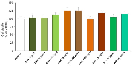
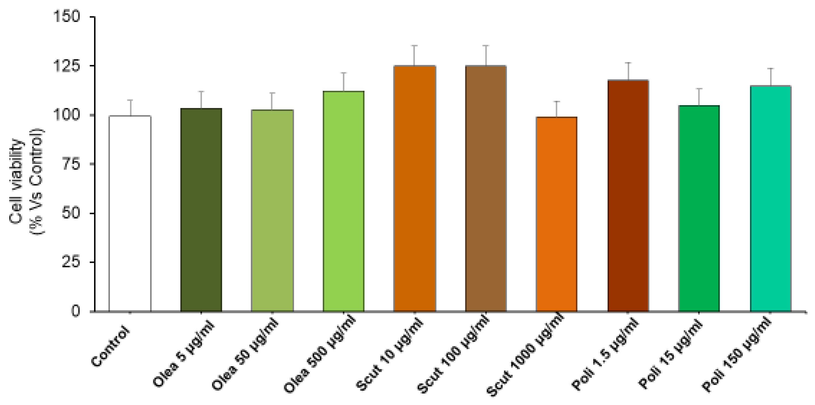
H9c2 cells were treated for 24 h with increasing concentrations of Olea europaea (5, 50, and 500 µg/mL), S. baicalensis (10, 100, and 1000 µg/mL), and policosanol (1.5, 15, and 150 µg/mL) extracts to assess potential cytotoxicity. Our present results (Figure 1), showing that Olea europaea, S. baicalensis, and policosanol extracts did not modify the viability of H9c2 cells at any concentration, are in agreement with previous studies showing that S. baicalensis and Olea europaea extracts did not affect the viability of normal cells in vitro [33,34]. The present findings highlight the absence of cytotoxic effects in these experimental conditions and support further investigations about their protective properties against oxidative stress and inflammation.
Figure 1.
Toxicity of Olea europaea, S. baicalensis and policosanol extracts. The graph shows the cell viability (%) of H9c2 treated with Olea europaea (5, 50, and 500 µg/mL), S. baicalensis (10, 100 and 1000 µg/mL), policosanol (1.5, 15 and 150 µg/mL) extracts. All the experiments were performed in triplicate at minimum (n = 9). Data are shown as the mean ± SEM. Statistical significance was calculated by one-way ANOVA followed by a Bonferroni post-test.
3.2. Evaluation of the Cell Viability Preservation Against H2O2-Induced Cell Damage in H9c2 Cells
Both inflammation and oxidative stress were reported to be critically involved in a number of CVDs, such as fibrosis, heart failure, left ventricular hypertrophy, diastolic dysfunction, and hypertension, as well as ischemia/reperfusion damage [35].
In the present study, we evaluated the preventive effects induced by Olea europaea, S. baicalensis, and policosanol extracts alone and in combination (MIX-1, MIX-2, MIX-3) in an in vitro model of H2O2-induced oxidative stress, in immortalized rat cardiomyoblast H9c2 cells.
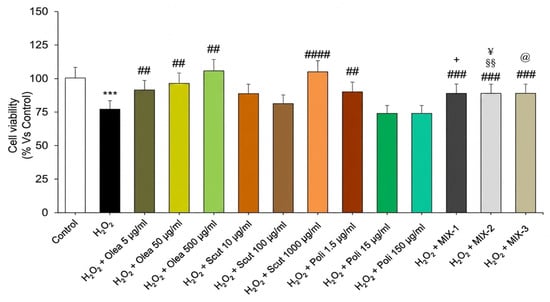
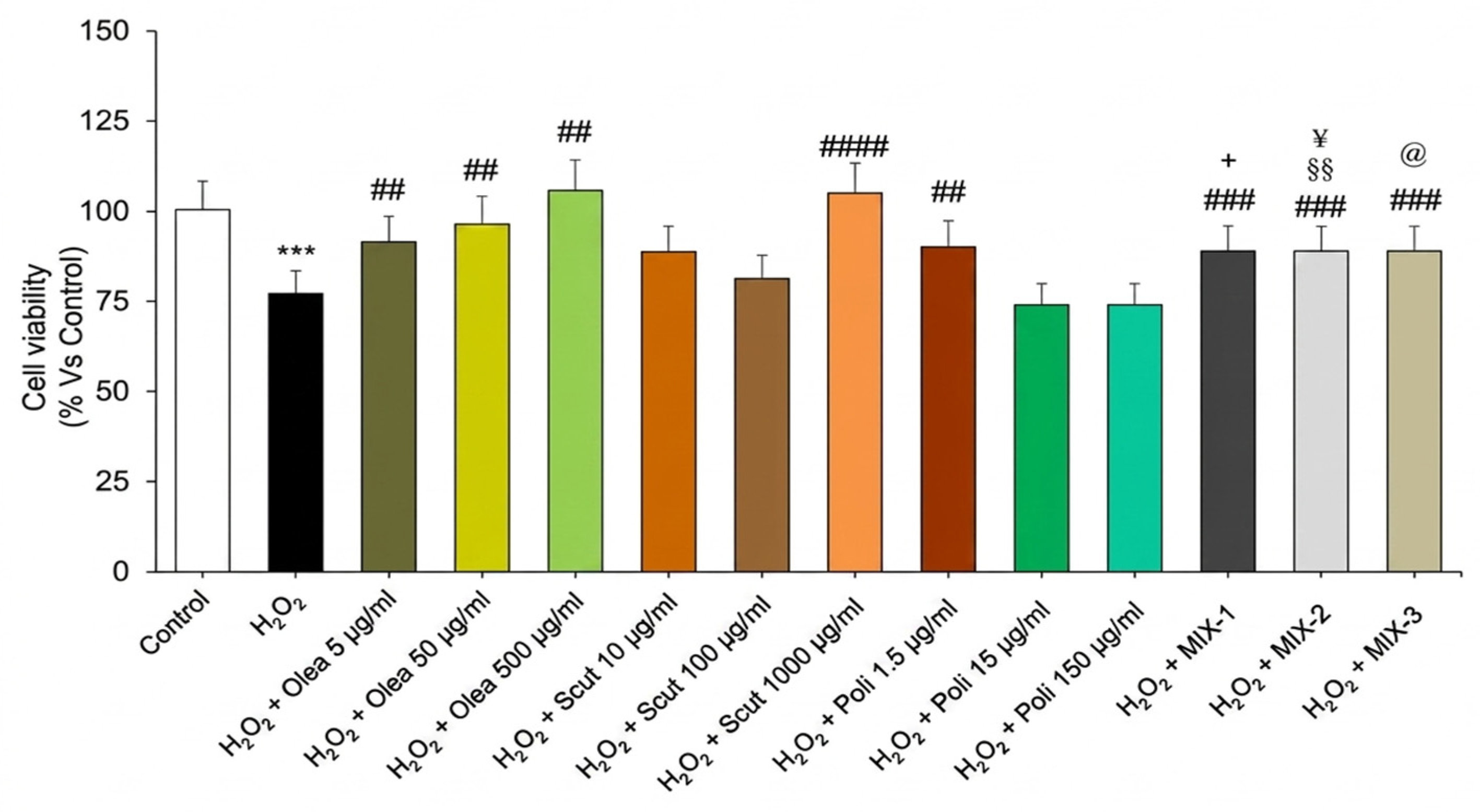
The potential preventive effects of Olea europaea, S. baicalensis, and policosanol extracts, as well as MIX-1, MIX-2, and MIX-3, against an oxidative stimulus observed after the administration of H2O2 were tested on H9c2 cells. The treatment with H2O2 induced a significant decrease in cell viability (Figure 2). This decrease was significantly prevented, in a concentration-dependent manner, by pre-incubation with Olea europaea extract, in the entire concentration range evaluated (Figure 2). S. baicalensis extract exhibited a significant preventive effect only at the highest concentration tested (Figure 2). In contrast, policosanols showed a significant preventive effect only at the lowest concentration tested, while higher concentrations did not yield significant protection (Figure 2).
Figure 2.
Preventive effect of Olea europaea, S. baicalensis, and policosanol extracts, as well as if MIX-1, MIX-2 and MIX-3, against H2O2-induced cell death. Graphs the show cell viability (%) of H9c2 pre-incubated with Olea europaea (5, 50, and 500 µg/mL), S. baicalensis (10, 100 and 1000 µg/mL), and policosanol (1.5, 15 and 150 µg/mL) extracts, as well as of MIX-1, MIX-2 and MIX-3, followed by exposure to H2O2. All the experiments were performed in triplicate at minimum (n = 9). Data are displayed as the mean ± SEM. Statistical significance was calculated by one-way ANOVA followed by a Bonferroni post-test. * indicates a significant difference vs. the control (*** p < 0.0001); # indicates significant difference vs. H2O2 (## p < 0.01, ### p < 0.001, #### p < 0.0001); § indicates a significant difference vs. policosanols at 15 µg/mL (§§ p < 0.01); @ indicates a significant difference vs. policosanols at 150 µg/mL (@ p < 0.05); + indicates a significant difference vs. S. baicalensis at 10 µg/mL (+ p < 0.05); ¥ indicates a significant difference vs. S. baicalensis at 100 µg/mL (p < 0.05).
Furthermore, pre-incubation with MIX-1, MIX-2 and MIX-3 significantly protected H9c2 cells against oxidative stimuli (Figure 2).
Notably, MIX-1 exhibited greater cytoprotection than S. baicalensis did at 10 µg/mL, while MIX-2 exhibited greater cytoprotection than both S. baicalensis at 100 µg/mL and policosanols at 15 µg/mL. Furthermore, MIX-3 showed a marked improvement in efficacy compared with policosanols at 150 µg/mL (Figure 2).
These observations suggest a possible additive interaction between the components in the mixtures. Such an additive effect is particularly relevant in oxidative stress-related pathologies, in which multifaceted mechanisms are involved, and single-agent therapies may not provide adequate protection.
3.3. Protective Effects of Olea europaea, S. baicalensis, and Policosanol Extracts, as Well as of MIX-1, MIX-2 and MIX-3, on Mouse Heart Specimens Stimulated with LPS
Finally, we investigated the protective effects induced by Olea europaea, S. baicalensis, and policosanol extracts, and their combinations in three dosages, MIX-1, MIX-2 and MIX-3, in mouse heart specimens treated with LPS, which constitutes a validated experimental model with which to study the activities of plant-derived extracts in modulating inflammatory response and oxidative stress [27,28].
Previous studies have also suggested the role of iNOS as a main contributor in CVDs like arteriosclerosis [36]. Moreover, iNOS activity has been suggested to be associated with endothelial dysfunction, as confirmed by previous studies showing increased iNOS protein expression in mice with LPS-induced endothelial dysfunction [37].
In addition, the protective effects induced by a core antioxidant enzyme, CAT, in contrasting atherosclerosis, endothelial dysfunction, and myocardial damage have been demonstrated in several reports [38,39]. Furthermore, CAT deficiency is also associated with various oxidative stress-related diseases such as CVDs [40,41], suggesting a link between reduced antioxidant defenses, increased oxidative stress, and CVD progression.
The imbalance between antioxidant capacity, including increased CAT activity, and ROS generation, is also well known to contribute to protein, lipid, and DNA oxidative damage, exacerbating CVDs [42].
Interestingly, Schnabeland and collaborators (2005) reported a relationship between GPx-1 activity in whole-blood and CVD development and severity [43]. In addition, GPx-1 levels were suggested to be a useful marker for CV event detection, and acute myocardial infarction incidence was increased in patients with GPx-1 activity impairment [44]. Furthermore, a reduction in GPx levels was found in patients with CVD compared to those of controls, further confirming the crucial role of oxidative stress in CVD onset and progression [45].
In this context, we evaluated the effects of Olea europaea, S. baicalensis, and policosanol extracts, as well as of MIX-1, MIX-2 and MIX-3, on the gene expression of pro- and anti-oxidant mediators, such as GPx, CAT and iNOS, on isolated LPS-exposed heart specimens, by RT-PCR analysis.
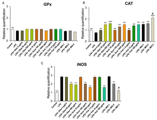
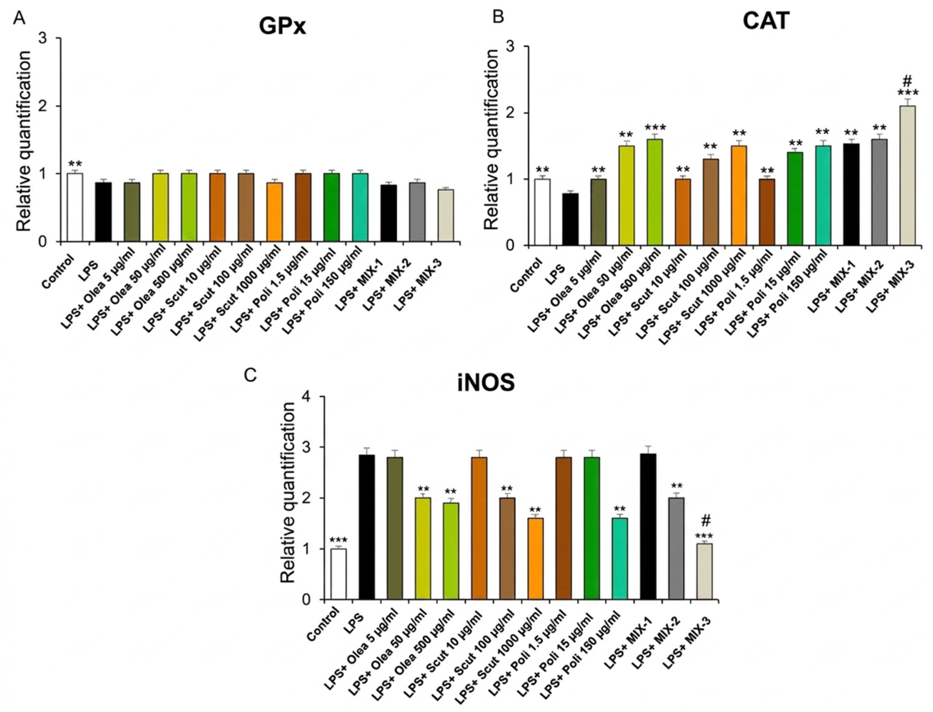
Our present findings showed that Olea europaea, S. baicalensis and policosanol extracts, as well as their combinations, increased CAT gene expression at all concentrations tested but did not influence GPx mRNA levels. In addition, Olea europaea, S. baicalensis and policosanol extracts were able to contrast the release of the pro-oxidative marker iNOS, induced by LPS, in the mouse heart, only at a higher concentration (Figure 3). Interestingly, their combination reduced the gene expression of iNOS; however, MIX-3 was more effective at decreasing CAT and iNOS gene expression compared to MIX-1 and MIX-2 (Figure 3).
Figure 3.
Effects of Olea europaea, S. baicalensis, and policosanol extracts, as well as of MIX-1, MIX-2 and MIX-3, on LPS-induced glutathione peroxidase (GPx) (A), catalase (CAT) (B), and inducible nitric oxide synthase (iNOS) (C) gene expression (RQ, relative quantification) in mouse heart specimens (n = 30, 6 for each experimental group). Data are shown as the mean ± SEM. Statistical significance was calculated by one-way ANOVA followed by a Bonferroni post-test. * p < 0.05, ** p < 0.005; *** p < 0.01 vs. LPS; # p < 0.05 vs. LPS + MIX-1 and LPS + MIX-2.
In line with this, olive leaf extracts were found to be capable of exerting protective effects in contrasting lipid peroxidation and increasing the levels of antioxidant enzymes, such as CAT, in the human cervical adenocarcinoma HeLa cell line [46]. In particular, the cardioprotective activity of oleuropein, the main secoiridoid from olive tree, is well known. In this context, the beneficial activities of oleuropein could be attributed to its antioxidant capacity, as well as to its modulatory abilities, for several targets, including NO, iNOS, interleukin (IL)-8, and TNFα [47]. The antioxidant effects induced by S. baicalensis are also well known. More recently, a significant improvement in antioxidant function, along with a significant decrease in inflammation, including reduced γ-glutamyltransferase and TNF-α, as well as increased CAT serum levels, was found in mice treated with S. baicalensis extract [48]. Moreover, the antioxidant activities of policosanols have also been reported and confirmed via decreasing ROS formation as well as increasing CAT activity [49].
Inflammation was suggested to be critically involved in CVD development and progression [50,51]. Notably, pro-inflammatory biomarkers could exert a potential role in predicting the risk for CVD developing and could correlate with CVD severity [52].
Increased blood inflammatory cytokines were reported in heart failure, as previously reported [53,54]. Moreover, NF-kB was shown to be involved in the transcription of different proinflammatory cytokines, including IL-6 and TNF-α [55], which play a key role in mediating cardiac dysfunction [56]. Recently, higher circulating levels of TNF-α and IL-1β were also found in patients with myocarditis [57].
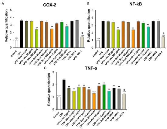
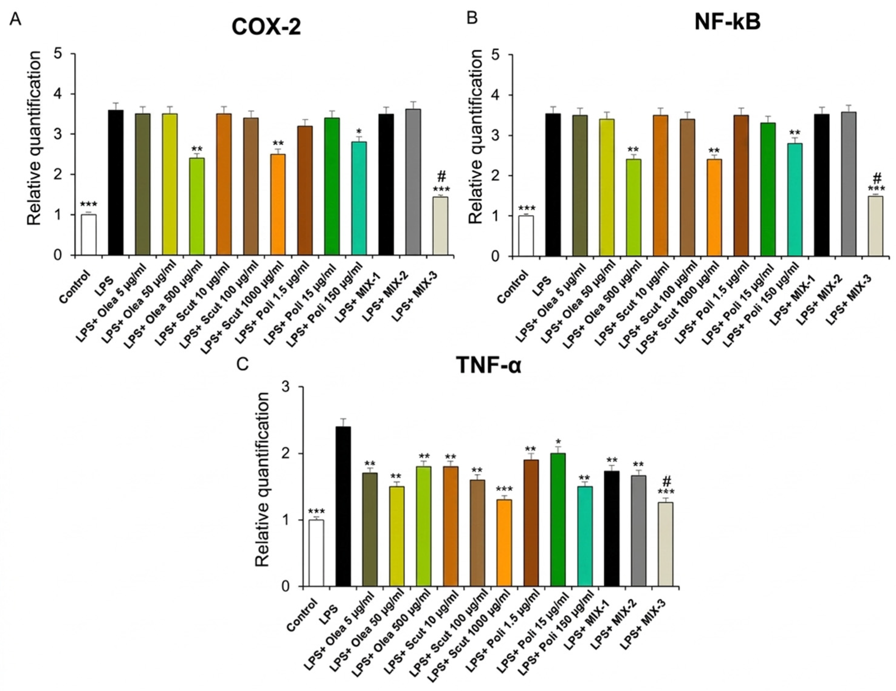
In our ex vivo experimental model, we also reported that Olea europaea, S. baicalensis and policosanol extracts, at higher concentrations, as well as MIX-3, significantly inhibited COX-2 and NF-kB gene expression (Figure 4, panels A and B). Moreover, the single constituents, as well as their combinations, MIX-1, MIX-2 and MIX-3, were able to decrease TNF-α gene expression (Figure 4, panel C). Interestingly, MIX-3 was more effective at decreasing the mRNA levels of all pro-inflammatory markers investigated (Figure 4), thus confirming the cardioprotective effects induced by the single extracts, as well as by their combinations.
Figure 4.
Effects of Olea europaea, S. baicalensis, and policosanol extracts, as well as of MIX-1, MIX-2 and MIX-3, on LPS-induced cyclooxygenase-2 (COX-2) (A), nuclear factor kB (NF-kB) (B), and tumor necrosis factor-α (TNF-α) (C) gene expression (RQ, relative quantification) in mouse heart specimens (n = 30, 6 for each experimental group). Data are shown as the mean ± SEM. Statistical significance was calculated by one-way ANOVA followed by a Bonferroni post-test. ** p < 0.005; *** p < 0.01 vs. LPS; # p < 0.05 vs. LPS + MIX-1 and LPS + MIX-2.
Accordingly, Burja and collaborators (2019) showed that olive leaf extract was able to inhibit levels of pro-inflammatory cytokines, such as TNF-α, IL-1β, IL-6, and IL-8, in human arterial endothelial cells [58]. In addition, oleuropein, suggested to be the major component responsible for the anti-inflammatory effects of olive leaf extract, reduced COX-2 gene expression, as well as that of NF-kB signaling pathways also involved in the inflammation process [59]. Furthermore, water extracts of S. baicalensis were also found to suppress the production of several pro-inflammatory cytokines, including TNF-α, IL-6, and IL-1β, as well as COX-2 mRNA levels, in LPS-induced macrophages [60].
Interestingly, the combination of S. baicalensis and Sophora japonica L. was found to exert antioxidant and anti-hypertensive activities [61]. Furthermore, the combination of oleuropein, hydroxytyrosol, and oleocanthal induces cardioprotective, and anti-atherosclerotic activities in vivo in mice [62].
Our data align with recent pharmacological trends emphasizing the use of natural multi-component formulations to manage subclinical cardiac inflammation. The downregulation of pro-inflammatory mediators observed in our model provides a strong rationale for further investigating the potential use of our combinations as a complementary strategy in the prevention of inflammatory-based cardiovascular diseases.
On the basis of these results, we performed a final step in our experimental paradigm, with the aim to further study the possible cardioprotective effects of MIX-1, MIX-2 and MIX-3, with particular regard to their activities on the gene expression of cardiac biomarkers.
In addition to pro-inflammatory and pro-oxidant factors, cTnI and cTnT, which are highly expressed in cardiomyocytes, were strongly associated with heart failure and CVD death [63]. Furthermore, an association was reported between increased levels of BNP and a greater risk for adverse short- and long-term outcomes in heart failure. Moreover, BNP levels were found to be increased in perimyocarditis [64].
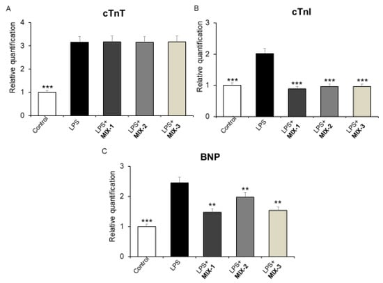
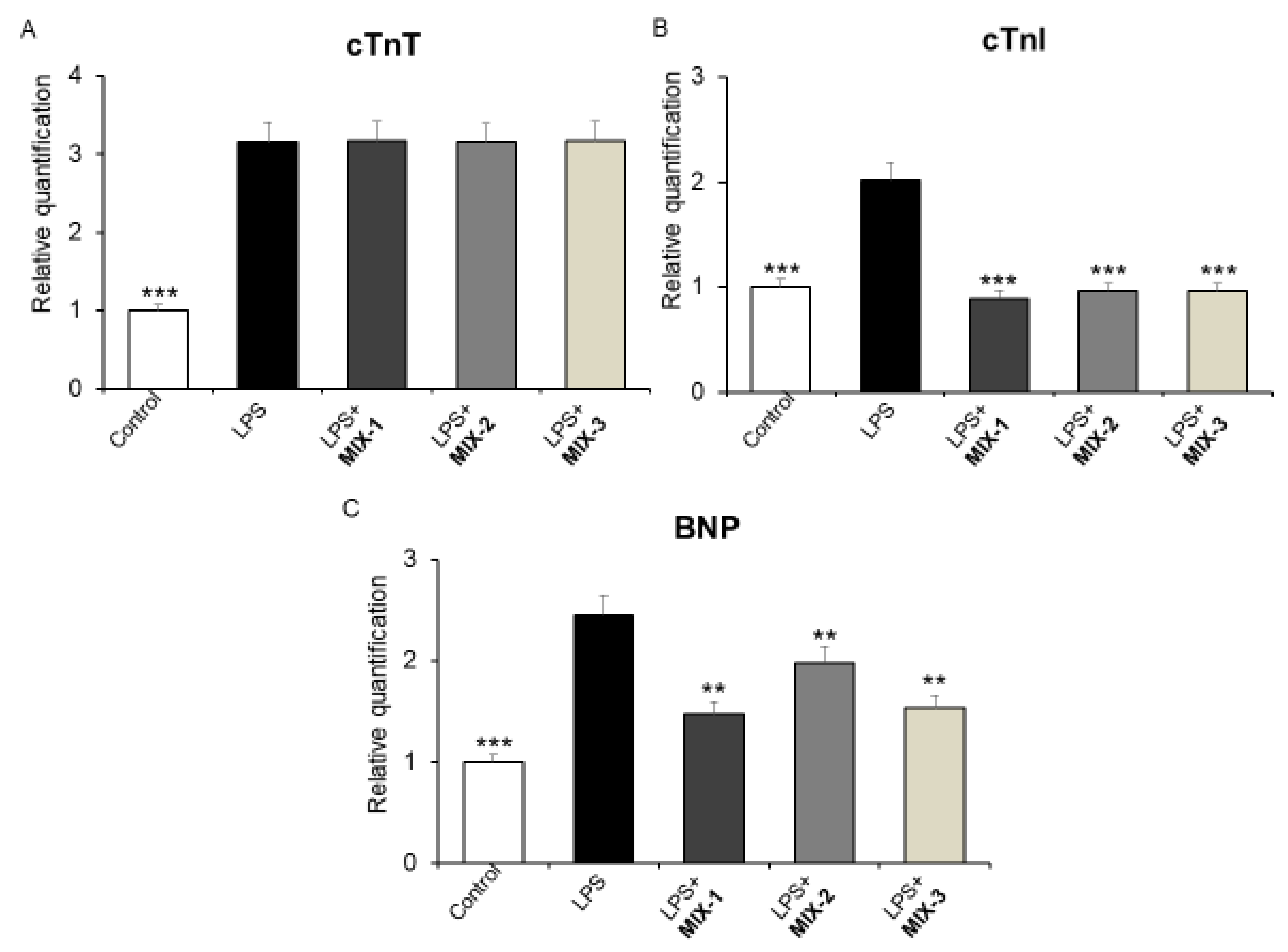
In the present study, we showed that the combination inhibited the LPS-induced gene expression of cTnI and BNP in mouse heart specimens at all concentrations (Figure 5, panel B and C), without affecting cTnT gene expression.
Figure 5.
Effects of MIX-1, MIX-2 and MIX-3 on LPS-induced cardiac troponin T (cTnT) (A), cardiac troponin I (cTnI) (B), and natriuretic peptide (BNP) (C) gene expression (RQ, relative quantification), in mouse heart specimens (n = 30, 6 for each experimental group). Data are shown as the mean ± SEM. Statistical significance was calculated by one-way ANOVA followed by a Bonferroni post-test. ** p < 0.005; *** p < 0.01 vs. LPS.
Interestingly, cTnI is considered the preferred cardiac-specific biomarker for detecting cardiac injury, and increased cTnI was observed in myocardial injury [65]. Accordingly, herbal extracts were found to be effective at decreasing cTnI serum levels in diabetic rats; this could be related to their protective activities in diabetic cardiomyopathy [66]. Notably, pretreatment with oleuropein was able to counteract the increased serum levels of cTnI in rats with acute myocardial infarction [67], thus confirming its cardioprotective activity. While this does not directly indicate a reduction in cTnI gene expression, we can suggest that the suppression of LPS-induced cTnI mRNA levels following treatment with our combination could be related to its potential protective effects on the heart. Moreover, considering the experimental in vitro and ex vivo paradigms used, it was not possible to measure circulating levels of the same markers.
Our findings are also in agreement with those of Asghari and collaborators (2022), who reported that oleuropein could exert protective effects in contrasting fibrosis and reducing the gene expression of markers of myocardial hypertrophy, such as atrial natriuretic peptide and BNP, thus improving cardiac function in streptozotocin-induced diabetic rats [68]. The significant reduction in BNP and cTnI mRNA levels suggests that MIX-1, MIX-2 and MIX-3 could exert protective effects against the structural and functional myocardial stress induced by LPS.
3.4. Preventive Effects of MIX-1, MIX-2 and MIX-3 Against H2O2-Induced Intracellular ROS Production
Oxidative stress has been suggested to be involved in several CVDs, including heart failure, myocardial infarction, and ischemia/reperfusion damage [69].
In particular, ROSs play a pivotal role in the stimulation of endothelial dysfunction, vascular remodeling, and inflammation [70]. In agreement, increased cellular ROS production was shown to be involved in LDL oxidation [71], as well as in the pathogenesis and development of several CVDs such as atherosclerosis and heart failure [2,72]. Furthermore, higher cardiac troponin levels are strongly associated with cardiac muscle damage or stress. Moreover, H2O2 could contribute to this damage by enhancing oxidative stress, particularly in myocardial infarction or stress-induced cardiomyopathy [72].
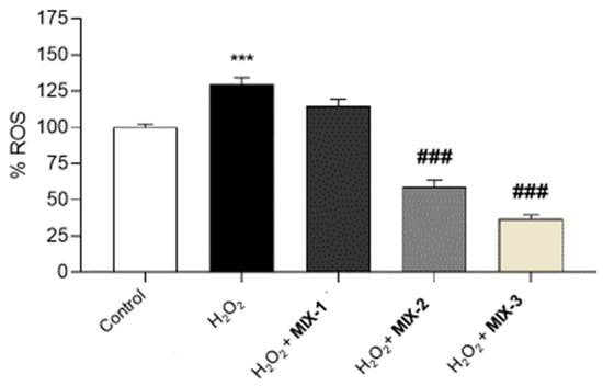
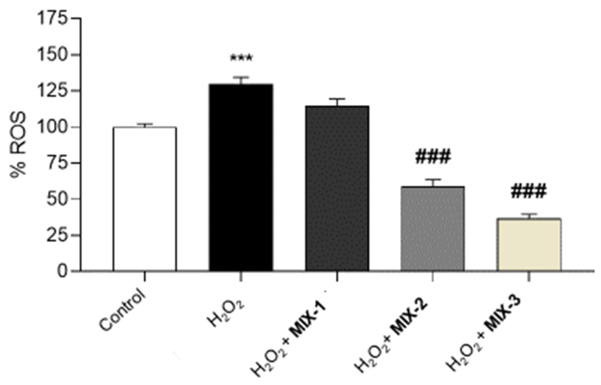
Our present findings showed that the treatment of H9c2 cells with H2O2 significantly increases intracellular ROS levels. However, pre-incubation with MIX-2 and MIX-3 significantly reduced ROS levels, suggesting preventive effects induced by the combination. The observed reduction is not significant at the lower concentration (MIX-1) (Figure 6).
Figure 6.
Evaluation of the preventive effects of MIX-1, MIX-2 and MIX-3 against H2O2-induced intracellular ROS production. The histograms show the levels of intracellular ROS (%) in H9c2 pre-treated with a control or MIX-1, MIX-2 and MIX-3, and then exposed to H2O2. All the experiments were performed in triplicate at minimum (n = 9). Data are displayed as the mean ± SEM. * indicates significant statistical differences vs. the control (*** p < 0.001), while # indicates statistical differences vs. the H2O2 treated-cells pre-incubated with the control (### p < 0.001).
Ultimately, the observed cardioprotective activities were not found to be associated with a single constituent but rather with various bioactive compounds. In this context, we can speculate that oleuropein from Olea europaea may act in combination with baicalin from S. baicalensis to inhibit pro-inflammatory and pro-oxidant signaling pathways. Moreover, the inclusion of policosanols may contribute to the stabilization of cellular membranes, thereby creating a multi-target therapeutic approach that addresses inflammatory process, oxidative stress, and structural damage simultaneously.
However, it is important to specify that our in vitro and ex vivo experimental models could not reflect, in vivo, the effects of the tested preparations on living organisms, due to limited absorption in the intestines and the metabolism of the preparation.
4. Conclusions
In conclusion, our results demonstrated that the combination MIX-3 exerts protective activities on the heart, as confirmed by the inhibitory effects on multiple markers involved in inflammatory processes and oxidative stress, including NF-kB, COX-2, TNF-α, iNOS, and CAT, as well as cardiac injury markers, such as cTnI and BNP, in mouse heart specimens treated with LPS. The combination may contribute to reduce ROS production and cardiac inflammation, as well as modulate key cardiac biomarkers that play a crucial role in CVD diagnosis and prognosis.
These findings suggest that this plant-based combination could be a promising candidate for the development of innovative functional foods and nutraceutical strategies aimed at supporting cardiovascular health and preventing oxidative stress-related cardiac dysfunction.
Further studies, including clinical trials, are needed to further confirm the activity of the combination as well as to evaluate its safety.
Supplementary Materials
The following supporting information can be downloaded at https://www.mdpi.com/article/10.3390/foods15091500/s1.
Author Contributions
Conceptualization, L.R., G.B. and S.L.; methodology, G.B., A.P.C., D.C., A.A., A.C. (Annalisa Chiavaroli), G.G., V.C. (Valentina Citi), S.V., A.P. and A.C. (Alessandro Cama); software, G.B., A.P.C., D.C. and G.O.; validation, L.R., G.O., V.C. (Valentina Citi) and S.L.; formal analysis, L.R., G.B., A.P.C., D.C. and S.L.; investigation, A.A., A.C. (Annalisa Chiavaroli), G.G. and V.C. (Valentina Citi); resources, L.R. and S.L.; data Curation, L.R. and S.L.; writing—original draft preparation, L.R., A.M., L.B. and S.L.; writing—review and editing, L.R., A.M., L.B. and S.L.; visualization, V.C. (Vincenzo Calderone) and L.B.; supervision, L.R., L.B., V.C. (Vincenzo Calderone) and S.L.; project administration, L.R., L.B. and S.L.; funding acquisition, L.R., L.B. and S.L. All authors have read and agreed to the published version of the manuscript.
Funding
This research was funded by Difass International Spa (Rimini, Cerasolo, Italy) (Grant 2024) (principal investigator: Sheila Leone; coordinators: Luigi Brunetti, Lucia Recinella). The novel patented combination (DIF2STAT®) constituting Olea europaea, Scutellaria baicalensis, and policosanol extracts was kindly provided by Difass International Spa (Rimini, Cerasolo, Italy).
Institutional Review Board Statement
Housing conditions and experimentation procedures were strictly in agreement with the European Community ethical regulations (EU Directive no. 26/2014) on the care of animals for scientific research. Accordingly to the recognized principles of “Replacement, Refinement and Reduction in Animals in Research”, the study protocol was approved by the local ethical committee (‘G. d’Annunzio’ University, Chieti, Italy) and Italian Health Ministry (Project no. F4738.N.ZDZ and 16 April 2025).
Informed Consent Statement
Not applicable.
Data Availability Statement
The original contributions presented in this study are included in the article/Supplementary Materials. Further inquiries can be directed to the corresponding author.
Conflicts of Interest
The authors declare that this study received funding from Difass International Spa. The funders were not involved in the study design; in the collection, analysis, and interpretation of data; in the writing of this article; or in the decision to submit it for publication.
Abbreviations
The following abbreviations are used in this manuscript:
| CVD | Cardiovascular diseases |
| BNP | B-type natriuretic peptide |
| cTnI | Cardiac troponin I |
| cTnT | Cardiac troponin T |
| ROS | Reactive oxygen species |
| LPS | Lipopolisaccharide |
| GPx | Glutathione peroxidase |
| CAT | Catalase |
| iNOS | Inducible nitric oxide synthase |
| COX-2 | Cyclooxygenase-2 |
| TNFα | Tumor necrosis factor α |
| NFkB | Nuclear factor-kB |
| IL | Interleukin |
References
- Sorrento, D.; Iaccarino, G. Inflammation and Cardiovascular Diseases: The Most Recent Findings. Int. J. Mol. Sci. 2019, 20, 3879. [Google Scholar] [CrossRef] [Scilit] [PubMed]
- Kattoor, A.J.; Pothineni, N.V.K.; Palagiri, D.; Mehta, J.L. Oxidative Stress in Atherosclerosis. Curr. Atheroscler. Rep. 2017, 19, 42. [Google Scholar] [CrossRef] [Scilit] [PubMed]
- Wróbel-Nowicka, K.; Wojciechowska, C.; Jacheć, W.; Zalewska, M.; Romuk, E. The Role of Oxidative Stress and Inflammatory Parameters in Heart Failure. Medicina 2024, 60, 760. [Google Scholar] [CrossRef] [Scilit]
- Lassègue, B.; Griendling, K.K. Reactive Oxygen Species in Hypertension: An Update. Am. J. Hypertens. 2004, 17, 852–860. [Google Scholar] [CrossRef] [Scilit]
- Ridker, P.M.; Everett, B.M.; Thuren, T.; MacFadyen, J.G.; Chang, W.H.; Ballantyne, C.; Fonseca, F.; Nicolau, J.; Koenig, W.; Anker, S.D.; et al. Antiinflammatory Therapy with Canakinumab for Atherosclerotic Disease. N. Engl. J. Med. 2017, 377, 1119–1131. [Google Scholar] [CrossRef] [Scilit]
- Ghazizadeh, H.; Mirinezhad, M.R.; Seyedi, S.M.R.; Sadabadi, F.; Ahmadnezhad, M.; Jaberi, N.; Pasdar, A.; Ferns, G.A.; Esmaily, H.; Ghayour-Mobarhan, M. Prognostic Factors Associating with Pro-Oxidant-Antioxidant Balance; Neutrophils to Lymphocytes Ratio, Vitamin D, Heat Shock Protein 27, and Red Cell Distribution Width. Arch. Med. Res. 2020, 51, 261–267. [Google Scholar] [CrossRef] [Scilit]
- Dhingra, R.; Vasan, R.S. Biomarkers in Cardiovascular Disease: Statistical Assessment and Section on Key Novel Heart Failure Biomarkers. Trends Cardiovasc. Med. 2017, 27, 123–133. [Google Scholar] [CrossRef] [Scilit]
- Netala, V.R.; Teertam, S.K.; Li, H.; Zhang, Z. A Comprehensive Review of Cardiovascular Disease Management: Cardiac Biomarkers, Imaging Modalities, Pharmacotherapy, Surgical Interventions, and Herbal Remedies. Cells 2024, 13, 1471. [Google Scholar] [CrossRef] [Scilit]
- Godos, J.; Romano, G.L.; Laudani, S.; Gozzo, L.; Guerrera, I.; Dominguez Azpíroz, I.; Martínez Diaz, R.; Quiles, J.L.; Battino, M.; Drago, F.; et al. Flavan-3-Ols and Vascular Health: Clinical Evidence and Mechanisms of Action. Nutrients 2024, 16, 2471. [Google Scholar] [CrossRef] [Scilit]
- Salvo, A.; Tuttolomondo, A. The Role of Olive Oil in Cardiometabolic Risk. Metabolites 2025, 15, 190. [Google Scholar] [CrossRef] [Scilit] [PubMed]
- Soulaimani, B.; Varoni, E.; Iriti, M.; Mezrioui, N.-E.; Hassani, L.; Abbad, A. Synergistic Anticandidal Effects of Six Essential Oils in Combination with Fluconazole or Amphotericin B against Four Clinically Isolated Candida Strains. Antibiotics 2021, 10, 1049. [Google Scholar] [CrossRef] [Scilit]
- Zhao, T.; Tang, H.; Xie, L.; Zheng, Y.; Ma, Z.; Sun, Q.; Li, X. Scutellaria baicalensis Georgi. (Lamiaceae): A Review of Its Traditional Uses, Botany, Phytochemistry, Pharmacology and Toxicology. J. Pharm. Pharmacol. 2019, 71, 1353–1369. [Google Scholar] [CrossRef] [Scilit] [PubMed]
- Sonawane, S.K.S.; Balmik, A.A.B.; Boral, S.R.; Ramasamy, S.R.; Chinnathambi, S.C. Baicalein Suppresses Repeat Tau Fibrillization by Sequestering Oligomers. Arch. Biochem. Biophys. 2019, 675, 108119. [Google Scholar] [CrossRef] [Scilit] [PubMed]
- Zhu, Q.; Zhuang, X.; Lu, J. Neuroprotective Effects of Baicalein in Animal Models of Parkinson’s Disease: A Systematic Review of Experimental Studies. Phytomedicine 2019, 55, 302–309. [Google Scholar] [CrossRef] [Scilit]
- Song, J.; Zhang, L.; Xu, Y.; Yang, D.; Zhang, L.; Yang, S.; Zhang, W.; Wang, J.; Tian, S.; Yang, S.; et al. The Comprehensive Study on the Therapeutic Effects of Baicalein for the Treatment of COVID-19 in Vivo and in Vitro. Biochem. Pharmacol. 2021, 183, 114302. [Google Scholar] [CrossRef] [Scilit]
- Janeczko, M.; Gmur, D.; Kochanowicz, E.; Górka, K.; Skrzypek, T. Inhibitory Effect of a Combination of Baicalein and Quercetin Flavonoids against Candida Albicans Strains Isolated from the Female Reproductive System. Fungal Biol. 2022, 126, 407–420. [Google Scholar] [CrossRef] [Scilit]
- Panou, A.A.; Karabagias, I.K. Olive Leaf Extracts as a Medicinal Beverage: Origin, Physico-Chemical Properties, and Bio-Functionality. Beverages 2025, 11, 66. [Google Scholar] [CrossRef] [Scilit]
- de Oliveira, N.M.; Machado, J.; Chéu, M.H.; Lopes, L.; Criado, M.B. Therapeutic Potential of Olive Leaf Extracts: A Comprehensive Review. Appl. Biosci. 2024, 3, 392–425. [Google Scholar] [CrossRef] [Scilit]
- Romani, A.; Ieri, F.; Urciuoli, S.; Noce, A.; Marrone, G.; Nediani, C.; Bernini, R. Health Effects of Phenolic Compounds Found in Extra-Virgin Olive Oil, By-Products, and Leaf of Olea europaea L. Nutrients 2019, 11, 1776. [Google Scholar] [CrossRef] [Scilit]
- Romero, M.; Toral, M.; Gomez-Guzman, M.; Jimenez, R.; Galindo, P.; Sanchez, M.; Olivares, M.; Galvez, J.; Duarte, J. Antihypertensive effects of oleuropein-enriched olive oil extract in spontaneously hypertensive rats. Food Funct. 2016, 7, 584–593. [Google Scholar] [CrossRef] [Scilit]
- Wang, L.; Geng, L.; Jiang, C.; Gong, D.; Liu, D.; Yoshimura, H.; Zhong, L. The Anti-Atherosclerotic Effect of Olive Leaf Extract Is Related to Suppressed Inflammatory Response in Rabbits with Experimental Atherosclerosis. Eur. J. Nutr. 2008, 47, 235–243. [Google Scholar] [CrossRef] [Scilit]
- Al-Azzawie, H.F.; Alhamdani, M.-S.S. Hypoglycemic and Antioxidant Effect of Oleuropein in Alloxan-Diabetic Rabbits. Life Sci. 2006, 78, 1371–1377. [Google Scholar] [CrossRef] [Scilit]
- Scheffler, A.; Rauwald, H.W.; Kampa, B.; Mann, U.; Mohr, F.W.; Dhein, S. Olea europaea Leaf Extract Exerts L-Type Ca2+ Channel Antagonistic Effects. J. Ethnopharmacol. 2008, 120, 233–240. [Google Scholar] [CrossRef] [Scilit]
- Weerawatanakorn, M.; Meerod, K.; Wongwaiwech, D.; Ho, C.-T. Policosanols: Chemistry, Occurrence, and Health Effects. Curr. Pharmacol. Rep. 2019, 5, 131–149. [Google Scholar] [CrossRef] [Scilit]
- Olatunji, L.K.; Jimoh, A.O.; Tukur, U.M.; Imam, M.U. A Review of the Effects of Policosanol on Metabolic Syndrome. Clin. Complement. Med. Pharmacol. 2022, 2, 100058. [Google Scholar] [CrossRef] [Scilit]
- Varady, K.A.; Wang, Y.; Jones, P.J.H. Role of Policosanols in the Prevention and Treatment of Cardiovascular Disease. Nutr. Rev. 2003, 61, 376–383. [Google Scholar] [CrossRef] [Scilit] [PubMed]
- Recinella, L.; Chiavaroli, A.; Masciulli, F.; Fraschetti, C.; Filippi, A.; Cesa, S.; Cairone, F.; Gorica, E.; De Leo, M.; Braca, A.; et al. Protective Effects Induced by a Hydroalcoholic Allium Sativum Extract in Isolated Mouse Heart. Nutrients 2021, 13, 2332. [Google Scholar] [CrossRef] [Scilit] [PubMed]
- Recinella, L.; Libero, M.L.; Citi, V.; Chiavaroli, A.; Martelli, A.; Foligni, R.; Mannozzi, C.; Acquaviva, A.; Di Simone, S.C.; Calderone, V.; et al. Anti-Inflammatory and Vasorelaxant Effects Induced by an Aqueous Aged Black Garlic Extract Supplemented with Vitamins D, C, and B12 on Cardiovascular System. Foods 2023, 12, 1558. [Google Scholar] [CrossRef] [Scilit]
- Panera, N.; Gnani, D.; Piermarini, E.; Petrini, S.; Bertini, E.; Nobili, V.; Pastore, A.; Piemonte, F.; Alisi, A. High Concentrations of H2O2 Trigger Hypertrophic Cascade and Phosphatase and Tensin Homologue (PTEN) Glutathionylation in H9c2 Cardiomyocytes. Exp. Mol. Pathol. 2016, 100, 199–206. [Google Scholar] [CrossRef] [Scilit]
- Recinella, L.; Gorica, E.; Chiavaroli, A.; Fraschetti, C.; Filippi, A.; Cesa, S.; Cairone, F.; Martelli, A.; Calderone, V.; Veschi, S.; et al. Anti-Inflammatory and Antioxidant Effects Induced by Allium sativum L. Extracts on an Ex Vivo Experimental Model of Ulcerative Colitis. Foods 2022, 11, 3559. [Google Scholar] [CrossRef] [Scilit]
- Livak, K.J.; Schmittgen, T.D. Analysis of Relative Gene Expression Data Using Real-Time Quantitative PCR and the 2−ΔΔCT Method. Methods 2001, 25, 402–408. [Google Scholar] [CrossRef] [Scilit]
- Charan, J.; Kantharia, N.D. How to Calculate Sample Size in Animal Studies? J. Pharmacol. Pharmacother. 2013, 4, 303–306. [Google Scholar] [CrossRef] [Scilit]
- Luca, T.; Malfa, G.A.; Siracusa, L.; La Mantia, A.; Bianchi, S.; Napoli, E.; Puleo, S.; Sergi, A.; Acquaviva, R.; Castorina, S. Redox State Modulatory Activity and Cytotoxicity of Olea europaea L. (Oleaceae) Leaves Extract Enriched in Polyphenols Using Macroporous Resin. Antioxidants 2024, 13, 73. [Google Scholar] [CrossRef] [Scilit]
- Wu, T.W.-H.; Lin, T.-Y.; Yang, P.-M.; Li, W.-T.; Yeh, C.-T.; Pan, T.-L. Scutellaria baicalensis Induces Cell Apoptosis and Elicits Mesenchymal-Epithelial Transition to Alleviate Metastatic Hepatocellular Carcinoma via Modulating HSP90β. Int. J. Mol. Sci. 2024, 25, 3073. [Google Scholar] [CrossRef] [Scilit]
- García, N.; Zazueta, C.; Aguilera-Aguirre, L. Oxidative Stress and Inflammation in Cardiovascular Disease. Oxid. Med. Cell. Longev. 2017, 2017, 5853238. [Google Scholar] [CrossRef] [Scilit]
- Lind, M.; Hayes, A.; Caprnda, M.; Petrovic, D.; Rodrigo, L.; Kruzliak, P.; Zulli, A.K. Inducible Nitric Oxide Synthase: Good or Bad? Biomed. Pharmacother. 2017, 93, 370–375. [Google Scholar] [CrossRef] [Scilit]
- Chauhan, S.D.; Seggara, G.; Vo, P.A.; Macallister, R.J.; Hobbs, A.J.; Ahluwalia, A. Protection against Lipopolysaccharide-Induced Endothelial Dysfunction in Resistance and Conduit Vasculature of iNOS Knockout Mice. FASEB J. 2003, 17, 773–775. [Google Scholar] [CrossRef] [Scilit]
- Sharifi-Rad, M.; Anil Kumar, N.V.; Zucca, P.; Varoni, E.M.; Dini, L.; Panzarini, E.; Rajkovic, J.; Tsouh Fokou, P.V.; Azzini, E.; Peluso, I.; et al. Lifestyle, Oxidative Stress, and Antioxidants: Back and Forth in the Pathophysiology of Chronic Diseases. Front. Physiol. 2020, 11, 694. [Google Scholar] [CrossRef] [Scilit]
- Senoner, T.; Dichtl, W. Oxidative Stress in Cardiovascular Diseases: Still a Therapeutic Target? Nutrients 2019, 11, 2090. [Google Scholar] [CrossRef] [Scilit]
- Nandi, A.; Yan, L.-J.; Jana, C.K.; Das, N. Role of Catalase in Oxidative Stress- and Age-Associated Degenerative Diseases. Oxid. Med. Cell. Longev. 2019, 2019, 9613090. [Google Scholar] [CrossRef] [Scilit]
- Forman, H.J.; Zhang, H. Targeting Oxidative Stress in Disease: Promise and Limitations of Antioxidant Therapy. Nat. Rev. Drug Discov. 2021, 20, 689–709. [Google Scholar] [CrossRef] [Scilit]
- Juan, C.A.; Pérez de la Lastra, J.M.; Plou, F.J.; Pérez-Lebeña, E. The Chemistry of Reactive Oxygen Species (ROS) Revisited: Outlining Their Role in Biological Macromolecules (DNA, Lipids and Proteins) and Induced Pathologies. Int. J. Mol. Sci. 2021, 22, 4642. [Google Scholar] [CrossRef] [Scilit]
- Schnabel, R.; Lackner, K.J.; Rupprecht, H.J.; Espinola-Klein, C.; Torzewski, M.; Lubos, E.; Bickel, C.; Cambien, F.; Tiret, L.; Münzel, T.; et al. Glutathione Peroxidase-1 and Homocysteine for Cardiovascular Risk Prediction: Results from the AtheroGene Study. J. Am. Coll. Cardiol. 2005, 45, 1631–1637. [Google Scholar] [CrossRef] [Scilit]
- Cheng, M.; Chen, C.; Ho, H.; Li, J.; Chiu, D.T. Effect of Acute Myocardial Infarction on Erythrocytic Glutathione Peroxidase 1 Activity and Plasma Vitamin e Levels. Am. J. Cardiol. 2009, 103, 471–475. [Google Scholar] [CrossRef] [Scilit]
- Karajibani, M.; Hashemi, M.; Montazerifar, F.; Bolouri, A.; Dikshit, M. The Status of Glutathione Peroxidase, Superoxide Dismutase, Vitamins A, C, E and Malondialdehyde in Patients with Cardiovascular Disease in Zahedan, Southeast Iran. J. Nutr. Sci. Vitaminol. 2009, 55, 309–316. [Google Scholar] [CrossRef] [Scilit]
- Pennisi, R.; Ben Amor, I.; Gargouri, B.; Attia, H.; Zaabi, R.; Chira, A.B.; Saoudi, M.; Piperno, A.; Trischitta, P.; Tamburello, M.P.; et al. Analysis of Antioxidant and Antiviral Effects of Olive (Olea europaea L.) Leaf Extracts and Pure Compound Using Cancer Cell Model. Biomolecules 2023, 13, 238. [Google Scholar] [CrossRef] [Scilit]
- Castejón, M.L.; Montoya, T.; Alarcón-de-la-Lastra, C.; Sánchez-Hidalgo, M. Potential Protective Role Exerted by Secoiridoids from Olea europaea L. in Cancer, Cardiovascular, Neurodegenerative, Aging-Related, and Immunoinflammatory Diseases. Antioxidants 2020, 9, 149. [Google Scholar] [CrossRef] [Scilit]
- Zhang, S.; Lv, H.; Cai, X.; Tang, S.; Zhong, R.; Chen, L.; Zhang, H. Effects of the Compound Extracts of Caprifoliaceae and Scutellaria baicalensis Georgi on the Intestinal Microbiota and Antioxidant Function. Front. Microbiol. 2024, 14, 1305414. [Google Scholar] [CrossRef] [Scilit]
- Ohta, Y.; Ohashi, K.; Matsura, T.; Tokunaga, K.; Kitagawa, A.; Yamada, K. Octacosanol Attenuates Disrupted Hepatic Reactive Oxygen Species Metabolism Associated with Acute Liver Injury Progression in Rats Intoxicated with Carbon Tetrachloride. J. Clin. Biochem. Nutr. 2008, 42, 118–125. [Google Scholar] [CrossRef] [Scilit]
- Ruparelia, N.; Choudhury, R. Inflammation and Atherosclerosis: What Is on the Horizon? Heart 2020, 106, 80–85. [Google Scholar] [CrossRef] [Scilit]
- Ministrini, S.; Carbone, F.; Montecucco, F. Updating Concepts on Atherosclerotic Inflammation: From Pathophysiology to Treatment. Eur. J. Clin. Investig. 2021, 51, e13467. [Google Scholar] [CrossRef] [Scilit] [PubMed]
- Zakynthinos, E.; Pappa, N. Inflammatory Biomarkers in Coronary Artery Disease. J. Cardiol. 2009, 53, 317–333. [Google Scholar] [CrossRef] [Scilit] [PubMed]
- Matsumori, A. Targeting Inflammation in the Diagnosis, Management, and Prevention of Cardiovascular Diseases. Glob. Heart 2022, 17, 80. [Google Scholar] [CrossRef] [Scilit]
- Matsumori, A. Anti-Inflammatory Therapy for Heart Failure. Curr. Opin. Pharmacol. 2004, 4, 171–176. [Google Scholar] [CrossRef] [Scilit]
- Liu, T.; Zhang, L.; Joo, D.; Sun, S.S.-C. NF-κB Signaling in Inflammation. Siginal Transduct. Target. Ther. 2017, 2, 17023. [Google Scholar] [CrossRef] [Scilit]
- Gullestad, L.; Ueland, T.; Vinge, L.E.; Finsen, A.; Yndestad, A.; Aukrust, P. Inflammatory Cytokines in Heart Failure: Mediators and Markers. Cardiology 2012, 122, 23–35. [Google Scholar] [CrossRef] [Scilit]
- Matsumori, A. Nuclear Factor-κB Is a Prime Candidate for the Diagnosis and Control of Inflammatory Cardiovascular Disease. Eur. Cardiol. 2023, 18, e40. [Google Scholar] [CrossRef] [Scilit] [PubMed]
- Burja, B.; Kuret, T.; Janko, T.; Topalović, D.; Živković, L.; Mrak-Poljšak, K.; Spremo-Potparević, B.; Žigon, P.; Distler, O.; Čučnik, S.; et al. Olive Leaf Extract Attenuates Inflammatory Activation and DNA Damage in Human Arterial Endothelial Cells. Front. Cardiovasc. Med. 2019, 6, 56. [Google Scholar] [CrossRef] [Scilit]
- Qabaha, K.; Al-Rimawi, F.; Qasem, A.; Naser, S.A. Oleuropein Is Responsible for the Major Anti-Inflammatory Effects of Olive Leaf Extract. J. Med. Food 2018, 21, 302–305. [Google Scholar] [CrossRef] [Scilit]
- Yoon, S.-B.; Lee, Y.-J.; Park, S.K.; Kim, H.-C.; Bae, H.; Kim, H.M.; Ko, S.-G.; Choi, H.Y.; Oh, M.S.; Park, W. Anti-Inflammatory Effects of Scutellaria baicalensis Water Extract on LPS-Activated RAW 264.7 Macrophages. J. Ethnopharmacol. 2009, 125, 286–290. [Google Scholar] [CrossRef] [Scilit]
- Guan, Y.; Chen, K.; Quan, D.; Kang, L.; Yang, D.; Wu, H.; Yan, M.; Wu, S.; Lv, L.; Zhang, G. The Combination of Scutellaria baicalensis Georgi and Sophora japonica L. Ameliorate Renal Function by Regulating Gut Microbiota in Spontaneously Hypertensive Rats. Front. Pharmacol. 2020, 11, 575294. [Google Scholar] [CrossRef] [Scilit] [PubMed]
- Christodoulou, A.; Nikolaou, P.-E.; Symeonidi, L.; Katogiannis, K.; Pechlivani, L.; Nikou, T.; Varela, A.; Chania, C.; Zerikiotis, S.; Efentakis, P.; et al. Cardioprotective Potential of Oleuropein, Hydroxytyrosol, Oleocanthal and Their Combination: Unravelling Complementary Effects on Acute Myocardial Infarction and Metabolic Syndrome. Redox Biol. 2024, 76, 103311. [Google Scholar] [CrossRef] [Scilit]
- Welsh, P.; Preiss, D.; Hayward, C.; Shah, A.S.V.; McAllister, D.; Briggs, A.; Boachie, C.; McConnachie, A.; Padmanabhan, S.; Welsh, C.; et al. Cardiac Troponin T and Troponin I in the General Population. Circulation 2019, 139, 2754–2764. [Google Scholar] [CrossRef] [Scilit]
- Grabowski, M.; Karpiński, G.; Filipiak, K.J.; Rdzanek, A.; Pietrasik, A.; Wretowski, D.; Rudowski, R.; Opolski, G. Diagnostic Value of BNP in Suspected Perimyocarditis—A Preliminary Report. Kardiol. Pol. 2004, 61, 451–458. [Google Scholar]
- Mingels, A.M.; Mills, N.L.; Mueller, C. Cardiac Troponin T and I: Back to Basics. Eur. Heart J. Acute Cardiovasc. Care 2023, 12, 631–632. [Google Scholar] [CrossRef] [Scilit]
- Yu, Y.; Gao, M.; Xu, H. Ginger Extract and Omega-3 Fatty Acids Supplementation: A Promising Strategy to Improve Diabetic Cardiomyopathy. Physiol. Res. 2024, 73, 351–367. [Google Scholar] [CrossRef] [Scilit]
- Janahmadi, Z.; Nekooeian, A.A.; Moaref, A.R.; Emamghoreishi, M. Oleuropein Offers Cardioprotection in Rats with Acute Myocardial Infarction. Cardiovasc. Toxicol. 2015, 15, 61–68. [Google Scholar] [CrossRef] [Scilit]
- Asghari, A.A.; Mahmoudabady, M.; Mousavi Emadi, Z.; Hosseini, S.J.; Salmani, H. Cardiac Hypertrophy and Fibrosis Were Attenuated by Olive Leaf Extract Treatment in a Rat Model of Diabetes. J. Food Biochem. 2022, 46, e14494. [Google Scholar] [CrossRef] [Scilit]
- Dubois-Deruy, E.; Peugnet, V.; Turkieh, A.; Pinet, F. Oxidative Stress in Cardiovascular Diseases. Antioxidants 2020, 9, 864. [Google Scholar] [CrossRef] [Scilit] [PubMed]
- Sena, C.M.; Leandro, A.; Azul, L.; Seiça, R.; Perry, G. Vascular Oxidative Stress: Impact and Therapeutic Approaches. Front. Physiol. 2018, 9, 1668. [Google Scholar] [CrossRef] [Scilit] [PubMed]
- Samman Tahhan, A.; Sandesara, P.B.; Hayek, S.S.; Alkhoder, A.; Chivukula, K.; Hammadah, M.; Mohamed-Kelli, H.; O’Neal, W.T.; Topel, M.; Ghasemzadeh, N.; et al. Association between Oxidative Stress and Atrial Fibrillation. Heart Rhythm 2017, 14, 1849–1855. [Google Scholar] [CrossRef] [Scilit] [PubMed]
- Baradaran, A.; Nasri, H.; Rafieian-Kopaei, M. Oxidative Stress and Hypertension: Possibility of Hypertension Therapy with Antioxidants. J. Res. Med. Sci. 2014, 19, 358–367. [Google Scholar] [PubMed]

Disclaimer/Publisher’s Note: The statements, opinions and data contained in all publications are solely those of the individual author(s) and contributor(s) and not of MDPI and/or the editor(s). MDPI and/or the editor(s) disclaim responsibility for any injury to people or property resulting from any ideas, methods, instructions or products referred to in the content. |
© 2026 by the authors. Licensee MDPI, Basel, Switzerland. This article is an open access article distributed under the terms and conditions of the Creative Commons Attribution (CC BY) license.