


Abstract
Citral exhibits favorable broad-spectrum antibacterial activity; however, it is prone to oxidative degradation or structural changes. To improve its stability and practical applicability, citral-loaded microcapsules were prepared using sodium carboxymethyl starch (CMS) and sodium caseinate (CS) via emulsification and freeze-drying. We then investigated the effects of the CMS-to-CS mass ratio on the physicochemical properties and microstructure of the microcapsules, and systematically evaluated the antibacterial activity and underlying mechanisms of the citral-loaded microcapsules against typical foodborne pathogenic bacteria and food-related bacteria. The results showed that when the CMS-to-CS mass ratio was 3:1, the microcapsules prepared exhibited the highest encapsulation efficiency (83.87%). The molecular interactions between citral and the wall materials were confirmed. The citral-loaded microcapsules demonstrated good thermal stability and a compact morphology with dense blocks. Furthermore, treatment with the citral-loaded microcapsules led to the leakage of intracellular contents and compromised the cell membrane integrity of Staphylococcus aureus, thereby inhibiting its normal physiological functions, as well as effectively disrupting bacterial aggregation at high concentrations. These findings offer a valuable reference for future studies aimed at improving the stability of citral when used as an antibacterial agent and at enhancing its practical application value.
1. Introduction
Citral (CA) is a fat-soluble open-chain monoterpene aldehyde and is composed of two cis-trans isomers, namely geranial and neral [1]. CA has been widely used in the food preservation industry due to its special citrus aroma, distinctive biological activities, and potent antimicrobial and antioxidant properties. For example, it has been demonstrated to possess effective antibacterial activities against Staphylococcus aureus, Escherichia coli, and Aspergillus flavus [2]. However, CA is unstable under various conditions due to its sensitivity to light and heat, which has largely limited its wide application [1]. Biofilms are clusters of microbial cells encased in extracellular polymeric substances. Foodborne pathogens can form biofilms on food-contact surfaces under favorable conditions. Thus, studying the inhibition of biofilms by CA provides insight into its antimicrobial efficacy [3]. Various strategies have been developed to enhance its stability. For instance, microencapsulation, a technology that entraps an active ingredient within a wall material to form particles at the nano-, micro-, or even millimeter-scale, offers an effective approach. Microcapsules can encapsulate bioactive compounds and protect them from humidity, oxygen, light, and other environmental factors. Many antibacterial agents suffer from drawbacks including instability and flavor changes, which can result in decreased efectiveness in products or the generation of off-flavors. Microencapsulation can improve substance stability, control the release of volatile compounds, and mask undesirable odors [4], making it suitable for developing novel food preservatives and freshness-keeping agents [5].
Previous research has documented various encapsulation strategies for CA, including the use of single wall materials such as chitosan or sodium alginate, as well as dual-wall systems such as chitosan-sodium alginate [6]. In another study, a cyclodextrin–metal–organic framework (CD-MOF) was utilized as the wall material to form CDMOF-citral inclusion complex via a vapor diffusion process [7]. CA microcapsules were prepared using dextrin as the encapsulant via spray drying, which demonstrated significant antibacterial effects against E. coli, S. aureus, and B. cereus [8]. To date, relevant research has been primarily focused on exploring different types of wall materials and methods for the microencapsulation of CA, and most studies still remain at the stage of optimizing the preparation processes. However, there have been few reports on the interaction mechanisms between wall materials during microcapsule formation and how they correlate with and regulate the structural and functional properties of the resulting microcapsules.
Sodium carboxymethyl starch (CMS) is an anionic starch ether with well recognized thickening, swelling, and emulsion-stabilizing properties [9]. It can form a three-dimensional network structure by establishing hydrogen bonds with hydrophilic colloids, thereby enhancing system stability [10]. For instance, nanoparticles formed by combining CMS with zein can improve the thermal stability, dispersibility, and antioxidant activity of rutin, while also exhibiting favorable sustained-release characteristics of rutin [11]. In previous studies, sodium caseinate (CS) has often been combined with various materials including γ-cyclodextrin, alginate [12], and Tremella fuciformis polysaccharide [13] for diverse applications. However, the properties of a composite system composed of CMS and CS remain poorly characterized, and there has been no report on the application of this specific combined wall system to the encapsulation of CA in microcapsules to date.
Based on preliminary screening of hydrophilic colloids, this study selected positively charged CS to be composited with CMS for the fabrication of CA-loaded microcapsules via the freeze-drying method, where CA was used as the core material and CMS and CS composed a composite wall material. The study then systematically characterized the physicochemical properties and microstructure of the resultant microcapsules, and evaluated their antibacterial activities against the typical foodborne pathogenic bacteria and food-related bacteria. The findings are expected to offer a valuable reference for future studies aimed at improving the stability of citral when used as an antibacterial agent and at enhancing its practical application value.
2. Materials and Methods
2.1. Materials and Chemicals
CA was supplied by Shangdong Ke Yuan Biochemical Co., Ltd. (Laizhou, China) CS (BR) and CMS (AR) were obtained from Shangdong Ke Yuan Biochemical Co., Ltd. Petroleum ether was purchased from Changshu Hongsheng Fine Chemical Co., Ltd. (Changshu, China). Luria–Bertani (LB) broth medium and agar powder were obtained from Sangon Biotech Co., Ltd. (Beijing, China) Potato dextrose agar (PDA) culture medium was procured from Beijing Solarbio Science & Technology Co., Ltd. (Beijing, China).
2.2. Instruments and Equipment
The following instruments were used in this study: an LGJ-18S freeze dryer (Beijing Songyuan Huaxing Technology Development Co., Ltd., Beijing, China); an Allegra X-30R high-pressure homogenizer (Beckman Coulter, Inc., Indianapolis, IN, USA); an SU3800 scanning electron microscope (Hitachi, Tokyo, Japan); an IS50 Fourier transform infrared (FTIR) spectrometer (Suzhou Ops Plasma Technology Co., Ltd., Suzhou, China); a DSC300 differential scanning calorimeter (NETZSCH-Geratebau GmbH, Selb, Germany); an HVE-50 autoclave (Hirayama Manufacturing Co., Kasukabe-Shi, Japan); an IS-RDV1 constant temperature incubator shaker (Crystal Technology & Industries, Inc., Addison, TX, USA); an LSM 900 laser scanning confocal microscope (Carl Zeiss AG, Oberkochen, Germany); and an SW-CJ-2FD clean bench (Suzhou Antai Air Technology Co., Ltd., Suzhou, China).
2.3. Experimental Methods
2.3.1. Preparation of Microcapsules
The microcapsules were prepared with the method described by Zang et al. with minor modifications [14]. Briefly, CMS and CS were separately dissolved in distilled water under high-speed stirring to obtain corresponding stock solutions. The two solutions were then mixed at different CMS-to-CS mass ratios (9:1, 7:1, 5:1, 3:1, 1:1, 1:3, 1:5, 1:7, and 1:9), followed by stirring at 1200 rpm for 15 min to form a homogeneous CMS-CS composite emulsion (Miccra, Buggingen, Germany, MICCRA D-9). The emulsion was subsequently subjected to hydration at 4 °C for 24 h for complete interaction between CMS and CS. After hydration, the mixture was homogenized in a high-speed homogenizer at 10,000 rpm for 15 min (Beckman Coulter, Inc., USA, Allegra X-30R). The homogenized emulsion was then placed in an ice-water bath, and CA was added dropwise at a rate of approximately one drop per second. The mass ratio of the solute in the composite emulsion to the added CA was precisely controlled to form a CA-CMS-CS composite emulsion. This final emulsion was subjected to further homogenization, rapidly frozen in a −80 °C cold trap, and ultimately lyophilized in a vacuum freeze dryer to obtain the CA-loaded microcapsules (Beijing Songyuan Huaxing Technology Development Co., Ltd. LGJ-18S).
2.3.2. Determination of Encapsulation Efficiency
The encapsulation efficiency of the microcapsules was determined with the method described by Zou et al. with minor modifications [15]. Briefly, an accurate mass of 1.000–2.000 g of the prepared microcapsule powder was precisely weighed, and washed with 30 mL of petroleum ether via vortex oscillation for 3 min, followed by filtration. The solid residue on the filter paper was further washed with 10 mL of petroleum ether, which was repeated in triplicate. All filtrates were combined and transferred into a pre-weighed beaker, followed by evaporation of petroleum ether in a 70 °C constant-temperature water bath. When no obvious liquid petroleum ether remained, the beaker was placed in an oven at 70 °C for drying. The mass of the beaker was recorded every 0.5 h until the mass difference between two consecutive weightings was less than 0.1 mg. The surface oil content was subsequently calculated using Equation (1) based on the final mass.
where M1 is the mass of the beaker after drying (g), m0 is the mass of the empty beaker (g), and M is the mass of the sample (g).
The total essential oil content was determined using the method described by Li et al. with some modifications [16]. An accurately weighed portion of microcapsule powder (1.000–2.000 g) was completely dissolved in 10 mL distilled water at 60 °C. The solution was transferred to a separatory funnel, mixed with 1 mL of ammonia solution, followed by vigorous shaking. Subsequently, 10 mL of absolute ethanol was added and mixed thoroughly, followed by the addition of 25 mL diethyl ether. After shaking and venting, 25 mL of petroleum ether was added. The mixture was left to stand for phase separation. The upper organic layer was collected, and the mixed solvents (petroleum ether and diethyl ether) were evaporated on a water bath at 70 °C. The residue was further dried to a constant weight in an oven at 70 °C. The final mass was precisely recorded, and the total essential oil content was calculated with Equation (2).
where M2 is the mass of the beaker after drying (g), m0 is the mass of the empty beaker (g), and M is the mass of the sample (g).
where M1 is the surface oil content (g), and m2 is the total essential oil content (g).
2.3.3. Particle Size Determination of CA-Loaded Microcapsules
The particle size was determined according to the method described by Qi et al. [17] with minor modifications. Briefly, 0.1 g of CA-loaded microcapsule samples prepared with different mass ratios was dissolved in 100 mL of distilled water. Then, 0.1 mL of the solution was taken and diluted 100-fold. One milliliter of the diluted solution was used to measure the average particle size using a laser diffraction particle size analyzer (Mastersizer 2000, Malvern Instruments Ltd., Malvern, UK). All measurements were performed in triplicate. The results are presented as the volume-weighted mean diameter D[4,3], and a histogram of the particle size distribution of CA-loaded microcapsules prepared with different mass ratios is plotted accordingly.
2.3.4. Fourier Transform Infrared (FTIR) Spectroscopy Characterization of Microcapsules
The FTIR analysis was carried out with the method described by Li et al. with minor modifications [18]. Briefly, an appropriate amount of microcapsule powder was thoroughly mixed and ground with 150 mg of pre-dried potassium bromide. The mixture was then compressed into a uniform, translucent pellet using a hydraulic press. The FTIR spectra were collected using a Fourier transform infrared spectrometer over the wavenumber range of 400 to 4000 cm−1.
2.3.5. Thermal Stability Analysis of Microcapsules
Thermal stability analysis was performed with the method reported by Jiang et al. with minor modifications [19]. A 3–10 mg sample was accurately weighed into an alumina crucible and subjected to characterization using a simultaneous thermal analyzer. The measurement was conducted under a nitrogen atmosphere (flow rate: 50 mL/min) from 30 to 600 °C at a constant heating rate of 10 °C/min.
2.3.6. Morphological Observation
The surface morphology of the microcapsules was observed using a scanning electron microscope (SEM) with minor modifications as described by Karaogul and Ugurtay [20]. Briefly, approximately 10 mg of CA microcapsule powder was evenly dispersed onto a sample stub pre-coated with conductive adhesive. The excess powder was gently removed by air blowing. The stub was then sputter-coated with a thin layer of gold prior to imaging. Observations were performed using an SEM operated at an accelerating voltage of 15 kV.
2.3.7. Determination of Inhibition Zones, MIC, and MBC
For the preparation of culture media, LB broth was prepared by dissolving 2.5 g of LB broth powder in 100 mL of distilled water in a 250 mL reagent bottle. The mixture was stirred to homogeneity and sterilized by autoclaving at 121 °C for 15 min. For LB solid medium, 1.5 g of agar powder was added to the aforementioned mixture prior to autoclaving. Following sterilization, the medium was cooled and poured into sterile disposable Petri dishes. Potato dextrose agar (PDA) solid medium was prepared by dissolving 4.6 g of PDA powder in 100 mL of distilled water, followed by autoclaving at 115 °C for 20 min. The sterilized PDA medium was subsequently cooled and poured into sterile Petri dishes. To prepare bacterial suspensions, a single colony of S. aureus, B. subtilis, and L. monocytogenes was collected from their respective solid media and inoculated into four separate tubes containing LB broth, with the fourth tube serving as a blank control. All tubes were incubated overnight under shaking at a constant temperature. For sample sterilization and preparation, the microcapsule powder was sterilized under UV light for 30 min. A stock solution of the sample was prepared with 5% ethanol for subsequent analyses.
For the inhibition zone test, the bacterial suspension was diluted to 105 CFU/mL and evenly spread onto LB solid medium. Wells with a diameter of 10 mm were punched into the agar using a sterile cork borer. Sterilized sample solutions were then added into the corresponding wells. Following incubation of the plates at a constant temperature for 24 h, the diameter of the inhibition zones was observed, measured, and recorded according to the method reported by Jafari et al. [21].
For minimum inhibitory concentration (MIC) test, the experiment consisted of a sample group, control group 1 (positive control), and control group 2 (solvent control). The bacterial suspensions were adjusted to 105 CFU/mL with LB broth. An initial sample solution at a concentration of 50 mg/mL was prepared and subjected to two-fold serial dilutions in a 96-well plate. Each well was then inoculated with an equal volume of the adjusted bacterial suspension. The plate was incubated at 37 °C for 18 h. Following incubation, the wells were photographed and visually inspected. The lowest concentration of the sample to the first clear well adjacent to turbid wells was recorded as the MIC.
For minimum bactericidal concentration (MBC) test, using the same dilution and incubation protocols as described in the MIC test, the samples were taken from the first turbid well and two preceding wells after incubation. Each sample was spread onto LB agar plates, which were then incubated at 37 °C for 18 h. Following incubation, the number of colonies were counted and recorded. The sample concentration corresponding to the plate showing ≤5 CFU was defined as the MBC.
2.3.8. Determination of Nucleic Acid Concentration
LB broth was prepared by dissolving 2.5 g of LB broth powder in 100 mL of distilled water in a 250 mL reagent bottle. The mixture was stirred to homogeneity and sterilized by autoclaving at 121 °C for 15 min. Two aliquots of the prepared LB broth were transferred into separate test tubes: one was inoculated with a single colony of S. aureus picked from a solid medium, while the other remained uninoculated to serve as a blank control. Both tubes were incubated overnight under shaking. The microcapsule powder was sterilized under UV light for 30 min. A stock solution of the sample was subsequently prepared with 5% ethanol for subsequent analyses. After overnight incubation, the bacterial culture was centrifuged at 8000 rpm for 10 min. The bacterial pellet was resuspended in physiological saline and adjusted to a concentration of 106 CFU/mL. The bacterial suspension was treated with the sample solution at 1 × MIC, with an untreated control group set in parallel. According to the method reported by Zhang et al. [22] for optical density measurement, the mixtures were incubated at 37 °C with shaking at 200 rpm for 9 h. At hourly intervals, 0.5 mL aliquots of the culture were sampled, centrifuged at 6000 rpm for 10 min, and the absorbance of the supernatant was measured at 260 nm.
2.3.9. Analysis of Bacterial Biofilm Structure
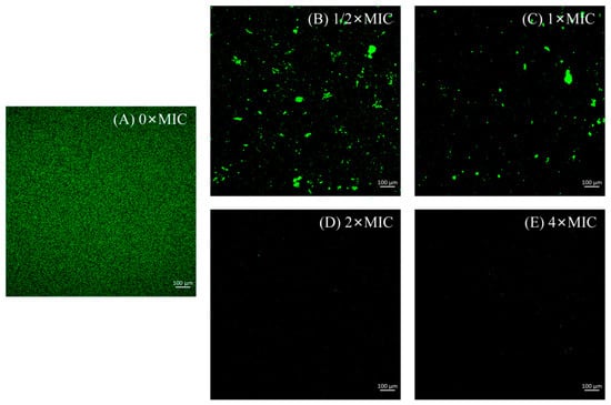
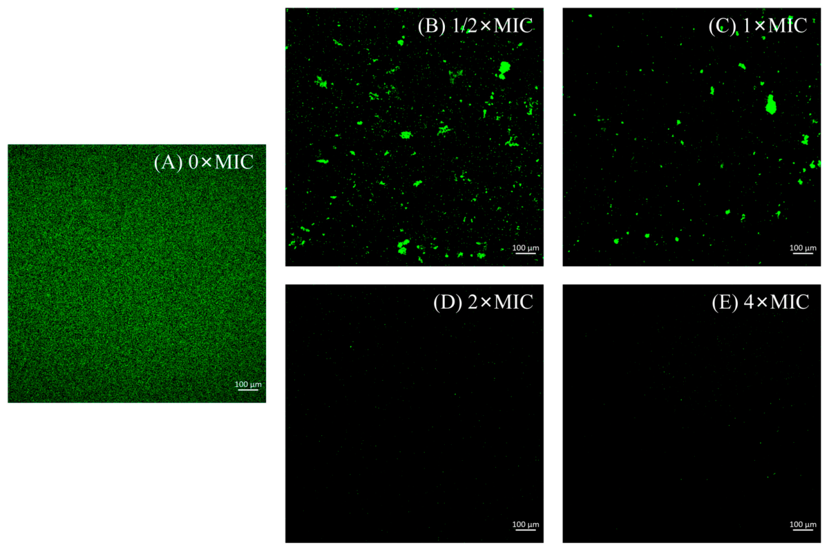
S. aureus suspension was diluted to 106 CFU/mL using LB broth. The diluted suspension was transferred to confocal dishes and incubated at 37 °C for 72 h to facilitate biofilm formation. Microcapsule solutions at concentrations of 1/2 × MIC, 1 × MIC, 2 × MIC, and 4 × MIC, as well as the required staining solutions, were prepared in advance. Following the incubation period, the formed biofilms were gently washed twice with sterile phosphate-buffered saline. The prepared sample solutions were then added to the corresponding confocal dishes, while the control group was treated with LB broth containing 5% ethanol. After 24 h incubation with the samples, the biofilms were washed again twice with sterile PBS. Subsequently, the biofilms were stained by applying SYTO 9 and propidium iodide onto their surface, followed by incubation at 37 °C in the dark for 15 min. After staining, excess dye was removed by washing with PBS. The biofilms were finally observed and imaged using excitation wavelengths of 488 nm and 561 nm [23].
2.3.10. Scanning Electron Microscopy Observation
The bacterial suspension of S. aureus was treated with the microcapsule sample solution at a final concentration of 1 × MIC, while the control group was treated with 5% ethanol solution. After incubation at 37 °C for 18 h, the bacterial cells were harvested and fixed overnight at 4 °C with 2% glutaraldehyde. The samples were then rinsed three times with PBS buffer, followed by gradient dehydration in a graded ethanol series (30%, 50%, 70%, 90%, and 100%) for 15 min at each concentration. Subsequently, the samples were dried using a critical point dryer, sputter-coated with gold, and observed under SEM to characterize the morphological alterations of the bacterial cells [24].
2.4. Statictical Analysis
Data processing and plotting were performed using Origin 2024 software (OriginLab Corporation, Northampton, MA, USA). Data visualization was performed with GraphPad Prism 8.0 (GraphPad Software, Boston, MA, USA). SPSS 19.0 software was used to perform one-way analysis to identify significant differences among treaments. A least significant difference test was used to determine significant differences in the means at p < 0.05.
3. Results and Discussion
3.1. Citral-Loaded Microcapsule Characterization
3.1.1. Microcapsule Encapsulation Efficiency for CA
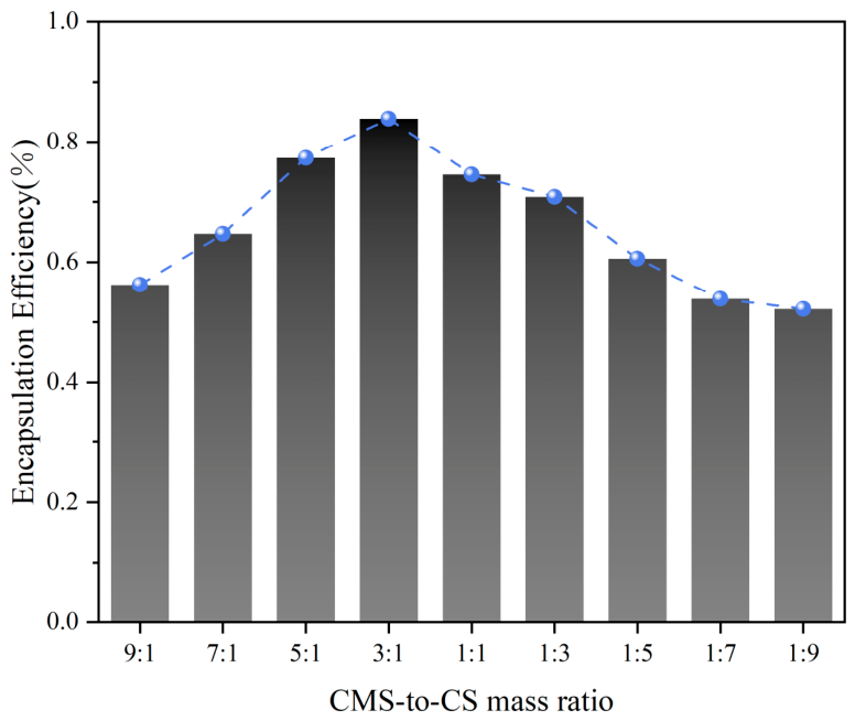
As known in Figure 1, in the present study, we blended CMS and CS at various mass ratios (9:1, 7:1, 5:1, 3:1, 1:1, 1:3, 1:5, 1:7, and 1:9), which achieved an encapsulation efficiency of 56.25%, 64.71%, 77.36%, 83.87%, 74.55%, 70.83%, 60.61%, 53.85%, and 52.17% for CA, respectively. The encapsulation efficiency reached the maximum (83.87%) at the CMS-to-CS mass ratio of 3:1, and gradually decreased thereafter. This decrease might be ascribed to the fact that an excessive concentration of emulsifier with increasing CS ratio may elevate the emulsion viscosity, restrict molecular mobility, and thereby impede the efficient incorporation of CA into the CMS-CS composite matrix. This is consistent with the previous research findings. A combination of CMS with hydrophilic colloids can enhance the stability of emulsion systems. CS is a positively charged hydrophilic colloid and is expected to improve the system stability upon complexation with CMS. It should be noted that the mass ratio of the two components has a profound influence on the functional performance of the composite matrix [10].
Figure 1.
CA encapsulation efficiency of microcapsules with different CMS-to-CS mass ratios.
3.1.2. Particle Size Analysis of CA-Loaded Microcapsules
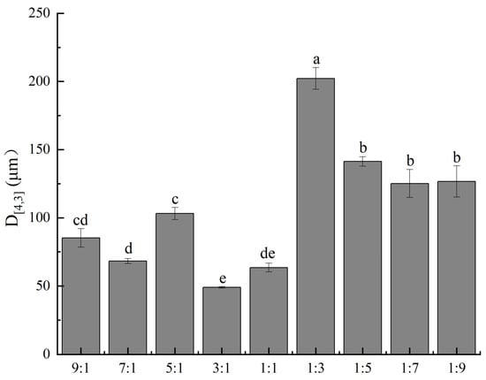
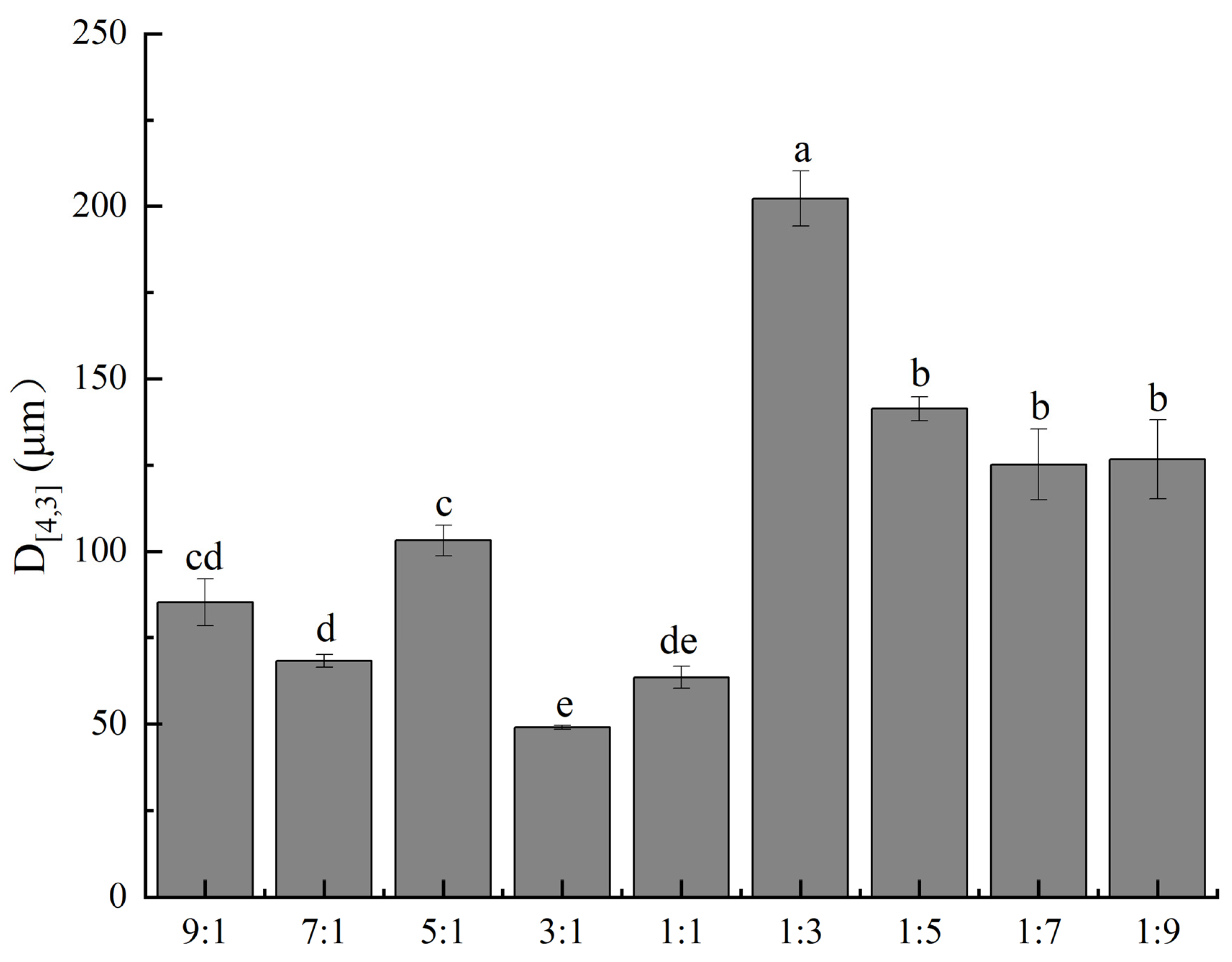
Particle size directly influences the encapsulation efficiency, release behavior, and stability of CA-loaded microcapsules, and is therefore a key parameter for evaluating their physical properties. In this study, the particle size distribution of CA-loaded microcapsules prepared with different mass ratios was determined using a laser diffraction particle size analyzer, aiming to assess their physical performance and provide a structural basis for subsequent application in preservation systems. The results are shown in Figure 2. Significant differences in particle size were observed among the CA-loaded microcapsules prepared with different mass ratios. Groups with a higher proportion of CS exhibited markedly larger particle sizes than those with a higher proportion of CMS. This is primarily because CMS is a negatively charged anionic polysaccharide, whereas CS can carry different charges depending on pH conditions. When the CS content is high, the excess positive charge in the composite system weakens the electrostatic repulsion between molecules, making molecular chains more prone to aggregation and thus forming larger particles. In addition, an excessive amount of CS prevents the formation of a stable three-dimensional network structure with CMS, resulting in a loose interfacial film that cannot effectively resist droplet coalescence. Furthermore, hydrophobic interactions and hydrogen bonding among protein molecular chains promote molecular aggregation, which directly leads to an increase in the particle size of CA-loaded microcapsules. The smallest particle size (49.045 um) was observed at a CMS-to-CS mass ratio of 3:1. This indicates that at this ratio, the electrostatic complexation between CMS and CS is most effective, producing the most compact and uniform interfacial film structure, which can efficiently encapsulate citral into fine particles and resist coalescence under drying conditions. From the perspective of drug release, a smaller particle size of CA-loaded microcapsules corresponds to a larger specific surface area, which leads to a faster release rate of the core material, whereas a larger particle size prolongs the diffusion path of the core material. These findings further confirm that the CA-loaded microcapsules prepared at the 3:1 mass ratio exhibit the best encapsulation performance and citral release behavior, making it the optimal ratio.
Figure 2.
Volume-weighted average particle size of CA-loaded microcapsules with different wall material qualities. Vertical bars indicate the standard errors of three replications. Different lowercase letters at same storage tiome points are significantly different.
3.1.3. FTIR Spectra of Microcapsules
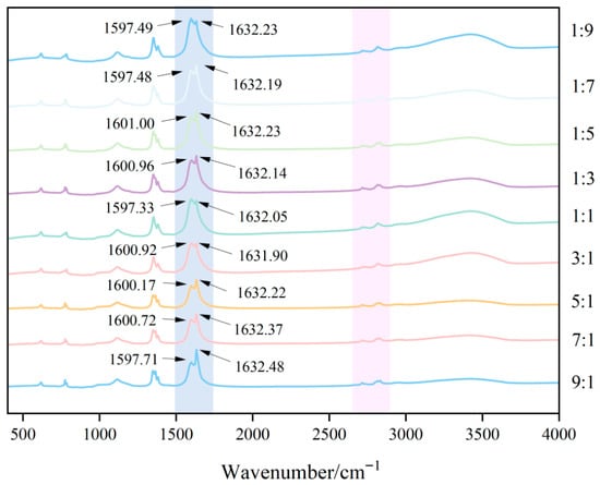
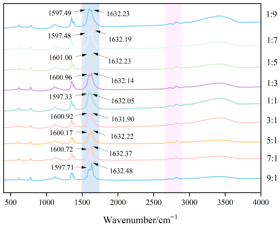
FTIR spectroscopy was utilized to characterize the variations in functional groups and intermolecular interactions within the biopolymer matrix (Figure 3). The characteristic FTIR absorption peaks of CMS included the O–H stretching vibration (~3400 cm−1), C–H stretching vibrations of methyl and methylene groups (~2920 cm−1 and ~2850 cm−1), and C=O stretching vibration of the carboxyl group (~1610–1630 cm−1) [25]. CS showed characteristic peaks corresponding to the overlapping N–H and O–H stretching vibration (~3400 cm−1), C–H stretching vibrations (~2920 cm−1 and ~2850 cm−1), and the amide I band derived from the C=O stretching vibration of peptide bonds (~1630–1650 cm−1) [26]. These results indicated that CMS and CS have highly similar peak positions. For CA, the characteristic absorption peaks are ascribed to the C=C stretching vibration (~1640–1680 cm−1) and the aldehyde C–H stretching vibrations (~2720–2740 cm−1 and ~2820 cm−1) [27]. The FTIR spectra of the CA-CMS-CS microcapsules prepared at different mass ratios are presented in Figure 3. The spectra displayed characteristic peaks corresponding to the C=O stretching vibration at approximately 1600 cm−1 and the overlapping O–H/N–H stretching vibration around 3400 cm−1, which represent the key fingerprint features of the CMS and CS wall materials. Furthermore, the characteristic peaks attributed to CA were also observed, including the C=C stretching vibration near 1630 cm−1 and the distinctive aldehyde C–H stretching vibrations at approximately 2717 cm−1 and 2815 cm−1. This is consistent with the previous research findings. While the overall spectral profiles generally remained consistent, a special trend was noted for the peak intensity at 1630 cm−1 (assigned to the C=C vibration of CA), which first decreased and then increased with increasing CS ratio, and reached the minimum at the CMS-to-CS mass ratio of 3:1. This attenuation trend of the CA characteristic peak is most likely ascribed to its effective encapsulation within the composite wall matrix. This phenomenon is in good agreement with the results for encapsulation efficiency, where the maximum encapsulation efficiency (83.87%) was obtained at the 3:1 CMS-to-CS mass ratio. The subsequent increase in peak intensity at higher CS ratios corresponds well with the downward trend of encapsulation efficiency, implying weakened shielding and less effective entrapment of CA within the microcapsules.
Figure 3.
FTIR spectra of CA-loaded microcapsules with different CMS-to-CS mass ratios.
3.1.4. Citral-Loaded Microcapsules Thermal Stability Analysis
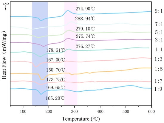
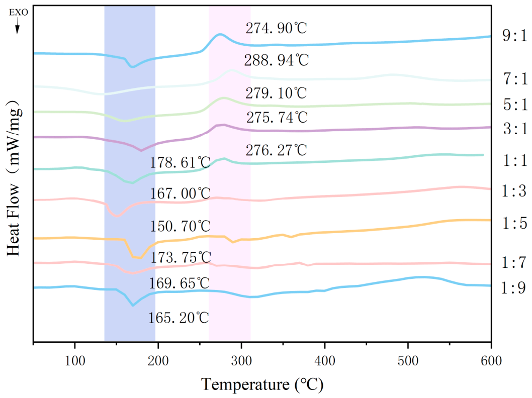
The DSC curves of CA-loaded microcapsules prepared at different CMS-to-CS mass ratios are displayed in Figure 4. For samples with CMS-to-CS ratios of 9:1, 7:1, 5:1, 3:1, and 1:1, a single endothermic peak was observed during the heating process at the temperature of 274.90 °C, 288.94 °C, 279.10 °C, 275.74 °C, and 276.27 °C, respectively. These endothermic characteristics probably correspond to the melting temperature of the microcapsule matrix. In contrast, samples with CMS-to-CS mass ratios of 1:9, 1:7, 1:5, 1:3, 1:1, and 3:1 exhibited a single exothermic peak during the heating process at the temperature of 165.20 °C, 169.65 °C, 173.75 °C, 150.70 °C, 167.61 °C, and 178.61 °C, respectively. This phenomenon is probably associated with the oxidation of CA released from the microcapsules with increasing temperature [28]. These results indicated that the CA-loaded microcapsules could remain relatively stable at temperatures below 150 °C. This provides guidance for the actual application scenarios of this microcapsule. Among all samples, the sample with a CMS-to-CS mass ratio of 3:1 exhibited the highest exothermic peak temperature (178.61 °C), implying that it possesses the most superior thermal stability.
Figure 4.
DSC curves of CA-loaded microcapsules with different CMS-to-CS mass ratios.
3.1.5. Morphology of Microcapsules
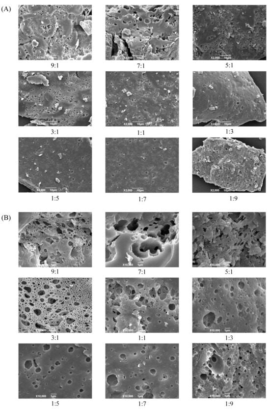
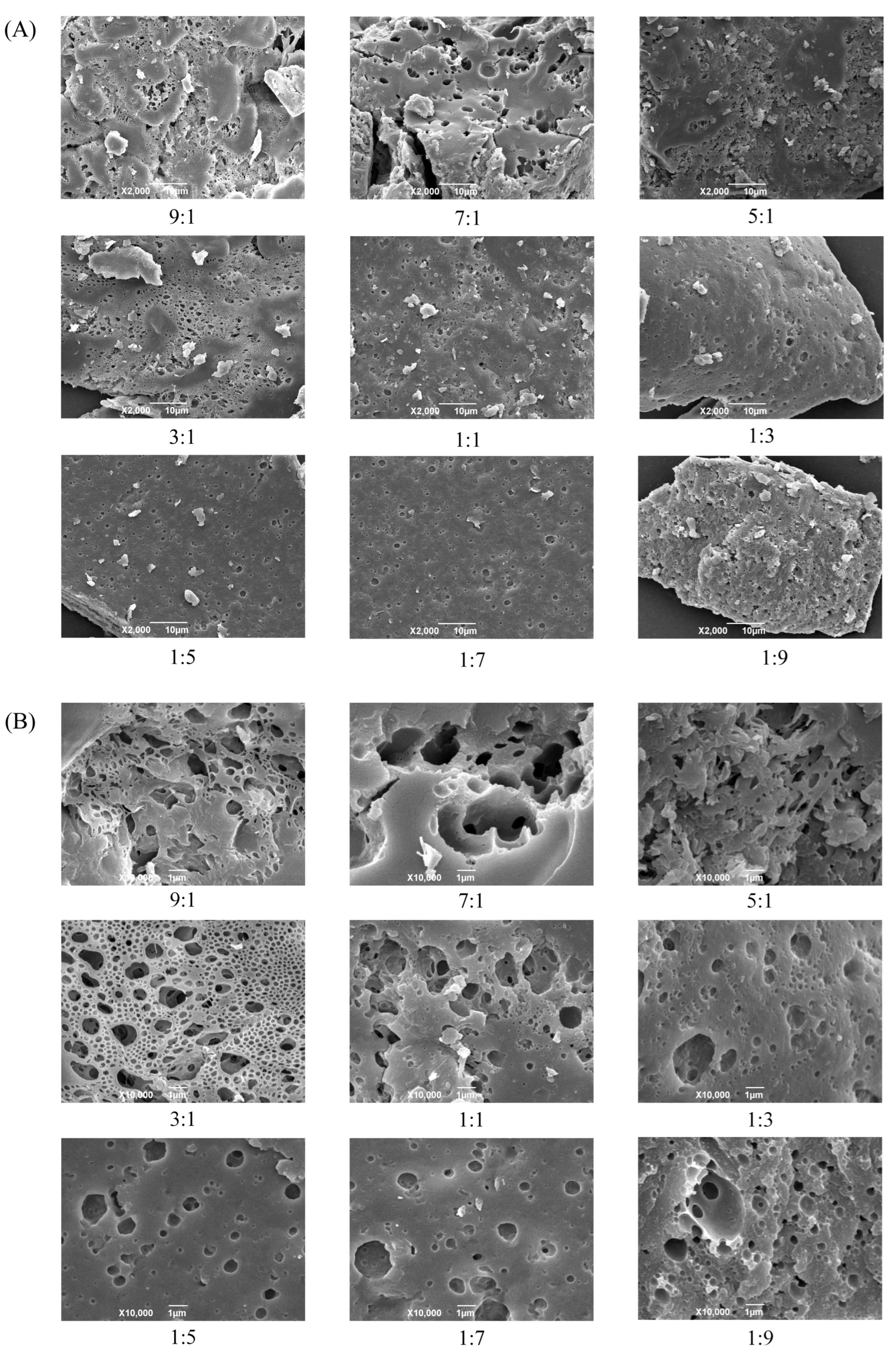
Figure 5 displays the SEM images of CA-loaded microcapsules with different CMS-to-CS mass ratios. The CA-loaded microcapsules showed irregular blocks of varying sizes on the surface, while pristine CMS and CS exhibited a uniform spherical morphology, indicating that significant intermolecular interactions occurred between CA and the CMS-CS composite matrix during the encapsulation process. It can be preliminarily speculated that intermolecular interactions among the three components may occur during the freeze-drying procedure, which modifies the morphology of CMS and CS and generates a new phase, ultimately resulting in a distinctive microcapsule structure. Furthermore, at identical magnification, the surfaces of microcapsules prepared at all CMS-to-CS mass ratios displayed pores and channels with diverse diameters. With increasing ratio of CS, the number and size of these surface pores gradually decreased. The microcapsules with CMS-to-CS mass ratios of 9:1, 7:1, and 5:1 exhibited fairly rough surfaces with complex architectures and heterogeneous pore size distributions, which could be attributed to the excessively high CMS ratios and inadequate emulsifier (CS) dosage. In contrast, the microcapsules with a CMS-to-CS mass ratio of 3:1 possessed more uniformly distributed pores, as well as a smaller average diameter and more orderly pore size distribution, which is in good agreement with the DSC analysis results.
Figure 5.
SEM Images of CA-loaded microcapsules with different CMS-to-CS mass ratios ((A): ×2000, (B): ×10,000).
3.2. Antibacterial Mechanism Analysis of Microcapsules
3.2.1. Analysis of Inhibition Zone, MIC, and MBC
Measurement of inhibition zones revealed that the CA-loaded microcapsules displayed significant antibacterial activity against S. aureus, B. subtilis, and L. monocytogenes, particularly against B. subtilis. This observation validated the efficient release of high concentrations of bioactive compounds from the microcapsules during incubation. MIC, the lowest concentration capable of inhibiting 99.9% of microbial growth, can reflect the antimicrobial potency of an agent, with a lower value signifying a stronger inhibitory efficacy [21]. As known in Table 1, the MIC and MBC assay results revealed that the MIC of CA-loaded microcapsules was 6.25, 3.13, and 6.25 mg/mL against S. aureus, B. subtilis, and L. monocytogenes, with corresponding MBC values of 12.5, 6.25, and 25 mg/mL, respectively. Notably, the MBC for the first two bacteria was exactly twice their MIC. Numerous studies have demonstrated that CA exhibits inhibitory activities against plant pathogens, foodborne pathogens, wood-decay fungi, and molds [29], making it a promising candidate for antimicrobial preservation in food applications. Nevertheless, CA possesses unsaturated bonds such as C=C and C=O moieties, which endow it with high instability and volatility. Encapsulation of CA within microcapsules can improve its stability, achieve controlled release of its antimicrobial constituents, and therefore enhance its antibacterial efficacy.
Table 1.
Minimum inhibitory concentration and minimum bactericidal concentration of CA-loaded microcapsules against typical foodborne pathogenic bacteria and food-related bacteria.
3.2.2. Effect of Microcapsules on Bacterial Nucleic Acid Concentration
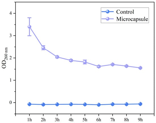
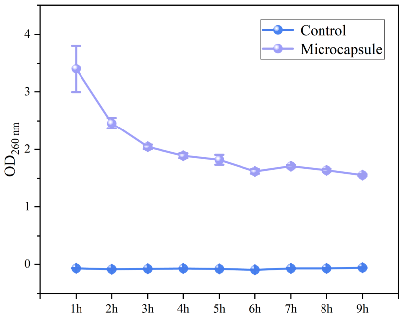
The results of OD260 measurement are presented in Figure 6. As the incubation time of S. aureus increased, the nucleic acid concentration in the control group exhibited no significant variation, which is indicative of normal bacterial growth; in contrast, that in the supernatant of the S. aureus suspension treated with CA-loaded microcapsules continuously decreased over time. 1× MIC was sufficient to completely inhibit bacterial growth. At the initial stage of treatment, transient cell membrane damage and nucleic acid leakage probably occurred, resulting in a relatively high initial OD260 value. Subsequently, the bacteria showed progressive death and autolysis due to metabolic inhibition. The nucleases released from the lysed cells might degrade the nucleic acids, contributing to the observed continuous decrease in OD260 value in later incubation. The destruction of DNA can impede gene expression, ultimately causing bacterial death [22]. The progressively decreasing OD260 value suggested DNA fragmentation, demonstrating that the CA-loaded microcapsule treatment not only inhibits S. aureus growth but also induces its cell death and nucleic acid degradation via the sustained release of CA, this is consistent with the previous research findings [22].
Figure 6.
Changes in nucleic acid macromolecules in the supernatant of Staphylococcus aureus culture medium.
3.2.3. Effect of Microcapsules on Bacterial Biofilm Permeability
Figure 7 shows the biofilms formed by S. aureus in both the control group (0× MIC) and groups treated with microcapsule solutions at 1/2× MIC, 1× MIC, 2× MIC, and 4× MIC. In CLSM analysis, a reduction or shrinkage of fluorescent regions signifies a decrease in the bacterial population within the biofilm [30]. In the 0× MIC group, the S. aureus biofilm displayed a dense and intact three-dimensional architecture, where all cells existed as viable single units with no visible aggregates. In the 1/2× MIC and 1× MIC groups, the three-dimensional architecture of the biofilm was completely disrupted, accompanied by a significantly diminishing count of single cells. Concurrently, a small number of green fluorescent aggregates emerged, implying that the bacterial cells initiated a collective resistance mechanism in response to the hostile environment induced by the CA-loaded microcapsules, which probably involves enhancement of cell-to-cell adhesion to counteract the penetration and antibacterial action of CA. In the 2× MIC and 4× MIC groups, only a minimal number of single cells were observable, demonstrating that the CA-loaded microcapsules can not only kill individual bacterial cells but also effectively dismantle the aggregated structures formed by the bacteria. Overall, the bactericidal efficacy of the CA-loaded microcapsules is concentration-dependent: at sub-inhibitory concentrations, they induce bacterial aggregation as a defensive response, whereas at high concentrations, they completely dismantle bacterial aggregates and eradicate the residual bacteria. These findings reveal the antimicrobial mechanism of the CA-loaded microcapsules from the perspectives of both cellular morphology and structural integrity.
Figure 7.
Inhibitory effects of CA-loaded microcapsules at different folds of MIC on Staphylococcus aureus biofilms.
3.2.4. Effect of Microcapsules on Bacterial Morphology
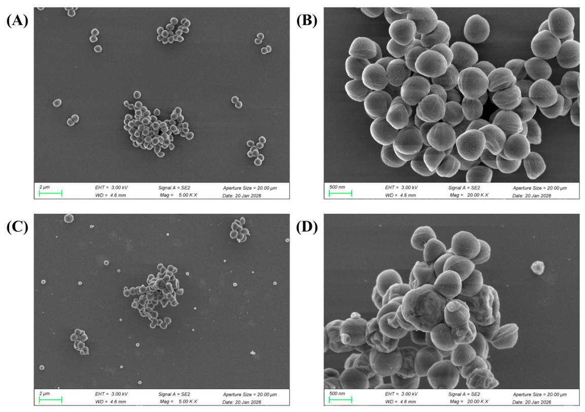
SEM observations revealed distinct morphological alterations in S. aureus cells after treatment with CA-loaded microcapsules. As known in Figure 8, the untreated cells (control group) were plump with comparatively smooth surfaces, exhibiting typical, intact spherical morphological characteristics of staphylococcal cocci, where no cell lysis or leakage of intracellular contents was detected. In contrast, co-culturing with CA-loaded microcapsules at 1× MIC resulted in distinct morphological changes in S. aureus. The bacterial surfaces became rough and uneven, exhibiting widespread shrinkage with an irregular spherical shape. These observations suggested that CA probably interferes with the synthesis or cross-linking of peptidoglycan within the bacterial cell wall [29]. Furthermore, pronounced concavities were visible on the surface of many cells. In severe cases, excessive indentation can lead to cell rupture and conspicuous leakage of intracellular contents. CA is a hydrophobic terpenoid aldehyde, and may be integrated into the lipid bilayer of the bacterial cytoplasmic membrane, thereby elevating its permeability. Such disruption will give rise to an imbalance in transmembrane ion gradients, ultimately compromising the membrane integrity. Therefore, it can be inferred that CA inhibits the normal growth of S. aureus by impeding cell wall biosynthesis and compromising the cell membrane integrity, this is consistent with the previous research findings [29].
Figure 8.
Scanning electron micrographs of Staphylococcus aureus. (A,B): Control group, S. aureus cells untreated with CA-loaded microcapsules. (C,D): S. aureus cells treated with 1× MIC.
4. Conclusions
This study investigated the physicochemical properties, thermal stability, and antibacterial efficacy of CA-loaded microcapsules fabricated with a composite wall system consisting of CMS and CS. A CMS-to-CS mass ratio of 3:1 was identified as the optimal ratio, which resulted in the most uniform distribution of pore size and number on the microcapsule surface, the lowest intensity of the characteristic peak of CA, and thereby an encapsulation efficiency exceeding 80%, as well as excellent thermal stability of the microcapsule at temperatures up to 150 °C. Moreover, the CA-loaded microcapsules exhibited potent antibacterial activities against typical foodborne pathogenic bacteria and food-related bacteria, specifically S. aureus, B. subtilis, and L. monocytogenes. Microplate assays indicated that the CA-loaded microcapsule treatment induced cell death in S. aureus and promoted nucleic acid degradation through the sustained release of CA. The CA-loaded microcapsules could not only eradicate planktonic bacterial cells but also effectively disrupt the aggregated biofilm architecture, probably through cleavage of DNA and impairment of cell membrane integrity. These modifications alter the cellular morphology and structural integrity of S. aureus, thereby interfering with its intrinsic physiological functions and ultimately culminating in cell death. These findings offer a valuable reference for future studies aimed at improving the stability of citral when used as an antibacterial agent and at enhancing its practical application value.
Author Contributions
J.H.: Writing—review and editing, Writing—original draft, Methodology, Investigation. H.W.: Methodology, Investigation. L.W.: Methodology, Funding acquisition. X.L.: Methodology, Funding acquisition. All authors have read and agreed to the published version of the manuscript.
Funding
This work was supported by the Yunnan Provincial Agricultural Basic Research Joint Special Project [Grant Number: 202401BD070001-077] and the Hubei Provincial Science & Technology Talent Service Enterprise Project “Research and Product Development of Clean Label Pickled Vegetable Production Technology” [Grant Number: 2025DJB015].
Institutional Review Board Statement
Not applicable.
Informed Consent Statement
Not applicable.
Data Availability Statement
The original contributions presented in this study are included in the article. Further inquiries can be directed to the corresponding authors.
Conflicts of Interest
The authors declare no conflicts of interest.
References
- Li, Y.; Mei, J.; Xie, J. Citral: Bioactivity, Metabolism, Delivery Systems, and Food Preservation Applications. Compr. Rev. Food Sci. Food Saf. 2025, 24, e70168. [Google Scholar] [CrossRef] [Scilit]
- Shan, Y.; Chen, S.; Zhang, J.; Du, C.; Liu, C.; Yang, F.; Yin, W.; Shao, Y.; Wang, Y. Preparation of Citral Compound and Its Bamboo Antimildew Properties. Polymers 2022, 14, 4691. [Google Scholar] [CrossRef] [Scilit] [PubMed]
- Unal Turhan, E.; Koca, E.E. Predictive Modeling for Inactivation of Escherichia coli Biofilm with Combined Treatment of Thermosonication and Organic Acid on Polystyrene Surface. Foods 2024, 13, 4002. [Google Scholar] [CrossRef] [Scilit] [PubMed]
- Niu, Y.; Wu, J.; Kang, Y.; Zhao, Q.; Xiao, Z.; Zhao, D. Encapsulation technique and application progress of mechanical stimuli-responsive microcapsules. Prog. Org. Coat. 2023, 176, 107390. [Google Scholar] [CrossRef] [Scilit]
- Li, Z.; Zheng, M.; He, P.; Gong, W.; Liu, Z.; Xu, C.; Tai, Z. Citral Essential Oil-Loaded Microcapsules by Simple Coacervation and Its Application on Peach Preservation. ACS Omega 2022, 7, 42181–42190. [Google Scholar] [CrossRef] [Scilit]
- Wu, W.; Li, Z.; Feng, D.; Tang, Q.; Liu, S.; Lin, W. Dissipative Particle Dynamics: Simulation of Chitosan–Citral Microcapsules. Polymers 2025, 17, 678. [Google Scholar] [CrossRef] [Scilit]
- Kathuria, A.; Kumar, L.; Gaikwad, K.K.; Badawy, A.E.; Kivy, M.B. Micro-encapsulation of citral using edible γ-cyclodextrin metal organic framework. J. Incl. Phenom. Macrocycl. Chem. 2024, 104, 99–108. [Google Scholar] [CrossRef] [Scilit]
- Yoplac, I.; Vargas, L.; Robert, P.; Hidalgo, A. Characterization and antimicrobial activity of microencapsulated citral with dextrin by spray drying. Heliyon 2021, 7, e06737. [Google Scholar] [CrossRef] [Scilit]
- Cai, X.; Wang, Y.; Du, X.; Xing, X.; Zhu, G. Stability of pH-responsive Pickering emulsion stabilized by carboxymethyl starch/xanthan gum combinations. Food Hydrocoll. 2020, 109, 106093. [Google Scholar] [CrossRef] [Scilit]
- Cai, X.; Du, X.; Zhu, G.; Chen, Q.; Cao, C. Synergistic effect and mechanism of stabilization of Pickering emulsion by carboxymethyl starch and xanthan gum. Int. J. Biol. Macromol. 2024, 283, 137560. [Google Scholar] [CrossRef] [Scilit]
- Xu, W.; Li, C.; McClements, D.J.; Xu, Z.; Zhang, Z.; Zhang, R.; Qiu, C.; Zhao, J.; Jin, Z.; Chen, L. Construction, characterization and application of rutin loaded zein—Carboxymethyl starch sodium nanoparticles. Int. J. Biol. Macromol. 2025, 302, 140355. [Google Scholar] [CrossRef] [Scilit]
- Wang, C.; Li, X.; Sang, S.; Julian McClements, D.; Chen, L.; Long, J.; Jiao, A.; Wang, J.; Jin, Z.; Qiu, C. Preparation, characterization and in vitro digestive behaviors of emulsions synergistically stabilized by γ-cyclodextrin/sodium caseinate/alginate. Food Res. Int. 2022, 160, 111634. [Google Scholar] [CrossRef] [Scilit] [PubMed]
- Lin, Y.; Xu, Q.; Mao, Y.; Shao, P.; Li, X. Synergistic effects of Tremella polysaccharide and sodium caseinate: A promising strategy for regulating water distribution in fat-replaced foods. J. Future Foods 2025. [Google Scholar] [CrossRef] [Scilit]
- Lin, Y.; Xu, J.; Gao, X. Development of antioxidant sodium alginate gel beads encapsulating curcumin/gum Arabic/gelatin microcapsules. Food Hydrocoll. 2024, 152, 109901. [Google Scholar] [CrossRef] [Scilit]
- Zou, X.; Pan, Y.; Hu, J.; Zhang, K.; Yang, X.; Nwankwo, J.A.; Guo, X.; Lin, Y.; Liu, W. Effects of different soluble dietary fibers on oil encapsulation: Comparison of emulsion and microcapsule properties. Food Chem. 2025, 479, 143842. [Google Scholar] [CrossRef] [Scilit]
- Li, Y.; Zhao, W.; Wang, Y.; Xie, Y.; Li, J.; He, J.; Wang, C.; De Souza, C.; Zhang, L.; Lin, K. Development of low-allergenicity algal oil microcapsules with high encapsulation efficiency using extensively hydrolyzed whey protein. Int. J. Biol. Macromol. 2025, 303, 140540. [Google Scholar] [CrossRef] [Scilit]
- Qi, J.-R.; Song, L.-W.; Zeng, W.-Q.; Liao, J.-S. Citrus fiber for the stabilization of O/W emulsion through combination of Pickering effect and fiber-based network. Food Chem. 2021, 343, 128523. [Google Scholar] [CrossRef] [Scilit]
- Qian, J.; Chen, Y.; Wang, Q.; Zhao, X.; Yang, H.; Gong, F.; Guo, H. Preparation and antimicrobial activity of pectin-chitosan embedding nisin microcapsules. Eur. Polym. J. 2021, 157, 110676. [Google Scholar] [CrossRef] [Scilit]
- Jiang, Y.; Wang, Q.; Tian, S.; Luo, Z.; Wang, D.; Bai, Y.; Lu, W.; Zhao, T. Preparation of NaF Microcapsules for High-Temperature Thermal Storage. ACS Omega 2022, 7, 24688–24694. [Google Scholar] [CrossRef] [Scilit]
- Karaogul, E.; Ugurtay, A. Unveiling modeling and SEM/XRD insights into enhanced antibacterial, antioxidant, and bioactive potentials of Micro-encapsulated Pistacia vera hull extract. Food Chem. 2025, 477, 143510. [Google Scholar] [CrossRef] [Scilit]
- Jafari, S.; Karami, Z.; Shiekh, K.A.; Kijpatanasilp, I.; Worobo, R.W.; Assatarakul, K. Ultrasound-Assisted Extraction of Bioactive Compounds from Cocoa Shell and Their Encapsulation in Gum Arabic and Maltodextrin: A Technology to Produce Functional Food Ingredients. Foods 2023, 12, 412. [Google Scholar] [CrossRef] [Scilit] [PubMed]
- Zhang, L.; Zhang, M.; Ju, R.; Bhandari, B.; Liu, K. Antibacterial mechanisms of star anise essential oil microcapsules encapsulated by rice protein-depolymerized pectin electrostatic complexation and its application in crab meatballs. Int. J. Food Microbiol. 2023, 384, 109963. [Google Scholar] [CrossRef] [Scilit] [PubMed]
- Gao, X.; Liu, J.; Li, B.; Xie, J. Antibacterial Activity and Antibacterial Mechanism of Lemon Verbena Essential Oil. Molecules 2023, 28, 3102. [Google Scholar] [CrossRef] [Scilit] [PubMed]
- Zhang, J.J.; Qu, L.B.; Bi, Y.F.; Pan, C.X.; Yang, R.; Zeng, H.J. Antibacterial activity and mechanism of chloroform fraction from aqueous extract of mugwort leaves (Artemisia argyi L.) against Staphylococcus aureus. Lett. Appl. Microbiol. 2022, 74, 893–900. [Google Scholar] [CrossRef] [Scilit]
- Huangfu, Z.; Sun, W.; Chen, C.; Liu, R. New insight into effect of cation bridging on the differential bonding mechanism of sodium carboxymethyl starch in molybdenite-talc separation. Miner. Eng. 2025, 232, 109532. [Google Scholar] [CrossRef] [Scilit]
- Sun, M.-C.; Wang, J.-T.; Wang, Y.-D.; Hu, L.-Y.; Yu, S.-Q.; Zhang, T.; Zhao, C. Structure-function modulation of sodium caseinate: Insights from non-covalent reuterin binding. Food Chem. 2025, 493, 146051. [Google Scholar] [CrossRef] [Scilit]
- Vilas Dhumal, C.; Pal, K.; Sarkar, P. Synthesis, characterization, and antimicrobial efficacy of composite films from guar gum/sago starch/whey protein isolate loaded with carvacrol, citral and carvacrol-citral mixture. J. Mater. Sci. Mater. Med. 2019, 30, 117. [Google Scholar] [CrossRef] [Scilit]
- Gür, F.; Duygu Kaya, E.; Gür, B.; Türkhan, A.; Onganer, Y. Preparation of bio-electrodes via Langmuir-Blodgett technique for pharmaceutical and waste industries and their biosensor application. Colloid Surf. A Physicochem. Eng. Aspects 2019, 583, 124005, Corrigendum to Colloids Surf. A Physicochem. Eng. Asp. 2020, 586, 124135. [Google Scholar] [CrossRef] [Scilit]
- Cui, K.; Wang, Y.; Wang, M.; Zhao, T.; Zhang, F.; He, L.; Zhou, L. Inhibitory activity and antioomycete mechanism of citral against Phytophthora capsici. Pestic. Biochem. Physiol. 2024, 204, 106067. [Google Scholar] [CrossRef] [Scilit]
- Bai, M.; Li, C.; Cui, H.; Lin, L. Preparation of self-assembling Litsea cubeba essential oil/diphenylalanine peptide micro/nanotubes with enhanced antibacterial properties against Staphylococcus aureus biofilm. LWT 2021, 146, 111394. [Google Scholar] [CrossRef] [Scilit]
Disclaimer/Publisher’s Note: The statements, opinions and data contained in all publications are solely those of the individual author(s) and contributor(s) and not of MDPI and/or the editor(s). MDPI and/or the editor(s) disclaim responsibility for any injury to people or property resulting from any ideas, methods, instructions or products referred to in the content. |
© 2026 by the authors. Licensee MDPI, Basel, Switzerland. This article is an open access article distributed under the terms and conditions of the Creative Commons Attribution (CC BY) license.