Next Issue
Volume 10, February
Previous Issue
Volume 9, December


Dent. J., Volume 10, Issue 1 (January 2022) – 13 articles

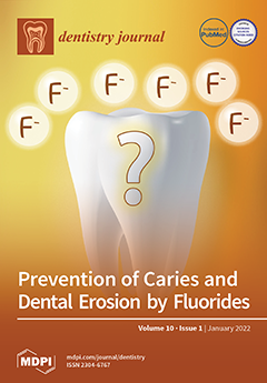
Cover Story (view full-size image): Dental erosion is a common problem in dentistry. It is defined as the loss of tooth mineral by the attack of acids that do not result from caries. The proposed protective actions of fluorides include the formation of an acid-resistant fluoride-rich surface layer and a fluoride-induced surface hardening of the tooth surface. A comprehensive literature study on the interaction between fluoride and tooth surfaces (e.g., by toothpastes or mouthwashes) is discussed. The analytical techniques available to address this question are presented. In summary, the amount of fluoride that is incorporated into teeth is very low (a few µg mm2). This is unlikely to protect a tooth against an attack by acids, be it from acidic agents (erosion) or from acid-producing cariogenic bacteria. View this paper
  • Issues are regarded as officially published after their release is announced to the table of contents alert mailing list.
  • You may sign up for e-mail alerts to receive table of contents of newly released issues.
  • PDF is the official format for papers published in both, html and pdf forms. To view the papers in pdf format, click on the "PDF Full-text" link, and use the free Adobe Reader to open them.
Order results
Result details
Section
Select all
Export citation of selected articles as:
16 pages, 2270 KiB  
Review
Simulating the Intraoral Aging of Dental Bonding Agents: A Narrative Review
by Tomas Vilde, Cameron A. Stewart and Yoav Finer
Dent. J. 2022, 10(1), 13; https://doi.org/10.3390/dj10010013 - 15 Jan 2022
Cited by 6 | Viewed by 3380
Abstract
Despite their popularity, resin composite restorations fail earlier and at higher rates than comparable amalgam restorations. One of the reasons for these rates of failure are the properties of current dental bonding agents. Modern bonding agents are vulnerable to gradual chemical and mechanical [...] Read more.
Despite their popularity, resin composite restorations fail earlier and at higher rates than comparable amalgam restorations. One of the reasons for these rates of failure are the properties of current dental bonding agents. Modern bonding agents are vulnerable to gradual chemical and mechanical degradation from a number of avenues such as daily use in chewing, catalytic hydrolysis facilitated by salivary or bacterial enzymes, and thermal fluctuations. These stressors have been found to work synergistically, all contributing to the deterioration and eventual failure of the hybrid layer. Due to the expense and difficulty in conducting in vivo experiments, in vitro protocols meant to accurately simulate the oral environment’s stressors are important in the development of bonding agents and materials that are more resistant to these processes of degradation. This narrative review serves to summarize the currently employed methods of aging dental materials and critically appraise them in the context of our knowledge of the oral environment’s parameters. Full article
(This article belongs to the Section Dental Materials)
Show Figures

Figure 1

15 pages, 4371 KiB  
Article
Influence of Framework Material and Posterior Implant Angulation in Full-Arch All-on-4 Implant-Supported Prosthesis Stress Concentration
by João Paulo Mendes Tribst, Dayana Campanelli de Morais, Jefferson David Melo de Matos, Guilherme da Rocha Scalzer Lopes, Amanda Maria de Oliveira Dal Piva, Alexandre Luiz Souto Borges, Marco Antonio Bottino, Antonio Lanzotti, Massimo Martorelli and Pietro Ausiello
Dent. J. 2022, 10(1), 12; https://doi.org/10.3390/dj10010012 - 14 Jan 2022
Cited by 24 | Viewed by 6809
Abstract
This study evaluated the influence of distal implants angulation and framework material in the stress concentration of an All-on-4 full-arch prosthesis. A full-arch implant-supported prosthesis 3D model was created with different distal implant angulations and cantilever arms (30° with 10-mm cantilever; 45° with [...] Read more.
This study evaluated the influence of distal implants angulation and framework material in the stress concentration of an All-on-4 full-arch prosthesis. A full-arch implant-supported prosthesis 3D model was created with different distal implant angulations and cantilever arms (30° with 10-mm cantilever; 45° with 10-mm cantilever and 45° with 6-mm cantilever) and framework materials (Cobalt–chrome [CoCr alloy], Yttria-stabilized tetragonal zirconia polycrystal [Y-TZP] and polyetheretherketone [PEEK]). Each solid was imported to computer-aided engineering software, and tetrahedral elements formed the mesh. Material properties were assigned to each solid with isotropic and homogeneous behavior. The contacts were considered bonded. A vertical load of 200 N was applied in the distal region of the cantilever arm, and stress was evaluated in Von Misses (σVM) for prosthesis components and the Maximum (σMAX) and Minimum (σMIN) Principal Stresses for the bone. Distal implants angled in 45° with a 10-mm cantilever arm showed the highest stress concentration for all structures with higher stress magnitudes when the PEEK framework was considered. However, distal implants angled in 45° with a 6-mm cantilever arm showed promising mechanical responses with the lowest stress peaks. For the All-on-4 concept, a 45° distal implants angulation is only beneficial if it is possible to reduce the cantilever’s length; otherwise, the use of 30° should be considered. Comparing with PEEK, the YTZP and CoCr concentrated stress in the framework structure, reducing the stress in the prosthetic screw. Full article
Show Figures

Figure 1

8 pages, 1808 KiB  
Article
An In-Vitro Evaluation of Articulation Accuracy for Digitally Milled Models vs. Conventional Gypsum Casts
by Jason D. Lee, German O. Gallucci and Sang J. Lee
Dent. J. 2022, 10(1), 11; https://doi.org/10.3390/dj10010011 - 11 Jan 2022
Cited by 7 | Viewed by 3632
Abstract
With the advent of a digital workflow in dentistry, the inter-occlusal articulation of digital models is now possible through various means. The Cadent iTero intraoral scanner uses a buccal scan in maximum intercuspation to record the maxillomandibular relationship. This in-vitro study compares the [...] Read more.
With the advent of a digital workflow in dentistry, the inter-occlusal articulation of digital models is now possible through various means. The Cadent iTero intraoral scanner uses a buccal scan in maximum intercuspation to record the maxillomandibular relationship. This in-vitro study compares the occlusion derived from conventionally articulated stone casts versus that of digitally articulated quadrant milled models. Thirty sets of stone casts poured from full arch polyvinyl siloxane impressions (Group A) and thirty sets of polyurethane quadrant models milled from digital impressions (Group B) were used for this study. The full arch stone casts were hand-articulated and mounted on semi-adjustable articulators, while the digitally derived models were pre-mounted from the milling center based on the data obtained from the buccal scanning procedure. A T-scan sensor was used to obtain a bite registration from each set of models in both groups. The T-scan data derived from groups A and B were compared to that from the master model to evaluate the reproducibility of the occlusion in the two groups. A statistically significant difference of the contact region surface area was found on #11 of the digitally articulated models compared to the master. An analysis of the force distribution also showed a tendency for a heavier distribution on the more anterior #11 tooth for the digitally articulated models. Within the limitations of this study, the use of a digitally articulated quadrant model system may result in a loss of accuracy, in terms of occlusion, the further anteriorly the tooth to be restored is located. Care must be taken to consider the sources of inaccuracies in the digital workflow to minimize them for a more efficient and effective restorative process. Full article
(This article belongs to the Special Issue Feature Papers in Dentistry Journal in 2021)
Show Figures

Figure 1

12 pages, 453 KiB  
Article
Impact of Impaired Glucose Metabolism on Periodontitis Progression over Three Years
by Oelisoa M. Andriankaja, Kaumudi Joshipura, Francisco Muñoz, Bruce A. Dye, Frank B. Hu and Cynthia M. Pérez
Dent. J. 2022, 10(1), 10; https://doi.org/10.3390/dj10010010 - 7 Jan 2022
Viewed by 2523
Abstract
We evaluated the relationship between glucose abnormalities and periodontitis in overweight/obese individuals. Eight hundred and seventy (870) diabetes-free participants aged 40–65 years completed the three-year follow-up in the San Juan Overweight Adults Longitudinal Study. The ADA thresholds for fasting and 2-h post-load glucose [...] Read more.
We evaluated the relationship between glucose abnormalities and periodontitis in overweight/obese individuals. Eight hundred and seventy (870) diabetes-free participants aged 40–65 years completed the three-year follow-up in the San Juan Overweight Adults Longitudinal Study. The ADA thresholds for fasting and 2-h post-load glucose and HbA1c were used to define prediabetes. The NHANES methods were used to assess periodontitis. Multivariable linear regression was used to evaluate the relationship between baseline glucose metabolism measures and periodontitis at follow-up, adjusting for potential confounders. There was no association between impaired glucose measures and mean pocket depth (PD), mean clinical attachment loss (CAL), or mean percent of sites ≥5 mm PD. Impaired glucose tolerance (IGT) was associated with a lower mean percent of sites ≥5 mm CAL (β = −1.6, p = 0.037). Prediabetes and impaired fasting glucose (IFG) were associated with improvement in mean percent of sites ≥5 mm PD (β = −1.4, p = 0.022; β = −1.6, p = 0.032; respectively). IFG and IGT were associated with improvement in mean percent of sites with ≥5 mm CAL (β = −1.6, p = 0.038; β = −1.9, p = 0.020; respectively). In conclusion, there were no consistent associations between baseline prediabetes or insulin resistance and periodontitis progression over a three-year period. Full article
(This article belongs to the Section Oral Hygiene, Periodontology and Peri-implant Diseases)
Show Figures

Figure 1

9 pages, 6339 KiB  
Case Report
Atypical Peripheral Ossifying Fibroma of the Mandible
by Tomislav Katanec, Lea Budak, Davor Brajdić and Dragana Gabrić
Dent. J. 2022, 10(1), 9; https://doi.org/10.3390/dj10010009 - 6 Jan 2022
Cited by 4 | Viewed by 6410
Abstract
Peripheral ossifying fibroma (POF) is a benign localized lesion originating from gingival and alveolar oral mucosa. Its origin can be cells of periodontal ligament. The lesions usually develop in women in their twenties. POF is a complex clinical and histological diagnosis due to [...] Read more.
Peripheral ossifying fibroma (POF) is a benign localized lesion originating from gingival and alveolar oral mucosa. Its origin can be cells of periodontal ligament. The lesions usually develop in women in their twenties. POF is a complex clinical and histological diagnosis due to its shared characteristics with many other conditions. In this paper, we presented a case of an atypical peripheral ossifying fibroma (POF) in the left lateral part of the mandible in a 70-year-old male patient who had two semicircular bridges supported on four implants in the upper and lower jaws. A review of CBCT and orthopedic imaging showed no visible intraosseous changes. Histological analysis revealed the diagnosis of POF. The case in question is interesting, as elaborated on in the discussion section of this paper because POF is usually found in female patients aged between 20 and 30 years. Full article
(This article belongs to the Section Dental Education)
Show Figures

Figure 1

9 pages, 247 KiB  
Article
Risk Indicators and Treatment Needs of Children 2–5 Years of Age Receiving Dental Treatment under General Anesthesia in Saskatchewan
by Alyssa Weninger, Erica Seebach, Jordyn Broz, Carol Nagle, Jessica Lieffers, Petros Papagerakis and Keith Da Silva
Dent. J. 2022, 10(1), 8; https://doi.org/10.3390/dj10010008 - 6 Jan 2022
Cited by 5 | Viewed by 2779
Abstract
Background: When compared to national averages in Canada, Saskatchewan has one of the highest rates of dental treatment under general anesthesia (GA) and average costs per child. Thus, the purpose of this cross-sectional study is to explore the risk indicators and treatment needs [...] Read more.
Background: When compared to national averages in Canada, Saskatchewan has one of the highest rates of dental treatment under general anesthesia (GA) and average costs per child. Thus, the purpose of this cross-sectional study is to explore the risk indicators and treatment needs of children receiving dental treatment under GA in Saskatchewan. Methods: In this cross-sectional study, we recruited caregivers of children between 24 and 71 months of age in Saskatoon, Canada. Caregivers completed a 40-item questionnaire, which was supplemented with clinical data and then subject to statistical analysis (independent t-tests and one-way ANOVA). Results: A total of 90 caregiver/child dyads were enrolled with the mean age for children being 49.5 ± 12.3 months. The mean age of a child’s first dental visit was 34.7 ± 15.3 months with only 37.9% of children having a dental home. The mean deft index was 11.7 ± 3.4, with an average of 10.9 ± 3.5 teeth receiving treatment. Additionally, location of primary residence (p = 0.03), family income (p = 0.04), family size (p = 0.01), parental education (p = 0.03), dental home (p = 0.04), and body mass index (p = 0.04) had a statistically significant association with a higher mean deft. Conclusions: Our cross-sectional study confirms that children who require dental treatment under GA have a high burden of disease. While individual risk indicators such as diet and oral hygiene play a role in the progression of early childhood caries (ECC), we also demonstrate that children who do not have access to early preventive visits or a dental home are at a higher risk. In addition to improving motivation for oral hygiene at home and nutritional education, improving access to oral health care should be addressed in strategies to reduce ECC. Full article
(This article belongs to the Special Issue Feature Papers in Dentistry Journal in 2021)
16 pages, 361 KiB  
Review
The Effects of Physical Exercise on Saliva Composition: A Comprehensive Review
by Panagiotis Ntovas, Nikolaos Loumprinis, Panagiotis Maniatakos, Loukia Margaritidi and Christos Rahiotis
Dent. J. 2022, 10(1), 7; https://doi.org/10.3390/dj10010007 - 5 Jan 2022
Cited by 14 | Viewed by 7172
Abstract
Saliva consists of organic and inorganic constituents. During exercise, analysis of the saliva can provide valuable information regarding training stress, adaptation and exercise performance. The objective of the present article was to review the effect of physical exercise on saliva composition. The shift [...] Read more.
Saliva consists of organic and inorganic constituents. During exercise, analysis of the saliva can provide valuable information regarding training stress, adaptation and exercise performance. The objective of the present article was to review the effect of physical exercise on saliva composition. The shift in the composition of the saliva, during and after a workout, reflects the benefits of exercise, its potential risks and the capability of the saliva to serve as a health indicator. The type and the frequency of training, the physical condition and the athletes’ general health influence the hormones, immunoglobulins and saliva enzymes. The correlation between saliva and physical exercise has to be further investigated and the available knowledge to be applied for the benefit of the athletes during sports activities. Full article
13 pages, 269 KiB  
Review
Prevention of Caries and Dental Erosion by Fluorides—A Critical Discussion Based on Physico-Chemical Data and Principles
by Matthias Epple, Joachim Enax and Frederic Meyer
Dent. J. 2022, 10(1), 6; https://doi.org/10.3390/dj10010006 - 5 Jan 2022
Cited by 20 | Viewed by 4186
Abstract
Dental erosion is a common problem in dentistry. It is defined as the loss of tooth mineral by the attack of acids that do not result from caries. From a physico-chemical point of view, the nature of the corroding acids only plays a [...] Read more.
Dental erosion is a common problem in dentistry. It is defined as the loss of tooth mineral by the attack of acids that do not result from caries. From a physico-chemical point of view, the nature of the corroding acids only plays a minor role. A protective effect of fluorides, to prevent caries and dental erosion, is frequently claimed in the literature. The proposed modes of action of fluorides include, for example, the formation of an acid-resistant fluoride-rich surface layer and a fluoride-induced surface hardening of the tooth surface. We performed a comprehensive literature study on the available data on the interaction between fluoride and tooth surfaces (e.g., by toothpastes or mouthwashes). These data are discussed in the light of general chemical considerations on fluoride incorporation and the acid solubility of teeth. The analytical techniques available to address this question are presented and discussed with respect to their capabilities. In summary, the amount of fluoride that is incorporated into teeth is very low (a few µg mm−2), and is unlikely to protect a tooth against an attack by acids, be it from acidic agents (erosion) or from acid-producing cariogenic bacteria. Full article
12 pages, 241 KiB  
Article
Knowledge and Attitude towards Orthodontic Treatment among Non-Orthodontic Specialists: An Online Survey in Croatia
by Sandra Brkanović, Marina Lapter Varga and Senka Meštrović
Dent. J. 2022, 10(1), 5; https://doi.org/10.3390/dj10010005 - 3 Jan 2022
Cited by 2 | Viewed by 2913
Abstract
Objectives: The aim of this study was to examine the knowledge and attitudes towards orthodontic treatment among non-orthodontic specialists. Methods: A web-based survey was formulated for non-orthodontic dental specialists to respond to statements regarding an orthodontic treatment. It contained 20 multiple-choice questions with [...] Read more.
Objectives: The aim of this study was to examine the knowledge and attitudes towards orthodontic treatment among non-orthodontic specialists. Methods: A web-based survey was formulated for non-orthodontic dental specialists to respond to statements regarding an orthodontic treatment. It contained 20 multiple-choice questions with three or more possible answers. Two hundred and fifty questionnaires were sent via email, with explanatory letters, to randomly selected non-orthodontic Croatian dental specialists. Data were assessed using IBAM SPSS 23.0. and p < 0.01 was considered significant. Results: The results indicate that the majority of respondents were well informed about principles and practices in orthodontics. All the respondents (100%) were aware that malocclusions can affect a patient’s facial aesthetic and masticatory function. The results also showed statistically significant differences in answers about contraindications for orthodontics therapy among different non-orthodontic specialists (p < 0.01). Private health practitioners were better informed about the ideal time for the first orthodontic appointment (74.2%) and that implants and periodontal problems are not contraindications for orthodontic treatment (over 70%), in comparison with public health practitioners. Conclusion: Non-orthodontic specialists in this sample exhibit encouraging awareness and knowledge of the principals and practices of orthodontic treatment. Additional improving of practitioners’ knowledge and awareness can help patients with malocclusion to decide upon orthodontic treatment at earlier stages and avoid later complications in the future. Full article
(This article belongs to the Section Dental Education)
10 pages, 2167 KiB  
Article
SATB2 and MDM2 Immunoexpression and Diagnostic Role in Primary Osteosarcomas of the Jaw
by Adepitan A. Owosho, Adeola M. Ladeji, Olufunlola M. Adesina, Kehinde E. Adebiyi, Mofoluwaso A. Olajide, Toluwaniyin Okunade, Jacob Palmer, Temitope Kehinde, Jeffrey A. Vos, Grayson Cole and Kurt F. Summersgill
Dent. J. 2022, 10(1), 4; https://doi.org/10.3390/dj10010004 - 30 Dec 2021
Cited by 4 | Viewed by 5311
Abstract
Primary osteosarcomas of the jaw (OSJ) are rare, accounting for 6% of all osteosarcomas. This study aims to determine the value of SATB2 and MDM2 immunohistochemistry (IHC) in differentiating OSJ from other jawbone mimickers, such as benign fibro-osseous lesions (BFOLs) of the jaw [...] Read more.
Primary osteosarcomas of the jaw (OSJ) are rare, accounting for 6% of all osteosarcomas. This study aims to determine the value of SATB2 and MDM2 immunohistochemistry (IHC) in differentiating OSJ from other jawbone mimickers, such as benign fibro-osseous lesions (BFOLs) of the jaw or Ewing sarcoma of the jaw. Certain subsets of osteosarcoma harbor a supernumerary ring and/or giant marker chromosomes with amplification of the 12q13–15 region, including the murine double-minute type 2 (MDM2) and cyclin-dependent kinase 4 (CDK4) genes. Special AT-rich sequence-binding protein 2 (SATB2) is an immunophenotypic marker for osteoblastic differentiation. Cases of OSJ, BFOLs (ossifying fibroma and fibrous dysplasia) of the jaw, and Ewing sarcoma of the jaw were retrieved from the Departments of Oral Pathology and Oral Medicine, Faculty of Dentistry, Obafemi Awolowo University and Lagos State University College of Medicine, Nigeria. All OSJ retrieved showed histologic features of high-grade osteosarcoma. IHC for SATB2 (clone EP281) and MDM2 (clone IF2), as well as fluorescence in situ hybridization (FISH) for MDM2 amplification, were performed on all cases. SATB2 was expressed in a strong intensity and diffuse staining pattern in all cases (11 OSJ, including a small-cell variant, 7 ossifying fibromas, and 5 fibrous dysplasias) except in Ewing sarcoma, where it was negative in neoplastic cells. MDM2 was expressed in a weak to moderate intensity and scattered focal to limited diffuse staining pattern in 27% (3/11) of cases of OSJ and negative in all BFOLs and the Ewing sarcoma. MDM2 amplification was negative by FISH in interpretable cases. In conclusion, the three cases of high-grade OSJs that expressed MDM2 may have undergone transformation from a low-grade osteosarcoma (LGOS). SATB2 is not a dependable diagnostic marker to differentiate OSJ from BFOLs of the jaw; however, it could serve as a valuable diagnostic marker in differentiating the small-cell variant of OSJ from Ewing sarcoma of the jaw, while MDM2 may be a useful diagnostic marker in differentiating OSJ from BFOLs of the jaw, especially in the case of an LGOS or high-grade transformed osteosarcoma. Full article
Show Figures

Figure 1

13 pages, 3428 KiB  
Communication
Biomechanical Behavior Evaluation of a Novel Hybrid Occlusal Splint-Mouthguard for Contact Sports: 3D-FEA
by Les Kalman, Amanda Maria de Oliveira Dal Piva, Talita Suelen de Queiroz and João Paulo Mendes Tribst
Dent. J. 2022, 10(1), 3; https://doi.org/10.3390/dj10010003 - 30 Dec 2021
Cited by 7 | Viewed by 3241
Abstract
Background: Orofacial injuries are common occurrences during contact sports activities. However, there is an absence of data regarding the performance of hybrid occlusal splint mouthguards (HMG), especially during compressive loading. This study amid to evaluate the biomechanical effects of wearing a conventional custom [...] Read more.
Background: Orofacial injuries are common occurrences during contact sports activities. However, there is an absence of data regarding the performance of hybrid occlusal splint mouthguards (HMG), especially during compressive loading. This study amid to evaluate the biomechanical effects of wearing a conventional custom mouthguard (MG) or the HMG on the teeth, bone, and the device itself. Methods: To evaluate the total deformation and stress concentration, a skull model was selected and duplicated to receive two different designs of mouthguard device: one model received a MG with 4-mm thickness and the other received a novel HMG with the same thickness. Both models were subdivided into finite elements. The frictionless contacts were used, and a nonlinear analysis was performed simulating the compressive loading in occlusion. Results: The results were presented in von-Mises stress maps (MPa) and total deformation (mm). A higher stress concentration in teeth was observed for the model with the conventional MG, while the HMG design displayed a promising mechanical response with lower stress magnitude. The HMG design displayed a higher magnitude of stress on its occlusal portion (7.05 MPa) than the MG design (6.19 MPa). Conclusion: The hybrid mouthguard (HMG) reduced (1) jaw displacement during chewing and (2) the generated stresses in maxillary and mandibular teeth. Full article
(This article belongs to the Special Issue Sport and Dental Traumatology)
Show Figures

Figure 1

11 pages, 2264 KiB  
Article
Sinus Mucosa Thinning and Perforations after Sinus Lifting Performed with Different Xenografts: A Histological Analysis in Rabbits
by Riccardo Favero, Karol Alí Apaza Alccayhuaman, Daniele Botticelli, Samuel Porfirio Xavier, Vitor Ferreira Balan, Veronica Macchi and Raffaele De Caro
Dent. J. 2022, 10(1), 2; https://doi.org/10.3390/dj10010002 - 28 Dec 2021
Cited by 7 | Viewed by 2283
Abstract
Background: Experimental studies have shown a progressive thinning and perforations of the sinus mucosa associated with sharpened edges and the cutting projections of graft particles used simultaneously for maxillary sinus augmentation. Hence, the aim of the present study was to evaluate the damaging [...] Read more.
Background: Experimental studies have shown a progressive thinning and perforations of the sinus mucosa associated with sharpened edges and the cutting projections of graft particles used simultaneously for maxillary sinus augmentation. Hence, the aim of the present study was to evaluate the damaging effects of two different bovine grafts on the sinus mucosa after sinus augmentation. Methods: Twenty New Zealand rabbits received a bilateral sinus lifting using, as fillers, two different types of deproteinized bovine bone in granules, one processed at low temperature (low-T group), and the other at high temperature (high-T group). Thinned mucosa sites (<40 µm) and perforations were evaluated in the sinus mucosa that were in contact with graft granules after 2 and 10 weeks, in ten animals per period. Results: After 2 weeks of healing, the number of thinned mucosa sites was 118 in the low-T group, and 149 in the high-T group (p = 0.191). At the 10-week assessment, the thinned sites increased to 237 and 195 sites, respectively. The numbers of sinus mucosa perforations after 2 weeks were eight and three in the low-T and high-T group, respectively. At the 10-week evaluation, the perforations increased to 19 in the low-T group, and to 14 in the high-T group. Conclusions: The contact with bovine xenografts yielded thinning and perforations of the sinus mucosa. Despite the differences in characteristics and dimensions, no differences were found between the two xenografts in the numbers of thinning mucosa sites and perforations. However, a trend of more events was found in the low-T compared to the high-T group. Full article
Show Figures

Figure 1

16 pages, 1169 KiB  
Article
A Qualitative Exploration of Existing Reflective Practices Used by Undergraduate Dental Students in Paediatric Dentistry
by Faith Campbell, Kirsten Jack and Helen Rogers
Dent. J. 2022, 10(1), 1; https://doi.org/10.3390/dj10010001 - 23 Dec 2021
Cited by 5 | Viewed by 3331
Abstract
Background: Reflection is increasingly significant for dental students and professionals and is a continuing requirement of dental regulatory bodies. There is a paucity of evidence regarding how best to facilitate deep reflection for dental students. This study explored whether the use of clinical [...] Read more.
Background: Reflection is increasingly significant for dental students and professionals and is a continuing requirement of dental regulatory bodies. There is a paucity of evidence regarding how best to facilitate deep reflection for dental students. This study explored whether the use of clinical logbooks in undergraduate clinical attachments in Paediatric Dentistry was facilitating deep reflection. Methods: This qualitative study used individual interviews for data collection. This was conducted at the University of Sheffield with third year undergraduate dental students and clinical teaching staff. Interviews were immediately transcribed verbatim. A reflexive approach to thematic analysis was used to co-constitute the data, enabling the development of the thematic framework. Results: The sample compromised 10 students and 4 educators. Thematic analysis generated 4 key themes: understanding of reflection, preparation for reflection, importance of learning through experience, and suggestions for development. The findings indicated that students perceived that they were not being supported in engaging in deep reflection by the use of a clinical logbook and that greater preparation for reflection would be beneficial. Conclusions: The current study revealed that using clinical logbooks during clinical attachments in Paediatric Dentistry was not facilitating deep reflection. Further research is required to explore how deep reflection can be facilitated for undergraduate dental students undertaking clinical learning. Full article
(This article belongs to the Special Issue Dental Education)
Show Figures

Figure 1

Previous Issue
Next Issue
Back to TopTop