


Abstract
Optical force originates from momentum transfer in the interaction between light and matter, forming the physical foundation of optical tweezers. Over the past four decades, optical tweezers have undergone a paradigm shift from passive manipulation tools to active platforms for bioregulation. This review focuses on the latest advances in the field of optical mechanics, ranging from fundamental theory to biological applications. First, the fundamentals of light scattering in different regimes and the resultant optical forces, as well as the conditions for stable trapping, are described. Then, advanced optical tweezer platforms developed on various physical principles are reviewed. Applications from single-molecule force spectroscopy to optical probing of cellular mechanosensitive responses are discussed. At the end, future directions toward in vivo manipulation, high-throughput automation, and intelligent real-time feedback control are provided.
1. Introduction
The interaction between light and matter is one of the most fundamental physical processes in nature [1,2]. When a beam of light irradiates a micro/nanoparticle, the linear momentum carried by the photon changes during the scattering process [3]. This momentum change is transferred to the particle in the form of an optical force, which manifests as two components: gradient force and scattering force [4]. It is based on this core principle—the controllable transfer of photon momentum to an object through light scattering—that Ashkin A. et al. first demonstrated the stable trapping of a single-beam gradient force optical trap in 1986 [5,6], marking the birth of optical tweezers technology. In the nearly forty years since then, optical tweezers, with their unique advantages such as non-contact manipulation [7], sub-piconewton force resolution, and natural compatibility with transparent windows of biological samples, have gradually evolved from a precision measuring tool in a physics laboratory into a general research platform spanning multiple disciplines such as physics, materials science, soft matter, and biomedicine [8,9,10].
A variety of optical manipulation platforms have been developed to meet the diverse demands ranging from single-particle trapping to multiphysics-assisted manipulation. The most widely used platform is the conventional single-beam gradient force optical trap (COTs) [11], which employs a high-numerical-aperture objective to tightly focus a laser beam into a three-dimensional potential well and has become a standard tool for trapping dielectric microspheres, cells, and subcellular structures [12]. Building on this, holographic optical tweezers (HOTs) [13], which dynamically shape the wavefront via a spatial light modulator (SLM), enable the independent control of hundreds of optical traps, offering new possibilities for coordinated multi-particle assembly and the study of intercellular mechanical interactions [14]. Optical fiber tweezers (OFTs) [15], by integrating the trapping beam into a fiber probe, leverage their miniaturized and flexible structure to access deep tissues or integrate with microfluidic chips, allowing for the trapping and mechanical characterization of single bacteria [16]. In the realm of near-field manipulation, schemes such as photonic crystal resonators [17], plasmonic nanostructures [18], and photonic nanojets have been utilized to confine optical fields beyond the diffraction limit, enabling the precise positioning and manipulation of nanoparticles and even single molecules [19,20]. Meanwhile, the introduction of structured light fields, including optical vortices carrying orbital angular momentum [21], non-diffracting Bessel beams [22], and self-accelerating Airy beams, has given rise to novel light-matter interaction mechanisms such as optical torque [23], optical pulling force, and optical binding force [24], thereby extending the physical paradigm beyond purely gradient-force trapping. Furthermore, the coupling of photothermal, optoelectronic, and optical effects has led to low-power, multifunctional indirect manipulation platforms that significantly reduce the required light intensity [25,26], providing favorable conditions for long-term in vivo manipulation. The parallel development of these technological approaches collectively constitutes the current toolkit of optical manipulation [27].
Notably, the role of optical tweezers has evolved from that of a purely passive measurement tool into a means of actively modulating the state and behavior of biological systems [28]. Over the past decade, researchers have begun using optical tweezers to actively intervene in cellular signal transduction, as shown in Table 1. At the single-molecule level, optical tweezers are integrated with modalities such as fluorescence imaging and magnetic manipulation; they employ controlled mechanical perturbations to actively elucidate the dynamic evolution of DNA topology or the dynamic exchange behavior of polymerases during replication [29,30]. At the cellular level, optical forces act directly upon key nodes of signaling pathways: for instance, by applying localized pulling forces to cell membranes to activate mechanosensitive ion channels [31], or by manipulating ligand-modified microspheres to probe mechanosensitive receptor dynamics at the single-cell level [32,33]. These advances illustrate the emerging capability of optical tweezers to directly probe and locally modulate mechanosensitive signaling events [34].
Table 1.
Evolution of optical tweezers: from trapping platforms to biological applications.
This review, structured around the unifying picture of light scattering, systematically examines how advanced optical tweezer technologies enable the transition from physical confinement to biophysical interrogation of living cells. While many previous reviews have been organized primarily by trapping platform or biological targets [40], this review takes a light scattering-centric approach, summarizing the latest advances in near-field plasma trapping, nanopore-based force spectroscopy, and multiphysics-assisted platforms, pushing the limits of size, resolution, and flux in optical manipulation beyond the scope of earlier reviews. The review is organized as follows. Section 2 establishes a scale-dependent description of light scattering, spanning the Rayleigh, Mie, and geometrical optics regimes and elucidates the generation of optical forces along with the conditions for stable three-dimensional trapping. Section 3 surveys the evolution of key technological platforms, including single-beam gradient traps, holographic tweezers, fiber-based tweezers, structured-light tweezers, and multiphysics-assisted schemes. Section 4 follows a logical progression of “measurement, extension, and modulation”, highlighting single-molecule force spectroscopy with far-field tweezers, label-free single-molecule detection enabled by near-field tweezers beyond the diffraction limit, and the direct optical modulation of mechanosensitive channels and local membrane receptors. Section 5 discusses the challenges and opportunities ahead, ranging from deep-tissue manipulation and high-throughput automation to intelligent real-time feedback control, with an outlook toward light-guided tissue regeneration and targeted therapy.
2. Fundamentals of Light Scattering and Optical Force
The scattering behavior of a particle exposed to an incident electromagnetic field is characterized by two key physical parameters: the size parameter x = 2πrparticle/λ, where rparticle is the particle radius, and λ is the wavelength of the trapping wavelength in vacuum [41], and the relative refractive index m = np/nm, where np and nm are the refractive indices of the particle and the medium, respectively. Depending on the value of x, three distinct scattering regimes are conventionally identified, each requiring a different theoretical framework to describe the resulting optical forces [42]. Yet all three share the same fundamental origin: the transfer of photon momentum upon scattering.
2.1. Scale Dependence of Light Scattering: Rayleigh, Mie, and Geometric Optics Approximations
Rayleigh scattering (rparticle ≪ λ, i.e., x ≪ 1). When the particle is much smaller than the trapping wavelength, the incident electromagnetic field can be considered spatially uniform across the particle volume [43]. In this regime, the particle behaves as an induced electric dipole that oscillates in phase with the driving field and moves toward the region of highest field gradient, the beam focus, to minimize its potential energy. Using the Clausius-Mossotti relation for a homogeneous spherical dielectric particle, the time-averaged optical gradient force (Fg) takes the form [44]:
where n0 is the refractive index of the surrounding medium, r is the particle radius, c is the speed of light in vacuum, and I(r) is the time-averaged intensity. Notably, Fg scales linearly with the intensity gradient and with the third power of the particle radius, i.e., it is proportional to the particle volume, making the stable trapping of nanoparticles with radii below approximately 10–20 nm increasingly challenging at practically attainable laser powers.
Complementing the gradient force, the particle also experiences an optical scattering force (Fs) arising from radiation pressure, which, in the Rayleigh limit, is described as:
where k = 2π/λ is the wavenumber of the trapping beam. Fs is proportional to the optical intensity and directed along the beam axis, thus displacing the particle’s equilibrium position slightly downstream from the point of maximum intensity. Crucially, Fs scales with the sixth power of the particle radius, whereas Fg scales with only the third power. This disparity in size scaling has important practical consequences: as the particle size decreases, both forces diminish rapidly in absolute magnitude, demanding higher laser powers to maintain a stable trap, yet the accompanying increase in photodamage sets a practical lower bound on trappable particle size. Beyond these deterministic optical forces, trapped particles are also subject to random thermal fluctuations arising from collisions with surrounding solvent molecules, which tend to eject the particle from the trap. Stable trapping therefore requires that the optical potential well depth significantly exceeds the thermal energy scale kbT, where kb is the Boltzmann constant, and T is the particle temperature. Notably, for particles below approximately 20 nm, the primary limitation is not Fs destabilization, but the rapid reduction in the gradient potential well depth U, which is proportional to r3. When U falls below approximately 10 kbT, thermal fluctuations are sufficient to eject the particle from the trap regardless of the scattering force [45,46].
Mie scattering (rparticle ≈ λ, i.e., x ≈ 1). For particles whose size is comparable to the incident wavelength [41], which includes most biological objects of practical interest, such as bacteria, yeast cells, and organelles, the Rayleigh dipole approximation is no longer valid. Mie theory provides the rigorous analytical solution for the scattering of a plane electromagnetic wave by a homogeneous isotropic sphere, expressed as an infinite series of vector spherical harmonics [47]. In this intermediate regime, electric and magnetic multipoles of various orders contribute to the scattered field, and the angular intensity distribution becomes complex. The time-averaged optical force on the particle is obtained by integrating the Maxwell stress tensor over a closed surface S surrounding the particle [48]:
where n is the outward unit normal vector, and the time-averaged Maxwell stress tensor ⟨TM⟩ is given by:
where EE* and HH* denote the outer product of the optical fields, I is the unit dyadic, and ε and μ are the electric permittivity and magnetic permeability of the surroundings, respectively.
For biological cells trapped by near-infrared lasers focused through high-NA objectives, stable three-dimensional confinement is routinely achieved in the Mie regime, provided the refractive index of the particle exceeds that of the medium (np > nm) and the intensity gradient is sufficiently steep to generate a net restoring force that overcomes the forward radiation pressure in the axial direction.
Geometrical optics approximation (rparticle ≫ λ, i.e., x ≫ 1). When the particle radius greatly exceeds the trapping wavelength, the full electromagnetic expansion becomes computationally expensive, but a much simpler description becomes valid: the ray-optics approximation. In this regime, the incident laser beam is treated as a bundle of individual rays, each carrying momentum proportional to its intensity. As a ray strikes the particle surface, it undergoes reflection and refraction; each change in photon direction alters the photon momentum, and the corresponding reaction force is imparted to the particle. Summing the contributions of all rays, weighted by the Fresnel coefficients at each interface, yields the total optical force [49]. In a tightly focused beam, off-axis rays are refracted twice, emerging with a net direction change toward the optical axis and pulling the particle toward the focus: this is the ray-optics analog of the gradient force. Rays passing near the particle center undergo minimal deflection and produce a net forward push, the analog of the scattering force. Stable three-dimensional trapping is achieved when the convergent rays dominate, which requires a sufficiently large focusing cone angle, i.e., a high-NA objective. A. Ashkin showed that for a polystyrene sphere in water, NA > 1.0 is necessary for single-beam trapping; typical biological experiments employ objectives with NA between 1.2 and 1.4 [6]. The ray-optics model yields quantitatively accurate forces for large particles and offers the most intuitive picture of the trapping mechanism. It is crucial to note that this description is strictly valid only when x ≫ 1; applying it to the Mie regime provides qualitative insight at best, and quantitative analysis there must rely on the electromagnetic methods described in the preceding subsection.
The above theoretical framework, while comprehensive, acquires its full utility when linked to the specific scattering regimes relevant to distinct experimental platforms. In the Rayleigh regime, where the particle size is much smaller than the wavelength, the trapping force scales with particle volume, making the trapping of nanoscale objects intrinsically inefficient [50]. Near-field plasmonic and photonic crystal tweezers overcome this limitation by exploiting highly localized field gradients that greatly enhance the gradient force per unit intensity, effectively circumventing the unfavorable volume scaling. In the Mie regime, where the particle size is comparable to or larger than the wavelength, trapping forces scale with the particle cross-section and are well suited for manipulating single cells, organelles, and dielectric beads; this regime underpins conventional single-beam optical tweezers as well as holographic and multi-trap configurations. For particles substantially larger than the wavelength [51], the ray-optics approximation provides a tractable description of force generation; this regime is particularly relevant to mesoscale bead handles commonly used as force transducers in single-molecule force spectroscopy, where the bead diameter far exceeds the trapping wavelength. By recognizing these regime-platform correspondences, the subsequent discussion of specific technologies can be understood as targeted engineering solutions to the distinct physical constraints posed by each scattering regime.
2.2. Competition Between Gradient and Scattering Forces: Conditions for Stable Trapping
Although the mathematical description of optical forces differs across the three scattering regimes outlined in Section 2.1, the underlying physics of stable trapping is governed everywhere by the same principle: the competition between a conservative gradient force Fg that confines the particle toward the intensity maximum, and a non-conservative scattering force Fs that pushes it along the beam propagation direction. The origin of both components can be traced to a single mechanism: the transfer of photon momentum upon light scattering. Yet they play opposing roles in trap formation. Fg arises from the spatial variation in the electromagnetic energy density and always points toward the focus for particles with np > nm; it is solely responsible for the three-dimensional confining potential. Fs, by contrast, originates from the direct momentum transfer of photons that are reflected or forward-scattered, and acts as a persistent destabilizing radiation pressure. A stable trap forms only when the axial component of Fg exceeds Fs, a condition that, across all regimes, demands a sufficiently steep intensity gradient and therefore a high-numerical-aperture focusing geometry [43,52]. The three regimes differ only in how this competition is quantified: analytically via Equations (1) and (2) in the Rayleigh limit, numerically through the Maxwell stress tensor in the Mie regime, and geometrically through ray momentum accounting in the ray-optics limit. In a single-beam gradient trap, the equilibrium position of a trapped particle is displaced slightly downstream from the focal center, where the axial gradient restoring force balances the forward radiation pressure. In counter-propagating dual-beam traps, however, the axial scattering forces can be balanced such that the equilibrium position is located near the midpoint between the two foci, and in standing-wave or interference-based traps the axial equilibrium position is governed by the intensity nodes [53].
2.3. Trap Stiffness Calibration and Force Spectroscopy
For small displacements from equilibrium, the optical trap behaves as a linear Hookean spring [54]:
where κ is the trap stiffness (in newtons per meter) and x is the displacement vector from the equilibrium position. The stiffness is the single most important parameter characterizing an optical trap: it determines the accessible force range, the spatial resolution of position tracking, and the bandwidth of time-resolved force measurements. Any quantitative biophysical experiment built upon optical tweezers is only as reliable as its stiffness calibration. Three standard calibration methods are widely employed. The most widely employed approach is the equipartition method. By recording the thermal fluctuations of a trapped bead and applying the equipartition theorem, one obtains:
where ⟨x2⟩ is the variance of the position distribution along the axis of interest. This method is simple to implement but sensitive to low-frequency drift. To quantitatively assess the impact of drift and determine the optimal measurement time window for calibration, Allan variance analysis provides a straightforward and robust method [55]. The Allan variance, originally developed to characterize the frequency stability of oscillators, has been applied to optical tweezers experiments to distinguish different noise sources, especially low-frequency drift, which is difficult to pinpoint precisely because the resolution of traditional Fourier analysis is below ~0.1 Hz.
The power spectral density (PSD) method fits the Lorentzian spectrum of Brownian motion to extract both the corner frequency and the detector sensitivity; it is regarded as the gold standard due to its self-consistency and immunity to drift [56]. The drag force method directly measures the steady-state displacement under a known Stokes drag, probing the linearity of the trap over a wide force range. Once calibrated, optical tweezers function as a quantitative force transducer with sub-piconewton resolution and sub-nanometer spatial precision, the foundation for the single-molecule force spectroscopy and cellular mechanobiology experiments discussed in Section 4.
3. Advanced Optical Tweezers Technology Platform: From Single Beam to Multi-Physical Field Synergy
3.1. Conventional Optical Tweezers
COTs employ a highly focused laser beam to exert gradient forces that stably trap and manipulate dielectric particles ranging from micrometers to nanometres in three dimensions. This technique was first demonstrated by Ashkin A. et al. in 1986 and has since served as a cornerstone of optical manipulation [6].
The physical mechanism of COTs originates from momentum transfer during light-matter interaction. When a tightly focused Gaussian beam impinges on a dielectric particle, the resulting momentum exchange generates a mechanical force (Figure 1a) [57]. This force can be decomposed into two orthogonal components: a gradient force arising from the non-uniform intensity distribution of the beam, which pulls the particle towards the focal center where the intensity gradient is maximal-this provides the necessary restoring force for three-dimensional confinement. The scattering force originates from reflection and refraction at the particle surface, which acts along the beam propagation direction and tends to expel the particle from the focal region [7,35,58]. Stable three-dimensional trapping is therefore achieved only when the gradient force exceeds the scattering force. A COTs system typically comprises a laser source, beam-expanding optics, a high-numerical-aperture objective, a three-dimensional positioning stage, and a position detection unit (Figure 1b) [59]. Early implementations focused on transparent dielectric microspheres (polystyrene or silica, several to ten micrometers in diameter) [60,61,62]. Trapped particles can be translated, rotated, or precisely positioned by adjusting the beam path or moving the sample stage, offering high precision with minimal invasiveness-an advantage well suited for delicate biological samples such as cells, viral particles and single biomolecules. For example, conventional tweezers were used to stretch DNA molecules and probe their elasticity (Figure 1c) [8]. With continued technological development, the repertoire of trappable objects has expanded to include biological specimens, colloidal particles and metallic nanostructures. Scherer N.F. et al. demonstrated three-dimensional trapping of individual gold nanowires using a single-beam trap [63]. Notably, the trapped nanowires align perpendicular to the laser polarization direction (Figure 1d).
Figure 1.
Principles and applications of COTs. (a) Schematic illustration of the principle. Reproduced from ref. [57] with permission from Springer Nature, copyright 2021. (b) Setup of a conventional optical tweezers system. Reproduced from ref. [59] with permission from the International Society for Optics and Photonics, copyright 2021. (c) DNA manipulation with COTs. (d) Gold nanowire trapping and alignment. Reproduced from ref. [63] with permission from American Chemical Society, copyright 2013.
COTs are characterized by their simplicity in design, theoretical clarity and operational ease–serving both as an ideal model system for studying light-matter mechanical interactions and as the benchmark against which all novel trapping configurations are evaluated. Although a single focused Gaussian beam provides one primary optical trap, multiple conventional traps can be generated without wavefront shaping by splitting the beam, rapidly time-sharing a single beam among several positions, or steering the focus with acousto-optic deflectors. Holographic optical tweezers, discussed in Section 3.2, extend this capability by independently controlling many traps with a spatial light modulator. Furthermore, the diffraction-limited focal spot precludes stable trapping of nanoscale objects. These inherent drawbacks have spurred the development of advanced variants, including HOTs, OFTs and Multi-physics-assisted indirect optical tweezers.
3.2. Holographic Optical Tweezers
The COTs enabled non-contact micro-nano manipulation, but their single-trap configuration precludes multi-particle parallel control and complex optical field design-a limitation that spurred the development of HOTs. HOTs employ a spatial light modulator to phase-modulate the incident beam; computer-generated holograms then reconstruct an arbitrary array of traps near the focal plane for parallel manipulation of multiple microparticles [64]. Compared with conventional single-beam traps, HOTs provide programmable, real-time control over the number, position, intensity and polarization of each trap.
In 1998, D.G. Grier and co-workers pioneered the use of holographic elements to split a single laser beam into an array of optical tweezers, enabling parallel particle trapping and marking the transition from single-trap to multi-trap optical manipulation [65]. In a typical configuration, the laser beam is phase-modulated by a spatial light modulator and subsequently focused to generate a programmable trap array (Figure 2a), with observation and control facilitated by a camera and displacement stage [66]. Leveraging multi-trap parallelism and flexible optical-field reconstruction, HOTs have found broad applications in microfluidic particle sorting and assembly, studies of soft-matter phase transitions, single-cell arrayed capture, and optically driven micromotor manipulation [67,68]. For example, Nedev S. et al. combined holographic multi-beam generation with resonant optical forces to develop a high-speed, high-precision, maskless nanoparticle imprinting technique capable of arranging arbitrary patterns within one second with a positioning accuracy of several tens of nanometres (Figure 2b) [69]. To achieve high-precision three-dimensional position and orientation control, Landenberger B. et al. employed multiple holographic traps to locate and track refractive-index-enhanced regions, demonstrating three-dimensional rotational manipulation of cell clusters (Figure 2c) [70].
Figure 2.
Setup and Applications HOTs. (a) Typical experimental setup of HOTs. Reproduced from ref. [66] with permission from Elsevier, copyright 2024. (b) Patterning using HOTs. Reproduced from ref. [69] with permission from American Chemical Society, copyright 2011. (c) Three-dimensional manipulation of cells using HOTs. Reproduced from ref. [70] with permission from Springer Nature, copyright 2021.
Beyond parallel trapping, HOTs have expanded the dimensionality of optical control by incorporating structured light fields-including vortex, Bessel and Airy beams-that impart orbital angular momentum and enable self-accelerating trajectories [71,72,73]. Yuan Z. et al. demonstrated longitudinally tunable vortex beams capable of independently controlling the rotation direction and speed of particles at different axial positions within a single beam [74] (Figure 3a). Qu Z. et al. exploited the self-accelerating nature of Airy beams to switch between particle “trapping” and “propulsion” modes by adjusting the inner radius parameter, achieving high-purity sorting of four microparticle types based on refractive-index differences [75] (Figure 3b). In terms of scalability, Holman A. et al. employed holographic metasurfaces to generate arrays of 360,000 optical traps with high uniformity and arbitrary patterns, surpassing the conventional capacity limit of roughly ten thousand traps [76] (Figure 3c). Tan L. et al. further used metasurfaces to generate three-dimensional vector beam arrays with independently tunable axial intensity and polarization states [77] (Figure 3d). At the system level, Li X. et al. introduced optical tweezer slice microscopy-a fully optical three-dimensional imaging approach combining trapping, optical sectioning and laser scanning-to address the challenge of suspended imaging specimens [78] (Figure 3e). Collectively, these developments indicate that HOTs are evolving from a single-particle manipulation tool into a comprehensive opto-manipulation platform that integrates multidimensional light-field control, large-scale parallel trapping and system-level functionality.
Figure 3.
Trapping and manipulation of particles using a structured light field. (a) Manipulation of particles by vortex beams. Reproduced from ref. [74] with permission from Optica Publishing Group, copyright 2026. (b) Sorting of particles using Airy beams. Reproduced from ref. [75] with permission from American Chemical Society, copyright 2025. (c) Construction of optical trap array using HOTs. (d) Three-dimensional vector beam array of HOTs,the arrow indicates the direction along which both intensity and polarization profiles evolve during propagation. Reproduced from ref. [77] with permission from Springer Nature, copyright 2026. (e) Optical tweezer-sectioning microscopy. Reproduced from ref. [78] with permission from the American Association for the Advancement of Science, copyright 2025.
3.3. Waveguide Tweezers
COTs, which rely on bulky free-space optics, are inherently difficult to implement in confined or remote environments. This limitation has motivated the development of fiber-based optical tweezers, which employ a tapered microstructure at the fiber tip to self-focus the laser beam, generating a gradient-force trap in the vicinity of the tip for particle trapping and manipulation [79,80]. As illustrated in Figure 4a, the laser beam focused by the tapered fiber end creates a three-dimensional gradient-force trap near the focal point: the transverse gradient force pulls particles towards the optical axis, while the axial gradient force confines them at the focus, enabling stable trapping [81]. Meanwhile, adjusting the beam parameters or fiber position induces a scattering force that propels particles along the axis. This fiber-based approach belongs to the broader family of waveguide tweezers, which encompass diverse near-field trapping platforms, including plasmonic and photonic crystal tweezers. These utilize evanescent fields generated by light-guiding modes [82].
Figure 4.
Principles and applications of OFTs. (a) The optical trapping principle of OFTs. Reproduced from ref. [81] with permission from Springer Nature, copyright 2012. (b) OFTs for single-cell labeling and analysis. Reproduced from ref. [83] with permission from John Wiley and Sons, copyright 2017. (c) OFTs as a living cell near-field optical probe. Reproduced from ref. [84] with permission from American Chemical Society, copyright 2018. (d) OFTs as a biological amplifier. Reproduced from ref. [85] with permission from Springer Nature, copyright 2019. (e) Red blood cell waveguide as a living biological sensor and micromotor. Reproduced from ref. [86] with permission from John Wiley and Sons, copyright 2019.
In recent years, OFTs have achieved significant advances in single-cell and single-particle manipulation and functional applications. Xin H. et al. used this tool to simultaneously trap upconversion nanoparticles and bacteria, enabling fluorescence labeling of single bacteria and collection of reflected-light signals (Figure 4b) [83]. Li Y. et al. extended this capability to assembly applications by sequentially trapping yeast cells and chains of Lactobacillus acidophilus to construct high-aspect-ratio biological nanolances, which, through near-field optics, allowed sensitive detection of localized fluorescence on the surface of leukemia cells (Figure 4c) [84]. As shown in Figure 4d, the same group also demonstrated that a living cell trapped at the fiber tip can serve as a microspherical lens-a “biolens”-that focuses incident light to a sub-diffraction-limited spot, enabling real-time super-resolution imaging of nanostructures [85]. Furthermore, as shown in Figure 4e, Li Y. et al. employed dual OFTs to trap red blood cells and assemble them into a one-dimensional waveguide along the optical axis, simultaneously achieving biosensing and microfluidic transport via living red blood cells, with functional feasibility verified in vivo [86]. In addition to the applications mentioned above, nanophotonic standing wave arrays have enabled the stretching and unwinding of single DNA molecules with forces up to ~20 pN and a resolution down to the base pair [87]; while silicon photonic optical phased arrays have enabled trapping at distances exceeding 5 mm, effectively avoiding surface adhesion problems and allowing cell manipulation in standard sample chambers [88].
3.4. Multi-Physics-Assisted Indirect Optical Tweezers
Unlike direct optical traps that confine particles via gradient and scattering forces, multi-physics-assisted indirect optical tweezers rely on auxiliary fields induced by light to manipulate targets through intermediate mechanisms. Representative techniques include optoelectronic tweezers (OETs), photothermal tweezers and photoacoustic tweezers, etc.
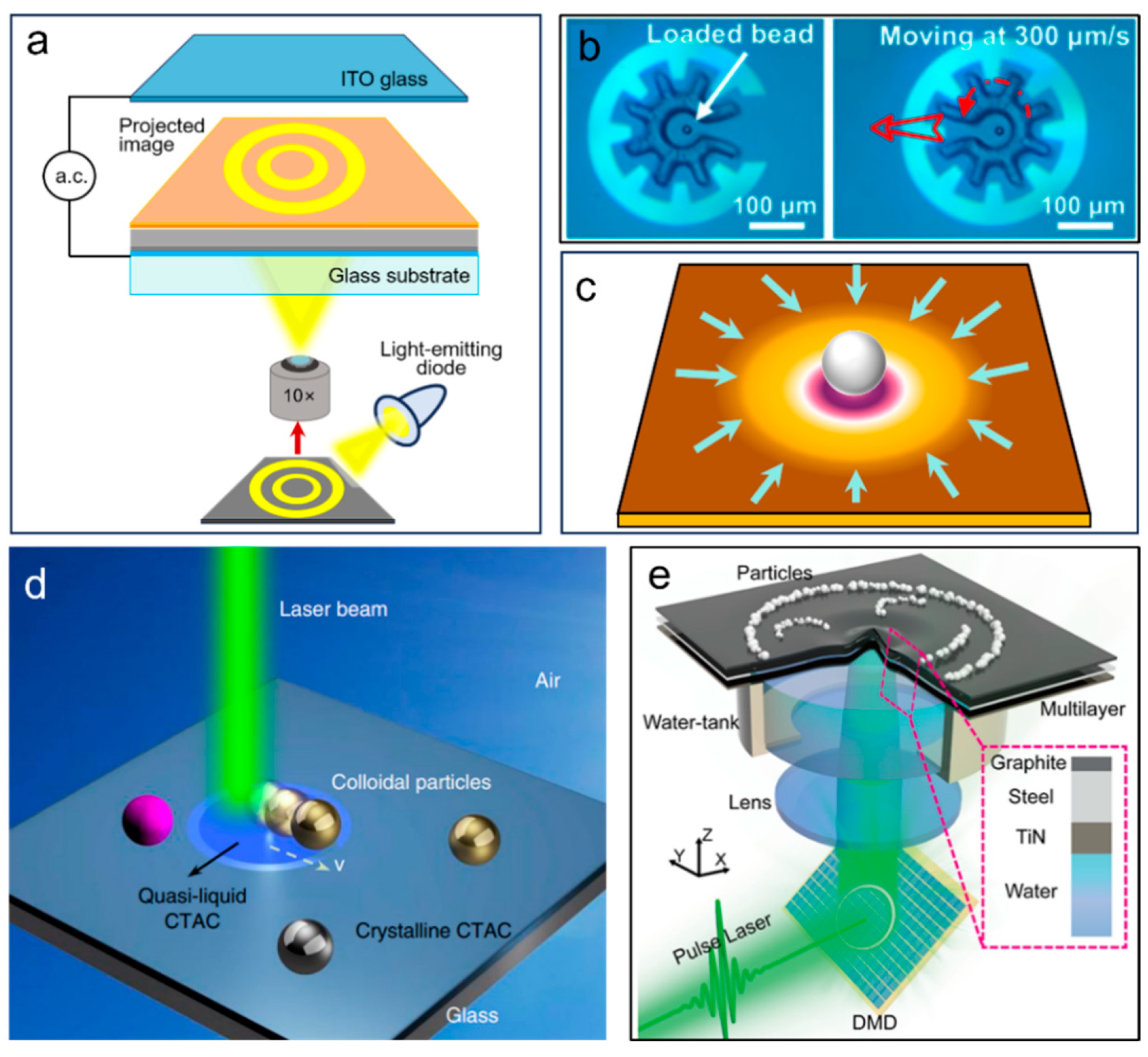
OETs operate on the light-induced dielectrophoretic effect: illuminating a photoconductive material generates a non-uniform electric field that drives particle migration via dielectrophoretic forces [89,90]. OETs offer key advantages-low optical power, high throughput and programmability and have become a major branch of optical manipulation since their introduction by Chiou P. Y. et al. in 2005, with broad applications in single-cell analysis, microfluidics, biomedical diagnostics and micro/nanofabrication (Figure 5a) [91]. Recent advances have extended OETs from fundamental manipulation towards high-precision, automated operation in complex environments. Zhang S. et al., for instance, have demonstrated a microrobot-mediated indirect manipulation mode, in which mobile microrobots are actuated to flexibly maneuver secondary targets such as cells and microspheres (Figure 5b) [92].
Opto-thermophoretic tweezers harness localized temperature gradients generated by photothermal materials under illumination to induce thermoelectric fields, enabling precise trapping and transport of metallic nanoparticles in solution at exceptionally low laser powers [93,94]. Lin L. et al. demonstrated this approach by using laser-heated porous gold films to create localized thermal gradients; the resulting thermoelectric fields allowed single-particle trapping and dynamic transport along arbitrary trajectories for gold, silver and other metal nanoparticles (Figure 5c) [95]. Li J. et al. further extended the operational environment from liquids to solid substrates through a photothermal-gated photon-pushing technique (Figure 5d) [96]. In this scheme, laser heating induces a solid–liquid phase transition in a pre-coated surfactant layer, instantaneously eliminating frictional resistance between particles and the substrate; the optical scattering force then drives nanoparticles along predefined paths on the solid surface.
Photoacoustic tweezers, operating on the photoacoustic effect, employ programmable pulsed lasers to excite localized Lamb waves in multilayered films, inducing microscale particles to jump and assemble into arbitrary patterns-thereby achieving high-throughput, reconfigurable and liquid-free manipulation [97,98]. As shown in Figure 5e, Zhang R. et al. demonstrated the versatility of this technique across multiple scales: complex patterning and frame-by-frame animation reconstruction over a large area with high resolution (~270 μm) within seconds; precise positioning of small particle ensembles with an accuracy of ~120 μm; and extension to three-dimensional microprinting, delivering a driving force of up to 80 μN and accommodating particles of diverse materials [99].
Figure 5.
Multi-physics-assisted indirect optical tweezers. (a) Typical setup of OETs. (b) Optoelectronic microrobot constructed by OETs,the red arrow indicates the translation direction of the bead-loaded microrobot. Reproduced from ref. [92] with permission from the National Academy of Sciences, copyright 2019. (c) Opto-thermoelectric nanotweezers. (d) Optical nanomanipulation on solid substrates. Reproduced from ref. [96] with permission from Springer Nature, copyright 2019. (e) Photoacoustic tweezers. Reproduced from ref. [99] with permission from Springer Nature, copyright 2024.
Magnetically assisted optical tweezers (opto-magnetic tweezers) represent a typical multi-physics-assisted indirect optical manipulation strategy that combines optical gradient confinement with magnetic field-driven actuation [100]. In this scheme, a focused laser beam immobilizes a magnetic microbead in space, while an external magnetic field supplies adjustable tensile forces and torsional torques, enabling synchronous mechanical characterization of chiral biomolecules such as DNA. This hybrid approach offers several distinct advantages: optical trapping ensures stable sample positioning without requiring excessively high laser power, thereby minimizing photothermal damage to biological specimens; meanwhile, magnetic torque generation avoids additional optical absorption by the sample, permitting long-duration single-molecule dynamic observations. Representative implementations of this platform have integrated optical traps, electromagnetic tweezers, and fluorescence readout to enable correlated measurements of molecular stretching, rotation, and structural dynamics. Subsequent upgrades incorporating dual independent optical traps have further suppressed lateral sample drift, achieving high-precision torsional measurements of chromatin fibers that are inaccessible to conventional single optical tweezers.
The diverse optical trapping platforms described above each offer distinct advantages and face specific limitations, making the choice of platform highly dependent on the target application. Table 2 provides a comprehensive comparison of these key platforms across standardized criteria, including trapping efficiency, force range, trappable particle size, required optical power, biological compatibility and representative application scenarios.
Table 2.
Comparison of key optical trapping platforms.
3.5. Hybrid and Multimodal Optical Manipulation Systems
Optical tweezers are evolving from independent mechanical manipulation tools into multimodal platforms integrating manipulation, imaging, and characterization. The core logic of this development lies in the fact that optical tweezers share the same optical path with various detection technologies, thereby achieving a synchronous “manipulation-readout” working mode. At the fluorescence imaging level, Whitley et al. comprehensively reviewed double-trap optical tweezers (“fleezers”) combined with single-molecule fluorescence detection, achieving simultaneous measurement of sub-nanometer displacement and sub-pico-Newton force resolution [101,102]. The technique combining optical slicing with optical manipulation is the light sheet optical tweezers (LOT) platform, which can be used for long-term cell capture and imaging [103]. In 2013, Heller et al. combined a one-dimensional STED nanomicroscope with double-trap optical tweezers, achieving a spatial resolution of 50 nm on protein-densely covered DNA, and were able to track individual DNA-binding proteins in real time at high protein concentrations [104]. At the label-free detection level, combining fiber optic tweezers with Raman spectroscopy and artificial neural networks has enabled real-time capture and label-free molecular fingerprint analysis of individual microorganisms in microfluidic channels [105]. Choi et al. demonstrated tomographic imaging of single suspended live cells using optical tweezers combined with full-field optical coherence tomography [106]. Sarshar et al. developed an integrated system of optical tweezers and quantitative interferometric phase microscopy, achieving piconewton-level force resolution and imaging of nanoscale structures in biological samples [107]. For even higher integration, Shepherd et al. reported the COMBI-Tweeze system [100], which integrates optical tweezers, magnetic tweezers, and fluorescence microscopy on a single platform, enabling time-resolved visualization of the formation and relaxation of higher-order structural motifs (such as plectonemes) in DNA molecules. These hybrid systems share the common characteristic of being able to read structural and chemical information from samples while maintaining the mechanical precision of optical tweezers, representing an important direction in the development of optical tweezers from a “single-mode force tool” to a “multi-mode mechano-optical characterization platform”.
4. Optical Forces in Action: From Single-Molecule Manipulation to Cellular Mechanosensing
4.1. Far-Field Optical Tweezers for Single-Molecule Force Spectroscopy
Single-molecule force spectroscopy represents the most mature biophysical application of optical tweezers. In a far-field optical trap, the gradient force and the scattering force act in concert: the gradient force confines a micron-sized dielectric bead within a three-dimensional potential well, while the scattering force displaces the equilibrium position of the bead slightly downstream from the focal center, establishing a linear relationship between the axial displacement and the restoring force. With sub-nanometer spatial resolution and sub-piconewton force sensitivity [108], optical tweezers enable the investigation of dynamic processes in biological macromolecules, including the elastic properties of DNA and RNA [109,110], protein folding and unfolding [111,112], and the mechanochemical coupling of molecular motors [113].
Direct optical manipulation of individual proteins or nucleic acids is challenging because these nanometer-scale biomolecules scatter light too weakly to be stably trapped at room temperature. The standard experimental strategy overcomes this limitation by tethering the molecule of interest to a micron-sized dielectric bead via chemical linkers, such as biotin-streptavidin, digoxigenin-antibody, or covalent protein-DNA conjugates. The bead serves as a force handle: it is trapped by the optical tweezers, and the force acting on the tethered molecule is read out from the bead displacement relative to the trap center. Several trap configurations have been developed for this purpose, including single-beam optical tweezers [57,114] (Figure 6a), angular optical tweezers [114,115] (Figure 6b), and four-trap optical tweezers [116] (Figure 6c). When the trap or the sample stage is moved, the bead deviates from the trap center, and the restoring force is measured from the deflection of the trapping beam.
Among these configurations, the angular optical trap (AOT) is particularly suited for studying torsional mechanics of DNA and DNA-processing enzymes, as it enables real-time rotation of the trapped particle and direct measurement of the torque exerted on the tethered molecule. A critical requirement for the AOT is that the trapping particle must exhibit optical anisotropy to generate a restoring torque when its orientation deviates from the polarization axis of the trapping beam. Conventionally, this has been achieved using birefringent quartz cylinders, whose extraordinary optical axis is perpendicular to the cylinder axis [117]. However, quartz cylinders suffer from limited maximum rotation rates due to the coupling between viscous drag and angular stiffness. Recent advances have introduced an alternative design based on shape anisotropy: metamaterial elliptical cylinders made of alternating SiO2 and Si3N4 layers generate optical torque via alignment of their major axis with the polarization direction, while offering tunable refractive index and reduced size [118]. These novel particles permit faster rotation without slippage and improve torque resolution, thereby expanding the scope of AOT-based single-molecule studies of DNA topological processes.
DNA has served as the benchmark system for optical force spectroscopy since the foundational work of Smith S. B. et al., who established the force-extension curve of λ-DNA and revealed a highly cooperative transition at approximately 65 pN, where B-DNA converts to a stable form with a 5.8 Å rise per base pair [119]. Building on this framework, researchers have investigated the mechanical modulation of DNA by associated proteins, including RNA polymerase, ribosomes, and DNA translocases. Recent efforts have shifted from isolated DNA molecules toward more physiologically relevant chromatin systems. Shepherd J. W. et al. developed the COMBI-Tweeze platform, which integrates optical tweezers, magnetic tweezers, and fluorescence microscopy to simultaneously apply force and torque to a single DNA molecule, enabling the first direct visualization of the dynamic formation and relaxation of plectonemic supercoils (Figure 6d) [100]. In 2024, the Sun group employed a correlated optical tweezers-fluorescence platform to study the T7 replisome and demonstrated that the replicative helicase, assisted by single-stranded binding protein gp2.5, facilitates replisome bypass of G-quadruplex structures [120].
Figure 6.
Single-molecule force spectroscopy using optical tweezers. (a) Schematic diagrams of measuring DNA molecules using a single optical tweezer (I), an optical tweezer-pipette (II), and a dual optical tweezers “dumbbell” structure (III), respectively. (b) Schematic diagram of measuring DNA molecules using angular optical tweezers. Reproduced from ref. [114] with permission from Wiley, copyright 2016. (c) Schematic diagram of manipulating two DNA molecules using four optical traps. (d) Schematic diagram of the tethered construct formed in the Combi-Tweez system. Reproduced from ref. [100] with permission from Springer Nature, copyright 2024. (e) A schematic diagram of optical tweezers experiments using DNA origami or dsDNA (illustrated) handles. Reproduced from ref. [121] with permission from The American Association for the Advancement of Science, copyright 2025.
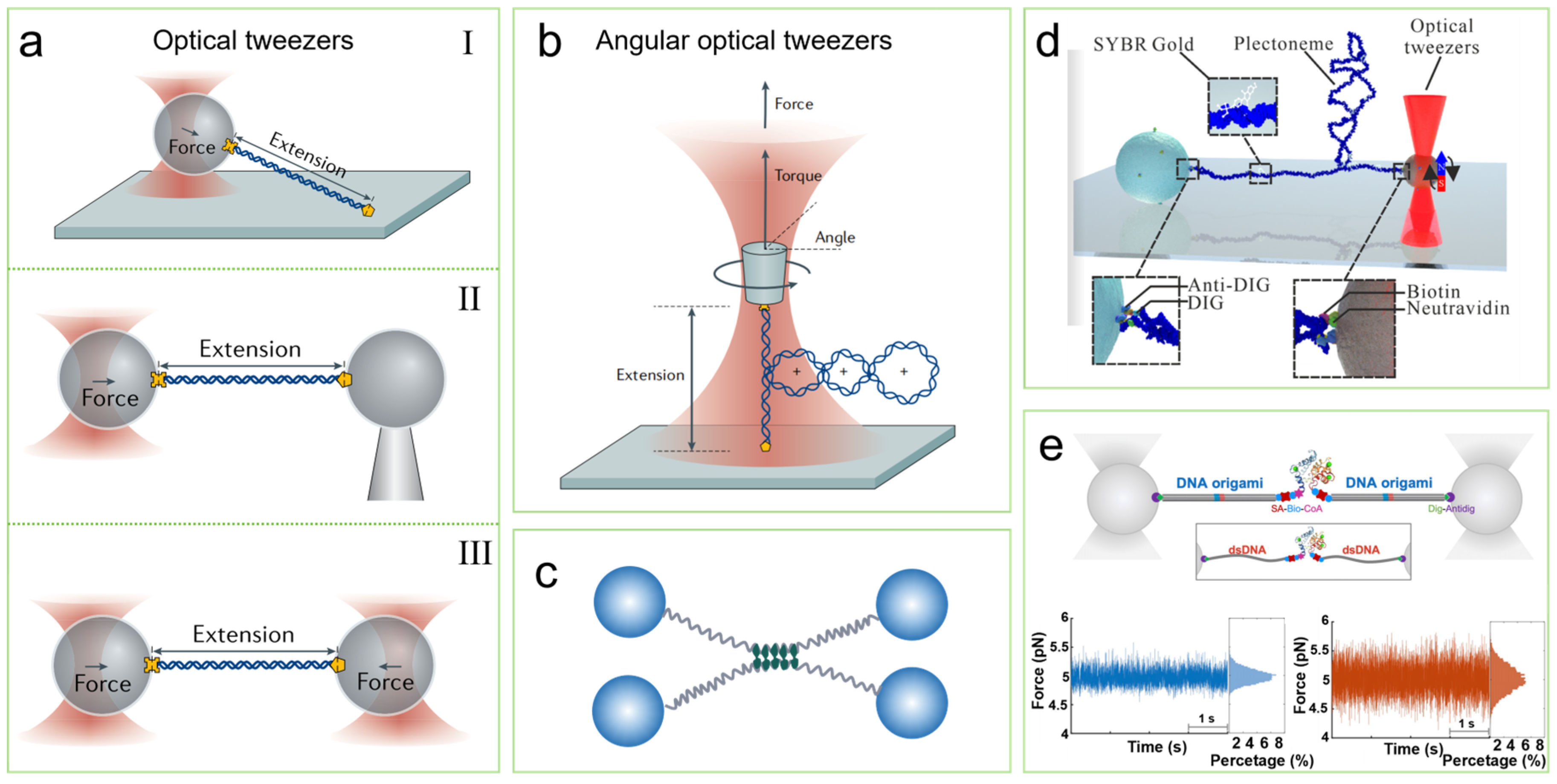
Protein force spectroscopy confronts a more complex free-energy landscape than DNA, with multiple short-lived intermediates and parallel folding pathways, often modulated by regulatory factors. Gao Y. et al. used dual-trap optical tweezers to investigate the folding dynamics of the SNARE complex under the regulation of complexin-1, revealing that complexin-1 orchestrates SNARE assembly through coordinated multi-domain interactions [112]. In 2025, Zhang H. et al. combined DNA origami nanostructures as rigid handles with AlphaFold structural analysis, capturing two hidden intermediates with lifetimes below 5 ms along the folding pathway of calmodulin and revealing a stepwise domain-folding mechanism (Figure 6e) [121]. More recently, Castro F. C. et al. (2026) employed time-shared optical tweezers microrheology to measure the liquid-to-solid phase transition of intracellular protein condensates during aging, demonstrating that mutation of nuclear envelope proteins accelerates the softening of the cytoplasm in C. elegans intestinal cells [122].
Molecular motors are enzymatic machines that convert chemical energy into mechanical work, driving essential cellular processes including DNA replication, transcription, cell division, and intracellular transport. The unique value of optical tweezers in motor protein research lies in their ability to simultaneously measure stepping distance, velocity, and stall force while resolving the chemomechanical coupling mechanism at the single-molecule level. In 1993, Svoboda K. et al. first demonstrated that individual kinesin-1 motors move in discrete 8 nm steps along microtubules, and further established that the force and velocity of a single kinesin molecule depend on the ATP concentration [123,124,125]. In 2025, Patrick E. M. et al. combined high-resolution optical tweezers with single-molecule fluorescence to resolve the assembly and unwinding dynamics of the DEAD-box helicase Ded1p on RNA duplexes, revealing that single Ded1p protomers initially bind to the single-stranded RNA tail adjacent to the duplex and subsequently recruit additional protomers to unwind the duplex cooperatively in steps of 4–6 base pairs [126]. Beyond classical motors, optical tweezers have recently been applied to chromatin remodeling complexes. Mihardja S. et al. investigated the mechanical stability of single nucleosomes and demonstrated the distinct interaction forces between histones and peripheral versus internal DNA [127]. Hall M. A. et al. further discovered an approximately 5 bp periodicity in the DNA-histone interactions within nucleosomes [128]. Zhang Y. et al. used optical tweezers to monitor the action of the yeast RSC chromatin remodeling complex at the single-nucleosome level in real time, measuring the translocation rate and confirming that RSC is a nucleosome-dependent ATPase translocase [129]. These studies extend optical tweezers-based motor research from classical cytoskeletal and nucleic acid motors into the realm of epigenetic regulation, establishing optical tweezers as a powerful tool for investigating the mechanisms of ATP-dependent chromatin remodeling.
4.2. Near-Field Optical Tweezers for Direct Single-Molecule Trapping
A fundamental constraint of the far-field force spectroscopy experiments described in Section 4.1 is that the molecule of interest cannot be directly trapped. For particles much smaller than the trapping wavelength, the gradient force decays steeply with particle volume (Fg ∝ a3), becoming insufficient to overcome Brownian motion at room temperature. Although increasing the laser power can deepen the trapping potential, the accompanying photothermal damage is prohibitive for biological specimens. Near-field optics circumvents this limitation by confining light to subwavelength scales through evanescent fields. The evanescent field decays exponentially from the interface with a characteristic decay length on the order of hundreds of nanometers, generating an intensity gradient far steeper than that achievable with a focused far-field beam. This physical property enables the stable trapping of individual nanoparticles and single biomolecules without the tethered bead that is indispensable in far-field force spectroscopy.
Plasmonic tweezers exploit this mechanism through localized surface plasmon resonance (LSPR) in metallic nanostructures [130], where the collective oscillation of free electrons confines the optical field into a sub-diffraction-limited hot spot with a field enhancement factor of 102–103 [46]. Novotny L. et al. first proposed this concept theoretically in 1997, predicting that a gold tip with a radius of approximately 5 nm under 810 nm illumination could achieve a field enhancement of roughly 3000, sufficient to stably trap particles on the order of 10 nm [52]. An early experimental demonstration was provided by Grigorenko A. N. et al. in 2008 [131], who fabricated an array of closely spaced gold nanopillar pairs on a glass substrate. The electromagnetic coupling between the nanopillars in each pair produced a new resonance mode with an intensity peak in the nanoscale gap that was significantly higher than the field around an isolated nanopillar and well below the diffraction limit in volume. The resulting average trapping force was approximately ten times that of a conventional optical trap under equivalent conditions, enabling the efficient capture of particles much smaller than the incident wavelength. Subsequently, in 2009, Juan M. L. et al. proposed the self-induced back-action (SIBA) plasmonic optical tweezer technique [132,133]. By utilizing a double-nanopore structure to generate transmission resonance, this technique enables the trapped object to play an active role in enhancing the restoring force. Researchers successfully trapped polystyrene microspheres with diameters of 100 nm and 50 nm using incident powers of only 0.7 mW and 1.9 mW. Compared to conventional optical tweezers, SIBA not only significantly reduces the minimum incident light intensity but also lowers the local field intensity within the trap by an order of magnitude. Gordon R. et al. exploited this platform to trap individual bovine serum albumin molecules with a hydrodynamic radius of only 3.4 nm; the stepwise changes in the transmitted optical signal allowed real-time discrimination between folded and unfolded conformational states (Figure 7a) [134]. The same approach was extended to single streptavidin molecules, resolving conformational fluctuations with and without ligand binding through autocorrelation analysis. The ultimate size limit of plasmonic trapping was demonstrated in 2020 by the groups of Tian and Hong, who developed a mechanically controllable break junction platform integrated with resonant laser excitation (Figure 7b). The precisely controlled metallic nanogap generated a field enhancement sufficient to capture and release individual oligo (phenylene ethynylene) molecules of approximately 2 nm in solution at room temperature (Figure 7c) [135]. The pronounced dependence of the capture efficiency on laser wavelength and polarization confirmed that the trapping mechanism relied on plasmonic resonance rather than thermal effects (Figure 7d,e).
Figure 7.
Manipulation of a single protein molecule using plasmonic optical tweezers. (a) Diagram of the experimental setup and results for manipulating BSA protein molecules using dual-nanopore plasmonic optical tweezers. Reproduced from ref. [134]. With permission from American Chemical Society, copyright 2012. (b) Diagram of the experimental setup for manipulating oligo (phenylene ethynylene) molecules using gold-nanotip plasmonic optical tweezers. (c) Spatial distribution of the logarithm of the electric field enhancement. (d) Plots of the optical trapping potential for light with different wavelengths and polarization states. (e) Wavelength-controlled single-molecule trapping [135]. Reproduced from ref. [135] with permission from Elsevier, copyright 2020.
A practical limitation of plasmonic tweezers is the photothermal heating inherent to metallic nanostructures, where ohmic losses can elevate the local temperature by tens of Kelvin. Photonic crystal cavity tweezers offer an alternative near-field trapping strategy that circumvents this issue through purely dielectric resonance. By confining light via total internal reflection and photonic bandgap effects, photonic crystal cavities generate intense evanescent fields without the ohmic heating associated with metals. Mandal S. et al. employed one-dimensional silicon photonic crystal cavities to trap and transport 48 nm dielectric nanoparticles along a waveguide [17]. Unlike plasmonic structures, photonic crystal cavities also provide simultaneous optical sensing capability: the resonance wavelength shifts measurably upon particle binding, enabling label-free detection of molecular interactions in real time.
While the approaches described above seek to suppress photothermal effects, an alternative strategy exploits these effects as an active driving mechanism. The photothermal effect, arising from photon-to-phonon conversion in illuminated nanostructures, has traditionally been regarded as detrimental to stable optical manipulation. Zheng Y. et al. demonstrated that optical heating of a thermoplasmonic substrate can induce spatial separation of dissolved ions within the laser spot (Figure 8a), generating a directed thermoelectric field that enables low-power manipulation of metal nanoparticles across a wide range of materials, sizes, and shapes, with optical intensities approximately three orders of magnitude lower than those required by conventional optical tweezers [95,136]. However, this approach still confines the target object within the high-intensity region of the laser beam, exposing optically sensitive specimens to potential photodamage. Addressing this limitation, Ndukaife C. J. and co-workers recently developed opto-thermo-electrohydrodynamic tweezers (OTET) [137], a multi-physics coupling platform that simultaneously achieves trapping and dynamic manipulation of nanoparticles while positioning them several micrometers away from the region of highest optical intensity (Figure 8b,c). At this displaced location, both the optical intensity and the photothermal effects experienced by the target become nearly negligible. This technique has demonstrated the rapid capture of biomolecules smaller than 10 nm at femtomolar concentrations (Figure 8d), offering a fundamentally new route to low-damage, high-precision manipulation of single biomolecules [138].
Figure 8.
Multiphysics coupling technology for particle manipulation. (a) Schematic illustration of opto-thermoelectric trapping of colloidal particles in a hydrogel solution and immobilization of the trapped particles by UV crosslinking. Reproduced from ref. [136] with permission from the American Chemical Society, copyright 2018. (b) Schematic illustration of the forces and fluid flows in the opto-thermo-electrohydrodynamic trapping system. (c) Electric field profile when a laser is focused at the center of the nanohole array. (d) Experimental images of capturing a single BSA molecule using opto-thermo-electro-hydrodynamic tweezers; the red dot indicates the laser position, while the blue rectangle highlights the molecule (appearing as a tiny, bright spot). Reproduced from ref. [138] with permission from CLP and SPIE, copyright 2023.
While near-field trapping pushes the size limit of optical manipulation from micron-scale beads to single molecules, its capture region remains confined to the subwavelength vicinity of the nanostructured interface. For applications requiring high-throughput parallel processing, multi-physics synergistic platforms complement near-field tweezers by incorporating auxiliary forces from thermal, electric, or hydrodynamic effects. The spatial decay of these secondary fields is governed by thermal diffusion lengths or electrode spacings rather than the exponential decay of the evanescent field, extending the manipulation range from nanometers to tens of micrometers while maintaining low optical power. Optothermal tweezers exploit laser-induced temperature gradients on plasmonic substrates to generate thermoelectric fields that drive charged particles toward hot spots via thermophoresis, achieving capture radii of tens of micrometers with microwatt-level power.
4.3. Optical Force-Mediated Probing of Cellular Mechanosensing
The single-molecule force spectroscopy discussed in Section 4.1 establishes optical tweezers as a precision measurement tool. Yet their capability extends beyond passive measurement: the piconewton force resolution and nanometer spatial precision that enable quantitative force spectroscopy also make optical tweezers an exquisitely controllable mechanical stimulator [139]. Unlike substrate stretching, fluid shear, or atomic force microscopy indentation [140,141,142], optical tweezers deliver localized forces to specific subcellular targets with calibrated magnitude and precisely defined temporal patterns. This transition from measuring cellular forces to using optical forces as a designed input to probe local mechanotransduction defines the theme of this section.
Cells continuously sense mechanical cues from their microenvironment and convert them into biochemical signals, a process termed mechanotransduction [143]. The Patapoutian group identified Piezo1 and Piezo2 as the essential pore-forming subunits of vertebrate mechanosensitive cation channels through RNA interference, patch-clamp recording [144], and expression profiling, and further delineated their distinct roles in touch and pain sensation [144,145]. Piezo1, in particular, has become a central focus of mechanotransduction research owing to its rapid activation and inactivation on the millisecond timescale and its high sensitivity to piconewton-level mechanical forces [146]. Mechanical stimulation of these channels induces calcium influx across the plasma membrane, thereby transducing a physical signal into a biochemical signal [147].
Optical tweezers are uniquely suited to deliver such mechanical stimuli. The force range accessible to optical tweezers, which ranges from sub-piconewton to hundreds of piconewtons, coincides precisely with the activation thresholds of mechanosensitive ion channels and the force scales of receptor-ligand interactions. It is worth noting that optically generated mechanical stimuli can also be delivered through mechanisms distinct from photon momentum transfer [148]. Photoacoustic neuromodulation utilizes pulsed light to irradiate a light absorber, generating a localized ultrasonic field through photothermal conversion to stimulate neurons [149,150]. Researchers have combined photoacoustic materials (such as graphite, epoxy resin, carbon nanotubes, and PDMS composites) with optical fiber carriers, using photoacoustic-coated optical fibers to achieve neural stimulation (Figure 9a–d) [149]. However, photoacoustic stimulation does not permit the simultaneous, calibrated force measurement that is inherent to optical tweezers, nor can it achieve the molecular-scale force resolution required for single-receptor activation. Falleroni F. et al. used oscillating optical tweezers to maneuver a microsphere against the plasma membrane and demonstrated that a single local 13 pN compression triggered an intracellular calcium transient, whereas repetitive compression induced a more sustained response accompanied by mechano-induced activation of CAMKII and the small GTPase RhoA (Figure 9e) [151]. However, conventional Gaussian optical traps require continuous-wave laser irradiation at relatively high power to maintain stable trapping [57], leading to photothermal accumulation that poses a non-negligible physiological perturbation. Moreover, the single-point trapping configuration limits the capacity for parallel, programmable control of multiple particles along complex three-dimensional trajectories, hindering the efficient delivery of multiplexed, time-sequenced mechanical stimulation [31].
Beyond acute calcium signaling, optical tweezers have been applied to modulate neuronal morphology and connectivity over extended timescales. The growth cone at the tip of a developing axon navigates through complex chemical and mechanical cues to form precise neural circuits. Wu et al. demonstrated that optical tweezers can directly guide the growth direction of axons by applying piconewton-scale forces to growth cone filopodia, redirecting neuronal extension in vitro without contact or chemical manipulation [152,153]. In their implementation, an optically trapped birefringent bead positioned near the growth cone was rotated by controlling the polarization of the trapping beam, and the resulting hydrodynamic flow was harnessed to steer axonal outgrowth. This approach achieved directional control of neuronal growth over distances of tens of micrometers, offering a mechanical guidance strategy that complements biochemical patterning. Although this method does not directly activate mechanosensitive channels via membrane deformation, it demonstrates that optical tweezers can exert sustained mechanical influence on neuronal architecture. This capability is distinct from the transient calcium transients discussed above, yet equally relevant to understanding how physical forces shape cellular behavior.
Figure 9.
Mechanical regulation of cellular mechanical signaling. (a–c) Fluorescence images of neurons before and after stimulation using a photoacoustic-coated optical fiber. (d) Calcium signal trace of a target neuron subjected to single-pulse stimulation. Reproduced from ref. [149] with permission from Springer Nature, copyright 2021. (e) Schematic diagram of an oscillating optical tweezer manipulating a microsphere to locally compress the cell membrane. The inset shows that applying high-intensity force to the cell membrane elicits a rapidly activating inward current (black trace); the red trace indicates the force applied to the cell membrane via the bead. The figure on the right shows the mechanotransduction pathway. Reproduced from ref. [151]. With permission from Elsevier, copyright 2022. (f) Schematic illustration of precision neural stimulation by optomechanical bio-dart. The mechanical stimulation on the cell membrane results in the activation of the Piezo1 channel, with an increase in Ca2+ influx and inward current. The bright-field (left) and fluorescent (right) images of a neural cell with Piezo1 channels identified in green fluorescence, and the nuclei in blue. Reproduced from ref. [154] with permission from Springer Nature, copyright 2024.
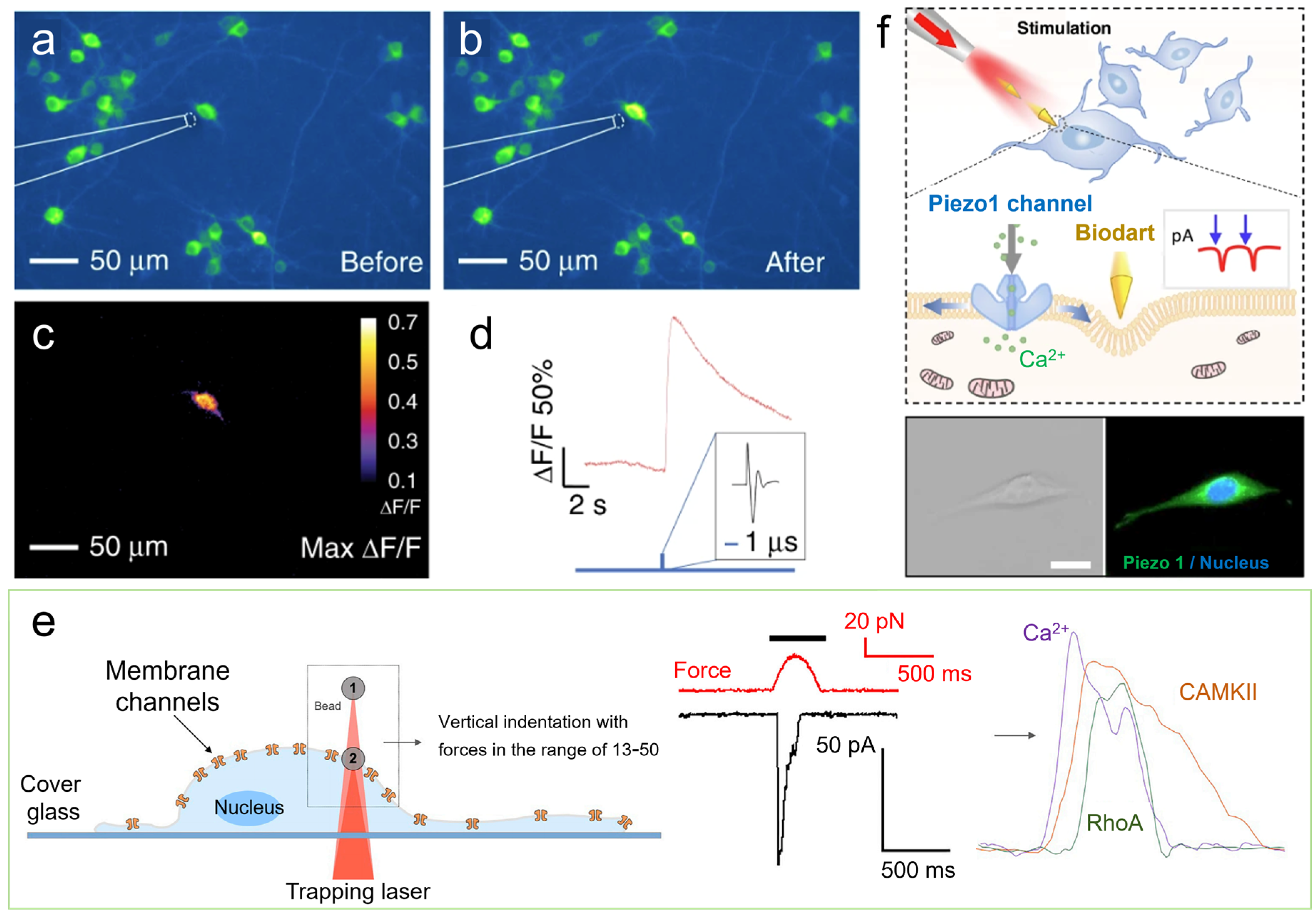
Two recent developments address these limitations. Xin H. et al. proposed the concept of “optically controlled biological darts” in which optical scattering forces transmitted through a fiber probe propel a microparticle toward the cell membrane (Figure 9f), generating a transient mechanical pressure with submicron spatial precision and enabling the selective activation of mechanosensitive ion channels for precise neural stimulation [154]. This approach enhances the spatial localization of the mechanical stimulus to the subcellular scale. In a complementary strategy, transparent resin microspheres serving as microlenses have been positioned by optical tweezers at specific intracellular targets, where they focus incident light into a subwavelength photonic nanojet to stimulate target neurons with subcellular precision; simultaneously, the microspheres enhance the fluorescence imaging signals of lysosomes and mitochondria, enabling concurrent stimulation and imaging [155]. These advances illustrate a broader trajectory: optical tweezers are evolving from a passive mechanical readout tool into an active modulatory platform capable of probing and locally modulating mechanosensitive signaling at the single-cell level.
5. Challenges and Outlook
The preceding sections have traced optical tweezers from the light scattering mechanisms that generate optical forces, through the platforms that harness them, to their applications in single-molecule force spectroscopy, near-field trapping, and cellular mechanosensing. Several challenges define the current boundaries of the field and point toward future directions.
A fundamental question concerns the lower size limit of optical trapping. Under specific experimental conditions, namely a plasmonic nanogap with strong local field enhancement, a particular molecular polarizability, and room-temperature solution operation, near-field plasmonic tweezers have pushed the directly trappable size to approximately 2 nm. This result, however, should not be regarded as a universal lower size limit for optical trapping, because the minimum trappable size depends on particle polarizability, local field enhancement, temperature, and the required trapping duration [135]. Whether this limit can be further lowered depends not solely on achieving stronger field enhancement, but on the competition between the optical gradient force and thermal fluctuations. Strategies that reduce the effective temperature of the particle, successfully employed in vacuum optical traps in atomic physics, remain difficult to translate to aqueous biological environments. Cryogenic near-field trapping may offer a pathway, though its applicability to dynamic biological processes is constrained by the requirement of vitrified samples.
A second challenge is the intrinsic trade-off between optical force and photodamage, both of which scale with incident intensity. Because the 1064 nm wavelength lies within the near-infrared “optical window”, water and most biomolecules exhibit extremely low absorption in this band, making it the standard wavelength for bio-phototrapping. Notably, water absorbs about two orders of magnitude less at 1064 nm than at 1550 nm. While continuous-wave 1064 nm light trapping at moderate power causes minimal damage to DNA or cells, it does produce localized heating, requiring careful power optimization [153]. Multi-physics platforms have reduced the required optical power by orders of magnitude by shifting force generation to secondary fields. Purely dielectric photonic crystal cavities avoid the ohmic heating associated with metallic nanostructures, but residual optical absorption in the dielectric material and in the surrounding aqueous environment can still produce local heating, and the temperature rise must be assessed for each specific structure and operating power. The development of new materials with high refractive index contrast and negligible absorption at biocompatible wavelengths, combined with real-time nanothermometry, could further expand the safe operating window for long-term manipulation of living specimens.
For the optical regulation of cellular signaling, a distinct challenge arises from the biological variability of the response to a calibrated force input. Identical mechanical stimuli can produce different outcomes depending on receptor expression, cytoskeletal state, and mechanical history. Addressing this will require closed-loop platforms that integrate optical force delivery with real-time readouts of downstream signaling, enabling the applied force to be adjusted dynamically based on the measured cellular response.
The transition from single-particle experiments to robust, high-throughput platforms remains an engineering challenge. Machine learning has been applied to automate cell classification and trap optimization, and integrated photonics offers routes toward chip-scale beam delivery. The convergence of these technologies with microfluidics may ultimately yield autonomous systems capable of operating outside specialized laboratories.
In closing, the trajectory traced in this Review reflects a field that has matured from physical principle to multifaceted biophotonics technology. The challenges that remain are those of refinement, integration, and translation. Their resolution will determine whether optical tweezers remain a specialized laboratory technique or become a broadly enabling platform for molecular biophysics, mechanobiology, and clinical medicine.
Author Contributions
J.H. conducted the literature survey, wrote the original draft, and prepared the figures and table. T.W. and Y.Z. conceived the framework and scope of the review. J.H. supervised the writing process, provided critical revisions, and finalized the manuscript. All authors contributed to the discussion and revision of the near-field trapping and cellular mechanosensing sections. All authors have read and agreed to the published version of the manuscript.
Funding
National Natural Science Foundation of China (62305132), High-level Talent Research Start-up Project Funding of Henan Academy of Sciences (251820021), The Fundamental Research Fund of Henan Academy of Sciences (20250620006), Science and Technology Key Project of Henan Province (252102310365), Joint Fund of Henan Province Science and Technology R&D Program (245200810104; 252103810383).
Institutional Review Board Statement
Not applicable.
Informed Consent Statement
Not applicable.
Data Availability Statement
No new data were created or analyzed in this study. Data sharing is not applicable to this article.
Acknowledgments
The authors would like to acknowledge the assistance of Zhiqiang Chen, Tingting Min and Jiafen Zhou in the literature retrieval, preliminary screening, as well as the collection, sorting and standardized archiving of research-related image materials.
Conflicts of Interest
The authors declare no conflicts of interest.
References
- Rivera, N.; Kaminer, I. Light–Matter Interactions with Photonic Quasiparticles. Nat. Rev. Phys. 2020, 2, 538–561. [Google Scholar] [CrossRef] [Scilit]
- Saleem, A.; Afzal, I.; Javed, Y.; Jamil, Y. Fundamentals of Light–Matter Interaction, Modern Luminescence from Fundamental Concepts to Materials and Applications; Elsevier: Amsterdam, The Netherlands, 2023; pp. 185–218. [Google Scholar] [CrossRef] [Scilit]
- Tsuboi, Y.; Naka, S.; Yamanishi, D.; Nagai, T.; Yuyama, K.-I.; Shoji, T.; Ohtani, B.; Tamura, M.; Iida, T.; Kameyama, T. Optical Trapping of Nanocrystals at Oil/Water Interfaces: Implications for Photocatalysis. ACS Appl. Nano Mater. 2021, 4, 11743–11752. [Google Scholar] [CrossRef] [Scilit]
- Stevenson, D.J.; Gunn-Moore, F.; Dholakia, K. Light Forces the Pace: Optical Manipulation for Biophotonics. J. Biomed. Opt. 2010, 15, 041503. [Google Scholar] [CrossRef] [Scilit] [PubMed]
- Ashkin, A. Optical Trapping and Manipulation of Neutral Particles Using Lasers. Proc. Natl. Acad. Sci. USA 1997, 94, 4853–4860. [Google Scholar] [CrossRef] [Scilit] [PubMed]
- Ashkin, A.; Dziedzic, J.M.; Bjorkholm, J.E.; Chu, S. Observation of a Single-Beam Gradient Force Optical Trap for Dielectric Particles. Opt. Lett. 1986, 11, 288–290. [Google Scholar] [CrossRef] [Scilit] [PubMed]
- Grier, D.G. A Revolution in Optical Manipulation. Nature 2003, 424, 810–816. [Google Scholar] [CrossRef] [Scilit] [PubMed]
- Fazal, F.M.; Block, S.M. Optical Tweezers Study Life under Tension. Nat. Photonics 2011, 5, 318–321. [Google Scholar] [CrossRef] [Scilit] [PubMed]
- Otte, E.; Denz, C. Optical Trapping Gets Structure: Structured Light for Advanced Optical Manipulation. Appl. Phys. Rev. 2020, 7, 041308. [Google Scholar] [CrossRef] [Scilit]
- Zhu, R.; Avsievich, T.; Popov, A.; Meglinski, I. Optical Tweezers in Studies of Red Blood Cells. Cells 2020, 9, 545. [Google Scholar] [CrossRef] [Scilit] [PubMed]
- Daly, M.; Sergides, M.; Nic Chormaic, S. Optical Trapping and Manipulation of Micrometer and Submicrometer Particles. Laser Photonics Rev. 2015, 9, 309–329. [Google Scholar] [CrossRef] [Scilit]
- Ramadier, G.; Chakraborty, S.; Fonquernie, I.; Dessauvages, T.T.; Boudoux, C.; Brun, Y.V.; Berne, C.; Weiss, L.E. Single-Cell Probing of Nanoscale Bacterial Adhesion in Real-Time Using Optical Tweezers. ACS Nano 2026, 20, 16919–16929. [Google Scholar] [CrossRef] [Scilit] [PubMed]
- Chen, H.-C.; Cheng, C.-J. Holographic Optical Tweezers: Techniques and Biomedical Applications. Appl. Sci. 2022, 12, 10244. [Google Scholar] [CrossRef] [Scilit]
- Li, J.; Du, G. Manipulation and Assembly of ZnO Nanowires with Single Holographic Optical Tweezers System. Appl. Opt. 2014, 53, 351–355. [Google Scholar] [CrossRef] [Scilit] [PubMed]
- Liu, S.; Li, Z.; Weng, Z.; Li, Y.; Shui, L.; Jiao, Z.; Chen, Y.; Luo, A.; Xing, X.; He, S. Miniaturized Optical Fiber Tweezers for Cell Separation by Optical Force. Opt. Lett. 2019, 44, 1868–1871. [Google Scholar] [CrossRef] [Scilit] [PubMed]
- Faudry, E.; Fick, J. Optical Trapping with Nanostructured Optical Fibers and Motility Analysis of Pseudomonas Aeruginosa. Eur. Biophys. J. 2025, 54, 277–287. [Google Scholar] [CrossRef] [Scilit] [PubMed]
- Mandal, S.; Serey, X.; Erickson, D. Nanomanipulation Using Silicon Photonic Crystal Resonators. Nano Lett. 2010, 10, 99–104. [Google Scholar] [CrossRef] [Scilit] [PubMed]
- Gordon, R. Biosensing with Nanoaperture Optical Tweezers. Opt. Laser Technol. 2019, 109, 328–335. [Google Scholar] [CrossRef] [Scilit]
- Li, Y.-C.; Xin, H.-B.; Lei, H.-X.; Liu, L.-L.; Li, Y.-Z.; Zhang, Y.; Li, B.-J. Manipulation and Detection of Single Nanoparticles and Biomolecules by a Photonic Nanojet. Light Sci. Appl. 2016, 5, e16176. [Google Scholar] [CrossRef] [Scilit] [PubMed]
- Li, H.; Gong, Z.; He, T.; Wu, T.; Li, Y.; Zhang, Y. Single-Molecule Manipulation and Detection by WGM-Coupled Photonic Nanojets. Discov. Nano 2025, 20, 73. [Google Scholar] [CrossRef] [Scilit] [PubMed]
- Wang, B.; Che, Z.; Cheng, C.; Tong, C.; Shi, L.; Shen, Y.; Bliokh, K.Y.; Zi, J. Topological Water-Wave Structures Manipulating Particles. Nature 2025, 638, 394–400. [Google Scholar] [CrossRef] [Scilit] [PubMed]
- Rui, G.; Wang, X.; Cui, Y. Manipulation of Metallic Nanoparticle with Evanescent Vortex Bessel Beam. Opt. Express 2015, 23, 25707–25716. [Google Scholar] [CrossRef] [Scilit] [PubMed]
- Zhao, Z.; Zang, W.; Tian, J. Optical Trapping and Manipulation of Mie Particles with Airy Beam. J. Opt. 2016, 18, 025607. [Google Scholar] [CrossRef] [Scilit]
- Yan, Z.; Shah, R.A.; Chado, G.; Gray, S.K.; Pelton, M.; Scherer, N.F. Guiding Spatial Arrangements of Silver Nanoparticles by Optical Binding Interactions in Shaped Light Fields. ACS Nano 2013, 7, 1790–1802. [Google Scholar] [CrossRef] [Scilit] [PubMed]
- Chen, S.; Zhang, C.; Shen, Y.; Pan, J.; Xu, H.; Dou, X.; Wu, X.; Min, C.; Zhang, Y.; Yuan, X. Opto-Thermoelectric Tweezers Based on Reduced Graphene Oxide. Adv. Opt. Mater. 2026, 14, e03576. [Google Scholar] [CrossRef] [Scilit]
- Zhu, R.; Gu, Z.; Shen, T.; Liu, Y.; Shi, Z.; Linghu, S.; Gu, F. In Situ Dynamic Spatial Reconfiguration of Nanoplasmonics Using Photothermal-Shock Tweezers. Laser Photonics Rev. 2025, 19, e02268. [Google Scholar] [CrossRef] [Scilit]
- Ohta, A.T.; Chiou, P.-Y.; Phan, H.L.; Sherwood, S.W.; Yang, J.M.; Lau, A.N.; Hsu, H.-Y.; Jamshidi, A.; Wu, M.C. Optically Controlled Cell Discrimination and Trapping Using Optoelectronic Tweezers. IEEE J. Sel. Top. Quantum Electron. 2007, 13, 235–243. [Google Scholar] [CrossRef] [Scilit]
- Wang, D.; Zhang, T.; Shi, J.; Wang, Y.; Liu, B.; Ding, L.; Chen, C.; Zhang, W.; Zheng, J.; Chen, J. Intracellular Manipulation by Particle Swarm Optimized Optical Tweezers. Laser Photonics Rev. 2026, 20, e02473. [Google Scholar] [CrossRef] [Scilit]
- Bustamante, C.; Alexander, L.; Maciuba, K.; Kaiser, C.M. Single-Molecule Studies of Protein Folding with Optical Tweezers. Annu. Rev. Biochem. 2020, 89, 443–470. [Google Scholar] [CrossRef] [Scilit] [PubMed]
- Shabestari, M.H.; Meijering, A.; Roos, W.; Wuite, G.; Peterman, E. Recent Advances in Biological Single-Molecule Applications of Optical Tweezers and Fluorescence Microscopy. Methods Enzymol. 2017, 582, 85–119. [Google Scholar] [CrossRef] [Scilit] [PubMed]
- Li, Y.-Y.; Li, H.; Zheng, Y.; Xu, D.-D.; Liu, L.; Liu, A.; Li, T.; Pang, D.-W.; Tang, H.-W. Optical Tweezer-Driven Mechanotransduction: Probing Pn-Scale Forces and Calcium-Mediated Redox Signaling in Single Endothelial Cells. ACS Nano 2025, 19, 16084–16095. [Google Scholar] [CrossRef] [Scilit] [PubMed]
- Sergides, M.; Perego, L.; Galgani, T.; Arbore, C.; Pavone, F.; Capitanio, M. Probing Mechanotransduction in Living Cells by Optical Tweezers and FRET-Based Molecular Force Microscopy. Eur. Phys. J. Plus 2021, 136, 316. [Google Scholar] [CrossRef] [Scilit]
- Stilgoe, A.B.; Favre-Bulle, I.A.; Watson, M.L.; Gomez-Godinez, V.; Berns, M.W.; Preece, D.; Rubinsztein-Dunlop, H. Shining Light in Mechanobiology: Optical Tweezers, Scissors, and Beyond. ACS Photonics 2024, 11, 917–940. [Google Scholar] [CrossRef] [Scilit] [PubMed]
- Arbore, C.; Perego, L.; Sergides, M.; Capitanio, M. Probing Force in Living Cells with Optical Tweezers: From Single-Molecule Mechanics to Cell Mechanotransduction. Biophys. Rev. 2019, 11, 765–782. [Google Scholar] [CrossRef] [Scilit] [PubMed]
- Ashkin, A. Trapping of Atoms by Resonance Radiation Pressure. Phys. Rev. Lett. 1978, 40, 729. [Google Scholar] [CrossRef] [Scilit]
- Constable, A.; Kim, J.; Mervis, J.; Zarinetchi, F.; Prentiss, M. Demonstration of a Fiber-Optical Light-Force Trap. Opt. Lett. 1993, 18, 1867–1869. [Google Scholar] [CrossRef] [Scilit] [PubMed]
- Taylor, R.; Hnatovsky, C. Particle Trapping in 3-D Using a Single Fiber Probe with an Annular Light Distribution. Opt. Express 2003, 11, 2775–2782. [Google Scholar] [CrossRef] [Scilit] [PubMed]
- Shen, Z.; Liu, N. Optical Tweezers with Optical Vortex Based on Deep Learning. ACS Photonics 2025, 12, 2212–2218. [Google Scholar] [CrossRef] [Scilit]
- Selin, M.; Ciarlo, A.; Pesce, G.; Bengtsson, L.; Camunas-Soler, J.; Sundar Rajan, V.; Westerlund, F.; Wilhelmsson, L.M.; Pastor, I.; Ritort, F. Smarttrap: Automated Precision Experiments with Optical Tweezers. Nat. Methods 2026, 23, 1368–1378. [Google Scholar] [CrossRef] [Scilit] [PubMed]
- Xin, H.; Li, Y.; Liu, Y.C.; Zhang, Y.; Xiao, Y.F.; Li, B. Optical Forces: From Fundamental to Biological Applications. Adv. Mater. 2020, 32, 2001994. [Google Scholar] [CrossRef] [Scilit] [PubMed]
- Stilgoe, A.B.; Nieminen, T.A.; Knöner, G.; Heckenberg, N.R.; Rubinsztein-Dunlop, H. The Effect of Mie Resonances on Trapping in Optical Tweezers. Opt. Express 2008, 16, 15039–15051. [Google Scholar] [CrossRef] [Scilit] [PubMed]
- Spesyvtseva, S.E.S.; Dholakia, K. Trapping in a Material World. ACS Photonics 2016, 3, 719–736. [Google Scholar] [CrossRef] [Scilit]
- Mazolli, A.; Maia Neto, P.; Nussenzveig, H.M. Theory of Trapping Forces in Optical Tweezers. Proc. R. Soc. London. Ser. A Math. Phys. Eng. Sci. 2003, 459, 3021–3041. [Google Scholar] [CrossRef] [Scilit]
- Nieminen, T.A.; Knöner, G.; Heckenberg, N.R.; Rubinsztein-Dunlop, H. Physics of Optical Tweezers. Methods Cell Biol. 2007, 82, 207–236. [Google Scholar] [CrossRef] [Scilit] [PubMed]
- Lehmuskero, A.; Johansson, P.; Rubinsztein-Dunlop, H.; Tong, L.; Kall, M. Laser Trapping of Colloidal Metal Nanoparticles. ACS Nano 2015, 9, 3453–3469. [Google Scholar] [CrossRef] [Scilit] [PubMed]
- Zhou, L.-M.; Sun, W.; Tao, Z.-Q.; Xiong, N.-J.; Huang, C.; Jiang, X.-Y.; Ren, Y.-X.; Yang, Y.; Shi, Y.-Z.; Hu, J.-G. Subwavelength-Scale Off-Axis Optical Nanomanipulation within Gaussian-Beam Traps. Nanophotonics 2025, 14, 219–228. [Google Scholar] [CrossRef] [Scilit] [PubMed]
- Mishchenko, M.I. Radiation Force Caused by Scattering, Absorption, and Emission of Light by Nonspherical Particles. J. Quant. Spectrosc. Radiat. Transf. 2001, 70, 811–816. [Google Scholar] [CrossRef] [Scilit]
- Saija, R.; Iatı, M.A.; Giusto, A.; Denti, P.; Borghese, F. Transverse Components of the Radiation Force on Nonspherical Particles in the T-Matrix Formalism. J. Quant. Spectrosc. Radiat. Transf. 2005, 94, 163–179. [Google Scholar] [CrossRef] [Scilit]
- Ashkin, A. Forces of a Single-Beam Gradient Laser Trap on a Dielectric Sphere in the Ray Optics Regime. Biophys. J. 1992, 61, 569–582. [Google Scholar] [CrossRef] [Scilit] [PubMed]
- Ren, Y.; Chen, Q.; He, M.; Zhang, X.; Qi, H.; Yan, Y. Plasmonic Optical Tweezers for Particle Manipulation: Principles, Methods, and Applications. ACS Nano 2021, 15, 6105–6128. [Google Scholar] [CrossRef] [Scilit] [PubMed]
- Descharmes, N.; Dharanipathy, U.P.; Diao, Z.; Tonin, M.; Houdré, R. Single Particle Detection, Manipulation and Analysis with Resonant Optical Trapping in Photonic Crystals. Lab A Chip 2013, 13, 3268–3274. [Google Scholar] [CrossRef] [Scilit] [PubMed]
- Novotny, L.; Bian, R.X.; Xie, X.S. Theory of Nanometric Optical Tweezers. Phys. Rev. Lett. 1997, 79, 645. [Google Scholar] [CrossRef] [Scilit]
- Zemánek, P.; Jonáš, A.; Liška, M. Simplified Description of Optical Forces Acting on a Nanoparticle in the Gaussian Standing Wave. J. Opt. Soc. Am. A 2002, 19, 1025–1034. [Google Scholar] [CrossRef] [Scilit] [PubMed]
- Dholakia, K.; Čižmár, T. Shaping the Future of Manipulation. Nat. Photonics 2011, 5, 335–342. [Google Scholar] [CrossRef] [Scilit]
- Czerwinski, F.; Richardson, A.C.; Oddershede, L.B. Quantifying Noise in Optical Tweezers by Allan Variance. Opt. Express 2009, 17, 13255–13269. [Google Scholar] [CrossRef] [Scilit] [PubMed]
- Huisstede, J.; Werf, K.v.d.; Bennink, M.L.; Subramaniam, V. Force Detection in Optical Tweezers Using Backscattered Light. Opt. Express 2005, 13, 1113–1123. [Google Scholar] [CrossRef] [Scilit] [PubMed]
- Bustamante, C.J.; Chemla, Y.R.; Liu, S.; Wang, M.D. Optical Tweezers in Single-Molecule Biophysics. Nat. Rev. Methods Prim. 2021, 1, 25. [Google Scholar] [CrossRef] [Scilit] [PubMed]
- Ashkin, A.; Dziedzic, J.M. Optical Trapping and Manipulation of Viruses and Bacteria. Science 1987, 235, 1517–1520. [Google Scholar] [CrossRef] [Scilit] [PubMed]
- Yang, Y.; Ren, Y.-X.; Chen, M.; Arita, Y.; Rosales-Guzmán, C. Optical Trapping with Structured Light: A Review. Adv. Photonics 2021, 3, 034001. [Google Scholar] [CrossRef] [Scilit]
- MacDonald, M.P.; Spalding, G.C.; Dholakia, K. Microfluidic Sorting in an Optical Lattice. Nature 2003, 426, 421–424. [Google Scholar] [CrossRef] [Scilit] [PubMed]
- Ashok, P.C.; Dholakia, K. Optical Trapping for Analytical Biotechnology. Curr. Opin. Biotechnol. 2012, 23, 16–21. [Google Scholar] [CrossRef] [Scilit] [PubMed]
- Hart, S.J.; Terray, A.V. Refractive-Index-Driven Separation of Colloidal Polymer Particles Using Optical Chromatography. Appl. Phys. Lett. 2003, 83, 5316–5318. [Google Scholar] [CrossRef] [Scilit]
- Yan, Z.; Pelton, M.; Vigderman, L.; Zubarev, E.R.; Scherer, N.F. Why Single-Beam Optical Tweezers Trap Gold Nanowires in Three Dimensions. ACS Nano 2013, 7, 8794–8800. [Google Scholar] [CrossRef] [Scilit] [PubMed]
- Curtis, J.E.; Koss, B.A.; Grier, D.G. Dynamic Holographic Optical Tweezers. Opt. Commun. 2002, 207, 169–175. [Google Scholar] [CrossRef] [Scilit]
- Dufresne, E.R.; Grier, D.G. Optical Tweezer Arrays and Optical Substrates Created with Diffractive Optics. Rev. Sci. Instrum. 1998, 69, 1974–1977. [Google Scholar] [CrossRef] [Scilit]
- Xie, X.; Wang, X.; Min, C.; Ma, H.; Yuan, Y.; Zhou, Z.; Zhang, Y.; Bu, J.; Yuan, X. Single-Particle Trapping and Dynamic Manipulation with Holographic Optical Surface-Wave Tweezers. Photonics Res. 2021, 10, 166–173. [Google Scholar] [CrossRef] [Scilit]
- Liesener, J.; Reicherter, M.; Haist, T.; Tiziani, H.J. Multi-Functional Optical Tweezers Using Computer-Generated Holograms. Opt. Commun. 2000, 185, 77–82. [Google Scholar] [CrossRef] [Scilit]
- Kirkham, G.R.; Britchford, E.; Upton, T.; Ware, J.; Gibson, G.M.; Devaud, Y.; Ehrbar, M.; Padgett, M.; Allen, S.; Buttery, L.D. Precision Assembly of Complex Cellular Microenvironments Using Holographic Optical Tweezers. Sci. Rep. 2015, 5, 8577. [Google Scholar] [CrossRef] [Scilit] [PubMed]
- Nedev, S.; Urban, A.S.; Lutich, A.A.; Feldmann, J. Optical Force Stamping Lithography. Nano Lett. 2011, 11, 5066–5070. [Google Scholar] [CrossRef] [Scilit] [PubMed]
- Landenberger, B.; Yatish; Rohrbach, A. Towards Non-Blind Optical Tweezing by Finding 3d Refractive Index Changes through Off-Focus Interferometric Tracking. Nat. Commun. 2021, 12, 6922. [Google Scholar] [CrossRef] [Scilit] [PubMed]
- Dienerowitz, M.; Mazilu, M.; Reece, P.J.; Krauss, T.F.; Dholakia, K. Optical Vortex Trap for Resonant Confinement of Metal Nanoparticles. Opt. Express 2008, 16, 4991–4999. [Google Scholar] [CrossRef] [Scilit] [PubMed]
- Qu, Z.; Wang, K.; Feng, C.; Wu, T.; Zhang, Y.; Zhao, X.; Wang, J.-L. Optofluidic Sorting of Microparticles Using Airy Beams. Int. J. Optomechatronics 2024, 18, 2375538. [Google Scholar] [CrossRef] [Scilit]
- Marago, O.M.; Jones, P.H.; Gucciardi, P.G.; Volpe, G.; Ferrari, A.C. Optical Trapping and Manipulation of Nanostructures. Nat. Nanotechnol. 2013, 8, 807–819. [Google Scholar] [CrossRef] [Scilit] [PubMed]
- Yuan, Z.; Yan, W.; Ren, Z.-C.; Wang, X.-L.; Ding, J.; Wang, H.-T. Longitudinally Tuned Oam Vortex Beams for Advanced Particle Manipulation. Optica 2026, 13, 651–660. [Google Scholar] [CrossRef] [Scilit]
- Qu, Z.; Zhang, Y.; Wang, K.; Feng, C.; Wu, T.; Zhang, Y.; Xu, X.; Yao, B.; Zhao, X.; Wang, J.-L. High-Precision Refractive Index-Based Microparticle Sorting Using Airy Beams. Anal. Chem. 2025, 97, 20761–20770. [Google Scholar] [CrossRef] [Scilit] [PubMed]
- Holman, A.; Xu, Y.; Sun, X.; Wu, J.; Wang, M.; Zhu, Z.; Seo, B.; Yu, N.; Will, S. Trapping of Single Atoms in Metasurface Optical Tweezer Arrays. Nature 2026, 649, 859–865. [Google Scholar] [CrossRef] [Scilit] [PubMed]
- Tan, L.; Huo, P.; Lin, P.; Ren, Y.; Qi, H.; Fang, L.; Wang, Y.; Ou, J.; Lu, Y.; Xu, T. Longitudinally Engineered Metasurfaces for 3d Vectorial Holography. Light Sci. Appl. 2026, 15, 36. [Google Scholar] [CrossRef] [Scilit] [PubMed]
- Li, X.; Dan, D.; Zavatski, S.; Gao, W.; Zhang, Q.; Zhou, Y.; Qian, J.; Yang, Y.; Yu, X.; Yan, S. Optical Tweeze-Sectioning Microscopy for 3d Imaging and Manipulation of Suspended Cells. Sci. Adv. 2025, 11, eadx3900. [Google Scholar] [CrossRef] [Scilit] [PubMed]
- Hu, Z.; Wang, J.; Liang, J. Manipulation and Arrangement of Biological and Dielectric Particles by a Lensed Fiber Probe. Opt. Express 2004, 12, 4123–4128. [Google Scholar] [CrossRef] [Scilit] [PubMed]
- Lou, Y.; Wu, D.; Pang, Y. Optical Trapping and Manipulation Using Optical Fibers. Adv. Fiber Mater. 2019, 1, 83–100. [Google Scholar] [CrossRef] [Scilit]
- Xin, H.; Xu, R.; Li, B. Optical Trapping, Driving and Arrangement of Particles Using a Tapered Fibre Probe. Sci. Rep. 2012, 2, 818. [Google Scholar] [CrossRef] [Scilit] [PubMed]
- Soltani, M.; Lin, J.; Forties, R.A.; Inman, J.T.; Saraf, S.N.; Fulbright, R.M.; Lipson, M.; Wang, M.D. Nanophotonic Trapping for Precise Manipulation of Biomolecular Arrays. Nat. Nanotechnol. 2014, 9, 448–452. [Google Scholar] [CrossRef] [Scilit] [PubMed]
- Xin, H.; Li, Y.; Xu, D.; Zhang, Y.; Chen, C.H.; Li, B. Single Upconversion Nanoparticle–Bacterium Cotrapping for Single-Bacterium Labeling and Analysis. Small 2017, 13, 1603418. [Google Scholar] [CrossRef] [Scilit] [PubMed]
- Li, Y.; Xin, H.; Zhang, Y.; Lei, H.; Zhang, T.; Ye, H.; Saenz, J.J.; Qiu, C.-W.; Li, B. Living Nanospear for near-Field Optical Probing. ACS Nano 2018, 12, 10703–10711. [Google Scholar] [CrossRef] [Scilit] [PubMed]
- Li, Y.; Liu, X.; Li, B. Single-Cell Biomagnifier for Optical Nanoscopes and Nanotweezers. Light Sci. Appl. 2019, 8, 61. [Google Scholar] [CrossRef] [Scilit] [PubMed]
- Li, Y.; Liu, X.; Xu, X.; Xin, H.; Zhang, Y.; Li, B. Red-Blood-Cell Waveguide as a Living Biosensor and Micromotor. Adv. Funct. Mater. 2019, 29, 1905568. [Google Scholar] [CrossRef] [Scilit]
- Ye, F.; Inman, J.T.; Hong, Y.; Hall, P.M.; Wang, M.D. Resonator Nanophotonic Standing-Wave Array Trap for Single-Molecule Manipulation and Measurement. Nat. Commun. 2022, 13, 77. [Google Scholar] [CrossRef] [Scilit] [PubMed]
- Sneh, T.; Corsetti, S.; Notaros, M.; Kikkeri, K.; Voldman, J.; Notaros, J. Optical Tweezing of Microparticles and Cells Using Silicon-Photonics-Based Optical Phased Arrays. Nat. Commun. 2024, 15, 8493. [Google Scholar] [CrossRef] [Scilit] [PubMed]
- Wu, M.C. Optoelectronic Tweezers. Nat. Photonics 2011, 5, 322–324. [Google Scholar] [CrossRef] [Scilit]
- Zhang, S.; Xu, B.; Elsayed, M.; Nan, F.; Liang, W.; Valley, J.K.; Liu, L.; Huang, Q.; Wu, M.C.; Wheeler, A.R. Optoelectronic Tweezers: A Versatile Toolbox for Nano-/Micro-Manipulation. Chem. Soc. Rev. 2022, 51, 9203–9242. [Google Scholar] [CrossRef] [Scilit] [PubMed]
- Chiou, P.Y.; Ohta, A.T.; Wu, M.C. Massively Parallel Manipulation of Single Cells and Microparticles Using Optical Images. Nature 2005, 436, 370–372. [Google Scholar] [CrossRef] [Scilit] [PubMed]
- Zhang, S.; Scott, E.Y.; Singh, J.; Chen, Y.; Zhang, Y.; Elsayed, M.; Chamberlain, M.D.; Shakiba, N.; Adams, K.; Yu, S. The Optoelectronic Microrobot: A Versatile Toolbox for Micromanipulation. Proc. Natl. Acad. Sci. USA 2019, 116, 14823–14828. [Google Scholar] [CrossRef] [Scilit] [PubMed]
- Lin, L.; Peng, X.; Wei, X.; Mao, Z.; Xie, C.; Zheng, Y. Thermophoretic Tweezers for Low-Power and Versatile Manipulation of Biological Cells. ACS Nano 2017, 11, 3147–3154. [Google Scholar] [CrossRef] [Scilit] [PubMed]
- Liu, S.; Lin, L.; Sun, H.-B. Opto-Thermophoretic Manipulation. ACS Nano 2021, 15, 5925–5943. [Google Scholar] [CrossRef] [Scilit] [PubMed]
- Lin, L.; Wang, M.; Peng, X.; Lissek, E.N.; Mao, Z.; Scarabelli, L.; Adkins, E.; Coskun, S.; Unalan, H.E.; Korgel, B.A. Opto-Thermoelectric Nanotweezers. Nat. Photonics 2018, 12, 195–201. [Google Scholar] [CrossRef] [Scilit] [PubMed]
- Li, J.; Liu, Y.; Lin, L.; Wang, M.; Jiang, T.; Guo, J.; Ding, H.; Kollipara, P.S.; Inoue, Y.; Fan, D. Optical Nanomanipulation on Solid Substrates Via Optothermally-Gated Photon Nudging. Nat. Commun. 2019, 10, 5672. [Google Scholar] [CrossRef] [Scilit] [PubMed]
- Zharov, V.; Malinsky, T.; Kurten, R. Photoacoustic Tweezers with a Pulsed Laser: Theory and Experiments. J. Phys. D. Appl. Phys. 2005, 38, 2662–2674. [Google Scholar] [CrossRef] [Scilit]
- Zhao, Z.; Xia, J.; Huang, T.J.; Zou, J. Ring-Shaped Photoacoustic Tweezers for Single Particle Manipulation. Opt. Lett. 2022, 47, 826–829. [Google Scholar] [CrossRef] [Scilit] [PubMed]
- Zhang, R.; Zhao, X.; Li, J.; Zhou, D.; Guo, H.; Li, Z.-Y.; Li, F. Programmable Photoacoustic Patterning of Microparticles in Air. Nat. Commun. 2024, 15, 3250. [Google Scholar] [CrossRef] [Scilit] [PubMed]
- Shepherd, J.W.; Guilbaud, S.; Zhou, Z.; Howard, J.A.; Burman, M.; Schaefer, C.; Kerrigan, A.; Steele-King, C.; Noy, A.; Leake, M.C. Correlating Fluorescence Microscopy, Optical and Magnetic Tweezers to Study Single Chiral Biopolymers Such as DNA. Nat. Commun. 2024, 15, 2748. [Google Scholar] [CrossRef] [Scilit] [PubMed]
- Whitley, K.D.; Comstock, M.J.; Chemla, Y.R. High-Resolution “Fleezers”: Dual-Trap Optical Tweezers Combined with Single-Molecule Fluorescence Detection, Optical Tweezers: Methods and Protocols; Springer: Berlin/Heidelberg, Germany, 2016; pp. 183–256. [Google Scholar] [CrossRef] [Scilit] [PubMed]
- Yadav, R.; Senanayake, K.B.; Comstock, M.J. High-Resolution Optical Tweezers Combined with Multicolor Single-Molecule Microscopy, Optical Tweezers: Methods and Protocols; Springer: Berlin/Heidelberg, Germany, 2022; pp. 141–240. [Google Scholar] [CrossRef] [Scilit] [PubMed]
- Mondal, P.P.; Baro, N.; Singh, A.; Joshi, P.; Basumatary, J. Lightsheet Optical Tweezer (Lot) for Optical Manipulation of Microscopic Particles and Live Cells. Sci. Rep. 2022, 12, 10229. [Google Scholar] [CrossRef] [Scilit] [PubMed]
- Heller, I.; Sitters, G.; Broekmans, O.D.; Farge, G.; Menges, C.; Wende, W.; Hell, S.W.; Peterman, E.J.; Wuite, G.J. Sted Nanoscopy Combined with Optical Tweezers Reveals Protein Dynamics on Densely Covered DNA. Nat. Methods 2013, 10, 910–916. [Google Scholar] [CrossRef] [Scilit] [PubMed]
- Lin, C.; Li, X.; Wu, T.; Xu, J.; Gong, Z.; Chen, T.; Li, B.; Li, Y.; Guo, J.; Zhang, Y. Optofluidic Identification of Single Microorganisms Using Fiber-Optical-Tweezer-Based Raman Spectroscopy with Artificial Neural Network. BMEMat 2023, 1, e12007. [Google Scholar] [CrossRef] [Scilit]
- Choi, W.J.; Park, K.S.; Eom, T.J.; Oh, M.-K.; Lee, B.H. Tomographic Imaging of a Suspending Single Live Cell Using Optical Tweezer-Combined Full-Field Optical Coherence Tomography. Opt. Lett. 2012, 37, 2784–2786. [Google Scholar] [CrossRef] [Scilit] [PubMed]
- Utadiya, S.; Joshi, S.; Patel, N.; Patel, C.; Joglekar, M.; Cahhniwal, V.; O’Connor, T.; Javidi, B.; Anand, A. Integrated Self-Referencing Single Shot Digital Holographic Microscope and Optical Tweezer. Light Adv. Manuf. 2022, 3, 453–465. [Google Scholar] [CrossRef] [Scilit]
- Zhang, X.; Ma, L.; Zhang, Y. High-Resolution Optical Tweezers for Single-Molecule Manipulation. Yale J. Biol. Med. 2013, 86, 367. [Google Scholar] [PubMed]
- Liphardt, J.; Onoa, B.; Smith, S.B.; Tinoco, I., Jr.; Bustamante, C. Reversible Unfolding of Single RNA Molecules by Mechanical Force. Science 2001, 292, 733–737. [Google Scholar] [CrossRef] [Scilit] [PubMed]
- Bustamante, C.; Bryant, Z.; Smith, S.B. Ten Years of Tension: Single-Molecule DNA Mechanics. Nature 2003, 421, 423–427. [Google Scholar] [CrossRef] [Scilit] [PubMed]
- Stigler, J.; Ziegler, F.; Gieseke, A.; Gebhardt, J.C.M.; Rief, M. The Complex Folding Network of Single Calmodulin Molecules. Science 2011, 334, 512–516. [Google Scholar] [CrossRef] [Scilit] [PubMed]
- Gao, Y.; Zorman, S.; Gundersen, G.; Xi, Z.; Ma, L.; Sirinakis, G.; Rothman, J.E.; Zhang, Y. Single Reconstituted Neuronal Snare Complexes Zipper in Three Distinct Stages. Science 2012, 337, 1340–1343. [Google Scholar] [CrossRef] [Scilit] [PubMed]
- Wang, M.D.; Schnitzer, M.J.; Yin, H.; Landick, R.; Gelles, J.; Block, S.M. Force and Velocity Measured for Single Molecules of RNA Polymerase. Science 1998, 282, 902–907. [Google Scholar] [CrossRef] [Scilit] [PubMed]
- Chemla, Y.R. High-Resolution, Hybrid Optical Trapping Methods, and Their Application to Nucleic Acid Processing Proteins. Biopolymers 2016, 105, 704–714. [Google Scholar] [CrossRef] [Scilit] [PubMed]
- Deufel, C.; Forth, S.; Simmons, C.R.; Dejgosha, S.; Wang, M.D. Nanofabricated Quartz Cylinders for Angular Trapping: DNA Supercoiling Torque Detection. Nat. Methods 2007, 4, 223–225. [Google Scholar] [CrossRef] [Scilit] [PubMed]
- Heller, I.; Laurens, N.; Vorselen, D.; Broekmans, O.D.; Biebricher, A.S.; King, G.A.; Brouwer, I.; Wuite, G.J.; Peterman, E.J. Versatile Quadruple-Trap Optical Tweezers for Dual DNA Experiments, Optical Tweezers: Methods and Protocols; Springer: Berlin/Heidelberg, Germany, 2016; pp. 257–272. [Google Scholar] [CrossRef] [Scilit] [PubMed]
- Hong, Y.; Ye, F.; Qian, J.; Gao, X.; Inman, J.T.; Wang, M.D. Optical Torque Calculations and Measurements for DNA Torsional Studies. Biophys. J. 2024, 123, 3080–3089. [Google Scholar] [CrossRef] [Scilit] [PubMed]
- Hong, Y.; Ye, F.; Gao, X.; Inman, J.T.; Wang, M.D. Tunable Elliptical Cylinders for Rotational Mechanical Studies of Single DNA Molecules. Sci. Adv. 2024, 10, eadr4519. [Google Scholar] [CrossRef] [Scilit] [PubMed]
- Smith, S.B.; Cui, Y.; Bustamante, C. Overstretching B-DNA: The Elastic Response of Individual Double-Stranded and Single-Stranded DNA Molecules. Science 1996, 271, 795–799. [Google Scholar] [CrossRef] [Scilit] [PubMed]
- Guo, L.; Bao, Y.; Zhao, Y.; Ren, Z.; Bi, L.; Zhang, X.; Liu, C.; Hou, X.M.; Wang, M.D.; Sun, B. Joint Efforts of Replicative Helicase and Ssb Ensure Inherent Replicative Tolerance of G-Quadruplex. Adv. Sci. 2024, 11, 2307696. [Google Scholar] [CrossRef] [Scilit] [PubMed]
- Zhang, H.; Cañari-Chumpitaz, C.; Alexander, L.; Zhang, H.; Fan, C.; Bustamante, C. DNA Origami–Enhanced Force Spectroscopy and Alphafold Structural Analyses Reveal the Folding Landscape of Calcium-Binding Proteins. Sci. Adv. 2025, 11, eadv1962. [Google Scholar] [CrossRef] [Scilit] [PubMed]
- Catala-Castro, F.; Ortiz-Vásquez, S.; Martínez-Fernández, C.; Pezzano, F.; Garcia-Cabau, C.; Fernández-Campo, M.; Sanfeliu-Cerdán, N.; Jiménez-Delgado, S.; Salvatella, X.; Ruprecht, V. Measuring Age-Dependent Viscoelasticity of Organelles, Cells and Organisms with Time-Shared Optical Tweezer Microrheology. Nat. Nanotechnol. 2025, 20, 411–420. [Google Scholar] [CrossRef] [Scilit] [PubMed]
- Svoboda, K.; Schmidt, C.F.; Schnapp, B.J.; Block, S.M. Direct Observation of Kinesin Stepping by Optical Trapping Interferometry. Nature 1993, 365, 721–727. [Google Scholar] [CrossRef] [Scilit] [PubMed]
- Cecconi, C.; Shank, E.A.; Bustamante, C.; Marqusee, S. Direct Observation of the Three-State Folding of a Single Protein Molecule. Science 2005, 309, 2057–2060. [Google Scholar] [CrossRef] [Scilit] [PubMed]
- Schnitzer, M.J.; Block, S.M. Kinesin Hydrolyses One ATP Per 8-Nm Step. Nature 1997, 388, 386–390. [Google Scholar] [CrossRef] [Scilit] [PubMed]
- Patrick, E.M.; Yadav, R.; Senanayake, K.; Cotter, K.; Putnam, A.A.; Jankowsky, E.; Comstock, M.J. High-Resolution Fleezers Reveal Duplex Opening and Stepwise Assembly by an Oligomer of the Dead-Box Helicase Ded1p. Nat. Commun. 2025, 16, 1015. [Google Scholar] [CrossRef] [Scilit] [PubMed]
- Mihardja, S.; Spakowitz, A.J.; Zhang, Y.; Bustamante, C. Effect of Force on Mononucleosomal Dynamics. Proc. Natl. Acad. Sci. USA 2006, 103, 15871–15876. [Google Scholar] [CrossRef] [Scilit] [PubMed]
- Hall, M.A.; Shundrovsky, A.; Bai, L.; Fulbright, R.M.; Lis, J.T.; Wang, M.D. High-Resolution Dynamic Mapping of Histone-DNA Interactions in a Nucleosome. Nat. Struct. Mol. Biol. 2009, 16, 124–129. [Google Scholar] [CrossRef] [Scilit] [PubMed]
- Zhang, Y.; Smith, C.L.; Saha, A.; Grill, S.W.; Mihardja, S.; Smith, S.B.; Cairns, B.R.; Peterson, C.L.; Bustamante, C. DNA Translocation and Loop Formation Mechanism of Chromatin Remodeling by Swi/Snf and Rsc. Mol. Cell 2006, 24, 559–568. [Google Scholar] [CrossRef] [Scilit] [PubMed]
- Juan, M.L.; Righini, M.; Quidant, R. Plasmon Nano-Optical Tweezers. Nat. Photonics 2011, 5, 349–356. [Google Scholar] [CrossRef] [Scilit]
- Grigorenko, A.N.; Roberts, N.W.; Dickinson, M.R.; Zhang, Y. Nanometric Optical Tweezers Based on Nanostructured Substrates. Nat. Photonics 2008, 2, 365–370. [Google Scholar] [CrossRef] [Scilit]
- Juan, M.L.; Gordon, R.; Pang, Y.; Eftekhari, F.; Quidant, R. Self-Induced Back-Action Optical Trapping of Dielectric Nanoparticles. Nat. Phys. 2009, 5, 915–919. [Google Scholar] [CrossRef] [Scilit]
- Chen, C.; Juan, M.L.; Li, Y.; Maes, G.; Borghs, G.; Van Dorpe, P.; Quidant, R. Enhanced Optical Trapping and Arrangement of Nano-Objects in a Plasmonic Nanocavity. Nano Lett. 2012, 12, 125–132. [Google Scholar] [CrossRef] [Scilit] [PubMed]
- Pang, Y.; Gordon, R. Optical Trapping of a Single Protein. Nano Lett. 2012, 12, 402–406. [Google Scholar] [CrossRef] [Scilit] [PubMed]
- Zhan, C.; Wang, G.; Yi, J.; Wei, J.-Y.; Li, Z.-H.; Chen, Z.-B.; Shi, J.; Yang, Y.; Hong, W.; Tian, Z.-Q. Single-Molecule Plasmonic Optical Trapping. Matter 2020, 3, 1350–1360. [Google Scholar] [CrossRef] [Scilit]
- Peng, X.; Li, J.; Lin, L.; Liu, Y.; Zheng, Y. Opto-Thermophoretic Manipulation and Construction of Colloidal Superstructures in Photocurable Hydrogels. ACS Appl. Nano Mater. 2018, 1, 3998–4004. [Google Scholar] [CrossRef] [Scilit] [PubMed]
- Hong, C.; Yang, S.; Ndukaife, J.C. Stand-Off Trapping and Manipulation of Sub-10 Nm Objects and Biomolecules Using Opto-Thermo-Electrohydrodynamic Tweezers. Nat. Nanotechnol. 2020, 15, 908–913. [Google Scholar] [CrossRef] [Scilit] [PubMed]
- Jia, Q.; Lyu, W.; Yan, W.; Tang, W.; Lu, J.; Qiu, M. Optical Manipulation: From Fluid to Solid Domains. Photonics Insights 2023, 2, R05. [Google Scholar] [CrossRef] [Scilit]
- Zheng, Y.; Xu, D.; Li, Y.; Liu, L.; Fu, D.; Li, T.; Liu, A.; Liu, Y.; Pang, D.; Tang, H. Force-Regulated Multimotor Coordination Drives Bidirectional Lipid Droplets Transport in Tunneling Nanotubes. ACS Nano 2026, 20, 11887–11898. [Google Scholar] [CrossRef] [Scilit] [PubMed]
- Hou, X.; Jing, J.; Jiang, Y.; Huang, X.; Xian, Q.; Lei, T.; Zhu, J.; Wong, K.F.; Zhao, X.; Su, M. Nanobubble-Actuated Ultrasound Neuromodulation for Selectively Shaping Behavior in Mice. Nat. Commun. 2024, 15, 2253. [Google Scholar] [CrossRef] [Scilit] [PubMed]
- Humphrey, J.D.; Dufresne, E.R.; Schwartz, M.A. Mechanotransduction and Extracellular Matrix Homeostasis. Nat. Rev. Mol. Cell Biol. 2014, 15, 802–812. [Google Scholar] [CrossRef] [Scilit] [PubMed]
- Lee, J.K.; Huwe, L.W.; Paschos, N.; Aryaei, A.; Gegg, C.A.; Hu, J.C.; Athanasiou, K.A. Tension Stimulation Drives Tissue Formation in Scaffold-Free Systems. Nat. Mater. 2017, 16, 864–873. [Google Scholar] [CrossRef] [Scilit] [PubMed]
- Wang, N.; Butler, J.P.; Ingber, D.E. Mechanotransduction across the Cell Surface and through the Cytoskeleton. Science 1993, 260, 1124–1127. [Google Scholar] [CrossRef] [Scilit] [PubMed]
- Coste, B.; Mathur, J.; Schmidt, M.; Earley, T.J.; Ranade, S.; Petrus, M.J.; Dubin, A.E.; Patapoutian, A. Piezo1 and Piezo2 Are Essential Components of Distinct Mechanically Activated Cation Channels. Science 2010, 330, 55–60. [Google Scholar] [CrossRef] [Scilit] [PubMed]
- Kefauver, J.M.; Ward, A.B.; Patapoutian, A. Discoveries in Structure and Physiology of Mechanically Activated Ion Channels. Nature 2020, 587, 567–576. [Google Scholar] [CrossRef] [Scilit] [PubMed]
- Cox, C.D.; Bavi, N.; Martinac, B. Origin of the Force: The Force-from-Lipids Principle Applied to Piezo Channels. Curr. Top. Membr. 2017, 79, 59–96. [Google Scholar] [CrossRef] [Scilit] [PubMed]
- Qiu, Z.; Guo, J.; Kala, S.; Zhu, J.; Xian, Q.; Qiu, W.; Li, G.; Zhu, T.; Meng, L.; Zhang, R. The Mechanosensitive Ion Channel Piezo1 Significantly Mediates in Vitro Ultrasonic Stimulation of Neurons. iScience 2019, 21, 448–457. [Google Scholar] [CrossRef] [Scilit] [PubMed]
- Wang, W.; Wu, Z.; He, Q. Swimming Nanorobots for Opening a Cell Membrane Mechanically. View 2020, 1, 20200005. [Google Scholar] [CrossRef] [Scilit]
- Shi, L.; Jiang, Y.; Fernandez, F.R.; Chen, G.; Lan, L.; Man, H.Y.; White, J.A.; Cheng, J.-X.; Yang, C. Non-Genetic Photoacoustic Stimulation of Single Neurons by a Tapered Fiber Optoacoustic Emitter. Light Sci. Appl. 2021, 10, 143. [Google Scholar] [CrossRef] [Scilit] [PubMed]
- Jiang, Y.; Lee, H.J.; Lan, L.; Tseng, H.-a.; Yang, C.; Man, H.-Y.; Han, X.; Cheng, J.-X. Optoacoustic Brain Stimulation at Submillimeter Spatial Precision. Nat. Commun. 2020, 11, 881. [Google Scholar] [CrossRef] [Scilit] [PubMed]
- Falleroni, F.; Bocchero, U.; Mortal, S.; Li, Y.; Ye, Z.; Cojoc, D.; Torre, V. Mechanotransduction in Hippocampal Neurons Operates under Localized Low Piconewton Forces. iScience 2022, 25, 103807. [Google Scholar] [CrossRef] [Scilit] [PubMed]
- Wu, T.; Nieminen, T.A.; Mohanty, S.; Miotke, J.; Meyer, R.L.; Rubinsztein-Dunlop, H.; Berns, M.W. A Photon-Driven Micromotor Can Direct Nerve Fibre Growth. Nat. Photonics 2012, 6, 62–67. [Google Scholar] [CrossRef] [Scilit]
- Zhao, Y.; Liu, X.; Gong, Z.; Xu, J.; Wu, T.; Wu, H.; Guo, J.; Li, Y.; Li, B.; Zhang, Y. Light-Driven Micronavigators for Directional Migration of Cells. Laser Photonics Rev. 2024, 18, 2400058. [Google Scholar] [CrossRef] [Scilit]
- Zhu, G.; Xiong, J.; Li, X.; He, Z.; Zhong, S.; Chen, J.; Shi, Y.; Pan, T.; Zhang, L.; Li, B. Neural Stimulation and Modulation with Sub-Cellular Precision by Optomechanical Bio-Dart. Light Sci. Appl. 2024, 13, 258. [Google Scholar] [CrossRef] [Scilit] [PubMed]
- Li, H.; Chen, X.; Wu, T.; Gong, Z.; Guo, J.; Bai, X.; Li, J.; Zhang, Y.; Li, Y.; Li, B. Stimulation and Imaging of Neural Cells Via Photonic Nanojets. Photonics Res. 2024, 12, 1604–1609. [Google Scholar] [CrossRef] [Scilit]
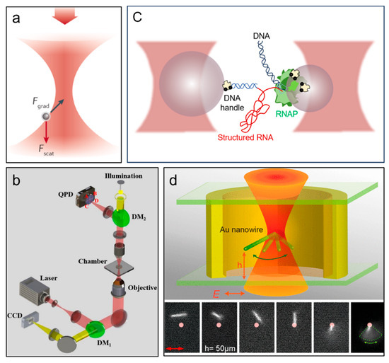
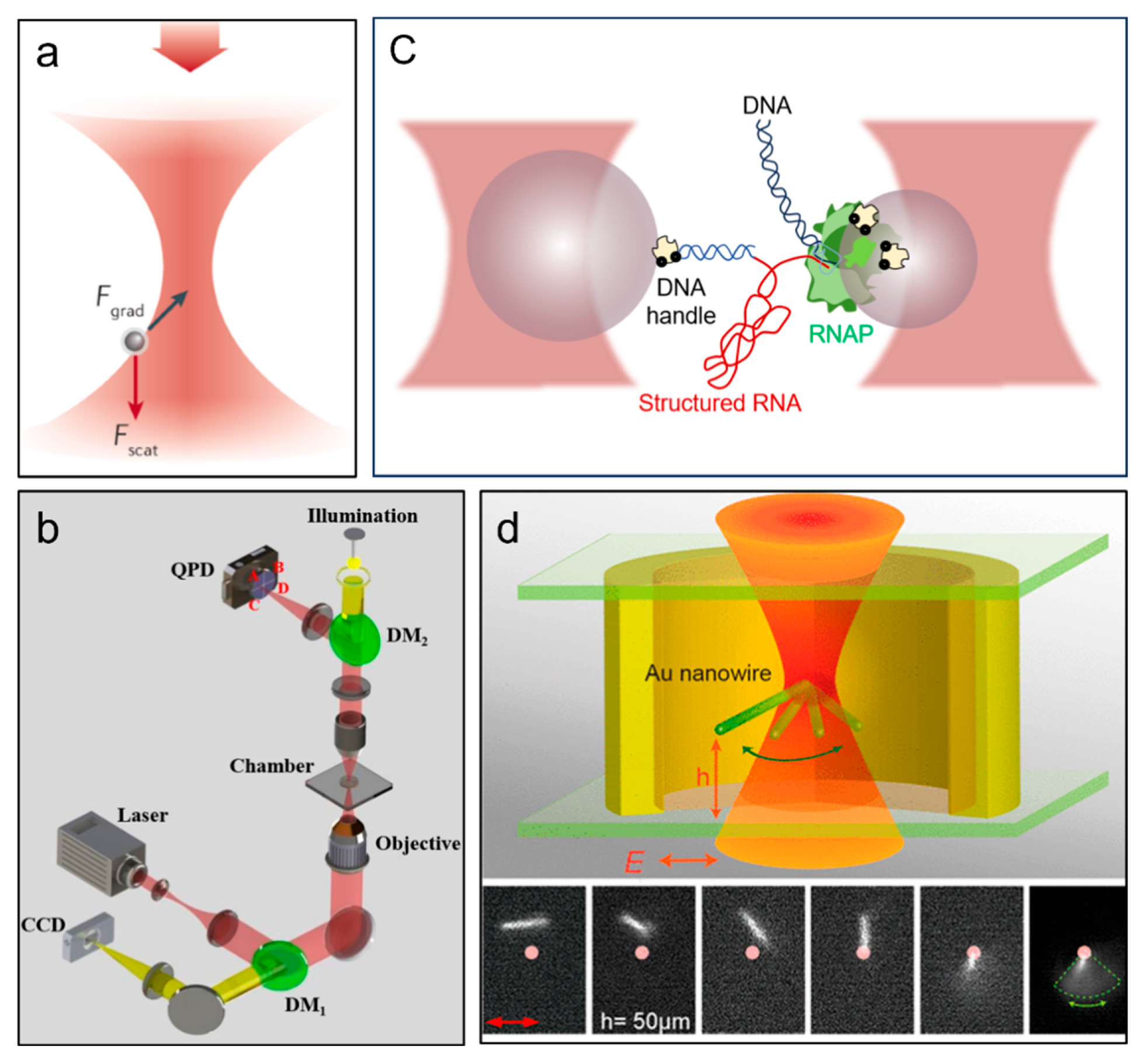
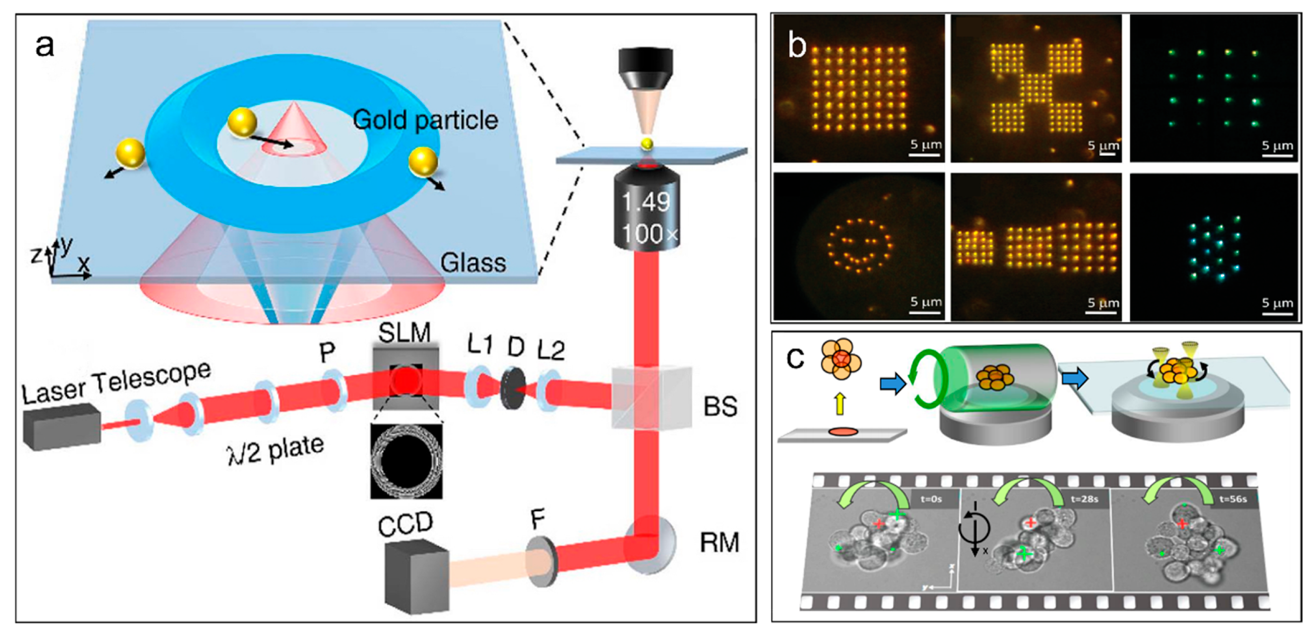
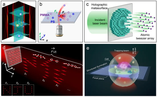
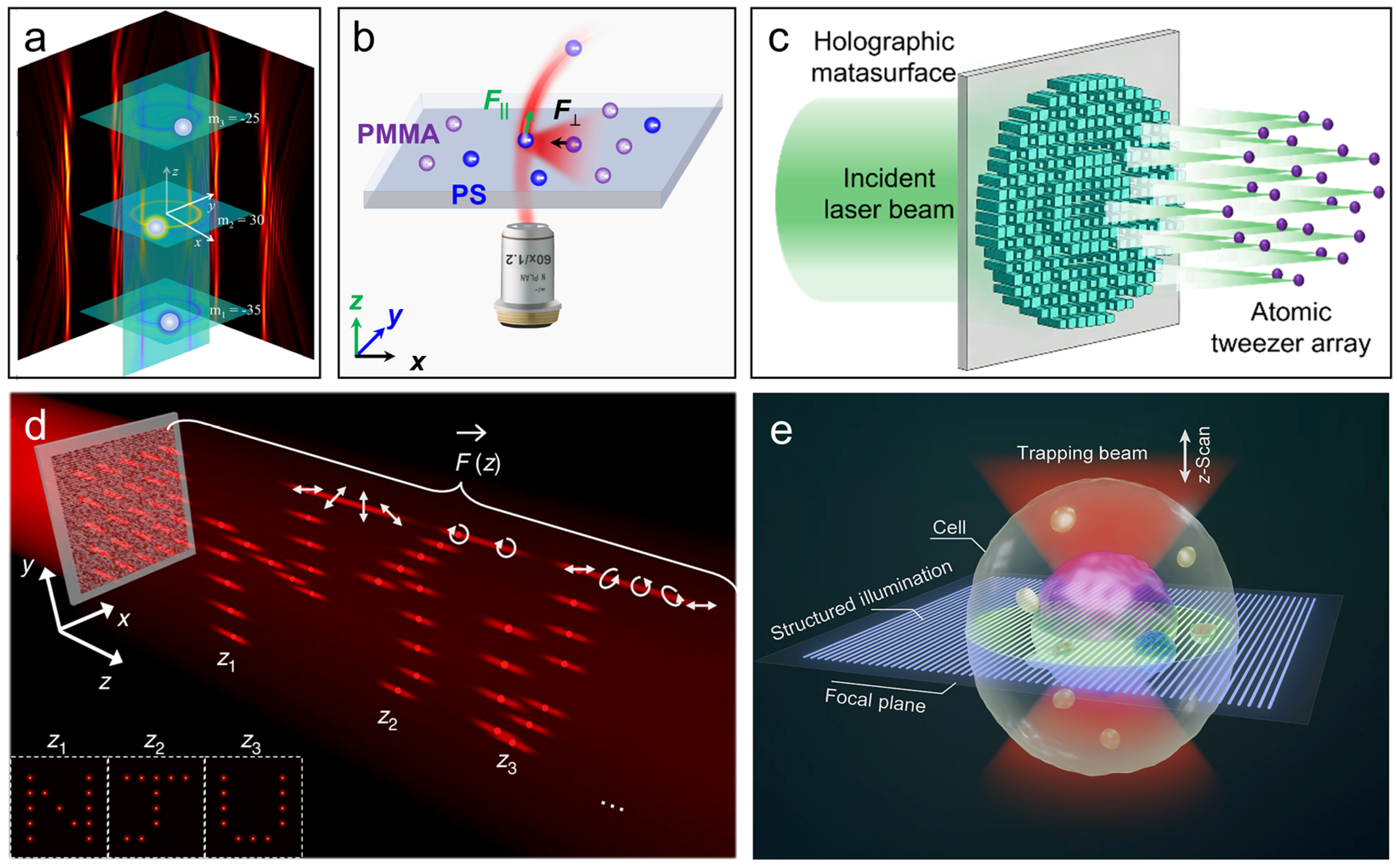
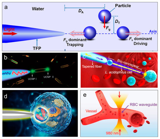
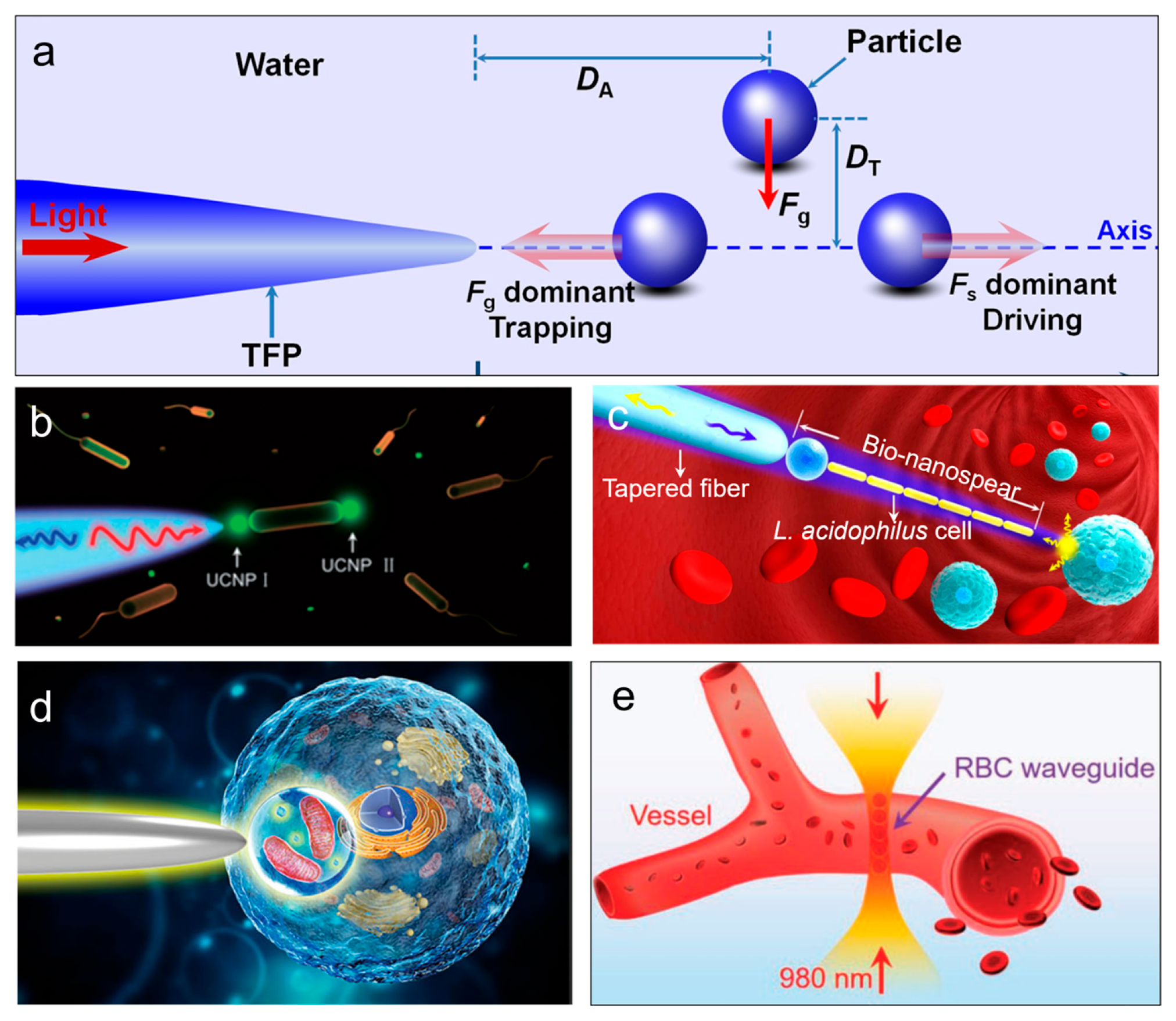
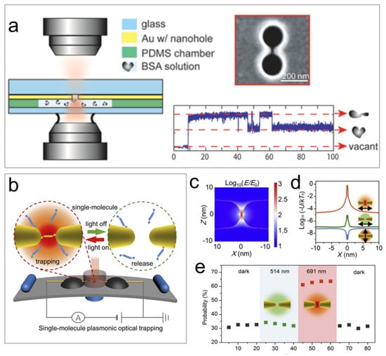
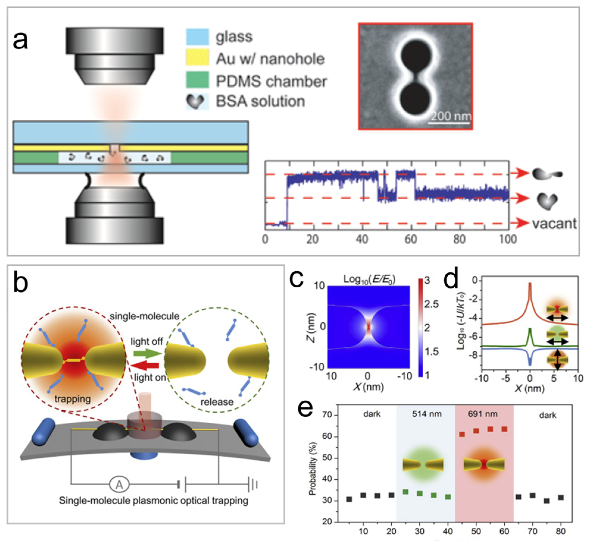
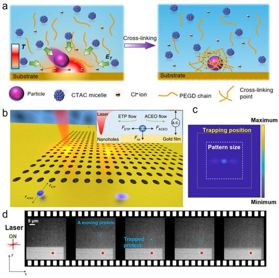
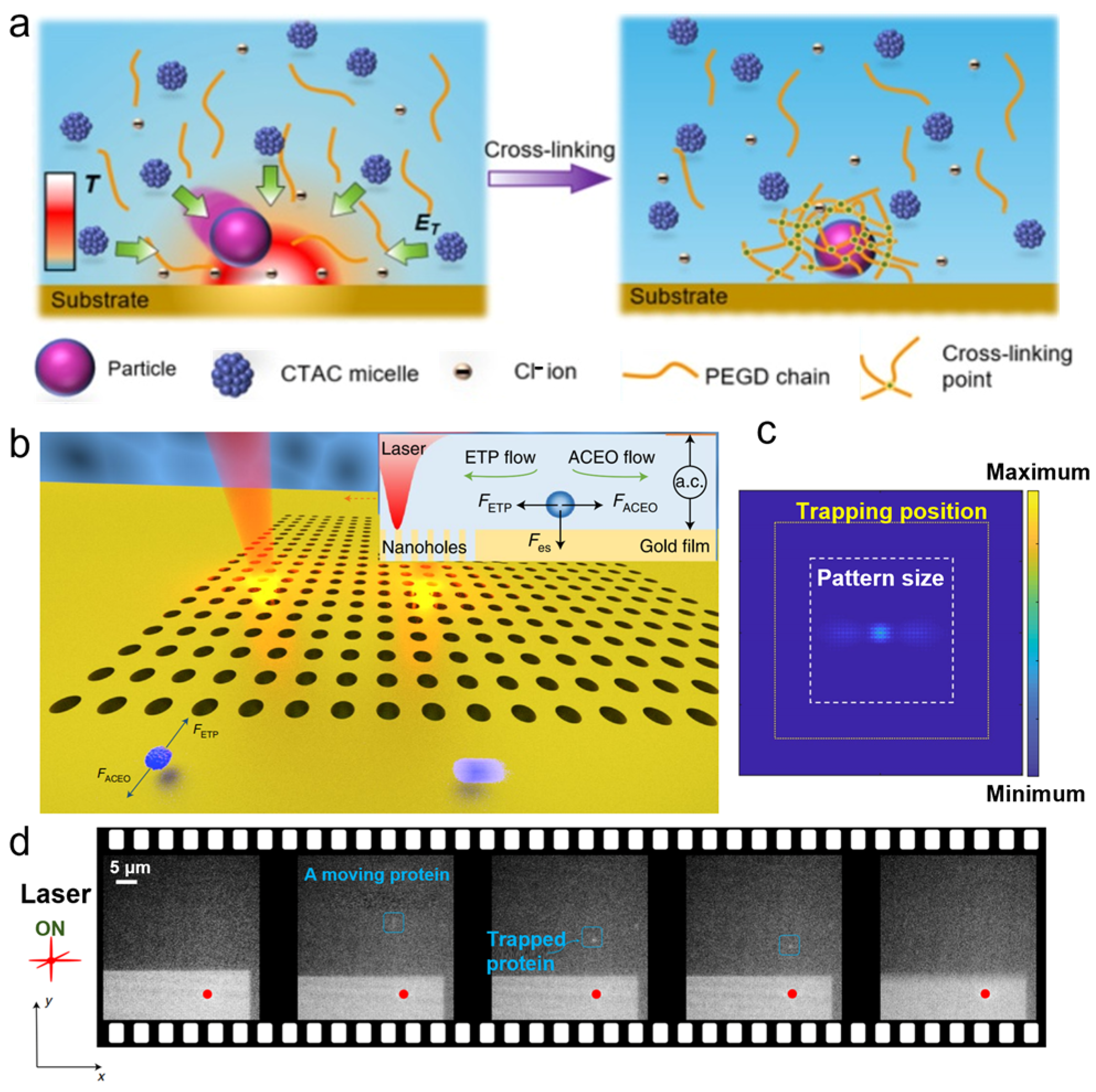

Disclaimer/Publisher’s Note: The statements, opinions and data contained in all publications are solely those of the individual author(s) and contributor(s) and not of MDPI and/or the editor(s). MDPI and/or the editor(s) disclaim responsibility for any injury to people or property resulting from any ideas, methods, instructions or products referred to in the content. |
© 2026 by the authors. Licensee MDPI, Basel, Switzerland. This article is an open access article distributed under the terms and conditions of the Creative Commons Attribution (CC BY) license.