


Abstract
The rapid expansion of RNA therapeutics requires analytical methods that resolve molecular integrity, heterogeneity, and process-related impurities. Capillary electrophoresis (CE) provides direct RNA separation with low sample consumption and can be coupled with laser-induced fluorescence (LIF) or mass spectrometry (MS). This review assesses CE across RNA therapeutic development, release testing, and process analytics. We examine CE separation methods and platform formats for mRNA integrity and poly(A) tail analysis, size- and structure-related impurity profiling, circular RNA purity evaluation, RNA modification analysis by CE-MS, and aptamer discovery. We also discuss multi-capillary systems, microchip electrophoresis (MCE), automation, artificial intelligence (AI)-assisted data analysis, and bioprocess integration. CE is already highly useful for selected quality control tasks, especially mRNA integrity, poly(A) tail profiling, and circular RNA purity, but broader routine adoption requires improved sensitivity, standardization, method transfer, and regulatory acceptance.
1. Introduction
The rapid emergence of RNA therapeutics has reshaped modern biomedicine. Following the clinical success of messenger RNA (mRNA) vaccines against severe acute respiratory syndrome coronavirus 2 (SARS-CoV-2), RNA-based medicines have expanded to include protein replacement therapy, cancer immunotherapy, gene regulation, and regenerative medicine [1,2,3,4]. This therapeutic landscape now spans multiple molecular formats, including mRNA, small interfering RNA (siRNA), antisense oligonucleotides (ASOs), microRNAs (miRNAs), and circular RNA (circRNA). As the field advances toward large-scale manufacturing and broader clinical translation, the demand for robust analytical platforms capable of accurately characterizing RNA products has become increasingly urgent.
Compared with traditional small-molecule drugs and protein biologics, RNA therapeutics present distinct analytical challenges. RNA molecules are chemically labile, highly susceptible to ribonuclease-mediated degradation, and structurally dynamic because of extensive secondary and tertiary folding [5,6,7]. Moreover, synthetic and in vitro transcribed RNA products often display substantial heterogeneity, including truncated transcripts, nicked species, double-stranded RNA (dsRNA) impurities, variable poly(A) tail lengths, and incomplete or heterogeneous nucleotide modifications [8,9,10]. These attributes directly affect translation efficiency, immunogenicity, stability, potency, and product safety, creating a strong need for analytical methods that can resolve RNA integrity, purity, conformation, and process-related heterogeneity with high resolution and minimal bias.
Current analytical strategies provide complementary but incomplete information. Reverse transcription polymerase chain reaction (RT-PCR) and quantitative PCR offer high sensitivity for sequence-specific detection but do not directly reveal native RNA integrity, size heterogeneity, or structural state; reverse transcription and amplification can also bias structured or chemically modified templates [11]. Slab gel electrophoresis, microfluidic bioanalyzers, liquid chromatography, and MS provide other forms of information. CE directly separates RNA and can be coupled with ultraviolet (UV) absorbance, LIF, or MS detection [12,13,14,15]. CE has become increasingly relevant to RNA therapeutics. Capillary gel electrophoresis (CGE) is used for mRNA integrity, purity, and poly(A) tail analysis, whereas capillary zone electrophoresis (CZE) supports selected process measurements, including monitoring during in vitro transcription (IVT). CE-based workflows also address circular RNA purity, RNA modification analysis by CE-MS, and systematic evolution of ligands by exponential enrichment (SELEX), with CE used as the partitioning step in CE-SELEX [16,17,18,19]. Microchip electrophoresis (MCE), multi-capillary systems, automation, and AI-assisted analysis are extending throughput and workflow integration [20,21].
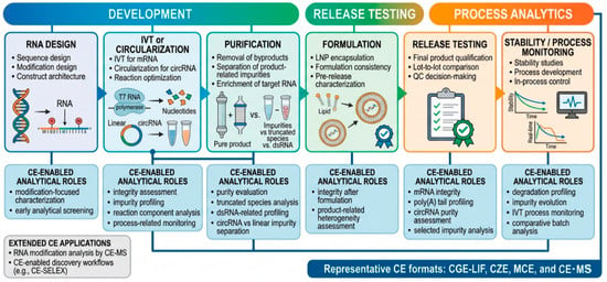
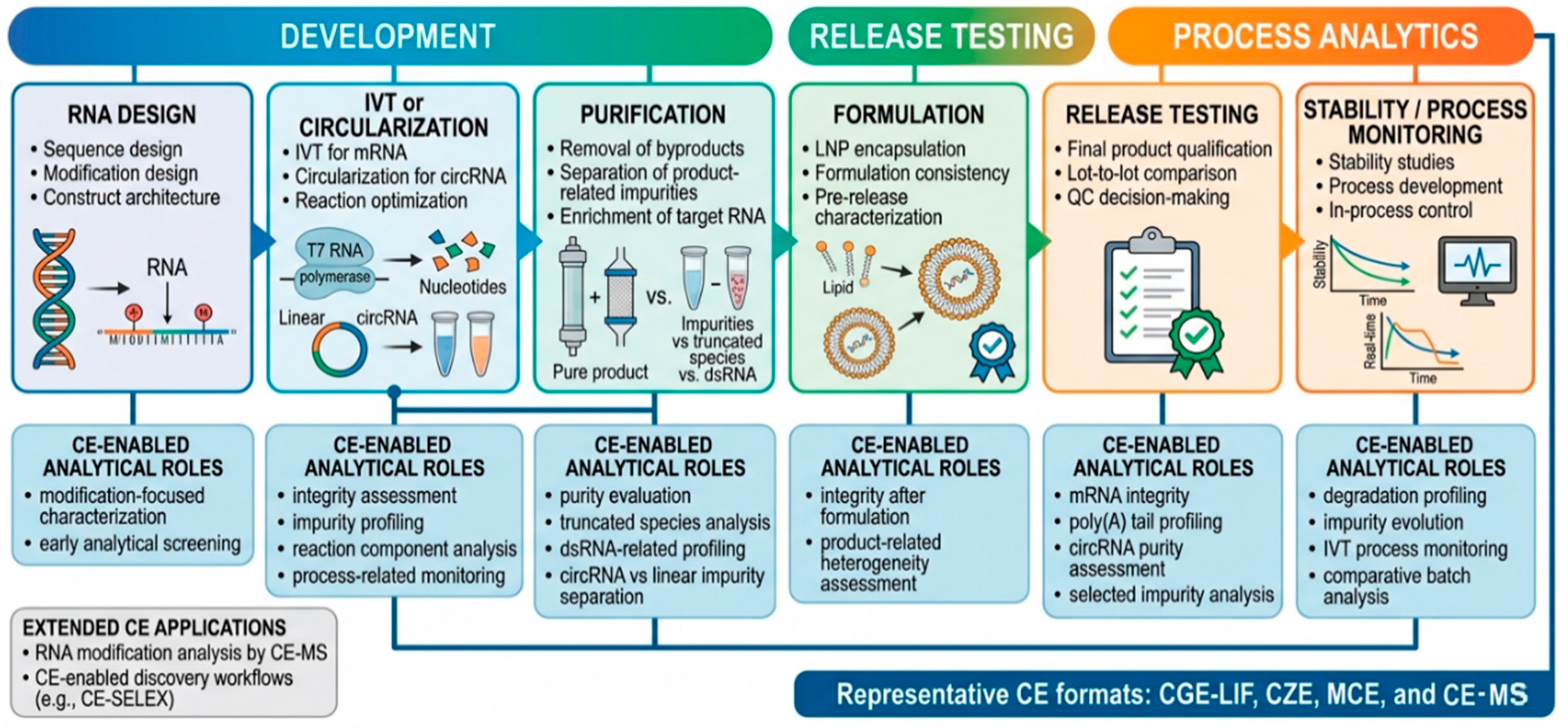
This review asks whether CE can function as a core analytical platform for RNA therapeutic development, release testing, and process analytics. We first define the relevant analytical demands, separation methods, platform formats, and detection techniques; then evaluate applications in mRNA quality control, circular RNA purity, RNA modification analysis, and aptamer discovery. Finally, we examine enabling technologies and the remaining limitations to routine implementation. Figure 1 summarizes CE-supported analytical roles across the RNA therapeutics lifecycle.
Figure 1.
Roles of capillary electrophoresis across the RNA therapeutics lifecycle, including synthesis, purification, formulation, release testing, and process monitoring. Representative CE separation methods include CGE coupled with LIF detection, CZE coupled with UV or LIF detection, microchip electrophoresis, and CE coupled with MS.
2. Analytical Demands of RNA Therapeutics and the Role of Capillary Electrophoresis
2.1. Analytical Attributes That Matter in RNA Therapeutics
As highlighted by the National Institute of Standards and Technology (NIST) reference material framework, critical quality attributes (CQAs) for mRNA therapeutics extend beyond sequence identity to intactness, 5′ capping efficiency, poly(A) tail length, and product- and process-related impurities. RNA integrity, size heterogeneity, purity, and double-stranded RNA (dsRNA) byproducts can affect safety and efficacy, while poly(A) tail length and distribution influence stability and translational output [22].
2.2. CE Separation Methods Relevant to RNA Analysis
According to the International Union of Pure and Applied Chemistry (IUPAC) [23,24], electromigration separation methods should be distinguished based on their underlying principles. Relevant methods include CZE, CGE, capillary isotachophoresis (CITP), capillary isoelectric focusing (CIEF), capillary electrokinetic chromatography (CEKC), and capillary electrochromatography (CEC) [25]. CGE is the most mature for therapeutic RNA analysis, enabling high-resolution size-based separation for integrity assessment, impurity profiling, and poly(A) tail analysis. CZE is well-suited for process-related measurements, such as monitoring IVT reaction components [18]. MCE offers miniaturized formats for screening and high-throughput workflows [9]. CE-MS is valuable for RNA modification analysis [14]. These separation methods can be coupled with various detection techniques, including UV absorbance, LIF, and MS, each providing a different balance of sensitivity, selectivity, and information content. In practice, these combinations are complementary: UV favors simplicity [26], LIF supports quality control applications [27], and MS offers the highest structural specificity [28].
2.3. Matching CE Separation Methods and Detection Techniques to Specific Analytical Tasks
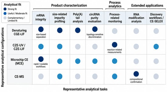
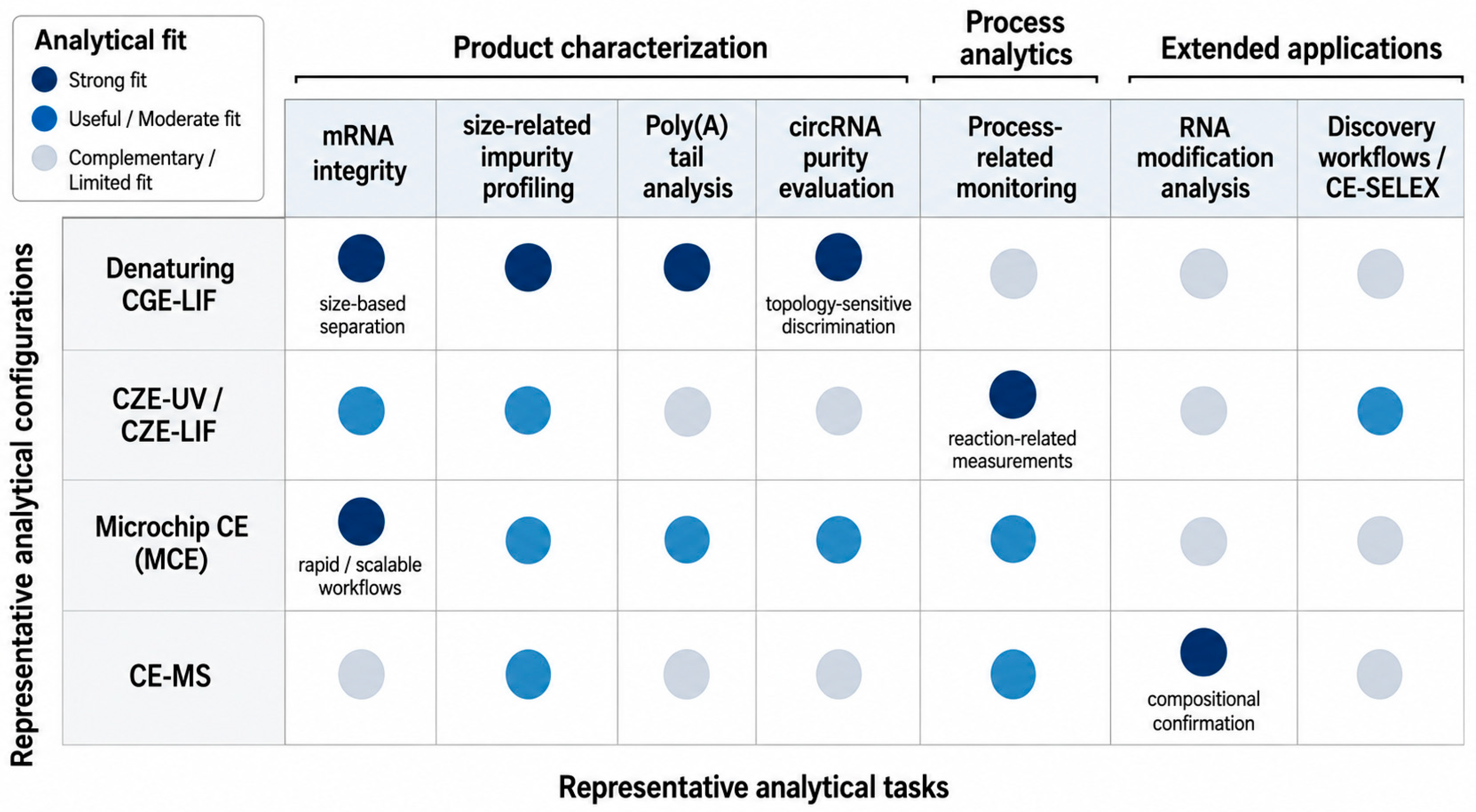
No single analytical configuration is optimal for all RNA therapeutic applications. For full-length mRNA integrity and size-related impurity profiling, denaturing CGE coupled with LIF detection is generally most suitable [29,30,31]. Poly(A) tails are commonly isolated by enzymatic processing and sized by CGE-LIF [16,32], while optimized CGE-LIF can distinguish intact circular RNA from linear and nicked species [17]. CZE coupled with UV or LIF detection is useful for selected process measurements, including ionic components, during IVT [10,18]. CE-MS is preferred when molecular identity or modification status must be confirmed [33,34]. MCE and multi-capillary systems are advantageous when speed, throughput, or miniaturization is the priority [9,35,36]. Figure 2 summarizes the suitability of these analytical configurations for key tasks.
Figure 2.
Matching CE separation methods, platform formats, and detection techniques to key analytical tasks in RNA therapeutics. Denaturing CGE coupled with LIF detection is suitable for mRNA integrity assessment, size-related impurity profiling, poly(A) tail analysis, and circular RNA purity evaluation. CZE coupled with UV or LIF detection is applicable to selected process-related measurements, MCE supports rapid and high-throughput workflows, and CE-MS provides molecular information for RNA modification and structural characterization.
3. Capillary Electrophoresis for mRNA Therapeutics Quality Control
3.1. Why mRNA Therapeutics Require Dedicated Analytical Control
Among RNA-based medicines, mRNA therapeutics impose particularly demanding analytical requirements because product performance depends not only on sequence identity but also on molecular integrity, compositional consistency, and process-related purity [37,38]. Therapeutic mRNAs are long, structurally dynamic molecules produced by IVT, a process that can generate substantial heterogeneity during synthesis and downstream processing. Analytical assessment must therefore extend beyond identity confirmation to attributes directly linked to product quality, safety, and efficacy [39]. A major challenge is that the desired full-length transcript is often accompanied by structurally related variants, including truncated products, degradation fragments, nicked species, and dsRNA byproducts [40,41]. Poly(A) tail length and distribution further influence RNA stability and translational output, while nucleotide modification consistency affects immune activation and expression performance [42,43]. These factors make the analytical landscape of mRNA therapeutics considerably more complex than simple sequence verification.
3.2. Mapping CE to the Critical Quality Attributes of mRNA Therapeutics
For mRNA-based medicines, CQAs include molecular integrity, size heterogeneity, purity, poly(A) tail characteristics, selected impurity classes, and process-related indicators [22,44]. CE is most directly applicable to integrity, size-related heterogeneity, and aspects of purity. Denaturing CGE-LIF resolves full-length mRNA from truncated, degraded, or related shorter species [45,46], while enzymatic workflows enable the electrophoretic sizing of poly(A)-containing fragments [16,32]. CZE can monitor selected IVT reaction components [18], and CE-MS or orthogonal methods are required for detailed modification or compositional analysis [14]. Table 1 maps these attributes to representative CE-based configurations.
Table 1.
Critical quality attributes of mRNA therapeutics and corresponding CE strategies.
3.3. CE for mRNA Integrity and Purity Assessment
Among mRNA therapeutic CQAs, integrity and purity are where CE is most directly applicable. Because therapeutic performance depends on intact full-length transcripts [47], analytical methods must distinguish the desired product from truncated, degraded, or closely related shorter species. This makes denaturing CGE-LIF particularly well-suited to mRNA quality assessment. Under denaturing conditions, CGE separates RNA primarily by size while minimizing higher-order structure effects, enabling the resolution of full-length mRNA from degradation products, incomplete transcripts, and related byproducts [48,49]. CE is therefore highly useful for batch comparison, degradation profiling, and stability-indicating analysis.
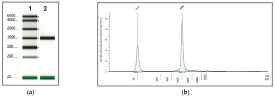
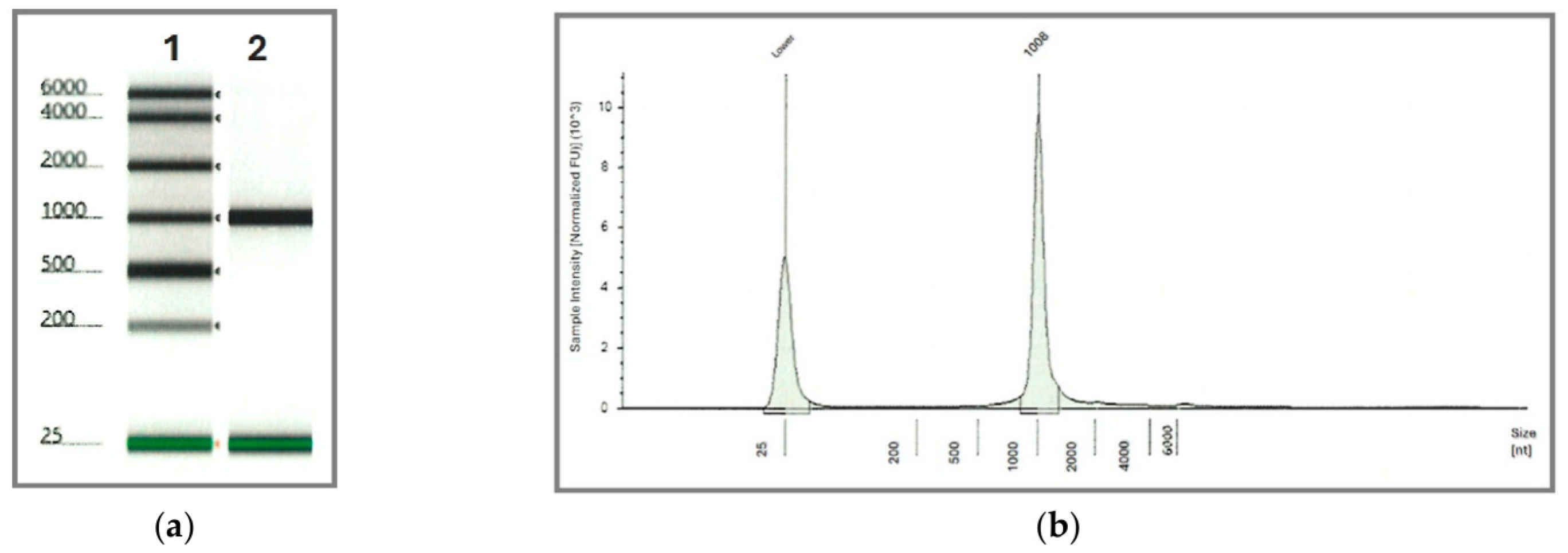
Recent representative studies have helped define the analytical maturity of CE in this area. Warzak et al. described CE methods specifically developed for assessing IVT mRNA size and purity as critical quality attributes, thereby positioning CE as a practical tool for therapeutic mRNA quality assessment [10]. Su et al. subsequently reported a high-throughput CGE workflow that integrated mRNA integrity analysis with poly(A) tail characterization, illustrating the transition from low-throughput characterization toward more scalable and industrially relevant formats [16]. Tran et al. further showed that reliable CGE-based integrity assessment depends not only on separation performance but also on careful control of robustness, repeatability, and transferability [45]. Rivera-Santiago et al. similarly employed automated CE (TapeStation, Agilent, Santa Clara, CA, USA) to verify the integrity and identity of synthetic hGH mRNA following encapsulation, demonstrating the broad utility of CE-based platforms for mRNA quality assessment in non-vaccine therapeutic contexts (Figure 3) [50]. mRNA purity also includes structurally related impurities generated during transcription, purification, formulation, or storage. Comparative work by Lardellier et al. showed that CE directly resolves intact mRNA and functionally relevant shorter RNA species that are not readily characterized by sequence-dependent inference [47]. Together, these studies support CE as a practical method for mRNA integrity and size-related purity assessment within broader manufacturing-control strategies [51].
Figure 3.
mRNA integrity assessment by automated capillary electrophoresis. (a) Gel image showing a single, sharp band at the expected size (~1000 nt), indicating high purity and absence of degradation. (b) Representative electropherogram with a sharp peak at 1008 nt, confirming the expected transcript size and structural integrity [50].
3.4. CE for Poly(A) Tail Profiling and Related Structural Attributes
Among the compositional features of mRNA therapeutics, the poly(A) tail is especially important because it influences RNA stability, translational efficiency, and overall product performance. Analytical assessment must address not only average length but also distribution, as heterogeneity may affect functional consistency across batches [42]. CE is well-suited to this task when combined with appropriate sample preparation. In many workflows, mRNA is enzymatically processed to generate a poly(A)-containing fragment, which is then selectively enriched and sized electrophoretically. In this context, CGE provides a high-resolution platform for resolving poly(A)-derived fragments and determining length distribution with good analytical precision.
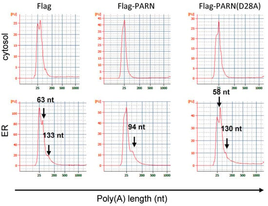
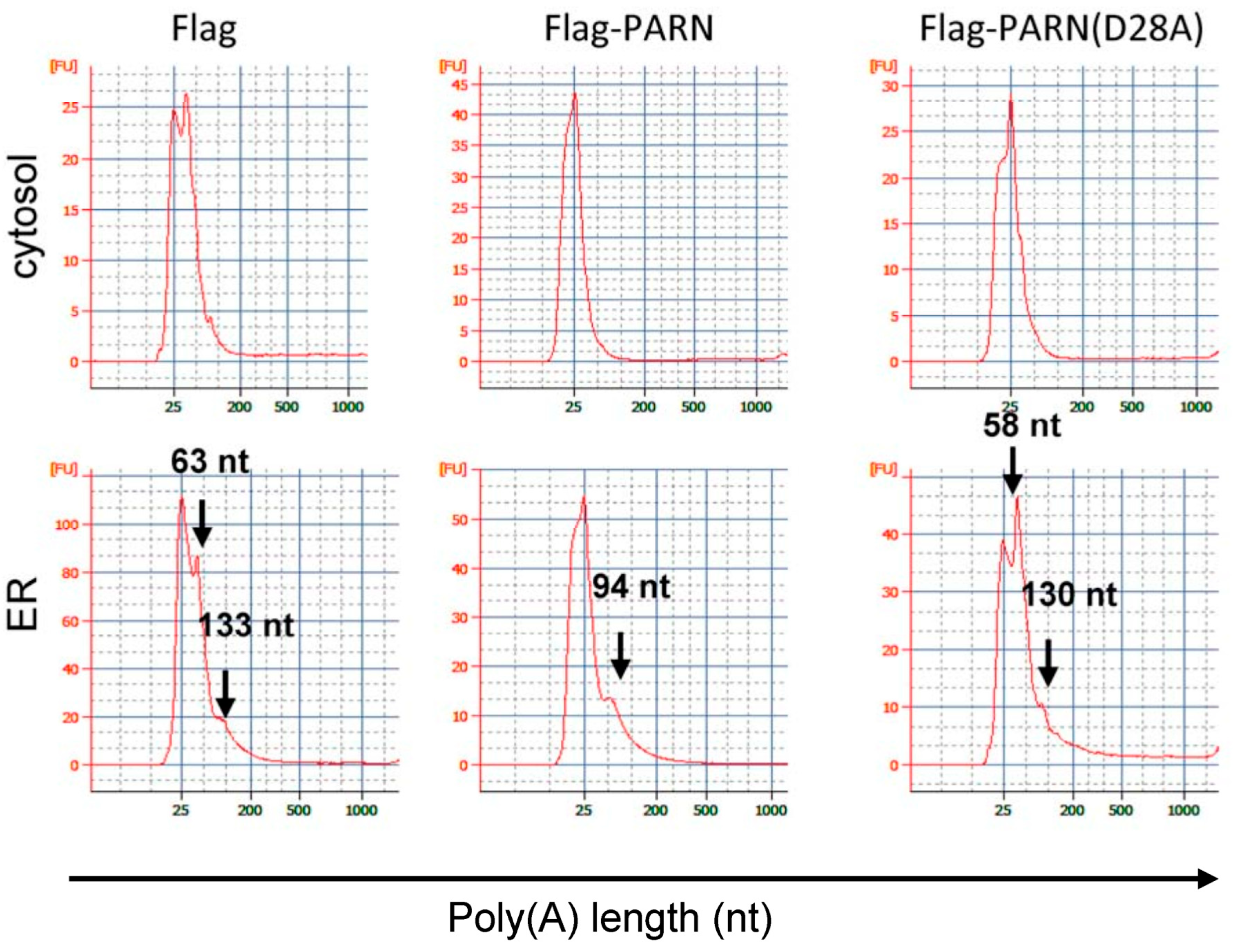
Representative studies show that this field has progressed from general fragment sizing toward more rigorous tail-length characterization. Microchip CE has also been employed for poly(A) tail profiling; for instance, Duan et al. utilized the Agilent 2100 bioanalyzer to analyze poly(A) tail length distribution of ER-associated mRNAs, demonstrating the utility of CE-based platforms for rapid poly(A) tail analysis, albeit with lower resolution compared to traditional gel-based methods (Figure 4) [52]. Su et al. demonstrated that poly(A) tail analysis can be incorporated into a broader high-throughput CGE workflow for therapeutic mRNA, thereby linking transcript integrity analysis and tail-length characterization within a unified analytical platform (Figure 4) [16]. Di Grandi et al. further reported a single-nucleotide resolution CGE workflow for poly(A) tail characterization in mRNA therapeutics and vaccines, showing that CE can move beyond broad distribution profiling to highly resolved length analysis with direct relevance to product development and comparability assessment [32]. This level of resolution is particularly important because poly(A) tail heterogeneity may be functionally meaningful even when average tail length appears similar across samples. Poly(A) tail profiling, therefore, supports development-stage optimization, lot comparison, and comparability assessment when both length resolution and workflow scalability are required.
Figure 4.
Poly(A) tail length analysis by microchip capillary electrophoresis. Poly(A)+ RNAs extracted from the ER fractions of HEK-293T cells with overexpressed Flag-PARN or Flag-PARN(D28A) were analyzed using the Agilent 2100 bioanalyzer with the Agilent RNA 6000 Pico Kit. The profiles show the poly(A) tail length distribution of ER-associated mRNAs under different conditions, confirming the results obtained by traditional gel electrophoresis [52].
3.5. CE in Impurity Assessment and Process Analytics
Beyond integrity and poly(A) profiling, CE also contributes to impurity assessment and process analytics in mRNA therapeutics [22]. In vitro transcription and downstream processing can generate unintended species, including abortive transcripts, degradation fragments, and dsRNA byproducts. Some of these impurities are analytically challenging because they are structurally similar to the desired product, making orthogonal separation methods especially valuable [43]. CE is particularly useful when impurity species differ sufficiently in size, conformation, or electrophoretic behavior from the target mRNA. In such cases, electrophoretic profiling can reveal impurity patterns not captured by simple bulk measurements and can support comparison across process conditions, purification steps, or stability time points. Although CE may not fully replace highly specific assays for all impurity classes, it provides an important complementary view of product-related heterogeneity. CZE also supports process analytics. Wang et al. monitored magnesium-ion consumption during IVT, linking reaction composition to transcription behavior and providing development-stage information before final-product characterization [18].
3.6. Current Role and Limitations of CE in mRNA Quality Control
CE is closest to routine use for mRNA integrity, size heterogeneity, and poly(A) tail profiling [16]. Other attributes, including modification status and some impurity classes, still require CE-MS or orthogonal methods. Routine release testing depends on robustness, transferability, throughput, standardization, inter-laboratory reproducibility, and sensitivity to low-abundance species. Multi-capillary, microchip, and automated workflows improve throughput but do not remove these validation requirements.
4. Capillary Electrophoresis for Circular RNA Characterization
4.1. Why circRNA Presents Distinct Analytical Challenges
Compared with linear RNA therapeutics, circular RNA presents a distinct analytical challenge because its defining attribute is topological integrity rather than sequence alone. Intact covalently closed molecules must be distinguished from linear precursors and nicked species that may have similar sequence and length but different functional consequences [53]. Sample handling and separation conditions can also alter circular integrity, so reliable analysis requires topology-sensitive discrimination with controlled structural artifacts.
4.2. CE for the Separation of circRNA from Linear and Nicked Impurities
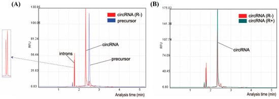
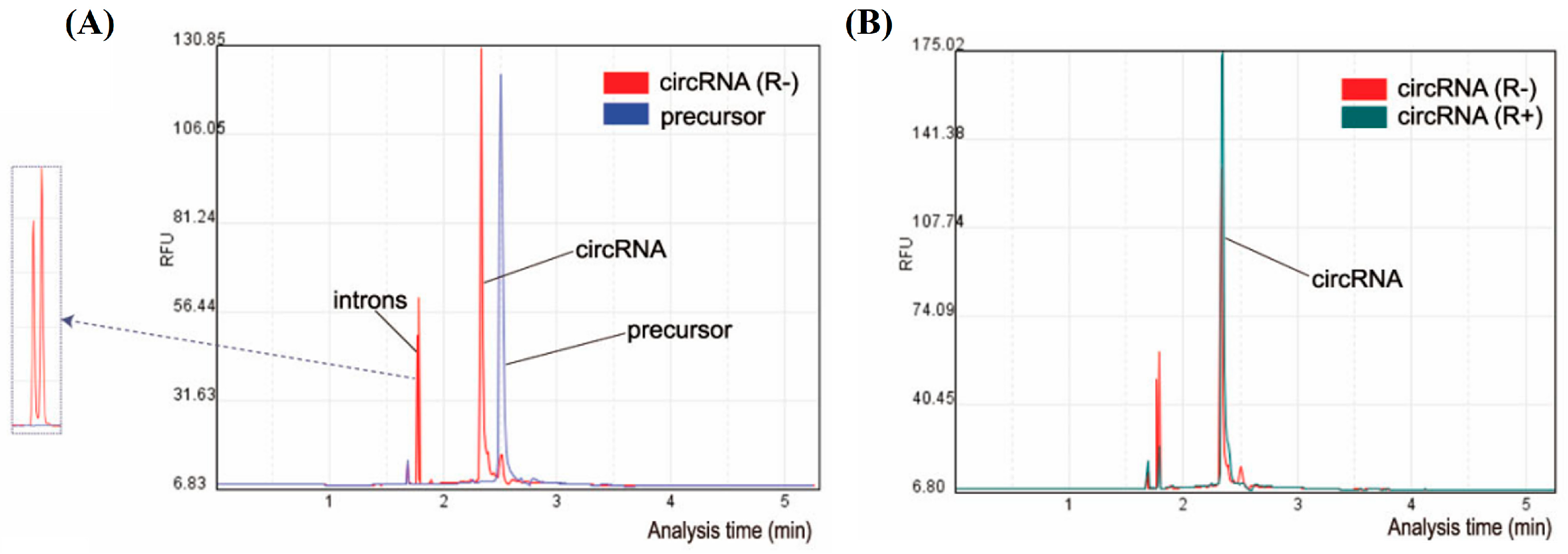
A central task in circRNA therapeutics is separating intact circular RNA from linear precursor and nicked byproducts because these species may differ markedly in functional value while remaining highly similar in sequence and length [54]. Methods relying primarily on bulk nucleic acid signal often lack the resolution needed for meaningful purity assessment [55]. CGE is particularly valuable here because it resolves RNA species based on electrophoretic behavior shaped by both size and topology. Recent work has moved this area beyond qualitative separation toward analytically robust performance metrics. Zheng et al. developed a CGE-LIF method that achieved high-resolution separation of circRNA from its linear and nicked impurities, with a separation resolution greater than 5.8 and excellent repeatability with RSD values below 0.04% (Figure 5) [17]. Their study also showed that migration behavior depends strongly on sample pretreatment, separation voltage, and gel matrix composition, indicating that circRNA purity analysis cannot be treated as a simple extension of conventional linear RNA sizing. Sun et al. also employed capillary electrophoresis to monitor circRNA circularization efficiency by resolving circular RNA from its linear precursor, demonstrating the utility of CE for assessing circRNA formation and purity [56]. This capability is especially important in circularization workflows, where incomplete conversion and backbone damage can generate complex product mixtures. Pulido et al. showed that multi-capillary electrophoresis can separate circular RNA from linear precursor species in a higher-throughput format, indicating movement from analytical feasibility toward more process-relevant implementation [53].
Figure 5.
Capillary electrophoresis analysis of circRNA circularization and purification. (A) Overlay of CE profiles showing IVT precursor RNA (blue) and circularized RNA (red). The disappearance of the precursor peak and the appearance of a new circRNA peak indicate successful circularization, with an estimated circularization efficiency of approximately 85%. Arrows indicate excised intron fragments released during circularization. (B) Overlay of CE profiles showing circular RNA before (blue) and after (red) RNase R treatment. Removal of residual linear RNA species results in highly purified circular RNA products [56].
4.3. Analytical Implications of circRNA Migration Behavior
The analytical behavior of circRNA in CE is shaped by molecular size, topology-dependent migration, and structural sensitivity during separation [17,57]. This distinguishes circRNA from linear RNA analyses, where denaturing size-based separation is often the primary objective. In circRNA systems, the migration difference between intact circular molecules and linear species is itself part of the analytical signal, meaning that separation conditions must preserve rather than eliminate this distinction. If sample preparation or electrophoretic conditions disrupt circular integrity, the advantage of topology-sensitive separation may be lost, making intact circRNA difficult to distinguish from linear or nicked forms. CircRNA analysis by CE, therefore, requires a careful balance between separation efficiency, structural preservation, and artifact minimization.
This interpretation is reinforced by recent analytical and mechanistic studies. Sun et al. reviewed methods for evaluating RNA circularization efficiency and highlighted that topology-sensitive discrimination remains a central unresolved challenge in circRNA characterization [58]. Jiang and Kjems further showed that RNA denaturation itself can underlie circRNA separation behavior, emphasizing that apparent analytical resolution may reflect not only intrinsic topology but also structure-dependent transformations occurring under the separation conditions [59]. Together with the mechanistic observations reported by Zheng et al. [17], these findings show that circRNA electropherograms must be interpreted with explicit attention to method-induced structural effects rather than assuming that migration differences automatically reflect native topology alone. Robust circular RNA methods must therefore evaluate both separation resolution and preservation of molecular topology during pretreatment and migration.
4.4. Current Relevance and Limitations of CE in circRNA Analysis
Zheng et al. reported a CGE-LIF method with a separation resolution greater than 5.8 and relative standard deviation (RSD) values below 0.04%, demonstrating strong discrimination and repeatability under optimized conditions [17]. Pulido et al. further showed that multi-capillary formats can support higher throughput circular RNA purity assessment [53]. However, sample handling, denaturation, and prolonged migration can alter the molecular state responsible for the separation [59]. Standardized workflows, control of structure-dependent artifacts, and orthogonal compositional confirmation remain necessary.
5. Beyond Primary Quality Control: CE-MS for RNA Modification Analysis
5.1. Why RNA Modification Analysis Matters in RNA Therapeutics and Biology
RNA modification analysis extends CE’s role beyond primary quality control into molecular-level characterization. Chemical modifications are widespread in natural RNAs and influence RNA stability, structure, translation, localization, and protein interactions. In biological systems, these modifications are recognized as important regulators of gene expression and cellular function [60]. In RNA therapeutics, modification analysis is especially important because chemically modified nucleosides are often deliberately introduced to improve product performance, including reduced innate immune activation, enhanced translational efficiency, and increased stability [14]. Analytical characterization must, therefore, not only address the correct sequence but also the consistent introduction of intended modifications without unintended heterogeneity. Whereas mRNA integrity and poly(A) analysis mainly address population-level size heterogeneity, modification analysis addresses chemical heterogeneity within the RNA molecule. Coupling CE with MS, therefore, extends electrophoretic analysis from size-based quality assessment to molecular-level structural characterization.
5.2. CE-MS as a Platform for RNA Modification Analysis
CE-MS combines electrophoretic separation with molecular-mass and compositional information, making it suitable for modified nucleosides and heterogeneous RNA-derived components [61]. Its routine implementation is constrained by interface design, ionization compatibility, analyte stability, and data interpretation; it is therefore most valuable when electrophoretic resolution must be paired with structural confirmation.
5.3. Representative Applications of CE-MS in Modified RNA Analysis
CE-MS applications highlight the value of this platform, where chemical specificity is essential. One major use is the detection and characterization of modified nucleosides from enzymatically digested RNA, where CE-MS separates closely related components and confirms their identities by mass. This is particularly relevant for studying modification patterns in tRNA, miRNA, and other highly modified RNA classes [62]. In RNA therapeutics, CE-MS can also evaluate the presence and consistency of intended chemical modifications in synthetic or IVT RNA products, making it an important complement to size-based and impurity-focused assays.
Representative studies illustrate the analytical depth that CE-MS can provide. Lechner et al. demonstrated sheathless CE-high-resolution MS for the characterization of post-transcriptional RNA modifications and achieved sequence coverages ranging from 68% to 97% across four tRNAs while requiring only picomole quantities of RNA, highlighting the combination of structural specificity and low sample consumption that makes CE-MS attractive for modification-focused analysis [14]. Yu et al. further improved analytical sensitivity by establishing a sheathless CE-MS/MS workflow for simultaneous determination of eight RNA-modified nucleotides from only 500 pg to 5 ng of total RNA, showing that CE-MS can support trace-level and multiplexed modification analysis [61]. Although not developed specifically for therapeutic manufacturing, Khan et al. also demonstrated direct CE-MS detection of endogenous microRNAs and their post-transcriptional modifications in serum, reinforcing the broader capability of CE-MS to resolve chemically heterogeneous RNA systems in complex samples [62]. For chemically heterogeneous RNA fragments, CE-MS provides molecular identification that UV or fluorescence detection alone cannot supply [63]. Its principal value is confirmation of modification identity, abundance, or heterogeneity rather than electrophoretic pattern recognition alone.
5.4. Current Opportunities and Limitations of CE-MS in This Area
CE-MS is one of the most promising CE extensions for RNA analysis, particularly when the key question involves chemical modification rather than size-related heterogeneity. Its main advantage lies in combining high-efficiency separation with compositional confirmation, enabling structural specificity beyond that available from conventional UV or LIF detection. This makes CE-MS especially valuable for modified nucleosides, chemically heterogeneous RNA fragments, and modification-focused characterization of therapeutic RNA products. The current literature suggests that its greatest strength emerges when electrophoretic resolution and mass-based confirmation are both required, such as in the discrimination of closely related modified nucleosides or low-abundance heterogeneous digestion products. However, it remains more specialized and less implementation-ready than CE workflows used for integrity or purity assessment. Successful use depends on technically demanding factors such as interface design, ionization compatibility, analyte stability, and data interpretation, which limit accessibility for routine high-throughput workflows and complicate inter-laboratory standardization. Accordingly, CE-MS is best viewed not as a primary standalone solution, but as a high-value complementary platform for modification-focused analysis when conventional electrophoretic readouts are insufficiently specific. Its broader impact in RNA therapeutics will depend on continued improvements in robustness, sensitivity, usability, and workflow standardization.
6. Beyond Primary Quality Control: CE-Enabled Discovery Workflows and CE-SELEX
6.1. Why CE Is Relevant to Aptamer Discovery Workflows
CE extends its role from analytical characterization into selection-enabled molecular discovery for aptamers, short single-stranded nucleic acids that bind targets with high affinity and specificity. Their discovery traditionally relies on iterative enrichment, such as SELEX, where the key challenge is efficiently distinguishing target-bound sequences from a large pool of unbound molecules [64]. This separation problem aligns well with CE’s strengths. By resolving free nucleic acids from target-bound complexes based on electrophoretic mobility differences, CE enables selection in free solution without immobilization-based partitioning. This avoids immobilization artifacts that can alter target conformation, introduce steric hindrance, or reduce selection efficiency. Here, CE contributes not as a routine quality-control tool but as a platform for rapid enrichment and efficient partitioning during aptamer discovery. Aptamer selection thus exemplifies how CE supports RNA-related biotechnology beyond primary therapeutic characterization while remaining connected to the broader analytical capabilities discussed in this review.
6.2. CE-SELEX as an Accelerated Selection Strategy
CE-SELEX uses electrophoretic separation as the key partitioning step in aptamer selection [65], in contrast to conventional SELEX, which requires repeated cycles of binding, separation, amplification, and regeneration. By contrast, CE-SELEX reduces selection rounds by exploiting mobility differences between free nucleic acids and target-bound complexes [66]. Compared with conventional SELEX workflows that often require 8~15 rounds and may extend over several months, CE-SELEX can, in many cases, reduce selection to only 1~4 rounds completed within days to weeks, making it one of the most compelling examples of CE as an enabling biotechnology platform. This strategy offers several advantages: selection occurs in free solution, preserving native target conformation and avoiding immobilization-related artifacts; high CE separation efficiency allows precise partitioning of bound and unbound populations, accelerating enrichment of high-affinity candidates [67]; and rapid analytical feedback shortens the overall discovery timeline.
Recent advances have further expanded the CE-SELEX toolkit. For instance, the concept of ideal-filter capillary electrophoresis (IFCE) has been introduced as a highly efficient partitioning method for selecting protein binders from oligonucleotide libraries, enabling one-step aptamer selection [68,69]. Furthermore, kinetic CE methods have been developed to screen oligonucleotide libraries for protein binders, providing additional dimensions for selection based on binding kinetics [70]. These approaches, along with other advancements in SELEX technology [71], demonstrate the ongoing evolution of CE-based selection strategies. Suzuki et al. have also reported a reliable post-SELEX design of bivalent aptamers through systematic CE exploration of ternary DNA-protein complexes [72].
This acceleration is well illustrated by representative studies. Mendonsa and Bowser provided an early proof of principle showing that capillary electrophoresis could be used for in vitro selection of high-affinity DNA ligands in far fewer rounds than conventional workflows [73]. More recently, Zhao et al. reported a three-step evolutionary enhanced CE-SELEX strategy for exosome vesicle aptamer selection, demonstrating that CE-based selection can be incorporated into more sophisticated multi-step enrichment schemes for complex biological targets. Accordingly, CE-SELEX is not simply a variation in SELEX but a selection strategy fundamentally shaped by electrophoretic resolution. Its relevance lies in translating CE’s strengths, including high-efficiency separation, low sample consumption, and free-solution analysis, from RNA characterization into molecular discovery applications.
6.3. Representative Applications in Protein and Small-Molecule Targets
CE-SELEX demonstrates its value across both protein and small-molecule targets. In protein-targeted workflows, CE-based partitioning accelerates aptamer enrichment by separating bound complexes from excess unbound oligonucleotides with high efficiency, thereby reducing iterative selection rounds [74]. CE-SELEX has also been extended to small-molecule targets, although this remains more challenging because small molecules often induce weaker mobility shifts upon binding, making electrophoretic discrimination less pronounced than in protein-based systems. Nevertheless, CE remains valuable under carefully optimized conditions, as its high resolving power and free-solution format can still support efficient partitioning in analytically demanding target classes.
Quantitative examples illustrate the practical impact of this approach. Zhu et al. reported high-efficiency CE-based selection of bovine lactoferrin aptamers in only two CE rounds, achieving a bulk dissociation constant of 0.19 ± 0.04 μM, compared with 0.39 ± 0.03 μM under conventional CZE conditions; the final selected aptamer showed a Kd of 20.74 ± 6.89 nM and more than 1000-fold specificity over non-target proteins [75]. Aeksiri et al. likewise showed that CE can be used to analyze aptamer interactions with wild-type and mutant HIV-1 reverse transcriptase, illustrating the value of CE-based workflows when measurable mobility differences exist between free and bound states [76]. By contrast, Hu et al. emphasized that small-molecule aptamer selection remains more difficult because mobility shifts are often weaker and the analytical window for confident partitioning is narrower [19]. Overall, CE-SELEX is strongest in target systems where binding generates sufficiently resolvable electrophoretic changes, whereas its broader extension to more challenging targets still depends on careful workflow design, target choice, and method optimization.
6.4. Current Opportunities and Limitations of CE-SELEX
CE-SELEX exemplifies how CE enables discovery-oriented workflows beyond conventional quality control. Its main opportunity lies in using high-efficiency electrophoretic partitioning to shorten selection time, reduce enrichment rounds, and avoid immobilization-related artifacts, making it attractive for targets where free-solution selection and rapid feedback improve workflow efficiency. Compared with conventional SELEX, CE-SELEX can substantially accelerate the critical partitioning step and is particularly valuable when preserving native target conformation or minimizing surface-induced bias is important for aptamer selection quality.
However, CE-SELEX also has clear limitations. The small sample volumes typical of CE can constrain library representation, while successful selection depends on sufficient mobility differences between free and bound species [74], a condition more readily achieved for proteins than for many small molecules. Method optimization, target stability, and nonspecific interactions therefore have a major influence on performance. Accordingly, CE-SELEX is best viewed not as a universally superior replacement for conventional SELEX, but as a highly effective enabling strategy in analytically favorable systems. Its greatest value lies in improving selection efficiency rather than replacing the broader discovery pipeline, and its broader generalization will depend on target-dependent electrophoretic behavior, workflow design, and the balance between partitioning efficiency and library coverage.
7. What Is Driving CE Toward Broader Deployment in RNA Therapeutics?
7.1. Throughput Expansion: Multi-Capillary and High-Throughput CE
Expanding analytical throughput is a key driver of broader CE deployment in RNA therapeutics. Traditional single capillary CE offers high resolution and low sample consumption, but its sample-by-sample format limits practicality in workflows requiring comparison of multiple batches, process conditions, or stability time points. This is especially relevant in RNA therapeutics, where development and manufacturing involve large numbers of analytically similar samples that must be evaluated within short timeframes [16,53]. Multi-capillary and high-throughput CE systems address this limitation by enabling parallel or semi-parallel analysis across multiple channels, improving sample turnaround and analytical consistency. For applications such as mRNA integrity assessment, poly(A) tail profiling, or circRNA purity evaluation, these platforms reduce a key historical barrier: limited throughput relative to industrial needs. Throughput expansion is not merely an operational improvement. As CE handles larger sample sets more efficiently, its role can shift from a specialized confirmatory method toward a platform that supports comparative decision-making, lot-to-lot assessment, and routine analytical workflows, thereby enabling broader implementation in RNA therapeutics.
7.2. Miniaturization and Microchip CE
Miniaturization, particularly through microchip CE (MCE), is a second major driver of broader CE deployment. By transferring separation from conventional capillaries to microfabricated channels, MCE reduces analysis time, lowers reagent and sample consumption, and enables more compact, integrated formats. These features are highly attractive in RNA therapeutics, where rapid turnaround and efficient sample use are important during method development, screening, and process optimization [77,78,79,80]. The significance of MCE lies not only in miniaturization but in changing how CE can be deployed. Miniaturized systems are more compatible with workflow integration, shorter analytical cycles, and portable or near-process formats. Although MCE may not always match the resolving power of conventional CE for demanding long-RNA separations, it offers clear advantages where speed, simplicity, and integration are prioritized. More broadly, it expands the practical scope of electrophoretic analysis and supports CE’s evolution into a more flexible and scalable platform beyond conventional benchtop configurations.
7.3. Workflow Automation and Integration with Bioprocessing
Broader CE deployment depends on incorporation into automated workflows and bioprocessing environments. In RNA therapeutics, analytical methods must support process development, impurity monitoring, stability studies, and data-rich decision-making beyond final product characterization, creating demand for reproducible, transferable methods with reduced manual handling [16,18]. Automation improves consistency in sample preparation, injection, separation, and data handling. Combined with plate-based workflows, robotic liquid handling, or integrated instrument control, CE becomes better suited to repetitive, high-volume tasks, which is especially important for RNA products because handling variability can affect both sample integrity and comparative analysis across conditions. Integration with bioprocessing is equally important. CE has growing relevance in applications connecting directly to upstream and downstream manufacturing decisions, including process monitoring, impurity tracking, and development-stage optimization. The more effectively CE interfaces with automated preparation, digital data systems, and process-oriented workflows, the more likely it is to function as a practical platform across development, release testing, and process analytics rather than an isolated characterization tool.
7.4. AI-Assisted Data Analysis and Digital Interpretation
AI-assisted data analysis is another important factor supporting broader CE deployment. A longstanding limitation of CE is that complex electropherograms often require expert manual interpretation, especially when peaks overlap, impurity patterns are subtle, or multiple samples must be compared. This reduces analytical scalability even when the separation itself is technically strong [21,81,82,83]. AI approaches improve consistency and speed of signal processing, pattern recognition, peak assignment, and comparative interpretation. In RNA therapeutics, this is particularly relevant for mRNA integrity profiling, circRNA impurity assessment, and workflows where analytical value depends on detecting small but meaningful differences in electropherogram structure. As datasets become larger and more complex, standardizing interpretation becomes increasingly important. AI in CE is not merely a convenience but part of a broader shift toward making CE more scalable, reproducible, and decision-oriented. By reducing operator dependence and enabling more robust interpretation of complex datasets, AI-assisted analysis strengthens CE as a platform technology for high-throughput, process-oriented, and industrially relevant RNA workflows.
8. Remaining Bottlenecks and Future Directions
8.1. Sensitivity and Low-Abundance Analytes
Despite its strong separation capability, analytical sensitivity remains a key limitation of CE in RNA applications, especially for low-abundance analytes, trace impurities, and RNA species in complex matrices. Although LIF and CE-MS have significantly improved sensitivity, the small sample volumes characteristic of CE constrain the absolute amount of analyte introduced [15,61,84]. This limitation has practical implications across several application areas. Sensitivity is generally sufficient for routine mRNA integrity analysis when the target RNA is abundant, but lower-level impurities, rare species, and subtle signals in modification-focused or process-related measurements remain challenging. Future progress depends not only on better detectors but also on improved preconcentration strategies, sample preparation workflows, and interface designs that preserve CE’s separation advantages while increasing detectability. Sensitivity will likely remain a defining factor in determining which CE applications become routine and which remain specialized.
8.2. Robustness, Standardization, and Method Transfer
Another major bottleneck is the need for greater method robustness and standardization. For CE to function as a broader platform in RNA therapeutics, methods must be reproducible across instruments, operators, and analytical settings. This is especially important for quality-control applications, where routine implementation depends on stable performance, transferable protocols, and clearly defined system suitability criteria [10,16]. CE methods can be sensitive to changes in capillary condition, polymer matrix, buffer formulation, temperature control, and sample handling—variables that may be manageable in research settings but become far more consequential in release-oriented or regulated workflows. The challenge is not simply achieving high performance once, but ensuring reliable and repeatable results. Future advances in standardized reagents, validated workflows, instrument harmonization, and data interpretation criteria will be essential for wider adoption. Without stronger robustness and transferability, CE may remain highly valuable scientifically while falling short of full platform-level implementation in routine RNA analytics.
8.3. Regulatory Acceptance and Routine Implementation
The future position of CE in RNA therapeutics also depends on its regulatory acceptance and practical suitability for routine implementation. Even analytically strong methods do not automatically become core platforms unless validated, documented, and integrated into quality systems consistent with regulated biopharmaceutical environments. This is especially relevant for release testing, stability studies, and comparability assessment [9,10,16]. CE is advancing in this direction, particularly for integrity and poly(A) analysis. However, broader routine use will require CE-based assays to demonstrate not only analytical performance, such as accuracy, precision, robustness, and inter-laboratory consistency, but also method clarity, transferability, and supporting reference frameworks needed for regulatory confidence. The inclusion of regulatory frameworks such as ICH Q2(R2) (validation of analytical procedures) and USP general chapters (e.g., <1058> Analytical Instrument Qualification) would further strengthen the applied perspective of CE in regulated environments [85,86]. Future progress requires stronger alignment between CE method development and validation-oriented frameworks. As this alignment improves, CE is likely to move closer to routine implementation in selected RNA therapeutic workflows, particularly where its separation-based advantages are difficult to replicate with alternative methods.
8.4. Complementarity with PCR, LC-MS, and Other Orthogonal Platforms
A final strategic issue is how CE should be positioned relative to PCR, LC-MS, and other orthogonal platforms [87]. CE’s future in RNA therapeutics is unlikely to depend on replacing these methods outright. Instead, its long-term value lies in functioning as a complementary platform addressing analytical questions poorly served by amplification-based or bulk compositional approaches alone. PCR-based methods remain dominant for highly sensitive sequence-specific detection, while LC-MS provides powerful compositional and structural information in modification-focused applications. By contrast, CE is strongest where the analytical task requires direct resolution of size-related heterogeneity, structurally related impurities, or topology-sensitive differences among RNA species. These strengths make CE particularly important for mRNA integrity assessment [47], circRNA purity evaluation, and selected process-related measurements.
To provide a more quantitative perspective on the comparative performance of CE versus alternative techniques, we highlight key analytical parameters. For mRNA integrity assessment, CE offers superior resolution of size-related heterogeneity (single-nucleotide resolution in some cases) and direct visualization of degradation profiles, whereas LC-MS provides complementary compositional information but typically requires extensive sample preparation. For poly(A) tail analysis, CE achieves single-nucleotide resolution with a relatively simple workflow, while LC-MS offers higher mass accuracy but lower throughput for distribution profiling. For impurity profiling, CE is highly effective for size-based discrimination of product-related impurities, whereas LC-MS is superior for identification of unknown species. Table 2 summarizes these complementary roles in RNA therapeutics.
Table 2.
Complementary roles of CE and other analytical platforms in RNA therapeutics.
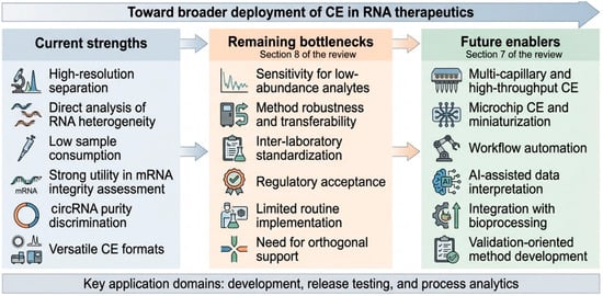
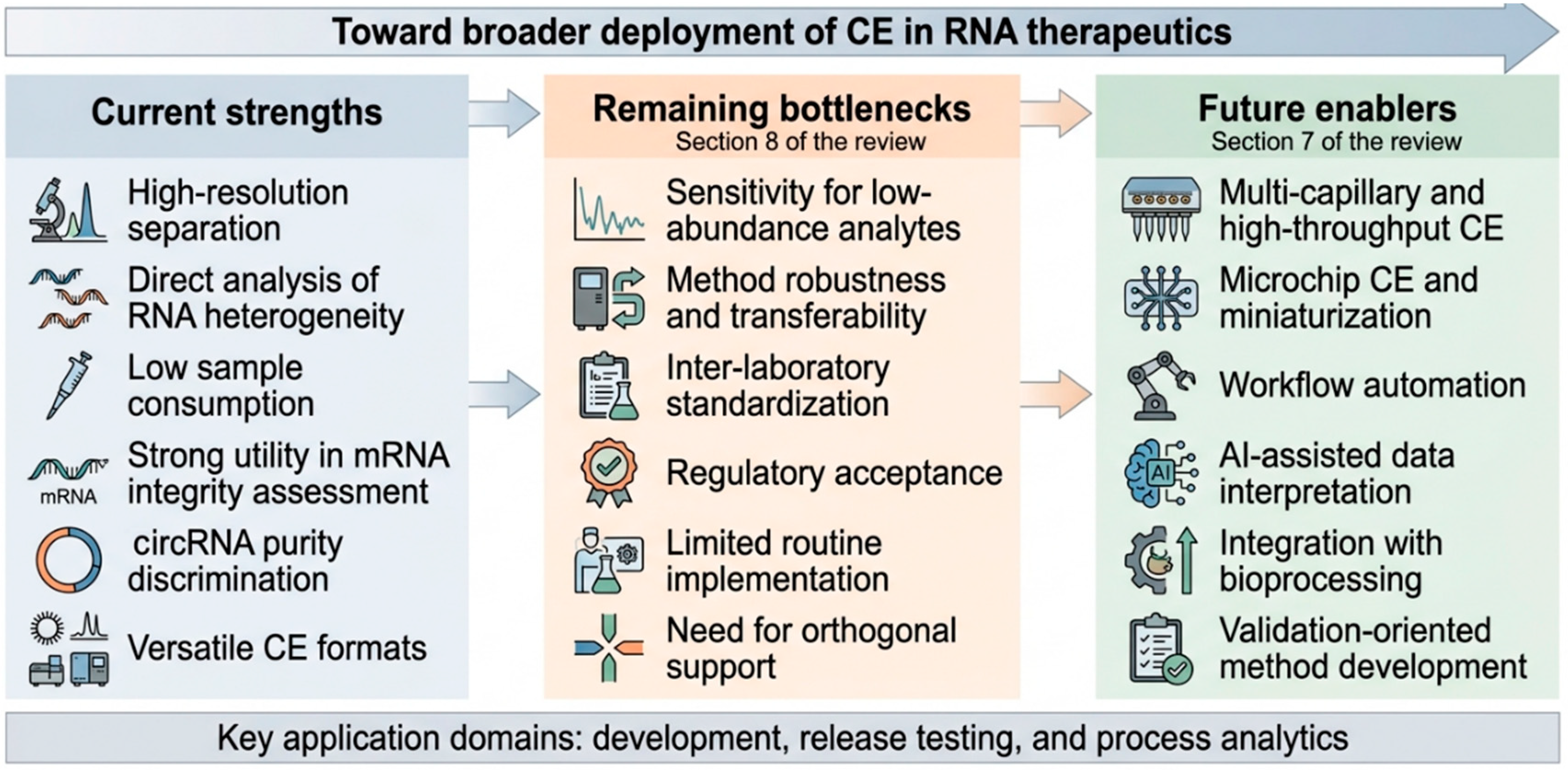
This complementarity is increasingly supported by comparative evidence rather than only conceptual reasoning. For example, recent comparative analysis of mRNA integrity methods has shown that CE-based workflows retain a distinctive advantage when the analytical objective is direct discrimination of intact transcripts, shorter RNA species, and functionally relevant heterogeneity, whereas amplification-based methods remain stronger for highly sensitive sequence-directed detection, and LC-MS remains unmatched when detailed compositional confirmation is required [47]. Furthermore, Barros et al. demonstrated that for the same mRNA product, CE measured degradation rates approximately 50% higher than IP-RP-HPLC, reflecting their fundamentally different separation mechanisms [87]. These quantitative differences underscore that each method provides a unique analytical window for integrity assessment and that CE is not simply interchangeable with chromatographic approaches. Such comparative data strengthen the argument for CE’s distinct role within an integrated analytical ecosystem. Figure 6 summarizes the opportunities and bottlenecks shaping broader CE deployment in RNA therapeutics. Accordingly, CE’s current trajectory is best understood not as displacement of PCR or LC-MS, but as increasing indispensability in the subset of analytical questions for which direct separation-based evidence is the most informative and operationally relevant readout.
Figure 6.
From orthogonal method to broader platform: opportunities and bottlenecks for capillary electrophoresis in RNA therapeutics. Capillary electrophoresis already offers key strengths in RNA therapeutics, including high-resolution separation, direct analysis of RNA heterogeneity, and utility in applications such as mRNA integrity assessment and circRNA purity discrimination. Broader deployment is still limited by challenges related to sensitivity, robustness, standardization, and regulatory acceptance. Future progress will depend on advances in throughput, miniaturization, automation, AI-assisted interpretation, and integration with bioprocessing workflows.
9. Conclusions
CE has evolved into an increasingly important analytical platform for RNA therapeutics, particularly where direct resolution of size-related heterogeneity, product-related impurities, and topology-sensitive differences is essential. This review asked whether CE can serve as a core platform for RNA therapeutic development, release testing, and process analytics. Based on the available evidence, the answer is affirmative in selected areas but still conditional. CE is closest to routine utility in mRNA integrity assessment, poly(A) tail profiling, and circRNA purity evaluation, where its high-resolution separation and quality-control compatibility provide clear advantages. In these areas, CE functions as a near-core platform in many industrial settings. In other contexts, especially RNA modification analysis and broader cross-platform workflows, CE is best viewed as a complementary or orthogonal method rather than a standalone solution.
Several advances are poised to accelerate broader deployment. Multi-capillary and high-throughput CE systems address throughput limitations. Microchip CE enables faster, lower-volume workflows. Automation and bioprocessing integration make CE more suitable for process monitoring. AI-assisted data analysis reduces operator dependence and improves interpretation consistency. However, three major bottlenecks remain: sensitivity for low-abundance analytes, robustness and standardization for method transfer, and regulatory acceptance for routine release testing. Addressing these bottlenecks will require concerted efforts in method validation according to established frameworks (e.g., ICH Q2(R2)), development of certified reference materials, and inter-laboratory collaborative studies to establish performance benchmarks. Taken together, these opportunities and constraints define a clear trajectory: CE is moving toward broader adoption, but the pace and extent of its progress will depend on how effectively the remaining bottlenecks are addressed. Considering this assessment, CE is well-positioned in selected RNA therapeutic applications. How far it advances toward full core-platform status will depend on continued progress in throughput, automation, standardization, and regulatory alignment. For now, CE is best viewed as a complementary near-core platform for mRNA and circRNA quality control in industrial settings, while recognizing that orthogonal methods remain essential for other critical quality attributes.
Author Contributions
Z.L. and Y.Y. designed the research. B.Y., X.W., W.B., C.M., S.Y. and Y.J. finished the data collection. B.Y., X.W., W.B., C.M., S.Y., Y.J. and Z.L. drafted the manuscript. B.Y., X.W., W.B., C.M., S.Y., Y.J. and Z.L. helped with writing and with analyzing the results. Y.Y. and Z.L. revised the draft and contributed new reagents and analytic tools. All authors have read and agreed to the published version of the manuscript.
Funding
This program was funded by Shanghai Pudong New Area Health Commission (PW2024A-39).
Data Availability Statement
No new data were created or analyzed in this study. Data sharing is not applicable to this article.
Acknowledgments
This work was supported by the Shanghai Municipal Innovation and Entrepreneurship Training Program.
Conflicts of Interest
The authors declare no conflicts of interest.
References
- Chaudhary, N.; Weissman, D.; Whitehead, K.A. mRNA vaccines for infectious diseases: Principles, delivery and clinical translation. Nat. Rev. Drug Discov. 2021, 20, 817–838. [Google Scholar] [CrossRef] [Scilit] [PubMed]
- Hogan, M.J.; Pardi, N. mRNA vaccines in the COVID-19 pandemic and beyond. Annu. Rev. Med. 2022, 73, 17–39. [Google Scholar] [CrossRef] [Scilit] [PubMed]
- Damase, T.R.; Sukhovershin, R.; Boada, C.; Taraballi, F.; Pettigrew, R.I.; Cooke, J.P. The limitless future of RNA therapeutics. Front. Bioeng. Biotechnol. 2021, 9, 628137. [Google Scholar] [CrossRef] [Scilit] [PubMed]
- Winkle, M.; El-Daly, S.M.; Fabbri, M.; Calin, G.A. Noncoding RNA therapeutics—Challenges and potential solutions. Nat. Rev. Drug Discov. 2021, 20, 629–651. [Google Scholar] [CrossRef] [Scilit] [PubMed]
- Watts, J.M.; Dang, K.K.; Gorelick, R.J.; Leonard, C.W.; Bess, J.W., Jr.; Swanstrom, R.; Burch, C.L.; Weeks, K.M. Architecture and secondary structure of an entire HIV-1 RNA genome. Nature 2009, 460, 711–716. [Google Scholar] [CrossRef] [Scilit] [PubMed]
- Romero-López, C.; Ramos-Lorente, S.E.; Berzal-Herranz, A. In vitro methods to decipher the structure of viral RNA genomes. Pharmaceuticals 2021, 14, 1192. [Google Scholar] [CrossRef] [Scilit] [PubMed]
- Solayman, M.; Litfin, T.; Singh, J.; Paliwal, K.; Zhou, Y.; Zhan, J. Probing RNA structures and functions by solvent accessibility: An overview from experimental and computational perspectives. Brief. Bioinform. 2022, 23, bbac112. [Google Scholar] [CrossRef] [Scilit] [PubMed]
- Martini, P.G.; Guey, L.T. A new era for rare genetic diseases: Messenger RNA therapy. Hum. Gene Ther. 2019, 30, 1180–1189. [Google Scholar] [CrossRef] [Scilit] [PubMed]
- Raffaele, J.; Loughney, J.W.; Rustandi, R.R. Development of a microchip capillary electrophoresis method for determination of the purity and integrity of mRNA in lipid nanoparticle vaccines. Electrophoresis 2022, 43, 1101–1106. [Google Scholar] [CrossRef] [Scilit] [PubMed]
- Warzak, D.A.; Pike, W.A.; Luttgeharm, K.D. Capillary electrophoresis methods for determining the IVT mRNA critical quality attributes of size and purity. SLAS Technol. 2023, 28, 369–374. [Google Scholar] [CrossRef] [Scilit] [PubMed]
- Chen, B.; Yuan, B.-F.; Feng, Y.-Q. Analytical methods for deciphering RNA modifications. Anal. Chem. 2018, 91, 743–756. [Google Scholar] [CrossRef] [Scilit] [PubMed]
- Voeten, R.L.; Ventouri, I.K.; Haselberg, R.; Somsen, G.W. Capillary electrophoresis: Trends and recent advances. Anal. Chem. 2018, 90, 1464–1481. [Google Scholar] [CrossRef] [Scilit] [PubMed]
- Li, Y.; Miao, S.; Tan, J.; Zhang, Q.; Chen, D.D.Y. Capillary electrophoresis: A three-year literature review. Anal. Chem. 2024, 96, 7799–7816. [Google Scholar] [CrossRef] [Scilit] [PubMed]
- Lechner, A.; Wolff, P.; Leize-Wagner, E.; Francois, Y.-N. Characterization of post-transcriptional RNA modifications by sheathless capillary electrophoresis–high resolution mass spectrometry. Anal. Chem. 2020, 92, 7363–7370. [Google Scholar] [CrossRef] [Scilit] [PubMed]
- Kawai, T. Recent advances in trace bioanalysis by capillary electrophoresis. Anal. Sci. 2021, 37, 27–36. [Google Scholar] [CrossRef] [Scilit] [PubMed]
- Su, W.-C.; Yu, Z.; Lieu, R.; Freund, E.; Hokeness, K.; Gu, Y.; Kohler, J.; Chen, T.; Fan, Y.; Zhang, K. Developing a high-throughput capillary gel electrophoresis workflow for mRNA integrity and poly (a) tail analysis. J. Chromatogr. A 2025, 1761, 466387. [Google Scholar] [CrossRef] [Scilit] [PubMed]
- Zheng, W.; Liu, D.; Chen, H.; Zhao, J.; Zhang, S. A methodological and mechanistic exploration to precise separation of circular RNA and nicked RNA by capillary gel electrophoresis with laser-induced fluorescence. J. Chromatogr. A 2026, 1772, 466809. [Google Scholar] [CrossRef] [Scilit] [PubMed]
- Wang, Y.; Li, L.; Sutton, A.T.; Tu, Q.; Zhao, K.; Wen, E.; Osborn, J.; Singh, A.; Gunsch, M.J.; Rustandi, R.R.; et al. Development of a capillary zone electrophoresis method to monitor magnesium ion consumption during in vitro transcription for mRNA production. Anal. Bioanal. Chem. 2024, 416, 2941–2949. [Google Scholar] [CrossRef] [Scilit] [PubMed]
- Hu, Y.; Jiang, G.; Wen, Y.; Shao, Y.; Yang, G.; Qu, F. Selection of aptamers targeting small molecules by capillary electrophoresis: Advances, challenges, and prospects. Biotechnol. Adv. 2025, 78, 108491. [Google Scholar] [CrossRef] [Scilit] [PubMed]
- Ragab, M.A.; El-Kimary, E.I. Recent advances and applications of microfluidic capillary electrophoresis: A comprehensive review (2017–mid 2019). Crit. Rev. Anal. Chem. 2021, 51, 709–741. [Google Scholar] [PubMed]
- Ali, I.; Messali, M.; Gogolashvili, A.; Giunashvili, L. Advances in artificial intelligence and machine learning in capillary electrophoresis. Anal. Methods 2025, 17, 9304–9318. [Google Scholar] [CrossRef] [Scilit] [PubMed]
- Camperi, J.; Lippold, S.; Ayalew, L.; Roper, B.; Shao, S.; Freund, E.; Nissenbaum, A.; Galan, C.; Cao, Q.; Yang, F.; et al. Comprehensive impurity profiling of mRNA: Evaluating current technologies and advanced analytical techniques. Anal. Chem. 2024, 96, 3886–3897. [Google Scholar] [CrossRef] [Scilit] [PubMed]
- Riekkola, M.-L.; Joensson, J.A.; Smith, R.M. Terminology for analytical capillary electromigration techniques. Pure Appl. Chem. 2004, 76, 443–451. [Google Scholar] [CrossRef] [Scilit]
- Maryutina, T.A.; Savonina, E.Y.; Fedotov, P.S.; Smith, R.M.; Siren, H.; Hibbert, D.B. Terminology of separation methods (IUPAC Recommendations 2017). Pure Appl. Chem. 2018, 90, 181–231. [Google Scholar]
- Santos, I.C.; Brodbelt, J.S. Recent developments in the characterization of nucleic acids by liquid chromatography, capillary electrophoresis, ion mobility, and mass spectrometry (2010–2020). J. Sep. Sci. 2021, 44, 340–372. [Google Scholar] [CrossRef] [Scilit] [PubMed]
- Vásconez, J.; Pero-Gascon, R.; Giménez, E.; Benavente, F. Comparison of capillary electrophoresis and zwitterionic-hydrophilic interaction capillary liquid chromatography with ultraviolet and mass spectrometry detection for the analysis of microRNA biomarkers. Talanta 2020, 219, 121263. [Google Scholar] [CrossRef] [Scilit] [PubMed]
- Yamaguchi, Y.; Li, Z.; Zhu, X.; Liu, C.; Zhang, D.; Dou, X. Polyethylene oxide (PEO) and polyethylene glycol (PEG) polymer sieving matrix for RNA capillary electrophoresis. PLoS ONE 2015, 10, e0123406. [Google Scholar] [CrossRef] [Scilit] [PubMed]
- Stolz, A.; Jooß, K.; Höcker, O.; Römer, J.; Schlecht, J.; Neusüß, C. Recent advances in capillary electrophoresis-mass spectrometry: Instrumentation, methodology and applications. Electrophoresis 2019, 40, 79–112. [Google Scholar] [PubMed]
- Sumitomo, K.; Sasaki, M.; Yamaguchi, Y. Acetic acid denaturing for RNA capillary polymer electrophoresis. Electrophoresis 2009, 30, 1538–1543. [Google Scholar] [CrossRef] [Scilit] [PubMed]
- Li, Z.; Dou, X.; Ni, Y.; Sumitomo, K.; Yamaguchi, Y. Acetic acid denaturing pulsed field capillary electrophoresis for RNA separation. Electrophoresis 2010, 31, 3531–3536. [Google Scholar] [CrossRef] [Scilit] [PubMed]
- Li, Z.; Liu, C.; Zhang, D.; Luo, S.; Yamaguchi, Y. Capillary electrophoresis of RNA in hydroxyethylcellulose polymer with various molecular weights. J. Chromatogr. B 2016, 1011, 114–120. [Google Scholar] [CrossRef] [Scilit]
- Di Grandi, D.; Dayeh, D.M.; Kaur, K.; Chen, Y.; Henderson, S.; Moon, Y.; Bhowmick, A.; Ihnat, P.M.; Fu, Y.; Muthusamy, K.; et al. A single-nucleotide resolution capillary gel electrophoresis workflow for poly(A) tail characterization in the development of mRNA therapeutics and vaccines. J. Pharm. Biomed. Anal. 2023, 236, 115692. [Google Scholar] [CrossRef] [Scilit] [PubMed]
- Wang, A.; Zhao, S.; Tian, M.; Yang, J.; Yang, L. Ultrasensitive and multiplexed microRNA analysis: Integrated RNA microreactor-capillary electrophoresis-mass spectrometry platform. Talanta 2026, 296, 128441. [Google Scholar] [CrossRef] [Scilit] [PubMed]
- Rollo, D.; Kulkarni, A.; Yu, K.; Fabris, D. Investigating the Merits of Microfluidic Capillary Zone Electrophoresis–Mass Spectrometry (CZE-MS) in the Bottom-Up Characterization of Larger RNAs. J. Am. Soc. Mass Spectrom. 2024, 35, 561–574. [Google Scholar] [CrossRef] [Scilit] [PubMed]
- Zeng, Y.; Wang, P.; Yang, B.; Xu, Y.; Wang, Y.; Li, Z.; Yamaguchi, Y. A Brief Review on the Analysis of dsDNA, RNA, Amino Acids and Bacteria by Capillary Electrophoresis. Bioengineering 2025, 12, 1306. [Google Scholar] [CrossRef] [Scilit] [PubMed]
- Xie, Q.; Chen, J.; Zhang, J.; Chu, Z.; Zhang, F.; Wang, Q. Quantification of multiple microRNAs by microchip electrophoresis assisted by strand displacement amplification. J. Chromatogr. A 2024, 1730, 465087. [Google Scholar] [CrossRef] [Scilit] [PubMed]
- Camperi, J.; Chatla, K.; Freund, E.; Galan, C.; Lippold, S.; Guilbaud, A. Current analytical strategies for mRNA-based therapeutics. Molecules 2025, 30, 1629. [Google Scholar] [CrossRef] [Scilit] [PubMed]
- Guimaraes, G.J.; Kim, J.; Bartlett, M.G. Characterization of mRNA therapeutics. Mass Spectrom. Rev. 2024, 43, 1066–1090. [Google Scholar] [PubMed]
- Zhang, C.; Zhang, B. RNA therapeutics: Updates and future potential. Sci. China Life Sci. 2023, 66, 12–30. [Google Scholar] [PubMed]
- Lenk, R.; Kleindienst, W.; Szabó, G.T.; Baiersdörfer, M.; Boros, G.; Keller, J.M.; Mahiny, A.J.; Vlatkovic, I. Understanding the impact of in vitro transcription byproducts and contaminants. Front. Mol. Biosci. 2024, 11, 1426129. [Google Scholar] [CrossRef] [Scilit] [PubMed]
- Camperi, J.; Roper, B.; Freund, E.; Leylek, R.; Nissenbaum, A.; Galan, C.; Caothien, R.; Hu, Z.; Ko, P.; Lee, A.; et al. Exploring the impact of in vitro-transcribed mRNA impurities on cellular responses. Anal. Chem. 2024, 96, 17789–17799. [Google Scholar] [CrossRef] [Scilit] [PubMed]
- Nicholson, A.L.; Pasquinelli, A.E. Tales of detailed poly (A) tails. Trends Cell Biol. 2019, 29, 191–200. [Google Scholar] [CrossRef] [Scilit] [PubMed]
- Liu, X.; Hu, C.; He, Q.; Bai, Y.; Zhang, X.; Fu, Z.; Ma, X.; Xu, M.; Liang, Z.; Mao, Q. Research progress on immune mechanism and control strategy of dsRNA impurities in mRNA vaccine. Expert Rev. Vaccines 2025, 24, 457–469. [Google Scholar] [CrossRef] [Scilit] [PubMed]
- Webb, A.L.; Welbourne, E.N.; Evans, C.A.; Dickman, M.J. Characterisation and analysis of mRNA critical quality attributes using liquid chromatography based methods. J. Chromatogr. A 2025, 1745, 465724. [Google Scholar] [CrossRef] [Scilit] [PubMed]
- Tran, J.P.; Gao, J.; Lansdell, C.; Lorbetskie, B.; Johnston, M.J.; Wang, L.; Li, X.; Lu, H. A Comprehensive Evaluation of Analytical Method Parameters Critical to the Reliable Assessment of Therapeutic mRNA Integrity by Capillary Gel Electrophoresis. Electrophoresis 2025, 46, 365–375. [Google Scholar] [CrossRef] [Scilit] [PubMed]
- Lu, T.; Klein, L.J.; Ha, S.; Rustandi, R.R. High-Resolution capillary electrophoresis separation of large RNA under non-aqueous conditions. J. Chromatogr. A 2020, 1618, 460875. [Google Scholar] [CrossRef] [Scilit] [PubMed]
- Lardellier, P.; Malburet, C.; Morani, M.; Willemin, T.; Rol-Moreno, J.; Noir, A.; Fertier-Prizzon, S.; Francois-Heude, M. Comparative study of analytical methods for assessing mRNA integrity and identification of functional mRNA oligomers. Sci. Rep. 2025, 15, 43557. [Google Scholar] [CrossRef] [Scilit] [PubMed]
- Mantri, P.; Juneja, B.; Henderson, S.; Koufos, E.; Moon, Y.; Dayeh, D.M.; Di Grandi, D.; Fu, Y.; Muthusamy, K.; Ihnat, P.M.; et al. Comparison of capillary electrophoresis-based methods for the analytical characterization of purity and stability of in vitro transcribed mRNA. J. Pharm. Biomed. Anal. 2024, 249, 116352. [Google Scholar] [CrossRef] [Scilit] [PubMed]
- Pulido, M.A.; Gutierrez, M.; Wang, F.; Borgos, S.E.; Parot, J. mRNA-LNP Nucleic Acid Assessment from Distinct Formulations by Multi-Capillary Gel Electrophoresis. Sciex Application Note. 2022. Available online: https://sciex.com/content/dam/SCIEX/pdf/tech-notes/biopharma/mrna-lnp-nucleic-acid-assessment.pdf (accessed on 10 June 2026).
- Rivera Santiago, C.; Quintanar Stephano, A.; Barrera Saldaña, H.A. Design, Synthesis, and Proof-of-Concept Bioassay of an Encapsulated mRNA for Human Growth Hormone. Curr. Issues Mol. Biol. 2026, 48, 647. [Google Scholar] [CrossRef] [Scilit]
- Rosa, S.S.; Prazeres, D.M.; Azevedo, A.M.; Marques, M.P. mRNA vaccines manufacturing: Challenges and bottlenecks. Vaccine 2021, 39, 2190–2200. [Google Scholar] [CrossRef] [Scilit] [PubMed]
- Duan, T.-L.; Jiao, H.; He, G.-J.; Yan, Y.-B. Translation Efficiency and Degradation of ER-Associated mRNAs Modulated by ER-Anchored poly(A)-Specific Ribonuclease (PARN). Cells 2020, 9, 162. [Google Scholar] [CrossRef] [Scilit] [PubMed]
- Pulido, M.; Holper, P.; Luo, J.; Baker, P.; Li, T.; Mollah, S. Separation of Circular RNA from the Linear Precursor by Multi-Capillary Electrophoresis. SciEx Technical Notes. 2023. Available online: https://sciex.com/content/dam/SCIEX/pdf/tech-notes/biopharma/RUO-MKT-02-27537-A-update.pdf (accessed on 12 June 2026).
- Guillen-Cuevas, K.; Lu, X.; Birtwistle, M.R.; Husson, S.M. Purifying circular RNA by ultrafiltration. Sep. Purif. Technol. 2025, 366, 132809. [Google Scholar] [CrossRef] [Scilit] [PubMed]
- Cheng, F.; Li, J.; Hu, C.; Bai, Y.; Liu, J.; Liu, D.; He, Q.; Jin, Q.; Mao, Q.; Liang, Z.; et al. Study on the characterization and degradation pattern of circular RNA vaccines using an HPLC method. Chemosensors 2024, 12, 120. [Google Scholar] [CrossRef] [Scilit]
- Sun, Y.; Zhang, Y.; Chen, W.; Chen, T.; Zhang, Y.; Zhang, S.; Lu, C. Enhancing Circular RNA Translation Efficiency Through Dual Internal Ribosome Entry Sites. Biology 2026, 15, 317. [Google Scholar] [CrossRef] [Scilit] [PubMed]
- Sun, Y.; Khimani, A.H.; Tong, Y.; Lu, Z.X. Analytical methods to evaluate RNA circularization efficiency. Electrophoresis 2024, 45, 2005–2011. [Google Scholar] [CrossRef] [Scilit] [PubMed]
- Jiang, Y.; Kjems, J. RNA denaturation underlies circular RNA separation. Nucleic Acids Res. 2025, 53, gkaf1160. [Google Scholar] [CrossRef] [Scilit] [PubMed]
- Barbieri, I.; Kouzarides, T. Role of RNA modifications in cancer. Nat. Rev. Cancer 2020, 20, 303–322. [Google Scholar] [CrossRef] [Scilit] [PubMed]
- Yu, Y.; Zhu, S.-H.; Yuan, F.; Zhang, X.-H.; Lu, Y.-Y.; Zhou, Y.-L.; Zhang, X.-X. Ultrasensitive and simultaneous determination of RNA modified nucleotides by sheathless interfaced capillary electrophoresis–tandem mass spectrometry. Chem. Commun. 2019, 55, 7595–7598. [Google Scholar] [CrossRef] [Scilit]
- Khan, N.; Mironov, G.; Berezovski, M.V. Direct detection of endogenous MicroRNAs and their post-transcriptional modifications in cancer serum by capillary electrophoresis-mass spectrometry. Anal. Bioanal. Chem. 2016, 408, 2891–2899. [Google Scholar] [CrossRef] [Scilit] [PubMed]
- Herbert, C.; Valesyan, S.; Kist, J.; Limbach, P.A. Analysis of RNA and its modifications. Annu. Rev. Anal. Chem. 2024, 17, 47–68. [Google Scholar] [CrossRef] [Scilit]
- Ravelet, C.; Grosset, C.; Peyrin, E. Liquid chromatography, electrochromatography and capillary electrophoresis applications of DNA and RNA aptamers. J. Chromatogr. A 2006, 1117, 1–10. [Google Scholar] [CrossRef] [Scilit] [PubMed]
- Zhu, C.; Yang, G.; Ghulam, M.; Li, L.; Qu, F. Evolution of multi-functional capillary electrophoresis for high-efficiency selection of aptamers. Biotechnol. Adv. 2019, 37, 107432. [Google Scholar] [CrossRef] [Scilit] [PubMed]
- Mendonsa, S.D.; Bowser, M.T. In vitro evolution of functional DNA using capillary electrophoresis. J. Am. Chem. Soc. 2004, 126, 20–21. [Google Scholar] [PubMed]
- Zhao, L.; Yang, G.; Zhu, C.; Li, L.; Zhao, Y.; Luan, Y.; Qu, F. Three-step evolutionary enhanced capillary electrophoresis-SELEX for aptamer selection of exosome vesicles. Talanta 2024, 267, 125203. [Google Scholar] [CrossRef] [Scilit] [PubMed]
- Le, A.T.; Krylova, S.M.; Krylov, S.N. Ideal-filter capillary electrophoresis: A highly efficient partitioning method for selection of protein binders from oligonucleotide libraries. Electrophoresis 2019, 40, 2553–2564. [Google Scholar] [CrossRef] [Scilit] [PubMed]
- Le, A.T.; Krylova, S.M.; Kanoatov, M.; Desai, S.; Krylov, S.N. Ideal-filter capillary electrophoresis (IFCE) facilitates the one-step selection of aptamers. Angew. Chem. 2019, 131, 2765–2769. [Google Scholar] [CrossRef] [Scilit]
- Teclemichael, E.; Le, A.T.; Krylova, S.M.; Wang, T.Y.; Krylov, S.N. Bulk Affinity Assays in Aptamer Selection: Challenges, Theory, and Workflow. Anal. Chem. 2022, 94, 15183–15188. [Google Scholar] [CrossRef] [Scilit] [PubMed]
- Feng, L.; Sun, Y.; Jia, W.; Yu, Y.; Liu, C.; Yang, J.; Luan, Y.; Chen, J.; Wang, F. Advancements in SELEX technology for aptamers and emerging applications in therapeutics and drug delivery. Biomolecules 2025, 15, 818. [Google Scholar] [CrossRef] [Scilit] [PubMed]
- Suzuki, Y.; Tsunehara, H.; Miyauchi, S.; Miyakawa, M.; Sugiyama, K.; Ishimitsu, M.; Tasaki-Handa, Y.; Yoshimoto, K.; Saito, S. Reliable Post-SELEX Design of Promising Bivalent Aptamers through the Systematic Capillary Electrophoresis Exploration of Ternary DNA–Protein Complexes. Anal. Chem. 2025, 97, 10978–10987. [Google Scholar] [CrossRef] [Scilit] [PubMed]
- Mendonsa, S.D.; Bowser, M.T. In vitro selection of high-affinity DNA ligands for human IgE using capillary electrophoresis. Anal. Chem. 2004, 76, 5387–5392. [Google Scholar] [CrossRef] [Scilit] [PubMed]
- Yang, G.; Tian, W.; Hu, Y.; Wen, Y.; Zhao, Y.; Jiang, G.; Liu, H.; Zhao, L.; Zhu, C.; Qu, F. Real-time visualization of “small Molecules-ssDNA” complexes for aptamer screening based on online competition CE-SELEX. Talanta 2025, 284, 127199. [Google Scholar] [CrossRef] [Scilit] [PubMed]
- Zhu, C.; Li, L.; Yang, G.; Irfan, M.; Wang, Z.; Fang, S.; Qu, F. High-efficiency selection of aptamers for bovine lactoferrin by capillary electrophoresis and its aptasensor application in milk powder. Talanta 2019, 205, 120088. [Google Scholar] [CrossRef] [Scilit] [PubMed]
- Aeksiri, N.; Warakulwit, C.; Hannongbua, S.; Unajak, S.; Choowongkomon, K. Use of capillary electrophoresis to study the binding interaction of aptamers with wild-Type, K103N, and double mutant (K103N/Y181C) HIV-1 RT: Studying the binding interaction of wild-type, K103N, and double mutant (K103N/Y181C) HIV-1 RT with aptamers by performing the capillary electrophoresis. Appl. Biochem. Biotechnol. 2017, 182, 546–558. [Google Scholar] [PubMed]
- Cong, H.; Xu, X.; Yu, B.; Yuan, H.; Peng, Q.; Tian, C. Recent progress in preparation and application of microfluidic chip electrophoresis. J. Micromech. Microeng. 2015, 25, 053001. [Google Scholar] [CrossRef] [Scilit]
- Wei, K.; Zhao, J.; Qin, Y.; Li, S.; Huang, Y.; Zhao, S. A novel multiplex signal amplification strategy based on microchip electrophoresis platform for the improved separation and detection of microRNAs. Talanta 2018, 189, 437–441. [Google Scholar] [CrossRef] [Scilit] [PubMed]
- Pan, J.-Z.; Fang, P.; Fang, X.-X.; Hu, T.-T.; Fang, J.; Fang, Q. A low-cost palmtop high-speed capillary electrophoresis bioanalyzer with laser induced fluorescence detection. Sci. Rep. 2018, 8, 1791. [Google Scholar] [CrossRef] [Scilit] [PubMed]
- Lee, J.W.; Lee, D.; Kim, Y.T.; Lee, E.Y.; Kim, D.H.; Seo, T.S. Low-cost and facile fabrication of a paper-based capillary electrophoresis microdevice for pathogen detection. Biosens. Bioelectron. 2017, 91, 388–392. [Google Scholar] [CrossRef] [Scilit] [PubMed]
- Karabiber, F.; McGinnis, J.L.; Favorov, O.V.; Weeks, K.M. QuShape: Rapid, accurate, and best-practices quantification of nucleic acid probing information, resolved by capillary electrophoresis. RNA 2013, 19, 63–73. [Google Scholar] [PubMed]
- Kim, H.; Cordero, P.; Das, R.; Yoon, S. HiTRACE-Web: An online tool for robust analysis of high-throughput capillary electrophoresis. Nucleic Acids Res. 2013, 41, W492–W498. [Google Scholar] [CrossRef] [Scilit] [PubMed]
- Gumna, J.; Zok, T.; Figurski, K.; Pachulska-Wieczorek, K.; Szachniuk, M. RNAthor–fast, accurate normalization, visualization and statistical analysis of RNA probing data resolved by capillary electrophoresis. PLoS ONE 2020, 15, e0239287. [Google Scholar] [CrossRef] [Scilit] [PubMed]
- Hu, L.; Krylova, S.M.; Liu, S.K.; Yousef, G.M.; Krylov, S.N. Necessity and challenges of sample preconcentration in analysis of multiple microRNAs by capillary electrophoresis. Anal. Chem. 2020, 92, 14251–14258. [Google Scholar] [CrossRef] [Scilit] [PubMed]
- IHT Guideline, Validation of Analytical Procedures Q2 (R2); ICH: Geneva, Switzerland, 2022; p. 1.
- Vuolo-Schuessler, L.; Newton, M.E.; Smith, P.; Burgess, C.; McDowall, R.D. Harmonizing USP <1058> and GAMP for analytical instrument qualification. Pharm. Eng. 2014, 34, 46–56. Available online: https://www.researchgate.net/profile/Christopher-Burgess-7/publication/292828121_Harmonizing_USP_1058_and_GAMP_for_analytical_instrument_qualification/links/609a7b1ca6fdccaebd213c96/Harmonizing-USP-1058-and-GAMP-for-analytical-instrument-qualification.pdf (accessed on 12 June 2026).
- Nogueira, S.S.; Samaridou, E.; Simon, J.; Frank, S.; Beck-Broichsitter, M.; Mehta, A. Analytical techniques for the characterization of nanoparticles for mRNA delivery. Eur. J. Pharm. Biopharm. 2024, 198, 114235. [Google Scholar] [CrossRef] [Scilit] [PubMed]
- Barros, C.H.; Alfaro, M.; Csiki-Fejer, A.; Bhatnagar, B.; Tschessalov, S.; Ferguson, S.; Barua, S.; Darvari, R.; Topp, E.M. Comparative analysis of mRNA degradation kinetics using chromatographic and electrophoretic methods. Mol. Pharm. 2025, 22, 3061–3072. [Google Scholar] [CrossRef] [Scilit] [PubMed]

Disclaimer/Publisher’s Note: The statements, opinions and data contained in all publications are solely those of the individual author(s) and contributor(s) and not of MDPI and/or the editor(s). MDPI and/or the editor(s) disclaim responsibility for any injury to people or property resulting from any ideas, methods, instructions or products referred to in the content. |
© 2026 by the authors. Licensee MDPI, Basel, Switzerland. This article is an open access article distributed under the terms and conditions of the Creative Commons Attribution (CC BY) license.