


Summary
Ultrasound has been used as a diagnostic tool by neurologists since the 1980s, when the first pioneers observed changed patterns of muscle echotexture in patients with muscle dystrophy. Since then, vast enhancements in ultrasound resolution and quality have added to its utility as an inexpensive, painless, real-time diagnostic instrument. By now, the value of ultrasound in peripheral entrapment neuropathies has been firmly established. Improved understanding in the field of neuromuscular diseases e.g., inflammatory neuropathies and motor neurone diseases, has positioned ultrasonography as a parameter of nerve and muscle morphology to complement traditional electrodiagnostic studies. But there is a catch: training and experience are essential. This is why recognised experts in the field and beginners met at the first Nerve and Muscle Ultrasound Hands-on Teaching Course on 6th and 7th of March 2015 in Lucerne, convened by local senior consultant neurologist Dr. Anne-Kathrin Peyer-Kauffmann [1].
Common nerve entrapments
After an in-depth review of relevant anatomical structures with live demonstrations and hands-on sessions in small groups (five people maximally), expert Prof. Einar Wilder-Smith, from the University Medical Centre at the Yong Loo Lin School of Medicine in Singapore, gave insight into his longstanding experience in peripheral nerve ultrasound and taught about its role in common nerve entrapments. Ultrasound can add to the sensitivity and specifity of electrodiagnostics in carpal tunnel syndrome (CTS) and correlates to the grade of electrodiagnostic abnormality. Its high positive predictive value enables use as a painless screening test for CTS, eliminating the need for electrodiagnostic studies in some patients [2]. The biomechanical aspects in ulnar entrapment neuropathy at the elbow can be revealed only by real-time ultrasound examination in flexion and extension, another unique feature of peripheral nerve ultrasound. The site of entrapment (cubital tunnel or ulnar groove) is reliably discernable and the responsible anatomic structures e.g., an anconeus epitrochlearis muscle, can be pointed out to the treating physician. Important aetiological and prognostic information can also be provided in the case of intraneural ganglion cysts, which can occur in diverse nerves, often mimicking conventional pressure palsies [3]. Ultrasound resolution is now high enough to depict in great detail even nerve entrapments at distal sites or involving small nerves, as was demonstrated in tarsal tunnel syndrome, entrapment of the deep radial nerve or the plantar nerves. Posttraumatic nerve continuity can often be best evaluated sonographically when electrodiagnostics are unhelpful or not applicable.
Dysimmune and hereditary neuropathies
As a clinician, one is often confronted with difficult and treatment-relevant distinctions between severe axonal and demyelinating neuropathies and even more so, between different types of demyelinating neuropathies. Alexander Grimm, senior consultant and expert in neuromuscular ultrasound from the University Hospital Basel, reported on his recently published findings on the differentiation of immune neuropathies by use of ultrasound. A key finding is that by measuring the enlargement of peripheral nerve cross-sectional areas (CSAs), demyelination can be detected in correlation with onion bulb formation in chronic demyelinating inflammatory neuropathies [4]. The dimension of CSA enlargement can also help differentiate between the different types of hereditary neuropathies, with hereditary motor and sensory neuropathy (HMSN) type Ia showing the largest engorgement of peripheral nerves. Characteristic additional features, which are currently being studied, are the intranerve variability index which helps in differentiating hereditary from acquired demyelinating polyneuropathies, and the CSA of the vagus nerve, also a determinant of autonomic involvement in Guillain-Barré Syndrome. Prof. Wilder-Smith added tropical flair by contributing with his experience on peripheral nerve changes in leprosy.
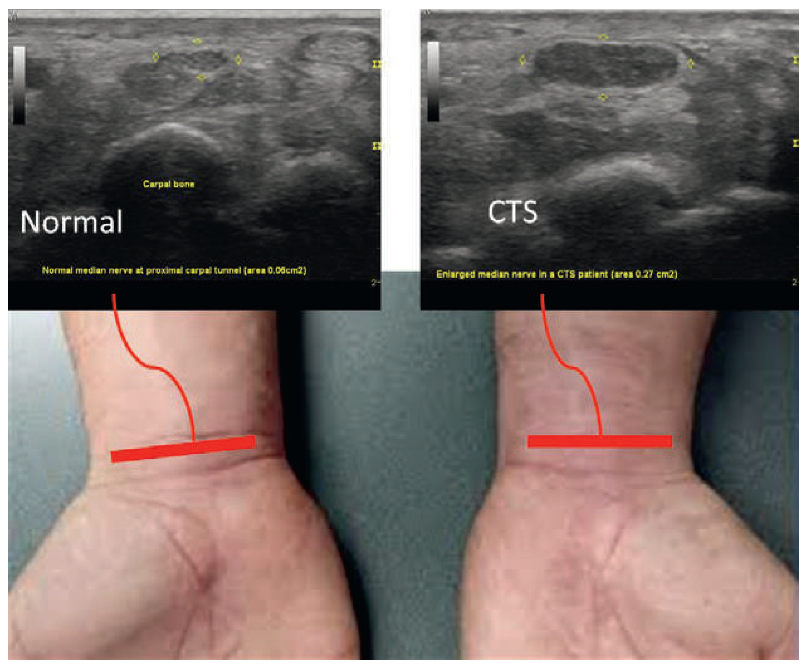
Figure 1.
Nerve ultrasound cross sectional image of the healthy median nerve (in picture: left) and in carpal tunnel syndrome (right). Note the significantly enlarged cross sectional area of 27 mm2 vs 6 mm2.
Muscle ultrasound
The youngest “up-and-comer” of peripheral sonography in neurology is muscular ultrasound, which has also gained impetus owing to recent technological advances. The muscle echogenicity classified by use of the Heckmatt-Score was established in the late 1980s in order to assess and (semi-)quantify neurogenic muscle degeneration patterns, often by-passing cost- and time-consuming muscle MRI. Nowadays, the resolution of muscle ultrasound is sufficient to detect fasciculations noninvasively and even in deep tissue structures, helping to diagnose amyotrophic lateral sclerosis (ALS) particularly when considering the new revised Awajii criteria [5]. Muscle ultrasound is set to develop into an important complementary technique for evaluating lower motor neuron disorders with the additional bonus of being painless as compared with standard needle myography. This, without decreasing diagnostic sensitivity and specifity [6]. Muscles may even show early sonographic changes before clinical paresis is manifest. Variation in homogeneity of muscle echotexture changes is an additional, useful, diagnostic feature in the distinction of neuromuscular disorders.
A sound training
Deliberately shutting out the sunny Lucerne mountain skyline, the participants focused on the ultrasound screens for hands-on experience of not-so-everyday neurological diseases in patients with adult onset Pompe’s disease, neurofibromatosis, Churg-Strauss syndrome and a brachial plexus gunshot wound, amongst others. Practical aspects were covered as well by Alexander Grimm, who instructed on how to write a peripheral sonography report, and by guest speaker senior consultant David Lorenzana, who entertainingly provided an anaesthetist’s perspective. After some practice sessions the participants were able to take home useful skills in evaluating neuromuscular disorders with ultrasound and were handed out their certification according to DEGUM/SSGUM/SSUM/ÖGUM standards. The course organisers are already looking forward to next year’s second Nerve and Muscle Ultrasound Teaching Course, presumably in early March 2016.
Peripheral neuromuscular ultrasound is an exciting and vibrant field that has filled some important gaps in the existing array of neurologic diagnostic tools. It will be interesting to see which new applications the future will bring. Research on the contribution of peripheral nerve and muscle ultrasound is generating so much clinically relevant classification and additional information, that concise practical hands-on workshops will form the basis of enabling crucial clinical translation.
Disclosures
No financial support and no other potential conflict of interest relevant to this article was reported.
References
- Programme at https://www.luks.ch/standorte/luzern/kliniken/zentrum-fuer-neurologie-und-neurorehabilitation-znn.html.
- Pastare D, Therimadasamy AK, Lee E, Wilder-Smith EP. Sonography versus nerve conduction studies in patients referred with a clinical diagnosis of carpal tunnel syndrome. J Clin Ultrasound. 2009 Sep;37(7):389–93.
- Schilg L, Hägele-Link S, Felbecker A, Gers B, Weber J, Tettenborn B et al. Nerve sonography of intraneural ganglia as cause painful peroneal palsies: a case series. T. Praxis (Bern 1994). 2014 Nov 26;103.
- Grimm A, Heiling B, Schumacher U, Witte OW, Axer H. Ultrasound differentiation of axonal and demyelinating neuropathies. Muscle Nerve. 2014 Dec;50(6):976–83.
- Misawa S, Noto Y, Shibuya K, Isose S, Sekiguchi Y, Nasu S et al. Ultrasonographic detection of fasciculations markedly increases diagnostic sensitivity of ALS. Neurology. 2011 Oct 18;77(16):1532–7.
- Grimm A, Prell T, Décard BF, Schumacher U, Witte OW, Axer H et al. Muscle ultrasonography as an additional diagnostic tool for the diagnosis of amyotrophic lateral sclerosis. J Clin Neurophysiol. 2015 Apr;126(4):820–7.
This is an open-access article distributed under the terms of the Creative Commons Attribution Non-Commercial No Derivatives (CC BY-NC-ND) 4.0 License.