


Summary
Thrombolytics have the potential to reduce disability of acute ischaemic stroke patients without increasing mortality, if patients are carefully selected. Per 1000 patients treated 110–150 less will be disabled. Intravenous thrombolysis with recombinant tissue plasminogen activator (rt-PA) given within 3 hours after stroke onset has been shown to reduce the rate of long-term-disability significantly. The clinical benefit of intraarterial thrombolysis in acute stroke patients with M1 or M2 occlusion of the middle cerebral artery, when performed up to six hours of symptom onset, has recently been proved in a randomised trial. In addition, several case series indicate that intraarterial thrombolysis and intravenous thrombolysis can be administered safely in clinical practice and improve clinical outcome when the occluded vessel is recanalised. In acute stroke due to basilar artery occlusion intraarterial thrombolysis is a therapeutic option at longer time intervals in specialist stroke centres. Several series of patients with basilar artery occlusion suggest that intraarterial thrombolysis has the potential to enhance the chances of basilar artery recanalisation and improve clinical outcome. In some of these studies, using various thrombolytic agents and intervals to treatment, an association between vessel recanalisation and clinical outcome was described. The percentage of patients with a favourable functional clinical outcome is more than 50% when early recanalisation of the basilar artery can be achieved and very low when the artery remains occluded. To date, no randomised controlled trial has compared intraarterial thrombolysis and intravenous thrombolysis. Intraarterial thrombolysis has several advantages: arteriography assesses the complete vessel status and collateral circulation.The thrombolytic medication can be applied directly into the thrombus. Vessel recanalisation as well as reocclusion are visualised directly, and in the case of rapid recanalisation, the infusion of the thrombolytic drug can be stopped before the maximum dose is applied. If pharmacological thrombolysis cannot be achieved, mechanical recanalisation procedures like thrombus perforation, thrombaspiration or percutaneous transluminal angioplasty are alternative or adjunctive strategies. In addition, mechanical recanalisation can sometimes be timesaving and achieved within a few minutes and faster than pharmacological reopening of the vessel. Limitations of intraarterial thrombolysis include the potential risk of arteriography, the time delay due to diagnostic arteriography and that it can only be applied at institutions with an experienced interventional neuroradiology team. Bleeding represents a hazard as with intravenous thrombolysis. Another major advantage of intraarterial thrombolysis is the extended time window up 6 hours compared to the 3 hours of intravenous thrombolysis.Therefore, intraarterial thrombolysis increases the number of patients who are eligible for thrombolysis. Nevertheless, campaigns and protocols for early referral to stroke centres and a standardised treatment algorithm are urgently needed. Educational programs for both the physicians and the public have the potential to increase the percentage of patients admitted to stroke centres within the time window for thrombolysis.
Introduction
Stroke is the third leading cause of death and the main cause of adult disability and ranks second as a cause of dementia in industrialised countries. In the last decade, with the administration of thrombolytic agents, treatment of patients with acute ischaemic stroke has been revolutionised. Intravenous thrombolysis with recombinant tissue plasminogen activator (rt-PA) has been shown to significantly reduce the rate of long-term-disability in acute stroke patients treated within 3 hours of symptom onset [1]. However, the majority of stroke patients are not eligible for intravenous thrombolysis because of the short time window. Local intraarterial thrombolysis involves selective administration of the fibrinolytic agent directly in the occluded cerebral artery.The clinical benefit of intraarterial thrombolysis in acute stroke patients with M1 or M2 occlusion of the middle cerebral artery within up to six hours of symptom onset has recently been proved in a randomised placebo-controlled trial [2].
In the light of these encouraging results, acute stroke has become a medical emergency involving more and more intensive care facilities.This review will focus on the benefit and limitations of early intraarterial reperfusion strategies and the role of critical care in acute stroke patients.
Intraarterial thrombolysis in the posterior circulation
Acute basilar artery occlusion is a serious condition that often leads to a severe disability or death [3,4]. Before the area of thrombolysis, mortality rates of up to 90% were reported. No randomised trials comparing conventional treatment with intraarterial thrombolysis have been performed until today. However, since the late 1980s several case series have suggested that intraarterial thrombolysis has the potential to improve basilar artery recanalisation and clinical outcome [5,6,7]. In some of these studies, using various thrombolytic agents and time intervals to treatment, an association between vessel recanalisation and clinical outcome was described. The percentage of patients with a favourable functional clinical outcome varies between 20 and 35% (Table 1) [5,6,7,8,9,10,11]. In the light of these results, in many European centres intraarterial thrombolysis is the treatment of choice in acute stroke due to basilar artery occlusion. However, intraarterial thrombolysis requires that the patient is admitted to an experienced stroke institution with access to cerebral arteriography and interventional neuroradiology.Therefore, both ASA and EUSI recommend that this therapeutic option should be done only as an experimental therapy in specialist stroke centres [12,13].
Table 1.
Literature review of thrombolysis in basilar artery occlusion: recanalisation and clinical outcome.
In our series we reported 40 patients with acute stroke following basilar artery occlusion treated within 12 hours of symptom onset [10]. The average time from symptom onset to thrombolysis was 5 hours 30 minutes. Complete or partial recanalisation of the basilar artery was achieved in 80% of the patients. Clinical outcome after three months was favourable (mRS ≤2) in 35%, 41% of the patients died. Low baseline NIHSS (National Institute of Health Stroke Scale) score on admission and vessel recanalisation were independent predictors of a favourable outcome. Only one of 15 patients with coma and tetraplegia before intraarterial thrombolysis recovered to an mRS grade 2 or less. However, in a series of Wijdicks et al. 2 patients with locked-in syndrome during several hours before intraarterial thrombolysis recovered fully after successful thrombolysis [14]. Therefore, in our opinion, patients with coma, quadriplegia and high NIHSS scores should not be excluded from intraarterial thrombolysis, although a poor outcome is more likely. The decision to give or to withhold intraarterial thrombolysis to such patients remains a matter of debate, and in such cases diffusion and perfusion MRI may be helpful to decide for or against [15]. However, the benefit of this approach has to be proven by further studies.
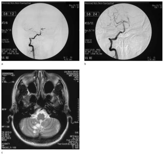
Figure 1 shows pre- and post-treatment angiograms and MRI of a successful intraarterial thrombolysis in a 48-year-old woman.
Figure 1.
Angiography of a 48-year-old woman with basilar artery occlusion before (a) and after (b) successful intraarterial thrombolysis. The basilar artery is completely recanalised after thrombolysis. Two days later T2-weighted MRI shows small ischaemic right-sided cerebellar lesions (c). The patient had no residual symptoms after 3 months.
Intraarterial thrombolysis in the anterior circulation
In the early 1980s case series of intraarterial thrombolysis in the anterior circulation with promising results were reported [16]. In 1999 a randomised trial showed significant clinical benefit of intraarterial recombinant prourokinase in acute stroke patients with M1 or M2 segment occlusion of the middle cerebral artery up to 6 hours from symptom onset [2]. Intraarterial thrombolysis restored vessel patency at least partially in two thirds of the patients with middle cerebral artery occlusion. In the placebo group partial or complete vessel recanalisation rate was only 18%. Mortality at three months was 25% in the recombinant prourokinase group and 27% in the control group. Symptomatic intracerebral haemorrhage occurred in 10% of the patients treated with intraarterial thrombolysis and low dose heparin and in 2% given low dose heparin and placebo. The high bleeding rate in the treatment group may be explained by the fact that only patients with M1 or M2 segment occlusion of the middle cerebral artery mostly with severe pretreatment neurological deficits (median NIHSS score 17) were included in this study. At three months 40% of prourokinase patients and 25% of controls had no significant disability (modified Rankin score 2 or less; p = 0.04). To date no second randomised trial has confirmed these results. Therefore, intraarterial thrombolysis is not yet FDA approved. The European Stroke Initiative (EUSI) states that intraarterial thrombolysis of acute middle cerebral artery occlusion with prourokinase in a 6-hour time window results in improved outcome (level of evidence II) [12]. However, the American Stroke Association (ASA) is more conservative [17]. The members of the stroke council of the American Stroke Association mentioned in recent guidelines for the early management of patients with ischaemic stroke that physicians with expertise in endovascular therapy are using intraarterial techniques to treat patients with acute ischaemic stroke secondary to occlusion of large intracranial arteries including the basilar or middle cerebral arteries [13]. They added that intraarterial thrombolysis is an option for treatment of selected patients with major stroke of <6 hours’ durations due to large vessel occlusions of the middle cerebral artery. However, they stated that it should be recognised that intraarterial thrombolysis is not FDA approved. Further, the drug (recombinant prourokinase) tested in the referenced prospective randomised trial of intraarterial thrombolysis is not available for clinical use. Therefore, extrapolation to the available thrombolytic drugs (rtPA in the USA, rtPA and urokinase in some European countries) is based on consensus as supported by case series data.
From May 1992 to December 2003 289 patients with acute stroke were treated with intraarterial thrombolysis using urokinase at our institution. Results of 100 patients with middle cerebral artery occlusion, 24 patients with internal carotid artery T-occlusion and 32 patients with basilar artery occlusion have been published earlier [10,18,19]. Indirect comparisons with the results of randomised trials showed favourable results in patients with middle cerebral artery occlusion indicating that intraarterial thrombolysis may be safe and effective also in routine clinical practice, being aware that differences in patient selection and vessel territories involved limit comparison with randomised trials. The percentage of favourable outcome and the mortality in our patients with middle cerebral artery occlusion treated within 6 hours of symptom onset is similar to the NINDS trial, despite earlier treatment (within 3 hours of symptom onset) in NINDS and similar baseline stroke severity. Even when the M1 or M2 segment of the middle cerebral artery was occluded, 59% of the patients had no significant disability after 3 months [18]. The partial or complete recanalisation rate of M1 or M2 occlusions was 79%, which is higher than in PROACT II (66%) and higher than after intravenous thrombolysis (48%) or in the spontaneous course (24%) [20]. The higher proportion of patients that regained functional independence compared with the PROACT II study might be explained by the lower median baseline NIHSS score of our patients (14 versus 17) and the shorter average time to treatment (234 versus 320 minutes). In addition, the lower age (59 versus 64 years) favoured our patients. Moreover, mechanical disruption of the clot, in addition to pharmacological thrombolysis, was performed in some of our patients leading to a higher recanalisation rate than in PROACT II. These promising results have to be confirmed by randomised trials.
In patients with carotid T-occlusion the prognosis is usually poor, even when intraarterial thrombolysis is performed. The results of the most important case series are summarised in Table 2 [19,21,22,23,24,25,26]. Of our 24 patients with carotid T-occlusion only 4 patients (17%) had a favorable outcome (mRS ≤2) after three months and 10 patients (42%) died. Sufficient collaterals and age <60 years were the only predictors of favourable clinical outcome [19].
Table 2.
Literature review of thrombolysis in carotid T-occlusion: recanalisation and clinical outcome.
Advantages and limitations of intraarterial thrombolysis
Until today, no randomised controlled trial comparing intraarterial thrombolysis with intravenous thrombolysis has been performed. The benefit of intravenous thrombolysis was proven in a large randomised trial with rt-PA administered within 3 hours of symptom onset [1]. Furthermore, a metaanalysis including the NINDS trial and the two large European thrombolysis studies (ECASS I, ECASS II) showed a clear benefit of intravenous thrombolysis during a maximal time window of 3 hours [27]. Per 1000 patients treated 110–150 less will be disabled. Based on these results rt-PA was approved by the US Food and Drug Administration (FDA) and the European Medicines Evaluation Agency (EMEA) for the treatment of acute ischaemic stroke within 3 hours of symptom onset. Some patients may benefit from intravenous thrombolysis even after 3 hours of symptom onset, however, American and European agencies have not approved this use of rt-PA [28]. Therefore, the narrow time window is a limitation for the use of intravenous thrombolysis in clinical practice [29]. Intraarterial thrombolysis can be administered with a time interval from symptom onset to treatment of up to 6 hours in patients with M1 or M2 segment middle cerebral artery occlusions. In our institution 20% of all acute stroke patients were treated with intraarterial thrombolysis. In addition, intraarterial thrombolysis has several advantages. Arteriography gives information on the vessel status and collateral circulation; the thrombolytic medication can be applied directly into the thrombus; vessel recanalisation as well as reocclusion is visualised; and in the case of rapid complete recanalisation, the infusion of the thrombolytic drug can be stopped before the maximum dose is applied. If pharmacological thrombolysis cannot be achieved, mechanical recanalisation procedures like thrombus perforation, thrombaspiration and percutaneous transluminal angioplasty are possible alternative or adjunctive strategies.
Limitations of intraarterial thrombolysis are the potential risk of arteriography, the time delay due to diagnostic arteriography and that it can only be applied at institutions with an experienced interventional neuroradiology service. However, the acute management by organised stroke teams has the potential to improve door-to-needle times [30]. Another potential approach is combined intravenous/intraarterial thrombolysis [31,32].The idea of this treatment is to give a part of the thrombolytic drug immediately after CT scan and then to proceed to intraarterial thrombolysis with injection of the remaining thrombolytic drug directly into the clot. However, further trials are required to prove the clinical benefit of this combined approach.
Indications and contraindications for intraarterial thrombolysis
Most stroke centres with experience in intraarterial thrombolysis use indications and contraindications similar to the PROACT II protocol [2]. Because recombinant prourokinase is neither available on the US nor the European market, agents used include urokinase and rt-PA in Europe and rt-PA in the USA.
According to the PROACT II protocol intraarterial thrombolysis can be performed if (1) new focal signs in the middle cerebral artery distribution allowing initiation of intraarterial thrombolysis within 6 hours of symptom onset are present; (2) baseline NIHSS score is >4 points, except for isolated heminanopia and aphasia; (3) age is 18 to 85 years; (4) occlusion of the M1 or M2 segment of the middle cerebral artery is visualized on arteriography. The most important exclusion criteria of the PROACT II protocol were as follows: (1) NIHSS score >30; (2) spontaneous rapidly improving neurological signs; (3) history of stroke within the previous 6 weeks; (4) seizure at symptom onset; (5) clinical presentation suggestive of subarachnoidal haemorrhage; (6) previous history of intracranial haemorrhage; (7) neoplasm;
(8) subarachnoidal haemorrhage; (9) septic embolism; (10) suspected lacunar stroke; (11) surgery, biopsy of a parenchymal organ, trauma with internal injuries or lumbar puncture within 30 days;
(12) head trauma within 30 days; (13) active or recent haemorrhage within 30 days; (14) known hemorrhagic diathesis; (15) baseline international normalised ration (INR) >1.7 or activated partial thromboplastin time more than 1.5 times normal or platelet count less than 100 × 109/L (100 × 103/µL);
(16) known contrast sensitivity; (17) uncontrolled hypertension (>180 mm Hg systolic or >100 mm Hg diastolic); (18) intracranial tumours on CT scan except small meningioma; (19) haemorrhage on CT scan; (20) significant mass effect with midline shift and early CT signs of ischaemia in more than one third of the middle cerebral artery territory on CT scan.
The role of intensive care medicine in acute stroke patients
Patient selection
The main reason for admission of stroke patients to the intensive care units are post interventional treatment after pharmacological or mechanical thrombolysis, clinical deterioration, disturbance of consciousness, requirement of intubation and mechanical ventilation, seizures, myocardial infarction and cardiac arrhythmia.
In our stroke centre intensive care facilities play an important role in the management of body temperature, ventilation, blood pressure, infections, increased intracranial pressure and seizures.
Body temperature
Increased body temperature is a predictor of poor outcome in acute stroke patients [33]. A recent meta-analysis indicated that elevated body temperature is associated with an increase in disability and mortality [34]. It is recommended to treat fever with antipyretic agents and/or physical measures. Acetaminophen was shown to lower body temperature in acute stroke patients by 0.3 to 0.4 °C in two small randomised phase II clinical trials [35,36]. Whether this therapeutic approach results in a clinical benefit remains open.
Data from animal studies indicate that systemic cooling has the potential to reduce the ischaemic brain damage [37].
The feasibility of systemic or endovascular moderate cooling to 32 to 33 °C in humans has been shown in small uncontrolled trials [38,39]. However, aggressive cooling has many potential side effects. The occurrence of complications like blood pressure instability, infections, coagulation disorders and cardiac arrhythmia may limit a potential benefit of this therapeutic approach in clinical practice.
Oxygen
Routine use of oxygen has not shown any clinical benefit in stroke patients until today. However, based on pathophysiological considerations supplemental oxygen is recommended in the case of hypoxaemia. The EUSI recommends a minimal target O2 saturation ≥92%, the ASA a minimal O2 saturation ≥95% [12,13].
Blood pressure management
Optimal blood pressure management in acute stroke remains controversial. According to the guidelines of the American Stroke Association (ASA) and the European Stroke Initiative (EUSI) antihypertensive agents should only be given if the systolic blood pressure is >220 mm Hg or the diastolic blood pressure is >120 mm Hg [12,13]. Before and during the first 24 hours after thrombolyis systolic blood pressure values >185 or diastolic blood pressures >110 mm Hg should be avoided.
Rapid lowering of blood pressure may be hazardous in acute stroke patients. When antihypertensive agents are used, it should be done cautiously. Recommended first-line agents include intravenous labetalol and orally administered nicardipin and captopril. In the case of severe diastolic hypertension intravenous infusion of nitroprussid may be preferred.
In patients with hypotension correction of hypovolaemia and optimising cardiac function are essential.
In a recent study induced hypertension using norepinephrine has been associated with a significant rise in blood flow velocity in the middle cerebral artery measured by transcranial ultrasound and cerebral perfusion pressure [40]. The safety and feasibility of norepinephrine-induced arterial hypertension in acute ischaemic stroke has been shown in a recent case series. Mean arterial pressure could be elevated by 10 to 20%. Maximal variability of systolic blood pressure was within 15% of the target values. Complications such as cardiac arrhythmia or intracerebral haemorrhage were rare [41]. However, the clinical benefit of this therapeutic approach has not yet been proven. In clinical practice we use vasopressor agents especially in patients with clinical deterioration associated with low blood pressure and a markedly reduced blood flow velocity of the middle cerebral artery measured by transcranial ultrasound.
Hyperglycaemia
Hyperglycaemia is associated with increased risk of mortality and poor functional recovery in nondiabetic stroke patients [42,43]. However, this may be at least partly due to diabetic comorbidity or increased initial stroke severity.
Possible explanations for a worse outcome in patients with hyperglycaemia include increased blood-brain-barrier permeability and tissue acidosis. Therefore, lowering of high blood glucose concentration with insulin is recommended. The recommended target blood glucose levels vary between <11 mmol/L (European Stroke Initiative) and <16.6 mmol/L (American Stroke Association) [12,13].
Increased intracranial pressure and brain oedema
Large infarctions in the middle cerebral artery territory with space-occupying brain oedema leading to elevated intracranial pressure and herniation usually within 2 to 5 days are called malignant middle cerebral artery infarctions. Clinical signs of malignant middle cerebral artery infarction are signs of brainstem herniation and deterioration of consciousness. The malignant, space-occupying middle cerebral artery infarction is associated with a mortality rate of up to 80% when only conventional treatment modalities are used [44].
Possible conservative treatment modalities of elevated intracranial pressure include corticosteroids, hyperventilation, osmotherapy (glycerol, mannitol, hypertonic saline solution, Tris-hydroxymethyl-aminomethane) and barbiturates. Corticosteroids have not proven any beneficial effects in acute stroke patients [45,46]. Corticosteroids may even be harmful, because infections are more common in patients with corticosteroid therapy.
Therefore, corticosteroids are not recommended in the management of cerebral oedema following acute ischaemic stroke [17].
Hyperventilation with induced hypocarbia has not proven any benefit in acute stroke patients either [47]. Induced hypocarbia may even be harmful by reducing the perfusion of the penumbra.
The goal of osmotherapy is to reduce brain volume by fluid shift by increasing serum osmolarity to about 315 to 320 mOsm/L [48,49]. We use mannitol 20% solution every 4 hours as first-line agent. However, the efficacy of this therapeutic approach has not been ascertained yet. In general, all these conservative treatment modalities are effective only over a short time and fail to prevent herniation. In some cases rebound intracranial hypertension has been described.
Decompressive surgery is likely to reduce mortality in these patients with some individuals achieving functional improvement [50]. Hemicraniectomy allows the swollen brain to expand and may prevent compression of the brainstem and preserve leptomeningeal collateral circulation. However, the effect on disability has to be proven in a randomised trial. Case series suggest that early craniectomy may be more effective than later decompressive surgery [51]. It has been recommended that the diameter of the craniectomy has to be at least 12 cm [51]. The identification of predictors of space-occupying oedema may be helpful to select patients for early craniectomy. Thomalla et al. showed that quantitative analysis of early DWI and PWI parameters can be helpful to predict malignant middle cerebral artery infarction; in addition, an NIHSS score ≥19 was highly sensitive in predicting malignant infarction [52]. Other possible predictors for malignant infarction include coma on admission, nausea, vomiting, elevated systolic blood pressure, history of hypertension, history of heart failure, CT signs of early ischaemia affecting more than 50% of the middle cerebral artery territory, hyperdense middle cerebral artery sign on CT, local brain swelling or midline shift on CT, or carotid T-occlusion on arteriography [26,53,54,55,56]. Possible complications of surgery include epidural or subdural haematoma, hygroma and infections [51]. In patients with severe ischaemic stroke, decompressive surgery resulted in lower mortality and lower complication rates compared with moderate hypothermia [57]. Both treatment modalities, however, were associated with a prolonged stay in the neurological intensive care unit. Because of the lack of randomised trials and clear selection criteria, there is no consensus on indications and contraindications of early decompressive craniectomy in patients with space-occupying middle cerebral artery infarction. Prospective randomised trials (HeADFIRST [Hemicraniectomy and Durotomy upon Deterioration for Massive Hemispheric Infarction Related Swelling Trial] and HAMLET [Hemicraniectomy After MCA infarction with Life-threatening Edema Trial]) are ongoing and will hopefully provide more information on this controversial subject. At the moment, the decision has to be taken individually in each patient considering ethical and medical aspects.
Also in space-occupying cerebellar infarction compressing the brainstem and leading to hydrocephalus, surgery can be life saving often with favourable functional clinical outcome [58].
Seizures
Seizures occur in about 5% of acute stroke patients within the first weeks and are more frequent in infarcts involving the cerebral cortex than in lacunar infarcts. Prophylactic use of anticonvulsants in patients with acute stroke who have not had seizures is not recommended.There is no consensus whether to start anticonvulsant treatment after a first seizure and which drug should be given. However, recurrent seizures after stroke should be treated with antiepileptic drugs as with any other condition [17,59].
Outlook for the future
At present, in clinical practice only a limited number of patients are eligible for thrombolysis [60]. Protocols for early referral to stroke centres and a standardised treatment algorithm are urgently needed. Educational programs for both the physicians and the public have the potential to increase the percentage of patients admitted to stroke centres within the time window for thrombolysis [61]. In addition, further trials to prove the efficacy of intraarterial thrombolysis and compare it with intravenous thrombolysis are urgently needed.
The identification of patient subgroups most likely to benefit from thrombolyis even within a longer time window, by means of perfusion CT, perfusion and diffusion MRI and MR angiography are subjects of current studies. New agents with neuroprotective effects have not yet proven to be beneficial in acute stroke. However, earlier administration of neuroprotective agents in combination with thrombolytic therapy may be successful in further trials. The efficacy of promising more invasive treatment strategies, including mechanical thrombus perforation, thrombaspiration, angioplasty, stent implantation, ultrasound-assisted thrombolysis or laser-induced thrombolysis have to be evaluated in larger trials [62,63,64,65].
References
- The NINDS rt-PA Stroke Study Group. Tissue plasminogen activator for acute ischemic stroke. N Engl J Med 1995;33:1581–7.
- Furlan A, Higashida R, Wechsler L, Gent M, Rowley H, Kase C, et al. Intra-arterial prourokinase for acute ischemic stroke. The PROACT II Study: a randomized controlled trial. JAMA 1999;282:2003–11.
- Castaigne P, Lhermitte F, Gautier JC, Escourolle R, Derouesne C, Der Agopian P, et al. Arterial occlusions in the vertebrobasilar system: a study of 44 patients with post-mortem data. Brain 1973;96:133–54.
- Archer CR, Horenstein S. Basilar artery occlusion: clinical and radiological correlation. Stroke 1977;8:383–90.
- Hacke W, Zeumer H, Ferbert A, Brückmann H, del Zoppo GJ. Intra-arterial thrombolytic therapy improves outcome in patients with acute vertebrobasilar occlusive disease. Stroke 1988;19:1216–22.
- Zeumer H, Freitag HJ, Grzyska U, Neunzig HP. Local intra-arterial fibrinolysis in acute vertebrobasilar occlusion. Neuroradiology 1989;31:336–40.
- Brandt T, von Kummer R, Müller-Küppers M, Hacke W. Thrombolytic therapy of acute basilar artery occlusion. Stroke 1996;27:875–81.
- Becker KJ, Monsein LH, Ulatowski J, Mirski M, Williams M, Hanley DF. Intraarterial thrombolysis in vertebrobasilar occlusion. AJNR Am J Neuroradiol 1996;17:255–62.
- Cross DT, Moran CJ, Akins PT, Angtuaco EE, Derdeyn CP, Diringer MN. Collateral circulation and outcome after basilar artery thrombolysis. AJNR Am J Neuroradiol 1998;19:1557–63.
- Arnold M, Nedeltchev K, Schroth G, Baumgartner RW, Remonda L, Loher TJ, et al. Clinical and radiological predictors of recanalization and outcome in 40 patients with acute basilar arter y occlusion treated with intraarterial thrombolysis. J Neurol Neurosurg Psychiatry 2004;75:857–62.
- Huemer M, Niederwieser V, Ladurner G. Thrombolytic treatment for acute occlusion of the basilar artery. J Neurol Neurosurg Psychiatry 1995;58:227–8.
- Hacke W, Kaste M, Bogousslavsky J, Brainin M, Chamorro A, Lees K, et al.; European Stroke Initiative Executive Committee and the EUSI Writing Committee. European Stroke Initiative Recommendations for Stroke Management – update 2003. Cerebrovasc Dis 2003;16:311–37.
- Adams HP Jr, Adams RJ, Brott T, del Zoppo GJ, Furlan A, Goldstein LB, et al. Guidelines for the early management of patients with ischaemic stroke. A scientific statement from the Stroke Council of the American Stroke Association. Stroke 2003;34:1056–83.
- Wijdicks EF, Nichols DA, Thielen KR, Fulgham JR, Brown RD Jr, Meissner I, et al. Intra-arterial thrombolysis in acute basilar artery thromboembolism: the initial Mayo Clinic experience. Mayo Clin Proc 1997;72:1005–13.
- du Mesnil de Rochemont R, Neumann-Haefelin T, Berkefeld J, Sitzer M, Lanfermann H. Magnetic resonance imaging in basilar artery occlusion. Arch Neurol 2002;59:398–402.
- del Zoppo GJ, Ferbert A, Otis S, Bruckmann H, Hacke W, Zyroff J, et al. Local intra-aterial fibrinolytic therapy in acute carotid territory stroke. A pilot study. Stroke 1988;19:307–13.
- Klijn CJ, Hankey GJ; American Stroke Association and European Stroke Initiative. Management of acute ischaemic stroke: new guidelines from the American Stroke Association and European Stroke Initiative. Lancet Neurol 2003;2:698–701.
- Arnold M, Schroth G, Nedeltchev K, Loher T, Remonda L, Stepper F, et al. Intra-arterial thrombolysis in 100 patients with acute stroke due to middle cerebral artery occlusion. Stroke 2002;33:1828–33.
- Arnold M, Nedeltchev K, Mattle HP, Loher TJ, Stepper F, Schroth G, et al. Intra-arterial thrombolysis in 24 consecutive patients with internal carotid artery T-occlusions. J Neurol Neurosurg Psychiatry 2003;74:739–42.
- Neumann-Haefelin T, du Mesnil de Rochemont R, Fiebach JB, Gass A, Nolte C, Kucinski T, et al. Kompetenznetz Schlaganfall Study Group. Effect of incomplete (spontaneous and postthrombolytic) recanalization after middle cerebral artery occlusion: a magnetic resonance imaging study. Stroke 2004;35:109–14.
- Zeumer H, Freitag H-J, Zanella F, Thie A, Arning C. Local intra-arterial fibrinolytic therapy in patients with stroke: urokinase versus recombinant tissue plasminogen activator (r-TPA). Neuroradiology 1993;35:159–62.
- Sasaki O, Takeuchi S, Koike T, Koizumi T, Tanaka R. Fibrinolytic therapy for acute embolic stroke: intravenous, intracarotid, and intra-arterial local approaches. Neurosurgery 1995;36:246–52.
- Bollaert PE, Bracard S, Boulanger T, Picard L, Larcan A. Early local intra-arterial thrombolysis for severe middle cerebral artery stroke. Cerebrovasc Dis 1995;5:292–6.
- Jansen O, von Kummer R, Forsting M, Hacke W, Sartor K. Thrombolytic therapy in acute occlusion of the intracranial internal carotid artery bifurcation. AJNR Am J Neuroradiol 1995;16:1977–86.
- Urbach H, Ries F, Ostertun B, Solymosi L. Local intraarterial fibrinolysis in thromboembolic “T” occlusions of the internal carotid artery. Neuroradiology 1997;39:105–10.
- Kucinski T, Koch C, Grzyska U, Freitag HJ, Kromer H, Zeumer H. The predictive value of early CT and angiography for fatal hemispheric swelling in acute stroke. AJNR Am J Neuroradiol 1998;19:839–46.
- Wardlaw JM, del Zoppo G, Yamaguchi T. Thrombolysis for acute ischemic stroke (Cochrane Review). In: The Chochrane Library, Issue 4, 1999. Oxford: Updated software.
- Hacke W, Donnan G, Fieschi C, Kaste M, von Kummer R, Broderick JP, et al. ATLANTIS Trials Investigators; ECASS Trials Investigators; NINDS rt-PA Study Group Investigators. Association of outcome with early stroke treatment: pooled analysis of ATLANTIS, ECASS, and NINDS rt-PA stroke trials. Lancet 2004;363:768–74.
- Schenkel J, Weimar C, Knoll T, Haberl RL, Busse O, Hamann GF, et al.; German Stroke Data Bank Collaborators. R1 – systemic thrombolysis in German stroke units – the experience from the German Stroke data bank. J Neurol 2003;250:320–4.
- Nedeltchev K, Arnold M, Brekenfeld C, Isenegger J, Remonda L, Schroth G, et al. Pre- and in-hospital delays from stroke onset to intra-arterial thrombolysis. Stroke 2003;34:1230–4.
- Lewandowski CA, Frankel M, Tomsick TA, Broderick J, Frey J, Clark W, et al. Combined intravenous and intraarterial r-TPA versus intra-arterial therapy of acute ischemic stroke: Emergency Management of Stroke (EMS) Bridging Trial. Stroke 1999;30:2598–605.
- Keris V, Rudnicka S, Vorona V, Enina G, Tilgale B, Fricbergs J. Combined intraarterial/intravenous thrombolysis for acute ischemic stroke. AJNR Am J Neuroradiol 2001;22:352–8.
- Reith J, Jorgensen HS, Pedersen PM, Nakayama H, Raaschou HO, Jeppesen LL, et al. Body temperature in acute stroke: relation to stroke severity, infarct size, mortality, and outcome. Lancet 1996;347:422–5.
- Hajat C, Hajat S, Sharma P. Effects of poststroke pyrexia on stroke outcome: a meta-analysis of studies in patients. Stroke 2000;31:410–4.
- Dippel DW, van Breda EJ, van Gemert HM, van der Worp HB, Meijer RJ, Kappelle LJ, et al. Effect of paracetamol (acetaminophen) on body temperature in acute ischemic stroke: a double-blind, randomized phase II clinical trial. Stroke 2001;32:1607–12.
- Dippel DW, van Breda EJ, van der Worp HB, van Gemert HM, Meijer RJ, Kappelle LJ, et al. Effect of paracetamol (acetaminophen) and ibuprofen on body temperature in acute ischemic stroke. PISA, a phase II double-blind randomized placebo-controlled trial. ISRCTN 98608690. BMC Cardiovasc Disord 2003;3:2.
- Ginsberg MD, Sternau LL, Globus YT, Dietrich WD, Busto R. Therapeutic modulation of brain temperature: relevance to ischemic brain injury. Cerebrovasc Brain Metab Rev 1992;4:189–225.
- Krieger DW, De Georgia MA, Abou-Chebl A, Andrefsky JC, Sila CA, Katzan IL, et al. Cooling for acute ischemic brain damage (cool aid): an open pilot study of induced hypothermia in acute ischemic stroke. Stroke 2001;32:1847–54.
- Georgiadis D, Schwarz S, Kollmar R, Schwab S. Endovascular cooling for moderate hypothermia in patients with acute stroke: first results of a novel approach. Stroke 2001;32:2550–3.
- Schwarz S, Georgiadis D, Aschoff A, Schwab S. Effects of induced hypertension on intracranial pressure and flow velocities of the middle cerebral arteries in patients with large hemispheric stroke. Stroke 2002;33:998–1004.
- Marzan AS, Hungerbühler HJ, Studer A, Baumgartner RW, Georgiadis D. Feasibility and safety of norepinephrineinduced arterial hypertension in acute ischemic stroke. Neurology 2004;62:1193–5.
- Capes SE, Hunt D, Malmberg K, Pathak P, Gerstein HC. Stress hyperglycemia and prognosis of stroke in nondiabetic and diabetic patients: a systematic overview. Stroke 2001;32:2426–32.
- Williams LS, Rotich J, Qi R, Fineberg N, Espay A, Bruno A, et al. Effects of admission hyperglycemia on mortality and costs in acute ischemic stroke. Neurology 2002;59:67–71.
- Hofmeijer J, van der Worp HB, Kappelle LJ. Treatment of space-occupying cerebral infarction. Crit Care Med 2003;31:617–25.
- Mulley G, Wilcox RG, Mitchell JR. Dexamethasone in acute stroke. Br Med J 1978;2:994–6.
- Norris JW. Steroid therapy in acute cerebral infarction. Arch Neuol 1976;33:69–71.
- Berrouschot J, Sterker M, Bettin S, Koster J, Schneider D. Mortality of space-occupying (‘malignant’) middle cerebral artery infarction under conservative intensive care. Intensive Care Med 1998;24:620–3.
- Manno EM, Adams RE, Derdeyn CP, Powers WJ, Diringer MN. The effects of mannitol on cerebral edema after large hemispheric cerebral infarct. Neurology 1999;52:583–7.
- Bereczki D, Mihalka L, Szatmari S, Fekete K, Di Cesar D, Fulesdi B, et al. Mannitol use in acute stroke: case fatality at 30 days and 1 year. Stroke 2003;34:1730–5.
- Schwab S, Steiner T, Aschoff A, Schwarz S, Steiner HH, Jansen O, et al. Early hemicraniectomy in patients with complete middle cerebral artery infarction. Stroke 1998;29:1888–93.
- Steiner T, Ringleb P, Hacke W. Treatment options for large hemispheric stroke. Neurology 2001;57(5 Suppl 2):61–88.
- Thomalla GJ, Kucinski T, Schoder V, Fiehler J, Knab R, Zeumer H, et al. Prediction of malignant middle cerebral artery infarction by early perfusion- and diffusion-weighted magnetic resonance imaging. Stroke 2003;34:1892–9.
- Pullicino PM, Alexandrov AV, Shelton JA, Alexandrova NA, Smurawska LT, Norris JW. Mass effect and death from severe acute stroke. Neurology 1997;49:1090–5.
- Krieger DW, Demchuk AM, Kasner SE, Jauss M, Hantson L. Early clinical and radiological predictors of fatal brain swelling in ischemic stroke. Stroke 1999;30:287–92.
- Manno EM, Nichols DA, Fulgham JR, Wijdicks EF. Computed tomographic determinants of neurologic deterioration in patients with large middle cerebral artery infarctions. Mayo Clin Proc 2003;78:156–60.
- Haring HP, Dilitz E, Pallua A, Hessenberger G, Kampfl A, Pfausler B, et al. Attenuated corticomedullary contrast: an early cerebral computed tomography sign indicating malignant middle cerebral artery infarction: a casecontrol study. Stroke 1999;30:1076–82.
- Georgiadis D, Schwarz S, Aschoff A, Schwab S. Hemicraniectomy and moderate hypothermia in patients with severe ischemic stroke. Stroke 2002;33:1584–8.
- Amarenco P. The spectrum of cerebellar infarctions. Neurology 1991;41:973–9.
- Kilpatrick CJ, Davis SM, Tress BM, Rossiter SC, Hopper JL, Vandendriesen ML. Epileptic seizures in acute stroke. Arch Neurol 1990;47:157–60.
- Katzan IL, Furlan AJ, Lloyd LE, Frank JI, Harper DL, Hinchey JA, et al. Use of tissue-type plasminogen activator for acute ischemic stroke: the Cleveland area experience. JAMA 2000;283:1151–8.
- Morgenstern LB, Staub L, Chan W, Wein TH, Bartholomew LK, King M, et al. Improving delivery of acute stroke therapy: the TLL Temple Foundation Stroke Project. Stroke 2002;33:160–6.
- Schroth G, Berlis A, Mayer T, Remonda L, Brekenfeld C, Ozdoba C, et al. Therapeutic interventional neuroradiology in acute stroke. Ther Umsch 2003;60:569–83.
- Song JK, Cacayorin ED, Campbell MS, Fisher S, Malkoff MD, Alexandrov AV, et al. Intracranial balloon angioplasty of acute terminal internal carotid artery occlusions. AJNR Am J Neuroradiol 2002;23:1308–12.
- Mayer TE, Hamann GF, Brueckmann H. Mechanical extraction of a basilar-artery embolus with the use of flow reversal and a microbasket. N Engl J Med 2002;347:769–70.
- Mahon BR, Nesbit GM, Barnwell SL, Clark W, Marotta TR, Weill A, et al. North American clinical experience with the EKOS MicroLysUS infusion catheter for the treatment of embolic stroke. AJNR Am J Neuroradiol 2003;24:534–8.

This is an open-access article distributed under the terms of the Creative Commons Attribution Non-Commercial No Derivatives (CC BY-NC-ND) 4.0 License.