


Summary
Within the last 15 years, image-guided minimally invasive endovascular treatment for ruptured or un-ruptured cerebral aneurysms has surpassed surgical treatment for a subset of aneurysms, as shown in recent large randomised trials (ISAT, ISUIA). Endovascular patient care occurs at equal or lower cost than surgery. Cerebral aneurysms are of variable pathogenesis and by far the largest group consists of saccular aneurysms formed at proximal cerebral vessel bifurcations. Likely based on a focal vessel wall weakness, fragile out-pouchings typically develop in areas of high shear stress and at areas of the vessel tree where there is higher intravascular pressure, i.e. along the proximal intracranial branching of the internal carotid artery and less frequently of the vertebro-basilar system. Initiation, growth and rupture of an aneurysm are likely driven by a combination of several factors, each contributing to a variable degree in each of the evolutionary steps of an aneurysm’s history. Current treatment modalities include for open surgical treatment various techniques to reconstruct the artery (clips) or to reinforce the aneurysm wall (wrapping) or to bypass circulation of the vessel carrying an aneurysm (aneurysm trapping and bypass surgery). Less invasive, endovascular treatment techniques have been based on the principles of aneurysm filling with coils or polymers or vessel occlusion (aneurysm trapping). More recently, vessel reconstruction with stents rather than aneurysm filling has been advocated as treatment (Lylyk et al., J Neurosurg 2002;97: 1306–13). Both coils and stent techniques aim to induce slowing of blood flow and initiation of thrombosis within the aneurysm. Secondary vessel wall repair with scar tissue occurs within weeks. With small vessel wall defects (focal defects, usually less than 3–4 mm in maximum diameter), coil implantation seems to allow successful repair under this principle, provided that the lumen of the aneurysm pouch is suitable for coil introduction. With larger vessel wall defects in proportion to the vessel diameter (segmental defects), either the vessel must be occluded if tolerated by the patient, or the vessel is reconstructed with the use of stent implants. Based on initial clinical experience, stents seem to offer advantages over coils alone in the endovascular repair of segmental arterial wall defects. However, more research is required to better understand this stent function and to adapt stent technology to the new role it is asked to play in the intracranial circulation: repairing vessels with an aneurysm rather than opening a stenosis as typically requested in coronary or peripheral vessels. The following text is an update on current concepts and treatment, and how medical informatics is about to influence patient management by better lesion understanding, treatment simulation, implant design and medical engineering.
Introduction
Image-guided minimally invasive treatment (IGMIT) for cerebral aneurysms has gone through a metamorphosis, evolving from diagnosis based on old-fashioned angiographic plain X-ray films and endovascular treatment with hand-made latex balloons to, currently, advanced 3D picture analysis and minimally invasive endovascular treatment surpassing open surgical treatment for a large subgroup of patients, as shown in recent large randomised trials (ISAT [1], ISUIA [2]). Within the last 15 years, improved image technology and specific implant design have translated into better patient care at a lower price.
And the field is heading for even a brighter future, including 3D image-based treatment simulation and, likely, custom-made implants – very similar to what we have seen at our dentists’ offices – at least those of us unfortunate enough to have needed to defy aging with a dental crown or implant. This trend has now become applicable to problems deeper within the human body thanks to major advents in medical imaging and informatics,
i.e. 3D-imaging, including advanced image postprocessing capabilities, and advanced material engineering. The following text is a brief diary of what has happened until now and where the field is currently moving for the diagnosis and the minimally invasive therapy of patients suffering from cerebral aneurysms.
Ruptured and un-ruptured aneurysms: what ISAT and ISUIA teach us
Cerebral aneurysms are of variable pathogenesis but by far the largest sub-group consists of saccular out-pouchings formed at vessel bifurcations. Likely based on a focal vessel wall weakness, aneurysms develop typically in areas of high shear stress and at areas of the vessel tree where there is high intravascular pressure, i.e. in proximity of the circle of Willis, especially in its anterior portion. Multiple factors contribute to aneurysm development and evolution and therefore it might be best to consider them as following the rules of a complex system – rules that still have to be better defined for cerebral aneurysms and where medical informatics could contribute significantly. Initiation, growth and rupture of an aneurysm are likely driven by a combination of several factors, each contributing to a variable degree in each of the evolutionary steps of an aneurysm’s history. Today, most aneurysms present with rupture or are incidentally found at small size (≤10 mm), but they can be large (>10 mm to <25 mm) or, rarely, giant size (≥25 mm), in which case they likely produce mass effect with irritation of nearby cranial nerves or signs of brain compression.
Cerebral aneurysms are discovered under a variety of clinical conditions, with the most threatening in form of aneurysm rupture producing intracranial haemorrhage.
Aneurysm rupture occurs in approximately 10/100 000 people per year. Of them, 4 will die of the initial haemorrhage and 6 will reach medical treatment.The ISAT, one of the first relevant largescale randomised trials on endovascular treatment of ruptured aneurysms, has shown that for a large subgroup of patients, endovascular treatment using coils bears a significantly lower risk than open surgery when looking at the outcome within the first year. These results continue to produce many discussions, however, since their publication they have started to profoundly change the medical management of patients presenting with a ruptured aneurysm worldwide at many medical centres.
Due to increased use of high-quality medical imaging, today more than ever before, un-ruptured aneurysms are discovered incidentally on crosssectional imaging such as CT/CTA or MRI/MRA. A large ongoing study, ISUIA, looking at the spontaneous evolution of un-ruptured aneurysms when compared to treatment risk, revealed that active treatment is only warranted for lesions of a location specific minimal size. In comparison to the outcome of surgery or endovascular treatment of the same, ISUIA further revealed in the recently published prospective part of the study that, with regard to treatment risk, conclusions similar to ISAT can be drawn: current endovascular treatment methods generally bear a lower treatment risk than surgical options. In addition, ISUIA showed that surgical risk increases for patients older than 50 years of age. In contrast, endovascular treatment risk does not increase until patient age is exceeding 70 years. The factors defining the risk of spontaneous rupture correlated mostly with size and location. Whether the only difference between un-ruptured and ruptured aneurysms is haemorrhage or whether other differences exist, such as biological aspects of the wall for instance, future studies will be needed to show. In summary, in view of ISUIA results, for un-ruptured aneurysms, the risk / benefit ratio may favour treatment over the natural history (with its risk of spontaneous haemorrhage) only for those aneurysms that are over a specific minimal size for any given location. Since the average size of ruptured aneurysms is similar to those of un-ruptured aneurysms, the ISUIA findings have led to discussions, limited up till now mostly to critiques expressed within the neurosurgical community. In view of the growing number of un-ruptured lesions discovered incidentally, it will become increasingly important to identify the subset of patients at risk of haemorrhage, i.e. those likely to benefit from a preemptive treatment strategy. Note that un-ruptured aneurysms are more frequently found than ruptured aneurysms and that un-ruptured aneurysms increase in incidence with age, reaching an estimated prevalence of up to 3% in the population older than 50 years.
Concluding from the recently published data of ISAT and ISUIA, it is clear that medical imaging and image-guided minimally invasive treatment have and will have an increasing role for the clinical management of patients suffering from cerebral aneurysms, both ruptured and un-ruptured.
To give more insight into current concepts of aneurysm imaging and treatment, we wish to focus further on some additional issues.
Stents for intracranial endovascular repair of aneurysms
Current treatment options and future trends
Surgical treatment uses various techniques to reconstruct the artery (with clips) or to reinforce the aneurysm wall (with wrapping) or to bypass circulation of the vessel carrying an aneurysm (aneurysm trapping and bypass surgery). Classically, endovascular treatment techniques have been based on the principles of aneurysm filling (with coils or polymers) or vessel occlusion (aneurysm trapping). More recently, vessel reconstruction (with stents) rather than aneurysm filling has been advocated as treatment [3]. Both techniques that are involving coils or stents aim to induce slowing of blood flow and through this are capable of initiating thrombosis within the aneurysmal cavity. Secondary vessel wall repair with scar tissue occurs within weeks (see illustrative case as shown in fig. 1–4). With small vessel wall defects (focal defects, usually less than 3–4 mm in maximum diameter), coil implantation seems to allow for successful repair along this principle, provided that the lumen of the aneurysm pouch is suitable for coil introduction and filling. When in proportion to the vessel diameter there is a larger vessel wall defect (segmental defects), aneurysm filling with coil use alone may lead to a temporary exclusion of the rupture point, however, recurrence is frequently observed likely due to unchanged haemodynamical exposure of an inadequately repaired large vessel wall defect. For such cases, to obtain a lasting repair, either the vessel must be occluded if tolerated by the patient, or if feasible, the vessel is reconstructed by the use of stent implants. Stents alone or in combination with coils induce clotting of the aneurysm lumen but leave side branches open. Stents then act as a scaffolding during the vessel wall repair. Finally, stents reconstruct the main flow pathway, correcting the flow direction and preventing recurrent irritation of a weak vessel wall area from happening – stents therefore act as a flow divertor. Based on initial clinical experience, stents seem to offer advantages over coils alone in the endovascular repair of segmental arterial wall defects. However, more research is required to better adapt stent technology to the new role it is asked to play in the intracranial circulation: repairing an aneurysm rather than opening a stenosis. Novel material technology increasingly allows the navigation of low profile delivery systems to safely deliver specially designed stent implants at the level of an aneurysm.
Therefore, as concluded at the first International Intracranial Stent Meeting (ICS’04, www.neuroimaging.ch) in a near future, 3D-imagebased treatment simulation may allow for computer-assisted design of stents adapted to patientspecific conditions and likely custom-made implants may be produced to match individual patient needs.
Current diagnostic imaging and treatment planning
In view of the current treatment trends, imaging will accordingly play a different role in the future. For ruptured aneurysms, CT identifies readily the haemorrhage location and extent, and with subsequent CT-angiography (CTA) an aneurysm can be found and analysed. Different methods of post-processing allow for identification, volumetric assessment, definition of extent of vessel defect and planning of treatment. This includes access planning, implant choice and overall risk/benefit assessment. Indeed, most of the time, an angiographic study is not any more necessary for decision making and surgical or increasingly endovascular treatment is undertaken based on CTA only.
For un-ruptured aneurysms, which are increasingly found as incidental findings on cross-sectional imaging (CT, MRI), there is need to assess factors that may lead to rupture allowing for adequate treatment decision taking.
Figure 1.
Illustrative case report. A 48-year-old woman, subarachnoid haemorrhage from posterior communicating artery aneurysm on the left side treated by surgical clipping. Secondary endovascular repair for additional aneurysms on the right ICA.
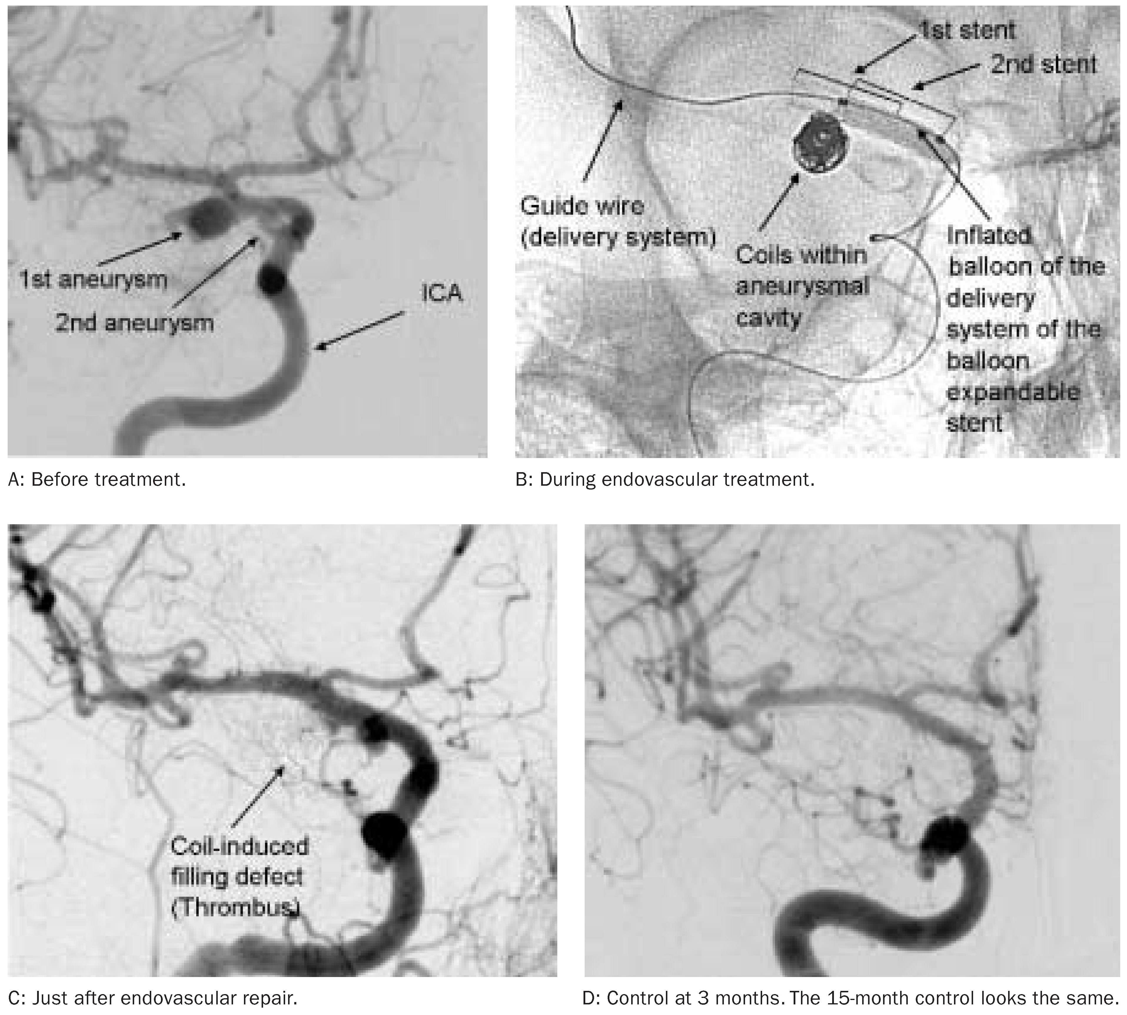
If endovascular treatment is contemplated for ruptured or un-ruptured aneurysms, digital subtraction angiography (DSA) is the imaging modality providing enhanced assessment capabilities. There is more anatomical detail and with 3D rotational angiography 3D reproduction of the diseased vessel segment including depiction of small perforating arteries and exact definition of the vessel defect is available. Based on this information, a stent treatment can be planned, if deemed necessary in view of large segmental defect to complement or replace the first principle of endovascular repair of cerebral aneurysm, i.e. filling by coil or liquid implant use.
Medical informatics is likely to influence patient management significantly by potentially allowing for simulation of some of the factors that critically govern the complex life of cerebral aneurysms. So for instance, simulation of flow effects prior and after virtual implantation of a
variety of stents may help to make the choice of the most suitable stent to likely solve the specific aneurysm condition of an individual patient. Such computer-assisted design (CAD) capabilities are likely to also have impact on implant design and medical engineering in the field of image-guided minimally invasive treatment (IGMIT) using stents or flow divertors alike. As engineering at nanotechnology dimensions becomes increasingly accessible, one can imagine to be able to produce CAD-based medical device design and translate such information through photo-lithographic material production technologies or alike into future medical implants.
Aneurysm research questions
The view of aneurysm treatment from inside the vessel gives rise to several questions. In view of ISAT and new treatment modalities, a first group must address the function of stents as a treatment adjunct, especially understanding the effect of stents on blood flow and feasibility of cerebral vessel reconstruction. To reach a higher level of treatment, for lesions with segmental wall defect, each unique aneurysm will need to be analysed to identify specific flow complexities and local factors, a corresponding treatment strategy tailored to optimally address these specific conditions, and custom materials such as stents may be constructed as needed to implement the strategy. Therefore, computer simulations will allow for testing of various treatment options and selection of the optimal approach.
In view of ISUIA, a second group of questions must aim to improve the risk stratification of unruptured lesions by incorporating concepts beyond just the size and location criteria described in the ISUIA study. Other relevant factors, at present inadequately understood, include external contact patterns, internal flow characteristics, patterns of aneurysm wall strain and the role of initial aneurysm growth pattern.The aneurysm wall itself is a crucial component of many of these concepts. A weak point of current aneurysm analysis is the inability to image the wall itself in vivo, and efforts to develop such a capability of indirect biomechanical assessment of the wall are likely to be important in the future for patients with unruptured aneurysms. This may include simulation of flow within patient-based imaging data to provide insight into factors such as pressure, shear stress and pulsation.
Conclusions
In conclusion, in view of ISAT and ISUIA, two large randomised trials that are still under way, current findings encourage further development of minimally invasive treatment modalities to enlarge endovascular wall repair capabilities. With the advent of increasingly suitable stents, among others, our group believes that endovascular treatment of aneurysms exhibit a trend to address repair of the cerebral artery rather than filling of the aneurysm by coils or liquid polymers. The topic of cerebral aneurysm is likely to be addressed increasingly with computer simulation capabilities based on patient-specific medical imaging data to plan and predict the result of minimally invasive repair of cerebral aneurysms. Medical informatics used for this purpose allows for definition of a variety of parameters, including lesion understanding, rupture risk assessment, treatment planning, stent choice and impact assessment, medical device design modifications and implant production, i.e. becomes part of future medical engineering in the interest of our patients.
Acknowledgments
We are grateful to Krisztina Baráth (CMU, UniGe), Jean Fasel (CMU, UniGe), Francis Cassot (INSERM), Akira Handa (HUG), Stephan Wetzel (HUG), Yann Lachenal (HUG), Karl-Olof Lövblad (HUG), Sadami Tsutsumi (Kyoto Univ.), Hiroo Iwata (Kyoto Univ.), Waro Taki (Mie Univ.), Fujimaro Ishida (Mie Univ.), Akira Takahashi (Tohoku Univ.), Bastien Chopard (CUI, UniGe), Rafik Ouared (CUI, UniGe), Miki Hirabayashi (AIST), Nikos Stergiopulos (EPFL), Luca Augsburger (EPFL, HUG), Edouard Fonck (EPFL, HUG), Alex Dommann (NTB), Marco Cuccinelli (NTB), Regis Rieu (ESM2), Patrick Flaud (LBHP), Philippe Dantan (LBHP), and to company support including Jesper Thregod (COOK-WCE), Drazenko Babic (Philips Medical Systems), Noureddine Frid (Caridatis), Louis Auer (Elastrat), Ralf Kröger (Fluent), and others. Concerning our clinical collaboration, we wish to express thanks to Nicolas de Tribolet, Alain Reverdin, Philippe Bijlenga, Amir Dedashdi and their collaborators at HUG.
References
- Molyneux, A; Kerr, R; Stratton, I; Sandercock, P; Clarke, M; Shrimpton, J; Holman, R; International Subarachnoid Aneurysm Trial (ISAT) Collaborative Group. International Subarachnoid Aneurysm Trial (ISAT) of neurosurgical clipping versus endovascular coiling in 2143 patients with ruptured intracranial aneurysms: a randomised trial. Lancet 2002, 360, 1267–74. [Google Scholar] [CrossRef] [Scilit] [PubMed]
- Wiebers, DO; Whisnant, JP; Huston, J, 3rd; Meissner, I; Brown, RD, Jr.; Piepgras, DG; et al. International Study of Unruptured Intracranial Aneurysms Investigators. Unruptured intracranial aneurysms: natural history, clinical outcome, and risks of surgical and endovascular treatment. Lancet 2003, 362, 103–10. [Google Scholar] [CrossRef] [Scilit] [PubMed]
- Lylyk, P; Cohen, JE; Ceratto, R; Ferrario, A; Miranda, C. Endovascular reconstruction of intracranial arteries by stent placement and combined techniques. J Neurosurg 2002, 97, 1306–13. [Google Scholar] [CrossRef] [Scilit] [PubMed]
This is an open-access article distributed under the terms of the Creative Commons Attribution Non-Commercial No Derivatives (CC BY-NC-ND) 4.0 License.