


Summary
Computed tomography (CT) including CT perfusion imaging and CT angiography has the capacity to assess stroke pathology on a functional and morphological level and can thus provide important information about patients with acute stroke. It excludes brain haemorrhage, assesses the extent of perfusion deficit, the extent of ischaemic damage, and the site and type of arterial obstruction. In patients with transient or mild symptoms, the assessment of vascular pathology and consecutive haemodynamic impairment is most important to guide treatment that will prevent disabling stroke. In patients with completed stroke, the early assessment of ischaemic damage is most important. Ischaemic brain tissue below the blood flow level of structural integrity takes up water immediately that causes a decrease in X-ray attenuation. Computed tomography has thus the specific advantage to identify the brain tissue that is irreversibly injured. If CT can exclude major ischaemic damage in acute stroke patients, reperfusion strategies may rescue brain function even after accepted therapeutic time windows.
Introduction
It is well established that computed tomography (CT) identifies patients with acute cerebral ischaemia among patients with a stroke syndrome and thus enables effective thrombolytic therapy [1]. It is a matter of debate, however, whether other information than the exclusion of haemorrhage provided by CT, CT angiography or CT perfusion imaging can really improve patients’ clinical outcome and reduce health costs.
In theory, CT like magnetic resonance imaging (MRI) can be clinically effective in acute stroke patients on 6 different levels [2]: (1) Brain imaging will reduce health care costs if it prevents disability and death of stroke victims. (2) Brain imaging will improve the clinical outcome of stroke patients if it can identify patients who will benefit from an effective treatment, e.g. thrombolysis. To prove the impact on clinical outcome, a randomised trial with 4 arms is required where the responses to treatment are measured in 2 arms in patients identified by certain image criteria and in 2 arms without the application of image criteria. (3) To identify patients who will benefit from a certain treatment, brain imaging must provide relevant information for the choice of treatment being not available from other sources. (4) This could be imaging techniques that allow excluding brain haemorrhage and other diseases that mimic ischaemic stroke, and allow assessing ischaemic oedema and perfusion disturbance, mass effect, arterial wall pathology and obstruction. (5) The imaging modality should be sensitive and specific for stroke pathology early after symptom onset. (6) This requires that the imaging modality is technically capable to reliably detect the relevant stroke pathology.
The technical capacity of CT is thus the basis for its clinical effectiveness. Technical capacity of CT is its capability to reproducibly display recognisable images that demonstrate a specific pathology with good intra- and inter-observer reliability [3]. The author will review in this article under which conditions the early detection of stroke pathology by CT may be relevant for stroke treatment and patient outcome and whether CT has the capacity to detect this pathology early.
Detection of ischaemic stroke pathology by CT
The stroke syndrome represents the sudden loss of normal brain function by various causes. The causes are focal hypoperfusion in about 85% of the patients, cerebral haemorrhage in 13% and other conditions in 2% of the patients. Focal cerebral hypoperfusion has different causes as well.Arterial obstruction will diminish cerebral blood flow (CBF) in its territory of blood supply if not compensated by the cerebro-vascular perfusion reserve, increase in perfusion pressure or collateral blood flow. The larger the arterial territory, the better are the chances for collateral arteries to sufficiently supply the territory with blood. An occlusion of the internal carotid artery (ICA) causes ischaemic stroke in only 10% of the patients, whereas the obstruction of the lenticulo-striatal arteries causes immediate basal ganglia infarction in all patients. If the arterial obstruction is not compensated, a sudden decrease in CBF will cause cerebral dysfunction, oedema and finally necrosis depending on the degree and time period of ischaemia [4]. Relatively mild degrees of brain ischaemia above CBF values of 25 ml per 100 g and min remain clinically silent although metabolic changes are already induced [5]. At the CBF threshold of 30 ml per 100 g and min, the extracellular fluid space begins to shrink causing impairment of water diffusion [6,7]. Brain dysfunction is observed in regions with a CBF below 20 ml per 100 g and min, and structural integrity is lost in regions with CBF values below 10 ml per 100 g and min [4,5,6,8]. Under worst conditions severe ischaemia is affecting the entire arterial territory. This type of infarction is, therefore, called “territorial” [9]. In case of partial compensation by collaterals, only patchy areas within the affected territory will become necrotic. Typically, arterial end supply areas are affected in the first place [10,11].These ischaemic lesions are called “haemodynamic” because the cerebral perfusion pressure directly determines their extent via the CBF in collateral arteries.
Arterial obstruction may be local or embolic. Sources for brain emboli are diseases of the heart, the aortic arch and brain supplying arteries that cause local thrombosis or allow emboli to cross from the venous into the arterial circulation. Local arterial obstruction is a result of arterial wall pathology – mainly atherosclerosis, but also inflammation, dissection and malformation – that causes the activation of coagulation cascades.
Depending on its location and extent, ischaemic lesions can be functionally silent or eloquent. Cerebral dysfunction indicates a high risk for a permanent neurological deficit in each patient, but does not specify the risk. Imaging can assess stroke pathology on a functional and morphological level and identify and specify an increased risk for stroke even in asymptomatic patients.
Functional CT imaging
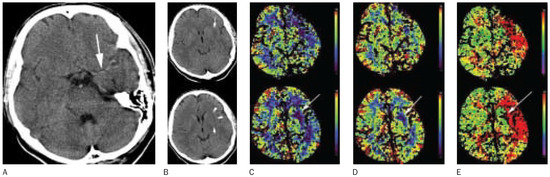
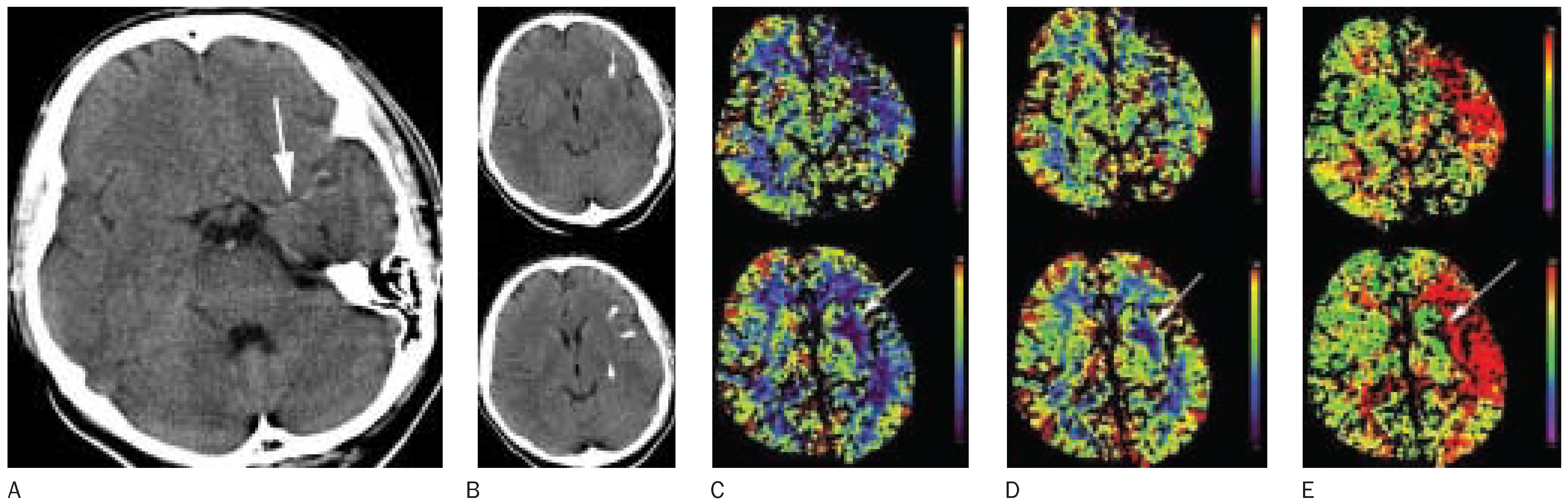
Computed tomography cannot identify the brain tissue that is functionally impaired because brain function is not associated with a change in X-ray attenuation large enough to be detected. After injection of a contrast bolus, however, CT can measure time-density curves in preselected brain sections that represent the blood flow determined change of tissue contrast in 1 to 4 brain sections during the first pass of the contrast agent. Quantification of CBF requires either a very rapid contrast infusion into the tissue voxel under observation, if the maximum slope method is applied [12], or the deconvolution of concentration curves by the function of arterial input into the tissue voxel [13,14]. Although the theoretical background allows approximations only, with modern computer technology both methods are quick and allow producing parameter images of CBF, cerebral blood volume (CBV), mean transit time (MTT) and time to peak (TTP) with good reliability and validity. Both methods of dynamic CT provide images with clear differences in the blood flow parameters for grey and white matter (Figure 1). With the single section method, areas of reduced CBF were detected with a sensitivity of 83% [12], and reactivity tests showed normal and impaired vasomotor reaction [13]. In the affected hemisphere of acute stroke patients, CBF values were reduced and MTT values increased, whereas CBV values were mainly reduced, but also increased in few patients [14]. Hypoattenuating brain tissue had a significantly decreased CBF and CBV of 13.1 ml per 100 g and min and 0.9 ml per 100 g, respectively [14]. Measurements of the inter-observer reliability found a κ coefficient of 0.73, 0.83 and 0.92 for CBV, CBF and MTT respectively. The mean intraobserver variability for the extent of CBV, CBF and MTT abnormalities were 10.1%, 8.9% and 7.6%, respectively [14]. In acute stroke patients the mean extent of regional MTT abnormalities was significantly greater than the mean extent of regional CBF abnormalities, which were significantly greater than regional CBV abnormalities [14]. Severe perfusion deficits on CBF maps were additionally characterised by marked reduction in CBV and undetectable bolus arrival times [12].
Figure 1.
Computed tomography in a 51-year-old man 4 hours after the onset of a severe right-sided hemiparesis and global aphasia. Doppler ultrasound is highly suspicious for a dissection of the left internal carotid artery. (A) shows a long thrombembolus within the middle cerebral artery trunk (arrow). (B) The un-enhanced CT shows a subtle hypoattenuating left lentiform nucleus (arrowheads). (C) Two sections of cerebral blood flow CT show hypoperfusion of the entire middle cerebral artery territory, most pronounced in the left lentiform nucleus (arrow). (D) The reduction in cerebral blood volume is restricted to the left lentiform nucleus (arrow). (E) The time-to-peak image shows a more homogenous delay of the contrast peak within the left middle cerebral artery territory than the perfusion impairment on the cerebral blood flow image.
The comparison between early perfusion abnormalities and the extent of the final infarct showed that MTT- or TTP-maps have the best sensitivity and CBV-maps the best specificity for brain tissue at risk to be injured by ischaemia [14,15]. Among the 22 acute stroke patients reported by Schramm et al., all 6 patients with normal TTP-map did not develop brain infarcts. These patients had normal CBV- and CBF-maps as well. In contrast, all 16 patients with TTP prolongation (>4 s) developed infarcts with the exception of 3 patients who were treated with thrombolysis. There were 2 patients with CBV and CBF abnormalities who did not develop brain infarcts after IV thrombolysis [15]. Based on this experience one may conclude that the high sensitivity of TTP-maps can be used to identify acute stroke patients without perfusion abnormalities who do not need treatment with thrombolytics if the selected CT section covers the affected brain region. We have no proof so far that perfusion thresholds detected by dynamic CT can identify an increased risk for thrombolysisassociated brain haemorrhage as it was shown for single photon emission CT [16,17].
Morphological CT imaging
Vascular pathology
Besides embolic occlusions, arterial wall pathologies as atherosclerosis, dissection, angiospasms, or inflammation of brain supplying arteries are the main causes for cerebral hypoperfusion and ischaemic stroke. The assessment of the type of arterial obstruction will guide secondary prophylaxis like anticoagulation or platelet inhibition and stent-protected angioplasty. The assessment of the site of arterial obstruction helps to estimate the territory of impaired perfusion, the risk of irreversible brain-tissue injury and the chances of reperfusion strategies [18]. In the case series of Schramm et al., all 11 patients with arterial occlusions on CT angiography had a perfusion deficit on CT perfusion imaging with the exception of one patient with basilar artery occlusion where the CT perfusion imaging section did not cover the basilar artery territory. These authors, however, observed 3 out of 11 patients without arterial occlusions on CT angiography but perfusion deficits and resulting infarcts on CT [15]. From this experience, arterial occlusion on CT angiography is always associated with perfusion impairment. A normal CT angiography does not exclude perfusion impairment, however. One may conclude then that CT perfusion imaging is more important than CT angiography when considering reperfusion strategies in acute stroke patients.
Non-contrast CT can detect thrombo-embolic occlusions of major cerebral arteries with high specificity but low sensitivity [19].Arterial thrombi may appear as hyperattenuating segments or spots compared to other segments of the same artery.The attenuation of a thrombus depends on its haematocrit [20]. Red thrombi are better visible on CT than white thrombi. The inter-observer agreement on such “hyperdense artery signs” varied between poor and moderate (κ = 0.20 and 0.63) [21,22,23].
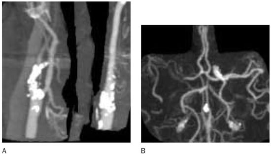
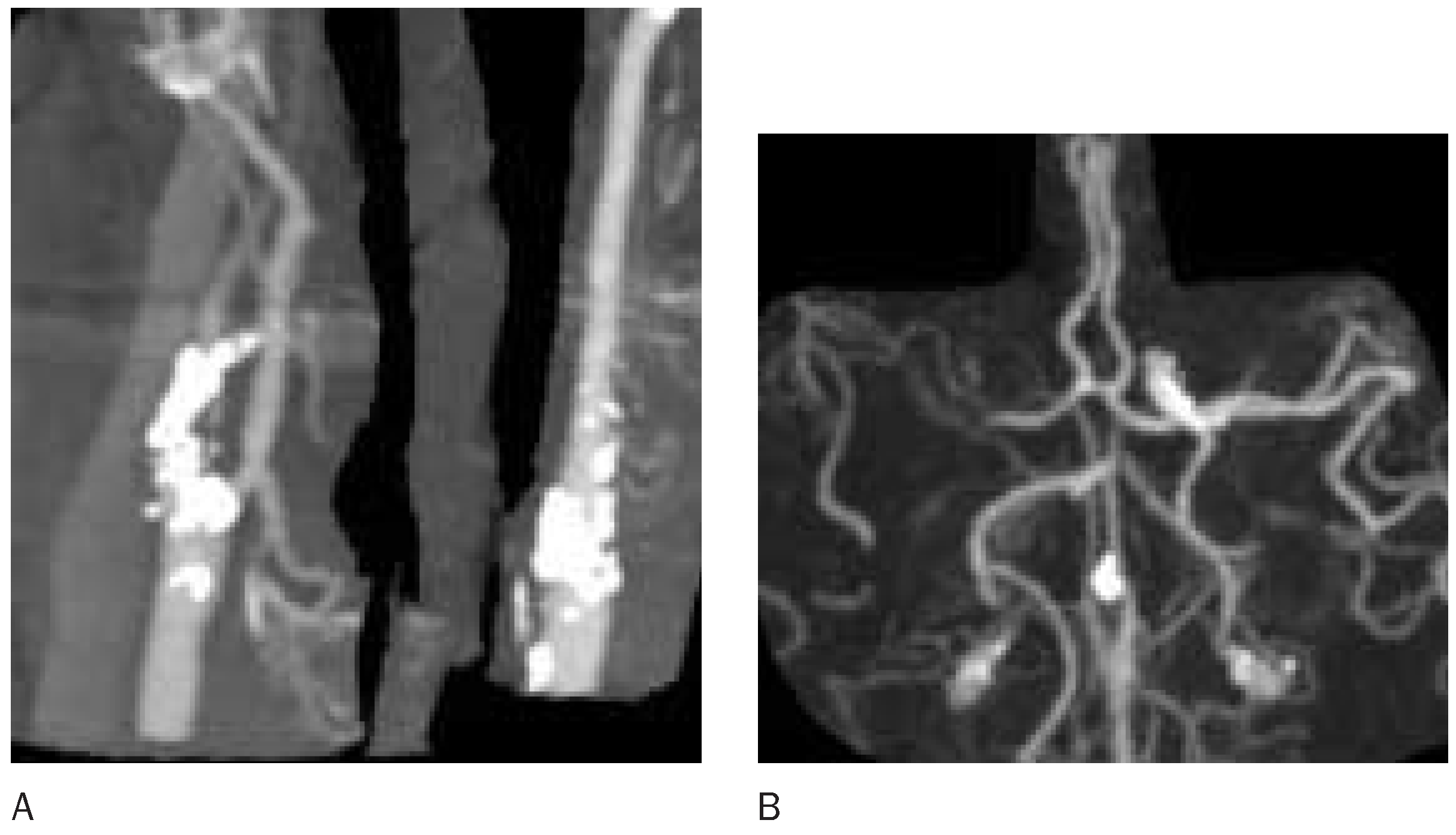
Without doubt, the assessment of cervical and cranial artery obstruction is more reliable and accurate with CT angiography than with unenhanced CT [24,25]. For CT angiography the brain is imaged during the arterial passage of a contrast bolus. The CT angiography source images provide information about brain-tissue enhancement and may identify regions with low CBV [26]. Cervical and cranial arteries can be reconstructed by various computer programs and segmented from the bones if required [27]. Plaque calcifications are nicely demonstrated (Figure 2).
Figure 2.
CT angiography detects severe calcifications at the origins of both internal carotid arteries with tight stenosis on the right side (A). Maximum intensity projection images of the intracranial arteries show less contrast in the right middle cerebral artery consecutive to the proximal ICA stenosis (B).
Brain-tissue pathology
Severe acute brain ischaemia with CBF values below 10 ml per 100 g and min causes immediate net uptake of water in grey matter and consecutively a decrease in X-ray attenuation [6,28,29,30,31,32,33]. Under experimental conditions, X-ray attenuation decreased by 1.06 Hounsfield Units (HU) per hour, reflecting a tissue water uptake of 0.62% per hour after occlusion of the middle cerebral artery [33]. Hypoattenuating grey matter loses its contrast to white matter and consequently its anatomical information. Such ischaemic oedema was, therefore, first characterised as “obscuration of lentiform nucleus” [34] or “loss of the insular ribbon” (Figure 1b) [35]. The CBF threshold for this type of ischaemic oedema is the same as for ischaemic tissue damage. Others confirmed that hypoattenuating brain tissue is associated with very low CBF on CT perfusion imaging [14] or positron emission tomography [36].The detection of hypoattenuating brain tissue by CT in acute stroke patients has thus a high predictive value and specificity for irreversible tissue injury [37].
Because of its subtlety, ischaemic oedema in its very early stage is recognised with only moderate inter-observer reliability [22,23,38]. The National Institute of Neurological Disorders and Stroke rt-PA stroke trialists and others demonstrated that training in CT reading considerably affects the sensitivity to detect ischaemic oedema [38,39]. Attempts were made to improve the capability of CT in detecting ischaemic oedema by performing a density-difference analysis between both cerebral hemispheres, by varying window width and centre level, and by using a quantitative score [40,41,42].
The frequency of positive CT findings in acute stroke may also be diminished by the prejudice that CT is negative within the first 24 to 48 hours after stroke that is still handed down in review articles [43] and books [44]. In contrast, many studies have described positive findings within the first 3–6 hours of stroke onset: Tomura et al. studied 25 patients with embolic cerebral infarction between 40 and 340 minutes after the onset of symptoms [34]. Twenty-three CT scans (92%) were positive with “obscuration of the lentiform nucleus” caused by hypodensity [34]. Bozzao et al. observed parenchymal hypodensity in 25 of 36 patients (69%) [45]. A “loss of the insular ribbon” was reported in 23 of 27 (85%) patients [35]. Horowitz et al. reported on hypodensity and mass effect in 56% of 50 scans [46]. When comparing MR versus CT imaging in identical patients within three hours of symptom onset, CT was positive in 19 patients (53%) and MRI in 18 patients (50%) with hemispheric stroke [47]. We reported 17 positive CT scans (68%) performed in a series of 25 patients with MCA trunk occlusion during the first two hours. The incidence of positive CT findings increased to 89% in the third hour after symptom onset and to 100% thereafter [19]. In another series of patients with hemispheric stroke, the incidence of early CT signs of infarction was 82% [48]. In patients selected for thrombolytic therapy, 12 of 23 patients (52%) had a parenchymal hypodensity on the CT performed within 3 hours of stroke onset [36]. In a series of 100 consecutive patients with MCA infarction, CT detected hypodensity of the lentiform nucleus in 48% and of the insular cortex in 59% of the patients within 14 hours of stroke onset [49]. In a recent randomised trial on prourokinase in patients with MCA occlusions, the investigators detected infarcts’ signs in 125 of 171 patients (73%) on CT within the first 6 hours [50].
Computed tomography cannot reliably detect other ischaemic changes that do not influence brain-tissue water content and consequently braintissue attenuation.
The enlargement of the cerebral cortex and the effacement of cerebral spinal fluid (CSF) spaces suggest brain-tissue swelling. CT may detect braintissue swelling without hypoattenuation for a short period early after arterial or venous obstruction. Compensatory arterial dilatation due to low perfusion pressure [51] or passive arterial dilatation due to high venous pressure [52] cause this type of swelling. Six neuroradiologists agreed on tissue swelling in 45 CT of acute stroke patients with a κ = 0.56–0.59 [22]. Cytotoxic oedema with a shift of extracellular water into the neurons with consecutive cells swelling and decrease in the extracellular fluid space in regions with CBF values of 30 ml per 100 g and min and lower is not detected by CT because it does not change tissue attenuation.
In summary, brain-tissue imaging by CT ignores the changes above the CBF threshold of irreversible injury with the exception of the very subtle findings of brain-tissue swelling due to other causes than ischaemic oedema. Unenhanced CT has a rather low sensitivity for brain ischaemia but a high specificity for irreversible ischaemic injury. The advantage of this constellation is that CT can tell us to which extent brain regions are already damaged when the patient presents with symptoms of acute ischaemic stroke. One may conclude that mainly patients will benefit from reperfusion therapy who have no or only small volumes of hypoattenuating brain tissue on CT but severe symptoms or extended perfusion deficits on CT perfusion imaging. Several studies showed that the response to thrombolytic therapy is associated with the extent of hypoattenuation on early CT [42,53,54].
Brain-tissue CT imaging can easily be combined with CT angiography and CT perfusion imaging adding only a few minutes of extra examination time to the un-enhanced CT. The doses and costs of contrast agent and additional X-ray radiation should be considered, however. One may question, therefore, whether all information that can be provided by CT is necessary in each stroke patient. We will here consider patients with findings or symptoms indicating an increased risk for stroke, patients with basilar artery symptoms and patients with completed hemispheric stroke.
Pending ischaemic stroke
Ischaemic attacks, a mild central neurological deficit or the incidental finding of cervical or cerebral arterial stenosis or occlusion is associated with an increased risk of disabling ischaemic stroke [55].
In this group of patients, CT perfusion imaging and CT angiography provide the most important information. A haemodynamic deficit increases the risk for stroke and should be treated by vessel reconstruction as soon as possible [55]. In patients without perfusion impairment cerebral embolism is more likely. The source of embolism should be identified and treated. Un-enhanced CT may provide additional information about a pattern of old ischaemic lesions. In summary, in patients with pending ischaemic stroke the information about brain perfusion and cerebral arteries is more important that the information about brain tissue.
Posterior circulation ischaemia
In patients with progressive dizziness, ataxia, tetraparesis and impaired consciousness, obstruction of vertebral or basilar arteries should be considered. Because of their high mortality [56], basilar artery occlusion or tight stenosis should be excluded by CT angiography in the first place. In this group of patients CT angiography provides the most valuable information. Perfusion CT is unnecessary for a decision about thrombolysis and CT tissue imaging is insensitive to assess the amount of brainstem damage early.
Completed hemispheric stroke
In patients with a full hemispheric stroke syndrome the risk of permanent brain dysfunction and death is immediate and increasing with time [1]. The chances of brain functions to recover are associated with the amount of brain tissue that can be rescued from ischaemic damage. In this group of patients brain-tissue imaging with CT provides the most valuable information because it detects the volume of brain tissue that is lost for reperfusion strategies. Patients with normal CT or only small amounts of hypoattenuating brain tissue (ischaemic oedema) may benefit most from reperfusion [42,54]. Because stroke severity already indicates the extent of brain perfusion disturbance, it is questionable whether CT perfusion imaging adds any important information. CT angiography may be useful to decide about the type of reperfusion strategy.
References
- The ATLANTIS, ECASS, and NINDS rt-PA Study Group Investigators. Association of outcome with early stroke treatment: pooled analysis of ATLANTIS, ECASS, and NINDS rt-PA stroke trials. Lancet 2004;363:768–74.
- Fryback D, Thornbury J. The efficacy of diagnostic imaging. Med Decis Making 1991;11:88–94.
- Powers W. Testing a test. A report card for DWI in acute stroke. Neurology 2000;54:1549–51.
- Jones TH, Morawetz RB, Crowell RM, Marcoux FW, FitzGibbon SJ, DeGirolami U, et al. Thresholds of focal cerebral ischemia in awake monkeys. J Neurosurg 1981;54:773–82.
- Hossmann KA. Viability thresholds and the penumbra of focal ischemia. Ann Neurol 1994;36:557–65.
- Schuier FJ, Hossmann KA. Experimental brain infarcts in cats. II. Ischemic brain edema. Stroke 1980;11:593–601.
- Wang Y, Hu W, Perez-Trepichio AD, Ng TC, Furlan AJ, Majors AW, et al. Brain tissue sodium is a ticking clock telling time after arterial occlusion in rat focal cerebral ischemia. Stroke 2000;31:1386–92.
- Heiss W, Rosner G. Functional recovery of cortical neurons as related to degree and duration of ischemia. Ann Neurol 1983;14:294–301.
- Ringelstein E, Zeumer H, Schneider R. Der Beitrag der zerebralen Computertomographie zur Differentialtypologie und Differentialtherapie des ischämischen Grosshirninfarkts. Fortschr Neurol Psychiatr 1985;53:315–36.
- Ringelstein E, Biniek R, Weiller C, Ammeling B, Nolte P, Thron A. Type and extent of hemispheric brain infarctions and clinical outcome in early and delayed middle cerebral artery recanalization. Neurology 1992;42:289–98.
- Zülch KJ. Cerebrovascular pathology and pathogenesis as a basis of neuroradiological diagnosis. In: Diethelm L, Heuck F, Olsson O, Strnad F, Vieten H, Zuppinger A, eds. Encyclopedia of Medical Radiology. Berlin, Heidelberg, New York: Springer; 1981. p. 1–192.
- Koenig M, Klotz E, Luka B, Venderink D, Spittler J, Heuser L. Perfusion CT of the brain: diagnostic approach for early detection of ischemic stroke. Radiology 1998;209:85–93.
- Nabavi DG, Cenic A, Craen RA, Gelb AW, Bennett JD, Kozak R, et al. CT assessment of cerebral perfusion: experimental and initial clinical experience. Radiology 1999;213:141–9.
- Eastwood JD, Lev MH, Azhari T, Lee TY, Barboriak DP, Delong DM, et al. CT perfusion scanning with deconvolution analysis: pilot study in patients with acute middle cerebral artery stroke. Radiology 2002;222:227–36.
- Schramm P, Schellinger PD, Klotz E, Kallenberg K, Fiebach JB, Kulkens S, et al. Comparison of perfusion computed tomography and computed angiography source images with perfusion-weighted imaging and diffusionweighted imaging in patients with acute stroke of less than 6 hours’ duration. Stroke 2004;35:1652–8.
- Ueda T, Hatakeyama T, Kumon Y, Sakaki S, Uraoka T. Evaluation of risk of hemorrhagic transformation in local intra-arterial thrombolysis in acute ischemic stroke by initial SPECT. Stroke 1994;25:298–303.
- Ueda T, Sakaki A, Yuh W, Nochide I, Ohta S. Outcome in acute stroke with successful intra-arterial thrombolysis and predictive value of initial single-photon-emissioncomputed tomography. J Cereb Blood Flow Metab 1999;19:99–108.
- Wildermuth S, Knauth M, Brandt T, Winter R, Sartor K, Hacke W. Role of CT angiography in patient selection for thrombolytic therapy in acute hemispheric stroke. Stroke 1998;29:935–8.
- von Kummer R, Meyding-Lamade U, Forsting M, Rosin L, Rieke K, Hacke W, et al. Sensitivity and prognostic value of early computed tomography in middle cerebral artery trunk occlusion. Am J Neuroradiol 1994;15:9–15.
- Kirchhof K, Welzel T, Mecke C, Zoubaa S, Sartor K. Differentiation of white, mixed, and red thrombi: value of CT in estimation of the prognosis of thrombolysisphantom study. Radiology 2003;228:126–30.
- Tomsick TA, Brott TG, Chambers AA, Fox AJ, Gaskill MF, Lukin RR, et al. Hyperdense middle artery sign on CT: efficacy in detecting middle cerebral artery thrombosis. Am J Neuroradiol 1990;11:473–7.
- von Kummer R, Holle R, Gizyska U, Hofmann E, Jansen O, Petersen D, et al. Interobserver agreement in assessing early CT signs of middle cerebral artery infarction. Am J Neuroradiol 1996;17:1743–8.
- Marks M, Holmgren E, Fox A, Patel S, von Kummer R, Froehlich J. Evaluation of early computed tomographic findings in acute ischemic stroke. Stroke 1999;30:389–92.
- Knauth M, Jansen O, von Kummer R, Wildermuth S, Forsting M, Sartor K. Sensitivität, Interrater-Reliabilität und prognostischer Wert der CT-Angiographie in der Diagnostik akuter intrakranieller Gefässverschlüsse. RöFo 1996;164:S21.
- Knauth M, von Kummer R, Jansen O, Hähnel S, Dörfler A, Sartor K. Potential of CT angiography in acute ischemic stroke. Am J Neuroradiol 1997;18:1001–10.
- Schramm P, Schellinger PD, Fiebach JB, Heiland S, Jansen O, Knauth M, et al. Comparison of CT and CT angiography source images with diffusion-weighted imaging in patients with acute stroke within 6 hours after onset. Stroke 2002;33:2426–32.
- Tomandl BF, Kostner NC, Schempershofe M, Huk WJ, Strauss C, Anker L, et al. CT angiography of intracranial aneurysms: a focus on postprocessing. Radiographics 2004;24:637.
- Hossmann KA, Schuier FJ. Experimental brain infarcts in cats. I. Pathophysiological observations. Stroke 1980;11:583–92.
- Watanabe O, West CR, Bremer A. Experimental regional cerebral ischemia in the middle cerebral artery territory in primates. Part 2: Effects on brain water and electrolytes in the early phase of MCA stroke. Stroke 1977;8:71–6.
- Gotoh O, Asano T, Koide T, Takakura K. Ischemic brain edema following occlusion of the middle cerebral artery in the rat. I: The time courses of the brain water, sodium, and potassium contents and blood-brain-barrier permeability to 125I-albumin. Stroke 1985;16:101–9.
- Todd N, Picozzi P, Crockard A, Russell R. Duration of ischemia influences the development and resolution of ischemic brain edema. Stroke 1986;17:466–71.
- Todd N, Picozzi P, Crockard A, Russell R. Reperfusion after cerebral ischemia: influence of duration of ischemia. Stroke 1986;17:460–5.
- Dzialowski I, Weber J, Doerfler A, Forsting M, von Kummer R. Brain tissue water uptake after middle cerebral artery occlusion assessed with CT. J Neuroimaging 2004;14:42–8.
- Tomura N, Uemura K, Inugami A, Fujita H, Higano S, Shishido F. Early CT finding in cerebral infarction. Radiology 1988;168:463–7.
- Truwit C, Barkovich A, Gean-Marton A, Hibri N, Norman D. Loss of the insular ribbon: another early CT sign of acute middle cerebral artery infarction. Radiology 1990;176:801–6.
- Grond M, von Kummer R, Sobesky J, Schmülling S, Heiss W-D. Early computed-tomography abnormalities in acute stroke. Lancet 1997;350:1595–6.
- von Kummer R, Bourquain H, Bastianello S, Bozzao L, Manelfe C, Meier D, et al. Early prediction of irreversible brain damage after ischemic stroke by computed tomography. Radiology 2001;219:95–100.
- Grotta JC, Chiu D, Lu M, Patel S, Levine SR, Tilley BC, et al. Agreement and variability in the interpretation of early CT changes in stroke patients qualifying for intravenous rtPA therapy. Stroke 1999;30:1528–33.
- von Kummer R. Effect of training in reading CT scans on patient selection for ECASS II. Neurology 1998;51:S50–2.
- Bendszus M, Urbach H, Meyer B, Schultheiss R, Solymosi L. Improved CT diagnosis of acute middle cerebral artery territory infarcts with density-difference analysis. Neuroradiology 1997;39:127–31.
- Lev MH, Farkas J, Gemmete JJ, Hossain ST, Hunter GJ, Koroshetz WJ, et al. Acute stroke: improved nonenhanced CT detection – benefits of soft-copy interpretation by using variable window width and center level settings. Radiology 1999;213:150–5.
- Barber P, Demchuk A, Zhang J, Buchan A. Validity and reliability of a quantitative computed tomography score in predicting outcome of hyperacute stroke before thrombolytic therapy. Lancet 2000;355:1670–4.
- Gilman S. Imaging of the brain. N Engl J Med 1998;338:812–20.
- Sorensen A, Reimer P. Cerebral MR Perfusion Imaging. Stuttgart, New York: Thieme; 2000.
- Bozzao L, Bastianello S, Fantozzi LM, Angeloni U, Argentino C, Fieschi C. Correlation of angiographic and sequential CT findings in patients with evolving cerebral infarction. Am J Neuroradiol 1989;10:1215–22.
- Horowitz SH, Zito JL, Donnarumma R, Patel M, Alvir J. Computed tomographic-angiographic findings within the first five hours of cerebral infarction. Stroke 1991;22:1245–53.
- Mohr JP, Biller J, Hilal SK, Yuh WT, Tatemichi TK, Hedges S, et al. Magnetic resonance versus computed tomographic imaging in acute stroke. Stroke 1995;26:807–12.
- von Kummer R, Nolte PN, Schnittger H, Thron A, Ringelstein EB. Detectability of hemispheric ischemic infarction by computed tomography within 6 hours after stroke. Neuroradiology 1996;38:31–3.
- Moulin T, Cattin F, Crepin-Leblond T, Tatu L, Chavot D, Piotin M, et al. Early CT signs in acute middle cerebral artery infarction: predictive value for subsequent infarct locations and outcome. Neurology 1996;47:355–75.
- Furlan A, Higashida R, Wechsler L, Gent M, Rowley H, Kase C, et al. Intra-arterial prourokinase for acute ischemic stroke. JAMA 1999;282:2003–11.
- Gibbs J, Wise R, Leenders K, Jones T. Evaluation of cerebral perfusion reserve in patients with carotid-artery occlusion. Lancet 1984;8372:310–4.
- Yuh WT, Simonson TM, Wang AM, Koci TM, Tali ET, Fisher DJ, et al. Venous sinus occlusive disease: MR findings. Am J Neuroradiol 1994;15:309–16.
- von Kummer R, Allen KL, Holle R, Bozzao L, Bastianello S, Manelfe C, et al. Acute stroke: usefulness of early CT findings before thrombolytic therapy. Radiology 1997;205:327–33.
- Hill MD, Rowley HA, Adler F, Eliasziw M, Furlan A, Higashida RT, et al. Selection of acute ischemic stroke patients for intra-arterial thrombolysis with pro-urokinase by using ASPECTS. Stroke 2003;34:1925–31.
- Blaser T, Hofmann K, Buerger T, Effenberger O, Wallesch C, Goertler M. Risk of stroke, transient ischemic attack, and vessel occlusion before endarteriectomy in patients with symptomatic severe carotid stenosis. Stroke 2002;33:1057–62.
- Brandt T, von Kummer R, Müller-Küppers M, Hacke W. Thrombolytic therapy of acute basilar occlusion: variables affecting recanalization and outcome. Stroke 1996;27:875–81.
This is an open-access article distributed under the terms of the Creative Commons Attribution Non-Commercial No Derivatives (CC BY-NC-ND) 4.0 License.