


Simple Summary
The publication of two discoveries [1, 2] set the ground for theories, experiments and clinical practice during the past 40 years in the neuro-science of memory. Since then, we know that memory is not supported by the total action of the entire brain but anatomically localizable like sensory and motor functions. It is the function of a neuronal network including both mediotemporal regions, particularly the hippocampal formations which were found to be indispensable for memory. Research with amnesic patients led to the next discovery that memory is not a unitary system but consists of at least five different sub-systems which are subserved by different, though overlapping neuronal networks. In the following, the function of the “pioneer structure” hippocampus in the human was pinned down to episodic memory alone and became even further specified to learning/consolidation, novelty detection, deep processing, associative learning, and—not treated in this article—spatial learning. To further elucidate the function(s) of the hippocampal region in human memory we carried out two Positron Emission Tomography (PET) experiments testing the above hypotheses. The findings were straight-forward: nothing challenged both hippocampal formations as much as establishing new semantic associations between previously unrelated words or pictures in memory.
Zusammenfassung
Die Veröffentlichung von zwei Entdeckungen [1,2] bereitete die Grundlage für Theorien, Experimente und die klinische Praxis während den vergangenen 40 Jahren in den Neurowissenschaften das Gedächtnisses. Seither wissen wir, dass Gedächtnis nicht auf der Funktion des gesamten Gehirns beruht, sondern anatomisch lokalisierbar ist wie sensorische und motorische Funktionen. Gedächtnis wird nämlich durch ein Netzwerk getragen, das die beiden mediotemporalen Regionen, besonders die hippocampalen Formationen, einschliesst, welche essentiell für das Gedächtnis sind. Die Forschung an amnestischen Patienten führte zur weiteren Entdeckung, dass Gedächtnis kein einheitliches System ist, sondern aus mindestens fünf verschiedenen Untersystemen besteht, denen unterschiedliche, doch teilweise überlappende neuronale Netzwerke zugrunde liegen. Im folgenden wurde die Funktion der «Pionierstruktur» Hippocampus beim Menschen eingeschränkt auf episodisches Gedächtnis allein und wurde sogar noch weiter spezifiziert auf das Lernen/Konsolidieren, das Entdecken von Neuem («novelty detection»), auf tiefes Verarbeiten, auf assoziatives Lernen und – in diesem Artikel nicht behandelt – räumliches Lernen. Zur weiteren Erhellung der Funktion(en) der hippocampalen Region im menschlichen Gedächtnis haben wir zwei Positronen-Emissions-Tomographie(PET)-Experimente durchgeführt und obige Hypothesen geprüft. Die Resultate waren eindeutig: Nichts forderte beide hippocampalen Formationen so sehr heraus wie das Herstellen und Speichern neuer semantischer Bezüge zwischen zuvor unbezogenen Wörtern und Bildern im Gedächtnis.
Schlüsselwörter: PET; Gedächtnis; Hippocampus; assoziatives Lernen
Introduction
The hippocampal formation is a bilateral, archicortical structure in the medial part of the temporal lobe (TL). It was once related to olfaction, later to emotion, and for the past 40 years to learning and memory. The influential reports by Scoville and Milner [1] and Penfield and Milner [2] established that the mediotemporal region in the human is crucial for memory functions. Up to that time, it was believed that there was no functional specialization for memory within the brain, but that memory depends upon the action of the entire brain. Scoville and Milner [1] reported nine patients with bilateral medial TL resections. In one of these patients (H. M.), surgery was a treatment for medically refractory epilepsy, in the other patients it was a treatment for psychotic disorders. Scoville extended the orbital undercutting to include the resection of medial temporal structures such as the amygdala, the uncus, and the pes of the hippocampus. He tried progressively larger bilateral resections extending up to 8 cm back from the tips of the TL. Two of these patients (H. M. and M. B.) who had received the largest bilateral resections (without orbital undercutting) developed a severe amnesic syndrome. Scoville and Milner reported a positive relationship between the extent of destruction of the medial temporal area and the degree of memory impairment. Specifically, they attributed the memory loss in these patients to the resection of the hippocampal formation.
Subsequent animal lesion studies, patient studies, and functional neuroimaging studies put the generality of the hippocampal memory hypothesis into question. Monkeys displayed intact recognition memory following bilateral hippocampal and amygdalar destruction [3,4], and monkeys significantly improved their recognition performance following the addition of a hippocampal removal to a rhinal cortex ablation [5]. In the human, it was discovered that there are at least five different memory systems, each supported by a separate, though partially overlapping neuronal network. The human hippocampus was found to subserve only one of these memory systems, namely episodic memory [6] which is the memory for autobiographical events and the time and place where they have happened [7]. But this notion is not unequivocal either, since no significant hippocampal activations could be identified during episodic learning and retrieval in most of the initial H215O PET studies with healthy volunteers (see «discussion»), and since only a partial or non-dramatic memory decrease was found in some studies of patients with bilateral mediotemporal damage [8,9,10]. Subsequent PET studies of episodic memory using different tasks were successful in activating the hippocampal regions significantly. Both these PET studies and the mentioned patient studies suggest that the hippocampus might subserve certain, but not all aspects of episodic memory. Thus, the role or roles of hippocampus within episodic memory needs further specification. To this aim, we conducted two H215O PET studies with healthy volunteers testing the following hypotheses about the function of the hippocampus in episodic memory: learning versus retrieval, novelty detection, associative learning versus single item learning, and depth of processing.
Learning versus retrieval
Squire and collaborators (e. g. [11,12]) claim that the consolidation of information from shortterm to long-term memory is a function of the hippocampal formation. The fact that retrograde amnesia is temporally graded in patients and experimental animals with amnesia due to bilateral hippocampal damage, is interpreted as evidence for a time-limited role of the hippocampal formation in memory storage. Thus, the hippocampal formation is initially indispensable for the retrieval of information, but after a certain time period during which consolidation takes place, information becomes independent of hippocampal function and can be retrieved in the absence of both hippocampal formations. Thus, the hippocampal formation is not considered a repository of permanent memory, but temporarily stores “aspects” of information held in short-term memory.
The hippocampal formation has several characteristics which make it a prime candidate for consolidation: it is reciprocally connected with the association cortices and exhibits long-term potentiation (LTP) which induces fast changes in the synaptic connectivity. Converging inputs arriving simultaneously in the hippocampus lead to an increase in synaptic efficacy lasting for hours to weeks. LTP induces cellular changes which allow for a well formed representation of the pattern of cortical coactivations on a single trial. The corticocortical connections between the simultaneously activated neocortical regions are less plastic and therefore need repeated simultaneous activation in order to become strengthened. The hippocampus forms a temporary memory trace holding the information about what neocortical regions were simultaneously activated when event “A” has been registered. Subsequently, the hippocampal formation repeatedly reactivates the cortical (and subcortical) sites in the same way that they were activated during encoding. It thereby helps strengthening the connections between these participating regions over time. This process is considered the neuronal correlate of consolidation. By the time the cortico-cortical connections between the storage sites of event “A” are established, event “A” can be retrieved without the participation of the hippocampi.
Associative learning versus single item learning
According to this hypothesis, the hippocampus is involved in learning the components of scenes and their relations (associative learning) rather than learning isolated single components (single item learning). This notion is supported by the anatomy and physiology of hippocampus (see “Learning versus retrieval”). If the hippocampus can store and reactivate patterns of coactivations, then the kind of learning that best suits these characteristics is learning associations between items.
The subjective experience at a given moment usually comprises the processing of diverse inputs through different sensory modalities, the appreciation of simultaneous bodily sensations, thoughts and emotions. The processing of these aspects requires activity in distributed cortical regions which needs to be bound in memory so that it can become reinstantiated later for the re-experience of all aspects of that moment. Binding several aspects of a scene might challenge the hippocampus more than just binding few or even storing only one.
Eichenbaum and collaborators [13] stress two characteristics which may depend on the specific ability of the hippocampal formation to store and reactivate patterns of coactivations, namely representational flexibility and compositionality: stored patterns of coactivations are believed to overlap with other stored patterns of coactivations in the hippocampal formation. This allows for the retrieval of an old event “A” on grounds of only one cue which is also part of a new event “B”. The activation of this cue may lead to the reactivation of the complete cortical-subcortical network which was involved in processing the experience of event “A” (= cued recall). Another feature of compositionality and flexibility is the hippocampal ability to either retrieve event “A” as a whole or to retrieve only a certain aspect of event “A”, like zooming in the recollection of only one component of event “A”. This is possible because the hippocampal representations are thought to include all constituent parts of an experience and theirrelations. Therefore, the hippocampal formation may reactivate all neocortical storage sites simultaneously and thus recover the experience of the complete event “A”, or it may reactivate only a part of the neocortical network and thereby recover only an aspect of event “A”. Accordingly, the hippocampal formation is not only binding information in memory but also unbinding it if necessary.
Novelty detection
This hypothesis claims that the hippocampal formation is engaged in the novelty assessment of items in our environment. Event-related potentials recorded from scalp and intracranial electrodes in humans (e. g. [14,15]), single neuron recording studies in animals [16,17], and functional neuroimaging studies in healthy volunteers (e. g. [18]) suggested that the hippocampal formation is part of a novelty detection network which comprises prefrontal, temporal, and parietal areas. Novelty assessment is considered an early stage of long-term memory encoding [18]. Before elaborate, meaning-based encoding processes become operational, perceived stimuli must be judged old or new. Obviously, there is no use in learning old items, since these are stored in memory already. Therefore, the probability of long-term storage of items increases with their novelty value. Following this hypothesis, the hippocampal formation subserves the selection of the to-be-learned material.
Depth of processing
The retrieval of stimuli is dependent on how they have been processed at the time of encoding. According to the levels of processing approach to learning [19], the semantic evaluation of stimuli, called deep processing, leads to a better retention than a purely sensory/perceptual analysis of stimuli which is called shallow processing. For example, if we pay attention to the meaning of a word, we can remember it better than if we pay attention to the word form or sound. Given that learning is most effective using a deep encoding strategy and that the hippocampus is thought to subserve learning, the hippocampal formation is likely to be more activated during deep than shallow processing of information. Indeed, Rugg and colleagues [20] found greater activation in the left hippocampus during the retrieval of words which had been deeply processed compared to words which had been shallowly processed.
PET experiments
We designed two H215O PET experiments measuring the regional cerebral blood flow during different learning and retrieval tasks to test the above outlined hypotheses. One experiment was performed with pictorial stimuli [21] and the other with verbal stimuli [22] using the following learning and retrieval tasks: deep single item learning, shallow single item learning, establishing inter-item associations between novel items, establishing inter-item associations between old (previously studied) items, retrieving recently formed associations between items, and detecting novel constellations of studied items. The use of the cognitive subtraction method for the statistical analysis of the PET data allowed us to isolate blood flow underlying the cognitive components of interest; these are encoding, retrieval, establishing associations, depth of processing, and novelty detection.
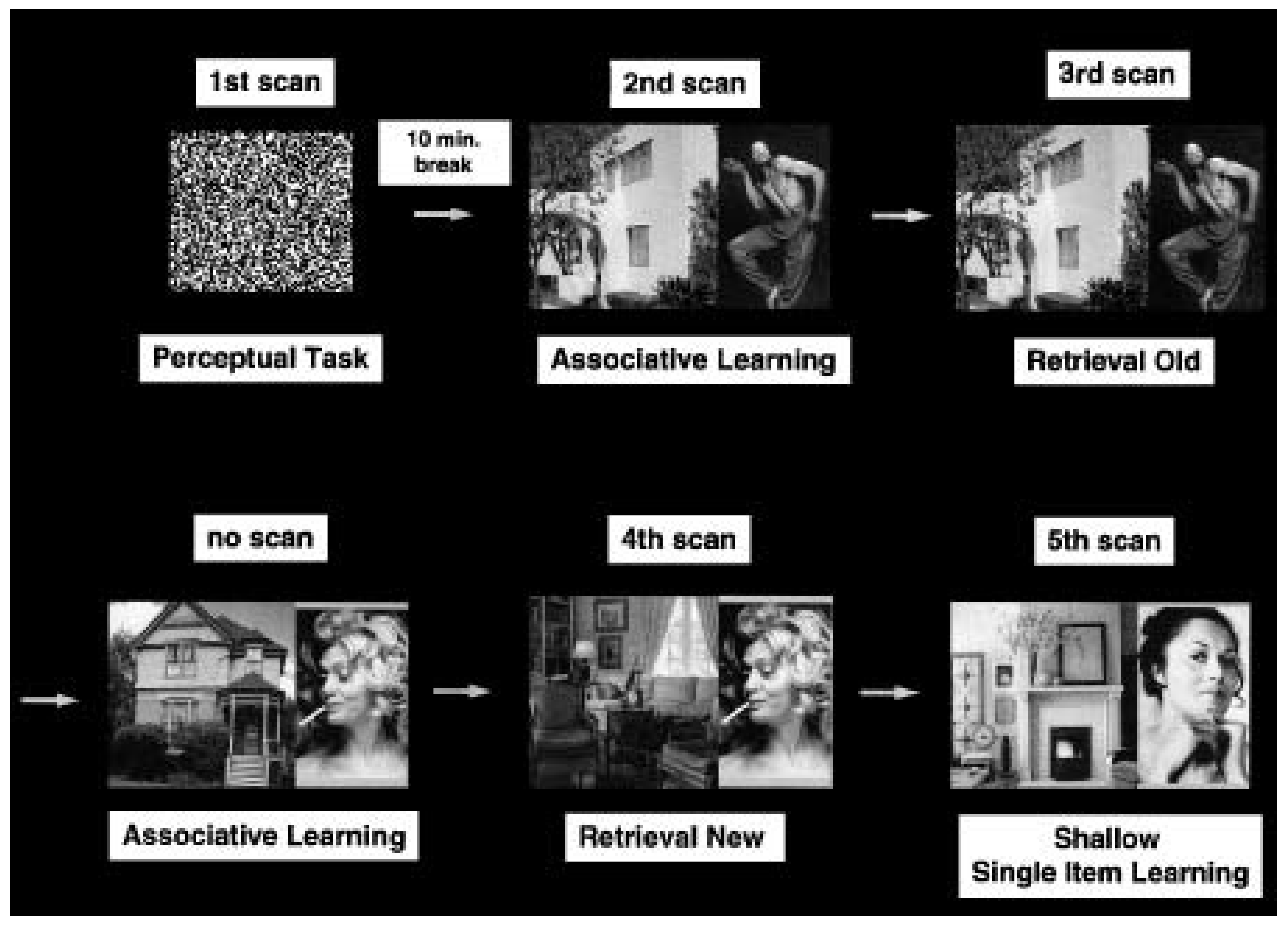
Our first PET experiment comprised two learning conditions, two retrieval conditions, and a perceptual task as reference (Figure 1). During both learning and retrieval the 12 subjects of this study were presented with photographs that contained houses next to individuals (Figure 1). Subjects were responding in all tasks by either pressing the right or the left key of a computer mouse according to the instruction. The first learning task required subjects to decide whether the presented individual might either be an inhabitant or a visitor of the house (Associative Learning). This instruction induces a semantic evaluation of the person and the house (deep processing). Moreover, the evaluation of whether the presented individual might live in the house or not leads to the establishment of a semantic link between the representation of the person and that of the house in episodic memory. The second learning task required subjects to determine the gender of the presented person and to decide whether an outside or an inside view of the house was presented (Shallow Single Item Learning). This task engages subjects in an evaluation of the person and the house independently of each other and therefore leads to the storage of the house and the person as separate entities. Thus, the first learning task should lead to a better retention of the house-person pairs than the second learning task.Associative Learning was carried out twice and was followed after 10 minutes by either of two retrieval tasks. In both retrieval tasks the studied houses and people were presented again, either combined as seen during study (Retrieval Old) or recombined to form new pairs (Retrieval New). The task in both retrieval conditions was to decide whether an old or a new combination was presented. Retrieval Old thus requires the recovery of stored associations, while Retrieval New requires a search for formed associations and leads to the detection of novel combinations. Old and new combinations were mixed during the prescanning period (30 s) and postscanning period (30 s). During scanning (60 s) either only old (Retrieval Old) or new combinations (Retrieval New) were presented. The Perceptual Task required subjects to judge whether a square or a rectangle (filled with black-and-white random dot patterns) was presented (Figure 1).
Figure 1.
Examples of stimuli and an example sequence of tasks. The sequence of tasks was varied across subjects to avoid order effects. Yet, the Associative Learning tasks always preceded the retrieval tasks to keep the delay time between encoding and retrieval at 10 minutes. Only one Associative Learning procedure was accompanied by scanning. Figure adapted from Henke, Buck, Weber, Wieser. Wiley & Sons, Inc.; 1997.
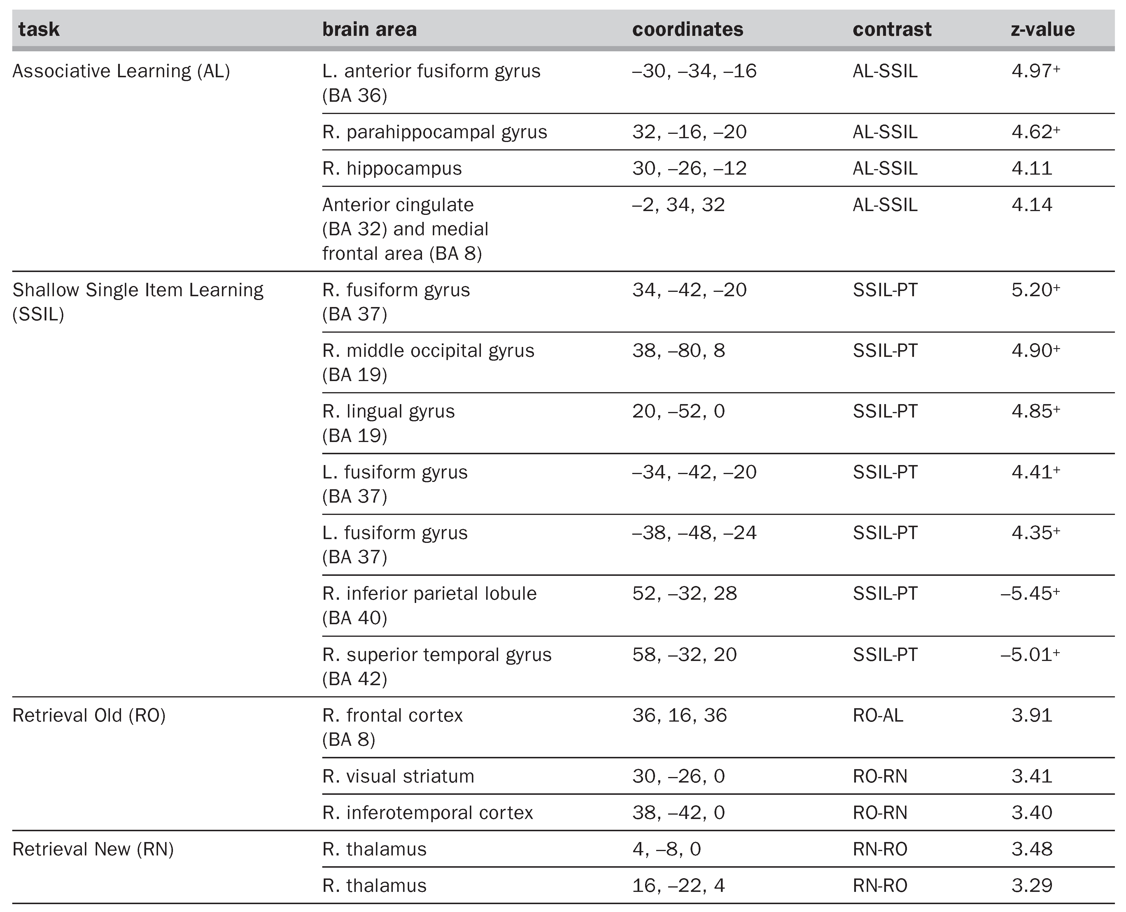
A behavioural pilot study confirmed the suitability of our learning instructions. The Associative Learning task led to a substantially better retention of house-person combinations than the Shallow Single Item Learning task. Pairwise subtraction analysis of the inter-subject averaged PET images was performed. The comparison of Associative Learning to Shallow Single Item Learning isolates net blood flow changes underlying the establishment of associations and depth of processing. This comparison yielded significant blood flow increases in the right parahippocampal gyrus and right hippocampal formation among other areas (
Table 1
). No brain area was significantly more activated during Shallow Single Item Learning than Associative Learning. Shallow Single Item Learning compared to the Perceptual Task revealed relative blood flow increases in higher visual areas, but not the hippocampal formation (
Table 1
). We therefore conclude that the hippocampal system supports depth of processing and/or the establishment of meaningful associations between items, while the higher visual areassubserve the formation of rather isolated visual memories. Activations due to the recovery of formed associations become apparent in the comparison of Retrieval Old with Retrieval New. This comparison did not reveal significant hippocampal activations. That the reactivation of associations 10 minutes after their formation did not lead to a recurrent activation in the hippocampal formation and parahippocampal gyrus is unexpected given that bilateral damage to the hippocampal formation usually abolishes recently formed memories. The comparison of Retrieval New to Retrieval Old reveals blood flow changes related to the detection of novel combinations of previously learned items. This comparison yielded no significant activations in the hippocampal region either.
Tabel 1.
Peaks of blood flow increases and decreases. The coordinates (x, y, z [36]) locate the maxima within an area of blood flow increase (positive z-values) or decrease (negative z-values) associated with a given contrast; + indicates p <0.05 (corrected for multiple comparisons), all other z-values correspond to p <0.001 (uncorrected for multiple comparisons). R, right; L, left; PT, Perceptual Task. Table adapted from Henke, Buck, Weber & Wieser. Wiley & Sons, Inc.; 1997.
Thus, the hippocampal formation was only activated during depth of processing and/or the establishment of associations between items. The additional comparison of the memory tasks to the Perceptual Task and to a baseline scan confirmed these results.
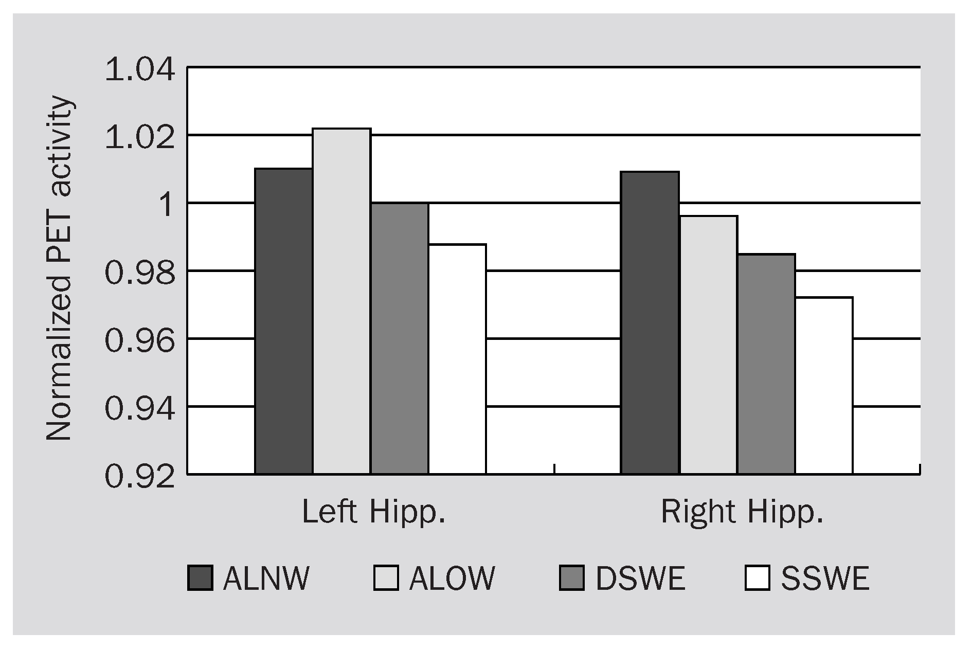
Unfortunately, in this first study we missed to test for hippocampal activations underlying depth of processing alone and the process of creating and establishing associations alone. Also, we could not determine whether depth of processing and/or associative learning by its nature activated the right rather than the left hippocampal area, or whether the found lateralization was induced by the nonverbal nature of the stimuli. These questions were answered by our second PET experiment in which we used pairs of abstract, unrelated nouns as stimulus material in all conditions. Since hippocampal activations were stronger during learning than retrieval in the first experiment, we used four different learning tasks in the second experiment and conducted the retrieval of the previously learned word pairs between scans. The experimental setup was very similar to that of our first PET study. The learning conditions were (1) Associative Learning of New Words (= words which had not been presented before in the experiment), (2) Associative Learning of Old Words (= words which had been presented for study preceding the respective learning scan), (3) Deep Single Word Encoding, and (4) Shallow Single Word Encoding. PET data was evaluated by pairwise subtraction and region-of-interest analyses. The subtraction of Deep Single Word Encoding from Associative Learning of New Words leaves blood flow which reflects the establishment of semantic associations between words.This comparison yielded significant activations in the pes of the right hippocampal formation.The subtraction of Associative Learning of Old Words from Associative Learning of New Words isolates blood flow underlying the detection of novelty. This contrast did not result in significant hippocampal activations. Comparing Deep to Shallow Single Word Encoding which tests the depth of processing hypothesis did not lead to significant activation differences in the hippocampal region either. Thus, only the process of creating and establishing semantic associations between words activated the hippocampus significantly. The region-of-interest analysis (Figure 2) shows the absolute activations in the pes of the left and right hippocampus and clarifies why the difference between Deep Single Word Encoding and Associative Learning of New Words yielded significance in the right rather than the left hippocampus: Single Word Encoding activated the left stronger than the right anterior hippocampus and thereby elevated the reference activations on the left side and thus decreasing the activation difference between the two tasks. In both hippocampi, Associative Word Learning increased the blood flow most, followed by Deep Single Word Encoding and Shallow Single Word Encoding. Interestingly, Associative Learning of New Words activated the left and right anterior hippocampus equally. This might suggest that both verbal and non-verbal associations had been created between words.
Figure 2.
Region-of-interest analysis within the left (Left Hipp.) and right (Right Hipp.) pes of hippocampus. Displayed is the normalized PET activity averaged over 12 subjects during each of the four learning tasks. ALNW, Associative Learning of New Words; ALOW, Associative Learning of Old Words; DSWE, Deep Single Word Encoding; SSWE, Shallow Single Word Encoding.
These results support Cohen and Eichenbaum’s [13] theory which is based on animal learning experiments and indicate its validity for encoding in the human brain. Our data predict that human amnesics with bilateral hippocampal damage would be more impaired on tests of associative memory than tests of single item memory. Indeed, it was reported that patients with damage to the hippocampal formation [23] suffered from a selective impairment in binding the constituent parts of individual stimuli in memory. The patients retained some ability to remember the studied stimulus parts, but not the combinations of parts.
Hippocampal and/or parahippocampal activations are typically reported in PET and fMRI studies of memory if the stimulus material was unfamiliar, complex, multi-facetted and nonverbal [24,25,26,27,28]. The reason for this selectivity might be the novelty of stimuli or the necessity to establish associations among the subcomponents of such stimuli in order to effectively learn them. On the other hand, many PET experiments using verbal stimuli failed to demonstrate significant hippocampal activations [29,30,31,32,33]. Although learning words or category-exemplar word pairs requires some associative learning such as binding the presentation of a word to its context, the demand for associative learning might not be as high to yield a hippocampal blood flow increase sufficient for statistical significance. Yet, the demand for associative learning can be increased – even with single words as stimuli – by use of a different learning instruction which requires the subject to form semantic associations between the presented words and concepts stored in memory. For example, Schacter et al. [34] reported right hippocampal activation during the recall of words. These words had been learned by counting the number of meanings associated with each word. The hippocampal activation during retrieval might thus reflect the recovery of previously formed associations. Similarly, Rugg et al. [20] found left hippocampal activation during the retrieval of words which had been encoded by producing a sentence which includes that word. This task binds the presented word into the selfcreated context of the sentence. At the time of retrieval then, the presented old word might reactivate the formed associations, i.e. the sentence context. Dolan and Fletcher [35] reported left hippocampal and parahippocampal activation during an associative learning task with “contextually new” word pairs. These are word pairs that have not been studied during two preceding runs of paired-associates learning. They found less left hippocampal activation if one word of each pair had been studied during the two preceding study runs, and again less hippocampal activation if the presented word pairs had been studied twice already. The authors interpret the left hippocampal activation during the study of new (= not yet learned) word pairs as a correlate of novelty detection: the newer the learning material (with reference to the two previously given study lists), the more hippocampal activation was present. An alternative interpretation is that hippocampal activation increased as a function of associative learning: previously learned word pairs needed less associative learning than new word pairs.
In conclusion, the hippocampal and parahippocampal activations found in other functional imaging studies of verbal or nonverbal memory can also be interpreted as correlates of establishing (or retrieving) previously formed semantic associations.
This research was supported by a scholarship (8210-040202) from the Swiss National Science Foundation (SNF) to the first author and by SNF grant 31-47’203.96. K. H. wishes to thank S. G. Müller for sharp and critical comments and inspiring discussions at all times.
References
- Scoville W, Milner B. Loss of recent memory after bilateral hippocampal lesions. J Neurol Neurosurg Psychiatr 1957;20:11–21.
- Penfield W, Milner B. Memory deficit produced by bilateral lesions in the hippocampal zone. Arch Neurol Psychiatr 1958;79:475–97.
- O’Boyle VJ Jr, Murray EA, Mishkin M. Effects of excitotoxic amygdalo-hippocampal lesions on visual recognition in rhesus monkeys. Soc Neurosci Abstr 1995;19:438.
- Murray EA. What have ablation studies told us about the neural substrates of stimulus memory? Sem Neurosci 1996;8:13–22.
- Meunier M, Hadfield W, Bachevalier J, Murray EA. Effects of rhinal cortex lesions combined with hippocampectomy on visual recognition memory in rhesus monkeys. J Neurophysiol 1996;75:1190–205.
- Vargha-Khadem F, Gadian DG, Watkins KE, Connelly A, Van Paesschen W, Mishkin M. Differential effects of early hippocampal pathology on episodic and semantic memory. Science 1997;277:376–80.
- Tulving E. Episodic and semantic memory. In: Tulving E, Donaldson W (editors). Organization of memory. New York: Academic; 1972. pp 381–403.
- Gol A, Fabisch GM. Effects of human hippocampal ablation. J Neurosurg 1967;26:390–8.
- Henke K, Wieser HG. Bilateral medial temporal lobe damage without amnesic syndrome: a case report. Epilepsy Res 1996;24:147–61.
- Henke K, Kroll NEA, Behniea H, Amaral DG, Miller MB, Rafal R, et al. Memory lost and regained following bilateral hippocampal damage. J Cog Neurosci. In press.
- Alvarez P, Squire LR. Memory consolidation and the medial temporal lobe: a simple network model. Proc Natl Acad Sci USA 1994;91:7041–5.
- Squire LS, Alvarez P. Retrograde amnesia and memory consolidation: a neurobiological perspective. Curr Opin Neurobiol 1995;5:169–77.
- Cohen NJ, Eichenbaum H. Memory, amnesia, and the hippocampal system. Cambridge, MA: MIT Press; 1993.
- Knight RT. Contribution of human hippocampal region to novelty detection. Nature 1996;383:256–9.
- Grunwald T, Lehnertz K, Heinze HJ, Helmstaedter C, Elger CE. Verbal novelty detection within the human hippocampus proper. Proc Natl Acad Sci USA 1998;95:3193–7.
- Fahy FL, Riches IP, Brown MW. Neuronal activity related to visual recognition memory: long-term memory and encoding of recency and familiarity information in the primate anterior and medial inferior temporal and rhinal cortex. Exp Brain Res 1993;96:457–72.
- Rolls ET, Cahusac PMB, Feigenbaum JD, Miyashita Y. Responses of single neurons in the hippocampus of the macaque related to recognition memory. Exp Brain Res 1993;93:299–306.
- Tulving E, Markowitsch HJ, Craik FIM, Habib R, Houle S. Novelty and familiarity activations in PET studies of memory encoding and retrieval. Cereb Cortex 1996;6:71–9.
- Craik FIM, Lockhart RS. Levels of processing: a framework for memory research. J Verb Learn Verb Behav 1972;11:671–84.
- Rugg MD, Fletcher PC, Frith CD, Frackowiak RSJ, Dolan RJ. Brain regions supporting intentional and incidential memory: a PET study. Neuro Report 1997;8:1283–7.
- Henke K, Buck A, Weber B, Wieser HG. Human hippocampus establishes associations in memory. Hippocampus 1997;7:249–56.
- Henke K, Weber B, Kneifel S, Wieser HG, Buck A. Human hippocampus associates information in memory. Proc Natl Acad Sci USA 1999; in press.
- Kroll NEA, Knight RT, Metcalfe J, Wolf ES, Tulving E. Cohesion failure as a source of memory illusions. J Mem Lang 1996;35:176–96.
- Kapur N, Friston KJ, Young A, Frith CD, Frackowiak RSJ. Activation of human hippocampal formation during memory for faces: a PET study. Cortex 1995;31:99–108.
- Haxby JV, Ungerleider LG, Horwitz B, Maisog JM, Rapoport SI, Grady CL. Face encoding and recognition in the human brain. Proc Natl Acad Sci USA 1996;93:922–7.
- Maguire EA, Frackowiak RSJ, Frith CD. Learning to find your way: a role for the human hippocampal formation. Proc R Soc Lond B Biol Sci 1996;263:1745–50.
- Stern CE, Corkin S, González RG, Guimarães AR, Baker JR, Jennings PJ, et al. The hippocampal formation participates in novel picture encoding: evidence from functional magnetic resonance imaging. Proc Natl Acad Sci USA 1996;93:8660–5.
- Gabrieli JDE, Brewer JB, Desmond JE, Glover GH. Separate neural bases of two fundamental memory processes in the human medial temporal lobe. Science 1997;276:264–6.
- Grasby PM, Frith CD, Friston KJ, Simpson J, Fletcher PC, Frackowiak RSJ, et al. A graded task approach to the functional mapping of brain areas implicated in auditoryverbal memory. Brain 1994;117:1271–82.
- Kapur S, Craik FIM, Tulving E, Wilson AA, Houle S, Brown GW. Neuroanatomical correlates of encoding in episodic memory: levels of processing effect. Proc Natl Acad Sci USA 1994;91:2008–11.
- Shallice T, Fletcher PC, Frith CD, Grasby PM, Frackowiak RSJ, Dolan RJ. Brain regions associated with acquisition and retrieval of verbal episodic memory. Nature 1994;368:633–5.
- Andreasen NC, O’Leary DS, Arndt S, Cizadlo T, Hurtig R, Rezai K, et al. Short-term and long-term verbal memory: a positron emission tomography study. Proc Natl Acad Sci USA 1995;92:5111–5.
- Fletcher PC, Frith CD, Grasby PM, Shallice T, Frackowiak RSJ, Dolan RJ. Brain systems for encoding and retrieval of auditory-verbal memory: an in vivo study in humans. Brain 1995;118:401–16.
- Schacter DL, Alpert NM, Savage CR, Rauch SL, Albert MS. Conscious recollection and the human hippocampal formation: evidence from positron emission tomography. Proc Natl Acad Sci USA 1996;93:321–5.
- Dolan RJ, Fletcher PC. Dissociating prefrontal and hippocampal function in episodic memory encoding. Nature 1997;388:582–5.
- Talairach J, Tournoux P. Co-planar stereotaxic atlas of the human brain. Stuttgart: Thieme; 1988.
This is an open-access article distributed under the terms of the Creative Commons Attribution Non-Commercial No Derivatives (CC BY-NC-ND) 4.0 License.