


Summary
The goal of epilepsy surgery is to excise the epileptogenic focus or to interrupt propagation of epileptiform activity. The prerequisite for the neurosurgical procedure is the exact localization of the epileptogenic focus in order to avoid postoperative morbidity. PET studies with 11C-Flumazenil have shown the involvement of the benzodiazepine receptors (BDR) in epilepsy. The aim of this study was to examine the clinical value of 123Iodine-Iomazenil (a derivate of Flumazenil) SPECT in relation to interictal rCBF SPECT, EEG and MRI findings in patients with therapy-resistant mesial temporal lobe epilepsy, candidates for epilepsy surgery. The results show that 123I-Iomazenil SPECT for the examination of the benzodiazepine receptors is a sensitive method for the preoperative localization of the epileptogenic focus: the results show a clearly higher sensitivity (89%, similar to ictal CBF SPECT) in comparison to the interictal CBF SPECT (25%). With a positive predictive value of 100%, 123I-Iomazenil SPECT allows a reliable localization of epileptogenic foci. Since an ictal CBF SPECT is rarely possible due to logistic reasons and due to high sensitivity of the 123I-Iomazenil SPECT, this method has become a routine examination during the intensive preoperative monitoring period.
Introduction
About 20–30% of the patients with epilepsy continue to have seizures despite adequate anti-epileptic therapy [1]. In a part of these patients a neurosurgical operation can lead to a real improvement. The objective of the presurgical evaluation is the correct localization of the epileptogenic focus (= region of brain responsible for generating epileptic seizures) [2]. For this reason, it is mandatory that the exact localization of the epileptogenic focus be determined preoperatively. The EEG monitoring is still the “gold standard” for the localization of the epileptogenic focus. However, in 20–50% it is difficult or impossible to localize the epileptogenic focus in the surface EEG. In these cases semi-invasive or invasive EEG monitoring is necessary [3,4,5]. Non-invasive functional and morphological methods have been developed for the precise localization of the epileptogenic focus in order to increase the safety and possibly the avoidance or at least the optimization of the invasive localization methods. PET studies with 11C-Flumazenil have shown the involvement of the benzodiazepine receptors (BDR) in epilepsy [6,7,8]. The aim of this study was to examine the clinical value of 123Iodine-Iomazenil (a derivate of Flumazenil) SPECT in relation to interictal rCBF SPECT, EEG and MRI findings in patients with therapy-resistant mesial temporal lobe epilepsy.
Patients and methods
Nine patients (females and males with an average age of 25.3 ± 9.6 yr. [s. d.]), suffering from therapy-resistant mesial temporal lobe epilepsy, were evaluated by long-term EEG (with scalp and foramen ovale electrodes), video monitoring and high-resolution MRI. Clinical observations were compatible with complex-partial epilepsy of mesial temporal lobe origin. In all subjects an interictal rCBF SPECT, using 99mTc-ECD, and a benzodiazepine receptor SPECT, using 123I-Iomazenil, were carried out. Data acquisition was performed using a three-head SPECT camera (Picker Prism 3000) respectively minutes after application of the radiopharmaceutical. The reconstruction was done iteratively. In addition to the visual evaluation, performed without knowledge of EEG and MRI results, there was a semiquantitative evaluation in the ROI (region of interest) technique with calculation of the asymmetry indices [AI] and comparison to those of healthy volunteers (n = 9), both for the benzo-diazepine receptor SPECT and the interictal rCBF study. A variation of >standard deviations was considered significant. After spatial standardization and normalization of the image data, 123I-Iomazenil SPECT was analyzed pixel-by-pixel in the subgroups, with right and left predominant deficiency according to asymmetry indices. Direct comparisons were performed with the normal group. The calculated t-maps were merged with the brain anatomy as shown by high-resolution MRI using a dedicated Computerized Brain Atlas (CBA) system.
All patients underwent a surgical procedure (7/9 an amygdalohippocampectomy and 2/9 an anterior temporal lobectomy) and all patients had been seizure free for at least months postoperative. We considered that the resected cerebral region correspond correctly with the epileptogenic focus, when the patients had been postoperatively seizure free for at least months.
Results
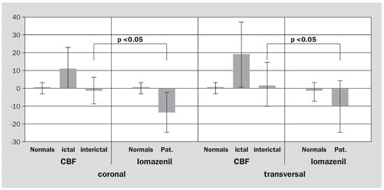
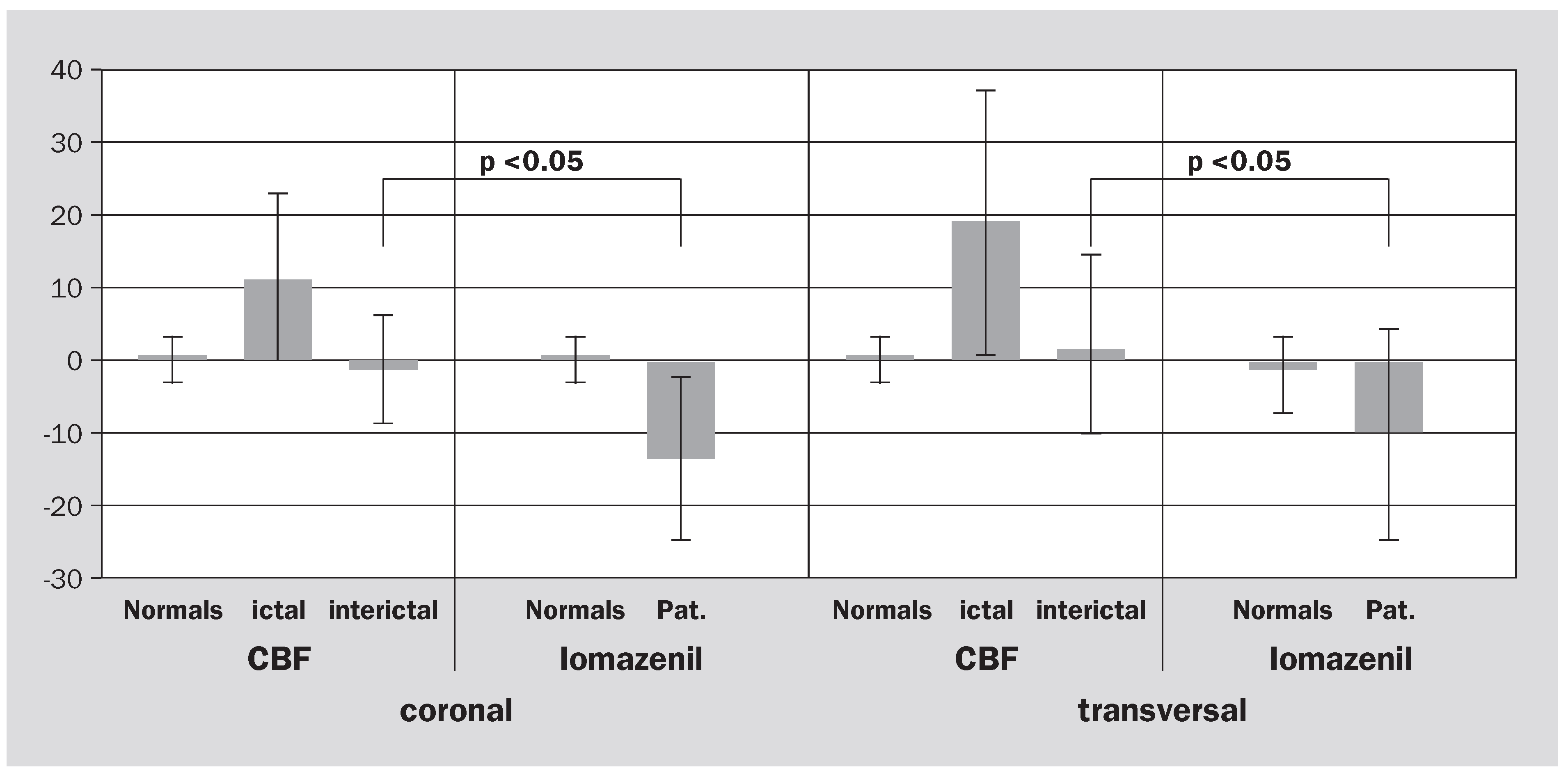
Ictal focal EEG activity over the mesial temporal region was obtained in all patients during intensive monitoring. High-resolution MRI revealed a focal anomaly in patients. In all but one patient the 123I-Iomazenil SPECT study showed reduced benzodiazepine receptors density in the mesial temporal lobe and localized the epileptogenic focus correctly in respect to the postoperative results. Thus, the 123I-Iomazenil SPECT study was concordant with EEG and MRI findings in 89% and 88% of the cases (Table 1). Considering interictal rCBF SPECT, the corresponding values were 22% and 25%, respectively (Table 1). Epileptogenic foci could be visually recognized more reliably in the 123I-Iomazenil SPECT studies since the asymmetry indices were significantly higher (–12.36 ± 10.8 vs. –2.82 ± 6.68, p <0.[Wilcoxon rank-sum test]) (Figure 1). Thus, 123I-Iomazenil SPECT had a sensitivity of 89% and a positive predictive value of 100%, whereas interictal rCBF SPECT was characterized by lower values of 25% and 67%, respectively. The Figures and show typical examples for CBF and 123I-Ioma-zenil SPECT findings. The Figure delineates deficiencies in patients subgroups by descriptive t-maps, on a pixel basis and integrated in the according MR-image slice of Computerized Brain Atlas.
Table 1.
Correlation of the SPECT findings with EEG and MRI. Focus localization in SPECT with regard to postoperative outcome.
Figure 1.
Asymmetry index mesial temporal in TLE (temporal lobe epilepsy) in coronal and transversal slices, significant higher asymmetry indices for 123I-Iomazenil vs. interictal CBF (w-test).
Figure 2.
Single case with the following semiology: epigastric aura, loss of consciousness, speech arrest, stiffness in the right arm, A = interictal CBF SPECT, no significant asymmetry, asymmetry indices < s. d. from normal; B = 123I-Iomazenil SPECT, significant asymmetry, decreased activity uptake left mesial temporal (arrow); right = MRI, FLAIR-signal increase left hippocampus.
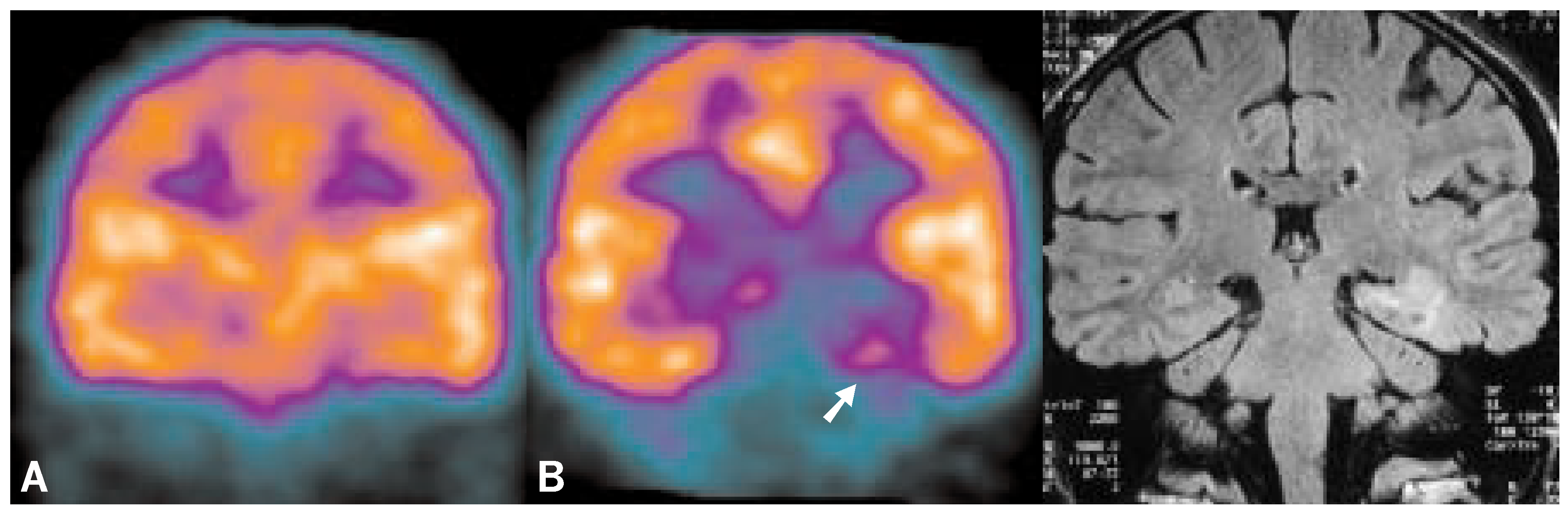
Figure 3.
Single case with the following semiology: loss of consciousness, mouth and hand automatisms, speech arrest, A = interictal CBF SPECT, decreased activity uptake right temporal (contralateral to EEG focus), clearly increased uptake in the striatum (constant finding in two examinations); B = 123I-Iomazenil SPECT, significant asymmetry, decreased activity uptake left mesial temporal (arrow); right = MRI normal.
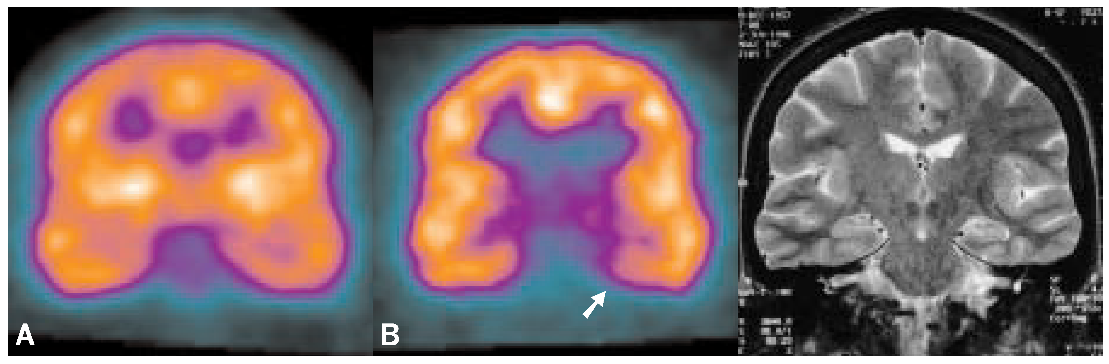
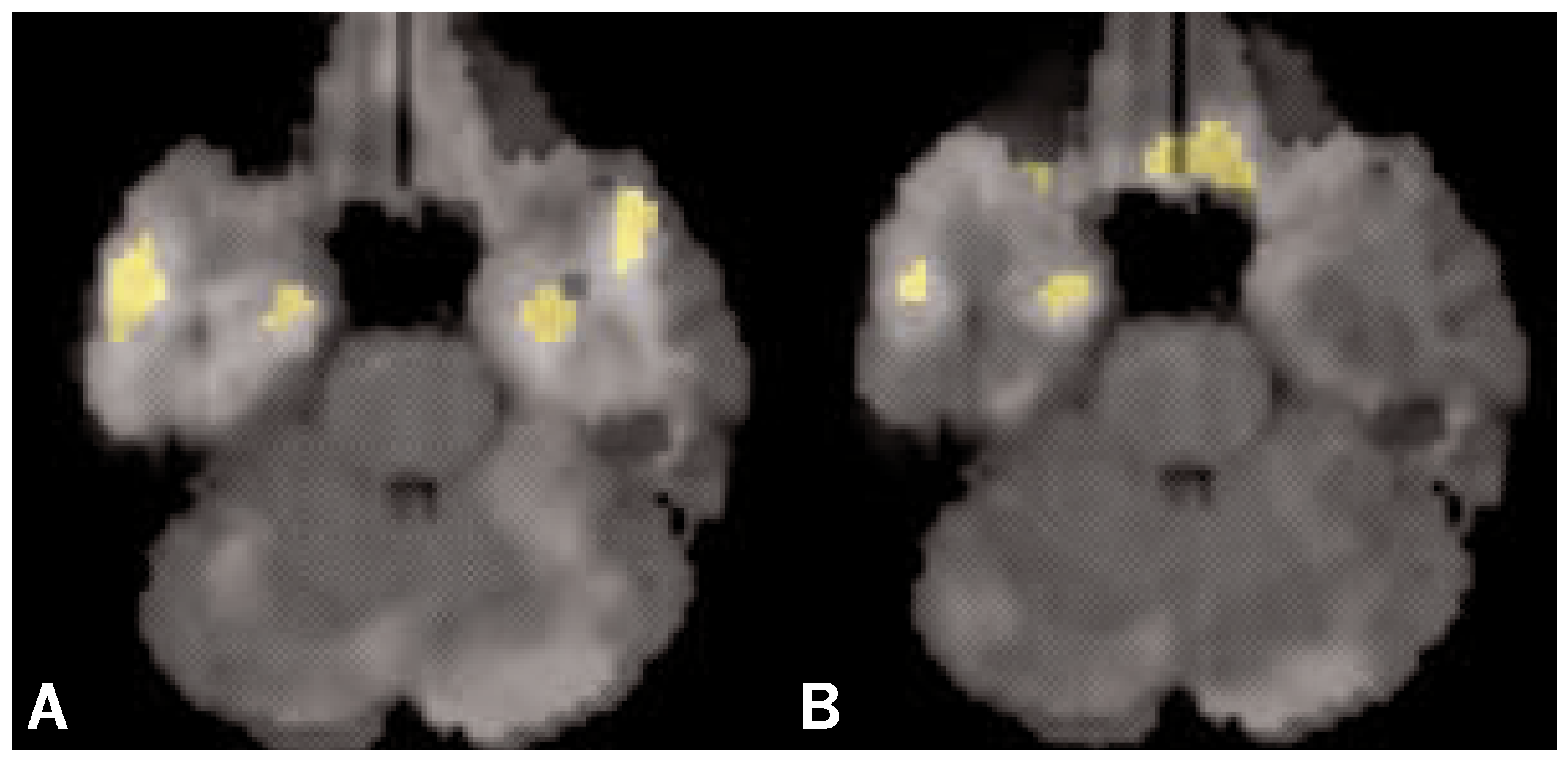
Figure 4.
Descriptive t-maps, on a pixel basis, integrated in the according MR-image slice, shows benzodiazepine receptors deficiencies in patients subgroups compared to normal volunteers [t = 3.0, p < 0.01]. A and B represent subgroups with benzodiazepine receptors deficiencies, as assessed by asymmetry indices, in the left (n = 5) and right mesial temporal (n = 4), respectively. Note: parametric t-map revealed a bilateral deficit in A.
Discussion
In the literature the average sensitivity for the interictal CBF SPECT is 44% (range 0–72%), for the ictal CBF SPECT 96% (range 67–100%), for FDG-PET 74%, Flumazenil PET 97%, CBF PET 47% and 123I-Iomazenil SPECT 85% [7,8,10,11]. Our results for the interictal CBF SPECT with 25% lie in the lower range of the published results. The 123I-Iomazenil SPECT with a sensitivity of 89% lies in the upper range of the published data. The results confirm the assumption that the reduction of the GABA-transmitted inhibition plays a key role in the spread of the seizure potential, and that the inhibitory transmission is disturbed in the epileptogenic focus [9,11,12]. 123I-Iomazenil shows a specific binding on the central benzodiazepine receptors at the neuronal membrane and reduced benzodiazepine receptor binding have been described in human epileptogenic foci [7,8,10,11,12], and, thus, possibly allows a more specific detection of the epileptogenic focus as the demonstration of the glucose metabolism or the cerebral blood flow. In the literature, there are various hypotheses as to the cause of the focal reduction of the benzodiazepine binding. Of the various theses, it could be that a loss of neurons is responsible, that there is a degeneration of the GABAergic neurons, a loss of the inhibitory synapses, down regulation of the benzodiazepine receptors or an irreversible binding of endogenous ligands on the benzodiazepine receptors [13,14,15,16].
Conclusion
The study of the benzodiazepine receptors density by 123I-Iomazenil brain SPECT gives valid information in candidates for epilepsy surgery and allows reliable localization of epileptogenic foci. In comparison to interictal rCBF SPECT, its sensitivity and positive predictive values are considerably higher and are comparable to ictal rCBF SPECT. 123I-Iomazenil SPECT is logistically simpler and finally less expensive than ictal ECD SPECT and gives complementary information to rCBF and metabolic data as derived from FDG PET. The sensitivity of benzodiazepine receptors SPECT using 123I-Iomazenil is comparable with PET studies using 11C-Flumazenil. A reliable spatial localization of the functional data is possible by displaying structures adapted to the individual anatomy on seizure localization and seizure propagation. 123I-Iomazenil SPECT should have a firm place in the preoperative diagnostic routine, particularly with equivocal EEG or MRI findings.
References
- Hauser, W.A.; Hesdorffer, D.C. Epilepsy: Frequency, Causes and Consequences; Demos Press: New York, 1990. [Google Scholar]
- Engel, J. Surgery for seizures. N. Engl. J. Med. 1996, 10, 647–652. [Google Scholar] [CrossRef] [Scilit] [PubMed]
- Rowe, C.C.; Berkovic, S.F.; Austin, M.C.; et al. Visual and quantitative analysis of interictal SPECT with technetium 99m-HMPAO in temporal lobe epilepsy. J. Nucl. Med. 1991, 32, 1688–1694. [Google Scholar] [PubMed]
- Shorvon, S. Epidemiology, classification, natural history, and genetics of epilepsy. Lancet 1990, 336, 93–96. [Google Scholar] [CrossRef] [Scilit] [PubMed]
- Engel, J.; Henry, T.R.; Risinger, M.W.; et al. Presurgical evaluation for partial epilepsy: Relative contributions of chronic depth-electrode recordings versus FDG-PET and scalpsphenoidal ictal EEG. Neurology 1990, 40, 1670–1677. [Google Scholar] [CrossRef] [Scilit] [PubMed]
- Bockisch, A.; Grünwald, F.; Elger, C.E.; Kaiser, W.A.; Biersack, H.J. Kernspinresonanz-Tomographie und HMPAO-SPECT in der Epilepsiediagnostik. Radiol. Diagn 1992, 33, 227–234. [Google Scholar]
- Savic, I.; Persson, A.; Roland, P.; Pauli, S.; Sedvall, G.; Widen, L. In vivo demonstration of reduced benzodiazepine receptor binding in human epileptic foci. Lancet 1988, II, 863–866. [Google Scholar] [CrossRef] [Scilit] [PubMed]
- Savic, I.; Ingvar, M.; Stone-Elander, S. Comparison of 11C-Flumazenil and 18FDG as PET markers of epileptic foci. J. Neurol. Neurosurg. Psychiatry 1993, 56, 615–621. [Google Scholar] [CrossRef] [Scilit] [PubMed]
- Henry, T.R.; Frey, K.A.; Sackellares, J.C.; et al. In vivo cerebral metabolism and central benzodiazepine-receptor binding in temporal lobe epilepsy. Neurology 1993, 43, 1998–2006. [Google Scholar] [CrossRef] [Scilit] [PubMed]
- Devous, M.D.; Thisted, R.A.; Morgan, G.F.; Leroy, R.F.; Rowe, C.C. SPECT brain imaging in epilepsy: a meta-analysis. J. Nucl. Med. 1998, 39, 285–293. [Google Scholar] [PubMed]
- Van Huffelen, A.C.; van Isselt, J.W.; van Veelen, C.W.M.; et al. Identification of the side of the epileptic focus with 123IIomazenil SPECT. Acta Neurochir. 1990, 50, 95–99. [Google Scholar]
- Ribak, C.E. Axon terminals of GABAergic chandelier cells are lost at epileptic foci. Brain Res. 1985, 326, 251–260. [Google Scholar] [CrossRef] [Scilit] [PubMed]
- Jibiki, I.; Yamaguchi, N. Epilepsy and SPECT. Neurosci. Biobehav. Rev. 1994, 2, 281–290. [Google Scholar] [CrossRef] [Scilit] [PubMed]
- Burdette, D.E.; Sakurai, S.Y.; Henry, T.R.; et al. Temporal lobe central benzodiazepine binding in unilateral mesial temporal lobe epilepsy. Neurology 1995, 45, 934–941. [Google Scholar] [CrossRef] [Scilit] [PubMed]
- Staedt, J.; Stoppe, G.; Kögler, A.; Steinhoff, B.J. Changes of central benzodiazepine receptor density in the course of anticonvulsant treatment in temporal lobe epilepsy. Seizure 1995, 4, 49–52. [Google Scholar] [CrossRef] [Scilit] [PubMed]
- Richardson, M.P.; Koepp, M.J.; Brooks, D.J.; Fish, D.R.; Duncan, J.S. Benzodiazepine receptors in focal epilepsy with cortical dysgenesis: An 11C-Flumazenil PET study. Ann. Neurol. 1996, 40, 188–198. [Google Scholar] [CrossRef] [Scilit] [PubMed]


Published under the copyright license “Attribution – Non-Commercial – NoDerivatives 4.0”. No commercial reuse without permission.