


Abstract
Vasconcellea jossei is an Andean fruit species of the Caricaceae family whose chemical composition remains poorly understood. This study aimed to characterize the phenolic composition, antioxidant capacity, and mineral composition of the peel and pulp of green and ripe V. jossei fruits. Total phenolic (TPC) and total flavonoid (TFC) contents were determined, as well as antioxidant capacity using DPPH and FRAP, the phenolic acid and flavonoid profile using HPLC-PDA, and the elemental composition. The peel exhibited higher phenolic compound contents and greater antioxidant capacity than the pulp, with the green peel showing 10.43 mg GAE/g DW of TPC, 2.57 mg QE/g DW of TFC, 10.81 µmol TE/g DW by DPPH, and 45.70 µmol TE/g DW by FRAP. HPLC-PDA analysis revealed differences in the distribution of phenolic compounds according to tissue type and maturity stage. Rutin was the predominant compound, reaching 189.54 mg/100 g DW in the green peel. TPC and TFC showed strong positive correlations with DPPH and FRAP, supporting the contribution of phenolic compounds to antioxidant capacity. Potassium (K) was the predominant element, with a maximum concentration of 43,900 mg/kg DW in the ripe pulp, accompanied by significant levels of magnesium (Mg) and phosphorus (P). Overall, the results indicate that tissue type and maturity stage were associated with differences in the chemical composition of V. jossei. The high rutin concentration and antioxidant capacity of the peel, along with the high mineral content of the ripe pulp, provide relevant information for the characterization of this understudied Andean species and support future strategies for the utilization and valorization of its different fractions.
1. Introduction
In recent decades, interest in native and underutilized fruits has increased considerably due to their potential as sources of bioactive compounds with applications in the food, nutraceutical, and pharmaceutical industries [1]. These species constitute an important reservoir of secondary metabolites and essential nutrients that, in addition to contributing to their nutritional value, exhibit diverse biological properties, including antioxidants, anti-inflammatory, antimicrobial, and cardioprotective activity [2]. In this context, chemically characterizing understudied fruit species is a relevant strategy for both biodiversity conservation and the development of new functional ingredients of natural origin [3].
Among the functional properties of fruits, antioxidant activity has gained relevance due to the growing evidence on the role of natural antioxidants in preventing diseases associated with oxidative damage [4]. This activity is closely related to the presence of bioactive metabolites, mainly phenolic compounds and flavonoids, whose concentration and composition vary by species, environmental conditions, plant tissue, and stage of fruit ripening [5,6]. Consequently, the evaluation of antioxidant capacity, along with the characterization of these metabolites, is a fundamental tool for determining the functional value of fruit species with food potential [7].
Oxidative stress, caused by an imbalance between reactive oxygen species (ROS) production and the body’s antioxidant mechanisms, is closely related to the development of chronic non-communicable diseases, such as cardiovascular diseases, diabetes mellitus, neurodegenerative disorders, and various types of cancer [8]. Therefore, there is a growing interest in identifying natural sources of antioxidants capable of neutralizing free radicals, reducing reactive oxygen species, and protecting biomolecules from oxidative damage [9]. Among the main compounds responsible for this activity are phenolic compounds, especially phenolic acids and flavonoids, whose antioxidant capacity depends on their chemical structure, the number and position of hydroxyl groups, and possible synergistic interactions with other metabolites present in the plant matrix [10].
The chemical composition of fruits is influenced by multiple factors, including the species, environmental conditions, agronomic management, and the stage of ripening [11]. During ripening, profound physiological and biochemical modifications affect the synthesis, transformation, and degradation of primary and secondary metabolites, altering the content of phenolic compounds, flavonoids, free amino acids, sugars, pigments, and minerals [12]. These variations directly affect the antioxidant capacity and nutritional quality of the fruit [3]. Likewise, different fruit tissues, such as pulp and peel, have distinct chemical profiles due to the physiological functions they play during growth and development [13].
The genus Vasconcellea (Caricaceae) comprises between 23 and 26 species, distributed mainly in the Andean region of South America, where they constitute an important component of biodiversity [14]. Various species of the genus, such as V. pubescens, V. candicans and V. quercifolia, have been the subject of research on their nutritional composition, the content of phenolic compounds, carotenoids, vitamin C, proteolytic enzymes and their antioxidant activity, which shows their potential as functional foods and plant genetic resources of economic interest [1,2,15]. However, the information available is very uneven between species, concentrating mainly on those of greatest commercial importance.
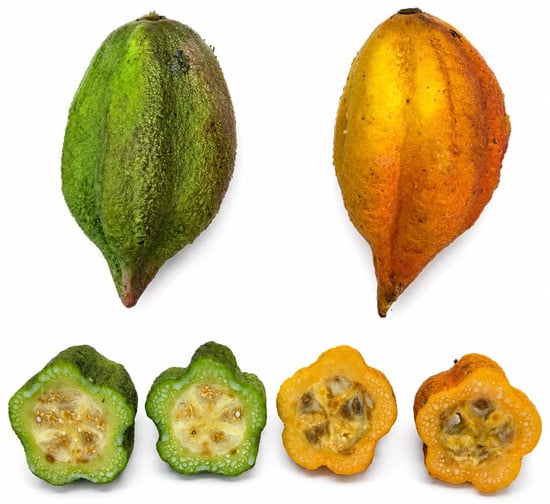
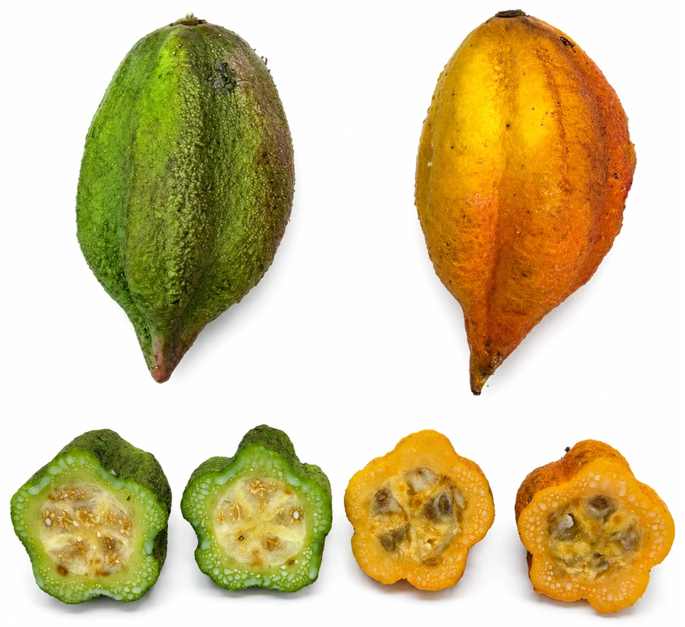
Among the least studied species is V. jossei (Figure 1), a fruit native to the Ecuadorian Andes, which grows between 2000 and 2500 m above sea level, where each plant produces between 20 and 50 fruits per growing cycle [14]. Studies published to date have focused mainly on taxonomic and botanical aspects, while its chemical composition, bioactive metabolite profile, mineral content, and antioxidant properties remain virtually unexplored. Therefore, the objective of this study was to characterize the antioxidant potential and chemical profile of the pulp and peel of green and ripe fruits of Vasconcellea jossei by evaluating phenolic compounds, flavonoids, macro- and microelements, as well as their antioxidant capacity. To the best of our knowledge, this work provides the first comprehensive characterization of this species.
Figure 1.
External appearance and cross-sectional view of green and ripe fruits of Vasconcellea jossei.
2. Materials and Methods
2.1. Chemicals and Reagents
Reagents and solvents. Folin–Ciocalteu phenol reagent, acetone (≥99%), methanol (≥99%), acetonitrile (≥99%), sodium carbonate (Na2CO3, ≥99.5%), hydrochloric acid (HCl, 37% solution), and trifluoroacetic acid (≥99%) were purchased from Merck (Darmstadt, Germany). Aluminum chloride (AlCl3, ≥98%), 1,1-diphenyl-2-picrylhydrazyl (DPPH, ≥97%), 2,4,6-tripyridyl-s-triazine (TPTZ, ≥98%), and ferric chloride (FeCl3, ≥97%) were purchased from Sigma Chemical Co. (St. Louis, MO, USA). Ethanol (≥99.5%; Haymankimia Group, Istanbul,, Türkiye), sodium acetate (≥99%; Mallinckrodt Chemicals, Mexico City, Mexico), formic acid (98%; Sisco Research Laboratories Pvt. Ltd. Mumbai, India), and nitric acid (HNO3, 69% solution; Fisher Chemical, Loughborough, UK) were also used. Reference standards. Trolox (6-hydroxy-2,5,7,8-tetramethylchroman-2-carboxylic acid, ≥97%), gallic acid (≥97.5%), syringic acid (≥95%), p-coumaric acid (≥98%), caffeic acid (≥98%), chlorogenic acid (≥95%), rutin (≥94%), quercetin (≥95%), naringenin (≥95%), kaempferol (≥90%, HPLC), epicatechin (≥90%), catechin (≥98%), and apigenin (≥97%) were purchased from Sigma Chemical Co. (St. Louis, MO, USA).
2.2. Plant Material
Vasconcellea jossei fruits were collected in June at Reserva El Corazón, Chimborazo Province, Ecuador (2°03′ S, 78°54′ W; 2300 m). Fruits were classified into two maturity stages (green and ripe) based on external peel color and firmness. Green fruits were characterized by predominantly green peel coloration and firm texture, whereas ripe fruits exhibited yellow to orange peel coloration and a softer texture. Fruits showing visible mechanical damage, fungal contamination, or signs of deterioration were excluded from the study.
After collection, the fruits were placed in a cooler and transported to the laboratory to minimize mechanical damage. Upon arrival, they were washed, dried with absorbent paper, and the peel and pulp were manually separated. Samples from the same maturity stage were separated according to tissue type into four experimental groups: green peel (G-PL), ripe peel (R-PL), green pulp (G-PP), and ripe pulp (R-PP). For each tissue and maturity stage, three independent biological replicates were analyzed, with each replicate corresponding to one individual fruit. Thus, three fruits were analyzed per tissue–maturity group, and each fruit was processed independently. The samples were subsequently freeze-dried, ground to obtain a homogeneous material, and stored at room temperature until analysis.
Voucher specimens were deposited in the Herbarium of the Centro de Investigaciones Biotecnológicas del Ecuador (CIBE), Guayaquil, Ecuador, under voucher number CIBE059. The plant material was taxonomically identified and authenticated by botanist Xavier Cornejo.
2.3. Extract Preparation
Pulp and peel from green and ripe V. jossei fruits were extracted following the method of Hoyos-Arbeláez et al. [16] with slight modifications. Briefly, 2 g of each sample were mixed with 16 mL of methanol: water (1:1, v/v) acidified to pH 2. The mixtures were vortexed for 1 min, sonicated for 15 min at room temperature, and centrifuged at 6000 rpm for 15 min. This extraction step was repeated three times, and the supernatants were collected after each extraction. The remaining residue was subsequently extracted three additional times with acetone: water (7:3, v/v) under the same conditions. The resulting supernatants were combined, filtered, and adjusted to a final volume of 100 mL with distilled water. The extracts were stored at −20 °C until further analysis.
2.4. Antioxidant Capacity Assays
2.4.1. 2,2-Diphenyl-1-Picrylhydrazyl Radical (DPPH) Assay
The DPPH radical scavenging capacity was evaluated using the DPPH assay (0.150 mM) as described by Thaipong et al. [17]. Briefly, 50 µL of each extract was mixed with 150 µL of a 0.15 mM DPPH solution prepared in methanol. The mixture was incubated in the dark for 30 min, and the absorbance was subsequently measured at 517 nm using a microplate reader (BioTek Synergy HTX, Winooski, VT, USA). Trolox solutions ranging from 25 to 200 µM were analyzed under the same conditions to construct the calibration curve. The antioxidant capacity was expressed as µmol Trolox equivalents per gram of dry weight (µmol TE/g DW).
2.4.2. Ferric Reducing Antioxidant Power (FRAP) Assay
The ferric reducing antioxidant power was determined according to the method described by Ghareeb et al. [18]. The FRAP reagent was freshly prepared by mixing 300 mM acetate buffer (pH 3.6), 10 mM 2,4,6-tripyridyl-s-triazine (TPTZ) prepared in 40 mM HCl, and 20 mM FeCl3 at a ratio of 10:1:1 (v/v/v). Subsequently, 20 µL of each extract was mixed with 180 µL of freshly prepared FRAP reagent in a 96-well microplate. The mixture was incubated in the dark for 30 min, and the absorbance was measured at 595 nm using a microplate reader (BioTek Synergy HTX, VT, USA). Trolox was used as the reference antioxidant to construct the calibration curve (150–1600 µM), and the results were expressed as µmol Trolox equivalents per gram of dry weight (µmol TE/g DW).
2.4.3. Total Phenolic Content
The total phenolic content (TPC) was determined using the Folin–Ciocalteu method as previously described by Ghareeb et al. [18]. Briefly, 20 µL of each extract was mixed with 100 µL of Folin–Ciocalteu reagent diluted 1:10 (v/v), followed by the addition of 80 µL of 7.5% Na2CO3 solution. The reaction mixture was incubated at room temperature for 60 min, and the absorbance was measured at 760 nm using a microplate reader (BioTek Synergy HTX, VT, USA). Gallic acid was used to construct the calibration curve (5–300 mg/L), and the results were expressed as milligrams of gallic acid equivalents per gram of dry weight (mg GAE/g DW).
2.4.4. Total Flavonoid Content
The total flavonoid content (TFC) was determined using the aluminum chloride colorimetric method [19]. Briefly, 100 µL of each extract was mixed with 100 µL of a 2% AlCl3 solution prepared in ethanol. The reaction mixture was incubated at room temperature for 60 min, after which the absorbance was measured at 420 nm using a microplate reader (BioTek Synergy HTX, VT, USA). Quercetin solutions ranging from 5 to 200 mg/L were used to construct the calibration curve, and the results were expressed as milligrams of quercetin equivalents per gram of dry weight (mg QE/g DW).
2.5. Analysis by HPLC
Polyphenol Profile
The polyphenolic profile of V. jossei extracts was analyzed using a Waters Arc™ high-performance liquid chromatography system equipped with a photodiode array (PDA) detector (Waters Corporation, Milford, MA, USA). Separation was performed on a Zorbax Eclipse XDB-C18 column (4.6 × 150 mm, 5 µm), and chromatographic data were processed using Empower™ 3 software. The method was adapted from Sánchez-Bonet et al. [20] with modifications. The mobile phase consisted of (A) methanol containing 0.2% formic acid and (B) HPLC-grade water containing 0.1% (v/v) formic acid. The injection volume was 10 µL, and the flow rate was maintained at 1.0 mL/min. The autosampler and column temperatures were set at 20 and 33 °C, respectively.
The gradient started at 20% A, increased to 32.5% A over 0–15 min, and then to 70% A from 15 to 35 min. The initial conditions were restored between 35 and 36 min and maintained until 39 min for column re-equilibration. Detection was performed at 280 and 320 nm. The calibration ranges were 0.10–20 mg/L for gallic acid (R2 = 0.9998) and syringic acid (R2 = 0.9997); 0.25–20 mg/L for p-coumaric acid (R2 = 0.9983), caffeic acid (R2 = 0.9973), and chlorogenic acid (R2 = 0.9989); 0.50–20 mg/L for apigenin (R2 = 0.9985); 0.10–20 mg/L for catechin (R2 = 0.9996) and epicatechin (R2 = 0.9998); 0.50–20 mg/L for kaempferol (R2 = 0.9985); 0.25–20 mg/L for naringenin (R2 = 0.9987); 0.50–20 mg/L for quercetin (R2 = 0.9969); and 0.25–20 mg/L for rutin (R2 = 0.9992). These coefficients of determination confirmed good linearity within the evaluated concentration ranges (Table S1). Polyphenolic compounds were identified by comparing their retention times and UV spectra with those of analytical standards and quantified using the corresponding calibration curves. Polyphenolic compound concentrations were expressed as mg/100 g dry weight (DW).
2.6. Macro and Microelement Analysis
Macro and microelement analysis was performed by the Soil and Plant Nutrition Laboratory of ESPOLTECH (Guayaquil, Ecuador). Approximately 0.500 g of dried sample was transferred to a Teflon digestion vessel, and 4 mL of concentrated HNO3 (69%) was added. The samples were allowed to predigest for 2–3 h and were subsequently subjected to microwave-assisted digestion using an Ethos UP system (Milestone SpA, Sorisole, Italy). The digestion program consisted of heating at 120 °C for 20 min, followed by 120 °C for 15 min, 200 °C for 15 min, and 200 °C for 10 min, with a maximum power of 1800 W. The samples were then allowed to cool for 20 min.
Following microwave digestion, the digests were transferred to borosilicate tubes using 5 mL of Type I water and subjected to a second digestion at 130 °C using a DigiPREP digestion system until the volume was reduced to approximately 1 mL. The digested samples were then transferred to polypropylene tubes and diluted to a final volume of 25 mL with Type I water.
Elemental determination was performed by inductively coupled plasma optical emission spectrometry (ICP-OES) using an Avio 550 Max system (PerkinElmerShelton, CT, USA). Calcium (Ca), magnesium (Mg), potassium (K), phosphorus (P), iron (Fe), boron (B), zinc (Zn), manganese (Mn), copper (Cu), molybdenum (Mo), sodium (Na), and sulfur (S) were determined at wavelengths of 317.933, 285.213, 766.490, 213.617, 238.204, 249.677, 206.200, 257.610, 327.393, 202.031, 589.592, and 181.975 nm, respectively. The ICP-OES was operated at a radiofrequency power of 1500 W, with plasma, auxiliary, and nebulizer gas flow rates of 8.0, 0.2, and 0.7 L/min, respectively. A cyclonic spray chamber and a ceramic torch injector (2.0 mm i.d.) were used. Ca, Mg, K, P, and Na were measured in radial view, whereas Fe, B, Zn, Mn, Cu, Mo, and S were determined in axial view. Results were expressed as mg/kg dry weight (DW).
2.7. Statistical Analysis
Three independent biological samples were analyzed for each tissue-maturity combination (three biological replicates per combination), and the results are expressed as mean ± standard deviation (SD). For TPC, TFC, DPPH, and FRAP, the effects of tissue type (peel and pulp), maturity stage (green and ripe), and their interaction (tissue × maturity) were evaluated using two-way analysis of variance (ANOVA). Tukey’s multiple-comparison test was used for pairwise comparisons among tissue–maturity combinations, with statistical significance established at p < 0.05. For HPLC-PDA data, statistical comparisons were performed only among groups in which the corresponding compound was detected and quantified. Pearson correlation analysis was performed using the individual values from all biological replicates across the four tissue–maturity combinations (12 observations in total) using Minitab 19 (Minitab LLC, State College, PA, USA). For HPLC-PDA compounds below the limit of detection (LOD), non-detected values were replaced with LOD/2 prior to correlation analysis. Graphical representations were generated using GraphPad Prism 8 (GraphPad Software, San Diego, CA, USA). Microsoft Excel (Microsoft 365, Microsoft Corporation, Redmond, WA, USA) was used for data organization and preliminary calculations.
3. Results
3.1. Phenolic Compound and Flavonoid Content and Antioxidant Capacity
The total phenolic content (TPC), total flavonoid content (TFC), and antioxidant capacity of the peel and pulp extracts of green and ripe fruits of V. jossei are presented in Table 1. Two-way ANOVA showed that tissue type had a significant effect on all evaluated variables. For TPC and FRAP, the main effect of maturity stage was not significant (p > 0.05), whereas the tissue × maturity interaction was significant (p < 0.05). In contrast, TFC and DPPH were significantly affected by tissue type, maturity stage, and their interaction (p < 0.001).
Table 1.
Total phenolic and flavonoid contents and antioxidant capacity of green and ripe peel and pulp of V. jossei.
The peel showed a higher total phenolic compound content than the pulp, regardless of the ripening stage. The green peel showed the highest value (10.43 ± 0.67 mg GAE/g DW), followed by the ripe peel (10.19 ± 0.16 mg GAE/g DW), with no significant differences between the two. In the pulp, the phenolic content increased numerically during ripening, rising from 7.73 ± 0.33 mg GAE/g DW in the green fruit to 8.73 ± 0.26 mg GAE/g DW in the ripe fruit. However, Tukey’s test did not indicate a significant difference between the two pulp maturity stages. TFC showed a different, tissue-dependent response to ripening, with the highest values recorded in the green peel (2.57 ± 0.04 mg QE/g DW) and the ripe peel (2.23 ± 0.04 mg QE/g DW). In the pulp, ripening significantly increased flavonoid content from 0.59 ± 0.00 to 1.27 ± 0.02 mg QE/g DW. In contrast, TFC decreased significantly during ripening in the peel. All four tissue–maturity combinations differed significantly according to Tukey’s test.
The antioxidant capacity determined by the DPPH assay ranged from 8.46 to 10.81 µmol TE/g DW. The green peel exhibited the highest antioxidant capacity (10.81 ± 0.01 µmol TE/g DW), followed by the ripe peel (10.61 ± 0.01 µmol TE/g DW), ripe pulp (9.57 ± 0.11 µmol TE/g DW), and green pulp (8.46 ± 0.04 µmol TE/g DW). Significant differences were observed among all tissue–maturity combinations. The reducing capacity evaluated by the FRAP assay was also significantly higher in the peel compared to the pulp. The green peel showed the highest value (45.70 ± 0.96 µmol TE/g DW), followed by the ripe peel (41.34 ± 0.04 µmol TE/g DW), while the ripe pulp (21.86 ± 0.75 µmol TE/g DW) showed a significantly greater reducing capacity than the green pulp (18.60 ± 1.28 µmol TE/g DW). The opposite direction of the ripening effect observed between peel and pulp is consistent with the significant tissue × maturity interaction detected for FRAP.
3.2. HPLC Polyphenols
Phenolic acids and flavonoids were identified by HPLC-PDA from their UV absorption spectra and subsequently quantified (Table 2, Figures S1–S5). Six compounds were detected: syringic acid, caffeic acid, apigenin, epicatechin, kaempferol, and rutin. The peel showed the highest concentrations, with rutin as the predominant compound: 189.54 ± 0.30 mg/100 g DW in green peel and 84.46 ± 0.26 mg/100 g DW in ripe peel. Caffeic acid, apigenin, and kaempferol also decreased significantly during ripening (p < 0.05). In the pulp, syringic acid increased from 1.83 ± 0.01 to 2.30 ± 0.00 mg/100 g DW, while apigenin and kaempferol were detected only in the green pulp and epicatechin exclusively in the ripe pulp.
Table 2.
Phenolic acids and flavonoids identified and quantified by HPLC-PDA of green and ripe peel and pulp of V. jossei (mg/100 g DW).
Pearson correlation analysis revealed strong positive associations between DPPH antioxidant capacity and total flavonoid content (TFC; r = 0.992), total phenolic content (TPC; r = 0.949), caffeic acid (r = 0.898), and rutin (r = 0.841). Similarly, FRAP was strongly positively correlated with caffeic acid (r = 0.982), TFC (r = 0.974), DPPH (r = 0.947), rutin (r = 0.932), and TPC (r = 0.898). In contrast, syringic acid showed a strong negative correlation with FRAP (r = −0.949). The complete Pearson correlation matrix is presented as a heatmap in Figure S6.
3.3. Mineral Content
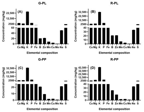
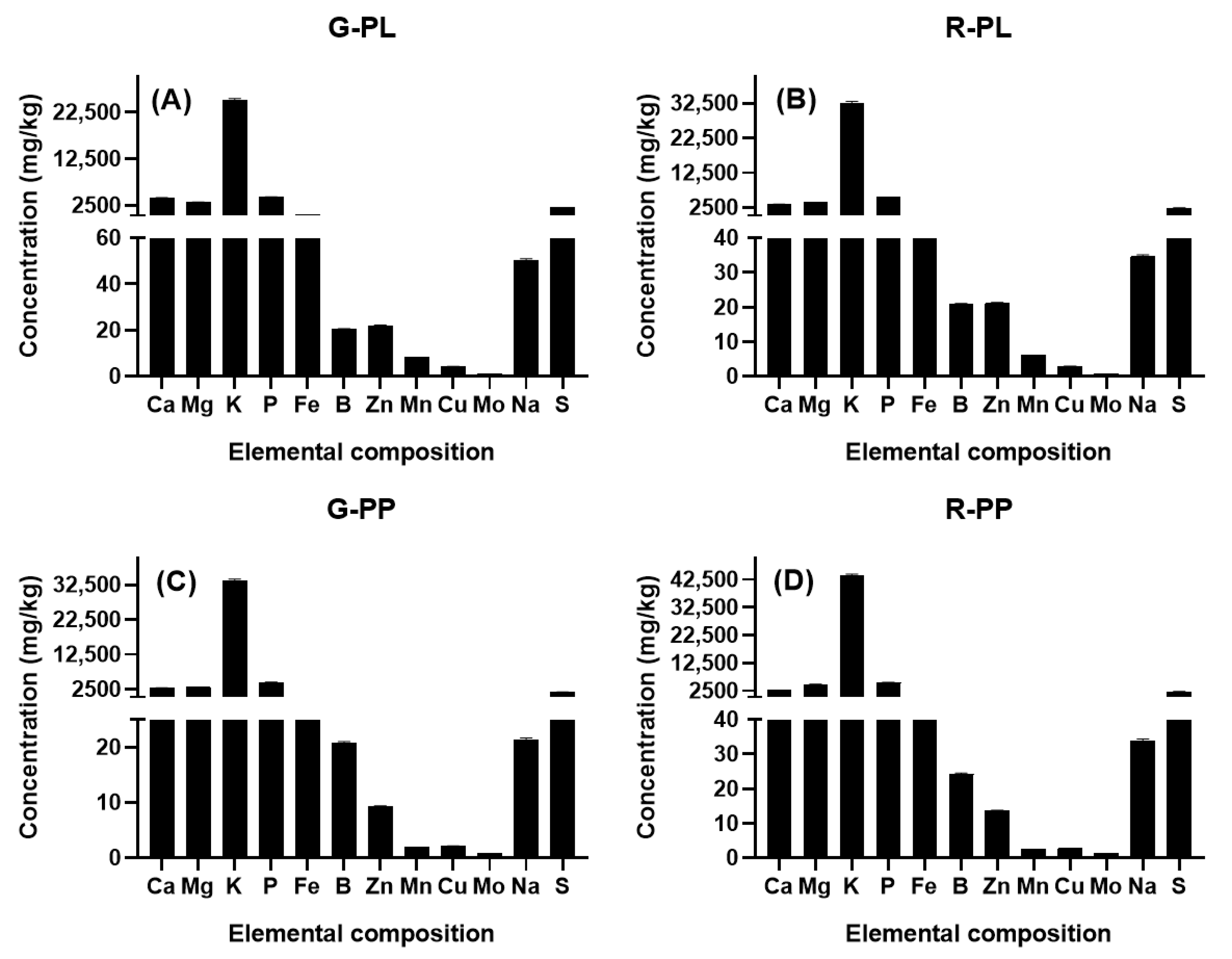
The mineral composition of the peel and pulp of green and ripe V. jossei fruits is presented in Table S2 and Figure 2. Among the macroelements analyzed, K was the predominant element in all samples, with concentrations between 25,100 and 43,900 mg/kg DW, followed by P, Mg, and Ca. The ripe pulp showed the highest concentrations of K (43,900 ± 590.61 mg/kg DW), Mg (4900 ± 65.92 mg/kg DW), and P (5500 ± 73.99 mg/kg DW), while the highest Ca content was recorded in the green peel (4100 ± 55.16 mg/kg DW).
Figure 2.
Mineral composition of the peel and pulp of green and ripe V. jossei fruits. Green peel: G-PL (A); ripe peel: R-PL (B); green pulp: G-PP (C); and ripe pulp: R-PP (D). Data are expressed as mean ± SD (n = 3). A discontinuous Y-axis was used to improve the visualization of elements occurring at different concentration ranges.
Among the microelements, iron (Fe) showed the greatest variation between tissues, reaching its highest concentration in the green peel (403.98 ± 5.43 mg/kg DW), followed by the ripe peel (153.14 ± 2.06 mg/kg DW), while considerably lower concentrations were observed in the pulp (42.80–59.06 mg/kg DW). The peel also showed higher concentrations of zinc (Zn), manganese (Mn), and copper (Cu) than the pulp.
4. Discussion
The content of phenolic compounds and flavonoids, as well as the antioxidant capacity, showed a clear dependence on the fruit part and, for some parameters, on the maturity stage of V. jossei. In general, the peel presented higher concentrations of bioactive compounds and greater antioxidant capacity than the pulp. However, the effect of maturity stage varied depending on the parameter evaluated. These results indicate that, particularly for total phenolic content (TPC), tissue type showed a more pronounced effect than maturity stage, with higher concentrations observed in the peel.
The TPC values found in the pulp of V. jossei are within or even above the range previously described for V. pubescens [21,22,23,24]. This result is particularly relevant because, in the present study, the peel was analyzed independently of the pulp, while several previous studies on Vasconcellea have evaluated the fruit or the pulp as the main matrix. Previous studies in V. pubescens have also shown that processing and drying conditions can considerably modify the apparent concentration of phenolic compounds [22].
The higher concentration of phenols in the peel could be related to the protective function of this tissue. As the fruit’s first barrier against environmental factors, the peel is more exposed to radiation, oxidative stress, microorganisms, and mechanical damage, which may contribute to the accumulation of secondary metabolites involved in defense mechanisms [25]. From a utilization perspective, this result also indicates that the peel should not be considered solely as waste from fruit processing, but as a potentially usable matrix for recovering bioactive compounds.
The behavior of flavonoids differed from that observed for total phenols. Green peel exhibited the highest TFC (2.57 ± 0.04 mg QE/g DW), followed by ripe peel (2.23 ± 0.04 mg QE/g DW), while in the pulp, ripening produced a significant increase from 0.59 ± 0.00 to 1.27 ± 0.02 mg QE/g DW. These values were comparable to those previously reported for V. pubescens [21,22,23,24]. In V. pubescens, flavonoid contents of approximately 2.37 mg QE/g of freeze-dried pulp have been reported [23], while different drying treatments resulted in values between approximately 0.71 and 1.26 mg QE/g DW [24]. Similarly, Vega-Gálvez et al. found differences in the flavonoid content of V. pubescens depending on the drying technology used [21]. In comparison, the green and ripe peels of V. jossei showed higher concentrations, while the ripe pulp presented values within the range described for V. pubescens. This reinforces the importance of considering fruit tissues separately when evaluating their potential as a source of bioactive compounds.
Two different methods were used to determine antioxidant capacity: the 2,2-diphenyl-1-picrylhydrazyl (DPPH) assay and the ferric reducing antioxidant power (FRAP) assay. Previous studies have shown that extracts from the genus Vasconcellea possess antioxidant capacity [26]. The DPPH capacity of V. jossei was lower than some values described for V. pubescens. In the latter species, an antioxidant capacity of approximately 20.1–31.6 µmol TE/g DW has been reported [21,22,23,24], while dehydration processes can considerably decrease these values. On the other hand, the FRAP assay showed a particularly marked separation between peel and pulp. The highest reducing capacity was observed in the green peel (45.70 ± 0.96 µmol TE/g DW), followed by the ripe peel (41.34 ± 0.05 µmol TE/g DW). In contrast, the green and ripe pulps showed 18.60 ± 1.81 and 21.86 ± 1.06 µmol TE/g DW, respectively, with a significant difference between them. Consequently, the reducing capacity of the peels was approximately twice that of the pulps. Furthermore, unlike TPC, where both peels belonged to the same statistical group, FRAP distinguished significantly between the green and ripe peel states, suggesting that ripening may be associated with changes in the composition of the antioxidants contributing to reducing capacity, even when total phenolic content remains relatively constant.
When comparing the phenolic profile of V. jossei with that described for other Caricaceae species, such as V. pubescens, V. × heilbornii, and Carica papaya, relevant differences were observed in both the abundance and distribution of the main phenolic metabolites. Rutin was the predominant compound in the peel of V. jossei, reaching 189.54 mg/100 g DW in the green state and decreasing to 84.46 mg/100 g DW during ripening. This high accumulation is of particular interest considering that glycosylated quercetin derivatives, including rutin, have been previously described as important components of the phenolic profile of Vasconcellea species, particularly V. pubescens [27]. In comparison, the levels described for C. papaya tissues are generally lower [28], indicating a higher accumulation of this glycosylated flavonol in the peel of V. jossei. Taken together, these results support the relevance of quercetin derivatives within the genus Vasconcellea and highlight the peel of V. jossei as a matrix with a high rutin content.
Interspecific differences were also observed in phenolic acid composition. While profiles including p-coumaric acid, ferulic acid, and various hydroxycinnamic acid derivatives have been described in C. papaya and V. pubescens [21,23,28,29], the profile determined by HPLC-PDA in V. jossei was more restricted. Caffeic acid was detected exclusively in the peel, decreasing from 5.13 to 3.58 mg/100 g DW during ripening, while syringic acid was present only in the pulp and increased from 1.83 to 2.30 mg/100 g DW. In contrast, gallic, p-coumaric, and chlorogenic acids were not detected under the analytical conditions used. Furthermore, the profile observed in V. jossei differs from that described for V. × pentagona (babaco), where other predominant phenolic compounds have been reported, including homovanillic acid and rosmarinic acid [30]. In contrast, the peel of V. jossei was characterized mainly by rutin, kaempferol, apigenin, and caffeic acid. These differences highlight the phytochemical diversity within Caricaceae and suggest that each species exhibits patterns of secondary metabolite accumulation. However, these comparisons should be interpreted with caution because studies differ in plant tissue, maturity stage, extraction procedures, chromatographic techniques, and how results are presented.
Taken together, the predominance of rutin and the presence of kaempferol, apigenin, and caffeic acid in the peel distinguish the phenolic profile of V. jossei from that described for other Caricaceae. The high concentration of rutin, particularly in the green peel, is also consistent with the greater antioxidant capacity observed in this tissue and with the strong correlation of this compound with DPPH and FRAP. These findings expand the available phytochemical knowledge for Andean species of Vasconcellea and highlight V. jossei, especially its peel, as a potential source of flavonoids of interest for future food and functional applications.
Mineral elements are essential nutrients that play a fundamental role in maintaining health and proper human body function [31]. These elements participate in numerous physiological and metabolic processes, including electrolyte balance, tissue formation and maintenance, energy metabolism, and the activity of various enzyme systems [32]. In this context, characterizing macro- and microelements in fruits allows evaluation of their nutritional composition and provides information on their potential as a dietary source of essential minerals.
The mineral composition of V. jossei showed significant differences between tissues and maturity stages, particularly notable for its high potassium content. This element was the predominant macroelement in all samples and reached its highest concentration in the ripe pulp (43,900 mg/kg DW), followed by the green pulp (33,700 mg/kg DW), the ripe peel (32,600 mg/kg DW), and the green peel (25,100 mg/kg DW). This trend may relate to K physiological roles in osmotic regulation, maintenance of ionic balance, and transport of photoassimilates to the fruit during development and ripening [33]. When comparing these results with other Caricaceae species, the concentration found in the ripe pulp of V. jossei was considerably higher than the values reported for commercial cultivars of Carica papaya, where contents of up to 8416 mg/kg DW in pulp [34] and 5617 mg/kg DW in peel [35] have been described, as well as that recorded in green fruit flour of V. quercifolia (29.56 mg/kg DW, converted from 26.25 ppm wet basis with 11.19% moisture content) [2]. For comparison on the same basis, the values reported for V. pulchra and V. × heilbornii [36] were converted to a dry-weight basis using the moisture contents reported in that study. The resulting K concentrations were approximately 37,051 and 16,564 mg/kg DW, respectively. Thus, the K content of ripe V. jossei pulp (43,900 mg/kg DW) was higher than that reported for both species, although the value for V. pulchra was of a similar magnitude.
Magnesium (Mg) also increased during ripening, particularly in the pulp, where its concentration rose from 3100 to 4900 mg/kg DW. The concentration found in the ripe pulp was higher than the values reported for V. pulchra (2110 mg/kg DW) and V. × heilbornii (1407 mg/kg DW) [36]. Similarly, it was higher than the 3.96 mg/kg DW reported for V. quercifolia (converted from 3.52 ppm wet basis) [2] and the 990 mg/kg DW reported for Carica papaya [34]. Phosphorus (P) followed a similar pattern in V. jossei, increasing from 4500 to 5500 mg/kg DW during pulp ripening. These increases are of interest due to the physiological functions of both elements: Mg participates as a cofactor of numerous enzymes [37], while P is involved in processes related to energy transfer and cellular metabolism [38].
Unlike K, Mg, and P, Ca showed a different trend during ripening. Its concentration decreased from 4100 to 3400 mg/kg DW in the peel and remained at 2900 mg/kg DW in both stages of pulp maturity. This differential behavior compared to other macroelements could be related to the low mobility of Ca through the phloem and its close association with structural components of plant tissues [39]. The contents determined in the ripe peel and pulp of V. jossei were higher than those reported for V. quercifolia fruit meal (6.04 mg/kg DW, converted from 5.36 ppm wet basis) [2], V. pulchra and V. × heilbornii pulp (1093 and 648 mg/kg DW) [36], and C. papaya peel (180.7 mg/kg DW) [35].
Another notable aspect of the mineral profile of V. jossei was the combination of a high potassium (K) concentration with a relatively low sodium (Na) content. The latter ranged from 21.38 to 33.94 mg/kg DW in the pulp and from 34.66 to 50.34 mg/kg DW in the peel. These values were lower than those described for C. papaya, where concentrations of 693 mg/kg DW in the pulp [34] and around 312 mg/kg DW in the peel [40] have been reported, as well as for the pulp of V. pulchra and V. × heilbornii (1668 and 633 mg/kg DW, respectively) [36]. Consequently, the ripe pulp of V. jossei exhibited a high K/Na ratio, derived primarily from its high potassium (K) content and low sodium (Na) concentration. This profile is a distinctive characteristic of the species’ mineral composition and enhances the nutritional value of its edible portion.
Overall, comparisons with other Caricaceae species reveal considerable interspecific variability in mineral composition, with V. jossei showing relatively high concentrations of potassium (K) and magnesium (Mg). Differences between the peel and pulp, together with ripening-related changes, further indicate that mineral distribution is not uniform within the fruit. The ripe pulp showed higher concentrations of K, Mg, and P, whereas the peel contained higher levels of Ca and several micronutrients, particularly Fe, Zn, and Mn. Together with the higher phenolic content and antioxidant capacity observed in the peel, these findings demonstrate the distinct chemical profiles of V. jossei tissues and provide a basis for further investigating the compositional value of both the edible pulp and the peel. Nevertheless, these findings should be interpreted considering the limitations of the present study, particularly the relatively small number of biological replicates. Moreover, although the compositional profile and in vitro antioxidant capacity provide valuable baseline information on this poorly studied species, they do not constitute direct evidence of nutritional benefits or biological effects. Therefore, further studies incorporating larger sample sizes, additional harvest periods, and appropriate biological assays are needed to validate these findings and determine the potential nutritional and functional relevance of V. jossei.
5. Conclusions
This study characterizes the phenolic composition, antioxidant capacity, and mineral composition of the peel and pulp of Vasconcellea jossei, revealing differences associated with both tissue type and ripening stage. The peel exhibited higher total phenolic and flavonoid contents and greater in vitro antioxidant capacity than the pulp, with the green peel showing the highest values for most of these parameters. HPLC-PDA analysis revealed a differentiated distribution of phenolic compounds, with rutin being the predominant compound in the peel, reaching a maximum concentration of 189.54 mg/100 g DW at the green stage, together with caffeic acid, apigenin, and kaempferol. In contrast, syringic acid was detected in the pulp, whereas epicatechin was detected exclusively in the ripe pulp. The strong positive correlations between total flavonoid content and selected phenolic compounds with DPPH and FRAP indicate close associations between the phenolic profile and antioxidant capacity. Regarding mineral composition, potassium (K) was the predominant element, reaching 43,900 mg/kg DW in the ripe pulp, together with relatively high concentrations of magnesium (Mg) and phosphorus (P), whereas the peel showed higher concentrations of several micronutrients. Overall, these findings establish the first baseline information on the chemical composition and in vitro antioxidant properties of V. jossei fruit and demonstrate the distinct chemical characteristics of its peel and pulp during ripening. The presence of phenolic compounds and mineral elements, together with the observed antioxidant capacity, highlights the scientific interest of this Andean fruit and provides a basis for exploring its nutritional and functional relevance. In particular, the phenolic compounds identified in the peel, together with its in vitro antioxidant capacity, suggest potential for future use as a source of bioactive compounds in food applications. Given the limited number of biological replicates and the in vitro nature of the antioxidant assays, these findings should be interpreted within the scope of the present study. Future studies incorporating broader sampling and complementary biological evaluations could further clarify the nutritional and functional relevance of V. jossei.
Supplementary Materials
The following supporting information can be downloaded at: https://www.mdpi.com/article/10.3390/pr14213543/s1, Table S1: Analytical performance parameters of the HPLC-PDA method for the determination of target polyphenols; Table S2: Elemental composition of the peel and pulp of green and ripe V. jossei fruits; Figure S1: HPLC-PDA chromatograms of the green peel extract of V. jossei recorded at 280 and 320 nm; Figure S2: HPLC-PDA chromatograms of the ripe peel extract of V. jossei recorded at 280 and 320 nm; Figure S3: HPLC-PDA chromatograms of the green pulp extract of V. jossei recorded at 280 and 320 nm; Figure S4: HPLC-PDA chromatograms of the ripe pulp extract of V. jossei recorded at 280 and 320 nm; Figure S5: HPLC-PDA chromatograms of 12 phenolic standards (10 mg/L) recorded at 280 and 320 nm; Figure S6: Heatmap of Pearson correlation coefficients among total phenolic content (TPC), total flavonoid content (TFC), antioxidant capacity (DPPH and FRAP), and individual phenolic compounds identified by HPLC-PDA in V. jossei.
Author Contributions
Conceptualization, R.V.; methodology, R.V., I.C.-G., R.R., C.J.-K. and L.B.-M.; software, R.V.; validation, R.V. and C.G.-V.; formal analysis, R.V., R.R., C.J.-K. and L.B.-M.; investigation, R.V., R.R., C.J.-K. and L.B.-M.; resources, R.V. and X.C.; data curation, R.V.; writing—original draft preparation, R.V.; writing—review and editing, R.V. and C.G.-V.; visualization, R.V.; supervision, R.V.; project administration, R.V. All authors have read and agreed to the published version of the manuscript.
Funding
This research was supported by the Centro de Investigaciones Biotecnológicas del Ecuador (CIBE) through Project CIBE-13-2023. Universidad del Alba provided financial support for the article processing charge (APC) associated with the publication of this study.
Data Availability Statement
The original contributions presented in this study are included in the article/Supplementary Material. Further inquiries can be directed to the corresponding authors.
Acknowledgments
The authors gratefully acknowledge the Centro de Investigaciones Biotecnológicas del Ecuador (CIBE) and Universidad del Alba for their institutional support, facilities, and resources that contributed to the execution and publication of this study.
Conflicts of Interest
The authors declare that they have no known competing financial interests or personal relationships that could have appeared to influence the work reported in this paper.
Abbreviations
The following abbreviations are used in this manuscript:
| Api | Apigenin |
| CafA | Caffeic acid |
| Cat | Catechin |
| ChlA | Chlorogenic acid |
| CouA | p-Coumaric acid |
| DPPH | 1,1-Diphenyl-2-picrylhydrazyl |
| DW | Dry weight |
| Epi | Epicatechin |
| FRAP | Ferric reducing antioxidant power |
| GAE | Gallic acid equivalents |
| GalA | Gallic acid |
| HPLC | High-performance liquid chromatography |
| HPLC-PDA | High-performance liquid chromatography coupled with photodiode array detection |
| Kae | Kaempferol |
| LOD | Limit of detection |
| Nan | Naringenin |
| ND | Not detected |
| PDA | Photodiode array detector |
| QE | Quercetin equivalents |
| Que | Quercetin |
| Rut | Rutin |
| SD | Standard deviation |
| SyrA | Syringic acid |
| TE | Trolox equivalents |
| TFC | Total flavonoid content |
| TPC | Total phenolic content |
| TPTZ | 2,4,6-Tripyridyl-s-triazine |
References
- Méndez, T.; Jara-Villacura, V.; Parra-Palma, C.; Morales-Quintana, L. Postharvest biology and quality preservation of Vasconcellea pubescens: Challenges and opportunities for reducing fruit losses. Horticulturae 2025, 11, 1165. [Google Scholar] [CrossRef] [Scilit]
- da Silva, L.d.F.F.; Rodrigues, K.F.; Ethur, E.M.; Hoehne, L.; de Souza, C.F.V.; Bonemann, D.H.; Ribeiro, A.S.; de Freitas, E.M. Nutritional potential of Vasconcellea quercifolia A. St.-Hil. green fruit flour. Braz. J. Food Technol. 2022, 25, e2021080. [Google Scholar] [CrossRef] [Scilit]
- Folharini, Z.F.; Orlandi, C.R.; Martini, M.C.; Bruxel, F.; Altamayer, T.; Brietzke, D.T.; Gonçalves, T.E.; Finatto, J.; Ethur, E.M.; de Moura, N.F.; et al. Nutritional characterization of Vasconcellea quercifolia A.St-Hil.: Potential for the development of functional food. Food Sci. Technol. 2019, 39, 432–438. [Google Scholar] [CrossRef] [Scilit]
- Shahidi, F.; Ambigaipalan, P. Phenolics and polyphenolics in foods, beverages and spices: Antioxidant activity and health effects—A review. J. Funct. Foods 2015, 18, 820–897. [Google Scholar] [CrossRef] [Scilit]
- Tomás-Barberán, F.A.; Espín, J.C. Phenolic compounds and related enzymes as determinants of quality in fruits and vegetables. J. Sci. Food Agric. 2001, 81, 853–876. [Google Scholar] [CrossRef] [Scilit]
- Kapasakalidis, P.G.; Rastall, R.A.; Gordon, M.H. Extraction of polyphenols from processed black currant (Ribes nigrum L.) residues. J. Agric. Food Chem. 2006, 54, 4016–4021. [Google Scholar] [CrossRef] [Scilit] [PubMed]
- Sadowska-Bartosz, I.; Bartosz, G. Evaluation of the antioxidant capacity of food products: Methods, applications and limitations. Processes 2022, 10, 2031. [Google Scholar] [CrossRef] [Scilit]
- Pino-Ramos, L.L.; Farias, D.R.; Olivares-Caro, L.; Mitsi, C.; Mardones, C.; Echeverria, J.; Avila, F.; Gutierrez, M. Chilean papaya (Vasconcellea pubescens A. DC.) residues as a source of bioactive compounds: Chemical composition, antioxidant capacity, and antiglycation effects. Heliyon 2024, 10, e38837. [Google Scholar] [CrossRef] [Scilit] [PubMed]
- Purohit, P.; Kataria, M.K. Phytochemicals screening, antioxidant and antimicrobial activity of Carica papaya leaf extracts. Int. J. Curr. Pharm. Res. 2024, 16, 95–98. [Google Scholar] [CrossRef] [Scilit]
- Martial-Didier, A.K.; Hubert, K.K.; Jean Parfait, K.E.; Kablan, T. Phytochemical Properties and Proximate Composition of Papaya (Carica papaya L. Var Solo 8) Peels. Turk. J. Agric.-Food Sci. Technol. 2017, 5, 676–680. [Google Scholar] [CrossRef] [Scilit]
- Costanzo, G.; Vitale, E.; Iesce, M.R.; Naviglio, D.; Amoresano, A.; Fontanarosa, C.; Spinelli, M.; Ciaravolo, M.; Arena, C. Antioxidant properties of pulp, peel and seeds of Phlegrean mandarin (Citrus reticulata Blanco) at different stages of fruit ripening. Antioxidants 2022, 11, 187. [Google Scholar] [CrossRef] [Scilit] [PubMed]
- Ovando-Martinez, M.; López-Teros, M.; Tortoledo-Ortiz, V.; Astiazarán-García, H.; Ayala-Zavala, J.F.; Villegas-Ochoa, M.A.; González-Aguilar, G.A. Effect of ripening on Physico-Chemical properties and bioactive compounds in papaya pulp, skin and seeds. Indian J. Nat. Prod. Resour. 2018, 9, 47–59. [Google Scholar]
- Chung, S.W.; Jang, Y.J.; Kim, S.; Kim, S.C. Spatial and compositional variations in fruit characteristics of papaya (Carica papaya cv. Tainung no. 2) during ripening. Plants 2023, 12, 1465. [Google Scholar] [CrossRef] [Scilit] [PubMed]
- Cornejo, X.; Cedeño, J.F. Vasconcellea jossei (Caricaceae): A new species of highland papaya with edible fruits from the Andes of Ecuador. Harv. Pap. Bot. 2024, 29, 59–62. [Google Scholar] [CrossRef] [Scilit]
- Gutiérrez-Román, A.I.F.; Santa Cruz-Carpio, C.M.; Velarde-Vilchez, M.; Nolasco-Cárdenas, O. Capacidad Antioxidante de La Pulpa de Vasconcellea candicans “Mito.”. Biotempo 2023, 20, 151–162. [Google Scholar] [CrossRef] [Scilit]
- Hoyos-Arbeláez, J.; Blandón-Naranjo, L.; Vázquez, M.; Contreras-Calderón, J. Antioxidant capacity of mango fruit (Mangifera indica). an electrochemical study as an approach to the spectrophotometric methods. Food Chem. 2018, 266, 435–440. [Google Scholar] [CrossRef] [Scilit] [PubMed]
- Thaipong, K.; Boonprakob, U.; Crosby, K.; Cisneros-Zevallos, L.; Hawkins Byrne, D. Comparison of ABTS, DPPH, FRAP, and ORAC assays for estimating antioxidant activity from guava fruit extracts. J. Food Compos. Anal. 2006, 19, 669–675. [Google Scholar] [CrossRef] [Scilit]
- Ghareeb, M.A.; Mohamed, T.; Saad, A.M.; Refahy, L.A.-G.; Sobeh, M.; Wink, M. HPLC-DAD-ESI-MS/MS analysis of fruits from Firmiana simplex (L.) and evaluation of their antioxidant and antigenotoxic properties. J. Pharm. Pharmacol. 2017, 70, 133–142. [Google Scholar] [CrossRef] [Scilit] [PubMed]
- Woisky, R.G.; Salatino, A. Analysis of Propolis: Some Parameters and Procedures for Chemical Quality Control. J. Apic. Res. 1998, 37(2), 99–105. Available online: https://www.tandfonline.com/doi/abs/10.1080/00218839.1998.11100961. [CrossRef] [Scilit]
- Sánchez-Bonet, D.; García-Oms, S.; Belda-Antolí, M.; Padrón-Sanz, C.; Lloris-Carsi, J.M.; Cejalvo-Lapeña, D. RP-HPLC-DAD determination of the differences in the polyphenol content of Fucus vesiculosus extracts with similar antioxidant activity. J. Chromatogr. B 2021, 1184, 122978. [Google Scholar] [CrossRef] [Scilit] [PubMed]
- Vega-Gálvez, A.; Poblete, J.; Quispe-Fuentes, I.; Uribe, E.; Bilbao-Sainz, C.; Pastén, A. Chemical and bioactive characterization of papaya (Vasconcellea pubescens) under different drying technologies: Evaluation of antioxidant and antidiabetic potential. J. Food Meas. Charact. 2019, 13, 1980–1990. [Google Scholar] [CrossRef] [Scilit]
- Vega-Gálvez, A.; Poblete, J.; Rojas-Carmona, R.; Uribe, E.; Pastén, A.; Goñi, M.G. Vacuum drying of Chilean papaya (Vasconcellea pubescens) fruit pulp: Effect of drying temperature on kinetics and quality parameters. J. Food Sci. Technol. 2021, 58, 1–11. [Google Scholar] [CrossRef] [Scilit] [PubMed]
- Vega-Gálvez, A.; Stucken, K.; Cantuarias, C.; Lamas, F.; García, V.; Pastén, A. Antimicrobial properties of papaya (Vasconcellea pubescens) subjected to Low-Temperature vacuum dehydration. Innov. Food Sci. Emerg. Technol. 2021, 67, 102563. [Google Scholar] [CrossRef] [Scilit]
- Vega-Gálvez, A.; Uribe, E.; Pastén, A.; Vega, M.; Poblete, J.; Bilbao-Sainz, C.; Chiou, B.-S. Low-Temperature vacuum drying as novel process to improve papaya (Vasconcellea pubescens) Nutritional-Functional properties. Future Foods 2022, 5, 100117. [Google Scholar] [CrossRef] [Scilit]
- Silva, M.A.; Albuquerque, T.G.; Ferreira, D.M.; Alves, R.C.; Oliveira, M.B.P.P.; Costa, H.S. Nutritional and bioactive profiling of Cucumis melo L. by-products: Towards a circular food economy. Molecules 2025, 30, 1287. [Google Scholar] [CrossRef] [Scilit] [PubMed]
- Aguirre-Rodríguez, A.; Duarte-Casar, R.; Rojas-Le-Fort, M.; Romero-Benavides, J.C. Food uses, functional activities, and bioactive compounds of three ecuadorian Vasconcellea fruits: Bibliometric analysis and review. J. Agric. Food Res. 2024, 17, 101244. [Google Scholar] [CrossRef] [Scilit]
- Simirgiotis, M.J.; Caligari, P.D.S.; Schmeda-Hirschmann, G. Identification of phenolic compounds from the fruits of the mountain papaya Vasconcellea pubescens A. DC. grown in chile by liquid Chromatography–UV Detection–Mass spectrometry. Food Chem. 2009, 115, 775–784. [Google Scholar] [CrossRef] [Scilit]
- Iordănescu, O.A.; Băla, M.; Gligor (Pane), D.; Zippenfening, S.E.; Cugerean, M.I.; Petroman, M.I.; Hădărugă, D.I.; Hădărugă, N.G.; Riviş, M. A DPPH· kinetic approach on the antioxidant activity of various parts and ripening levels of papaya (Carica papaya L.) ethanolic extracts. Plants 2021, 10, 1679. [Google Scholar] [CrossRef] [Scilit] [PubMed]
- Fuentes, Y.; Giovagnoli-Vicuña, C.; Faúndez, M.; Giordano, A. Microencapsulation of Chilean papaya waste extract and its impact on physicochemical and bioactive properties. Antioxidants 2023, 12, 1900. [Google Scholar] [CrossRef] [Scilit] [PubMed]
- Mihai, R.A.; Canchignia Guacollantes, M.G.; Vivanco Gonzaga, R.F.; Cubi-Insuaste, N.S.; Catana, R.D. Impact of babaco (Vasconcellea × pentagona (Heilborn) Mabb.) fruit ripening stages on phytochemical composition and biological properties. Metabolites 2024, 14, 718. [Google Scholar] [CrossRef] [Scilit] [PubMed]
- Razzaque, M.S.; Wimalawansa, S.J. Minerals and human health: From deficiency to toxicity. Nutrients 2025, 17, 454. [Google Scholar] [CrossRef] [Scilit] [PubMed]
- Weyh, C.; Krüger, K.; Peeling, P.; Castell, L. The role of minerals in the optimal functioning of the immune system. Nutrients 2022, 14, 644. [Google Scholar] [CrossRef] [Scilit] [PubMed]
- Johnson, R.; Vishwakarma, K.; Hossen, M.d.S.; Kumar, V.; Shackira, A.M.; Puthur, J.T.; Abdi, G.; Sarraf, M.; Hasanuzzaman, M. Potassium in plants: Growth regulation, signaling, and environmental stress tolerance. Plant Physiol. Biochem. 2022, 172, 56–69. [Google Scholar] [CrossRef] [Scilit] [PubMed]
- Leterme, P.; Buldgen, A.; Estrada, F.; Londoño, A.M. Mineral content of tropical fruits and unconventional foods of the Andes and the rain forest of Colombia. Food Chem. 2006, 95, 644–652. [Google Scholar] [CrossRef] [Scilit]
- Moses, M.O.; Olanrewaju, M.J. Proximate and selected mineral composition of ripe pawpaw (Carica papaya) seeds and skin. J. Sci. Innov. Res. 2018, 7, 75–77. [Google Scholar] [CrossRef] [Scilit]
- Matute Jaramillo, L.; Tirado Valladares, B. Análisis Bromatólogico de Vasconcellea pulchra VM Badillo y Vasconcellea × heilbornii VM Badillo Procedentes de La Provincia Bolívar, Ecuador. 2013. Available online: https://dspace.ups.edu.ec/handle/123456789/6008 (accessed on 17 August 2026).
- Ahmed, N.; Zhang, B.; Bozdar, B.; Chachar, S.; Rai, M.; Li, J.; Li, Y.; Hayat, F.; Chachar, Z.; Tu, P. The power of magnesium: Unlocking the potential for increased yield, quality, and stress tolerance of horticultural crops. Front. Plant Sci. 2023, 14, 1285512. [Google Scholar] [CrossRef] [Scilit] [PubMed]
- Sandoval-Vargas, J.M.; Macedo-Osorio, K.S.; Durán-Figueroa, N.V.; Garibay-Orijel, C.; Badillo-Corona, J.A. Chloroplast engineering of Chlamydomonas reinhardtii to use phosphite as phosphorus source. Algal Res. 2018, 33, 291–297. [Google Scholar] [CrossRef] [Scilit]
- Hocking, B.; Tyerman, S.D.; Burton, R.A.; Gilliham, M. Fruit calcium: Transport and physiology. Front. Plant Sci. 2016, 7, 569. [Google Scholar] [CrossRef] [Scilit] [PubMed]
- Akintunde, A.O.; Kolu, P.; Akintunde, I.A.; Adewole, S.A.; Akinboye, O.E.; Afodu, O.J.; Ndubuisi-Ogbonna, L.C.; Shobo, B.A. Evaluation of the nutritive values of Carica papaya fruit peels as A potential ingredient in livestock nutrition. Anim. Prod. 2022, 24, 104–113. [Google Scholar] [CrossRef] [Scilit]

Disclaimer/Publisher’s Note: The statements, opinions and data contained in all publications are solely those of the individual author(s) and contributor(s) and not of MDPI and/or the editor(s). MDPI and/or the editor(s) disclaim responsibility for any injury to people or property resulting from any ideas, methods, instructions or products referred to in the content. |
© 2026 by the authors. Licensee MDPI, Basel, Switzerland. This article is an open access article distributed under the terms and conditions of the Creative Commons Attribution (CC BY) license.