


Highlights
- This narrative review proposes a conceptual framework that organizes pediatric insulin resistance across interconnected biological domains rather than validated temporal stages.
- Candidate biomarkers differ substantially in the maturity of pediatric evidence, assay availability, and clinical readiness.
- Developmental, pubertal, skeletal, neuroendocrine, and socioeconomic factors modify metabolic risk across these domains.
- Prospective studies are needed before biomarker panels can be used for risk prediction, clinical classification, or biomarker-guided treatment.
Abstract
Background/Objectives: Insulin resistance (IR) in children and adolescents is heterogeneous and influenced by puberty, adiposity, growth, and developmental factors. Conventional measures such as fasting insulin and the homeostasis model assessment of insulin resistance (HOMA-IR) have limited pediatric specificity because of assay variability and the absence of universally accepted thresholds. This narrative review proposes a conceptual framework linking candidate biomarkers to biological domains relevant to pediatric IR. Methods: PubMed/MEDLINE and Google Scholar were searched through August 2026 using terms related to pediatric IR, obesity, puberty, and candidate biomarkers. Pediatric and adolescent studies were prioritized; adult studies were used selectively for mechanistic context. Biomarkers were selected as representative examples of biological domains, not as a validated diagnostic panel. Results: The framework comprises four interacting domains: intracellular insulin signaling (intracellular magnesium), adipose tissue dysfunction (leptin, adiponectin), inflammatory activation (lipocalin-2, white blood cell count, high-sensitivity C-reactive protein), and endothelial dysfunction (asymmetric dimethylarginine). Sonic hedgehog N-terminal fragment, osteocalcin, oxytocin, and central appetite-regulatory pathways are considered cross-cutting developmental, skeletal, and neuroendocrine modifiers. Evidence maturity, assay standardization, and clinical readiness differ substantially among markers. Conclusions: This framework is hypothesis-generating and does not represent a validated temporal sequence, diagnostic staging system, or routine biomarker panel. Longitudinal pediatric studies with standardized assays, puberty-specific reference ranges, and assessment of incremental clinical value are needed before biomarker-informed approaches can improve risk prediction, classification, or outcomes.
Keywords:
insulin resistance; obesity; biomarker; puberty; adipokine; magnesium; endothelial dysfunction 1. Introduction
Insulin resistance (IR) in childhood and adolescence is associated with an increased risk of dysglycemia, metabolic dysfunction-associated steatotic liver disease, dyslipidemia, hypertension, and later type 2 diabetes [1]. However, obesity, hyperinsulinemia, IR, impaired glucose tolerance, and type 2 diabetes are overlapping but noninterchangeable phenotypes. Not every child with obesity has IR, and biomarkers identified in established type 2 diabetes cannot automatically be interpreted as early predictors of pediatric IR.
Fasting insulin and the homeostasis model assessment of insulin resistance (HOMA-IR) are commonly used surrogate measures in pediatric research. Their interpretation, however, is influenced by age, sex, pubertal stage, adiposity, ethnicity, fasting conditions, biological variation, and inter-assay differences. Accordingly, fasting insulin and HOMA-IR should not be used in isolation to define an individual child’s metabolic status.
Recent studies, including those by Takaya et al., have broadened the biological perspective on pediatric IR by examining developmental signaling pathways, extracellular vesicles, and bone-derived endocrine regulation [2]. These observations support the view that pediatric IR is biologically heterogeneous and cannot be adequately characterized by a single surrogate measure. This review therefore presents a conceptual framework that organizes candidate biomarkers according to their principal biological domains. The framework is intended to complement, rather than replace, standard clinical assessment and to identify testable hypotheses for future pediatric research.
2. Literature Search and Biomarker Selection
This narrative review was informed by literature searches of PubMed/MEDLINE and Google Scholar through August 2026. Search concepts included “pediatric insulin resistance,” “childhood obesity,” “adolescent insulin resistance,” “magnesium,” “adiponectin,” “leptin,” “lipocalin-2,” “white blood cell count,” “ADMA,” “sonic hedgehog,” “osteocalcin,” “oxytocin,” “puberty,” “inflammation,” “endothelial dysfunction,” and “appetite regulation.” Priority was given to pediatric and adolescent studies. Adult studies were used selectively to provide mechanistic context when pediatric evidence was limited.
References were selected narratively on the basis of biological relevance, pediatric applicability, methodological quality, and relevance to the proposed framework. This article was not designed as a systematic review or meta-analysis. Biomarkers were selected as representative examples of distinct biological domains, not as a comprehensive diagnostic panel or as markers with equivalent levels of evidence. Established clinical variables—including adiposity, blood pressure, glucose measures, lipid profile, liver-related assessment, family history, pubertal status, diet, sleep, physical activity, and psychosocial context—remain essential in clinical care.
3. A Conceptual Framework of Interconnected Domains
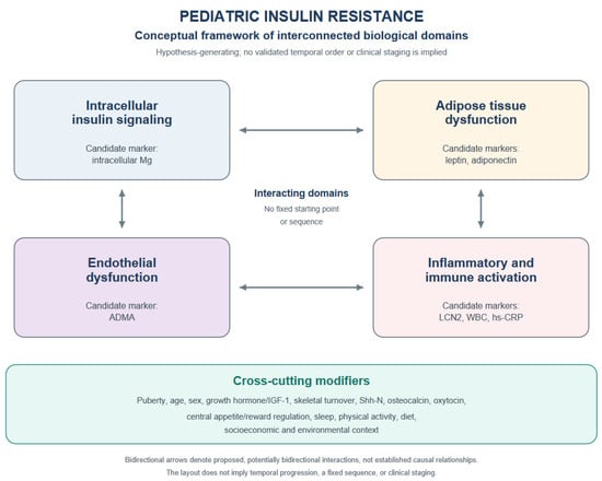
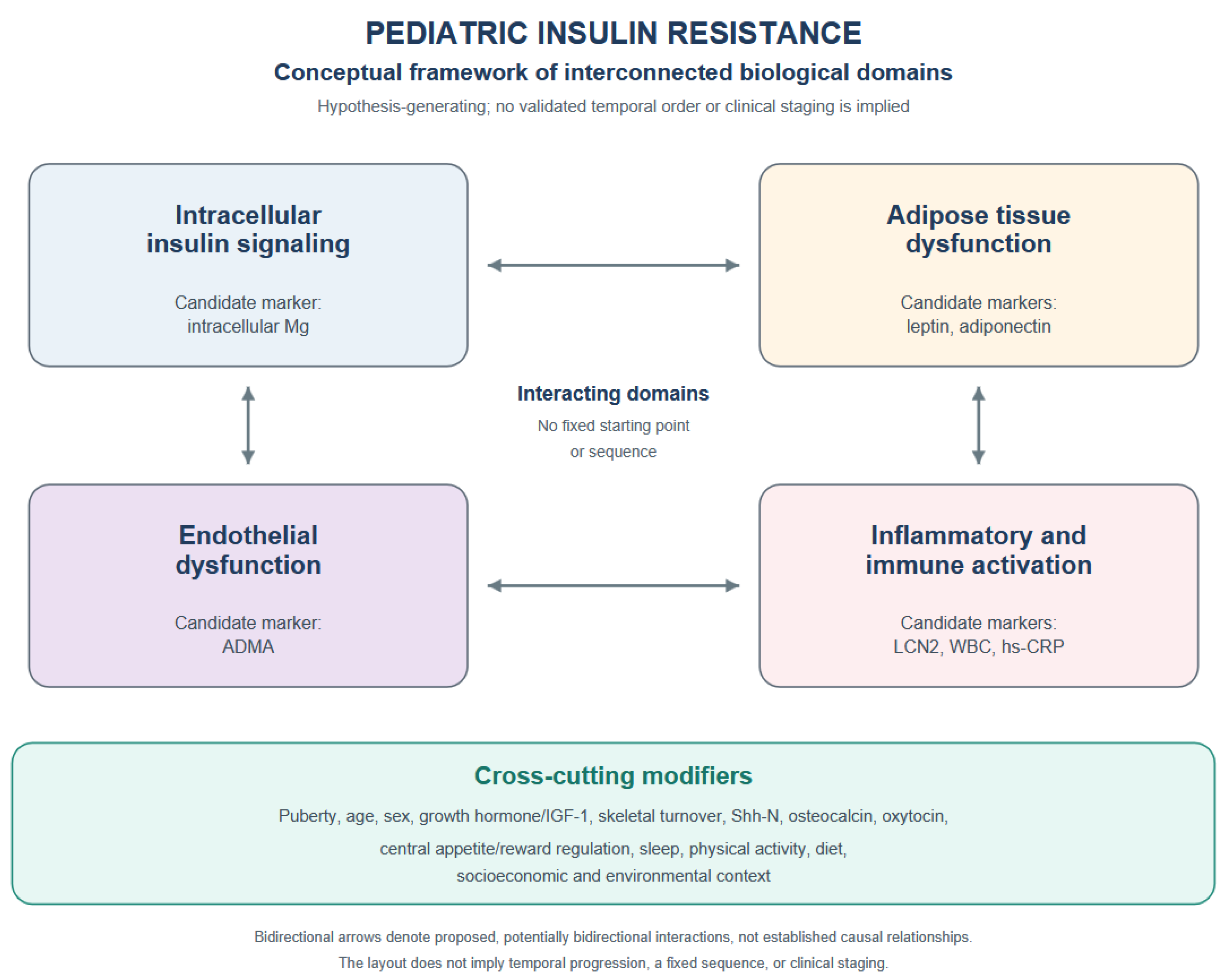
The proposed framework comprises four interacting biological domains and a group of cross-cutting modifiers. This organization is intended to facilitate the interpretation of mechanistic heterogeneity rather than to establish a temporal sequence. Figure 1 presents the domains in a non-linear layout without a fixed starting point or sequence. Bidirectional arrows denote proposed, potentially bidirectional interactions rather than established causal relationships.
Figure 1.
Conceptual framework of interconnected biological domains in pediatric insulin resistance. The four principal domains—intracellular insulin signaling, adipose tissue dysfunction, inflammatory and immune activation, and endothelial dysfunction—are arranged without a fixed starting point or sequence. Bidirectional arrows denote proposed, potentially bidirectional interactions rather than established causal relationships. Developmental, skeletal, neuroendocrine, behavioral, environmental, and socioeconomic influences are shown separately as cross-cutting modifiers. The framework is conceptual and hypothesis-generating; it does not represent a validated temporal cascade, clinical staging system, or equivalent biomarker maturity across domains. Candidate biomarkers are shown for illustrative purposes and should not be used alone to diagnose pediatric insulin resistance, assign a mechanistic stage, or direct treatment. Abbreviations: Mg, magnesium; LCN2, lipocalin-2; WBC, white blood cell count; hs-CRP, high-sensitivity C-reactive protein; ADMA, asymmetric dimethylarginine; Shh-N, Sonic hedgehog N-terminal fragment.
3.1. Intracellular Insulin Signaling and Magnesium
Insulin-stimulated glucose uptake in skeletal muscle and adipose tissue depends on signaling through insulin receptor substrates, phosphoinositide 3-kinase, Akt, and GLUT4 trafficking [3,4,5]. Magnesium is biologically relevant because it participates in ATP-dependent enzymatic reactions and kinase activity [6]. However, a mechanistic role does not establish intracellular magnesium deficiency as the earliest detectable abnormality in pediatric IR [7]. Serum magnesium is tightly regulated and may not reflect intracellular magnesium pools, whereas erythrocyte and peripheral blood mononuclear cell measurements remain insufficiently standardized for routine clinical use [8]. Dietary intake, renal function, medication exposure, gastrointestinal losses, and supplement use may further influence interpretation [9,10].
3.2. Adipose Tissue Dysfunction
Adipose tissue expansion can alter the secretion of leptin, adiponectin, free fatty acids, and inflammatory mediators. Leptin generally increases with fat mass, whereas adiponectin is often lower in pediatric obesity and metabolic risk states [11]. The adiponectin-to-leptin ratio may provide contextual information about adipose endocrine function, but it is strongly influenced by adiposity, sex, pubertal stage, and assay characteristics [12,13]. These markers should therefore be interpreted as indicators associated with adipose biology rather than as validated markers of a discrete disease stage.
3.3. Inflammatory and Immune Activation
Adipose tissue dysfunction may be accompanied by local and systemic inflammatory signaling [11]. Lipocalin-2 (LCN2) [14], white blood cell count [15], and high-sensitivity C-reactive protein [16] may reflect inflammatory activity. However, all are nonspecific and may be influenced by acute infection, chronic inflammatory conditions, medication exposure, and adiposity itself [16]. Their value in pediatric IR is therefore contextual and investigational rather than diagnostic.
3.4. Endothelial Dysfunction
Insulin has vascular actions that include stimulation of endothelial nitric oxide production. Asymmetric dimethylarginine (ADMA) is an endogenous inhibitor of nitric oxide synthase and is mechanistically relevant to endothelial dysfunction [17]. Nonetheless, evidence that ADMA prospectively identifies endothelial disease or clinically meaningful progression of IR in children remains limited [18,19]. ADMA should therefore be regarded as an investigational marker of vascular biology rather than as a routine component of pediatric risk assessment.
3.5. Cross-Cutting Developmental, Skeletal, Neuroendocrine, and Central Modifiers
Developmental, skeletal, and neuroendocrine factors are best understood as cross-cutting modifiers rather than as a terminal phase or discrete stage of IR. Puberty, sex steroids, growth hormone/insulin-like growth factor 1 signaling, skeletal turnover, and growth velocity may influence insulin sensitivity, adipokine concentrations, body composition, inflammatory markers, and vascular biology during childhood and adolescence [20,21].
Sonic hedgehog N-terminal fragment (Shh-N) is involved in developmental signaling and has been implicated in adipogenesis, inflammation, and energy metabolism [22]. In pediatric studies, its association with adiponectin and IR supports the possibility that developmental signaling may contribute to metabolic susceptibility [2]; however, the available evidence is limited and does not establish a causal or temporal role. Similarly, bone-derived hormones such as osteocalcin may link skeletal turnover to glucose metabolism [23]. Experimental studies suggest that osteocalcin can affect insulin secretion and insulin sensitivity [24], whereas interpretation in children is complicated by age-related and growth-related variation in bone turnover.
Neuroendocrine factors may also modify metabolic phenotypes. Oxytocin has been associated with energy balance and glucose metabolism, but its clinical relevance to pediatric IR remains exploratory [25]. Central appetite-regulatory pathways provide an additional mechanistic context: hypothalamic neuropeptide Y/agouti-related peptide neurons promote feeding, whereas proopiomelanocortin and cocaine- and amphetamine-regulated transcript pathways contribute to satiety-related signaling [26]. Leptin and insulin influence these circuits, although the translation of experimental findings into clinically useful pediatric biomarkers remains limited.
Mesolimbic reward pathways may interact with homeostatic appetite regulation and contribute to hedonic or reward-driven eating. However, the term “food addiction” remains conceptually and clinically contested and should not be used interchangeably with overeating or hedonic eating [27].
4. Puberty and Interpretation of Pediatric Insulin Resistance
Puberty is a major determinant of insulin sensitivity and should be incorporated into the interpretation of pediatric metabolic measures. Physiological pubertal insulin resistance may increase fasting insulin and HOMA-IR, particularly during mid-puberty, without necessarily indicating progressive pathological disease [20,21]. Interpretation should therefore take account of age, sex, Tanner stage, adiposity, body composition, growth velocity, ethnicity, family history, fasting conditions, and laboratory methods.
There are no universally accepted pediatric diagnostic thresholds for fasting insulin or HOMA-IR. Both measures are influenced by assay methodology and biological variation. In clinical settings, fasting insulin should not be interpreted as a stand-alone diagnostic test, and an isolated value should not be used to assign a child to a mechanistic domain. When metabolic complications are suspected, assessment should be guided by the clinical history, physical examination, blood pressure, glucose-related testing as indicated, lipid profile, liver-related assessment, and evaluation of obesity-related comorbidities [28].
5. Evidence Profile and Clinical Readiness
Table 1 summarizes candidate biomarkers by biological domain, pediatric evidence, longitudinal evidence, key limitations, and clinical readiness. It does not establish that their associations with insulin resistance are independent of adiposity. Adiposity-related confounding is explicitly noted for leptin/adiponectin and inflammatory markers in the Key limitations column; the reported associations should not be interpreted as demonstrating independent predictive value.
Table 1.
Candidate biomarkers differ in evidence maturity, assay feasibility, and clinical readiness. None of the listed markers should currently be used alone to diagnose pediatric insulin resistance, assign a mechanistic stage, or direct treatment.
6. Clinical Implications, Equity, and Feasibility
The framework is intended to complement, rather than replace, routine pediatric care. At present, no evidence demonstrates that the proposed panel improves diagnostic accuracy beyond accessible clinical assessment, prospectively predicts outcomes, identifies treatment-responsive phenotypes, or improves patient-centered outcomes. Specialized tests such as intracellular magnesium, LCN2, ADMA, Shh-N, and oxytocin may be unavailable or unaffordable in many settings.
Effective care for children with obesity or metabolic risk should not depend on access to specialized biomarkers. Core care should remain family-centered and should include nutritional support, opportunities for physical activity, sleep assessment, behavioral and psychological support, evaluation of obesity-related complications, and attention to food insecurity, safe environments for physical activity, the affordability of nutritious foods, and access to healthcare. Where resources are limited, growth trajectory, BMI percentile, blood pressure, clinical signs such as acanthosis nigricans, and routine laboratory investigations when indicated remain practical components of risk assessment.
Magnesium supplementation should not be recommended solely on the basis of this exploratory biomarker framework. Treatment of documented magnesium deficiency is distinct from unproven biomarker-guided supplementation for pediatric IR. Similarly, anti-inflammatory or other phenotype-directed interventions require prospective validation before they can be recommended on the basis of biomarker patterns.
7. Limitations and Future Research Priorities
This review has several limitations. It is a narrative review, and the cited evidence is heterogeneous with respect to study design, population, adiposity, pubertal status, laboratory methods, and definitions of IR. Much of the available evidence is cross-sectional. The model does not establish causality, temporal order, or a universal pathway from obesity to dysglycemia or type 2 diabetes. Several biomarkers have limited standardization, and their associations may not be independent of adiposity. Supplement use, dietary intake, intercurrent illness, and social determinants may further confound interpretation.
Priority research needs include longitudinal pediatric cohorts with repeated phenotyping across puberty; standardized assays; age-, sex-, and Tanner-stage-specific reference distributions; assessment of incremental value beyond accessible clinical measures; evaluation of cost and feasibility; and trials testing whether biomarker-informed approaches improve meaningful outcomes. These studies should include diverse populations and explicitly address equity and implementation in resource-constrained settings.
8. Conclusions
Pediatric insulin resistance is best understood as a heterogeneous and multisystem process rather than as a single glycemic abnormality. The proposed conceptual framework organizes candidate biomarkers across intracellular signaling, adipose tissue function, inflammation, endothelial biology, and cross-cutting developmental, skeletal, and neuroendocrine modifiers. It is not a validated temporal cascade, diagnostic staging system, or routine clinical panel. Its principal value lies in identifying gaps in knowledge, supporting biologically informed research hypotheses, and encouraging prospective studies to determine whether selected biomarkers add value beyond accessible clinical care.
Funding
This research received no external funding.
Institutional Review Board Statement
Not applicable.
Informed Consent Statement
Not applicable.
Data Availability Statement
No new data were created or analyzed in this study. Data sharing is not applicable to this article.
Acknowledgments
During the preparation of this manuscript, the author used an AI-assisted language tool, Perplexity AI, to improve grammar and syntax (The model selected was “Best”; accessed 10 September 2026). The author reviewed and edited the content and takes full responsibility for the final manuscript.
Conflicts of Interest
The author declares no conflicts of interest.
References
- Al-Beltagi, M.; Bediwy, A.S.; Saeed, N.K. Insulin resistance in paediatric age: Its magnitude and implications. World J. Diabetes 2022, 13, 282–307. [Google Scholar] [CrossRef] [Scilit] [PubMed]
- Takaya, J.; Tanabe, Y.; Kaneko, K. Sonic hedgehog N-terminal level correlates with adiponectin level and insulin resistance in adolescents. J. Pediatr. Endocrinol. Metab. 2023, 36, 126–131. [Google Scholar] [CrossRef] [Scilit] [PubMed]
- Watson, R.T.; Kanzaki, M.; Pessin, J.E. Regulated membrane trafficking of the insulin-responsive glucose transporter 4 in adipocyte. Endocr. Rev. 2004, 25, 177–204. [Google Scholar] [CrossRef] [Scilit] [PubMed]
- Bogan, J.S.; Kandror, K.V. Biogenesis and regulation of insulin-responsive vesicles containing GLUT4. Curr. Opin. Cell Biol. 2010, 22, 506–512. [Google Scholar] [CrossRef] [Scilit] [PubMed]
- van Gerwen, J.; Shun-Shion, A.S.; Fazakerley, D.J. Insulin signalling and GLUT4 trafficking in insulin resistance. Biochem. Soc. Trans. 2023, 51, 1057–1069. [Google Scholar] [CrossRef] [Scilit] [PubMed]
- Kostov, K. Effects of magnesium deficiency on mechanisms of insulin resistance in type 2 diabetes: Focusing on the processes of insulin secretion and signaling. Int. J. Mol. Sci. 2019, 20, 1351. [Google Scholar] [CrossRef] [Scilit] [PubMed]
- Huerta, M.G.; Roemmich, J.N.; Kington, M.L.; Bovbjerg, V.E.; Weltman, A.L.; Holmes, V.F.; Patrie, J.T.; Rogol, A.D.; Nadler, J.L. Magnesium deficiency is associated with insulin resistance in obese children. Diabetes Care 2005, 28, 1175–1181. [Google Scholar] [CrossRef] [Scilit] [PubMed]
- Arnaud, M.J. Update on the assessment of magnesium status. Br. J. Nutr. 2008, 99, S24–S36. [Google Scholar] [CrossRef] [Scilit] [PubMed]
- Barbagallo, M.; Dominguez, L.J.; Galioto, A.; Ferlisi, A.; Cani, C.; Malfa, L.; Pineo, A.; Busardo’, A.; Paolisso, G. Role of magnesium in insulin action, diabetes and cardio-metabolic syndrome X. Mol. Asp. Med. 2003, 24, 39–52. [Google Scholar] [CrossRef] [Scilit] [PubMed]
- Barbagallo, M.; Veronese, N.; Dominguez, L.J. Magnesium in type 2 diabetes mellitus, obesity, and metabolic syndrome. Nutrients 2022, 14, 714. [Google Scholar] [CrossRef] [Scilit] [PubMed]
- Reinehr, T.; Roth, C.; Menke, T.; Andler, W. Adiponectin before and after weight loss in obese children. J. Clin. Endocrinol. Metab. 2004, 89, 3790–3794. [Google Scholar] [CrossRef] [Scilit] [PubMed][Green Version]
- Diamond, F.B., Jr.; Cuthbertson, D.; Hanna, S.; Eichler, D. Correlates of adiponectin and the leptin/adiponectin ratio in obese and non-obese children. J. Pediatr. Endocrinol. Metab. 2004, 17, 1069–1075. [Google Scholar] [CrossRef] [Scilit] [PubMed]
- Mi, J.; Munkonda, M.N.; Li, M.; Zhang, M.X.; Zhao, X.Y.; Fouejeu, P.C.W.; Cianflone, K. Adiponectin and leptin metabolic biomarkers in Chinese children and adolescents. J. Obes. 2010, 2010, 892081. [Google Scholar] [CrossRef] [Scilit] [PubMed]
- Wang, Y.; Lam, K.S.; Kraegen, E.W.; Sweeney, G.; Zhang, J.; Tso, A.W.; Chow, W.-S.; Wat, N.M.; Xu, J.Y.; Hoo, R.L.; et al. Lipocalin-2 is an inflammatory marker closely associated with obesity, insulin resistance, and hyperglycemia in humans. Clin. Chem. 2007, 53, 34–41. [Google Scholar] [CrossRef] [Scilit] [PubMed]
- Takaya, J.; Tanabe, Y.; Nomura, N.; Mianami, M.; Onuma, C.; Yamagishi, M.; Kaneko, K. Platelet and white blood cell counts correlate with leptin and body mass index in Japanese adolescents. Clin. Pediatr. Endocrinol. 2024, 33, 207–213. [Google Scholar] [CrossRef] [Scilit] [PubMed]
- Bhatia, K.; Gupta, V.K.; Upadhyay, S.K. Obesity and type 2 diabetes as chronic inflammation: How does the cytokine evidence align? Front. Endocrinol. 2026, 17, 1721206. [Google Scholar] [CrossRef] [Scilit] [PubMed]
- Yu, C.; Zhang, M.; Xia, W. Asymmetric dimethylarginine mediates oxidative stress and atrial remodeling in HL-1 cells. Front. Med. 2025, 12, 1696845. [Google Scholar] [CrossRef] [Scilit] [PubMed]
- Takaya, J.; Tanabe, Y.; Kuroyanagi, Y.; Kaneko, K. Asymmetric dimethylarginine is negatively correlated with hyperglycemia in children. Endocr. J. 2015, 62, 551–556. [Google Scholar] [CrossRef] [Scilit] [PubMed]
- Toutouzas, K.; Riga, M.; Stefanadi, E.; Stefanadis, C. Asymmetric dimethylarginine (ADMA) and other endogenous nitric oxide synthase inhibitors as an important cause of vascular insulin resistance. Horm. Metab. Res. 2008, 40, 655–659. [Google Scholar] [CrossRef] [Scilit] [PubMed]
- Kelsey, M.M.; Zeitler, P.S. Insulin resistance of puberty. Curr. Diabetes Rep. 2016, 16, 64. [Google Scholar] [CrossRef] [Scilit] [PubMed]
- Goran, M.I.; Gower, B.A. Longitudinal study on puberty insulin resistance. Diabetes 2001, 50, 2444–2450. [Google Scholar] [CrossRef] [Scilit] [PubMed]
- Song, M.; Han, L.; Chen, F.F.; Wang, F.; Wang, F.; Zhang, L.; Wang, Z.-H.; Zhong, M.; Tang, M.-X.; Zhang, W. adipocyte-derived exosomes carrying Sonic Hedgehog mediates M1 macrophage polarization-induced insulin resistance via Ptch and PI3K pathways. Cell. Physiol. Biochem. 2018, 48, 1416–1432. [Google Scholar] [CrossRef] [Scilit] [PubMed]
- Motyl, K.J.; McCabe, L.R.; Schwartz, A.V. Bone and glucose metabolism: A two-way street. Arch. Biochem. Biophys. 2010, 503, 2–10. [Google Scholar] [CrossRef] [Scilit] [PubMed]
- Ferron, M.; Hinoi, E.; Karsenty, G.; Ducy, P. Osteocalcin differentially regulates beta cell and adipocyte gene expression and affects the development of metabolic diseases in wild-type mice. Proc. Natl. Acad. Sci. USA 2008, 105, 5266–5270. [Google Scholar] [CrossRef] [Scilit] [PubMed]
- Takaya, J.; Tanabe, Y.; Nomura, N.; Minami, M.; Nakai, Y.; Yamagishi, M.; Kaneko, K. The serum oxytocin correlated with serum leptin and body mass index in Japanese adolescents. Clin. Pediatr. Endocrinol. 2026, 35, 67–73. [Google Scholar] [CrossRef] [Scilit] [PubMed]
- Schwartz, M.W.; Woods, S.C.; Porte, D., Jr.; Seeley, R.J.; Baskin, D.G. Central nervous system control of food intake. Nature 2000, 404, 661–671. [Google Scholar] [CrossRef] [Scilit] [PubMed]
- Berthoud, H.R. Metabolic and hedonic drives in the neural control of appetite: Who is the boss? Curr. Opin. Neurobiol. 2011, 21, 888–896. [Google Scholar] [CrossRef] [Scilit] [PubMed]
- Tagi, V.M.; Giannini, C.; Chiarelli, F. Insulin resistance in children. Front. Endocrinol. 2019, 10, 342. [Google Scholar] [CrossRef] [Scilit] [PubMed]

Disclaimer/Publisher’s Note: The statements, opinions and data contained in all publications are solely those of the individual author(s) and contributor(s) and not of MDPI and/or the editor(s). MDPI and/or the editor(s) disclaim responsibility for any injury to people or property resulting from any ideas, methods, instructions or products referred to in the content. |
© 2026 by the author. Licensee MDPI, Basel, Switzerland. This article is an open access article distributed under the terms and conditions of the Creative Commons Attribution (CC BY) license.