


Abstract
Cancer remains a major global health challenge, and chemoresistance continues to limit the efficacy of antineoplastic therapies. Pentacyclic triterpenes, such as oleanolic acid (OA) and ursolic acid (UA), have attracted significant interest because of their diverse biological activities, including anticancer effects. UA and OA influence several processes associated with cancer development and progression, including tumor cell proliferation, growth-suppressive signaling, immune and inflammatory responses, migration and invasion, apoptosis, and cellular metabolism. Their biological activity also involves autophagy, a process closely interconnected with several cancer hallmarks and particularly relevant because of its context-dependent functions in tumor biology. While autophagy contributes to cellular homeostasis and can suppress early tumor development, established tumors can exploit this process to promote survival and adaptation to metabolic and therapeutic stress. This review critically examines the anticancer effects of UA and OA within the framework of the hallmarks of cancer, with particular emphasis on the molecular mechanisms underlying autophagy and its contribution to the cellular responses induced by these triterpenes. Evidence from cancer cell lines and animal models indicates that UA and OA affect interconnected signaling pathways involved in cancer-associated processes, while autophagy can either enhance or counteract their experimental anticancer effects, depending on the cellular and experimental context. This dual behavior provides a mechanistic basis for understanding the context-dependent activity of UA and OA and for exploring strategies that integrate autophagy modulation into future evaluations of these compounds as multitarget anticancer agents.
1. Introduction
Cancer is a leading cause of death worldwide. In 2022, 9.7 million cancer-related deaths and approximately 20 million new cases were recorded [1,2]. The global burden of cancer is projected to continue rising in the coming decades [3]. The most frequently diagnosed types of cancer include lung, breast, colorectal, prostate, stomach, and liver cancers [1,4]. Current therapeutic approaches include surgery, chemotherapy, radiotherapy, hormone therapy, immunotherapy, and targeted therapy. These treatments may be used alone or in combination [5,6]. However, chemoresistance remains a major cause of treatment failure and poses a significant obstacle to successful cancer treatment [7,8,9].
The search for new antitumor agents has increased interest in natural bioactive compounds, particularly those derived from medicinal plants. Several plant-derived compounds exhibit anticancer activity and can enhance the effects of conventional cancer treatments [10,11,12,13,14]. Secondary metabolites, such as polyphenols, phytosterols, saponins, and isoprenoids, have been extensively investigated [15,16,17]. Among them, pentacyclic triterpenoids are of particular interest because they can act on interconnected signaling pathways involved in cancer progression. Ursolic acid (UA) and oleanolic acid (OA) are two representative members of this group and exhibit antioxidant, anti-inflammatory, immunomodulatory, antimicrobial, and anticancer activities [18,19,20,21,22,23,24,25]. Among these biological activities, their anticancer effects have received significant attention. Both triterpenes can modulate multiple cellular processes involved in tumor development and progression [13,14,26,27].
The hallmarks of cancer provide a conceptual framework for understanding the biological capabilities acquired during tumor development and progression [28,29,30,31]. Within this framework, UA and OA have been reported to influence several cancer-associated processes, supporting their study as multitarget compounds rather than agents acting through a single molecular mechanism. One of the original hallmarks, “resisting cell death,” reflects the ability of cancer cells to evade mechanisms that would otherwise limit their survival [30]. Autophagy is particularly relevant in this context because it intersects with cell survival, metabolism, and several regulated cell-death mechanisms.
Autophagy is a highly conserved intracellular degradation pathway that removes damaged organelles and misfolded proteins, thereby contributing to cellular homeostasis. Although it is not itself a core hallmark of cancer, autophagy intersects with several hallmark-associated processes, particularly cell survival, metabolism, and regulated cell death. It is also interconnected with apoptosis, ferroptosis, and pyroptosis [29,32,33]. Autophagy plays a dual role in cancer. During the early stages of tumor development, it can exert tumor-suppressive effects by maintaining cellular homeostasis and genomic stability. In established tumors, however, autophagy can promote survival, proliferation, and adaptation to metabolic stress [34,35,36,37,38,39]. Consequently, the pharmacological modulation of autophagy has attracted considerable interest as a potential therapeutic strategy used against cancer [40,41,42,43].
This dual behavior is particularly relevant to UA and OA because both cytotoxic and cytoprotective autophagic responses have been reported following treatment with these triterpenes [44,45,46,47,48,49,50,51,52]. Thus, autophagy may not simply represent another molecular response to UA and OA but a mechanism capable of modifying the outcome of their anticancer activity.
Although the anticancer properties of UA and OA and their effects on autophagy have been extensively investigated, these findings have rarely been examined together within the current hallmarks of cancer framework while considering the context-dependent role of autophagy. This review therefore critically integrates the anticancer effects of UA and OA across the hallmarks of cancer, with particular emphasis on the molecular mechanisms regulating autophagy and how this process may enhance or limit the biological effects of these triterpenes.
2. Literature Search Approach
The literature search was conducted in PubMed and focused on studies published between 2016 and 2026. Earlier publications were also included to provide background information and relevant experimental evidence. The search addressed the anticancer effects of ursolic acid (UA) and oleanolic acid (OA), with particular emphasis on autophagy. Search terms included “hallmarks of cancer”, “cancer”, “autophagy”, “TGR5”, “FXR”, “tumor suppressors”, “methylation”, “immune evasion”, “replicative immortality”, “telomerase”, “inflammation”, “microbiomes”, “metastasis”, “angiogenesis”, “senescence”, “cellular metabolism”, “EMT”, “cell death”, “apoptosis”, “ferroptosis”, “pyroptosis”, “ursolic acid”, and “oleanolic acid”, combined using the Boolean operators AND and OR.
Original experimental studies using in vitro and in vivo models that evaluated the anticancer effects of UA or OA or their effects on autophagy in cancer models were included. Relevant review articles were also considered to provide conceptual context.
3. Ursolic and Oleanolic Acids: Chemical Characteristics and Natural Sources
OA and UA are naturally occurring pentacyclic triterpenes and structural isomers composed of 30 carbon atoms derived from six isoprene units [14,18,53,54,55]. Both compounds share the molecular formula C30H48O3 and have a molecular mass of approximately 456.7 g/mol. Their biosynthesis proceeds from 2,3-oxidosqualene through amyrin precursors: OA is derived from β-amyrin, whereas UA is derived from α-amyrin [56,57]. Structurally, OA and UA differ in the position of methyl-group substitution on the E-ring (Figure 1) [18,56,57]. Despite this subtle structural difference, the two isomers can exhibit distinct physicochemical and biological properties, which may influence their interactions with molecular targets [56,58].
Figure 1.
Chemical structures of oleanolic acid (OA) and ursolic acid (UA). Both compounds are pentacyclic triterpenoids with the molecular formula C30H48O3 and differ in the position of methyl-group substitution on the E-ring.
OA and UA are widely distributed in medicinal, aromatic, and edible plants. They have been identified in different plant tissues, including flowers, leaves, bark, fruits, and cuticular waxes [56,59,60,61,62,63,64,65]. Their abundance varies considerably among plant species and tissues [62]. Several plant sources contain higher concentrations of UA than OA, although the relative abundance of these triterpenes depends on both species and plant part.
The names of these triterpenes reflect the plant sources from which they were originally isolated. OA derives its name from Olea europaea, an important natural source of this triterpene [60], whereas UA was first isolated from the leaves and berries of Arctostaphylos uva-ursi [66]. Species of the Lamiaceae family, including rosemary, sage, and lavender, are also important natural sources of UA (Table 1) [18,67].
Table 1.
Natural plant sources and reported concentrations of oleanolic acid (OA) and ursolic acid (UA).
Several conventional and assisted extraction methods have been used to recover UA and OA from plant matrices, including ethanol maceration, ultrasound-assisted extraction with ethanol or methanol, microwave-assisted extraction, and heated n-heptane extraction [60,64,68,69,70,71]. Following extraction, chromatographic techniques, particularly high-performance liquid chromatography (HPLC), are widely used for their separation, identification, and quantification [59,62,64,65]. The widespread occurrence of UA and OA in edible and medicinal plants, together with established methods for their extraction and analysis, has facilitated investigation of their biological activities. Despite their close structural similarity, these isomers can produce different biological responses depending on concentration, cellular context, and the molecular pathways involved. These differences are particularly relevant when considering the signaling mechanisms associated with their anticancer activity.
4. Cell Signaling Pathways Modulated by Ursolic and Oleanolic Acids
UA and OA exhibit low water solubility and intestinal permeability, which contribute to their poor bioavailability [18,56]. Nevertheless, both triterpenes interact with molecular targets involved in inflammation, metabolism, and cell survival [18,57,72]. Among the receptors associated with these effects, takeda G-protein-coupled receptor 5 (TGR5) and farnesoid X receptor (FXR) have emerged as potential targets, although the strength of the evidence differs between receptors and experimental models [72,73].
TGR5 is a plasma membrane G-protein-coupled bile acid receptor involved in metabolic and inflammatory signaling. Functional evidence supports the ability of both OA and UA to activate TGR5 in specific experimental models. OA acted as a TGR5 agonist in gastric smooth muscle cells, activating G-protein alpha s subunit (Gαs)/ cyclic adenosine monophosphate (cAMP)/ protein kinase A (PKA) signaling and promoting muscle relaxation [74]. OA also activated TGR5 signaling through the Gαs/adenylyl cyclase/cAMP/PKA pathway in mouse and human pancreatic β cells, increasing intracellular Ca2+ levels and glucose-stimulated insulin secretion [75]. In inflammatory models, however, the evidence primarily supports modulation of TGR5-associated signaling rather than direct receptor activation. In piglets and intestinal epithelial cells exposed to enterotoxigenic Escherichia coli (ETEC), OA increased TGR5 expression and cAMP/PKA/ cAMP response element-binding protein (CREB) signaling while reducing nuclear factor kappa-light-chain-enhancer of activated B cells (NF-κB) activation and the expression of interleukin-1β (IL-1β), interleukin-6 (IL-6), and tumor necrosis factor alpha (TNF-α) [76]. Conversely, OA reduced TGR5 and Bcl-2-associated X protein (BAX) expression and increased B-cell lymphoma 2 (Bcl-2) expression in a murine model of intestinal injury, changes associated with reduced apoptosis and inflammation [77]. These observations show that the effects of OA on TGR5 expression and associated signaling can vary by tissue and experimental conditions.
Evidence also supports UA as a functional TGR5 agonist. UA increased cAMP levels and glucose uptake in TGR5-expressing cells, whereas these effects were absent in cells lacking TGR5. It also increased glucagon-like peptide-1 (GLP-1) secretion, an effect inhibited by TGR5 blockade, supporting a functional interaction between UA and this receptor [78]. Oral UA administration also increased glucagon-like peptide-2 (GLP-2) secretion in neonatal piglets receiving parenteral nutrition, although this response was not reproduced in a short-bowel model [79]. Thus, the downstream consequences of UA-mediated TGR5 activation may also vary with physiological context.
FXR is a bile acid-activated nuclear receptor involved in lipid, glucose, and bile acid metabolism. In contrast to TGR5, current evidence supports modulation of FXR signaling by OA but does not consistently establish OA as a direct FXR agonist. OA increased FXR expression in human umbilical vein endothelial cells exposed to oxidized low-density lipoprotein (ox-LDL), accompanied by increased production of Angiotensin-(1–7) [80]. In an ETEC-induced intestinal inflammation model, OA also increased FXR expression and enhanced FXR activation induced by chenodeoxycholic acid, a natural FXR ligand, without directly activating the receptor [76]. The biological consequences of this modulation are context-dependent, as FXR deletion protected against cholestatic liver injury induced by prolonged OA exposure in another experimental model [81]. Evidence linking UA to FXR is comparatively scarce, leaving its interaction with this receptor less clearly defined.
Overall, TGR5 and FXR provide examples of receptor-associated mechanisms through which UA and OA may influence cellular signaling beyond their direct effects on tumor cells. Functional evidence supports TGR5 activation by both triterpenes in specific experimental models, whereas the relationship between OA and FXR is more appropriately described as receptor modulation. These distinctions are important when considering how receptor-mediated signaling may intersect with the cancer-related pathways discussed below.
5. Anticancer Signaling Pathways Modulated by Ursolic and Oleanolic Acids
UA and OA modulate several signaling pathways involved in cancer development and progression, including phosphoinositide 3-kinase (PI3K)/protein kinase B (AKT)/mechanistic target of rapamycin (mTOR), adenosine monophosphate-activated protein kinase (AMPK)/mTOR, NF-κB, nuclear factor erythroid 2-related factor 2 (Nrf2), and signal transducer and activator of transcription 3 (STAT3). Changes in these interconnected pathways have been reported alongside effects on proliferation, survival, oxidative stress, inflammation, cell death, and autophagy (Figure 2). TGR5 and FXR provide additional receptor-related context, although most evidence involving these receptors comes from metabolic, inflammatory, or gastrointestinal models.
Figure 2.
Signaling pathways modulated by ursolic acid (UA) and oleanolic acid (OA) and their potential interactions with TGR5-associated signaling in cancer.
Evidence from human tissues provides observational support for investigating TGR5 in gastric cancer. Carino et al. [82] reported increased TGR5 and decreased FXR expression in neoplastic gastric tissues from patients with advanced disease compared with non-neoplastic gastric mucosa. These findings describe receptor expression in patient samples and do not demonstrate a therapeutic effect of OA. In the same study, experiments in gastric cancer cells revealed a similar receptor-expression pattern, and OA altered metastasis-associated gene expression without reducing cell viability [82].
TGR5 activates Gαs-dependent adenylyl cyclase, increasing intracellular cAMP and signaling through PKA and exchange protein directly activated by cAMP [83,84]. This signaling network can intersect with pathways involved in inflammation, survival, and autophagy. TGR5/cAMP signaling has been associated with suppression of NF-κB-dependent inflammatory responses and reduced expression of pro-inflammatory mediators [72,85]. UA reduces NF-κB signaling and pro-inflammatory cytokine production in breast cancer cells, whereas OA disrupts NF-κB signaling in gastric cancer cells [86,87]. These findings support UA and OA modulating NF-κB signaling in cancer models.
PI3K/AKT/mTOR and AMPK/mTOR represent another important signaling interface because they regulate cell growth, survival, metabolism, and autophagy. TGR5/cAMP signaling can interact with PKA, AMPK, and PI3K/AKT/mTOR in different cellular contexts [72,88]. Because mTOR is a central negative regulator of autophagy, these interactions provide a plausible molecular connection between receptor-mediated signaling and autophagic responses. Changes in PI3K/AKT/mTOR and AMPK/mTOR signaling following UA or OA treatment have been reported in several tumor models and are discussed in Section 6 and Section 7.
STAT3 is another relevant signaling component because it is involved in proliferation, migration, immune evasion, and tumor progression. OA reduced STAT3 expression in HeLa cells [89], whereas UA-mediated reduction of programmed death-ligand 1 (PD-L1) has been associated with STAT3-related signaling in lung cancer models [90,91]. OA also reduced PD-L1 expression in gastric cancer cells through a mechanism associated with ten-eleven translocation 3 (TET3) regulation [86]. These observations document changes in STAT3 signaling and PD-L1 expression following UA or OA treatment.
Nrf2 provides a particularly clear example of the context-dependent activity of these triterpenes. Nrf2 coordinates cellular antioxidant responses, but its role in cancer varies with tumor stage and cellular state: transient activation can protect cells from oxidative damage, whereas persistent Nrf2 activity in established tumors may support survival and treatment resistance. UA reduced Nrf2 expression when combined with cisplatin in hepatocellular carcinoma cells, consistent with a potential chemosensitizing effect [92]. Conversely, UA increased Nrf2 expression in epidermal cells in a chemopreventive model of skin carcinogenesis [93]. In lung cancer cells, PI3K/AKT inhibition with wortmannin modified Nrf2 responses to OA and UA, suggesting an interaction between PI3K-associated signaling and the antioxidant response under these treatment conditions [44]. Thus, Nrf2 modulation by UA and OA cannot be interpreted simply as either pro- or antitumor; its biological consequence depends on the cellular and experimental context.
Overall, UA and OA are associated with changes across an interconnected signaling network rather than a single molecular target. PI3K/AKT/mTOR, AMPK/mTOR, NF-κB, Nrf2, and STAT3 link these responses to several cancer-associated processes. TGR5 represents a potential upstream component, although its contribution to UA- or OA-associated signaling and autophagy in cancer cells remains to be established. These interconnected pathways provide a framework for examining the relationship between UA/OA treatment, autophagy, and the hallmarks of cancer discussed in Section 6 and Section 7.
6. Autophagy: From Homeostasis to Its Role in Cancer
6.1. Autophagy Process
Autophagy is a highly conserved catabolic process activated by cellular stresses such as nutrient deprivation, hypoxia, oxidative stress, infection, and exposure to drugs or bioactive compounds, including UA and OA [94]. Through lysosomal degradation and recycling of damaged organelles, proteins, and other cellular components, autophagy contributes to cellular homeostasis and provides metabolic substrates that can support cell survival under stress [32,33,95,96]. Autophagy also interacts with regulated cell-death pathways, including apoptosis and ferroptosis, and its biological outcome depends on the cellular and pathological context [95,97]. This context dependence is particularly relevant in cancer, where autophagy can either restrict tumor development or support the survival of established tumor cells.
Autophagy begins with phagophore formation and is tightly regulated by nutrient- and energy-sensing pathways. AMPK activation and mechanistic target of rapamycin complex 1 (mTORC1) inhibition promote activation of the unc-51-like kinase (ULK) complex, composed of ULK1/2, focal adhesion kinase family interacting protein of 200 kDa (FIP200), autophagy-related protein 13 (ATG13), and ATG101, initiating autophagosome formation [94,95,97,98]. The ULK complex activates the class III phosphatidylinositol 3-kinase (PI3KC3) complex, containing Beclin 1 (BECN1), vacuolar protein sorting 34 (VPS34), VPS15, and autophagy-related 14-like (ATG14L), which generates phosphatidylinositol 3-phosphate (PI3P) and promotes phagophore nucleation. PI3P recruits proteins such as WD repeat domain phosphoinositide-interacting protein 2 (WIPI2) and the ATG12–ATG5–ATG16L complex, facilitating membrane elongation [94,95,98]. During this process, microtubule-associated protein 1 light chain 3 (LC3) is cleaved to LC3-I and subsequently conjugated to phosphatidylethanolamine to generate LC3-II, which associates with autophagosomal membranes and participates in cargo recruitment and autophagosome formation [94,97,98]. Although LC3-II is widely used as an autophagy-associated marker, changes in LC3-II levels alone do not distinguish increased autophagosome formation from impaired lysosomal degradation; therefore, assessment of autophagic flux is necessary to interpret changes in autophagy accurately. Following autophagosome closure, fusion with lysosomes generates autolysosomes, where lysosomal hydrolases degrade the cargo and release metabolites that can be recycled for cellular metabolism [95,98] (Figure 3). Because several components of this regulatory network are modulated by UA and OA, understanding autophagic signaling and flux is essential for interpreting the effects of these triterpenes in cancer cells [44,45,47,48,49,50,51,52].
Figure 3.
Major stages and molecular regulators of autophagy. Autophagy proceeds through initiation, nucleation, elongation and autophagosome formation, lysosomal fusion, and cargo degradation. AMPK/mTORC1 signaling regulates the ULK complex, followed by PI3KC3-dependent PI3P formation and recruitment of autophagy-related proteins. LC3 lipidation generates membrane-associated LC3-II during autophagosome formation. Mature autophagosomes subsequently fuse with lysosomes, forming autolysosomes in which cellular cargo is degraded and recycled.
6.2. Autophagy’s Dual Role in Cancer Cells
In cancer, autophagy has a context-dependent dual role. During early tumor development, it can exert tumor-suppressive effects by maintaining cellular homeostasis, limiting oxidative damage, and preserving genomic stability [96,99]. Mitophagy is particularly relevant in this context because selectively removing dysfunctional mitochondria limits excessive reactive oxygen species (ROS) production and the resulting DNA damage and genomic instability [96,99]. Impaired autophagy can therefore favor the accumulation of damaged cellular components and increase susceptibility to genomic instability.
In established tumors, however, cancer cells can exploit autophagy to survive metabolic, oxidative, and therapeutic stress. By recycling intracellular components, autophagy provides metabolic substrates that support tumor cell survival under conditions of limited nutrients and oxygen [99,100]. Autophagy is also associated with migration, invasion, and metastatic progression, where it can support tumor cell survival and metabolic adaptation during dissemination and colonization of distant tissues [96,99,100].
This survival function can also contribute to resistance to anticancer therapies. Autophagy may protect tumor cells from treatment-induced stress and cell death, while mitophagy can remove damaged mitochondria, limit oxidative stress, and attenuate pro-apoptotic signaling [96,99,100]. However, autophagy does not invariably promote tumor survival; depending on tumor type, cellular state, and treatment conditions, excessive or sustained autophagic activity may instead reduce cell viability. This functional plasticity is particularly relevant when evaluating compounds such as UA and OA, whose modulation of autophagy can produce either cytoprotective or cytotoxic outcomes.
Autophagy may also intersect with mechanisms of tumor immune evasion, including PD-L1-associated signaling [101,102]. The relationship between autophagy and PD-L1 appears to be bidirectional and context-dependent, providing an additional mechanistic link between cellular stress responses and antitumor immunity. Understanding this interaction may help clarify how autophagy modulation influences tumor responses to therapy.
6.3. Ursolic and Oleanolic Acids as Modulators of Autophagy
OA and UA modulate autophagy-related signaling and markers in different cancer cell lines, although the direction and functional consequences of these changes vary among experimental models. OA treatment has been associated with the formation of autophagic vacuoles [44,48], changes in LC3 levels [44,46,48,52,103,104,105], and modulation of unc-51-like autophagy activating kinase 1 (ULK1) [49,104], p62 [44,103], and BECN1 [52]. OA also increased AMPK phosphorylation in colon and lung cancer cells [104,106], while activation of c-Jun N-terminal kinase (JNK) and inhibition of mTOR signaling were reported in lung and pancreatic cancer cells [46]. Together, these changes support the ability of OA to modulate signaling pathways involved in autophagy, although changes in individual autophagy-associated proteins do not by themselves establish increased autophagic flux.
UA also modulates autophagy-associated pathways in several tumor models. UA treatment increased AMPK phosphorylation in a concentration-dependent manner [107] and altered LC3 levels in different cancer cell lines [44,45,50,51,108]. Changes in p62 further illustrate the context-dependent nature of this response: UA treatment has been reported to increase p62 levels [44], whereas a concentration-dependent decrease was observed in esophageal squamous cell carcinoma cells [45]. Importantly, Fogde et al. [109] demonstrated inhibition of autophagic flux by UA in breast cancer cells, indicating that accumulation of autophagy-associated markers following UA treatment cannot be universally interpreted as autophagy induction [109].
OA may also modulate autophagy and directly influence tumor cell metabolism. In lung cancer cells, OA increased autophagic flux and promoted the lysosomal degradation of hypoxanthine-guanine phosphoribosyltransferase and 5′-nucleotidase, two enzymes involved in the purine salvage pathway. This response was mediated through the superoxide dismutase 1/ROS/AMPK/mTORC1/macroautophagy axis and contributed to suppression of purine salvage and cancer cell proliferation [104]. These findings indicate that UA and OA can affect different components of the autophagic machinery, but the resulting response may involve increased autophagic activity, altered autophagosome turnover, or impaired autophagic flux depending on the cellular model. Determining the functional consequence of this modulation therefore requires assessment of autophagic flux together with the effects of autophagy inhibition on tumor cell survival.
6.4. Autophagy: Friend or Foe in UA- and OA-Based Cancer Therapy?
To determine the functional role of autophagy in the anticancer effects of UA and OA, several studies have evaluated the consequences of pharmacological or genetic inhibition of the autophagic pathway (Table 2). In several cancer models, autophagy inhibition enhances the cytotoxic effects of these triterpenes, supporting a cytoprotective role for autophagy. Wortmannin further reduced cell viability in UA- and OA-treated lung cancer cells, providing pharmacological support for a protective autophagy-associated response [44], while 3-methyladenine (3-MA) enhanced the reduction in cell viability induced by OA in lung and pancreatic cancer cells [46]. In these models, genetic inhibition with siATG7 further supported the cytoprotective role of autophagy, while pharmacological modulation of JNK/mTOR signaling provided additional mechanistic evidence linking these pathways to the autophagic response [46]. Similar effects were observed following pharmacological or genetic inhibition of autophagy in UA-treated breast and lung cancer cells [50,51]. These findings indicate that, in these models, autophagy acts as an adaptive survival response that limits the anticancer effects of UA and OA.
Table 2.
Experimental evidence for the cytoprotective and cytotoxic roles of autophagy in the anticancer effects of ursolic and oleanolic acids.
Pharmacological interventions support the functional contribution of autophagy, although effects on other cellular processes may also influence treatment outcomes. Complementary genetic perturbations therefore strengthen confidence in assigning autophagy a protective or pro-death role. Flux measurements further clarify whether treatment increases autophagic turnover or impairs degradation.
Zhou et al. [52] also demonstrated a cytoprotective response in SMMC-7721 hepatocellular carcinoma cells. Inhibition of autophagy with 3-MA or chloroquine (CQ) enhanced OA-induced growth inhibition and apoptosis and further decreased mitochondrial membrane potential and ATP levels. Thus, blocking autophagy increased rather than attenuated the anticancer activity of OA, supporting a protective function of the autophagic response in this experimental setting [52].
However, autophagy can have the opposite functional consequence. In esophageal squamous cell carcinoma cells, autophagy inhibition increased cell viability after UA treatment, indicating that the autophagic response contributed to UA-induced cytotoxicity [45]. Shi et al. [48] reported a similar pro-death effect in HepG2 and SMC7721 hepatocellular carcinoma cells, where 3-MA or CQ attenuated OA-induced cell death. Notably, Shi et al. [48] and Zhou et al. [52] reported opposite functional roles of autophagy in OA-treated cells designated SMC7721 and SMMC-7721, respectively. In the former study, autophagy inhibition reduced OA-induced cell death, whereas in the latter it enhanced growth inhibition and apoptosis. The studies used different inhibitor conditions and treatment schedules. Zhou et al. [52] used a 12-h pretreatment with 2 mM 3-MA or 6.25 μM CQ before exposure to 30 μM OA for 24 h, whereas Shi et al. [48] evaluated 3-MA at 1–5 mM and CQ at 50 μM under assay-specific conditions. These differences may explain the divergent responses, but neither study directly tested their role. Both functional interpretations rely on pharmacological interventions, without complementary genetic perturbation or conclusive flux assessment.
The contrasting responses observed across these studies demonstrate that autophagy cannot be classified as uniformly cytoprotective or cytotoxic during UA or OA treatment. Instead, its functional role appears to depend on the tumor model and experimental context. UA and OA can influence ROS production, AMPK/mTOR signaling, and other pathways involved in autophagy regulation [45,48,104,106,107], but activation of these pathways does not by itself predict whether the resulting autophagic response will promote survival or contribute to cell death. Functional experiments combining assessment of autophagic flux with pharmacological or genetic inhibition are therefore particularly important for defining the biological role of autophagy in each model.
These findings position autophagy as a potential determinant of the cellular response to UA and OA rather than simply another pathway modulated by these triterpenes. Defining when autophagy protects tumor cells and when it contributes to their elimination may provide a mechanistic basis for future studies evaluating UA or OA in combination with autophagy-modulating strategies.
7. Ursolic and Oleanolic Acids as Anticancer Agents and Modulators of Hallmarks of Cancer
The hallmarks of cancer provide a conceptual framework for understanding the biological capabilities acquired during tumor development and progression [28,29,30,31]. Within this framework, UA and OA have been reported to modulate processes associated with sustained proliferative signaling, evasion of growth suppressors, nonmutational epigenetic reprogramming, avoidance of immune destruction, replicative immortality, tumor-promoting inflammation, polymorphic microbiomes, invasion and metastasis, tumor vascularization, cellular senescence, genome instability and mutation, resistance to cell death, deregulated cellular metabolism, and phenotypic plasticity. Based on the evidence discussed throughout this review, UA and OA may counteract or restrict several biological processes associated with cancer development and progression (Figure 4).
Figure 4.
Multitarget effects of ursolic acid (UA) and oleanolic acid (OA) on cancer-associated processes within the hallmarks of cancer framework. UA and OA modulate multiple interconnected processes involved in cancer development and progression, including proliferative signaling, growth suppression, epigenetic regulation, immune evasion, replicative potential, tumor-promoting inflammation, polymorphic microbiomes, invasion and metastasis, tumor vascularization, senescence, genomic stress, regulated cell death, cellular metabolism, and phenotypic plasticity. Hallmark terminology was conceptually adapted from Hanahan [29] to represent the effects of UA and OA discussed in this review. The upward arrow (↑) indicates and increase, activation, or upregulation, whereas the downward arrow (↓) indicates a decrease, inhibition, or downregulation of the indicated molecular targets or cellular processes.
The evidence discussed below is primarily derived from cancer cell lines and animal models. Human-derived cell lines are considered in vitro evidence, whereas findings from patient tissues are observational and should be distinguished from clinical studies evaluating UA or OA treatment. These experimental settings inform interpretations of each compound’s biological effects and potential therapeutic relevance.
Experimental concentrations should also be considered when interpreting therapeutic relevance, because nominal culture concentrations are not directly equivalent to systemic or tumor exposure. A comparison with available human pharmacokinetic data is provided in Section 8.
7.1. Ursolic and Oleanolic Acids Modulate Cellular Proliferation
Cancer cells exhibit sustained, uncontrolled proliferation, a defining hallmark of cancer [30,110]. Signaling networks involving AKT, mitogen-activated protein kinase (MAPK)/extracellular signal-regulated kinase (ERK), and mTOR strongly influence this phenotype [31,110]. In cancer cells, these pathways become aberrantly activated and dysregulated, promoting sustained proliferation while disrupting normal cellular homeostasis [30]. Both UA and OA can interfere with several components of these signaling networks, supporting their antiproliferative activity across different tumor models.
OA reduces viability and proliferation across multiple cancer cell lines, including breast, colorectal, prostate, lung, cervical, liver, and bladder cancers, as well as osteosarcoma, oral squamous cell carcinoma, and leukemia [48,49,52,91,103,104,105,106,111,112,113,114,115,116]. However, the magnitude of this effect varies considerably among tumor models. OA reduced bladder cancer cell viability by about 60% at 12 µM [49]. Conversely, human glioblastoma cells required 163.60 µg/mL OA to achieve 50% inhibition of cell viability (IC50) [116]. Furthermore, OA enhanced the cytotoxic effect of cisplatin in cervical cancer cells, suggesting a potential chemosensitizing effect [105].
UA has also been shown to reduce viability in various cancer cell lines, including breast, esophageal, prostate, lung, colorectal, papillary thyroid, and leukemia cells [45,50,51,87,91,107,108,109,111,117,118,119,120,121,122]. In pancreatic adenocarcinoma cells, an IC50 of 11.3 µM was reported for UA [123], whereas IC50 values of 231, 221, and 239 µg/mL were reported in different breast cancer cell lines [87]. Because these studies used different concentration units and experimental conditions, their IC50 values should not be directly interpreted as evidence of relative potency. Additionally, in breast cancer cells, UA decreased viability in a time- and concentration-dependent manner [121].
In vitro, OA, UA, and their combination were evaluated in colon cancer cell lines. The OA-UA combination decreased HT-29 colon cancer cell viability but increased SW620 cell viability, suggesting that responses to combined treatment may vary between cell lines [124].
In animal studies, no evidence of systemic toxicity was reported under the conditions evaluated in the corresponding colon cancer model [124]. Separately, UA reduced breast cancer cell proliferation in a zebrafish xenotransplantation model [121] and prostate cancer cell growth in a mouse xenograft model [118]. Likewise, OA inhibited tumor growth in mice bearing breast and lung cancer xenografts without changes in body weight or histological alterations in the liver and kidney [104]. These findings provide preclinical evidence of antitumor activity and preliminary safety observations, but do not establish efficacy or safety in patients.
Consistent with its antiproliferative activity, OA decreased expression of proliferating cell nuclear antigen, a marker of cell proliferation in breast and lung cancer [104]. OA stimulation in HeLa cells also decreased STAT3 and epidermal growth factor receptor (EGFR) expression [89]. Among the signaling mechanisms involved, the PI3K/AKT/mTOR pathway is particularly relevant because it regulates survival, growth, metabolism, invasion, metastasis, angiogenesis, and proliferation [88,125]. Changes in PI3K/AKT/mTOR signaling have been reported following OA or UA treatment in several cancer models, although their direction varies among studies [44,45,46,48,49,50,52,89,106,119]. These observations identify this pathway as a recurrent signaling response to the triterpenes, but do not uniformly establish its dependence or contribution to growth inhibition. Because this pathway also integrates cellular metabolism, protein synthesis, survival, and autophagy, modulating it provides a mechanistic link between proliferation inhibition and other responses discussed throughout this review.
The antiproliferative activity of UA and OA varies among tumor models, as reflected by the range of IC50 values reported across studies. Differences in tumor origin, treatment duration, viability assay, experimental conditions, and concentration units limit direct comparisons of potency between studies. Overall, the available evidence supports the antiproliferative activity of both triterpenes while indicating that the magnitude of their effects depends on the cellular and experimental context.
The contribution of autophagy to these antiproliferative responses has been investigated in selected models. For OA, autophagy-associated degradation of purine salvage enzymes provides a functional link between altered metabolism and reduced proliferation [104]. In contrast, inhibition of autophagy enhanced OA- or UA-induced cytotoxicity in other cancer cell models, supporting a protective response under those conditions [46,49,50,51]. These findings should be distinguished from studies reporting changes in PI3K/AKT/mTOR signaling without directly testing autophagy’s contribution to the phenotype. Thus, a shared signaling pathway does not establish that proliferation inhibition depends on autophagy.
7.2. Ursolic and Oleanolic Acids Modulate Tumor Suppressors and Cell-Cycle Regulators
Cell proliferation depends on coordinated cell-cycle progression, during which cellular components are duplicated before a parent cell divides into two daughter cells. The cell cycle comprises the G1, S, G2, and M phases, whereas non-proliferating cells may remain in the quiescent G0 phase [126].
Both UA and OA have been reported to alter cell-cycle progression, although the affected phase varies among cancer models. In A549 lung cancer cells, both triterpenes induced cell-cycle arrest in the S and G2/M phases, with greater G2/M arrest than that observed with cisplatin [91]. OA at 291 µM increased the proportion of breast cancer cells in the G0/G1 phase [112], while G0/G1 accumulation has also been reported following treatment with 10 µM UA in breast cancer cells [51,107]. OA also induced G2 arrest in prostate cancer cells, accompanied by changes in AKT, JNK, and ERK signaling, whereas G1 arrest in breast cancer and glioblastoma cells was associated with decreased expression of cyclin D1, cyclin-dependent kinase 4 (CDK4), cyclin E, and CDK2 [116]. OA further induced G2/M cell-cycle arrest in HepG2 models [127]. UA induced S-phase arrest in breast cancer cells [121]. Treatment with 200 µM OA also induced G1 arrest in lung and breast cancer cells [104]. In addition to these effects on cell-cycle progression, 50 µM OA increased the sub-G1 population in HeLa cells [89], whereas UA increased the sub-G1 population in esophageal squamous cell carcinoma cells in a concentration-dependent manner [45].
The variability in the cell-cycle phase affected by UA and OA indicates that these triterpenes do not behave as uniform phase-specific inhibitors. Rather, their effects on cell-cycle progression appear to depend on the tumor model and its molecular characteristics. Importantly, increases in the sub-G1 population should be distinguished from cell-cycle arrest because they may reflect DNA fragmentation associated with cell death. Despite these differences, the available evidence supports disruption of cell-cycle progression as one mechanism contributing to the antiproliferative activity of UA and OA.
Cancer cells can evade growth-suppressive signals through alterations in tumor suppressors and cell-cycle checkpoints [30,110]. Tumor protein p53 (TP53), phosphatase and tensin homolog (PTEN), p21, and p27 are among the regulators reported to be affected by UA and OA in experimental cancer models. TP53 responds to cellular stress, including DNA damage, and can induce p21-dependent cell-cycle arrest or promote apoptosis when damage cannot be adequately resolved [31,126,128]. UA increased TP53 and p21 levels in breast cancer cells following treatment with 10 µM UA [107].
PTEN negatively regulates PI3K/AKT signaling and thereby contributes to the control of cell proliferation and survival [88]. UA has been reported to suppress tumorigenesis associated with PTEN deficiency in a prostate cancer model [129], whereas OA increased PTEN expression in HeLa cells [89]. These findings provide a mechanistic connection between growth-suppressive signaling and the PI3K/AKT/mTOR pathway discussed in Section 7.1.
The cyclin-dependent kinase inhibitors p21 and p27 provide an additional link between tumor-suppressive signaling and cell-cycle arrest [130]. UA increased p27 levels in breast cancer cells [107], whereas OA increased p27 expression in HeLa cells [89] and increased p21 and p27 in prostate, breast, and glioblastoma models [116]. OA also decreased cyclin D, CDK1, CDK2, CDK4, and CDK7 expression in HeLa cells [89].
UA and OA also modulate proteins involved in cell survival. OA decreased the expression of the antiapoptotic protein Bcl-2 in colon [106], liver [52], and cervical cancer cells [105], whereas UA reduced Bcl-2 expression in lung [50], breast [87], and esophageal cancer cells [45]. Although Bcl-2 is not a tumor suppressor or a direct cell-cycle regulator, its downregulation links to the cell-death mechanisms discussed later in this review.
The effects of UA and OA on cell-cycle regulators and tumor-suppressive signaling provide a molecular basis for the cell-cycle arrest observed across different tumor models. Modulation of TP53, PTEN, p21, p27, cyclins, and CDKs can interfere with cell-cycle progression at multiple levels, while changes in Bcl-2 connect these responses with apoptotic signaling. Thus, the activity of both triterpenes appears to involve an interconnected network controlling proliferation, survival, and cell death rather than a single cell-cycle checkpoint.
7.3. Ursolic and Oleanolic Acids Modulate Non-Mutational Epigenetic Reprogramming
Non-mutational epigenetic reprogramming is considered an enabling characteristic that can facilitate the acquisition and maintenance of multiple hallmarks of cancer [28]. It involves changes in gene regulation without alterations in the DNA sequence and includes DNA and histone modifications, changes in chromatin organization, transcriptional regulation, and post-transcriptional and post-translational mechanisms [29]. The tumor microenvironment can also influence the cancer epigenome; for example, hypoxia can promote DNA hypermethylation by inhibiting TET demethylases [28].
UA can modulate epigenetic regulation by altering DNA methylation and the expression of epigenetic regulatory enzymes. In mouse epidermal cells, UA promoted demethylation of the first 15 CpG sites within the Nrf2 promoter and reduced the expression of the DNA methyltransferases DNMT1 and DNMT3A. These changes were associated with increased expression of Nrf2-regulated antioxidant enzymes, including heme oxygenase-1, NAD(P)H quinone oxidoreductase 1, and UDP-glucuronosyltransferase 1A1. This study provides evidence that UA can influence gene expression by modulating promoter methylation and epigenetic regulatory enzymes [93].
UA has also been associated with broader changes in DNA methylation in prostate cancer. Treatment with UA increased S-adenosylmethionine, a major methyl donor, and these metabolic changes were associated with alterations in CpG methylation and gene expression. Integrated epigenomic and transcriptomic analyses identified changes in cancer-related pathways, including Nrf2-mediated oxidative stress responses and C-X-C motif chemokine receptor 4 signaling. UA also inhibited tumor growth in a mouse xenograft model [118]. Although these findings connect metabolic alterations with epigenomic changes following UA treatment, the contribution of these epigenetic changes to tumor growth inhibition remains to be established.
For OA, Lu et al. [86] reported reduced PD-L1 expression accompanied by decreased expression of the DNA demethylase TET3 in gastric cancer cells. TET3 overexpression restored PD-L1 expression reduced by OA, supporting the involvement of TET3 in the epigenetic regulation of PD-L1 and linking this mechanism with tumor immune evasion [86].
Evidence for the epigenetic effects of UA and OA remains more limited than that available for their effects on proliferative and survival signaling. Nevertheless, current studies indicate that these triterpenes can influence DNA methylation and epigenetic regulatory enzymes, with potential consequences for oxidative stress responses, tumor-associated signaling, and immune-evasion mechanisms.
7.4. Ursolic and Oleanolic Acids Modulate Mechanisms of Tumor Immune Evasion
Immunosurveillance enables the immune system to recognize and eliminate transformed cells, particularly through cytotoxic lymphocytes such as CD8+ T cells and natural killer cells. However, cancer cells can evade immune-mediated elimination through multiple mechanisms, including the expression of inhibitory immune-checkpoint molecules [110]. Among these, PD-L1 plays an important role in tumor immune evasion by binding to PD-1 and suppressing T-cell activity, thereby limiting immune-mediated elimination of cancer cells [131].
Recent studies have shown that UA reduces PD-L1 expression in lung cancer cells [90,91], whereas OA reduces PD-L1 expression in gastric cancer cells [86]. These effects appear to involve different regulatory mechanisms. UA has been associated with modulation of STAT3-related signaling, whereas OA-mediated reduction of PD-L1 in gastric cancer cells was associated with decreased expression of the DNA demethylase TET3 [86,90,91]. This provides a mechanistic link between immune-evasion signaling and the epigenetic regulation discussed in Section 7.3.
The retinoid-related orphan receptor γ (RORγ) represents another potential target associated with tumor progression and immune evasion. Inhibition of RORγ has been associated with reduced proliferation, migration, invasion, and metastasis and with increased susceptibility of cancer cells to immune-mediated elimination [132]. UA has been shown to inhibit RORγ in prostate and breast cancer cells [133]. This suggests that the influence of UA on tumor immune evasion may extend beyond PD-L1 regulation, although the contribution of RORγ inhibition to antitumor immune-cell activity requires further characterization.
The available evidence therefore supports effects of UA and OA on molecular mechanisms involved in tumor immune evasion, particularly PD-L1 regulation, while UA may also act through RORγ. Importantly, OA also increased T-cell-mediated killing of gastric cancer cells in a co-culture model, an effect associated with PD-L1 downregulation [86]. However, evidence for broader effects on antitumor immune-cell responses remains limited. Thus, current findings support modulation of tumor susceptibility to immune-mediated elimination, while the effects of UA and OA on antitumor immunity require further characterization.
The interplay between autophagy and PD-L1 regulation provides a rationale for examining these processes together [101,102]. In the studies discussed, functional evidence concerns OA-enhanced T-cell-mediated killing [86], while its relationship with autophagy remains unclear.
7.5. Ursolic and Oleanolic Acids: Emerging Evidence Related to Replicative Immortality
Normal human somatic cells can complete a limited number of cell divisions before entering senescence or, under conditions of severe genomic instability, cellular crisis and cell death [30,31]. Cancer cells that overcome these barriers can acquire unlimited replicative potential, a property known as replicative immortality [29]. Telomeres play a central role in limiting replicative capacity because they progressively shorten with successive cell divisions. When telomeres become critically short, their protective function is compromised, triggering senescence or crisis [29,31]. Cancer cells can circumvent this limitation by maintaining telomere length, most commonly through telomerase activation, thereby sustaining their replicative capacity [29].
Evidence linking OA-related compounds to this hallmark has primarily come from an OA derivative rather than the parent compound. In pancreatic cancer cells, an OA derivative inhibited telomerase activity through a ROS-mediated mechanism, an effect associated with decreased cell proliferation and apoptosis induction [134,135]. Telomerase inhibition was accompanied by changes in transcription factors that regulate human telomerase reverse transcriptase (hTERT) expression, including Sp1, c-Myc, NF-κB, chromatin domain control region, E2F-1, and MAD1 [135]. In prostate cancer cells, the same OA derivative also decreased telomerase activity and hTERT expression, accompanied by inhibited cell proliferation and induced apoptosis [136].
In contrast, OA has been reported to increase telomerase activity in a non-tumor cellular model [137], indicating that OA can exert different effects on telomerase depending on the experimental model. Current evidence therefore supports telomerase-related effects of CDDO-Me, an OA derivative, rather than parent OA or UA. Whether the parent compounds limit replicative immortality in cancer remains a hypothesis requiring direct experimental validation.
7.6. Ursolic and Oleanolic Acids Modulate Tumor-Promoting Inflammation
Innate and adaptive immune cells are frequently found within or surrounding tumors, contributing to inflammatory processes that can influence tumor development and progression [31,138]. Chronic tumor-associated inflammation can support several cancer-associated processes by producing cytokines, growth factors, and ROS [31]. ROS generated within the inflammatory tumor microenvironment can also contribute to genomic damage and tumor evolution. However, this pro-tumorigenic role of ROS should be distinguished from oxidative stress induced directly by anticancer compounds, in which excessive ROS accumulation can exceed the antioxidant capacity of cancer cells and promote cell death [91,139].
Both UA and OA can induce oxidative stress in cancer cells under specific experimental conditions. OA increased ROS production in liver and lung cancer cells [48,104], whereas UA increased ROS levels in esophageal squamous cell carcinoma and breast cancer cells [45,107]. Both triterpenes also increased ROS production in lung cancer cells, an effect associated with decreased cell viability [44,91]. These effects are more appropriately interpreted as part of the cellular stress response induced by UA and OA than as direct evidence of modulation of tumor-promoting inflammation. Importantly, ROS can also intersect with autophagic signaling; for example, OA-induced ROS production in lung cancer cells was associated with AMPK activation and autophagy [104].
More direct evidence for modulation of tumor-promoting inflammation involves NF-κB, a transcription factor that regulates inflammatory signaling and whose persistent activation can promote the expression of inflammatory mediators associated with tumor progression [140]. UA has been reported to suppress this pathway in breast cancer cells. At concentrations of 160 and 320 µg/mL, UA decreased phosphorylated NF-κB levels and the expression of pro-inflammatory cytokines, including IL-1β and interleukin-18 (IL-18) [87]. These findings support the effect of UA on NF-κB-associated inflammatory signaling in the breast cancer model evaluated.
Thus, UA and OA can influence both oxidative and inflammatory responses in cancer models, but these effects should not be considered equivalent. The studies discussed here support UA-associated suppression of inflammatory signaling, whereas ROS induction by both triterpenes primarily reflects a cellular stress mechanism that may contribute to their cytotoxic and autophagy-related effects.
7.7. Ursolic and Oleanolic Acids and Polymorphic Microbiomes
The microbiome has emerged as an important component of cancer biology because microbial communities can influence tumor development, progression, and responses to therapy [28,29]. In this context, polymorphic microbiomes have been proposed as an enabling characteristic that can influence the acquisition and expression of other hallmarks of cancer. However, evidence directly linking UA- or OA-mediated microbiome modulation to anticancer effects remains limited.
Xue et al. [76] evaluated OA supplementation in piglets with intestinal inflammation induced by ETEC. ETEC infection decreased intestinal microbial richness and diversity and altered the abundance of several bacterial taxa, whereas OA supplementation modified the intestinal microbiota composition [76]. In an in vivo mouse model, UA significantly altered gut microbiota composition and metabolites; UA increased beneficial bacteria such as Odoribacter and Parabacteroides, as well as their metabolites, ornithine and lactucin [141]. In addition, administration of a bacterium of the genus Parabacteroides reduced tumor numbers in a colon cancer model in mice [142].
Although the studies directly evaluating OA and UA were not conducted in oncological models, they provide evidence that both triterpenes can influence intestinal microbial communities. The separate finding that administration of Parabacteroides reduced tumor numbers supports investigating this connection, but does not establish that microbiome changes mediate UA or OA anticancer effects [142]. An anticancer contribution of UA- or OA-induced microbiome modulation therefore remains hypothetical and requires validation in cancer models.
7.8. Ursolic and Oleanolic Acids Modulate Invasion and Metastasis
Invasion and metastasis enable cancer cells to spread from the primary tumor into adjacent tissues and subsequently colonize distant organs [143,144]. These processes involve coordinated changes in cell adhesion, migration, extracellular matrix interactions, and signaling pathways that facilitate tumor cell dissemination and establishment at secondary sites.
UA has been reported to reduce the migratory capacity of esophageal, breast, colon, and thyroid cancer cells in wound-healing assays [45,87,117,119,124,145]. Reduced migration and/or invasion has also been demonstrated using Transwell assays in several tumor models [45,87,108,117,119,122]. Similarly, OA decreased the migratory capacity of HeLa, osteosarcoma, and hepatocellular carcinoma cells in wound-healing assays [89,115,146], whereas Transwell assays further supported its inhibitory effects on migration and/or invasion [115,146,147]. Combined UA and OA treatment also decreased the migratory capacity of colon cancer cells [124]. Together, these findings indicate that both triterpenes can interfere with cellular behaviors required for tumor dissemination, although migration and invasion should be distinguished according to the experimental assay used.
In vivo evidence further supports these effects, particularly for UA. In a murine model of colon cancer metastasis to the lung, UA reduced lung metastasis. Mechanistically, this effect was associated with AKT inhibition and ubiquitination-mediated degradation of ADP-ribosylation factor-like protein 4C, accompanied by reduced migration and invasion [122]. UA also reduced breast cancer metastasis in zebrafish xenotransplantation and mouse models [121] and modulated metastasis-associated signaling pathways in a prostate cancer xenograft model [118].
Evidence for OA extends these effects to the establishment of the premetastatic niche, a permissive microenvironment that facilitates the colonization of distant tissues by disseminated tumor cells. In a murine model, OA suppressed lung premetastatic niche formation [147]. This observation suggests that OA may affect not only tumor-cell intrinsic migratory and invasive properties but also processes that prepare distant tissues for metastatic colonization. Together with the in vivo findings for UA, these results extend the effects of both triterpenes beyond changes in migration and invasion observed in cultured cells and support their potential to interfere with different stages of metastatic progression.
7.9. Ursolic and Oleanolic Acids Modulate Vascular Access and Angiogenesis
Tumor growth requires adequate vascular access to supply oxygen and nutrients and remove metabolic waste. Cancer cells can promote new blood vessel formation by producing proangiogenic factors, among which vascular endothelial growth factor (VEGF) is a major regulator of angiogenesis [29,30,31].
OA has been reported to modulate VEGF-associated signaling in different cancer models. In vitro, OA significantly reduced VEGF expression in lung cancer cells [114]. In vivo, OA reduced intratumoral microvessel density and VEGF-A expression in a colorectal cancer xenograft model, providing evidence of antiangiogenic activity in animals. In complementary in vitro experiments, OA also inhibited HUVEC proliferation, migration, and tube formation [148]. UA has similarly shown antiangiogenic activity in vitro in lung cancer models, accompanied by decreased VEGF expression through inhibition of the EGFR/Janus kinase 2 (JAK2)/STAT3 pathway [90]. Together, these findings provide functional and molecular evidence that both OA and UA can interfere with angiogenesis-associated processes in preclinical models.
7.10. Ursolic and Oleanolic Acids Modulate Cellular Senescence
Cellular senescence is a state of stable cell-cycle arrest that can be triggered by different forms of cellular stress [28,149]. Senescent cells undergo changes in morphology and metabolism and may develop a senescence-associated secretory phenotype (SASP), characterized by the secretion of inflammatory and other bioactive mediators [28,150]. Senescence has a dual role in cancer: it can suppress tumor growth by limiting the proliferation of damaged or transformed cells, whereas persistent senescent cells can influence the tumor microenvironment through SASP-associated factors that may promote inflammation and tumor progression [149,151].
UA increased β-galactosidase activity in breast cancer cells compared with untreated cells [107]. This increase is consistent with the development of a senescence-associated phenotype following UA treatment. Similarly, OA treatment of HeLa cells increased β-galactosidase activity and was associated with reduced cell proliferation, supporting the induction of a senescence-associated response [89].
These observations are relevant to therapy-induced senescence (TIS), a response to anticancer treatment that can initially restrict tumor cell proliferation. However, the long-term consequences of TIS are more complex because persistent senescent cells and the SASP have been associated with inflammation, therapeutic resistance, and tumor recurrence [152]. Thus, the senescence-associated responses induced by UA and OA may contribute to their antiproliferative effects, but the available studies do not establish whether these responses result in stable tumor suppression or whether treated cells develop a functional SASP with potential effects on the tumor microenvironment.
7.11. Ursolic and Oleanolic Acids Modulate Genomic Instability and Mutation
Genomic instability is a characteristic of cancer cells that can arise from defects in DNA repair mechanisms, telomere dysfunction, and other alterations that compromise genome integrity. It can result in mutations, chromosomal gains and losses, gene deletions, genome duplication, and chromosome fragmentation, thereby contributing to tumor evolution and the acquisition of other hallmarks of cancer [29,31].
UA has been shown to induce DNA damage and activate the DNA damage response (DDR) in cancer cells. In breast cancer cells, Lewinska et al. [107] reported that UA induced DNA single- and double-strand breaks accompanied by ataxia telangiectasia mutated phosphorylation, effects associated with cell-cycle arrest and cytotoxicity. Similarly, Tang et al. [153] showed that UA induced DNA damage in osteosarcoma cells, as evidenced by increased phosphorylated histone H2AX (γ-H2AX) levels, a marker of the cellular response to DNA double-strand breaks.
OA has also been associated with modulation of the cellular response to DNA damage. In HeLa cervical cancer cells, combined treatment with OA and camptothecin produced a greater reduction in cell viability than either treatment alone. OA influenced DNA repair pathway choice, shifting repair from homologous recombination gene conversion toward single-strand annealing, a non-conservative repair mechanism associated with increased mutagenicity [154]. These findings indicate that OA can influence DNA damage response and repair mechanisms and may modify the cellular response to DNA-damaging agents.
Thus, the effects of UA and OA on this cancer-associated process appear to involve different aspects of genome integrity. UA can increase DNA damage and activate DDR-associated mechanisms, whereas OA has been associated with modulation of DNA repair and the response to DNA-damaging treatment. These effects may contribute to cell-cycle arrest and loss of cancer cell viability, but they should be distinguished from restoration of genomic stability.
7.12. Ursolic and Oleanolic Acids Modulate Cellular Metabolism
Cancer cells reprogram their metabolism to meet the increased energetic and biosynthetic demands associated with sustained proliferation [29,31]. One of the best-characterized metabolic adaptations is aerobic glycolysis, or the Warburg effect, in which cancer cells preferentially use glycolysis and produce lactate, even in the presence of oxygen [30,31,155]. Hypoxia-inducible factor 1-alpha (HIF-1α) contributes to this metabolic phenotype by promoting the expression of glycolytic enzymes, glucose transporter 1 (GLUT1), and lactate dehydrogenase, thereby supporting glycolytic metabolism and tumor cell adaptation to hypoxic conditions [156].
OA has been shown to suppress glycolytic metabolism in different cancer models. In prostate and breast cancer cells, OA reduced glucose consumption and lactate production while increasing oxygen consumption, indicating a metabolic shift away from aerobic glycolysis [155,157]. These metabolic changes were associated with reduced cancer cell growth [155]. Similarly, OA decreased aerobic glycolysis in gastric cancer cells by reducing HIF-1α expression [156].
UA also modulates glycolytic metabolism. In breast cancer cells, UA decreased lactate production, consistent with reduced glycolytic activity [121]. UA also increased caveolin-1 (Cav-1) expression in breast cancer cells in both in vitro and in vivo models. Given the role of Cav-1 in cancer cell metabolism, this effect may also contribute to the metabolic changes associated with UA treatment [121,158]. Thus, both UA and OA can interfere with the glycolytic phenotype of cancer cells, although the molecular mechanisms and magnitude of these effects vary among tumor models.
Autophagy provides an additional mechanistic link between cellular metabolism and the anticancer effects of these triterpenes. By recycling intracellular components, autophagy can supply metabolic substrates that support cancer cell survival during nutrient deprivation and other forms of metabolic stress [29]. However, its metabolic consequences are context-dependent. D. Liu et al. [104] showed that OA-induced autophagy promoted degradation of key enzymes in the purine salvage pathway in vitro and in vivo, impairing purine recycling and restricting metabolic resources needed for cancer cell proliferation. This finding illustrates how OA-mediated autophagy can contribute directly to metabolic reprogramming and provides a mechanistic link between autophagy, nucleotide metabolism, and OA’s anticancer activity.
7.13. Ursolic and Oleanolic Acids Modulate Phenotypic Plasticity
Cellular plasticity refers to the ability of cells to change their identity and adopt different phenotypic states during development and in response to environmental signals [159]. In cancer, phenotypic plasticity can enable tumor cells to evade terminal differentiation and acquire alternative cellular states that favor tumor progression [28]. One of the best-characterized manifestations of phenotypic plasticity is epithelial–mesenchymal transition (EMT), during which epithelial cells acquire mesenchymal characteristics, including decreased expression of epithelial markers such as E-cadherin and increased expression of mesenchymal markers such as N-cadherin and vimentin [29,31].
UA has been shown to modulate EMT in several cancer models. In lung cancer cells, UA inhibited EMT by modulating transforming growth factor beta 1 (TGF-β1) signaling and was associated with reduced migration and invasion [160]. UA also inhibited EMT in colorectal and gastric cancer cells, with these changes being associated with reduced invasive and metastatic behavior [161,162].
OA has similarly been reported to modulate EMT. In hepatocellular carcinoma models, OA increased E-cadherin expression and decreased vimentin expression in both in vitro and in vivo experiments [146,163]. OA-mediated changes in EMT-associated markers have also been reported in ovarian cancer cells and were accompanied by reduced migratory and invasive behavior [164].
These findings support EMT as one component of UA- and OA-modulated phenotypic plasticity and provide a mechanistic link to their effects on migration, invasion, and metastasis. However, phenotypic plasticity encompasses processes beyond EMT, including changes in differentiation state and acquisition of stem-like phenotypes. Current evidence therefore supports modulation of EMT more strongly than more extensive effects of UA and OA on cancer cell plasticity.
7.14. Ursolic and Oleanolic Acids Modulate Regulated Cell Death
Cancer cells can acquire mechanisms that allow them to evade regulated cell death, thereby promoting tumor survival and progression. Different forms of regulated cell death, including apoptosis, ferroptosis, and pyroptosis, can eliminate cancer cells [29]. Autophagy is closely linked to these processes but plays a context-dependent role in cancer, as it can promote cell survival or contribute to cell death under specific conditions. As described in Section 6.3, UA and OA can modulate autophagy in cancer cells. Both triterpenes have also been reported to modulate different cell-death mechanisms, particularly apoptosis, thereby increasing the susceptibility of cancer cells to death. The available evidence for the effects of UA and OA on these mechanisms is discussed below.
7.14.1. Apoptosis
Apoptosis is a regulated cell death mechanism that eliminates damaged or unnecessary cells and can be initiated through extrinsic or intrinsic signaling pathways involving activation of caspase-8 and caspase-9, respectively [31,165]. Cancer cells often acquire mechanisms that evade apoptotic signaling, promoting survival and contributing to tumor progression [31]. Both OA and UA have been shown to induce apoptotic cell death in multiple cancer models.
OA induced apoptosis in HeLa cells, an effect associated with increased p53 levels [89]. Similarly, OA increased apoptosis in breast and prostate cancer and glioblastoma cells, accompanied by changes in apoptosis-associated proteins, including p53, cytochrome c, Bax, poly (ADP-ribose) polymerase 1 (PARP-1), and caspase-3 [116]. OA also induced apoptosis in breast and lung cancer cells [104] and in in vitro and in vivo hepatocellular carcinoma models, where it increased BAX expression, decreased Bcl-2 expression, and activated caspase-9 and caspase-3 [127]. Additional studies across different tumor models further support the proapoptotic activity of OA [49,52,106,112,115].
UA similarly induces apoptotic signaling in several cancer models. Increased apoptosis has been reported in lung, esophageal, and breast cancer cells following UA treatment [45,50,51,87,107]. In breast cancer cells, UA decreased Bcl-2 expression while increasing Bax and PARP-associated markers of apoptosis [121]. These findings indicate that UA and OA converge on key components of the intrinsic apoptotic pathway, particularly the balance between proapoptotic and antiapoptotic Bcl-2 family proteins, mitochondrial signaling, and caspase activation.
The apoptotic effects of UA and OA are closely interconnected with other mechanisms discussed throughout this review. Inhibition of PI3K/AKT/mTOR signaling, modulation of tumor suppressors, oxidative stress, and DNA damage can influence the apoptotic response to these triterpenes. Autophagy is particularly relevant because it can either limit or contribute to their cytotoxic effects depending on the tumor model and experimental conditions. In lung and pancreatic cancer cells, OA-induced autophagy increased resistance to apoptosis, whereas inhibition of autophagy enhanced OA-induced cytotoxicity [46]. A similar cytoprotective response has been reported for UA in non-small-cell lung cancer cells, where autophagy inhibition enhanced UA-induced apoptosis [50]. In contrast, OA-induced autophagy contributed to cell death in hepatocellular carcinoma cells [48]. Together, these studies indicate that autophagy can modify the apoptotic and cytotoxic responses to UA or OA in the models evaluated, with its functional contribution varying among experimental conditions.
7.14.2. Ferroptosis
Ferroptosis is a form of regulated cell death characterized by iron-dependent lipid peroxidation and disruption of cellular redox homeostasis [29,166]. Unlike apoptosis, ferroptosis is closely linked to iron metabolism, glutathione depletion, and impaired antioxidant mechanisms that protect membrane lipids from oxidative damage.
In vitro, UA induced ferroptosis in colorectal cancer cells, as evidenced by decreased glutathione levels and increased Fe2+, malondialdehyde, and lipid peroxidation. Mechanistically, UA decreased the expression of the system xc− components SLC7A11 and SLC3A2 and modulated the miR-214-3p/STAT3/glutathione peroxidase 4 (GPX4) axis. Ferroptosis involvement was further supported by ferrostatin-1 experiments and characteristic mitochondrial alterations. In the animal experiments reported in the same study, UA inhibited colorectal tumor growth [120]. Similarly, Yang et al. [167] showed that UA induced ferroptosis and reduced stem-like properties in spheroids derived from MDA-MB-231 and BT-549 triple-negative breast cancer cells. Ferrostatin-1 and deferoxamine attenuated UA-induced cell death, supporting ferroptosis involvement. In mice bearing xenografts derived from MDA-MB-231 spheroids, UA inhibited tumor growth, and both agents attenuated this effect. These responses were accompanied by increased KEAP1 expression and reduced NRF2 activation, consistent with an association between UA treatment and changes in KEAP1/NRF2 signaling.
A link between autophagy and ferroptosis has also been reported for UA. In osteosarcoma cells, Tang et al. [153] showed that UA promoted ferritinophagy, while treatment with UA combined with cisplatin increased intracellular Fe2+ accumulation and lipid peroxidation. These responses were attenuated by 3-MA or the iron chelator deferoxamine, providing pharmacological support for a connection between autophagy-associated iron mobilization and ferroptotic responses during combined treatment [153].
Additional evidence for UA comes from combination studies. UA combined with sorafenib promoted ferroptosis in cancer cells through a mechanism associated with SLC7A11 downregulation [168]. However, because UA was evaluated in combination with sorafenib, these findings should not be interpreted as evidence that UA alone induces ferroptosis. Thus, the studies discussed here support ferroptotic responses to UA alone and in combination treatments, with the contribution of each treatment distinguished above. Comparable evidence for parent OA remains limited.
7.14.3. Pyroptosis
Pyroptosis is an inflammatory form of regulated cell death characterized by gasdermin-mediated membrane permeabilization, cell swelling, and release of intracellular inflammatory components [169]. Compared with apoptosis and ferroptosis, evidence linking UA or OA to pyroptosis in cancer remains limited.
Luo et al. [170] demonstrated that UA induced both apoptosis and pyroptosis in Reh cells, a model of B-cell acute lymphoblastic leukemia. UA increased lactate dehydrogenase release and membrane permeability and activated components of the canonical pyroptotic pathway, including NLRP3, caspase-1, IL-1β, and gasdermin D (GSDMD). The increased GSDMD-N/GSDMD-FL and caspase-1/pro-caspase-1 ratios supported activation of caspase-1-dependent pyroptosis. These effects were associated with activation of JNK signaling [170].
These findings identify pyroptosis as an emerging mechanism that may contribute to the anticancer activity of UA. However, the evidence is currently restricted to a limited number of experimental models, and comparable evidence for parent OA remains insufficient. Therefore, pyroptosis should presently be considered an emerging rather than an established mechanism of UA-associated cancer cell death.
8. Future Perspectives and Conclusions
The evidence reviewed here supports UA and OA as multitarget compounds that can modulate interconnected processes involved in cancer development and progression. However, the strength of the evidence is not uniform across the hallmarks of cancer. The most consistent experimental findings concern inhibition of proliferation, induction of apoptosis, and changes in survival signaling. Several models also support effects on metabolism, migration, and invasion, although the mechanistic basis varies across studies. Ferroptosis, particularly in UA-treated models, and UA-associated pyroptosis represent emerging mechanisms. Evidence concerning replicative immortality largely involves OA derivatives, while microbiome-mediated anticancer effects and the contribution of TGR5/FXR remain hypotheses requiring further validation. This distinction identifies the better-supported cellular effects and the principal questions for further investigation.
A particularly relevant finding of this review is autophagy’s context-dependent role in the response to UA and OA. Autophagy can protect cancer cells from triterpene-induced cytotoxicity in some models, whereas in others it contributes to cell death. Therefore, future studies should determine autophagy’s functional role rather than relying solely on changes in autophagy-associated markers. Understanding when autophagy enhances or limits the anticancer effects of UA and OA may provide a rationale for combining these triterpenes with autophagy-modulating strategies.
Clinical investigation of UA has included phase I studies of liposomal formulations in healthy volunteers and patients with advanced solid tumors. Zhu et al. evaluated pharmacokinetics and safety [171], whereas Qian et al. investigated multiple-dose tolerability, pharmacokinetics, and exploratory antitumor activity [172]. Mean peak plasma UA concentrations following single intravenous doses in healthy volunteers ranged from approximately 4.0 to 7.6 μM [171]. In patients receiving repeated doses of 74 mg/m2, mean peak concentrations were approximately 3.5 μM on day 1 and 2.7 μM on day 14 [172]. These values were below several nominal UA concentrations used in the cellular autophagy studies summarized in Table 2, including 10–30 μM and 10 μg/mL (approximately 21.9 μM). This comparison highlights the relevance of exposure when interpreting experimental findings, although plasma concentrations do not establish free drug concentrations or tumor exposure. These studies provide early clinical information but do not establish anticancer efficacy. Moreover, findings obtained with liposomal UA should not be extrapolated to unformulated UA or to OA.
Translating these findings remains challenging because much of the available evidence comes from in vitro studies. Animal models provide additional evidence of antitumor activity, but these preclinical findings do not establish efficacy or safety in patients. The poor solubility and limited bioavailability of UA and OA may further restrict their therapeutic application. Future studies should integrate appropriate in vivo models with pharmacokinetic, safety, and tumor-selectivity analyses. Nanoformulations and other delivery strategies may help improve tumor exposure, while comparative studies in cancer and non-cancerous cells and tissues are needed to define therapeutic selectivity. Combination with established anticancer therapies also warrants investigation, using appropriate quantitative analyses to distinguish synergistic, additive, and antagonistic interactions.
In conclusion, UA and OA emerge as multitarget modulators of interconnected cancer-associated processes in experimental models, with evidence strength varying by compound and mechanism. Autophagy can modify their biological effects by supporting either cancer cell survival or cell death. Their future therapeutic relevance will depend on defining the molecular conditions in which they are most effective and overcoming limitations related to bioavailability, selectivity, and effective exposure. Rigorous mechanistic and preclinical studies will be essential to determine whether these experimental findings can support further clinical development.
Author Contributions
Conceptualization, A.L.-G., N.S.C.-J., J.C.H.-G. and B.E.G.-P.; investigation, A.L.-G.; writing—original draft preparation, A.L.-G., J.C.H.-G. and N.S.C.-J.; writing—review and editing, A.L.-G., K.C.G.-M., N.S.C.-J., J.C.H.-G., B.E.G.-P. and S.L.B.-U.; supervision, N.S.C.-J., J.C.H.-G., B.E.G.-P. and S.L.B.-U. All authors have read and agreed to the published version of the manuscript.
Funding
The APC was funded by Universidad Autónoma del Estado de Hidalgo.
Institutional Review Board Statement
Not applicable.
Informed Consent Statement
Not applicable.
Data Availability Statement
No new data were created or analyzed in this study. Data sharing is not applicable to this article.
Acknowledgments
A.L.-G. received scholarships from SECIHTI. N.S.C.-J., J.C.H.-G., S.L.B.-U. and K.C.G.-M. received scholarships from SECIHTI. B.E.G.-P. gratefully acknowledge the COFAA-IPN, E.D.I., and S.N.I.I. for their fellowships and financial support. The authors also acknowledge the Universidad Autónoma del Estado de Hidalgo and Instituto Politécnico Nacional.
Conflicts of Interest
The authors declare no conflicts of interest.
Abbreviations
The following abbreviations are used in this manuscript:
| 3-MA | 3-methyladenine |
| AKT | Protein kinase B |
| AMPK | Adenosine monophosphate-activated protein kinase |
| ATG101 | Autophagy-related protein 101 |
| ATG12 | Autophagy-related protein 12 |
| ATG13 | Autophagy-related protein 13 |
| ATG14L | Autophagy-related 14-like |
| ATG16L | Autophagy-related 16-like |
| ATG5 | Autophagy-related protein 5 |
| BAX | Bcl-2-associated X protein |
| Bcl-2 | B-cell lymphoma 2 |
| BECN1 | Beclin 1 |
| cAMP | Cyclic adenosine monophosphate |
| Cav-1 | Caveolin-1 |
| CDK1 | Cyclin-dependent kinase 1 |
| CDK2 | Cyclin-dependent kinase 2 |
| CDK4 | Cyclin-dependent kinase 4 |
| CDK7 | Cyclin-dependent kinase 7 |
| CQ | Chloroquine |
| CREB | cAMP response element-binding protein |
| DDR | DNA damage response |
| EGFR | Epidermal growth factor receptor |
| EMT | Epithelial–mesenchymal transition |
| ERK | Extracellular signal-regulated kinase |
| ETEC | Enterotoxigenic Escherichia coli |
| FIP200 | Focal adhesion kinase family interacting protein of 200 kDa |
| FXR | Farnesoid X receptor |
| GLP-1 | Glucagon-like peptide-1 |
| GLP-2 | Glucagon-like peptide-2 |
| GLUT1 | Glucose transporter 1 |
| GPX4 | Glutathione peroxidase 4 |
| GSDMD | Gasdermin D |
| Gαs | G-protein alpha s subunit |
| HIF-1α | Hypoxia-inducible factor 1-alpha |
| HPLC | High-performance liquid chromatography |
| hTERT | Human telomerase reverse transcriptase |
| IC50 | 50% inhibition of cell viability |
| IL-18 | Interleukin-18 |
| IL-1β | Interleukin-1β |
| IL-6 | Interleukin-6 |
| JAK2 | Janus kinase 2 |
| JNK | c-Jun N-terminal kinase |
| LC3 | Microtubule-associated protein 1 light chain 3 |
| MAPK | Mitogen-activated protein kinase |
| mTOR | mechanistic target of rapamycin |
| mTORC1 | Mechanistic target of rapamycin complex 1 |
| NF-κB | Nuclear factor kappa-light-chain-enhancer of activated B cells |
| NLRP3 | NOD-like receptor protein 3 |
| Nrf2 | Nuclear factor erythroid 2-related factor 2 |
| OA | Oleanolic acid |
| ox-LDL | Oxidized low-density lipoprotein |
| PARP-1 | Poly (ADP-ribose) polymerase 1 |
| PD-L1 | Programmed death-ligand 1 |
| PI3K | Phosphoinositide 3-kinase |
| PI3KC3 | Class III phosphatidylinositol 3-kinase |
| PI3P | Phosphatidylinositol 3-phosphate |
| PKA | Protein kinase A |
| PTEN | Phosphatase and tensin homolog |
| RORγ | Retinoid-related orphan receptor γ |
| ROS | Reactive oxygen species |
| SASP | Senescence-associated secretory phenotype |
| STAT3 | Signal transducer and activator of transcription 3 |
| TET3 | Ten-eleven translocation 3 |
| TGF-β1 | Transforming growth factor beta 1 |
| TGR5 | Takeda G-protein-coupled receptor 5 |
| TIS | Therapy-induced senescence |
| TNF-α | Tumor necrosis factor alpha |
| TP53 | Tumor protein p53 |
| UA | Ursolic acid |
| ULK | Unc-51-like kinase |
| ULK1 | Unc-51-like autophagy activating kinase 1 |
| VEGF | Vascular endothelial growth factor |
| VPS15 | Vacuolar protein sorting 15 |
| VPS34 | Vacuolar protein sorting 34 |
| WIPI2 | WD repeat domain phosphoinositide-interacting protein 2 |
| γ-H2AX | phosphorylated histone H2AX |
References
- Bray, F.; Laversanne, M.; Sung, H.; Ferlay, J.; Siegel, R.L.; Soerjomataram, I.; Jemal, A. Global Cancer Statistics 2022: GLOBOCAN Estimates of Incidence and Mortality Worldwide for 36 Cancers in 185 Countries. CA A Cancer J. Clin. 2024, 74, 229–263. [Google Scholar] [CrossRef] [Scilit] [PubMed]
- National Cancer Institute Cancer Statistics-NCI. Available online: https://www.cancer.gov/about-cancer/understanding/statistics (accessed on 29 May 2026).
- International Agency for Research on Cancer Cancer Tomorrow. Available online: https://gco.iarc.who.int/tomorrow/ (accessed on 1 June 2026).
- Ferlay, J.E.; Lam, F.; Laversanne, M.; Colombet, M.; Mery, L. Global Cancer Observatory: Cancer Today. Available online: https://gco.iarc.who.int/today/ (accessed on 1 June 2026).
- Liu, B.; Zhou, H.; Tan, L.; Siu, K.T.H.; Guan, X.-Y. Exploring Treatment Options in Cancer: Tumor Treatment Strategies. Signal Transduct. Target. Ther. 2024, 9, 1–44. [Google Scholar] [CrossRef] [Scilit] [PubMed]
- National Cancer Institute Types of Cancer Treatment-NCI. Available online: https://www.cancer.gov/about-cancer/treatment/types (accessed on 17 September 2026).
- Hwang, J.R.; Kim, W.Y.; Cho, Y.-J.; Ryu, J.-Y.; Choi, J.-J.; Jeong, S.Y.; Kim, M.-S.; Kim, J.H.; Paik, E.S.; Lee, Y.-Y.; et al. Chloroquine Reverses Chemoresistance via Upregulation of p21WAF1/CIP1 and Autophagy Inhibition in Ovarian Cancer. Cell Death Dis. 2020, 11, 1034. [Google Scholar] [CrossRef] [Scilit] [PubMed]
- Khan, S.U.; Fatima, K.; Aisha, S.; Malik, F. Unveiling the Mechanisms and Challenges of Cancer Drug Resistance. Cell Commun. Signal. 2024, 22, 109. [Google Scholar] [CrossRef] [Scilit] [PubMed]
- Lim, W.; Hwang, I.; Zhang, J.; Chen, Z.; Han, J.; Jeon, J.; Koo, B.-K.; Kim, S.; Lee, J.E.; Kim, Y.; et al. Exploration of Drug Resistance Mechanisms in Triple Negative Breast Cancer Cells Using a Microfluidic Device and Patient Tissues. eLife 2024, 12, RP88830. [Google Scholar] [CrossRef] [PubMed]
- González, A.S.C.; Valencia, M.G.; Cervantes-Villagrana, R.D.; Zapata, A.B.; Cervantes-Villagrana, A.R. Cytotoxic and Antitumor Effects of the Hydroalcoholic Extract of Tagetes Erecta in Lung Cancer Cells. Molecules 2023, 28, 7055. [Google Scholar] [CrossRef] [Scilit] [PubMed]
- Ko, M.M.; Na, S.W.; Yi, J.-M.; Jang, H.; Choi, C.M.; Lee, S.H.; Lee, S.Y.; Jeong, M.-K. Effects of Bojungikki-Tang on Immune Response and Clinical Outcomes in NSCLC Patients Receiving Immune Checkpoint Inhibitors: A Randomized Pilot Study. BMC Cancer 2025, 25, 1229. [Google Scholar] [CrossRef] [Scilit] [PubMed]
- Lu, X.; Wang, A.; Chen, D.; Yang, M.; Zhang, Y.; Liu, M.; Zhang, X.; Li, Y.; Liu, D.; Wang, Y.; et al. Gegen Qinlian Tablets Attenuate Immune-Related Adverse Events in NSCLC Patients: A Multi-Center Randomized Controlled Trial in China. Phytomedicine 2025, 145, 156968. [Google Scholar] [CrossRef] [Scilit] [PubMed]
- Sandeep; Ghosh, S. Chapter 12—Triterpenoids: Structural Diversity, Biosynthetic Pathway, and Bioactivity. In Studies in Natural Products Chemistry; Atta-ur-Rahman, Ed.; Bioactive Natural Products; Elsevier: Amsterdam, The Netherlands, 2020; Volume 67, pp. 411–461. [Google Scholar]
- Seo, D.Y.; Lee, S.R.; Heo, J.W.; No, M.H.; Rhee, B.D.; Ko, K.S.; Kwak, H.B.; Han, J. Ursolic Acid in Health and Disease. Korean J. Physiol. Pharmacol. 2018, 22, 235–248. [Google Scholar] [CrossRef] [Scilit] [PubMed]
- Arcusa, R.; Carrillo, J.Á.; Xandri-Martínez, R.; Cerdá, B.; Villaño, D.; Marhuenda, J.; Zafrilla, M.P. Effects of a Fruit and Vegetable-Based Nutraceutical on Biomarkers of Inflammation and Oxidative Status in the Plasma of a Healthy Population: A Placebo-Controlled, Double-Blind, and Randomized Clinical Trial. Molecules 2021, 26, 3604. [Google Scholar] [CrossRef] [Scilit] [PubMed]
- Gwon, M.-R.; Lee, J.H.; Lee, D.-H.; Park, Y.K.; Park, S.-J.; Lee, J.-W.; Lee, H.W.; Seong, S.J.; Yoon, Y.-R. Urinary Metabolomic Profiling to Identify the Impact of the Increased Intake of Whole Grains, Vegetables, and Fruits in Healthy Adolescents. Eur. J. Clin. Nutr. 2026, 80, 1070–1078. [Google Scholar] [CrossRef] [Scilit] [PubMed]
- Kumar, A.; P, N.; Kumar, M.; Jose, A.; Tomer, V.; Oz, E.; Proestos, C.; Zeng, M.; Elobeid, T.; K, S.; et al. Major Phytochemicals: Recent Advances in Health Benefits and Extraction Method. Molecules 2023, 28, 887. [Google Scholar] [CrossRef] [Scilit] [PubMed]
- Similie, D.; Minda, D.; Bora, L.; Kroškins, V.; Lugiņina, J.; Turks, M.; Dehelean, C.A.; Danciu, C. An Update on Pentacyclic Triterpenoids Ursolic and Oleanolic Acids and Related Derivatives as Anticancer Candidates. Antioxidants 2024, 13, 952. [Google Scholar] [CrossRef] [Scilit] [PubMed]
- Bai, X.; Lai, T.; Zhou, T.; Li, Y.; Li, X.; Zhang, H. In Vitro Antioxidant Activities of Phenols and Oleanolic Acid from Mango Peel and Their Cytotoxic Effect on A549 Cell Line. Molecules 2018, 23, 1395. [Google Scholar] [CrossRef] [Scilit] [PubMed]
- Choi, W.H.; Lee, I.A. The Mechanism of Action of Ursolic Acid as a Potential Anti-Toxoplasmosis Agent, and Its Immunomodulatory Effects. Pathogens 2019, 8, 61. [Google Scholar] [CrossRef] [Scilit] [PubMed]
- Jehangir, A.; Shahzad, M.; Shahid, K.; Waheed, A.; Ayub, F. Zinc and Iron Complexes of Oleanolic Acid, (OA) Attenuate Allergic Airway Inflammation in Rats. Inflammopharmacol 2019, 27, 1179–1192. [Google Scholar] [CrossRef] [Scilit] [PubMed]
- Qian, W.; Wang, W.; Zhang, J.; Wang, T.; Liu, M.; Yang, M.; Sun, Z.; Li, X.; Li, Y. Antimicrobial and Antibiofilm Activities of Ursolic Acid against Carbapenem-Resistant Klebsiella Pneumoniae. J. Antibiot. 2020, 73, 382–391. [Google Scholar] [CrossRef] [Scilit] [PubMed]
- Rai, S.N.; Zahra, W.; Singh, S.S.; Birla, H.; Keswani, C.; Dilnashin, H.; Rathore, A.S.; Singh, R.; Singh, R.K.; Singh, S.P. Anti-Inflammatory Activity of Ursolic Acid in MPTP-Induced Parkinsonian Mouse Model. Neurotox. Res. 2019, 36, 452–462. [Google Scholar] [CrossRef] [Scilit] [PubMed]
- Srinivasan, R.; Aruna, A.; Lee, J.S.; Kim, M.; Shivakumar, M.S.; Natarajan, D. Antioxidant and Antiproliferative Potential of Bioactive Molecules Ursolic Acid and Thujone Isolated from Memecylon Edule and Elaeagnus Indica and Their Inhibitory Effect on Topoisomerase II by Molecular Docking Approach. BioMed Res. Int. 2020, 2020, 8716927. [Google Scholar] [CrossRef] [Scilit] [PubMed]
- Yuan, Y.; Tian, Y.; Zhao, Z.; Gui, Y.; Wang, J.; Zhu, F.; Xue, G.; Gao, F.; Liu, Y.; Fan, H. Oleanolic Acid Alleviates Ulcerative Colitis by Remodeling Th17 Cells via Targeting HSP90β-ODC1 Pathway-Mediated Polyamine Synthesis. Phytomedicine 2025, 148, 157441. [Google Scholar] [CrossRef] [Scilit] [PubMed]
- Luo, H.; Vong, C.T.; Chen, H.; Gao, Y.; Lyu, P.; Qiu, L.; Zhao, M.; Liu, Q.; Cheng, Z.; Zou, J.; et al. Naturally Occurring Anti-Cancer Compounds: Shining from Chinese Herbal Medicine. Chin. Med. 2019, 14, 48. [Google Scholar] [CrossRef] [Scilit] [PubMed]
- Žiberna, L.; Šamec, D.; Mocan, A.; Nabavi, S.F.; Bishayee, A.; Farooqi, A.A.; Sureda, A.; Nabavi, S.M. Oleanolic Acid Alters Multiple Cell Signaling Pathways: Implication in Cancer Prevention and Therapy. Int. J. Mol. Sci. 2017, 18, 643. [Google Scholar] [CrossRef] [Scilit] [PubMed]
- Hanahan, D. Hallmarks of Cancer: New Dimensions. Cancer Discov. 2022, 12, 31–46. [Google Scholar] [CrossRef] [Scilit] [PubMed]
- Hanahan, D. Hallmarks of Cancer—Then and Now, and Beyond. Cell 2026, 189, 2254–2277. [Google Scholar] [CrossRef] [Scilit] [PubMed]
- Hanahan, D.; Weinberg, R.A. The Hallmarks of Cancer. Cell 2000, 100, 57–70. [Google Scholar] [CrossRef] [Scilit] [PubMed]
- Hanahan, D.; Weinberg, R.A. Hallmarks of Cancer: The Next Generation. Cell 2011, 144, 646–674. [Google Scholar] [CrossRef] [Scilit] [PubMed]
- Levine, B.; Kroemer, G. Autophagy in the Pathogenesis of Disease. Cell 2008, 132, 27–42. [Google Scholar] [CrossRef] [Scilit] [PubMed]
- Sharma, V.; Verma, S.; Seranova, E.; Sarkar, S.; Kumar, D. Selective Autophagy and Xenophagy in Infection and Disease. Front. Cell Dev. Biol. 2018, 6, 147. [Google Scholar] [CrossRef] [Scilit] [PubMed]
- Bu, W.; Hao, X.; Yang, T.; Wang, J.; Liu, Q.; Zhang, X.; Li, X.; Gong, Y.; Shao, C. Autophagy Contributes to the Maintenance of Genomic Integrity by Reducing Oxidative Stress. Oxidative Med. Cell. Longev. 2020, 2020, 2015920. [Google Scholar] [CrossRef] [Scilit] [PubMed]
- Delaney, J.R.; Patel, C.B.; Bapat, J.; Jones, C.M.; Ramos-Zapatero, M.; Ortell, K.K.; Tanios, R.; Haghighiabyaneh, M.; Axelrod, J.; DeStefano, J.W.; et al. Autophagy Gene Haploinsufficiency Drives Chromosome Instability, Increases Migration, and Promotes Early Ovarian Tumors. PLoS Genet. 2020, 16, e1008558. [Google Scholar] [CrossRef] [Scilit] [PubMed]
- Kim, J.; Lee, S.; Lee, M.-S. Suppressive Effect of Autocrine FGF21 on Autophagy-Deficient Hepatic Tumorigenesis. Front. Oncol. 2022, 12, 832804. [Google Scholar] [CrossRef] [Scilit] [PubMed]
- Kwantwi, L.B. The Dual Role of Autophagy in the Regulation of Cancer Treatment. Amino Acids 2024, 56, 7. [Google Scholar] [CrossRef] [Scilit] [PubMed]
- Li, Q.; Zan, L. Knockdown of ATG4A Inhibits Breast Cancer Progression and Promotes Tamoxifen Chemosensitivity by Suppressing Autophagy. Mol. Med. Rep. 2022, 25, 101. [Google Scholar] [CrossRef] [Scilit] [PubMed]
- Taraborrelli, L.; Şenbabaoğlu, Y.; Wang, L.; Lim, J.; Blake, K.; Kljavin, N.; Gierke, S.; Scherl, A.; Ziai, J.; McNamara, E.; et al. Tumor-Intrinsic Expression of the Autophagy Gene Atg16l1 Suppresses Anti-Tumor Immunity in Colorectal Cancer. Nat. Commun. 2023, 14, 5945. [Google Scholar] [CrossRef] [Scilit] [PubMed]
- Chude, C.I.; Amaravadi, R.K. Targeting Autophagy in Cancer: Update on Clinical Trials and Novel Inhibitors. Int. J. Mol. Sci. 2017, 18, 1279. [Google Scholar] [CrossRef] [Scilit] [PubMed]
- El Mahi, Y.; Nizami, Z.N.; Wali, A.F.; Al Neyadi, A.; Magramane, M.; Al Azzani, M.; Arafat, K.; Attoub, S.; Eid, A.H.; Iratni, R. Rhus Coriaria Induces Autophagic and Apoptotic Cell Death in Pancreatic Cancer Cells. Front. Pharmacol. 2024, 15, 1412565. [Google Scholar] [CrossRef] [Scilit] [PubMed]
- Mendez-Callejas, G.; Piñeros-Avila, M.; Celis, C.A.; Torrenegra, R.; Espinosa-Benitez, A.; Pestana-Nobles, R.; Yosa-Reyes, J. Natural 2′,4-Dihydroxy-4′,6′-Dimethoxy Chalcone Isolated from Chromolaena Tacotana Inhibits Breast Cancer Cell Growth through Autophagy and Mitochondrial Apoptosis. Plants 2024, 13, 570. [Google Scholar] [CrossRef] [Scilit] [PubMed]
- Onorati, A.V.; Dyczynski, M.; Ojha, R.; Amaravadi, R.K. Targeting Autophagy in Cancer. Cancer 2018, 124, 3307–3318. [Google Scholar] [CrossRef] [Scilit] [PubMed]
- Castrejón-Jiménez, N.S.; Leyva-Paredes, K.; Baltierra-Uribe, S.L.; Castillo-Cruz, J.; Campillo-Navarro, M.; Hernández-Pérez, A.D.; Luna-Angulo, A.B.; Chacón-Salinas, R.; Coral-Vázquez, R.M.; Estrada-García, I.; et al. Ursolic and Oleanolic Acids Induce Mitophagy in A549 Human Lung Cancer Cells. Molecules 2019, 24, 3444. [Google Scholar] [CrossRef] [Scilit] [PubMed]
- Lee, N.-R.; Meng, R.Y.; Rah, S.-Y.; Jin, H.; Ray, N.; Kim, S.-H.; Park, B.H.; Kim, S.M. Reactive Oxygen Species-Mediated Autophagy by Ursolic Acid Inhibits Growth and Metastasis of Esophageal Cancer Cells. Int. J. Mol. Sci. 2020, 21, 9409. [Google Scholar] [CrossRef] [Scilit] [PubMed]
- Liu, J.; Zheng, L.; Zhong, J.; Wu, N.; Liu, G.; Lin, X. Oleanolic Acid Induces Protective Autophagy in Cancer Cells through the JNK and mTOR Pathways. Oncol. Rep. 2014, 32, 567–572. [Google Scholar] [CrossRef] [Scilit] [PubMed]
- Nie, H.; Wang, Y.; Qin, Y.; Gong, X.-G. Oleanolic Acid Induces Autophagic Death in Human Gastric Cancer Cells in Vitro and in Vivo. Cell Biol. Int. 2016, 40, 770–778. [Google Scholar] [CrossRef] [Scilit] [PubMed]
- Shi, Y.; Song, Q.; Hu, D.; Zhuang, X.; Yu, S.; Teng, D. Oleanolic acid induced autophagic cell death in hepatocellular carcinoma cells via PI3K/Akt/mTOR and ROS-dependent pathway. Korean J. Physiol. Pharmacol. 2016, 20, 237. [Google Scholar] [CrossRef] [Scilit] [PubMed]
- Song, Y.; Zhang, P.; Sun, Y.; Li, X.; Chen, L.; Xiao, Y.; Xing, Y. AMPK Activation-Dependent Autophagy Compromises Oleanolic Acid-Induced Cytotoxicity in Human Bladder Cancer Cells. Oncotarget 2017, 8, 67942–67954. [Google Scholar] [CrossRef] [Scilit] [PubMed]
- Wang, M.; Yu, H.; Wu, R.; Chen, Z.-Y.; Hu, Q.; Zhang, Y.-F.; Gao, S.-H.; Zhou, G.-B. Autophagy inhibition enhances the inhibitory effects of ursolic acid on lung cancer cells. Int. J. Mol. Med. 2020, 46, 1816. [Google Scholar] [CrossRef] [Scilit] [PubMed]
- Zhao, C.; Yin, S.; Dong, Y.; Guo, X.; Fan, L.; Ye, M.; Hu, H. Autophagy-dependent EIF2AK3 activation compromises ursolic acid-induced apoptosis through upregulation of MCL1 in MCF-7 human breast cancer cells. Autophagy 2013, 9, 196. [Google Scholar] [CrossRef] [Scilit] [PubMed]
- Zhou, W.; Zeng, X.; Wu, X. Effect of Oleanolic Acid on Apoptosis and Autophagy of SMMC-7721 Hepatoma Cells. Med. Sci. Monit. Int. Med. J. Exp. Clin. Res. 2020, 26, e921606. [Google Scholar] [CrossRef] [Scilit] [PubMed]
- Castellano, J.M.; Ramos-Romero, S.; Perona, J.S. Oleanolic Acid: Extraction, Characterization and Biological Activity. Nutrients 2022, 14, 623. [Google Scholar] [CrossRef] [Scilit] [PubMed]
- Fernández-León, I.M. Bioquímica del Olivo: Triterpenos Pentacíclicos. Bachelor’s Thesis, Universidad de Jaén, Jaén, Spain, 2014. [Google Scholar]
- Zeng, L.R.; Pan, B.W.; Cai, J.; Liu, L.J.; Dong, Z.C.; Zhou, Y.; Feng, T.T.; Shi, Y. Construction, Structural Modification, and Bioactivity Evaluation of Pentacyclic Triterpenoid Privileged Scaffolds in Active Natural Products. RSC Adv. 2024, 14, 39436–39461. [Google Scholar] [CrossRef] [Scilit] [PubMed]
- Gudoityte, E.; Arandarcikaite, O.; Mazeikiene, I.; Bendokas, V.; Liobikas, J. Ursolic and Oleanolic Acids: Plant Metabolites with Neuroprotective Potential. Int. J. Mol. Sci. 2021, 22, 4599. [Google Scholar] [CrossRef] [Scilit] [PubMed]
- Pironi, A.M.; de Araújo, P.R.; Fernandes, M.A.; Salgado, H.R.N.; Chorilli, M. Characteristics, Biological Properties and Analytical Methods of Ursolic Acid: A Review. Crit. Rev. Anal. Chem. 2018, 48, 86–93. [Google Scholar] [CrossRef] [Scilit] [PubMed]
- Peng, W.; Ding, F.; Jiang, Y.-T.; Peng, Y.-K. Bioavailability and Activity of Natural Food Additive Triterpenoids as Influenced by Protein. J. Agric. Food Chem. 2014, 62, 2271–2283. [Google Scholar] [CrossRef] [Scilit] [PubMed]
- Gleńsk, M.; Gliński, J.A.; Włodarczyk, M.; Stefanowicz, P. Determination of Ursolic and Oleanolic Acid in Sambuci Fructus. Chem. Biodivers. 2014, 11, 1939–1944. [Google Scholar] [CrossRef] [Scilit] [PubMed]
- Jäger, S.; Trojan, H.; Kopp, T.; Laszczyk, M.N.; Scheffler, A. Pentacyclic Triterpene Distribution in Various Plants—Rich Sources for a New Group of Multi-Potent Plant Extracts. Molecules 2009, 14, 2016–2031. [Google Scholar] [CrossRef] [Scilit] [PubMed]
- Kowalski, R. Studies of Selected Plant Raw Materials as Alternative Sources of Triterpenes of Oleanolic and Ursolic Acid Types. J. Agric. Food Chem. 2007, 55, 656–662. [Google Scholar] [CrossRef] [Scilit] [PubMed]
- Ludeña-Huaman, M.A.; Ramos-Inquiltupa, D.A. Determination of the Content of Ursolic and Oleanolic Acid in the Cuticular Wax of Fruits of Different Species of Rosaceae. Rev. Colomb. Quím. 2019, 48, 15–20. [Google Scholar] [CrossRef] [Scilit]
- Staicu, V. Separation and Purification of Plant Extracts by Removal of Chlorophyll, Xanthophylls and Pigments Using Mineral Adsorbents in Liquid Chromatography (CLS). Curr. Trends Eng. Sci. 2023, 3, 1–8. [Google Scholar] [CrossRef] [Scilit]
- Xia, E.Q.; Wang, B.W.; Xu, X.R.; Zhu, L.; Song, Y.; Li, H.B. Microwave-Assisted Extraction of Oleanolic Acid and Ursolic Acid from Ligustrum Lucidum Ait. Int. J. Mol. Sci. 2011, 12, 5319–5329. [Google Scholar] [CrossRef] [Scilit] [PubMed]
- Zhou, C.; Chen, K.; Sun, C.; Chen, Q.; Zhang, W.; Li, X. Determination of Oleanolic Acid, Ursolic Acid and Amygdalin in the Flower of Eriobotrya Japonica Lindl. by HPLC. Biomed. Chromatogr. 2007, 21, 755–761. [Google Scholar] [CrossRef] [Scilit] [PubMed]
- Breitmaier, E. Tarazastanes and Ursanes. In Terpenes. Flavors, Fragances, Pharmaca, Pheromones; Wiley-VCH: Weinheim, Germany, 2006; p. 100. [Google Scholar]
- Staicu, V.; Tomescu, J.-A.; Calinescu, I. Bioavailability of Ursolic/Oleanolic Acid, with Therapeutic Potential in Chronic Diseases and Cancer. Adv. Clin. Exp. Med. 2024, 33, 1173–1178. [Google Scholar] [CrossRef] [Scilit] [PubMed]
- Fan, J.-P.; Liao, D.-D.; Zhang, X.-H. Ultrasonic Assisted Extraction of Ursolic Acid from Apple Pomace: A Novel and Facile Technique. Sep. Sci. Technol. 2016, 51, 1344–1350. [Google Scholar] [CrossRef] [Scilit]
- Fu, Q.; Zhang, L.; Cheng, N.; Jia, M.; Zhang, Y. Optimización de La Extracción de Ácidos Oleanólico y Ursólico de Flores de Granada (Punica granatum L.). Food Bioprod. Process. 2014, 92, 321–327. [Google Scholar] [CrossRef] [Scilit]
- Ludeña Huaman, M.A.; Tupa Quispe, A.L.; Huamán Quispe, R.I.; Serrano Flores, C.A.; Robles Caycho, J. A Simple Method to Obtain Ursolic Acid. Results Chem. 2021, 3, 100144. [Google Scholar] [CrossRef] [Scilit]
- Xia, E.-Q.; Yu, Y.-Y.; Xu, X.-R.; Deng, G.-F.; Guo, Y.-J.; Li, H.-B. Ultrasound-Assisted Extraction of Oleanolic Acid and Ursolic Acid from Ligustrum lucidum Ait. Ultrason. Sonochemistry 2012, 19, 772–776. [Google Scholar] [CrossRef] [Scilit] [PubMed]
- Guo, C.; Chen, W.-D.; Wang, Y.-D. TGR5, Not Only a Metabolic Regulator. Front. Physiol. 2016, 7, 646. [Google Scholar] [CrossRef] [Scilit] [PubMed]
- Chianese, G.; Golin-Pacheco, S.D.; Taglialatela-Scafati, O.; Collado, J.A.; Munoz, E.; Appendino, G.; Pollastro, F. Bioactive Triterpenoids from the Caffeine-Rich Plants Guayusa and Maté. Food Res. Int. 2019, 115, 504–510. [Google Scholar] [CrossRef] [Scilit] [PubMed]
- Rajagopal, S.; Kumar, D.P.; Mahavadi, S.; Bhattacharya, S.; Zhou, R.; Corvera, C.U.; Bunnett, N.W.; Grider, J.R.; Murthy, K.S. Activation of G Protein-Coupled Bile Acid Receptor, TGR5, Induces Smooth Muscle Relaxation via Both Epac- and PKA-Mediated Inhibition of RhoA/Rho Kinase Pathway. Am. J. Physiol. Gastrointest. Liver Physiol. 2013, 304, G527–G535. [Google Scholar] [CrossRef] [Scilit] [PubMed]
- Maczewsky, J.; Kaiser, J.; Gresch, A.; Gerst, F.; Düfer, M.; Krippeit-Drews, P.; Drews, G. TGR5 Activation Promotes Stimulus-Secretion Coupling of Pancreatic β-Cells via a PKA-Dependent Pathway. Diabetes 2019, 68, 324–336. [Google Scholar] [CrossRef] [Scilit] [PubMed]
- Xue, C.; Jia, H.; Cao, R.; Cai, W.; Hong, W.; Tu, J.; Wang, S.; Jiang, Q.; Bi, C.; Shan, A.; et al. Oleanolic Acid Improved Intestinal Immune Function by Activating and Potentiating Bile Acids Receptor Signaling in E. Coli-Challenged Piglets. J. Anim. Sci. Biotechnol. 2024, 15, 79. [Google Scholar] [CrossRef] [Scilit] [PubMed]
- Zhai, Z.; Liu, Y.; Niu, K.; Zeng, W.; Wang, R.; Guo, X.; Lin, C.; Hu, L. Oleanolic Acid Alleviate Intestinal Inflammation by Inhibiting Takeda G-Coupled Protein Receptor (TGR) 5 Mediated Cell Apoptosis. Food Funct. 2024, 15, 1963–1976. [Google Scholar] [CrossRef] [Scilit] [PubMed]
- Lo, S.-H.; Li, Y.; Cheng, K.C.; Niu, C.-S.; Cheng, J.-T.; Niu, H.-S. Ursolic Acid Activates the TGR5 Receptor to Enhance GLP-1 Secretion in Type 1-like Diabetic Rats. Naunyn Schmiedebergs Arch. Pharmacol. 2017, 390, 1097–1104. [Google Scholar] [CrossRef] [Scilit] [PubMed]
- Lin, S.; Stoll, B.; Robinson, J.; Pastor, J.J.; Marini, J.C.; Ipharraguerre, I.R.; Hartmann, B.; Holst, J.J.; Cruz, S.; Lau, P.; et al. Differential Action of TGR5 Agonists on GLP-2 Secretion and Promotion of Intestinal Adaptation in a Piglet Short Bowel Model. Am. J. Physiol. Gastrointest. Liver Physiol. 2019, 316, G641–G652. [Google Scholar] [CrossRef] [Scilit] [PubMed]
- Pan, Y.; Zhou, F.; Song, Z.; Huang, H.; Chen, Y.; Shen, Y.; Jia, Y.; Chen, J. Oleanolic Acid Protects against Pathogenesis of Atherosclerosis, Possibly via FXR-Mediated Angiotensin (Ang)-(1-7) Upregulation. Biomed. Pharmacother. 2018, 97, 1694–1700. [Google Scholar] [CrossRef] [Scilit] [PubMed]
- Feng, H.; Hu, Y.; Zhou, S.; Lu, Y. Farnesoid X Receptor Contributes to Oleanolic Acid-Induced Cholestatic Liver Injury in Mice. J. Appl. Toxicol. 2022, 42, 1323–1336. [Google Scholar] [CrossRef] [Scilit] [PubMed]
- Carino, A.; Graziosi, L.; D’Amore, C.; Cipriani, S.; Marchianò, S.; Marino, E.; Zampella, A.; Rende, M.; Mosci, P.; Distrutti, E.; et al. The Bile Acid Receptor GPBAR1 (TGR5) Is Expressed in Human Gastric Cancers and Promotes Epithelial-Mesenchymal Transition in Gastric Cancer Cell Lines. Oncotarget 2016, 7, 61021–61035. [Google Scholar] [CrossRef] [Scilit] [PubMed]
- Aslam, M.; Ladilov, Y. Emerging Role of cAMP/AMPK Signaling. Cells 2022, 11, 308. [Google Scholar] [CrossRef] [Scilit] [PubMed]
- Jewell, J.L.; Fu, V.; Hong, A.W.; Yu, F.-X.; Meng, D.; Melick, C.H.; Wang, H.; Lam, W.-L.M.; Yuan, H.-X.; Taylor, S.S.; et al. GPCR Signaling Inhibits mTORC1 via PKA Phosphorylation of Raptor. eLife 2019, 8, e43038. [Google Scholar] [CrossRef] [Scilit] [PubMed]
- Duboc, H.; Taché, Y.; Hofmann, A.F. The Bile Acid TGR5 Membrane Receptor: From Basic Research to Clinical Application. Dig. Liver Dis. 2014, 46, 302–312. [Google Scholar] [CrossRef] [Scilit] [PubMed]
- Lu, X.; Li, Y.; Yang, W.; Tao, M.; Dai, Y.; Xu, J.; Xu, Q. Inhibition of NF-κB Is Required for Oleanolic Acid to Downregulate PD-L1 by Promoting DNA Demethylation in Gastric Cancer Cells. J. Biochem. Mol. Toxicol. 2021, 35, e22621. [Google Scholar] [CrossRef] [Scilit] [PubMed]
- Luo, J.; Hu, Y.-L.; Wang, H. Ursolic acid inhibits breast cancer growth by inhibiting proliferation, inducing autophagy and apoptosis, and suppressing inflammatory responses via the PI3K/AKT and NF-κB signaling pathways in vitro. Exp. Ther. Med. 2017, 14, 3623. [Google Scholar] [CrossRef] [Scilit] [PubMed]
- Iksen; Pothongsrisit, S.; Pongrakhananon, V. Targeting the PI3K/AKT/mTOR Signaling Pathway in Lung Cancer: An Update Regarding Potential Drugs and Natural Products. Molecules 2021, 26, 4100. [Google Scholar] [CrossRef] [Scilit] [PubMed]
- Edathara, P.M.; Chintalapally, S.; Makani, V.K.K.; Pant, C.; Yerramsetty, S.; Rao, M.D.; Bhadra, M.P. Inhibitory Role of Oleanolic Acid and Esculetin in HeLa Cells Involve Multiple Signaling Pathways. Gene 2021, 771, 145370. [Google Scholar] [CrossRef] [Scilit] [PubMed]
- Kang, D.Y.; Sp, N.; Lee, J.-M.; Jang, K.-J. Antitumor Effects of Ursolic Acid through Mediating the Inhibition of STAT3/PD-L1 Signaling in Non-Small Cell Lung Cancer Cells. Biomedicines 2021, 9, 297. [Google Scholar] [CrossRef] [Scilit] [PubMed]
- Torres-Sánchez, A.; Torres, G.; Estrada, S.; Perez, D.; Garcia, C.; Milian, M.; Velazquez, E.; Molina, V.; Delgado, Y. Unraveling the Impact of Six Pentacyclic Triterpenes Regulating Metabolic Pathways on Lung Carcinoma Cells. Pharmaceuticals 2024, 17, 694. [Google Scholar] [CrossRef] [Scilit] [PubMed]
- Wu, S.; Zhang, T.; Du, J. Ursolic Acid Sensitizes Cisplatin-Resistant HepG2/DDP Cells to Cisplatin via Inhibiting Nrf2/ARE Pathway. Drug Des. Dev. Ther. 2016, 10, 3471–3481. [Google Scholar] [CrossRef] [Scilit] [PubMed]
- Kim, H.; Ramirez, C.N.; Su, Z.-Y.; Kong, A.-N.T. Epigenetic Modifications of Triterpenoid Ursolic Acid in Activating Nrf2 and Blocking Cellular Transformation of Mouse Epidermal Cells. J. Nutr. Biochem. 2016, 33, 54–62. [Google Scholar] [CrossRef] [Scilit] [PubMed]
- Ahmadi-Dehlaghi, F.; Mohammadi, P.; Valipour, E.; Pournaghi, P.; Kiani, S.; Mansouri, K. Autophagy: A Challengeable Paradox in Cancer Treatment. Cancer Med. 2023, 12, 11542–11569. [Google Scholar] [CrossRef] [Scilit] [PubMed]
- Yu, G.; Klionsky, D.J. Life and Death Decisions—The Many Faces of Autophagy in Cell Survival and Cell Death. Biomolecules 2022, 12, 866. [Google Scholar] [CrossRef] [Scilit] [PubMed]
- Yu, Y.S.; Kim, I.S.; Baek, S.H. Decoding the Dual Role of Autophagy in Cancer through Transcriptional and Epigenetic Regulation. FEBS Lett. 2025, 599, 2237–2249. [Google Scholar] [CrossRef] [Scilit] [PubMed]
- Liu, S.; Yao, S.; Yang, H.; Liu, S.; Wang, Y. Autophagy: Regulator of Cell Death. Cell Death Dis. 2023, 14, 648. [Google Scholar] [CrossRef] [Scilit] [PubMed]
- Levine, B.; Kroemer, G. Biological Functions of Autophagy Genes: A Disease Perspective. Cell 2019, 176, 11–42. [Google Scholar] [CrossRef] [Scilit] [PubMed]
- Lim, S.M.; Mohamad Hanif, E.A.; Chin, S.-F. Is Targeting Autophagy Mechanism in Cancer a Good Approach? The Possible Double-Edge Sword Effect. Cell Biosci. 2021, 11, 56. [Google Scholar] [CrossRef] [Scilit] [PubMed]
- Yun, C.W.; Jeon, J.; Go, G.; Lee, J.H.; Lee, S.H. The Dual Role of Autophagy in Cancer Development and a Therapeutic Strategy for Cancer by Targeting Autophagy. Int. J. Mol. Sci. 2021, 22, 179. [Google Scholar] [CrossRef] [Scilit] [PubMed]
- Cui, Y.; Shi, J.; Cui, Y.; Zhu, Z.; Zhu, W. The Relationship between Autophagy and PD-L1 and Their Role in Antitumor Therapy. Front. Immunol. 2023, 14, 1093558. [Google Scholar] [CrossRef] [Scilit] [PubMed]
- García-Pérez, B.E.; Pérez-Torres, C.; Baltierra-Uribe, S.L.; Castillo-Cruz, J.; Castrejón-Jiménez, N.S. Autophagy as a Target for Non-Immune Intrinsic Functions of Programmed Cell Death-Ligand 1 in Cancer. Int. J. Mol. Sci. 2023, 24, 15016. [Google Scholar] [CrossRef] [Scilit] [PubMed]
- Kim, Y.-S.; Lee, J.-C.; Lee, M.; Oh, H.-J.; An, W.G.; Sung, E.-S. Discovering Potential Anti-Oral Squamous Cell Carcinoma Mechanisms from Kochiae Fructus Using Network-Based Pharmacology Analysis and Experimental Validation. Life 2023, 13, 1300. [Google Scholar] [CrossRef] [Scilit] [PubMed]
- Liu, D.; Jin, X.; Yu, G.; Wang, M.; Liu, L.; Zhang, W.; Wu, J.; Wang, F.; Yang, J.; Luo, Q.; et al. Oleanolic Acid Blocks the Purine Salvage Pathway for Cancer Therapy by Inactivating SOD1 and Stimulating Lysosomal Proteolysis. Mol. Ther.-Oncolytics 2021, 23, 107–123. [Google Scholar] [CrossRef] [Scilit] [PubMed]
- Potočnjak, I.; Šimić, L.; Vukelić, I.; Domitrović, R. Oleanolic acid attenuates cisplatin-induced nephrotoxicity in mice and chemosensitizes human cervical cancer cells to cisplatin cytotoxicity. Food Chem. Toxicol. 2019, 132, 110676. [Google Scholar] [CrossRef] [Scilit] [PubMed]
- Hu, C.; Cao, Y.; Li, P.; Tang, X.; Yang, M.; Gu, S.; Xiong, K.; Li, T.; Xiao, T. Oleanolic Acid Induces Autophagy and Apoptosis via the AMPK-mTOR Signaling Pathway in Colon Cancer. J. Oncol. 2021, 2021, 8281718. [Google Scholar] [CrossRef] [Scilit] [PubMed]
- Lewinska, A.; Adamczyk-Grochala, J.; Kwasniewicz, E.; Deregowska, A.; Wnuk, M. Ursolic acid-mediated changes in glycolytic pathway promote cytotoxic autophagy and apoptosis in phenotypically different breast cancer cells. Apoptosis 2017, 22, 800. [Google Scholar] [CrossRef] [Scilit] [PubMed]
- Wang, Z.; Zhang, P.; Jiang, H.; Sun, B.; Luo, H.; Jia, A. Ursolic Acid Enhances the Sensitivity of MCF-7 and MDA-MB-231 Cells to Epirubicin by Modulating the Autophagy Pathway. Molecules 2022, 27, 3399. [Google Scholar] [CrossRef] [Scilit] [PubMed]
- Fogde, D.L.; Xavier, C.P.R.; Balnytė, K.; Holland, L.K.K.; Stahl-Meyer, K.; Dinant, C.; Corcelle-Termeau, E.; Pereira-Wilson, C.; Maeda, K.; Jäättelä, M. Ursolic Acid Impairs Cellular Lipid Homeostasis and Lysosomal Membrane Integrity in Breast Carcinoma Cells. Cells 2022, 11, 4079. [Google Scholar] [CrossRef] [Scilit] [PubMed]
- Limami, Y.; Pinon, A.; Wahnou, H.; Oudghiri, M.; Liagre, B.; Simon, A.; Duval, R.E. Ursolic Acid’s Alluring Journey: One Triterpenoid vs. Cancer Hallmarks. Molecules 2023, 28, 7897. [Google Scholar] [CrossRef] [Scilit] [PubMed]
- Wang, R.; Li, Y.; Hu, H.; Persoons, L.; Daelemans, D.; De Jonghe, S.; Luyten, W.; Krasniqi, B.; Dehaen, W. Antibacterial and Antitumoral Properties of 1,2,3-Triazolo Fused Triterpenes and Their Mechanism of Inhibiting the Proliferation of HL-60 Cells. Eur. J. Med. Chem. 2021, 224, 113727. [Google Scholar] [CrossRef] [Scilit] [PubMed]
- Avcı, C.B.; Sogutlu, F.; Ozates, N.P.; Shademan, B.; Gunduz, C. Enhanced Anti-Cancer Potency Using a Combination of Oleanolic Acid and Maslinic Acid to Control Treatment Resistance in Breast Cancer. Adv. Pharm. Bull. 2022, 13, 611. [Google Scholar] [CrossRef] [Scilit] [PubMed]
- Potočnjak, I.; Šimić, L.; Vukelić, I.; Batičić, L.; Domitrović, R. Oleanolic Acid Induces HCT116 Colon Cancer Cell Death through the P38/FOXO3a/Sirt6 Pathway. Chem.-Biol. Interact. 2022, 363, 110010. [Google Scholar] [CrossRef] [Scilit] [PubMed]
- Lúcio, K.A.; Rocha, G.d.G.; Monção-Ribeiro, L.C.; Fernandes, J.; Takiya, C.M.; Gattass, C.R. Oleanolic Acid Initiates Apoptosis in Non-Small Cell Lung Cancer Cell Lines and Reduces Metastasis of a B16F10 Melanoma Model In Vivo. PLoS ONE 2011, 6, e28596. [Google Scholar] [CrossRef] [Scilit] [PubMed]
- Chen, X.; Zhang, Y.; Zhang, S.; Wang, A.; Du, Q.; Wang, Z. Oleanolic Acid Inhibits Osteosarcoma Cell Proliferation and Invasion by Suppressing the SOX9/Wnt1 Signaling Pathway. Exp. Ther. Med. 2021, 21, 443. [Google Scholar] [CrossRef] [Scilit] [PubMed]
- Kim, G.-J.; Jo, H.-J.; Lee, K.-J.; Choi, J.W.; An, J.H. Oleanolic Acid Induces P53-Dependent Apoptosis via the ERK/JNK/AKT Pathway in Cancer Cell Lines in Prostatic Cancer Xenografts in Mice. Oncotarget 2018, 9, 26370–26386. [Google Scholar] [CrossRef] [Scilit] [PubMed]
- Cao, X.; He, Q. Ursolic Acid Inhibits Proliferation, Migration and Invasion of Human Papillary Thyroid Carcinoma Cells via CXCL12/CXCR4/CXCR7 Axis through Cancer-Associated Fibroblasts. Human. Exp. Toxicol. 2022, 41, 1–27. [Google Scholar] [CrossRef] [Scilit] [PubMed]
- Li, S.; Wu, R.; Wang, L.; Kuo, H.-C.D.; Sargsyan, D.; Zheng, X.; Wang, Y.; Su, X.; Kong, A.-N. Triterpenoid Ursolic Acid Drives Metabolic Rewiring and Epigenetic Reprogramming in Treatment/Prevention of Human Prostate Cancer. Mol. Carcinog. 2022, 61, 111–121. [Google Scholar] [CrossRef] [Scilit] [PubMed]
- Liao, W.-L.; Liu, Y.-F.; Ying, T.-H.; Shieh, J.-C.; Hung, Y.-T.; Lee, H.-J.; Shen, C.-Y.; Cheng, C.-W. Inhibitory Effects of Ursolic Acid on the Stemness and Progression of Human Breast Cancer Cells by Modulating Argonaute-2. Int. J. Mol. Sci. 2023, 24, 366. [Google Scholar] [CrossRef] [Scilit] [PubMed]
- Shi, Y.; Ma, C.; Tang, X.; Su, H.; Lu, Y.; Wei, J.; Li, L.; Huang, T.; Wang, X.; Qin, X.; et al. Ursolic Acid Induces Colorectal Cancer Cells Ferroptosis via Regulation of System Xc- and miR-214-3p/Stat3/GPX4 Axis. Front. Immunol. 2025, 16, 1674321. [Google Scholar] [CrossRef] [Scilit] [PubMed]
- Wang, S.; Chang, X.; Zhang, J.; Li, J.; Wang, N.; Yang, B.; Pan, B.; Zheng, Y.; Wang, X.; Ou, H.; et al. Ursolic Acid Inhibits Breast Cancer Metastasis by Suppressing Glycolytic Metabolism via Activating SP1/Caveolin-1 Signaling. Front. Oncol. 2021, 11, 745584. [Google Scholar] [CrossRef] [Scilit] [PubMed]
- Zhang, M.; Xiang, F.; Sun, Y.; Liu, R.; Li, Q.; Gu, Q.; Kang, X.; Wu, R. Ursolic Acid Inhibits the Metastasis of Colon Cancer by Downregulating ARL4C Expression. Oncol. Rep. 2023, 51, 27. [Google Scholar] [CrossRef] [Scilit] [PubMed]
- Markowski, A.; Jaromin, A.; Migdał, P.; Olczak, E.; Zygmunt, A.; Zaremba-Czogalla, M.; Pawlik, K.; Gubernator, J. Design and Development of a New Type of Hybrid PLGA/Lipid Nanoparticle as an Ursolic Acid Delivery System against Pancreatic Ductal Adenocarcinoma Cells. Int. J. Mol. Sci. 2022, 23, 5536. [Google Scholar] [CrossRef] [Scilit] [PubMed]
- Piet, M.; Paduch, R. Ursolic and Oleanolic Acids in Combination Therapy Inhibit Migration of Colon Cancer Cells through Down-Regulation of the uPA/uPAR-Dependent MMPs Pathway. Chem. -Biol. Interact. 2022, 368, 110202. [Google Scholar] [CrossRef] [Scilit] [PubMed]
- Peng, Y.; Wang, Y.; Zhou, C.; Mei, W.; Zeng, C. PI3K/Akt/mTOR Pathway and Its Role in Cancer Therapeutics: Are We Making Headway? Front. Oncol. 2022, 12, 819128. [Google Scholar] [CrossRef] [Scilit] [PubMed]
- Harnett, M.M.; Goodridge, H.S. Homeostasis Celular: Crecimiento, Diferenciación Celular y Cáncer. In Bioquímica Médica; Elsevier: Barcelona, Spain, 2011; pp. 593–603. [Google Scholar]
- Wang, X.; Bai, H.; Zhang, X.; Liu, J.; Cao, P.; Liao, N.; Zhang, W.; Wang, Z.; Hai, C. Inhibitory Effect of Oleanolic Acid on Hepatocellular Carcinoma via ERK-P53-Mediated Cell Cycle Arrest and Mitochondrial-Dependent Apoptosis. Carcinogenesis 2013, 34, 1323–1330. [Google Scholar] [CrossRef] [Scilit] [PubMed]
- Ferrándiz Díaz, N. Funciones de p21 en la Regulación Génica y en la Apoptosis por Inhibidores de Tirosina-Quinasa; Estudio en modelos de cáncer colorectal y leucemia mieloide: Santander, Spain, 2007. [Google Scholar]
- Wang, L.; Wang, C.; Sarwar, M.S.; Chou, P.; Wang, Y.; Su, X.; Kong, A.-N. PTEN-knockout regulates metabolic rewiring and epigenetic reprogramming in prostate cancer and chemoprevention by triterpenoid ursolic acid. FASEB J. Off. Publ. Fed. Am. Soc. Exp. Biol. 2022, 36, e22626. [Google Scholar] [CrossRef] [Scilit] [PubMed]
- Razavipour, S.F.; Harikumar, K.B.; Slingerland, J.M. P27 as a Transcriptional Regulator: New Roles in Development and Cancer. Cancer Res. 2020, 80, 3451–3458. [Google Scholar] [CrossRef] [Scilit] [PubMed]
- National Cancer Institute Definition of PD-L1-NCI Dictionary of Cancer Terms. Available online: https://www.cancer.gov/espanol/publicaciones/diccionarios/diccionario-cancer/def/pd-l1 (accessed on 1 June 2026).
- Zou, H.; Yang, N.; Zhang, X.; Chen, H.-W. RORγ Is a Context-Specific Master Regulator of Cholesterol Biosynthesis and an Emerging Therapeutic Target in Cancer and Autoimmune Diseases. Biochem. Pharmacol. 2022, 196, 114725. [Google Scholar] [CrossRef] [Scilit] [PubMed]
- Zou, H.; Yang, Y.; Chen, H.-W. Natural Compounds Ursolic Acid and Digoxin Exhibit Inhibitory Activities to Cancer Cells in RORγ-Dependent and -Independent Manner. Front. Pharmacol. 2023, 14, 1146741. [Google Scholar] [CrossRef] [Scilit] [PubMed]
- Deeb, D.; Gao, X.; Liu, Y.; Varma, N.R.S.; Arbab, A.S.; Gautam, S.C. Inhibition of Telomerase Activity by Oleanane Triterpenoid CDDO-Me in Pancreatic Cancer Cells Is ROS-Dependent. Molecules 2013, 18, 3250–3265. [Google Scholar] [CrossRef] [Scilit] [PubMed]
- Deeb, D.; Brigolin, C.; Gao, X.; Liu, Y.; Pindolia, K.R.; Gautam, S.C. Induction of Apoptosis in Pancreatic Cancer Cells by CDDO-Me Involves Repression of Telomerase through Epigenetic Pathways. J. Carcinog. Mutagen. 2014, 5, 177. [Google Scholar] [CrossRef] [PubMed]
- Liu, Y.; Gao, X.; Deeb, D.; Arbab, A.S.; Gautam, S.C. Telomerase Reverse Transcriptase (TERT) Is a Therapeutic Target of Oleanane Triterpenoid CDDO-Me in Prostate Cancer. Molecules 2012, 17, 14795–14809. [Google Scholar] [CrossRef] [Scilit] [PubMed]
- Tsoukalas, D.; Fragkiadaki, P.; Docea, A.O.; Alegakis, A.K.; Sarandi, E.; Thanasoula, M.; Spandidos, D.A.; Tsatsakis, A.; Razgonova, M.P.; Calina, D. Discovery of potent telomerase activators: Unfolding new therapeutic and anti-aging perspectives. Mol. Med. Rep. 2019, 20, 3701–3708. [Google Scholar] [CrossRef] [Scilit] [PubMed]
- National Cancer Institute Immunotherapy to Treat Cancer. Available online: https://www.cancer.gov/about-cancer/treatment/types/immunotherapy (accessed on 1 June 2026).
- Gupta, S.C.; Hevia, D.; Patchva, S.; Park, B.; Koh, W.; Aggarwal, B.B. Upsides and Downsides of Reactive Oxygen Species for Cancer: The Roles of Reactive Oxygen Species in Tumorigenesis, Prevention, and Therapy. Antioxid. Redox Signal. 2012, 16, 1295–1322. [Google Scholar] [CrossRef] [Scilit] [PubMed]
- National Cancer Institute Definition of NF-kB-NCI Dictionary of Cancer Terms. Available online: https://www.cancer.gov/espanol/publicaciones/diccionarios/diccionario-cancer/def/nf-kb (accessed on 1 June 2026).
- Zhao, M.; Cui, Y.; Wang, F.; Wu, F.; Li, C.; Liu, S.; Chen, B. Ursolic Acid Regulates Immune Balance, Modulates Gut Microbial Metabolism, and Improves Liver Health in Mice. Int. J. Mol. Sci. 2024, 25, 10623. [Google Scholar] [CrossRef] [Scilit] [PubMed]
- Liu, J.; Zhang, Y.; Xu, L.; Gu, G.; Dong, Z. Parabacteroides Johnsonii Inhibits the Onset and Progression of Colorectal Cancer by Modulating the Gut Microbiota. J. Transl. Med. 2025, 23, 734. [Google Scholar] [CrossRef] [Scilit] [PubMed]
- Gerstberger, S.; Jiang, Q.; Ganesh, K. Metastasis. Cell 2023, 186, 1564–1579. [Google Scholar] [CrossRef] [Scilit] [PubMed]
- National Cancer Institute Definition of Metastasis-NCI Dictionary of Cancer Terms-NCI. Available online: https://www.cancer.gov/publications/dictionaries/cancer-terms/def/metastasis (accessed on 17 September 2026).
- Zong, L.; Cheng, G.; Zhao, J.; Zhuang, X.; Zheng, Z.; Liu, Z.; Song, F. Inhibitory Effect of Ursolic Acid on the Migration and Invasion of Doxorubicin-Resistant Breast Cancer. Molecules 2022, 27, 1282. [Google Scholar] [CrossRef] [Scilit] [PubMed]
- He, Y.; Liu, X.; Huang, M.; Wei, Z.; Zhang, M.; He, M.; Zheng, Z.; Dong, H.; Liu, D. Oleanolic Acid Inhibits the Migration and Invasion of Hepatocellular Carcinoma Cells by Promoting microRNA-122 Expression. Die Pharm. -Int. J. Pharm. Sci. 2021, 76, 422–427. [Google Scholar] [CrossRef] [Scilit] [PubMed]
- Jia, W.; Liang, S.; Jin, M.; Li, S.; Yuan, J.; Zhang, J.; Lin, W.; Wang, Y.; Nie, S.; Ling, C.; et al. Oleanolic Acid Inhibits Hypoxic Tumor-Derived Exosomes-Induced Premetastatic Niche Formation in Hepatocellular Carcinoma by Targeting ERK1/2-NFκB Signaling. Phytomedicine 2024, 126, 155208. [Google Scholar] [CrossRef] [Scilit] [PubMed]
- Li, L.; Lin, J.; Sun, G.; Wei, L.; Shen, A.; Zhang, M.; Peng, J. Oleanolic Acid Inhibits Colorectal Cancer Angiogenesis in Vivo and in Vitro via Suppression of STAT3 and Hedgehog Pathways. Mol. Med. Rep. 2016, 13, 5276–5282. [Google Scholar] [CrossRef] [Scilit] [PubMed]
- National Institute on Aging Does Cellular Senescence Hold Secrets for Healthier Aging? Available online: https://www.nia.nih.gov/news/does-cellular-senescence-hold-secrets-healthier-aging#:~:text=What%20is%20cellular%20senescence%3F,chemicals%20that%20can%20trigger%20inflammation (accessed on 1 June 2026).
- Valieva, Y.; Ivanova, E.; Fayzullin, A.; Kurkov, A.; Igrunkova, A. Senescence-Associated β-Galactosidase Detection in Pathology. Diagnostics 2022, 12, 2309. [Google Scholar] [CrossRef] [Scilit] [PubMed]
- Ou, H.; Hoffmann, R.; González-López, C.; Doherty, G.J.; Korkola, J.E.; Muñoz-Espín, D. Cellular Senescence in Cancer: From Mechanisms to Detection. Mol. Oncol. 2021, 15, 2634–2671. [Google Scholar] [CrossRef] [Scilit] [PubMed]
- Liu, Y.; Lomeli, I.; Kron, S.J. Therapy-Induced Cellular Senescence: Potentiating Tumor Elimination or Driving Cancer Resistance and Recurrence? Cells 2024, 13, 1281. [Google Scholar] [CrossRef] [Scilit] [PubMed]
- Tang, Z.; Dong, H.; Li, T.; Wang, N.; Wei, X.; Wu, H.; Liu, Y.; Wang, W.; Guo, Z.; Xiao, X. The Synergistic Reducing Drug Resistance Effect of Cisplatin and Ursolic Acid on Osteosarcoma through a Multistep Mechanism Involving Ferritinophagy. Oxidative Med. Cell. Longev. 2021, 2021, 5192271. [Google Scholar] [CrossRef] [Scilit] [PubMed]
- Mazzarotti, G.; Cuomo, M.; Ragosta, M.C.; Russo, A.; D’Angelo, M.; Medugno, A.; Napolitano, G.M.; Iannuzzi, C.A.; Forte, I.M.; Camerlingo, R.; et al. Oleanolic Acid Modulates DNA Damage Response to Camptothecin Increasing Cancer Cell Death. Int. J. Mol. Sci. 2024, 25, 13475. [Google Scholar] [CrossRef] [Scilit] [PubMed]
- Liu, J.; Wu, N.; Ma, L.; Liu, M.; Liu, G.; Zhang, Y.; Lin, X. Oleanolic Acid Suppresses Aerobic Glycolysis in Cancer Cells by Switching Pyruvate Kinase Type M Isoforms. PLoS ONE 2014, 9, e91606. [Google Scholar] [CrossRef] [Scilit] [PubMed]
- Li, Y.; Xu, Q.; Yang, W.; Wu, T.; Lu, X. Oleanolic Acid Reduces Aerobic Glycolysis-Associated Proliferation by Inhibiting Yes-Associated Protein in Gastric Cancer Cells. Gene 2019, 712, 143956. [Google Scholar] [CrossRef] [Scilit] [PubMed]
- Amara, S.; Zheng, M.; Tiriveedhi, V. Oleanolic Acid Inhibits High Salt-Induced Exaggeration of Warburg-like Metabolism in Breast Cancer Cells. Cell Biochem. Biophys. 2016, 74, 427–434. [Google Scholar] [CrossRef] [Scilit] [PubMed]
- Nwosu, Z.C.; Ebert, M.P.; Dooley, S.; Meyer, C. Caveolin-1 in the Regulation of Cell Metabolism: A Cancer Perspective. Mol. Cancer 2016, 15, 71. [Google Scholar] [CrossRef] [Scilit] [PubMed]
- Yuan, S.; Norgard, R.J.; Stanger, B.Z. Cellular Plasticity in Cancer. Cancer Discov. 2019, 9, 837–851. [Google Scholar] [CrossRef] [Scilit] [PubMed]
- Ruan, J.S.; Zhou, H.; Yang, L.; Wang, L.; Jiang, Z.S.; Sun, H.; Wang, S.M. Ursolic Acid Attenuates TGF-Β1-Induced Epithelial–Mesenchymal Transition in NSCLC by Targeting Integrin αVβ5/MMPs Signaling. Oncol. Res. 2019, 27, 593–600. [Google Scholar] [CrossRef] [Scilit] [PubMed]
- Li, J.; Dai, C.; Shen, L. Ursolic Acid Inhibits Epithelial-Mesenchymal Transition through the Axl/NF-κB Pathway in Gastric Cancer Cells. Evid. Based Complement. Altern. Med. 2019, 2019, 2474805. [Google Scholar] [CrossRef] [Scilit] [PubMed]
- Wang, X.; Wang, T.; Yi, F.; Duan, C.; Wanwg, Q.; He, N.; Zhu, L.; Li, Q.; Deng, W. Ursolic Acid Inhibits Tumor Growth via Epithelial-to-Mesenchymal Transition in Colorectal Cancer Cells. Biol. Pharm. Bull. 2019, 42, 685–691. [Google Scholar] [CrossRef] [Scilit] [PubMed]
- Wang, H.; Zhong, W.; Zhao, J.; Zhang, H.; Zhang, Q.; Liang, Y.; Chen, S.; Liu, H.; Zong, S.; Tian, Y.; et al. Oleanolic Acid Inhibits Epithelial–Mesenchymal Transition of Hepatocellular Carcinoma by Promoting iNOS Dimerization. Mol. Cancer Ther. 2019, 18, 62–74. [Google Scholar] [CrossRef] [Scilit] [PubMed]
- Zeng, Z.; Yu, J.; Jiang, Z.; Zhao, N. Oleanolic Acid (OA) Targeting UNC5B Inhibits Proliferation and EMT of Ovarian Cancer Cell and Increases Chemotherapy Sensitivity of Niraparib. J. Oncol. 2022, 2022, 5887671. [Google Scholar] [CrossRef] [Scilit] [PubMed]
- Newton, K.; Strasser, A.; Kayagaki, N.; Dixit, V.M. Cell Death. Cell 2024, 187, 235–256. [Google Scholar] [CrossRef] [Scilit] [PubMed]
- Zhou, Q.; Meng, Y.; Li, D.; Yao, L.; Le, J.; Liu, Y.; Sun, Y.; Zeng, F.; Chen, X.; Deng, G. Ferroptosis in Cancer: From Molecular Mechanisms to Therapeutic Strategies. Signal Transduct. Target. Ther. 2024, 9, 55. [Google Scholar] [CrossRef] [Scilit] [PubMed]
- Yang, X.; Liang, B.; Zhang, L.; Zhang, M.; Ma, M.; Qing, L.; Yang, H.; Huang, G.; Zhao, J. Ursolic Acid Inhibits the Proliferation of Triple-negative Breast Cancer Stem-like Cells through NRF2-mediated Ferroptosis. Oncol. Rep. 2024, 52, 94. [Google Scholar] [CrossRef] [Scilit] [PubMed]
- Li, H.; Yu, Y.; Liu, Y.; Luo, Z.; Law, B.Y.K.; Zheng, Y.; Huang, X.; Li, W. Ursolic Acid Enhances the Antitumor Effects of Sorafenib Associated with Mcl-1-Related Apoptosis and SLC7A11-Dependent Ferroptosis in Human Cancer. Pharmacol. Res. 2022, 182, 106306. [Google Scholar] [CrossRef] [Scilit] [PubMed]
- Rao, Z.; Zhu, Y.; Yang, P.; Chen, Z.; Xia, Y.; Qiao, C.; Liu, W.; Deng, H.; Li, J.; Ning, P.; et al. Pyroptosis in inflammatory diseases and cancer. Theranostics 2022, 12, 4310–4329. [Google Scholar] [CrossRef] [Scilit] [PubMed]
- Luo, Y.; Xiang, J.; Tang, S.; Huang, S.; Zhou, Y.; Shen, H. Ursolic Acid Induces Apoptosis and Pyroptosis in Reh Cells by Upregulating of the JNK Signalling Pathway Based on Network Pharmacology and Experimental Validation. Heliyon 2023, 9, e23079. [Google Scholar] [CrossRef] [Scilit] [PubMed]
- Zhu, Z.; Qian, Z.; Yan, Z.; Zhao, C.; Wang, H.; Ying, G. A Phase I Pharmacokinetic Study of Ursolic Acid Nanoliposomes in Healthy Volunteers and Patients with Advanced Solid Tumors. Int. J. Nanomed. 2013, 8, 129–136. [Google Scholar] [CrossRef] [Scilit] [PubMed]
- Qian, Z.; Wang, X.; Song, Z.; Zhang, H.; Zhou, S.; Zhao, J.; Wang, H. A Phase I Trial to Evaluate the Multiple-Dose Safety and Antitumor Activity of Ursolic Acid Liposomes in Subjects with Advanced Solid Tumors. BioMed Res. Int. 2015, 2015, 809714. [Google Scholar] [CrossRef] [Scilit] [PubMed]
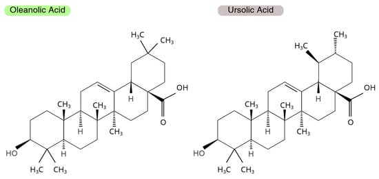
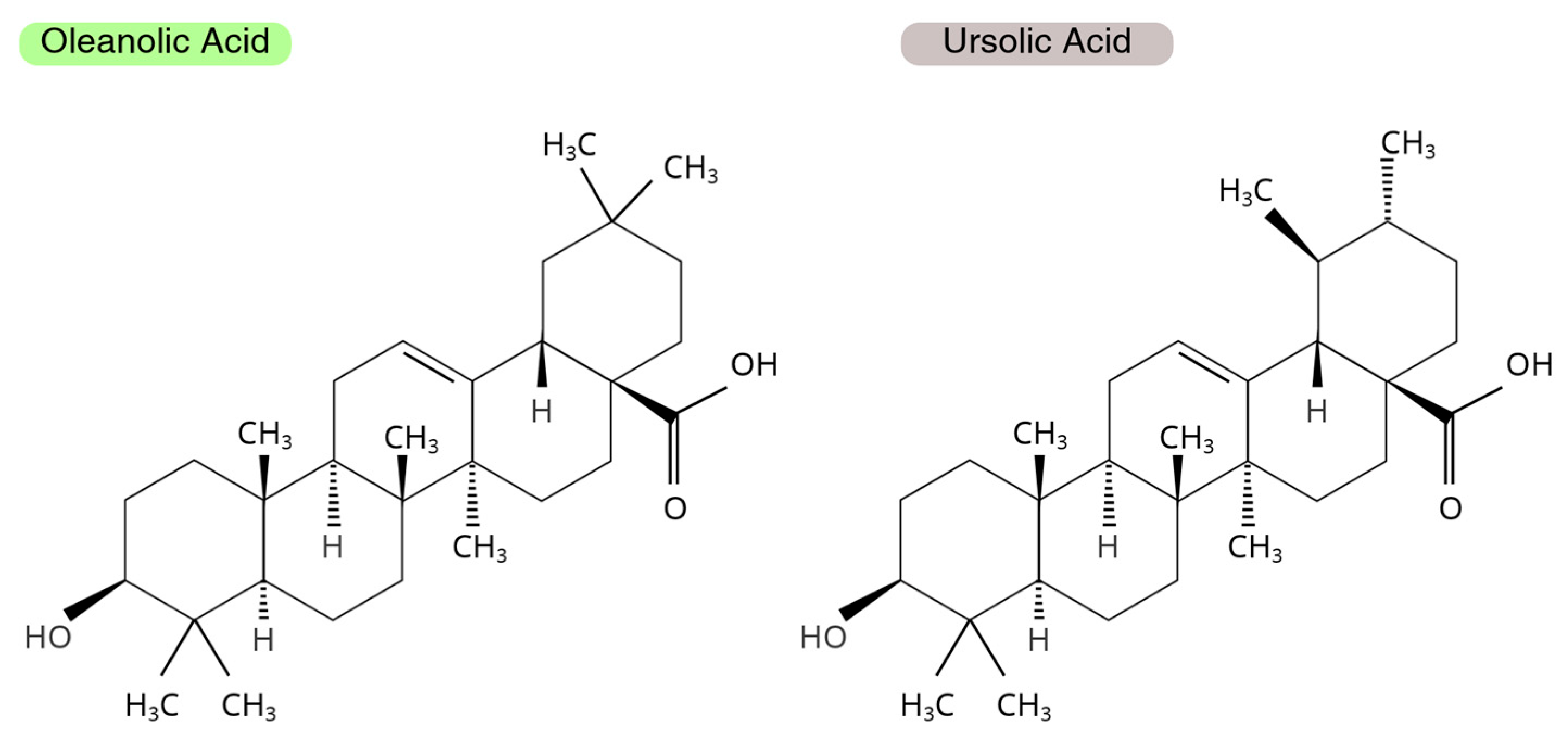
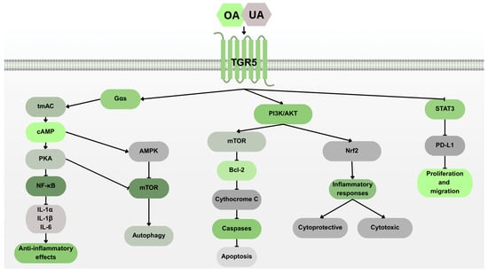
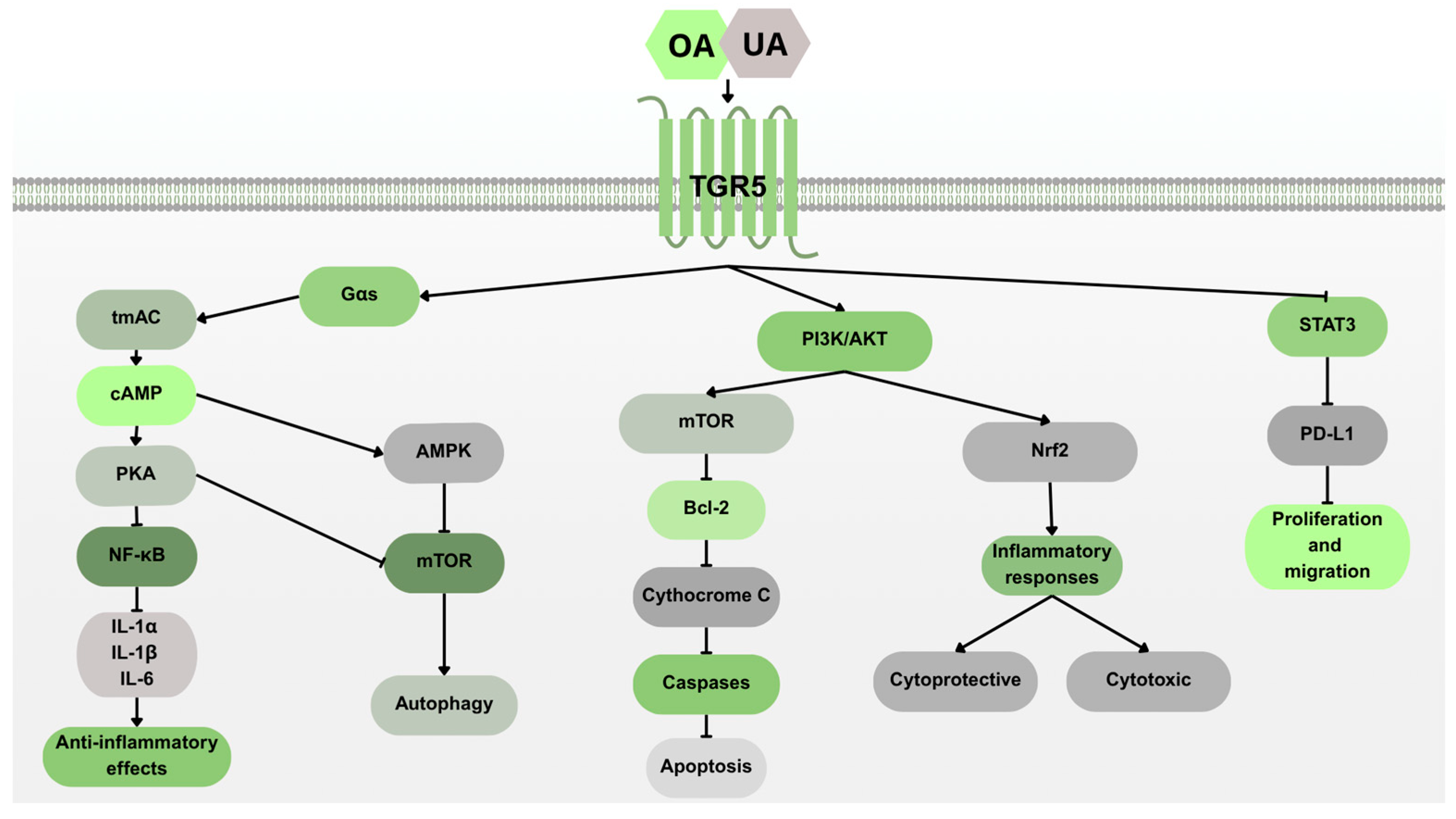
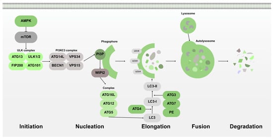
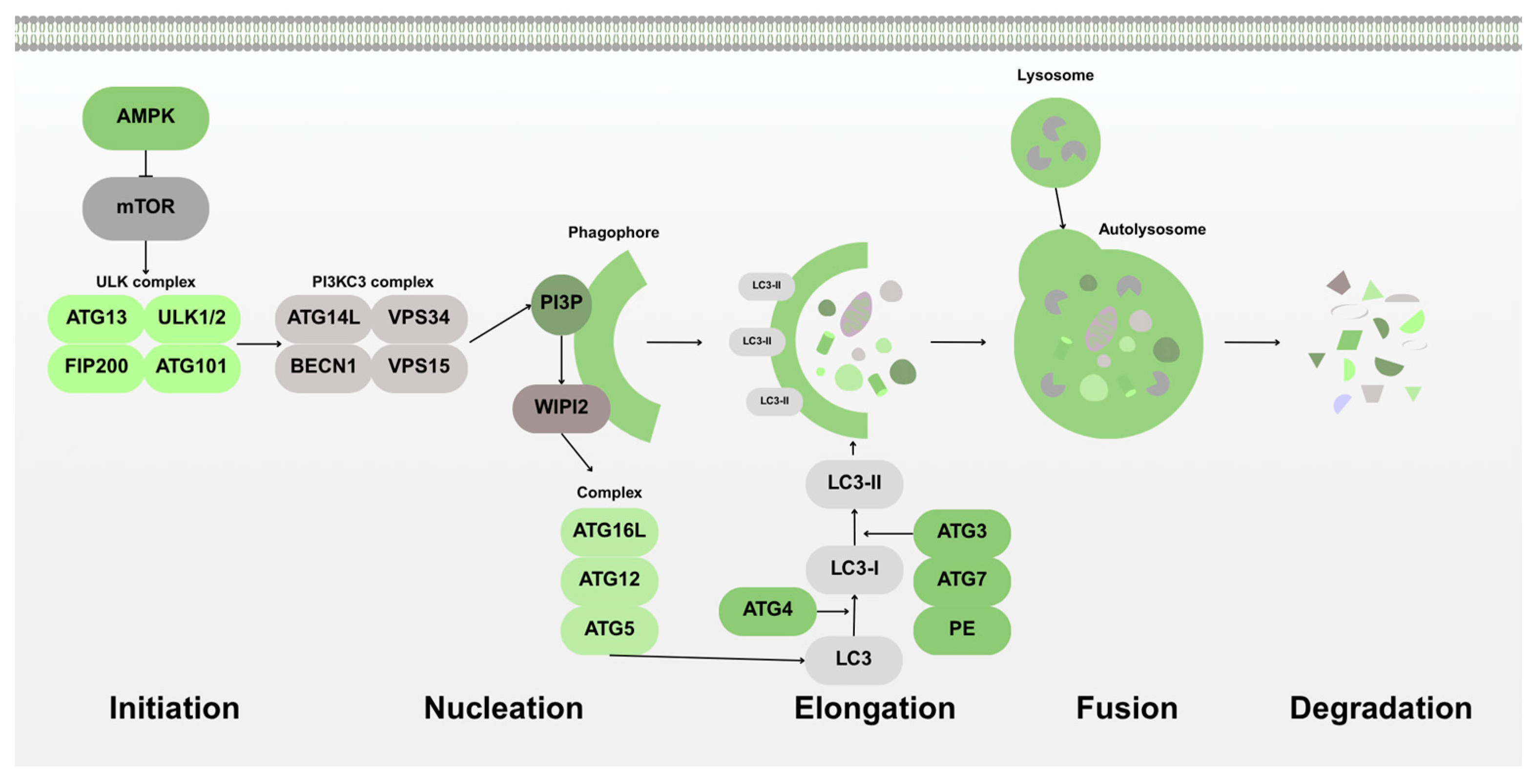
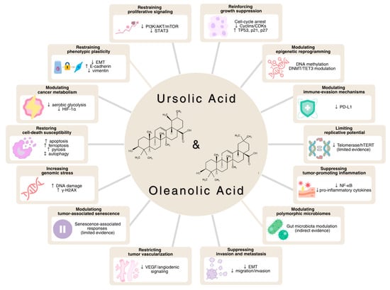
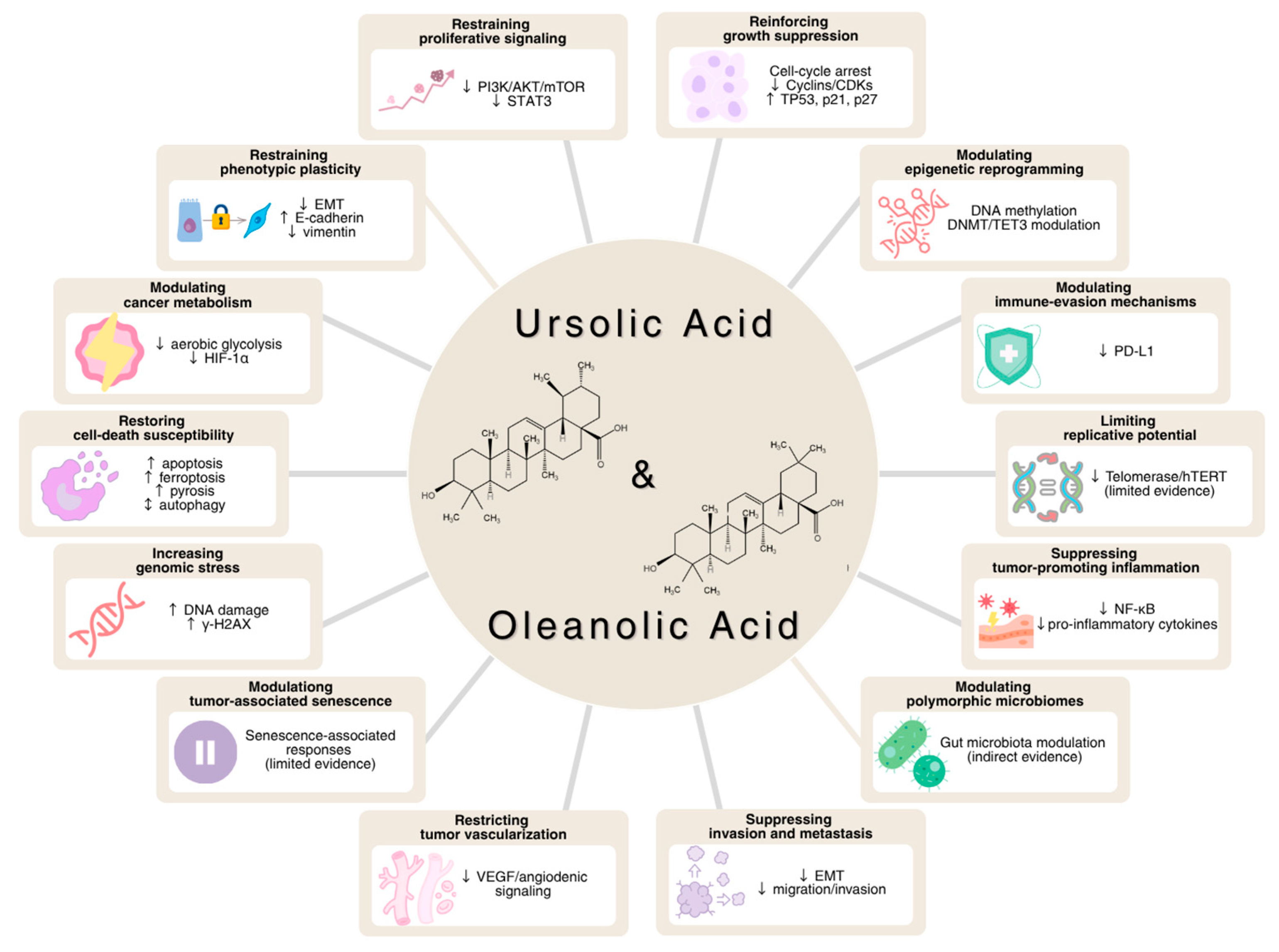

Disclaimer/Publisher’s Note: The statements, opinions and data contained in all publications are solely those of the individual author(s) and contributor(s) and not of MDPI and/or the editor(s). MDPI and/or the editor(s) disclaim responsibility for any injury to people or property resulting from any ideas, methods, instructions or products referred to in the content. |
© 2026 by the authors. Licensee MDPI, Basel, Switzerland. This article is an open access article distributed under the terms and conditions of the Creative Commons Attribution (CC BY) license.