


Abstract
Molecularly imprinted polymers combined with carbon nanomaterials have proven effective in constructing electrochemical sensors with high selectivity, sensitivity, and robustness. Herein, a polypyrrole-based molecularly imprinted electrochemical sensor was developed on a multi-walled carbon nanotube-modified glassy carbon electrode for homocysteine detection in human serum. The sensor was fabricated via drop-coating of sodium dodecyl sulfate-dispersed multi-walled carbon nanotubes, followed by in situ electropolymerization of pyrrole using homocysteine as the template. The morphology, interfacial properties, and electrochemical behavior of the electrode were systematically characterized by scanning electron microscopy and electrochemical techniques. Under optimized conditions, the sensor showed a linear response to homocysteine in the range of 1.0 × 10⁻¹⁰ mol/L to 1.0 × 10⁻⁵ mol/L, with a detection limit of 7.12 × 10⁻¹¹ mol/L (S/N = 3). The sensor also exhibited good selectivity against common interferents, as well as acceptable reproducibility and stability. Recovery tests in human serum yielded recoveries of 91.00~110.50% (average: 100.73%), demonstrating its potential for practical homocysteine analysis in complex biological matrices.
1. Introduction
Homocysteine (Hcy) is a non-essential sulfur-containing amino acid and a key intermediate in methionine and cysteine metabolism [1]. Elevated circulating Hcy levels are recognized as an independent risk factor for cardiovascular diseases [2], neurodegenerative disorders [3], pregnancy complications [4], chronic kidney disease [5], and neuropsychiatric conditions [6]. Therefore, accurate and rapid determination of Hcy is of great clinical significance for early screening and disease monitoring.
A variety of established homocysteine detection approaches have been developed, including high-performance liquid chromatography (HPLC) [7,8], enzyme-linked immunosorbent assay (ELISA) [9], chemiluminescence immunoassay [10], fluorescence sensing [11], mass spectrometry [12] and electrochemical biosensing [13,14,15,16]. Among them, electrochemical sensing has attracted extensive attention owing to its advantages of low cost, simple operation, fast response, high sensitivity, and easy miniaturization.
However, conventional electrochemical sensors often suffer from insufficient selectivity in complex biological samples. To address this limitation, molecular imprinting technology (MIT) has been integrated with electrochemical sensing to construct molecularly imprinted electrochemical sensors (MIECSs) [17,18]. Inspired by antibody–antigen recognition, MIT generates artificial recognition cavities in polymer matrices that match template molecules in size, shape, and functional groups [19]. Molecularly imprinted polymers (MIPs) feature high specificity, good mechanical stability, and excellent reusability, making them ideal for trace analyte detection in complex matrices [20,21].
To further improve sensing performance, carbon nanomaterials are widely employed as electrode modification materials owing to their large specific surface area and excellent electrical conductivity [22]. Among them, multi-walled carbon nanotubes (MWCNTs) exhibit outstanding conductivity, mechanical strength, and adsorption capacity [23,24], which can effectively increase electroactive surface area, accelerate electron transfer, and provide more anchoring sites for MIP growth.
Nevertheless, pristine MWCNTs inevitably suffer from severe agglomeration driven by intense van der Waals forces, which greatly reduces their effective electroactive area and hinders interfacial electron transfer [25]. Sodium dodecyl sulfate (SDS), a typical anionic surfactant, is therefore introduced to achieve homogeneous dispersion of MWCNTs. The hydrophobic alkyl segments of SDS anchor firmly on the nanotube surface, while outward-oriented negatively charged sulfate groups generate electrostatic repulsion to alleviate MWCNT stacking and aggregation. Moreover, the loosely interconnected MWCNT-SDS hybrids construct a porous conductive network on the electrode substrate, providing unobstructed mass transport pathways for redox species and further optimizing the interfacial electrochemical properties [26].
As a crucial functional component of molecularly imprinted electrochemical sensors, the polymer matrix also determines the recognition capability and structural stability of sensing films. Pyrrole (Py) has been regarded as an ideal electropolymerization monomer for sensor fabrication. It can be readily polymerized under mild and controllable electrochemical conditions to form conductive and biocompatible polypyrrole (PPy) films with favorable substrate adhesion [27]. During electropolymerization, target Hcy templates can be effectively encapsulated within the growing PPy network. After template elution, well-defined imprinted cavities matching the size and spatial configuration of Hcy are successfully constructed, endowing the sensor with specific molecular recognition ability [28]. Additionally, anionic SDS can serve as a dopant to integrate into the PPy backbone, which effectively modulates the interfacial physicochemical properties of the composite sensing film [29].
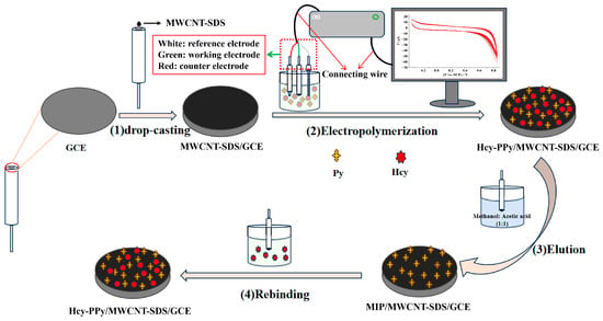
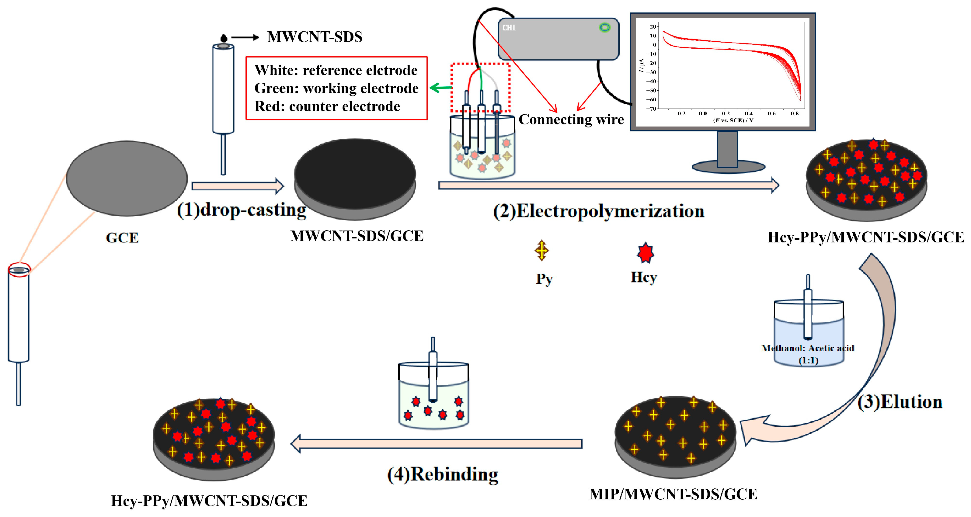
In this work, an MIECS was fabricated based on a polypyrrole and MWCNT-modified glassy carbon electrode (GCE) for the detection of Hcy. Sodium dodecyl sulfate was used to uniformly disperse carboxylated MWCNTs, and pyrrole was electropolymerized in the presence of Hcy templates. After template elution, specific recognition cavities were formed on the electrode surface. The morphology, interfacial properties, and sensing performance of the sensor were systematically investigated. Finally, the sensor was used for the determination of homocysteine in human serum samples, and the results were satisfactory. The fabrication process of the sensor and its specific recognition mechanism toward Hcy is schematically depicted in Scheme 1.
Scheme 1.
Schematic illustration of the preparation process of the Hcy MIECS and its specific recognition mechanism toward Hcy.
2. Materials and Methods
2.1. Chemical and Reagents
DL-HcySH (purity > 90%) was purchased from TCI (Shanghai) Development Co., Ltd. (Shanghai, China). Carboxylated multi-walled carbon nanotubes (Φ = 10–20 nm, l = 10–30 μm, purity > 95%) were purchased from Suzhou Tanfeng Graphene Technology Co., Ltd. (Suzhou, China). Pyrrole (Py, purity > 99.7%), sodium dodecyl sulfate (SDS, purity 92.5–100.5%), reduced glutathione (GSH, purity > 98%) and L-cysteine (L-Cys, purity > 98%) were obtained from Aladdin Industrial Corporation (Shanghai, China). High-purity nitrogen (purity ≥ 99.999%) was purchased from Wuhan Xiangyun Industry and Trade Co., Ltd. (Wuhan, China). KCl (purity ≥ 99.5%) was purchased from Tianjin Beilian Fine Chemicals Development Co., Ltd. (Tianjin, China). Acetic acid (CH3COOH, purity ≥ 99.5%) was purchased from Shanghai Titan Scientific Co., Ltd. (Shanghai, China). Nitric acid (HNO3, 65.0–68.0%), anhydrous ethanol (C2H5OH, purity ≥ 99.7%), methanol (CH3OH, purity ≥ 99.5%), K4[Fe(CN)6]·3H2O (purity ≥ 99.5%) and K3[Fe(CN)6] (purity ≥ 99.5%) were purchased from Sinopharm Chemical Reagent Co., Ltd. (Shanghai, China). Phosphate buffer solution (PBS) was purchased from Shanghai Yuanye Bio-Technology Co., Ltd. (Shanghai, China). Dopamine (DA) was obtained from Sigma (Shanghai, China). Normal human serum for scientific research was purchased from Nanjing Xinfan Biotechnology Co., Ltd. (Nanjing, China). All reagents were used without further purification. All solutions were prepared with ultrapure water.
An MWCNT-SDS dispersion with a concentration of 1.0 mg/mL was prepared through adding 10 mg MWCNT-COOH and 10 mg SDS into 10 mL ultrapure water and sonicating for 8 h.
2.2. Apparatus
Electrochemical measurements were primarily performed on a CHI660D workstation (Chenhua Instrument Co., Ltd., Shanghai, China). All electrochemical experiments, including condition optimization, mechanism investigation, and performance evaluations, employed a three-electrode system comprising a platinum wire counter electrode, a bare or modified GCE working electrode, and a saturated calomel electrode (SCE) as the reference electrode. GCE (CHI104, Φ = 3 mm), platinum wire counter electrode (CHI115, Φ = 0.5 mm, l = 30 mm), SCE (CHI150) and electrode polishing materials were purchased from Chenhua Instrument Co., Ltd. (Shanghai, China). Morphological characterization of electrode surfaces was conducted using an SU8010 field emission scanning electron microscope (Hitachi, Japan). Solution pH was adjusted with a pHS-3E pH meter (Shanghai Youke Instrument Co., Ltd., Shanghai, China). Ultrapure water for all experiments was supplied by a PGUV-10-AS purification system (Pinguan Instrument Equipment Co., Ltd., Wuhan, China). Additional apparatuses included a KQ3200DB ultrasonic cleaner (Kunshan Ultrasonic Instrument Co., Ltd., Kunshan, China), an FA2204B electronic balance (Shanghai Precision Scientific Instrument Co., Ltd., Shanghai, China), a 98-2 magnetic stirrer (Shanghai Meiyingpu Instrument Co., Ltd., Shanghai, China) and a Univer 320R refrigerated centrifuge (Zentrifugen, Andreas Hettich GmbH & Co., KG, Tuttlingen, Germany).
2.3. Preparation of Different Electrodes
To conduct comparative investigations, a series of modified GCEs were fabricated. Consistent pretreatment was performed on all GCEs prior to surface modification. Specifically, bare GCEs with a diameter of 3 mm were initially polished with 50 nm alumina (Al2O3) slurry until a mirror-finished surface was obtained. The electrode was then ultrasonically rinsed sequentially in 1:1 (v/v) dilute nitric acid, anhydrous ethanol and ultrapure water (1 min for each solvent) to eliminate surface impurities.
Preparation of MWCNT-SDS/GCE: A volume of 8.0 μL MWCNT-SDS aqueous dispersion with a concentration of 1 mg/mL was drop-cast onto the pretreated GCE surface and naturally air-dried at room temperature to fabricate the MWCNT-SDS-modified electrode (MWCNT-SDS/GCE).
Preparation of PPy/MWCNT-SDS/GCE: The polypyrrole (PPy) film was electropolymerized on the surface of MWCNT-SDS/GCE via cyclic voltammetry (CV). The electrolyte consisted of 0.01 mol/L PBS (pH 7.7) containing 3 mmol/L pyrrole (Py). Before electrochemical scanning, the electrolyte was purged with high-purity nitrogen for 10 min to remove dissolved oxygen. The CV measurement was carried out with a potential window ranging from −0.35 V to 0.85 V at a scan rate of 50 mV/s for 25 consecutive cycles.
Preparation of Hcy-PPy/MWCNT-SDS/GCE: The fabrication procedure of the Hcy-PPy composite film-modified electrode was similar to that of the PPy/MWCNT-SDS/GCE, except that 1.5 mmol/L Hcy was additionally incorporated into the electrolyte during electropolymerization.
Preparation of MIP/MWCNT-SDS/GCE: The obtained Hcy-PPy/MWCNT-SDS/GCE was immersed in a 1:1 (v/v) methanol–acetic acid mixed solution under continuous magnetic stirring for 8 h to thoroughly elute the embedded Hcy template molecules. After template removal, the molecularly imprinted polymer-based electrode (MIP/MWCNT-SDS/GCE) was successfully fabricated.
Preparation of NIP/MWCNT-SDS/GCE: The non-imprinted polymer electrode (NIP/MWCNT-SDS/GCE) was prepared through the identical fabrication procedure as the MIP/MWCNT-SDS/GCE, except that no Hcy template molecules were added into the polymerization solution.
Notably, the resulting NIP film was also immersed in a 1:1 (v/v) methanol–acetic acid mixed solution with continuous magnetic stirring for 8 h to undergo the same elution treatment, ensuring consistent experimental conditions. By contrast, the pure PPy/MWCNT-SDS/GCE required no elution procedure throughout the whole preparation process.
2.4. Electrochemical Detection of Hcy
For Hcy determination, the molecularly imprinted electrochemical sensor (MIP/MWCNT-SDS/GCE) adopts K3Fe(CN)6/K4Fe(CN)6 as the redox probe. Differential pulse voltammetry (DPV) superimposes potential pulses onto a linear potential ramp and records differential currents to suppress non-faradaic capacitive current. Owing to its high sensitivity, favorable redox peak resolution and low limit of detection, DPV is well suited for trace analyte quantification using electrochemical sensors [30]. Accordingly, DPV was selected as the detection technique in this work. All measurements were conducted within a potential window ranging from −0.2 V to 0.6 V, with a potential increment of 0.004 V, pulse amplitude of 0.05 V and pulse duration of 0.05 s.
Parameter optimization results are presented in the Supplementary Material. The optimal conditions for sensor fabrication and detection were determined as follows: 8 µL for drop-coating amount of MWCNT-SDS (Figure S1A); 2:1 for a molar ratio of functional monomers to template molecules (Figure S1B); pH = 7.7 for the pre-polymerization base electrolyte (Figure S1C); 25 for the number of polymerization cycles (Figure S1D); 8 h for the elution time (Figure S1E); and 420 s for the incubation time (Figure S1F).
3. Results and Discussion
3.1. Electrochemical Behavior of Different Electrodes
To investigate the recognition and response capabilities of different electrodes toward Hcy, DPV measurements were performed in an aqueous solution containing 1 mmol/L K3Fe(CN)6 and 0.2 mol/L KCl, using various differently modified GCEs as working electrodes. K3Fe(CN)6 yields a typical pair of redox peaks in cyclic voltammetry (CV). Considering that the anodic peak current is more sensitive than its cathodic counterpart, the anodic peak signal was adopted for data collection.
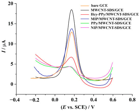
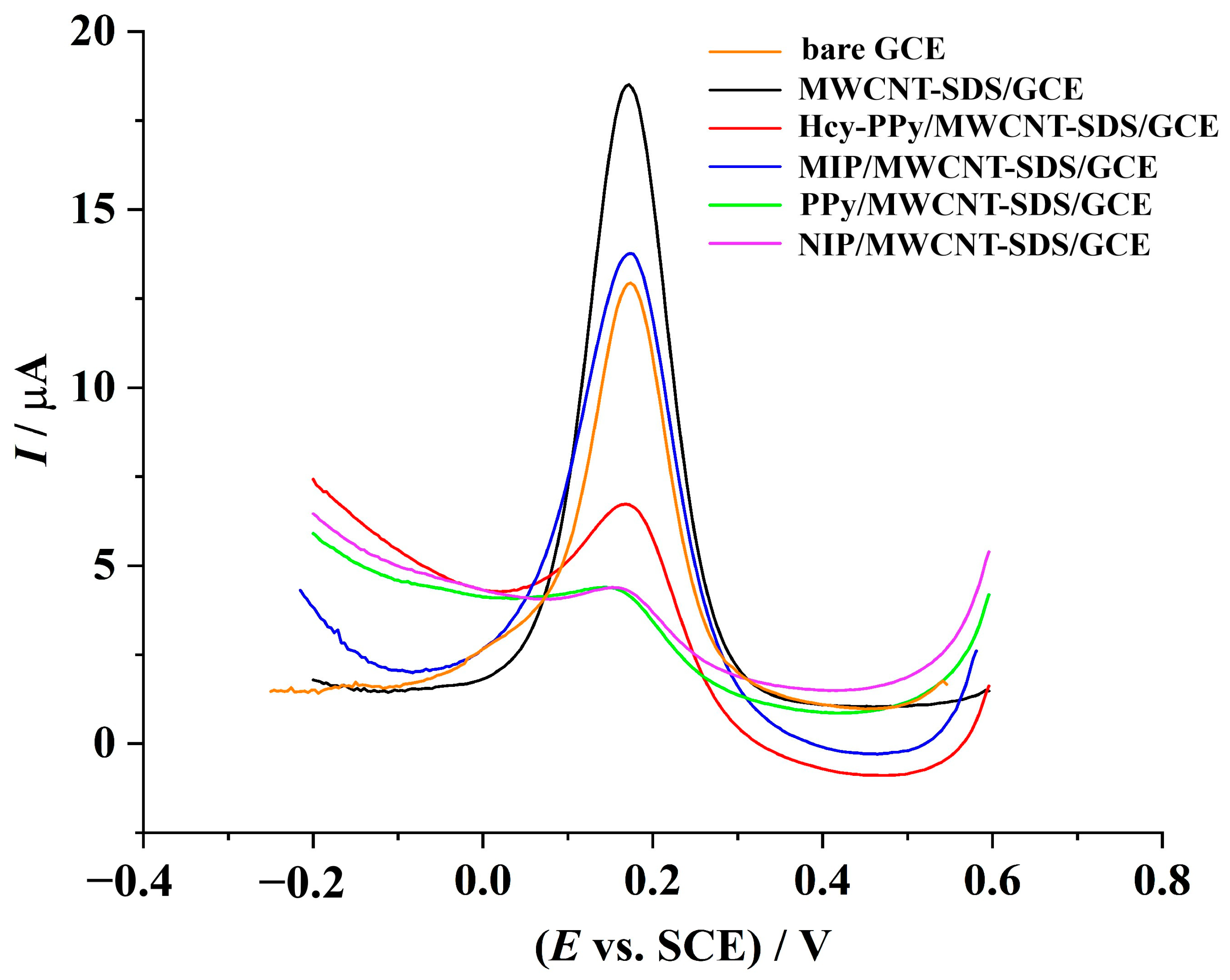
As shown in Figure 1, the electrochemical response of the probe on the MWCNT-SDS/GCE is obviously higher than that on other electrodes (the black line), which can be attributed to the large specific surface area and favorable electrical conductivity of MWCNTs. SDS endows the electrode with negative surface charges, which would otherwise repel [Fe(CN)6]3−. Nevertheless, abundant species in the electrolyte induce a prominent ionic shielding effect, substantially mitigating electrostatic repulsion toward anions. More importantly, SDS serves as an effective dispersant to inhibit the aggregation of MWCNTs, constructing a porous conductive network with a large electroactive area and facilitating the transport of redox probes.
Figure 1.
Differential pulse voltammograms of different electrodes in 1 mmol/L K3Fe(CN)6 and 0.2 mol/L KCl aqueous solution from −0.2 V to 0.6 V with a potential increment of 0.004 V, pulse amplitude of 0.05 V and pulse duration of 0.05 s.
After the electropolymerization of pyrrole, a continuous compact PPy film forms on the electrode surface introducing physical barriers for ion diffusion. Therefore, the response current on the PPy/MWCNT-SDS/GCE decreases significantly (the green line).
Hcy incorporated during electropolymerization tailors the micromorphology of polypyrrole films to form relatively loose porous structures. In addition, amphiprotic Hcy molecules adjust the film’s interfacial charge and weaken the SDS-triggered electrostatic repulsion against anionic ferricyanide probes. These predominant favorable contributions enable the Hcy-PPy/MWCNT-SDS/GCE to display enhanced electrochemical responses (the red line) relative to the PPy/MWCNT-SDS/GCE.
After template elution, the obtained MIP/MWCNT-SDS/GCE delivers an increased peak current (the blue line), since the formed imprinted cavities offer accessible diffusion pathways for redox probes. By contrast, the NIP/MWCNT-SDS/GCE exhibits a considerably lower peak current (the purple line) than the MIP electrode, verifying the successful construction of specific recognition cavities in the MIP film. Furthermore, there is nearly no change in the DPV responses of the PPy/MWCNT-SDS/GCE (the green line) and the NIP/MWCNT-SDS/GCE (the purple line), revealing that the PPy film is stable and unaffected throughout the elution procedure.
3.2. Morphological Characterization
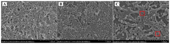
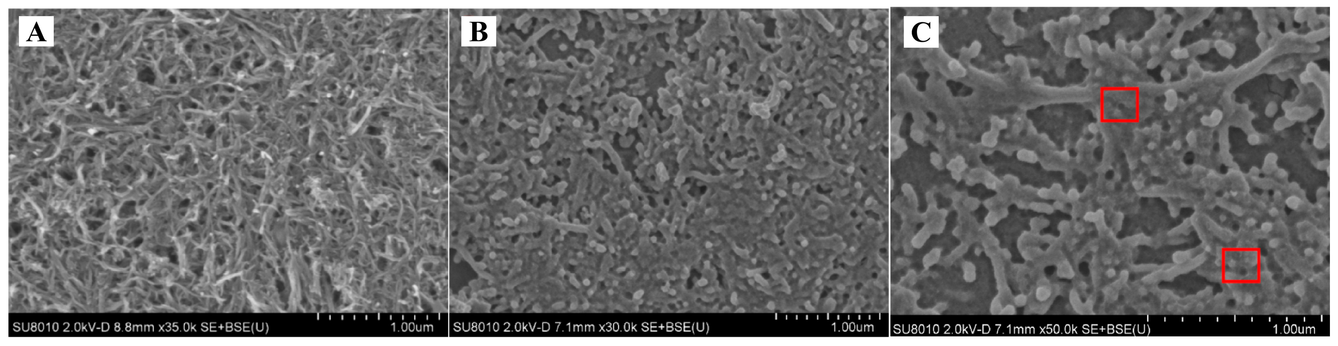
Scanning electron microscopy (SEM) is a prevalent surface characterization technique capable of providing high-resolution micrographs to visualize the surface microstructure and morphological features of materials. The surface morphologies of different modified electrodes were investigated by SEM. As displayed in Figure 2A, the surface of the MWCNT-SDS/GCE shows an interwoven, tangled network of MWCNTs. This well-dispersed nanotube network provides plentiful conductive paths and a large specific surface area, which creates favorable conditions for the subsequent electropolymerization and growth of the molecularly imprinted polymer (MIP). After the electropolymerization of pyrrole (Figure 2B), a continuous and uniform polypyrrole (PPy) film covers the carbon nanotube network. The original contours of the carbon nanotubes are partially encapsulated by the polymer, demonstrating the successful formation of the MIP composite film. After template molecule elution (Figure 2C), abundant irregular porous cavities can be clearly observed on the polymer surface (highlighted by red boxes). These cavities are geometrically complementary to template Hcy molecules, indicating the formation of specific imprinted recognition sites. The porous structure accelerates mass transfer of target Hcy molecules and supports specific binding between analytes and imprinted cavities, which plays a vital role in the sensing performance of the MIP sensor.
Figure 2.
SEM images of MWCNT-SDS/GCE (A), Hcy-PPy/MWCNT-SDS/GCE (B) and MIP/MWCNT-SDS/GCE (C).
3.3. Evaluation of Sensor Performance
3.3.1. Linear Range and Detection Limit
DPV was employed to evaluate the analytical performance of the as-fabricated MIECS for Hcy detection. For linear range measurement, the MIP/MWCNT-SDS/GCE was incubated in Hcy solutions with a series of concentrations for 420 s. Subsequently, the electrode was transferred into an aqueous solution containing 1 mmol/L K3[Fe(CN)6] and 0.2 mol/L KCl, and DPV signals were recorded. Measurements were performed sequentially from low to high Hcy concentrations. After each Hcy detection, the MIP/MWCNT-SDS/GCE required regeneration to release Hcy trapped within the imprinted cavities prior to the next measurement. Briefly, the electrode was thoroughly rinsed with ultrapure water for about 10–20 s to remove loosely adsorbed Hcy molecules. Afterwards, the electrode was immersed in 0.01 mol/L PBS (pH 7.7) for 2–5 min and subsequently soaked in ultrapure water for another 15–30 min to realize the desorption of specifically bound Hcy. The regeneration efficiency was verified by DPV measurements in 0.01 mol/L PBS (pH = 7.7). The electrode was ready for subsequent detection only after the peak current recovered to the baseline level.
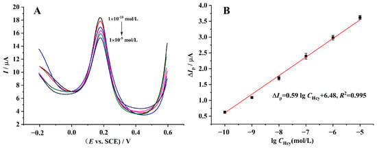
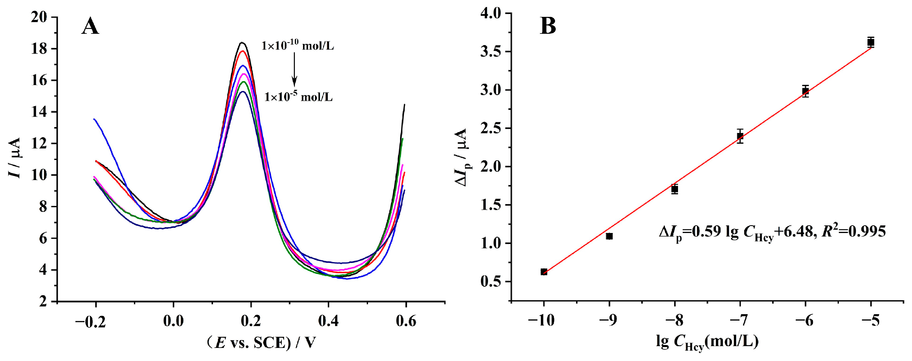
As depicted in Figure 3A, the peak current difference (ΔIp) at approximately 0.2 V decreased linearly with the increasing concentration of Hcy. The peak current difference ΔIp was calculated as ΔIp = Ip0 − Ip, where Ip0 and Ip represent the DPV peak current of the MIP electrode before and after incubation with Hcy solution, respectively. ΔIp was adopted rather than the raw peak current to eliminate the inherent deviation originating from electrode fabrication and quantitatively reflect the amount of specifically bound Hcy on the molecularly imprinted membrane. For quantitative analysis, a calibration curve of ΔIp versus the common logarithm of Hcy concentration (lgCHcy) in the range from 1 × 10⁻¹⁰ to 1 × 10⁻⁵ mol/L was established (Figure 3B). All DPV measurements were performed in triplicate, and the fitted linear regression equation is given as follows:
ΔIp = 0.59 lgCHcy + 6.48, R2 = 0.995
Figure 3.
(A) Differential pulse voltammograms recorded at the MIECS in an aqueous solution containing 1 mmol/L K3[Fe(CN)6] and 0.2 mol/L KCl. The electrodes were pre-incubated with Hcy solutions of varying concentrations ranging from 1 × 10⁻¹⁰ to 1 × 10⁻⁵ mol/L for 420 s prior to measurement. (B) Corresponding linear calibration plot for quantitative determination of Hcy using the as-fabricated MIECS.
Benefiting from this favorable linear response, the as-prepared MIECS delivers outstanding sensing performance with a limit of detection (LOD) calculated to be 7.12 × 10⁻¹¹ mol/L.
3.3.2. Reproducibility and Stability
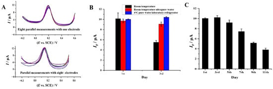
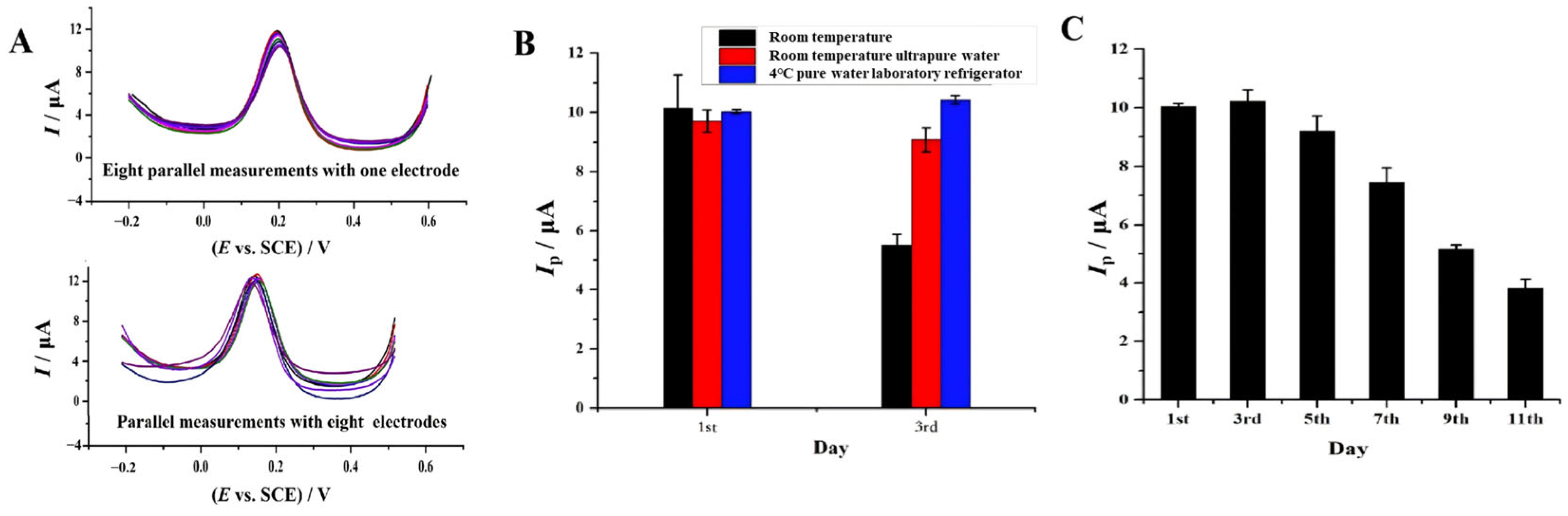
The reproducibility of the as-prepared MIECS was also assessed via DPV measurements. As presented in Figure 4A, eight parallel tests carried out on a single MIP/MWCNT-SDS/GCE delivered a relative standard deviation (RSD) of 7.9%. It should be clarified that the 8 h elution procedure was only adopted for the initial removal of template molecules after electropolymerization. Between parallel measurements, the electrode is rinsed with ultrapure water for 10–20 s, soaked in blank PBS buffer for 2–5 min and immersed in ultrapure water for 15–30 min. Short-time exposure to aqueous solutions cannot achieve this effect. The detection electrolyte contains KCl and K3[Fe(CN)6] serving as electrochemical redox probes, which merely diffuse in the bulk solution and the superficial layer of the modified membrane and cannot infiltrate the compact recognition cavities. A single DPV measurement lasts merely 12–18 s. Such an extremely short residence time of the electrode in the aqueous electrolyte is inadequate to break the stable specific intermolecular interactions between Hcy and the MIP matrix. Consequently, the specifically bound Hcy remains stably anchored inside the cavities and will not desorb into the test solution during electrochemical detection. Furthermore, eight independently fabricated MIP/MWCNT-SDS/GCEs were measured under identical experimental conditions, affording an inter-electrode RSD value of 6.5%. Collectively, these observations verify the favorable reproducibility of the constructed electrochemical sensor.
Figure 4.
(A) Evaluation of the reproducibility of the MIECS; (B) influence of the storage conditions on the electrochemical response of the sensor; (C) influence of the storage time on the electrochemical response of the sensor with the same storage conditions.
The preservation environment has a crucial influence on the long-term service performance of MIECSs. The storage stability of the as-fabricated sensor was systematically assessed under three different preservation modes, and the time-dependent signal changes are summarized in Figure 4B. On the first day of storage, the sensors under three conditions exhibited close initial peak currents, with negligible differences in the original response signal. After being stored for 3 days, obvious signal attenuation was observed for the electrode kept under ambient air at room temperature, whose peak current dropped markedly. By contrast, the electrode immersed in ultrapure water at room temperature displayed moderate signal loss, while the sensor preserved in ultrapure water inside a 4 °C laboratory refrigerator maintained the highest residual peak current and showed the weakest performance decay within the test period. This finding confirms that low-temperature aqueous environments can effectively stabilize the imprinted polymeric membrane structure, suppress the collapse of recognition cavities and electrode surface oxidation, and thus excellently retain the specific molecular recognition capability toward Hcy. Storing the modified electrodes in 4 °C ultrapure water is identified as the optimal preservation strategy for this MIECS.
Furthermore, the time-dependent stability was further evaluated under the optimal storage condition (Figure 4C). After 7 days of storage, the sensor retained 75% of its initial peak current response; the response decreased to less than 50% of the original value after 11 days. These findings demonstrate that the MIECS possesses acceptable short-term stability, but its stability remains insufficient for long-term storage. For practical applications, this level of short-term stability is adequate for routine laboratory batch assays and small-scale clinical sample testing, as electrodes can be fabricated weekly on demand. Nevertheless, the inadequate long-term storage performance limits its applicability to commercial, prefabricated disposable sensors, which will be addressed in future work by optimizing the polymer crosslinking degree and interfacial adhesion between the MIP layer and the nanomaterial substrate. Overall, storage conditions have a pronounced effect on the sensing stability of the electrode, and preservation in ultrapure water at 4 °C is recommended for short-term use.
3.3.3. Selectivity
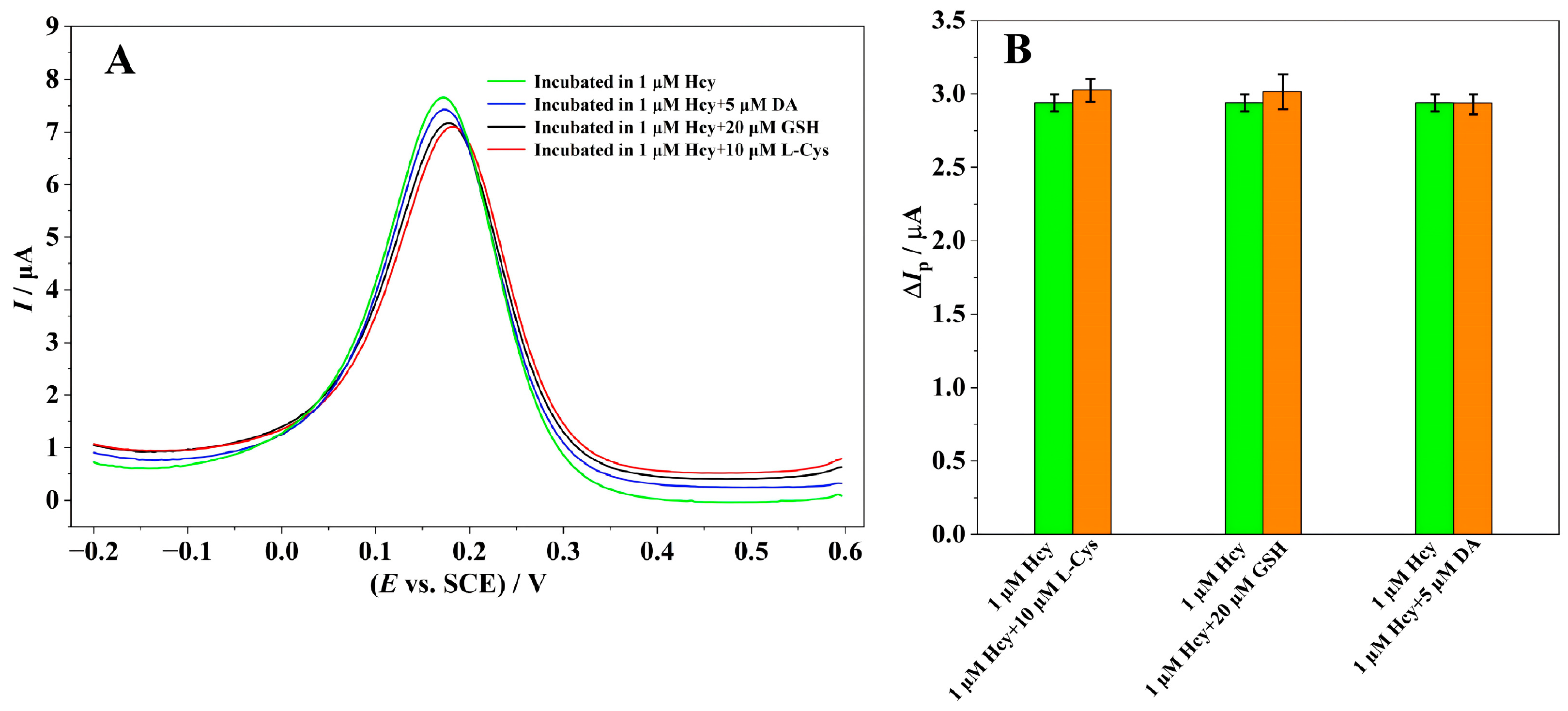
Selectivity is a vital performance metric for MIECSs, particularly for quantitative detection of target analytes in complex biological matrices such as serum. L-cysteine (L-Cys), reduced glutathione (GSH), and dopamine (DA), typical endogenous serum components with structural resemblance to Hcy, were selected as potential interferents to evaluate the specific recognition and anti-interference performance of the fabricated MIECS. And their working concentrations were determined based on their natural physiological proportional levels coexisting with Hcy in practical serum samples. For selectivity evaluation, the MIP/MWCNT-SDS/GCE was incubated in 1 μM Hcy solutions with or without interferents for 420 s. The electrode was then transferred into an aqueous electrolyte containing 1 mmol/L K3[Fe(CN)6] and 0.2 mol/L KCl for subsequent DPV measurement.
Figure 5A shows that, no matter whether incubation was carried out in the single Hcy solution or the mixture of Hcy and interferents, the DPV detection performed in the probe solution yielded a response peak consistently centered at around 0.2 V. As shown in Figure 5B, the bar chart evaluates the anti-interference performance of the fabricated MIP electrochemical sensor by comparing the DPV peak current change (ΔIp, μA) of 1 μM pure Hcy (green bars) with mixtures of 1 μM Hcy plus 10 μM L-Cys, 20 μM GSH and 5 μM DA (orange bars). Error bars denote standard deviations of three parallel tests, and the highly consistent current responses reveal trivial interference from the coexisting substances. Taking the peak current change (ΔIp) induced by 1 μmol/L Hcy as the reference baseline, the signal variations triggered by each interferent were all below 5%, verifying the favorable specific recognition capability of the MIECS.
Figure 5.
(A) Differential pulse voltammograms of the MIP/MWCNT-SDS/GCE incubated with 1 μM Hcy alone and co-incubated with 1 μM Hcy together with 5 μM DA, 20 μM GSH and 10 μM L-Cys, respectively; (B) evaluation of the selectivity of the sensor.
The satisfactory selectivity originates from the imprinted cavities with well-matched size, shape, and functional group distribution, which bind Hcy through weak intermolecular interactions and steric recognition. Such high selectivity against coexisting interferents endows the MIECS with great application potential for Hcy detection in real serum samples.
3.3.4. Determination of Hcy in Serum Samples
Spike recovery experiments were performed to evaluate the practical applicability of the proposed sensor in real biological matrices. Two batches of liquid normal human serum for scientific research (non-lyophilized) with different endogenous Hcy levels were adopted for the spike assay. The sample pretreatment procedure was as follows: 10 mL of serum was first centrifuged at 10,000 rpm for 20 min to remove impurities; then the supernatant was collected and filtered through a 0.45 μm microporous filter membrane before use. The pretreated serum samples were subsequently analyzed using the as-fabricated MIECS. Each spiked concentration level in Sample 1 and Sample 2 was measured with three parallel replicate tests (n = 3).
The calculation formula for spike recovery is Recovery (%) = [(Cfound − Coriginal)/Cspiked] × 100%, where Coriginal is the endogenous Hcy concentration in blank serum, Cspiked is the concentration of standard Hcy solution added into serum samples, Cfound is the total detected Hcy concentration after spiking standard solution. As shown in Table 1, Coriginal is 0.075 μM for Sample 1 and 0.014 μM for Sample 2. Cspiked corresponds to the data in the second column “Added (μM)”, and Cfound refers to the measured total Hcy concentration shown in the third column “Found (μM)”. Therefore, according to the above formula, the spiked recoveries were calculated and shown in the fourth column “Recovery (%)”. Overall, the Hcy spiked recoveries across the two serum samples fell within the range of 91.00–110.50%, with an average recovery of 100.73%. These results demonstrate the favorable detection accuracy of the proposed sensor for Hcy quantification in real serum matrices and validate its great prospect for practical biomedical testing.
Table 1.
Results of the recovery experiments.
4. Conclusions
In this work, a molecularly imprinted electrochemical sensor for the detection of Hcy was constructed based on a modified GCE via a facile drop-coating and in situ electropolymerization strategy. The introduction of SDS-dispersed MWCNTs effectively enlarged the electroactive surface area and improved the electrical conductivity of the electrode, providing a favorable substrate for the growth of the polypyrrole-based molecularly imprinted film. The surface morphology and interfacial electrochemical properties of the modified electrodes were systematically characterized by scanning electron microscopy and electrochemical techniques, and the sensing mechanism of Hcy at the nanocomposite interface was elucidated.
As summarized in Table 2, a series of electrochemical biosensors have been reported for Hcy detection. The MIP/Mxene@AuNPs/GCE exhibits outstanding analytical performance, featuring a lower detection limit and a linear range. Most other reported Hcy sensors in Table 2 are either limited by relatively high detection limits or narrow measurable concentration windows. Although our MIP/MWCNT-SDS/GCE cannot surpass the MIP/Mxene@AuNPs/GCE, it still obtains a favorable LOD of 7.12 × 10⁻⁵ μM, which is distinctly superior to the majority of the listed Hcy electrochemical sensors and biosensors. Compared with Mxene-based composites, the MWCNT-SDS modifier possesses prominent merits of low cost, facile synthesis and favorable aqueous dispersibility. Moreover, the developed sensor achieves reliable responses within the commonly concerned trace-level Hcy concentration range. Therefore, this platform provides a cost-effective and competitive alternative for practical Hcy quantitative analysis.
Table 2.
Comparison of analytical performance of different Hcy electrochemical and biosensors.
Supplementary Materials
The following supporting information can be downloaded at https://www.mdpi.com/article/10.3390/chemosensors14080187/s1: Figure S1: Influence of the drop-coating amount of MWCNT-SDS (A), the concentration ratio of functional monomer and template molecule (B), the pH value of the pre-polymerization base electrolyte (C), the number of MIP polymerization cycles (D), the elution time of template Hcy molecules (E) and the rebinding time for template Hcy molecules (F).
Author Contributions
Conceptualization, methodology, writing—review and editing, X.S.; investigation and writing—original draft preparation, X.S. and J.Y.; resources and visualization, S.Z. and D.Z.; software, X.S.; validation and formal analysis, J.Y. and S.Z.; supervision and project administration, D.Z. All authors have read and agreed to the published version of the manuscript.
Funding
This research was funded by the National Natural Science Foundation of China (No. 52472291), the Fundamental Research Funds for the Central Universities, South-Central Minzu University (No. CZZ24004), and the Enterprise-Commissioned Research Project from Wuhan Jinzhixiu Optoelectronic Technology Co., Ltd. (No. HZY25161).
Institutional Review Board Statement
Not applicable.
Informed Consent Statement
Not applicable.
Data Availability Statement
All data that support the findings of this study are included within the article and any Supplementary Files.
Conflicts of Interest
The funding sponsors had no role in the design of the study; in the collection, analyses, or interpretation of data; in the writing of the manuscript; or in the decision to publish the results.
References
- Brennerová, K.; Grešíková, M.; Behúlová, D.S.; Radová, S.; Bzdúch, V. Importance of homocysteine determination in differential diagnosis of rare causes of macrocytic anaemia. Biologia 2022, 77, 1103–1108. [Google Scholar] [CrossRef] [Scilit]
- Guieu, R.; Ruf, J.; Mottola, G. Hyperhomocysteinemia and cardiovascular diseases. Ann. Biol. Clin. 2022, 80, 7–14. [Google Scholar] [CrossRef] [Scilit] [PubMed]
- Yoganathan, S.; Bhasin, H.; Garg, D.; Malik, P.; Saini, A.G.; Chandran, M.; Korula, S.; Arunachal, G.; Danda, S.; Thomas, M.; et al. Childhood neurological disorders with hyperhomocystinemia: A case-based review. Pediatr. Neurol. 2023, 146, 26–30. [Google Scholar] [CrossRef] [Scilit] [PubMed]
- Dogan, G.O.; Sahin, O. Effects of maternal micronutrient levels and homocysteine on pregnancy complications and outcomes: A prospective cohort study. Clin. Exp. Obstet. Gynecol. 2025, 52, 27137. [Google Scholar] [CrossRef] [Scilit]
- Lin, H.; Liu, Y.; Geng, S.K.; Sun, Y.; Li, X.; Li, B.; Liu, Y. A Meta-Analysis of the Effects of Homocysteine-Lowering Therapy on Chronic Kidney Disease. Horm. Metab. Res. 2026, 58, 13–22. [Google Scholar] [PubMed]
- Pana, A. Homocysteine and Neuropsychiatric Disease. Psychiatr. Ann. 2015, 45, 463–468. [Google Scholar] [CrossRef] [Scilit]
- Omar, A.; Kadir, S.R.A.; Rahman, S.A.; Nordin, F.D.A.; Kamarudin, B.; Muhamad, N.J.; Mohamed, R.; Rozali, N.A.; Khalid, N.; Nazri, M.I.B.A.; et al. Validation of ion exchange high performance liquid chromatography (HPLC) method and its comparison with reverse phase HPLC for total homocysteine determination. SAINS Malays. 2021, 50, 1913–1920. [Google Scholar]
- Sarvutiene, J.; Prentice, U.; Ramanavicius, S.; Ramanavicius, A. Molecular imprinting technology for biomedical applications. Biotechnol. Adv. 2024, 71, 108318. [Google Scholar] [CrossRef] [Scilit] [PubMed]
- Li, H.; Dang, Y.; Yan, Y. Serum interleukin-17 A and homocysteine levels in children with autism. BMC Neurosci. 2024, 25, 17. [Google Scholar] [CrossRef] [Scilit] [PubMed]
- Liu, Y.K.; Li, Y.R.; Wang, R.; Chen, S.; Sun, N.; Qi, X. Development of a chemiluminescent immunoassay based on magnetic solid phase for quantification of homocysteine in human serum. BMC Biotechnol. 2024, 24, 77. [Google Scholar] [CrossRef] [Scilit] [PubMed]
- Huang, J.; Wei, F.; Cui, Y.; Hou, L.; Lin, T. Fluorescence immunosensor based on functional nanomaterials and its application in tumor biomarker detection. RSC Adv. 2022, 12, 31369–31379. [Google Scholar] [CrossRef] [Scilit] [PubMed]
- Piechocka, J.; Wrońska, M.; Chwatko, G.; Jakubowski, H.; Głowacki, R. Quantification of homocysteine thiolactone in human saliva and urine by gas chromatography-mass spectrometry. J. Chromatogr. B 2020, 1149, 122155. [Google Scholar] [CrossRef] [Scilit] [PubMed]
- Liu, J. Construction of an electrochemical molecularly imprinted sensor based on L-carnitine doped polypyrrole for vibrio parahaemolyticus detection. J. Clin. Med. Res. 2024, 5, 234. [Google Scholar] [CrossRef] [Scilit]
- Liu, B.; Lu, W.; Huang, Y.; Zhang, X.; Yuan, X. A label-free electrochemical sensor for the detection of two kinds of targets based on CRISPR/Cas12a system. Sens. Actuators B Chem. 2024, 406, 135406. [Google Scholar] [CrossRef] [Scilit]
- Wang, J.; Zhao, L.; Du, H.; He, W.; Xu, S.; Shao, Q.; Wang, L. Mechanism of SDBS functionalized PPy enhancing the electrochemical sensitivity of SPGE to dopamine and uric acid: Experimental and DFT investigations. J. Electroanal. Chem. 2024, 966, 118415. [Google Scholar] [CrossRef] [Scilit]
- Benchirouf, A.; Kanoun, O. Inkjet-printed multiwalled carbon vanotube dispersion as wireless passive strain sensor. Sensors 2024, 24, 1585. [Google Scholar] [CrossRef] [Scilit] [PubMed]
- Hasaneen, N.; Akhtarian, S.; Pulicharla, R.; Brar, S.K.; Rezai, P. Surface molecularly imprinted polymer-based sensors for antibiotic detection. TrAC Trends Anal. Chem. 2024, 170, 117389. [Google Scholar] [CrossRef] [Scilit]
- Han, S.; Ding, Y.; Teng, F.; Yao, A.; Leng, Q. Molecularly imprinted electrochemical sensor based on 3D-flower-like MoS2 decorated with silver nanoparticles for highly selective detection of butylated hydroxyanisole. Food Chem. 2022, 387, 132899. [Google Scholar] [CrossRef] [Scilit] [PubMed]
- Niu, J.; Du, M.; Wu, W.; Yang, J.; Chen, Q. Advances in the selection of functional monomers for molecularly imprinted polymers: A review. J. Sep. Sci. 2024, 47, 202400353. [Google Scholar] [CrossRef] [Scilit] [PubMed]
- Zhao, H.; Liu, S.; Kong, X.; Zou, R.; Zhou, D. Electropolymerized molecularly imprinted electrochemical sensor for sensitive and selective detection of an antipsychotic compound chlorpromazine. Int. J. Electrochem. Sci. 2025, 20, 101076. [Google Scholar] [CrossRef] [Scilit]
- Li, Y.; Luo, L.; Kong, Y.; Li, Y.; Wang, Q.; Wang, M.; Li, Y.; Davenport, A.; Li, B. Recent advances in molecularly imprinted polymer-based electrochemical sensors. Biosens. Bioelectron. 2024, 249, 116018. [Google Scholar] [CrossRef] [Scilit] [PubMed]
- Raimondo, M.; Donati, G.; Milano, G.; Guadagno, L. Hybrid composites based on carbon nanotubes and graphene nanosheets outperforming their single-nanofiller counterparts. FlatChem 2022, 36, 100431. [Google Scholar] [CrossRef] [Scilit]
- Geng, L.; Huang, J.; Zhai, H.; Shen, Z.; Han, J.; Yu, Y.; Fang, H.; Li, F.; Sun, X.; Guo, Y. Molecularly imprinted electrochemical sensor based on multi-walled carbon nanotubes for specific recognition and determination of chloramphenicol in milk. Microchem. J. 2022, 182, 107887. [Google Scholar] [CrossRef] [Scilit]
- Xu, Z.; Jiang, X.; Liu, S.; Yang, M. Sensitive and selective molecularly imprinted electrochemical sensor based on multi-walled carbon nanotubes for doxycycline hyclate determination. Chin. Chem. Lett. 2020, 31, 185–188. [Google Scholar] [CrossRef] [Scilit]
- Li, H.; Wang, G.; Wu, Y.; Jiang, N.; Niu, K. Functionalization of carbon nanotubes in polystyrene and properties of their composites: A Review. Polymers 2024, 16, 770. [Google Scholar] [CrossRef] [Scilit] [PubMed]
- Moulya, K.P.; Manjunatha, J.G. Affordable surfactant modified carbon nanotube sensor for the enhanced electrochemical detection of eugenol. Ionics 2024, 30, 8577–8590. [Google Scholar] [CrossRef] [Scilit]
- Zvirzdine, G.; Zukauskas, S.; Rucinskiene, A.; Mohsenzadeh, E.; Boguzaite, R.; Ramanaviciene, A.; Pogorielov, M.; Ratautaite, V.; Ramanavicius, A. Electrochemical salicylic acid sensor based on molecularly imprinted polypyrrole. ACS Appl. Mater. Interfaces 2025, 17, 57475–57485. [Google Scholar] [CrossRef] [Scilit] [PubMed]
- Deepa, J.R.; Jyothish, B.; Athira, V.S.; Mini, S.; Remya, T.S.; Nair, A.A. Molecularly imprinted electrochemical sensor enhanced with functionalized gold nanoparticle/graphene oxide composite for ultra-selective detection of homocysteine. Microchem. J. 2025, 218, 115310. [Google Scholar] [CrossRef] [Scilit]
- Samwang, T.; Watanabe, N.M.; Okamoto, Y.; Umakoshi, H. Exploring the influence of morphology on bipolaron–polaron ratios and conductivity in polypyrrole in the presence of surfactants. Molecules 2024, 29, 1197. [Google Scholar] [CrossRef] [Scilit] [PubMed]
- Li, Y.; Wang, H.; Zhang, L. Molecularly imprinted electrochemical sensor modified with MWCNTs for biomarker determination via differential pulse voltammetry. RSC Adv. 2025, 15, 19432–19441. [Google Scholar]
- Liu, M.; Pan, B.; Tang, S.; Wang, W.; Hou, H.; Xie, B.; Liang, A.; Luo, A. A Label-free molecularly imprinted electrochemical sensor based on mXene nanosheets modified by gold nanoparticles for sensitive and selective detection of homocysteine. J. Electrochem. Soc. 2022, 169, 087503. [Google Scholar] [CrossRef] [Scilit]
- Kongintr, U.; Lertanantawong, B.; Promptmas, C. A Label-free electrochemical biosensor for homocysteine detection using molecularly imprinted polymer and nanocomposite-modified electrodes. Polymers 2023, 15, 2241. [Google Scholar] [CrossRef] [Scilit] [PubMed]
- Wen, X.H.; Zhao, X.F.; Peng, B.F.; Yuan, K.P.; Li, X.X.; Zhu, L.Y.; Lu, H.L. Facile preparation of an electrochemical aptasensor based on Au NPs/graphene sponge for detection of homocysteine. Appl. Surf. Sci. 2021, 556, 149735. [Google Scholar] [CrossRef] [Scilit]
- Rajaram, R.; Mathiyarasu, J. An electrochemical sensor for homocysteine detection using gold nanoparticle incorporated reduced graphene oxide. Colloids Surf. B Biointerfaces 2018, 170, 109–114. [Google Scholar] [CrossRef] [Scilit] [PubMed]
- Wang, S.; Mi, Y.; Ren, G.; Chen, Y.; Wu, X. Assembly of a nanogold-assisted aptamer sensor for highly sensitive detection of homocysteine. Int. J. Electrochem. Sci. 2021, 16, 211129. [Google Scholar] [CrossRef] [Scilit]
- Hosseinzadeh, L.; Khoshroo, A.; Adib, K.; Rahimi-Nasrabadi, M.; Ahmadi, F. Determination of homocysteine using a dopamine-functionalized graphene composite. Microchem. J. 2021, 165, 106124. [Google Scholar] [CrossRef] [Scilit]
- Salehzadeh, H.; Mokhtari, B.; Nematollahi, D. Selective electrochemical determination of homocysteine in the presence of cysteine and glutathione. Electrochim. Acta 2014, 123, 353–361. [Google Scholar] [CrossRef] [Scilit]
- Soltani, H.; Beitollahi, H.; Hatefi-Mehrjardi, A.; Tajik, S.; Mahani, M.T. Nanostructured base electrochemical sensor for voltammetric determination of homocysteine using a modified single-walled carbon nanotubes paste electrode. Ionics 2014, 20, 1481–1488. [Google Scholar] [CrossRef] [Scilit]
- Chandran, N.; Bineesha, C.; Malavika, R.K.; Bayal, M.; Pilankatta, R.; Nair, S.S. CuS-Cu hybrid nanostructures and their applications in electrochemical sensing of homocysteine. Surf. Interfaces 2024, 44, 103769. [Google Scholar] [CrossRef] [Scilit]
- Liu, F.; Han, L.; Yang, Y.; Xue, Z.; Lu, X.; Liu, X. Designable synthesis of a novel layered MXene loaded gold nanocluster composite for efficient electrochemical sensing of homocysteine in biological samples. Chem. Eng. J. 2023, 461, 141928. [Google Scholar] [CrossRef] [Scilit]

Disclaimer/Publisher’s Note: The statements, opinions and data contained in all publications are solely those of the individual author(s) and contributor(s) and not of MDPI and/or the editor(s). MDPI and/or the editor(s) disclaim responsibility for any injury to people or property resulting from any ideas, methods, instructions or products referred to in the content. |
© 2026 by the authors. Licensee MDPI, Basel, Switzerland. This article is an open access article distributed under the terms and conditions of the Creative Commons Attribution (CC BY) license.