


Abstract
Tapered optical fiber-based surface-enhanced Raman scattering (SERS) probes have emerged as promising miniaturized platforms for chemical and molecular sensing by integrating optical excitation, plasmonic enhancement, and Raman signal collection within a single fiber architecture. Their enhanced light–matter interaction, compact geometry, and remote interrogation capability make them particularly attractive for in situ sensing in confined and complex environments. This review systematically examines recent advances in tapered optical fiber SERS probes, covering enhancement mechanisms, taper fabrication, plasmonic hotspot engineering, and analytical applications. Particular emphasis is placed on how taper geometry and plasmonic nanostructure organization jointly influence sensing performance. Fabrication and hotspot-engineering strategies are critically compared in terms of sensitivity, reproducibility, stability, fabrication complexity, and scalability. Representative applications in biomedical analysis, food safety, and environmental monitoring are further evaluated. Despite these advances, practical implementation remains constrained by insufficient hotspot reproducibility, quantitative reliability in complex matrices, long-term stability and antifouling performance, as well as the limited scalability of current fabrication protocols. Future progress will require balancing analytical sensitivity with reproducibility, robustness, and real-sample compatibility, while advancing deterministic hotspot engineering, selective recognition interfaces, standardized performance evaluation, intelligent spectral analysis, and Lab-on-Fiber integration. Together, these developments could accelerate the transition of tapered optical fiber SERS from laboratory-scale demonstrations to field-deployable platforms for remote and in situ molecular sensing.
1. Introduction
Since its discovery by Raman and Krishnan in 1928, Raman spectroscopy has become an indispensable analytical technique for probing molecular vibrations and structural information [1]. Owing to its capability to provide molecular fingerprints with minimal sample preparation, Raman spectroscopy has been widely applied in chemical analysis, biological detection, pharmaceutical research, environmental monitoring, and materials characterization [2,3,4,5,6,7,8]. However, the inherently weak Raman scattering cross-section results in low signal intensity, significantly limiting its sensitivity for trace-level detection. A major breakthrough was achieved in 1974 when Fleischmann and co-workers observed remarkably enhanced Raman signals from pyridine adsorbed on roughened silver electrodes [9,10,11,12,13]. This discovery subsequently led to the development of surface-enhanced Raman scattering (SERS), which dramatically improves Raman sensitivity by several orders of magnitude [14,15,16,17,18,19]. Depending on the substrate design and analyte properties, enhancement factors ranging from 1010 to 1015 can be achieved, enabling ultrasensitive molecular detection and even single-molecule analysis [20,21,22,23].
The enhancement mechanism of SERS is generally attributed to two contributions: electromagnetic enhancement (EM) and chemical enhancement (CM) [24,25,26]. The EM originates primarily from localized surface plasmon resonance (LSPR) excited within metallic nanostructures, resulting in highly concentrated electromagnetic fields in nanoscale regions commonly referred to as “hotspots” [27,28,29]. The CM arises from charge-transfer interactions between adsorbed molecules and substrate surfaces [30,31,32]. Among these mechanisms, electromagnetic enhancement is generally regarded as the dominant contributor to SERS performance [33,34,35]. In recent years, numerous highly sensitive SERS sensing platforms have been developed and widely applied in chemical analysis, food safety, environmental monitoring, biomedicine, and materials science [36,37,38,39,40,41]. However, despite their excellent sensitivity, conventional SERS analyses are typically performed on planar substrates and often require complex optical alignment and bulky instrumentation [42,43,44]. These limitations restrict its applicability in remote sensing, in situ monitoring, and real-time analysis in complex environments.
To address the limitations of conventional planar SERS platforms in remote sensing, in situ analysis, and operation in complex environments, fiber-based SERS platforms have attracted increasing attention in recent years [45,46,47]. Optical fibers offer several advantages, including low transmission loss, immunity to electromagnetic interference, compact size, mechanical flexibility, and the capability for long-distance delivery and collection of optical signals [48]. By integrating plasmonic nanostructures onto the fiber surface, SERS enhancement can be incorporated into the fiber platform, thereby enabling remote and in situ chemical detection [49,50,51,52,53]. To date, various fiber-based SERS configurations, including cylindrical [54,55,56], D-shaped [57,58,59], U-shaped [60,61,62,63,64], hollow-core [65,66,67], and tapered fibers, have been developed to enhance light–matter interactions and accommodate different sensing requirements.
Among these fiber-based SERS configurations, tapered optical fibers have received particular attention because of their distinctive geometry and optical-field modulation capability. As the fiber diameter gradually decreases along the tapered region, the guided modes are redistributed, allowing a larger fraction of the optical field to extend toward the fiber surface and into the surrounding medium. This redistribution enhances the evanescent field and strengthens local light–matter interactions. In addition, the tapered geometry can improve the coupling efficiency between guided optical modes and surface-supported plasmonic nanostructures [68,69,70,71]. Consequently, tapered optical fibers not only enable the delivery of excitation light and collection of Raman signals, but can also further enhance the SERS response through strengthened evanescent-field interactions and waveguide–plasmon coupling. These characteristics provide tapered optical fiber SERS probes with distinctive advantages over conventional planar SERS substrates and other fiber configurations, particularly for remote sensing, microscale analysis, and measurements in confined or difficult-to-access environments [72].
Recent advances in fiber micro/nanofabrication, plasmonic nanostructure engineering, and interfacial assembly have promoted the development of tapered optical fiber-based SERS from simple metal nanoparticle modification toward taper optimization, hotspot engineering, ordered assembly, and hierarchical plasmonic architectures [73,74,75,76,77]. These platforms have consequently been explored in biomedical analysis, food safety, and environmental sensing. However, existing reviews have mainly addressed fiber-optic SERS or optical fiber sensing from a broader perspective [78,79,80,81], whereas systematic discussions focused on tapered fibers, including their enhancement mechanisms, fabrication strategies, plasmonic hotspot construction, analytical performance, and practical applications, remain limited. Therefore, this review critically summarizes recent advances in tapered optical fiber-based SERS probes and discusses key challenges related to reproducibility, scalable fabrication, quantitative reliability, selective recognition, remote sensing, intelligent spectral analysis, and Lab-on-Fiber integration. As illustrated in Figure 1, tapered optical fiber SERS platforms integrate excitation delivery, plasmonic enhancement in the tapered sensing region, and Raman signal collection within a single fiber-optic architecture, enabling remote and in situ chemical and molecular sensing.
Figure 1.
Schematic illustration of a tapered optical fiber-based SERS sensing platform. (a) Overall experimental setup for remote Raman detection using a tapered optical fiber probe, adapted from Ref. [82]. (b) Local sensing mechanism at the tapered probe region, showing evanescent-field excitation, plasmonic hotspot formation, and Raman signal generation/collection, adapted from Ref. [83]. (c) Optical path diagram inside the probe, adapted from Ref. [84].
2. Fundamental Principles of Tapered Optical Fiber SERS Platforms
2.1. Evanescent-Field Enhancement in Tapered Optical Fibers
Tapered optical fibers are typically fabricated by chemical etching, flame-heated pulling, laser-assisted tapering, or arc-discharge processing, producing a gradually reduced fiber diameter along the taper region [85]. This geometric transition substantially modifies the propagation characteristics of guided modes. As the fiber diameter decreases, a larger fraction of the optical field extends beyond the fiber boundary into the surrounding medium as an evanescent field, thereby strengthening the interaction among guided light, surface-supported plasmonic nanostructures, and analyte molecules [86,87,88]. Meanwhile, the reduced cross-sectional area can increase the local optical energy density, further facilitating the excitation of localized surface plasmons on metallic nanostructures [89,90,91].
Unlike conventional planar SERS substrates, where plasmonic nanostructures are commonly excited by free-space illumination, tapered optical fiber SERS probes can utilize the evanescent field of guided modes to excite LSPR in surface-supported plasmonic nanostructures [92,93]. The extended tapered region provides a relatively large light–matter interaction interface and enables continuous coupling between guided light and plasmonic structures. This waveguide-mediated excitation configuration can also promote more spatially distributed Raman excitation and collection along the tapered region, which may help improve signal uniformity and measurement reproducibility under appropriately controlled nanostructure distributions [94,95]. Nevertheless, these improvements depend strongly on the uniformity of nanoparticle coverage and hotspot distribution, and substantial probe-to-probe signal variations may still arise when these parameters are insufficiently controlled.
In addition to the distribution of surface plasmonic nanostructures, the magnitude and spatial distribution of the evanescent field are also strongly governed by taper geometry, including the taper angle, waist or tip diameter, and taper length. An excessively gradual taper may provide insufficient field exposure, whereas a steep taper can introduce additional propagation losses and mode conversion. Consequently, optimizing the taper geometry is essential for balancing evanescent-field enhancement and optical transmission efficiency. Numerical simulations based on the finite-difference time-domain (FDTD) method have further shown that reducing the tip radius can markedly enhance field leakage and plasmonic excitation; in particular, substantial increases in local electric-field intensity have been reported when the tip radius decreases to the sub-200-nm regime [96]. These findings demonstrate that rational optimization of taper geometry is crucial for maximizing evanescent-field enhancement and improving the SERS performance of tapered optical fiber probes.
2.2. Electromagnetic and Chemical Enhancement Mechanisms
EM is generally regarded as the dominant mechanism responsible for the high sensitivity of SERS. It arises from the excitation of LSPR in metallic nanostructures, which produces strongly amplified local electromagnetic fields near the metal surface [97,98,99]. In tapered optical fiber SERS platforms, noble-metal nanostructures, particularly Ag and Au nanoparticles, are commonly immobilized on the tapered sensing region [100,101,102]. The evanescent field of the guided light can excite these plasmonic structures, while nanometer-scale gaps between adjacent nanoparticles induce strong plasmonic coupling and generate highly localized electromagnetic hotspots. Consequently, nanoparticle size, morphology, interparticle spacing, surface roughness, and spatial distribution strongly influence hotspot density and local field intensity, thereby determining the overall electromagnetic enhancement and SERS performance of the probe. The influence of plasmonic nanostructure geometry on electromagnetic enhancement has been further supported by numerical simulations of Au-nanoisland-decorated tapered optical fibers. Zheng et al. [103] demonstrated that variations in nanoisland morphology and surface distribution substantially altered the near-field enhancement volume, with an increased total near-field enhancement volume contributing to improved through-fiber SERS sensitivity. These results provide further numerical support for the importance of rational plasmonic nanostructure engineering in optimizing electromagnetic enhancement in tapered optical fiber SERS probes.
CM provides an additional contribution to the SERS effect and is primarily associated with electronic interactions and charge transfer between adsorbed molecules and the substrate surface [104]. These interactions can modify the electronic structure and polarizability of the adsorbed molecules, thereby increasing their Raman scattering cross-sections [30]. The magnitude of CM depends on factors such as molecular adsorption configuration, energy-level alignment, surface coordination, and the chemical nature of the sensing interface [105,106,107]. Accordingly, bimetallic nanostructures and metal–semiconductor hybrid systems can further modulate interfacial electronic structures and facilitate charge-transfer processes, providing additional opportunities for Raman enhancement and molecular recognition [108].
Although CM generally contributes less to the total SERS enhancement than EM, it can substantially influence adsorption selectivity, interfacial interactions, and analyte-dependent signal responses [109]. Therefore, the performance of tapered optical fiber SERS probes is governed not only by electromagnetic hotspot engineering but also by chemical interactions at the plasmonic sensing interface. Rational control of both plasmonic nanostructure geometry and interfacial chemistry is thus essential for achieving high sensitivity, selectivity, and reproducibility in these sensing platforms.
2.3. Waveguide–Plasmon Coupling Enhancement
A distinctive feature of tapered optical fiber SERS platforms is the coupling between guided optical modes and plasmonic modes supported by surface nanostructures [110]. Through the evanescent field, guided light can efficiently excite plasmonic resonances at the tapered sensing region, while Raman-scattered photons generated near plasmonic hotspots can be recoupled into the fiber and transmitted to the detector. This bidirectional light–plasmon interaction allows tapered optical fiber SERS probes to combine efficient excitation with effective signal collection within a single waveguide platform, providing clear advantages for remote and in situ sensing.
The efficiency of waveguide–plasmon coupling can be further improved through rational micro- and nanostructure engineering [111]. Huang et al. [112], for example, developed a spiral-grating-assisted tapered gold tip integrated with an optical fiber, in which grating-mediated momentum compensation promoted efficient excitation and propagation of surface plasmon polaritons (SPPs). As the SPPs propagated toward the tapered apex, the optical energy was progressively confined into a smaller volume, producing pronounced plasmonic nanofocusing and strongly localized electromagnetic fields. Such coupling between guided-wave excitation, propagating plasmons, and nanoscale field confinement provides an effective strategy for enhancing local optical intensity in fiber-based spectroscopic probes.
Waveguide–plasmon coupling is also strongly influenced by modal evolution within the tapered region. As the fiber diameter decreases, guided modes are continuously redistributed, leading to spatial variations in optical-field intensity and propagation behavior along the taper. Experimental studies have shown that the optical emission position along a tapered fiber can be tuned by changing the input propagation angle, while increasing the numerical aperture can extend the effective interaction region and improve spatial control of the optical field [113]. Although these mode-control concepts were originally explored in other fiber-optic applications, they provide useful design principles for optimizing excitation delivery, plasmonic coupling, and Raman signal collection in tapered optical fiber SERS probes. Overall, waveguide–plasmon coupling serves as an important bridge between fiber-guided light propagation and localized plasmonic enhancement, thereby contributing to the high sensitivity and remote sensing capability of tapered optical fiber SERS platforms. These fundamental enhancement processes and the associated plasmonic hotspot formation are schematically summarized in Figure 2.
Figure 2.
Schematic illustration of the fundamental SERS enhancement mechanisms. (a) Surface plasmon-induced electromagnetic-field enhancement near metallic nanostructures, adapted from Ref. [114]. (b) Energy-level diagram of Rayleigh, Stokes Raman, and anti-Stokes Raman scattering, adapted from Ref. [2]. (c) Electromagnetic coupling and hotspot formation between adjacent plasmonic nanoparticles, adapted from Ref. [115].
3. Structural Design Strategies of Tapered Optical Fiber SERS Probes
The performance of tapered optical fiber SERS probes is determined not only by the optical properties of the tapered geometry but also by the morphology, distribution, and stability of plasmonic nanostructures deposited on the fiber surface [116,117]. Therefore, both taper fabrication and SERS-active layer engineering are critical factors governing sensing performance. This section summarizes the major fabrication methods for tapered optical fibers and recent advances in hotspot engineering strategies. The main taper fabrication methods are schematically illustrated in Figure 3.
Figure 3.
Schematic illustrations of the main fabrication methods for tapered optical fibers: (a) hydrofluoric acid (HF) chemical etching; (b) flame-heated pulling; (c) arc-discharge tapering; and (d) CO2 laser-assisted tapering. Adapted from Refs. [81,89,118,119].
3.1. Fabrication of Tapered Optical Fibers
The fabrication of tapered optical fibers is a fundamental step in the development of fiber-based SERS probes. Various fabrication techniques have been developed to produce tapered structures with controllable geometries and optical properties, including hydrofluoric acid (HF) chemical etching, flame-heated pulling, CO2 laser-assisted tapering, and arc-discharge tapering [83,120,121,122,123,124].
Among these methods, HF chemical etching remains one of the most widely used approaches due to its simplicity, low equipment requirements, and low fabrication cost. In this method, tapered structures are generated through the selective dissolution of silica cladding and core materials by hydrofluoric acid. The taper geometry can be controlled by adjusting parameters such as HF concentration, etching duration, and fiber withdrawal speed. During the etching process, liquid-surface tension, gravity-assisted pulling, or mechanical translation can be employed to generate a gradually varying taper profile. Previous studies have demonstrated that taper-tip diameter, taper angle, and surface roughness strongly influence local electromagnetic-field distributions and SERS enhancement performance [125]. To improve fabrication reproducibility, various approaches, including dynamic liquid-level control, stepwise etching, and pull-assisted etching, have been developed [126]. Furthermore, optimization of HF concentration and etching kinetics can reduce surface defects and improve the uniformity of subsequent plasmonic nanostructure deposition. Despite its widespread use, HF etching still suffers from several limitations. The resulting taper geometry is often sensitive to environmental conditions and operational variations, leading to relatively poor reproducibility and mechanical robustness. Therefore, achieving precise structural control and standardized fabrication remains a major challenge for chemically etched tapered optical fibers.
Compared with chemical etching, flame-heated pulling techniques offer improved structural uniformity and reproducibility. In this approach, a localized region of the optical fiber is softened by flame heating while simultaneous mechanical stretching is applied, producing a smooth and symmetric taper profile. The taper geometry can be adjusted by controlling heating temperature, pulling speed, and stretching distance. CO2 laser-assisted tapering has emerged as an attractive alternative owing to its superior processing precision and automation capability. By utilizing localized laser heating instead of conventional flames, highly controllable taper geometries can be achieved. Recent studies employing response surface methodology (RSM) have demonstrated that laser power primarily determines the taper-tip diameter, whereas translation speed strongly influences taper angle [119]. Through multi-parameter optimization, taper-tip dimensions and taper angles can be controlled with fabrication errors below 7%, highlighting the potential of laser-assisted tapering for reproducible probe fabrication. Arc-discharge tapering represents another important fabrication strategy. In this method, localized heating is generated through electric arc discharge, enabling rapid and controllable fiber softening. Arc-discharge systems are widely available in commercial fiber splicers, making this approach attractive for low-cost and reproducible fabrication.
Different fabrication techniques produce tapered structures with distinct taper angles, surface morphologies, and optical transmission characteristics, which ultimately influence SERS performance. Systematic studies of tapered fiber probes with taper angles of approximately 4.7–10.2° have shown that intermediate taper angles can provide stronger SERS enhancement by balancing optical-field confinement, evanescent-field leakage, and propagation losses [50]. Excessively large taper angles may increase mode-radiation losses and photothermal effects, whereas excessively small angles can extend evanescent-field propagation and increase scattering and transmission losses. Consequently, the optimum taper geometry depends on both waveguide properties and plasmonic nanostructure configuration, with reported optimum taper angles typically falling within approximately 8–13° depending on nanoparticle coverage and probe architecture [127]. These results highlight taper geometry as a critical structural parameter that should be optimized together with plasmonic surface engineering. A comparison of the principal tapered-fiber fabrication methods, including their working principles, advantages, limitations, and representative references, is provided in Table 1.
Table 1.
Comparison of the main fabrication methods for tapered optical fibers.
3.2. Strategies for Constructing SERS-Active Hotspot Layers
The construction of SERS-active hotspot layers is one of the most important aspects of tapered optical fiber SERS probe design. When adjacent plasmonic nanoparticles are separated by nanometer-scale gaps, strong plasmonic coupling occurs, generating highly localized electromagnetic hotspots that dominate Raman signal enhancement. Although hotspots typically occupy only a small fraction of the substrate surface, they contribute the majority of the overall SERS signal [129,130,131]. Consequently, the fabrication of dense, uniformly distributed, and reproducible hotspot structures has become a primary objective in tapered optical fiber SERS research [132,133,134,135,136,137]. Various strategies have been developed for constructing plasmonic hotspot layers on optical fibers, including self-assembly, laser-induced chemical deposition (LICD), in situ growth, vacuum deposition [135,138], and advanced nanofabrication techniques such as photolithography [139,140], electron-beam lithography (EBL) [141,142], and focused ion beam (FIB) milling [143,144]. The representative strategies for constructing SERS-active hotspot layers on optical fibers are schematically summarized in Figure 4.
Figure 4.
Representative strategies for constructing SERS-active hotspot layers on optical fibers: (a) laser-induced chemical deposition (LICD) [145]; (b) in situ growth [146]; (c) self-assembly [147]; (d) vacuum deposition [135]; (e) electron-beam lithography (EBL) [148]; and (f) focused ion beam (FIB) milling [149]. Adapted from Refs. [135,144,145,146,147,148] with permission.
Among the above strategies, LICD is particularly suitable for tapered optical fiber SERS probes because it enables the direct and spatially controlled formation of plasmonic nanostructures on the fiber surface [116,150]. In LICD, laser irradiation induces the reduction of metal ions, such as Ag+ or Au3+, leading to localized deposition of plasmonic nanostructures. Compared with conventional sputtering or drop-casting techniques, LICD offers several advantages, including relatively low cost, simple instrumentation, spatially selective deposition, and efficient hotspot generation. The morphology and distribution of the deposited nanostructures are strongly influenced by laser power, irradiation time, and precursor concentration. Short irradiation times generally produce isolated nanoparticles, whereas prolonged deposition promotes the formation of hierarchical rough structures with a higher density of hotspots. Excessive deposition, however, may result in continuous metallic films, reducing the number of effective interparticle gaps and thereby weakening SERS enhancement. Similarly, excessively high laser power can induce rapid nanoparticle aggregation, which may adversely affect optical propagation and hotspot uniformity. Therefore, careful optimization of the deposition parameters is essential for balancing hotspot density, structural uniformity, and optical transmission, and ultimately for achieving high SERS performance.
Self-assembly represents another important class of hotspot-engineering strategies and includes electrostatic, evaporation-induced, light-assisted, and interfacial assembly methods [151,152,153]. Interfacial self-assembly further enables the formation of highly ordered nanoparticle monolayers or films at gas–liquid or liquid–liquid interfaces and has attracted increasing attention as an effective strategy for constructing high-performance SERS substrates [154,155,156]. Owing to the high mobility of colloidal building blocks at the interface, nanoparticles can reorganize into relatively uniform and closely packed structures through molecular interactions, capillary forces, and interfacial forces [157]. These assembled nanostructured films can subsequently be transferred onto tapered optical fibers, providing controllable surface coverage and dense interparticle junctions that are favorable for reproducible SERS hotspot formation. Experimental studies have demonstrated that various plasmonic nanostructures, including Ag nanocubes, Au nanospheres, Au nanorods, and Au@Ag core–shell nanorods, can be assembled on tapered optical fibers through electrostatic and interfacial assembly methods [158]. In particular, evaporation-induced stick–slip motion has been used to generate ring-like Au nanorod assemblies with high hotspot density and good reproducibility [145]. The Langmuir–Blodgett (LB) technique provides another controlled interfacial assembly route, enabling the transfer of relatively uniform nanostructured films formed at the air–water interface onto tapered fibers [159]. Despite these advantages, self-assembly methods remain susceptible to structural defects, crack formation, nonuniform particle distribution, and transfer-induced variability, which may limit probe-to-probe reproducibility and large-scale fabrication.
Compared with self-assembly and ex situ nanoparticle deposition, in situ growth strategies generally provide stronger nanoparticle adhesion, higher surface coverage, and improved structural stability. In these approaches, metal seeds are typically immobilized on the fiber surface, followed by controlled chemical growth of plasmonic nanostructures, enabling the direct formation of densely packed nanostructured layers. For example, silane-modified tapered fibers have been used as substrates for hydrothermal growth of silver nanoparticles, producing uniform nanoparticle coatings with abundant nanoscale junctions [82]. Seed-mediated growth has also been employed to construct dense silver nanosheet networks on tapered optical fibers. Preferential adsorption of citrate ions on specific crystallographic facets promotes anisotropic crystal growth, leading to interconnected two-dimensional nanosheet architectures with abundant edge-associated hotspots and enhanced multiple scattering within the taper region [84]. A representative fabrication process for such dense silver nanosheet layers, involving fiber etching, APTMS functionalization, silver-seed attachment, and subsequent chemical growth, is illustrated in Figure 5. Beyond single-metal nanostructures, bimetallic [160,161,162,163], core–shell [164,165,166,167], and metal–semiconductor hybrid architectures [168,169,170,171] have also been explored to balance electromagnetic enhancement, chemical stability, and long-term sensing performance. However, the resulting morphology and hotspot distribution are highly dependent on seed density, precursor concentration, growth time, and reaction kinetics, making precise control of structural uniformity and batch-to-batch reproducibility challenging.
Figure 5.
Schematic fabrication process for constructing dense silver nanosheets on tapered optical fiber surfaces. Adapted from Ref. [84].
In addition to hotspot density and structural stability, probe reusability has emerged as another important consideration for practical tapered optical fiber SERS sensing. Conventional SERS substrates often suffer from persistent analyte adsorption and memory effects, which limit repeated measurements. To address this issue, self-cleaning tapered optical fiber SERS probes have been developed by exploiting localized photothermal effects generated by silver nanoclusters under laser irradiation, enabling adsorbed molecules to be removed from the probe surface. Such probes have achieved detection limits as low as 10−11 M for Rhodamine 6G and analytical enhancement factors exceeding 108 while maintaining good stability over repeated detection–cleaning cycles [172]. These results demonstrate that integrating plasmonic enhancement with surface-regeneration functionality provides a promising strategy for improving the reusability and operational stability of tapered optical fiber SERS probes.
Overall, hotspot engineering remains a key determinant of the performance of tapered optical fiber SERS probes. Different fabrication strategies offer distinct trade-offs in hotspot density, structural controllability, reproducibility, fabrication complexity, stability, and scalability. Future advances in deterministic assembly, precision nanofabrication, and multifunctional material integration are expected to further improve the sensitivity, reproducibility, and practical applicability of tapered optical fiber SERS probes.
4. Applications of Tapered Optical Fiber SERS Platforms
Benefiting from their unique combination of localized electromagnetic enhancement, efficient optical transmission, miniaturized dimensions, and remote sensing capability, tapered optical fiber SERS platforms have attracted increasing interest in a broad range of analytical applications. In recent years, substantial progress has been achieved in biomedical diagnostics, food safety monitoring, environmental analysis, and chemical sensing. Representative applications are summarized in Table 2 and discussed in detail in the following sections.
Table 2.
Representative applications and analytical performance of tapered optical fiber-based SERS platforms.
4.1. Biomedical Detection
Driven by the growing demand for biomedical analysis, disease diagnosis, and in vivo monitoring, tapered optical fiber SERS platforms have gradually expanded from conventional chemical sensing applications to biological molecule detection, neural interfacing, and implantable biosensing technologies [181]. Compared with conventional planar SERS substrates, tapered optical fibers combine flexibility, miniature dimensions, and efficient optical signal transmission within a single platform [182]. They can simultaneously serve as excitation-light delivery channels and Raman signal collection pathways, making them particularly suitable for in situ sensing in deep tissues and complex physiological environments. Moreover, the enhanced evanescent field generated in the taper region strengthens the coupling between guided light and plasmonic nanostructures, thereby improving the excitation efficiency of localized hotspots and enhancing the detection sensitivity toward weakly scattering biomolecules.
Among various biomedical applications, the detection of neurotransmitters and other biologically important small molecules has received considerable attention. Neurotransmitters play critical roles in neuronal communication and are closely associated with numerous neurological disorders, yet their real-time monitoring in living tissues remains challenging because of their low physiological concentrations and complex biological environments. To address these challenges, Zheng and co-workers developed plasmonic neural probes based on tapered optical fibers decorated with gold nanoislands (Au NIs). Using a non-planar repeated dewetting strategy, densely distributed Au nanoislands with sub-10 nm nanogaps were fabricated on tapered optical fibers, generating abundant plasmonic hotspots for near-infrared SERS detection of dopamine and serotonin [171]. Benefiting from the through-fiber configuration, the probes simultaneously delivered excitation light and collected Raman signals while maintaining a minimally invasive probe geometry. Subsequent optimization of nanoisland morphology and interparticle spacing further improved sensing performance, enabling serotonin detection at concentrations as low as 10−7 M [103]. Beyond neurotransmitters, AuNP/ZnO hybrid nanostructures integrated onto tapered single-mode optical fibers and combined with ascorbate oxidase have also been explored for highly sensitive ascorbic acid detection [180], demonstrating the potential of integrating molecular recognition with enhanced optical sensing for metabolite analysis.
Single-cell analysis represents another emerging biomedical application. Wang et al. [174] developed a silver nanoparticle-modified tapered optical fiber SERS probe for intracellular and extracellular pH monitoring in living cells. Owing to its ultrafine tip diameter (<500 nm), the probe could be inserted into individual cells with minimal invasiveness. Simultaneous modification with pH-responsive Raman reporters enabled sensitive pH measurements at both cellular and subcellular levels. Experimental results revealed distinct intracellular–extracellular pH gradients between cancerous and normal cells and demonstrated significant pH differences between the nucleus and cytoplasm, highlighting the potential of tapered optical fiber SERS probes for single-cell analysis and intracellular biochemical sensing.
Beyond neurotransmitters and intracellular pH sensing, related fiber-based SERS platforms have also been explored for the detection of hormones, nucleic acids, viral biomarkers, and disease-related proteins, highlighting broader opportunities for extending tapered-fiber architectures toward multifunctional biomedical sensing systems [128,183]. Collectively, these advances demonstrate that tapered optical fiber SERS technology is evolving from conventional biomolecular analysis toward more sophisticated biomedical applications, including neurotransmitter sensing, single-cell analysis, implantable biosensors, and neural interfacing. With continued advances in hotspot engineering, near-infrared excitation, through-fiber detection architectures, and molecular recognition interfaces, these probes are expected to play increasingly important roles in precision medicine, neuroscience research, and personalized healthcare. However, their biomedical translation still requires further improvements in long-term biocompatibility, mechanical robustness, antifouling capability, tissue-penetration efficiency, and selective molecular recognition in complex physiological media.
4.2. Food Safety Analysis
Growing concerns over food safety and quality have increased the demand for rapid, sensitive, and on-site analytical methods for detecting contaminants and residues in food products [184,185,186]. In this context, tapered optical fiber SERS platforms have emerged as promising tools for food safety monitoring [187]. Compared with conventional chromatographic and mass spectrometric methods, SERS-based approaches offer advantages such as rapid analysis, minimal sample preparation, high sensitivity, and potential compatibility with portable instrumentation [188,189,190]. Accordingly, tapered optical fiber SERS probes have been increasingly explored for the detection of pesticide residues, veterinary drugs, and other food-related contaminants [191,192,193,194,195,196,197,198].
Pesticide residue analysis is among the most extensively investigated food-related applications of tapered optical fiber SERS probes. Tao et al. developed an evanescent-field-excited SERS probe based on Ag nanocube-modified tapered optical fibers. The enhanced evanescent field in the taper waist efficiently excited localized surface plasmon resonances in the Ag nanocubes, generating strong electromagnetic hotspots and enabling thiram detection down to 10−8 M [176]. The probe was further applied to thiram detection on tomato and cucumber surfaces, demonstrating its potential for agricultural product screening. Using a related Ag nanocube-assembled tapered fiber configuration, Huang and co-workers achieved methyl parathion detection down to 10−8 M in aqueous solution [158]. Together, these studies demonstrate the potential of nanocube-based tapered fiber architectures for sensitive pesticide detection, while further validation in complex food matrices remains necessary.
Tapered optical fiber SERS platforms have also been applied to veterinary drug residue monitoring. Dong et al. reported a silver nanocube-enhanced tapered optical fiber probe for levofloxacin detection in milk. Benefiting from evanescent-field enhancement and dense plasmonic hotspot formation, the probe achieved a detection limit of 2.77 × 10⁻⁶ M and enabled successful detection in spiked milk samples [177]. These results further illustrate the potential of tapered optical fiber SERS probes for rapid screening of chemical residues in food products.
Taken together, the miniaturized geometry, remote interrogation capability, and rapid molecular fingerprint readout of tapered optical fiber SERS probes make them attractive for real-time and on-site food safety monitoring. Nevertheless, most reported studies remain at the proof-of-concept stage and rely largely on model solutions, spiked samples, or simplified sample pretreatment. Future studies should therefore focus more strongly on matrix tolerance, quantitative accuracy, probe-to-probe reproducibility, recovery and validation in authentic food samples, and integration with portable Raman instrumentation to facilitate practical food-safety applications.
4.3. Environmental Monitoring
Rapid and sensitive detection of environmental pollutants is essential for protecting ecosystems and public health. Hazardous contaminants, including heavy metal ions, organic dyes, and pesticide residues, are often present at trace concentrations and therefore require highly sensitive analytical techniques for effective monitoring [199,200,201]. By combining localized electromagnetic enhancement with remote optical interrogation, tapered optical fiber SERS probes provide promising platforms for in situ and field-deployable environmental analysis.
Tapered optical fiber SERS platforms have demonstrated high sensitivity toward a variety of organic pollutants. Yang et al. [147] developed a tapered-fiber confined-enhanced Raman spectroscopy (TF-CERS) platform based on a self-assembled Ag nanoparticle film coupled with an AgCl confinement layer. This architecture promoted analyte enrichment and strengthened local electromagnetic confinement, enabling detection limits of 10−13 M for malachite green (MG), 10−12 M for Rhodamine 6G (R6G), 10−11 M for thiram, and 10−9 M for carbendazim. The platform was further evaluated in lake-water samples and enabled the discrimination of multiple contaminants in mixed systems, highlighting its potential for complex environmental analysis.
Heavy metal ion detection represents another important environmental application. Liu et al. [179] reported an AgNP-modified tapered optical fiber SERS probe for rapid Hg2+ determination in water. The sensing mechanism relies on redox and amalgamation reactions between Hg2+ and Ag nanoparticles, which consume the SERS-active Ag surface and reduce the number of effective hotspots, resulting in a measurable decrease in Raman intensity. The probe responded to Hg2+ over a concentration range of 10−12–10−4 M, with a linear response from 10−12 to 10−6 M and a detection limit of 5.15 × 10⁻¹³ M. Measurements in real lake-water samples showed good agreement with ICP–MS results, further demonstrating the feasibility of tapered optical fiber SERS probes for trace heavy-metal monitoring.
These studies demonstrate the strong potential of tapered optical fiber SERS platforms for environmental pollutant monitoring, particularly for trace organic contaminants and heavy metal ions in aqueous systems. Their combination of high sensitivity, remote interrogation, compact geometry, and compatibility with portable Raman instrumentation makes them attractive for in situ water-quality assessment and field-deployable analysis. Nevertheless, most reported studies still rely on spiked samples or controlled laboratory conditions. Future research should therefore focus on quantitative accuracy in complex environmental matrices, long-term probe stability, resistance to surface fouling, probe-to-probe reproducibility, multiplexed detection, and validation against established analytical methods.
5. Conclusions and Future Perspectives
Tapered optical fiber-based SERS probes have emerged as promising platforms for chemical and molecular sensing by integrating guided-light delivery, plasmonic enhancement, and Raman fingerprint collection within a compact fiber architecture. Considerable progress has been achieved in understanding evanescent-field enhancement and waveguide–plasmon coupling, optimizing taper geometries, engineering plasmonic hotspot structures, and extending these probes toward biomedical, food-safety, and environmental applications. Compared with conventional planar SERS substrates, tapered optical fibers provide a distinctive combination of enhanced light–matter interaction, remote excitation and signal collection, miniaturization, and compatibility with in situ measurements. These characteristics establish tapered fibers as an attractive architecture for remote and minimally invasive SERS sensing.
Nevertheless, several fundamental and practical challenges continue to limit their broader deployment. The spatial distribution and intensity of plasmonic hotspots remain highly sensitive to nanoparticle morphology, surface coverage, interparticle spacing, and taper geometry, resulting in probe-to-probe variability and limited quantitative reproducibility. Important trade-offs also exist between sensitivity, optical transmission, mechanical robustness, and fabrication complexity. Highly tapered structures can strengthen evanescent-field interactions but may suffer from increased fragility and propagation loss, whereas dense plasmonic coatings can improve SERS enhancement at the expense of optical transmission and structural uniformity. Likewise, advanced nanofabrication approaches can provide precise hotspot control but often involve high cost, low throughput, and limited scalability. For biomedical applications, long-term biocompatibility, antifouling performance, mechanical stability, and selective molecular recognition in complex physiological environments remain additional barriers.
Future progress should strike a balance between maximizing enhancement performance and ensuring reproducible and scalable probe fabrication. Deterministic nanoparticle assembly, controlled in situ growth, robust interfacial engineering, and multifunctional hybrid materials may help produce more uniform hotspot distributions while maintaining optical and mechanical stability [202,203,204,205]. Compared with more elaborate microstructured or side-access fiber architectures, tapered fibers are compatible with relatively simple and readily automated fabrication processes, facilitating reproducible control of taper geometry and batch production. However, geometric reproducibility alone does not guarantee consistent SERS performance, because plasmonic functionalization remains a major source of probe-to-probe variation. Scalable manufacturing must therefore be accompanied by standardized performance benchmarks to assess batch-to-batch consistency and enable meaningful comparison across different probe designs. Key parameters should include enhancement factor, limit of detection, linear dynamic range, probe-to-probe RSD, long-term stability, recovery in real samples, and detection time. Beyond improvements in fabrication reproducibility, another important direction is the integration of plasmonic nanostructures with selective molecular recognition interfaces, including aptamers, antibodies, molecularly imprinted polymers (MIPs), and enzyme-assisted systems [206,207,208,209]. Such recognition elements may improve molecular selectivity and suppress matrix interference in complex samples, thereby addressing one of the major limitations of current tapered optical fiber SERS platforms. Their incorporation could facilitate more reliable applications in biomedical diagnostics, food safety analysis, and environmental monitoring.
Artificial intelligence and machine learning provide complementary strategies for managing spectral complexity, background interference, and spectral overlap. These tools can further enhance feature extraction, classification, and quantitative prediction [210,211,212,213]. In parallel, Lab-on-Fiber concepts offer opportunities to integrate molecular recognition, analyte enrichment, microfluidic handling, plasmonic enhancement, and optical readout within a single miniaturized platform [214,215,216]. When combined with scalable probe fabrication and portable Raman instrumentation, these developments could facilitate field-deployable, multiplexed, and potentially automated sensing systems. Their practical value, however, will ultimately depend on maintaining reproducibility, robustness, and quantitative accuracy under real-world measurement conditions.
Taken together, the central challenge for tapered optical fiber SERS has shifted from merely maximizing electromagnetic enhancement toward maintaining reproducible plasmonic interfaces, surface stability, antifouling performance, and quantitative reliability under complex measurement conditions. Future progress will therefore require navigating trade-offs between sensitivity and the practical demands of reproducibility, robustness, and real-sample compatibility. Provided that these challenges are adequately addressed, continued advances in fiber engineering, plasmonic nanostructure design, surface chemistry, portable instrumentation, and intelligent spectral analysis could enable tapered optical fiber SERS platforms to move beyond laboratory-scale demonstrations toward practical tools for remote and in situ molecular sensing.
Author Contributions
Conceptualization, B.T. and B.Z.; Methodology, H.Z. and S.H.; Formal Analysis, S.H. and L.Z.; Investigation, B.T., H.Z., S.H. and L.Z.; Resources: S.H. and L.Z.; Data Curation, B.T. and S.H.; Writing—Original Draft Preparation, B.T. and H.Z.; Writing—Review and Editing, S.H., L.Z. and B.Z.; Visualization, B.T. and S.H.; Supervision, B.T. and B.Z.; Project Administration, B.T. and B.Z.; Funding Acquisition, B.T. and B.Z. All authors have read and agreed to the published version of the manuscript.
Funding
This research was funded by the Major Science and Technology Innovation Platform Project of Anhui Province (S202305a12020040), the Key Research Project of Higher Education Institutions of the Anhui Provincial Department of Education (2025AHGXZK30988), the Start-up Fund for Scientific Research of Bengbu University (2024YYX22QD), and the Industry–University–Research Cooperation Projects of Bengbu University (000160011 and 000009177).
Data Availability Statement
No new data were created or analyzed in this study. Data sharing is not applicable to this article.
Conflicts of Interest
The authors declare no conflicts of interest.
References
- Raman, C.V.; Krishnan, K.S. A New Type of Secondary Radiation. Nature 1928, 121, 501–502. [Google Scholar] [CrossRef] [Scilit]
- Gao, S.; Guo, Z.; Liu, Z. Recent Advances in Rational Design and Engineering of Signal-Amplifying Substrates for Surface-Enhanced Raman Scattering-Based Bioassays. Chemosensors 2023, 11, 461. [Google Scholar] [CrossRef] [Scilit]
- Guo, J.; Wang, M.; Yu, Q.; Zhang, H.; Wang, J.; Zhang, Q.; Qi, Y.; Wang, Z.; Liu, J.; Sun, H.; et al. Super-Broadband Stimulated Raman Scattering Spectroscopy and Imaging. Nat. Photon. 2026, 20, 444–451. [Google Scholar] [CrossRef] [Scilit]
- Wang, Y.-H.; Zheng, S.; Yang, W.-M.; Zhou, R.-Y.; He, Q.-F.; Radjenovic, P.; Dong, J.-C.; Li, S.; Zheng, J.; Yang, Z.-L.; et al. In Situ Raman Spectroscopy Reveals the Structure and Dissociation of Interfacial Water. Nature 2021, 600, 81–85. [Google Scholar] [CrossRef] [Scilit] [PubMed]
- Epstein, S.C.; Huff, A.R.; Winesett, E.S.; Londergan, C.H.; Charkoudian, L.K. Tracking Carrier Protein Motions with Raman Spectroscopy. Nat. Commun. 2019, 10, 2227. [Google Scholar] [CrossRef] [Scilit] [PubMed]
- Yilmaz, H.; Ramoji, A.; Winterfeld, A.; Salehi, H.; Ozkul, A.; Popp, J. Applications of Raman Spectroscopy in Pandemic Virology: A Comprehensive Review. ACS Photonics 2026, 13, 1568–1590. [Google Scholar] [CrossRef] [Scilit]
- Ettabib, M.A.; Marti, A.; Liu, Z.; Bowden, B.M.; Zervas, M.N.; Bartlett, P.N.; Wilkinson, J.S. Waveguide Enhanced Raman Spectroscopy for Biosensing: A Review. ACS Sens. 2021, 6, 2025–2045. [Google Scholar] [CrossRef] [Scilit] [PubMed]
- Martinez, L.; He, L. Detection of Mycotoxins in Food Using Surface-Enhanced Raman Spectroscopy: A Review. ACS Appl. Bio Mater. 2021, 4, 295–310. [Google Scholar] [CrossRef] [Scilit] [PubMed]
- Fleischmann, M.; Hendra, P.J.; McQuillan, A.J. Raman Spectra of Pyridine Adsorbed at a Silver Electrode. Chem. Phys. Lett. 1974, 26, 163–166. [Google Scholar] [CrossRef] [Scilit]
- Jeanmaire, D.L.; Van Duyne, R.P. Surface Raman Spectroelectrochemistry: Part I. Heterocyclic, Aromatic, and Aliphatic Amines Adsorbed on the Anodized Silver Electrode. J. Electroanal. Chem. Interfacial Electrochem. 1977, 84, 1–20. [Google Scholar] [CrossRef]
- Geddis, A.; Williams, H.; Bashir, S.; Malenfant, J.; Dubois, C.; Hamlet, L.; Masson, J.-F. Artificial Intelligence and Machine Learning for Plasmonic and Surface-Enhanced Sensing. Chem. Soc. Rev. 2026, 55, 3599–3644. [Google Scholar] [CrossRef] [Scilit] [PubMed]
- Yi, J.; You, E.-M.; Hu, R.; Wu, D.-Y.; Liu, G.-K.; Yang, Z.-L.; Zhang, H.; Gu, Y.; Wang, Y.-H.; Wang, X.; et al. Surface-Enhanced Raman Spectroscopy: A Half-Century Historical Perspective. Chem. Soc. Rev. 2025, 54, 1453–1551. [Google Scholar] [CrossRef] [Scilit] [PubMed]
- Yin, D.; Wang, S.; He, Y.; Liu, J.; Zhou, M.; Ouyang, J.; Liu, B.; Chen, H.-Y.; Liu, Z. Surface-Enhanced Raman Scattering Imaging of Cancer Cells and Tissues via Sialic Acid-Imprinted Nanotags. Chem. Commun. 2015, 51, 17696–17699. [Google Scholar] [CrossRef] [Scilit] [PubMed]
- Wang, X.; Zhu, L.; Zhu, Z.; Chang, S.; Qian, J.; Jiang, J.; Wang, X.; Li, A.; Jiang, L.; Cao, Y. Simultaneously Improved SERS Sensitivity and Thermal Stability on Ag Dendrites via Surface Protection by Atomic Layer Deposition. Appl. Surf. Sci. 2023, 611, 155626. [Google Scholar] [CrossRef] [Scilit]
- Liu, H.; Jiang, Z.; Hu, Z.; Zhang, B.; He, T.; Dong, X.; Sun, C.; Tian, J.; Jiang, W.; Pisanello, F.; et al. Spatiotemporal Raman Probing of Molecular Transport in Sub–2-Nm Plasmonic Quasi-2D Nanochannels. Sci. Adv. 2026, 12, eaec3641. [Google Scholar] [CrossRef] [Scilit] [PubMed]
- Murali, K.; Murugan, D.; Pathak, R.; Chauhan, A.; Munjal, S.; Usha, S.P.; Shrivastav, A.M. Forensic Detection: A Comprehensive Review of SERS Mechanisms and Applications. ACS Appl. Opt. Mater. 2026, 4, 695–722. [Google Scholar] [CrossRef] [Scilit]
- Chen, H.-Y.; Xi, C.-Y.; Xu, H.-B.; Ye, M.-J.; He, Y.; Chen, B.-B.; Li, D.-W. Reaction-Based SERS Probes for the Detection of Raman-Inactive Species. ACS Appl. Mater. Interfaces 2024, 16, 67134–67154. [Google Scholar] [CrossRef] [Scilit] [PubMed]
- Fu, J.; Zhang, H.; Xiang, Z.; Wu, L.; Jin, S. Biologically Inspired Superwetting Surface Enhanced Raman Scattering (SERS) Substrates. ACS Appl. Nano Mater. 2024, 7, 23337–23367. [Google Scholar] [CrossRef] [Scilit]
- Guo, Z.; Zhao, J.; He, H.; Liu, Z. Molecularly Imprinted and Cladded Nanotags Enable Specific SERS Bioimaging of Tyrosine Phosphorylation. Chem.–Asian J. 2022, 17, e202200844. [Google Scholar] [CrossRef] [Scilit] [PubMed]
- Vendamani, V.S.; Rao, S.V.S.N.; Pathak, A.P.; Soma, V.R. Silicon Nanostructures for Molecular Sensing: A Review. ACS Appl. Nano Mater. 2022, 5, 4550–4582. [Google Scholar] [CrossRef] [Scilit]
- Shi, L.; Liu, Y.; Li, X.; Zhang, H.; Wang, Z.; He, S.; Fan, D.; Huang, X.; Zi, Y.; Han, Y.; et al. Advances in Functional Nucleic Acid SERS Sensing Strategies. ACS Sens. 2025, 10, 1579–1599. [Google Scholar] [CrossRef] [Scilit] [PubMed]
- Zhou, T.; Chen, K.; Cao, K.; Zhou, X.; Yang, Z.; Cao, J.; Ma, C.; Hu, L. Superhydrophobic/-Philic SERS Platform Based on Femtosecond Laser-Induced Periodic Surface Structures and Ag Nanoparticles. ACS Appl. Nano Mater. 2024, 7, 25014–25024. [Google Scholar] [CrossRef] [Scilit]
- Jiang, L.; Niu, G.; Wu, H.; Zhao, J.; Liu, Y.; Xie, Z.; Yao, Q.; Yu, W.; Ren, W.; Zhao, G. Detection of K562 Leukemia Cells in Different States Using a Graphene-SERS Platform. ACS Appl. Nano Mater. 2021, 4, 8972–8978. [Google Scholar] [CrossRef] [Scilit]
- Liu, J.; He, H.; Xie, D.; Wen, Y.; Liu, Z. Probing Low-Copy-Number Proteins in Single Living Cells Using Single-Cell Plasmonic Immunosandwich Assays. Nat. Protoc. 2021, 16, 3522–3546. [Google Scholar] [CrossRef] [Scilit] [PubMed]
- Lin, L.L.; Alvarez-Puebla, R.; Liz-Marzán, L.M.; Trau, M.; Wang, J.; Fabris, L.; Wang, X.; Liu, G.; Xu, S.; Han, X.X.; et al. Surface-Enhanced Raman Spectroscopy for Biomedical Applications: Recent Advances and Future Challenges. ACS Appl. Mater. Interfaces 2025, 17, 16287–16379. [Google Scholar] [CrossRef] [Scilit] [PubMed]
- Tang, X.; Hao, Q.; Hou, X.; Lan, L.; Li, M.; Yao, L.; Zhao, X.; Ni, Z.; Fan, X.; Qiu, T. Exploring and Engineering 2D Transition Metal Dichalcogenides toward Ultimate SERS Performance. Adv. Mater. 2024, 36, 2312348. [Google Scholar] [CrossRef] [Scilit] [PubMed]
- Nanda, B.P.; Rani, P.; Paul, P.; Aman; Ganti, S.S.; Bhatia, R. Recent Trends and Impact of Localized Surface Plasmon Resonance (LSPR) and Surface-Enhanced Raman Spectroscopy (SERS) in Modern Analysis. J. Pharm. Anal. 2024, 14, 100959. [Google Scholar] [CrossRef] [Scilit] [PubMed]
- Tang, X.; Jiang, H.; Wen, R.; Xue, D.; Zeng, W.; Han, Y.; Wu, L. Advancements and Challenges on SERS-Based Multimodal Biosensors for Biotoxin Detection. Trends Food Sci. Technol. 2024, 152, 104672. [Google Scholar] [CrossRef] [Scilit]
- Zeng, W.; Fu, X.; Tang, X.; Liu, X.; Pan, F.; Wu, L. Novel Clean-Background Coffee Ring SERS Platform for Trace Toxin Analysis. Anal. Chem. 2025, 97, 7429–7438. [Google Scholar] [CrossRef] [Scilit] [PubMed]
- Chaudhry, I.; Hu, G.; Ye, H.; Jensen, L. Toward Modeling the Complexity of the Chemical Mechanism in SERS. ACS Nano 2024, 18, 20835–20850. [Google Scholar] [CrossRef] [Scilit] [PubMed]
- Sammi, H. Surface Enhanced Raman Spectroscopy: From Fundamental Principles to Applications in Environmental Safety. Next Res. 2025, 2, 100883. [Google Scholar] [CrossRef] [Scilit]
- Chen, R.; Jensen, L. Interpreting Chemical Enhancements of Surface-Enhanced Raman Scattering. Chem. Phys. Rev. 2023, 4, 021305. [Google Scholar] [CrossRef] [Scilit]
- Tian, J.; Jiao, X.; Guo, J.; Yu, Q.; Zhang, S.; Gu, G.; Sivashanmugan, K.; Kong, X. Quantitative Detection of Pyrene in Edible Oil via Plasmonic TLC-SERS Combined with Machine Learning Analysis. Biosensors 2025, 15, 477. [Google Scholar] [CrossRef] [Scilit] [PubMed]
- Nazempour, R.; Zhang, B.; Ye, Z.; Yin, L.; Lv, X.; Sheng, X. Emerging Applications of Optical Fiber-Based Devices for Brain Research. Adv. Fiber Mater. 2022, 4, 24–42. [Google Scholar] [CrossRef] [Scilit]
- Wang, J.; Wang, L.; Feng, J.; Tang, C.; Sun, X.; Peng, H. Long-Term In Vivo Monitoring of Chemicals with Fiber Sensors. Adv. Fiber Mater. 2021, 3, 47–58. [Google Scholar] [CrossRef] [Scilit]
- Rippa, M.; Sagnelli, D.; Vestri, A.; Marchesano, V.; Munari, B.; Carnicelli, D.; Varrone, E.; Brigotti, M.; Tozzoli, R.; Montalbano, M.; et al. Plasmonic Metasurfaces for Specific SERS Detection of Shiga Toxins. ACS Appl. Mater. Interfaces 2022, 14, 4969–4979. [Google Scholar] [CrossRef] [Scilit] [PubMed]
- Schlücker, S. Surface-Enhanced Raman Spectroscopy: Concepts and Chemical Applications. Angew. Chem. Int. Ed. 2014, 53, 4756–4795. [Google Scholar] [CrossRef] [Scilit] [PubMed]
- Che, L.; Tang, Z.; Xu, X.; Huang, H. Interfacial Self-Assembly of Au@Ag Nanoparticles on Graphene for High-Performance and Highly Reproducible SERS Detection of Prohibited Methylpyridine Isomers. Appl. Surf. Sci. 2026, 739, 166964. [Google Scholar] [CrossRef] [Scilit]
- Correa-Duarte, M.A.; Becerril-Castro, I.B.; Alvarez-Puebla, R.A. The Role of Porosity in SERS. Adv. Opt. Mater. 2025, 13, e01376. [Google Scholar] [CrossRef] [Scilit]
- Zong, C.; Xu, M.; Xu, L.-J.; Wei, T.; Ma, X.; Zheng, X.-S.; Hu, R.; Ren, B. Surface-Enhanced Raman Spectroscopy for Bioanalysis: Reliability and Challenges. Chem. Rev. 2018, 118, 4946–4980. [Google Scholar] [CrossRef] [Scilit] [PubMed]
- He, H.; Zhou, L.; Guo, Z.; Li, P.; Gao, S.; Liu, Z. Dual Biomimetic Recognition-Driven Plasmonic Nanogap-Enhanced Raman Scattering for Ultrasensitive Protein Fingerprinting and Quantitation. Nano Lett. 2022, 22, 9664–9671. [Google Scholar] [CrossRef] [Scilit] [PubMed]
- Laing, S.; Sloan-Dennison, S.; Faulds, K.; Graham, D. Surface Enhanced Raman Scattering for Biomolecular Sensing in Human Healthcare Monitoring. ACS Nano 2025, 19, 8381–8400. [Google Scholar] [CrossRef] [Scilit] [PubMed]
- Fu, W.; Chi, H.; Dai, X.; Zhu, H.; Mesias, V.S.D.; Liu, W.; Huang, J. Efficient Optical Plasmonic Tweezer-Controlled Single-Molecule SERS Characterization of pH-Dependent Amylin Species in Aqueous Milieus. Nat. Commun. 2023, 14, 6996. [Google Scholar] [CrossRef] [Scilit] [PubMed]
- Shang, L.; Liang, P.; Xu, L.; Xue, Y.; Liu, K.; Wang, Y.; Bao, X.; Chen, F.; Peng, H.; Wang, Y.; et al. Stable SERS Detection of Lactobacillus Fermentum Using Optical Tweezers in a Microfluidic Environment. Anal. Chem. 2023, 96, 248–255. [Google Scholar] [CrossRef] [Scilit] [PubMed]
- Yu, X.; Li, C.; Hu, D.J.J.; Milenko, K.; Wang, G.; Shum, P.; Xu, F.; Lu, Y.; Zhang, X. All-Fiber Online Raman Sensor with Enhancement via a Fabry–Perot Cavity. Opt. Lett. 2020, 45, 5760–5763. [Google Scholar] [CrossRef] [Scilit] [PubMed]
- Cai, C.; Zhou, F.; Chu, R.; Ye, H.; Zhang, C.; Shui, L.; Liu, Y. Rapid and Sensitive In-Situ Detection of Pesticide Residues in Real Tea Soup with Optical Fiber SERS Probes. J. Food Compos. Anal. 2024, 134, 106520. [Google Scholar] [CrossRef] [Scilit]
- Chu, R.; Tan, Y.; Zhou, F.; Liu, Y. Sensitivity-Enhanced Humidity Sensor Based on a Surface Core Fiber Decorated with Graphene Oxide. Sens. Actuators Rep. 2024, 8, 100207. [Google Scholar] [CrossRef] [Scilit]
- Gao, D.; Liu, J.; Liu, X.; He, K.; Ma, Z.; Liu, H.; Wang, J.; Zhang, Q.; Huang, Z.; Luo, M.; et al. Emerging Frontiers in SERS-Integrated Optical Waveguides: Advancing Portable and Ultra-Sensitive Detection for Trace Liquid Analysis. Light Sci. Appl. 2025, 14, 389. [Google Scholar] [CrossRef] [Scilit] [PubMed]
- Lei, T.; Deng, C.; Yang, X.; Feng, W. Surface-Enhanced Raman Scattering Optical Fiber Sensors: Advances and Prospects. J. Raman Spectrosc. 2025, 56, 1444–1456. [Google Scholar] [CrossRef] [Scilit]
- Yu, M.; Tian, Q.; He, G.; Cui, K.; Zhang, J. Surface-Enhanced Raman Scattering Fiber Probe Based on Silver Nanocubes. Adv. Fiber Mater. 2021, 3, 349–358. [Google Scholar] [CrossRef] [Scilit]
- Long, Y.; Li, H.; Du, Z.; Geng, M.; Liu, Z. Confined Gaussian-Distributed Electromagnetic Field of Tin(II) Chloride-Sensitized Surface-Enhanced Raman Scattering (SERS) Optical Fiber Probe: From Localized Surface Plasmon Resonance (LSPR) to Waveguide Propagation. J. Colloid Interface Sci. 2021, 581, 698–708. [Google Scholar] [CrossRef] [Scilit] [PubMed]
- Luo, J.; Sun, J.; Chen, H.; Zhao, C.; Ye, M. A Review of High-Sensitivity SERS-Active Photonic Crystal Fiber Sensors for Chemical and Biological Detection. Sensors 2025, 25, 6982. [Google Scholar] [CrossRef] [Scilit] [PubMed]
- Li, L.; Sheng, S.; Liu, Y.; Wen, J.; Song, C.; Chen, Z.; Xu, W.; Zhang, Z.; Fan, W.; Chen, C.; et al. Automatic and Continuous Blood Pressure Monitoring via an Optical-Fiber-Sensor-Assisted Smartwatch. PhotoniX 2023, 4, 21. [Google Scholar] [CrossRef] [Scilit]
- Gu, C.; Zhao, Z.; Shi, P. Development of Monolayer AuNPs Decorated on an Optical Fiber Facet for SERS Analysis. Appl. Opt. 2021, 60, 792–798. [Google Scholar] [CrossRef] [Scilit] [PubMed]
- Du, W.; Wei, S.; Li, N.; Hao, Z.; Li, Y.; Wang, M. Highly Sensitive Fiber Optic Enhanced Raman Scattering Sensor. Opt. Laser Technol. 2024, 168, 109879. [Google Scholar] [CrossRef] [Scilit]
- Huang, R.; Lian, S.; Li, J.; Feng, Y.; Bai, S.; Wu, T.; Ruan, M.; Wu, P.; Li, X.; Cai, S.; et al. High-Sensitivity and Throughput Optical Fiber SERS Probes Based on Laser-Induced Fractional Reaction Method. Results Phys. 2023, 48, 106410. [Google Scholar] [CrossRef] [Scilit]
- Yin, Z.; Geng, Y.; Xie, Q.; Hong, X.; Tan, X.; Chen, Y.; Wang, L.; Wang, W.; Li, X. Photoreduced Silver Nanoparticles Grown on Femtosecond Laser Ablated, D-Shaped Fiber Probe for Surface-Enhanced Raman Scattering. Appl. Opt. 2016, 55, 5408–5412. [Google Scholar] [CrossRef] [Scilit] [PubMed]
- Yang, K.; Wang, Z.; Zhang, J. High Sensitivity D-Shaped Fiber Decorated with Self-Assembly Ag Nanoparticles for Raman Enhancement via Collecting Backward and Vertical Direction Scattering Signals. Opt. Laser Technol. 2023, 164, 109533. [Google Scholar] [CrossRef] [Scilit]
- Bo, H.; Ke, Y.; Yong, Z.; Jie, Z. Microfluidic Integrated D-Shaped Optical Fiber SERS Probe with High Sensitivity and Ability of Multi-Molecule Detection. Opt. Express 2023, 31, 27304–27311. [Google Scholar] [CrossRef] [Scilit] [PubMed]
- Shenoy, A.G.; Sadani, K.; Nag, P. Optic Fiber Sensors with Tunable Sensitivities for Rapid Detection of Amatoxins in Water and Mushroom Derived Agro-Products. Food Res. Int. 2025, 203, 115885. [Google Scholar] [CrossRef] [Scilit] [PubMed]
- Danny, C.G.; Murugan, D.; Sai, V.V.R. Evanescent Wave Absorption in U-Bent Fiber Optic Sensors—A Ray-Optics Model for Evanescent Power and Experimental Validation. Appl. Opt. 2025, 64, 5997–6005. [Google Scholar] [CrossRef] [Scilit] [PubMed]
- Dutta, S.; Srivastava, S.; Menon, S.; Janakiraman, V.; Sai, V.V.R. Silver-Doped UiO-66 Metal–Organic Framework as U-Bent Optical Fiber Nanocoating for Trace Cadmium Ions Sensing in Wastewater. ACS Appl. Nano Mater. 2026, 9, 14913–14925. [Google Scholar] [CrossRef] [Scilit]
- Manivannan, P.; Alex, Z.C. Highly Selective U-Bend Fiber Optic Glucose Sensor Utilizing Glucose Oxidase (GOD) Enzyme as a Potential Bioreceptor for Visible and near-Infrared Regime Operation. Opt. Mater. 2026, 174, 117924. [Google Scholar] [CrossRef] [Scilit]
- Kong, X.; Li, M.; Xiao, W.; Li, Y.; Luo, Z.; Shen, J.-W.; Duan, Y. Ω-Shaped Fiber Optic LSPR Coated with Hybridized Nanolayers for Tumor Cell Sensing and Photothermal Treatment. Talanta 2024, 278, 126381. [Google Scholar] [CrossRef] [Scilit] [PubMed]
- Sun, Z.; Fang, X.; Kang, C.; Han, Y.; Zha, L.; Zhang, X. Plasmonic Ag/ZnO Nanoscale Villi in Microstructure Fibers for Sensitive and Reusable Surface-Enhanced Raman Scattering Sensing. ACS Appl. Nano Mater. 2022, 6, 714–719. [Google Scholar] [CrossRef] [Scilit]
- Kwon, H.J.; Son, H.; Choi, S.H.; Byun, K.M. Synergistic Combination of Convective Self-Assembly and Hollow Core Fiber for Sensitive SERS Detection of Glucose Molecules. Biomed. Opt. Express 2026, 17, 3016–3025. [Google Scholar] [CrossRef] [Scilit] [PubMed]
- Li, C.; Fang, X.; Li, H.; Zhang, X. Direct and Rapid Sensing of Per- and Polyfluoroalkyl Substances Using SERS-Active Optical Fibers. ACS Appl. Opt. Mater. 2024, 2, 610–616. [Google Scholar] [CrossRef] [Scilit]
- Balena, A.; Bianco, M.; Spagnolo, B.; Kashif, M.F.; Bramati, A.; De Vittorio, M.; Pisanello, F. Localized Temperature Monitoring in Mouse Brain during Light Delivery via a Non-Planar Tapered Fiber-Integrated µRTD Sensor. Adv. Mater. 2026, 38, e19655. [Google Scholar] [CrossRef] [Scilit] [PubMed]
- Huang, B.; Wang, Z.; Zhu, Y.; Zhang, J. Raman Enhancement Characteristics of Bimetallic Tapered Optical Fiber Surface-Enhanced Raman Scattering Probe. Acta Opt. Sin. 2023, 43, 2106003. [Google Scholar] [CrossRef] [Scilit]
- Chen, Z.; Dai, Z.; Chen, N.; Liu, S.; Pang, F.; Lu, B.; Wang, T. Gold Nanoparticles-Modified Tapered Fiber Nanoprobe for Remote SERS Detection. IEEE Photonics Technol. Lett. 2014, 26, 777–780. [Google Scholar] [CrossRef] [Scilit]
- Yu, Z.; Wang, Z.; Zhang, J. Preparation Optimization for a Silver Cavity Coupled Tapered Fiber SERS Probe with High Sensitivity. Opt. Mater. Express 2022, 12, 2835–2843. [Google Scholar] [CrossRef] [Scilit]
- Guo, S.; Albin, S. Transmission Property and Evanescent Wave Absorption of Cladded Multimode Fiber Tapers. Opt. Express OE 2003, 11, 215–223. [Google Scholar] [CrossRef] [Scilit] [PubMed]
- Peng, R.; Zhang, T.; Wang, S.; Liu, Z.; Pan, P.; Xu, X.; Song, Y.; Liu, X.; Yan, S.; Wang, J. Self-Assembly of Strain-Adaptable Surface-Enhanced Raman Scattering Substrate on Polydimethylsiloxane Nanowrinkles. Anal. Chem. 2024, 96, 10620–10629. [Google Scholar] [CrossRef] [Scilit] [PubMed]
- Zhao, Y.; Shi, L.; Tian, Y.; Zhang, L. Self-Assembly at Liquid-Liquid Interface: A New SERS Substrate for Analytical Sensing†. Chin. J. Chem. 2023, 41, 569–580. [Google Scholar] [CrossRef] [Scilit]
- Canning, A.J.; Li, J.Q.; Atta, S.; Wang, H.-N.; Vo-Dinh, T. Nanoplasmonics Biosensors: At the Frontiers of Biomedical Diagnostics. TrAC Trends Anal. Chem. 2024, 180, 117973. [Google Scholar] [CrossRef] [Scilit] [PubMed]
- Hardy, M.; Goldberg Oppenheimer, P. ‘When Is a Hotspot a Good Nanospot’–Review of Analytical and Hotspot-Dominated Surface Enhanced Raman Spectroscopy Nanoplatforms. Nanoscale 2024, 16, 3293–3323. [Google Scholar] [CrossRef] [Scilit] [PubMed]
- Wang, P.; Lin, H.; Cao, L.; Sui, J.; Wang, X.; Wang, K. Recent Advances in SERS Hotspot Regulatory Strategies and Their Applications in Food Safety Detection. Food Chem. 2025, 495, 146488. [Google Scholar] [CrossRef] [Scilit] [PubMed]
- Gangwar, R.K.; Pathak, A.K.; Chiavaioli, F.; Abu Bakar, M.H.; Kamil, Y.M.; Mahdi, M.A.; Singh, V.K. Optical Fiber SERS Sensors: Unveiling Advances, Challenges, and Applications in a Miniaturized Technology. Coord. Chem. Rev. 2024, 510, 215861. [Google Scholar] [CrossRef] [Scilit]
- Hjiri, M.; Mustapha, N.; Benamara, M. Review: Advanced Functional Materials for Photonic and Extreme-Environment Applications—Structure–Property–Performance Relationships. J. Mater. Sci. 2026, 61, 12196–12226. [Google Scholar] [CrossRef] [Scilit]
- Wu, Y.; Liu, X.; Ren, L.; Sun, Y.; Sun, Y.; Zhang, Y. Advances in Optical Fiber SERS Sensor for Biochemical Sensing Applications. Coord. Chem. Rev. 2026, 552, 217516. [Google Scholar] [CrossRef] [Scilit]
- Korposh, S.; James, S.W.; Lee, S.-W.; Tatam, R.P. Tapered Optical Fibre Sensors: Current Trends and Future Perspectives. Sensors 2019, 19, 2294. [Google Scholar] [CrossRef] [Scilit] [PubMed]
- Cao, J.; Zhao, D.; Mao, Q. A Highly Reproducible and Sensitive Fiber SERS Probe Fabricated by Direct Synthesis of Closely Packed AgNPs on the Silanized Fiber Taper. Analyst 2017, 142, 596–602. [Google Scholar] [CrossRef] [Scilit] [PubMed]
- Li, T.; Yu, Z.; Wang, Z.; Zhu, Y.; Zhang, J. Optimized Tapered Fiber Decorated by Ag Nanoparticles for Raman Measurement with High Sensitivity. Sensors 2021, 21, 2300. [Google Scholar] [CrossRef] [Scilit] [PubMed]
- Shi, T.; Feng, S.; Song, J.; Shi, H.; Li, J.; Wang, Y.; Liu, G. Growth of Dense Silver Nanosheets on Tapered Optical Fibers for Remote SERS Detection. Mater. Chem. Phys. 2025, 345, 131300. [Google Scholar] [CrossRef] [Scilit]
- Rego, G. Fibre Optic Devices Produced by Arc Discharges. J. Opt. 2010, 12, 113002. [Google Scholar] [CrossRef] [Scilit]
- Su, L.; Lee, T.H.; Elliott, S.R. Evanescent-Wave Excitation of Surface-Enhanced Raman Scattering Substrates by an Optical-Fiber Taper. Opt. Lett. 2009, 34, 2685–2687. [Google Scholar] [CrossRef] [Scilit] [PubMed]
- Yao, W.; Yan, X.; Liu, F.; Zhao, S.; Li, Z.; Wang, Y.; Si, Y.; Liu, M. Tapered Optical Fiber Photocatalysis Enhances Hydrogen Production. Nano Energy 2025, 142, 111234. [Google Scholar] [CrossRef] [Scilit]
- Zhang, W.; Lang, X.; Liu, X.; Li, G.; Singh, R.; Zhang, B.; Kumar, S. Advances in Tapered Optical Fiber Sensor Structures: From Conventional to Novel and Emerging. Biosensors 2023, 13, 644. [Google Scholar] [CrossRef] [Scilit] [PubMed]
- Silva, W.; Macedo, L.; Leal-Junior, A. Experimental and Analytical Analysis of Tapered Optical Fiber Tweezers. Opt. Commun. 2025, 577, 131319. [Google Scholar] [CrossRef] [Scilit]
- Alkhabet, M.M. Predictive Geometry-Optimized Tapered Optical Fiber for High-Sensitivity and Fast Hydrogen Detection. Measurement 2026, 278, 121755. [Google Scholar] [CrossRef] [Scilit]
- Zulkeflee, N.N.; Mohd Saing, S.H.; Kamil, Y.M.; Mansor, M.; Halim, N.A.; Zainol Abidin, N.H.; Mahdi, M.A. Multimode Tapered Fiber Sensor-Based Optical Response for the Detection of Nile Red-Stained Microplastics. Opt. Fiber Technol. 2025, 95, 104441. [Google Scholar] [CrossRef] [Scilit]
- Guo, P.; Wang, K.; Du, H.; Zhang, L.; Li, J.; Huo, L.; Li, S. Spectrally-Selective SPR Fiber-Optic Sensor for Simultaneous Monitoring of Glucose, pH, and Temperature in Biopharmaceutical Processes. Opt. Laser Technol. 2026, 196, 114645. [Google Scholar] [CrossRef] [Scilit]
- Borjikhani, P.; Granpayeh, N.; Zibaii, M.I. High Sensitivity Tapered Fiber Refractive Index Biosensor Using Hollow Gold Nanoparticles. Sci. Rep. 2025, 15, 1458. [Google Scholar] [CrossRef] [Scilit] [PubMed]
- Jing, J.; Liu, K.; Jiang, J.; Xu, T.; Wang, S.; Ma, J.; Zhang, Z.; Zhang, W.; Liu, T. Performance Improvement Approaches for Optical Fiber SPR Sensors and Their Sensing Applications. Photon. Res. 2022, 10, 126–147. [Google Scholar] [CrossRef] [Scilit]
- Chen, C.; Singh, R.; Huo, S.; Song, Y.; Wang, K.; Chiavaioli, F.; Hou, X. Evanescent Wave-Based Optical Biosensors for Innovations, Medical Application and Future Perspectives. J. Adv. Res. 2026, 82, 295–313. [Google Scholar] [CrossRef] [Scilit] [PubMed]
- Gu, C.; Meng, D.; Zhao, Z.; Yu, X. Design and Optimization of Tapered Optical Fiber Probes for SERS Utilizing FDTD Method. Plasmonics 2022, 17, 2205–2211. [Google Scholar] [CrossRef] [Scilit]
- Liyanage, T.; Lai, M.; Slaughter, G. Label-Free Tapered Optical Fiber Plasmonic Biosensor. Anal. Chim. Acta 2021, 1169, 338629. [Google Scholar] [CrossRef] [Scilit] [PubMed]
- Wang, Z.; Singh, R.; Marques, C.; Jha, R.; Zhang, B.; Kumar, S. Taper-in-Taper Fiber Structure-Based LSPR Sensor for Alanine Aminotransferase Detection. Opt. Express 2021, 29, 43793–43810. [Google Scholar] [CrossRef] [Scilit]
- Wang, Z.; Zhang, W.; Liu, X.; Li, M.; Lang, X.; Singh, R.; Marques, C.; Zhang, B.; Kumar, S. Novel Optical Fiber-Based Structures for Plasmonics Sensors. Biosensors 2022, 12, 1016. [Google Scholar] [CrossRef] [Scilit] [PubMed]
- Li, Z.; Yang, Q.; Ye, Q.; Wang, H.; Chen, H.; Zhang, Z.; Wang, Y.; Zhou, B.; Ran, Y.; Liu, K. A Fiber-Optic Surface-Enhanced Raman Scattering (SERS) Probe for Sensitive Endotoxin Detection with Reduced Limulus Amebocyte Lysate (LAL) Consumption. Adv. Sens. Res. 2026, 5, e00187. [Google Scholar] [CrossRef] [Scilit]
- Pan, Y.; Song, X.; Wang, S.; Li, S.; Wang, M. Highly Sensitive Fiber Optic SERS Probes Based on Semiconductor and Precious Metal Materials. Spectrochim. Acta Part A Mol. Biomol. Spectrosc. 2026, 346, 126860. [Google Scholar] [CrossRef] [Scilit] [PubMed]
- La Ngoc Tran, N.; Anh, D.T.; Truong, H.B.; Bach, T.N.; Seok, J.S.; Diep Trinh, T.N.; Rabani, I.; Thi Tran, N.H.; Ju, H. Plasmonic AuZIF-8 Hybrid as a High-Performance Optical Sensing Material: A Synergistic Approach with SERS and Optical Fiber Sensing for Trace Molecular Detection. Ceram. Int. 2025; in press. [CrossRef] [Scilit]
- Zheng, D.; Kashif, M.F.; Piscopo, L.; Collard, L.; Ciracì, C.; De Vittorio, M.; Pisanello, F. Tunable Nanoislands Decorated Tapered Optical Fibers Reveal Concurrent Contributions in Through-Fiber SERS Detection. ACS Photonics 2024, 11, 3774–3783. [Google Scholar] [CrossRef] [Scilit] [PubMed]
- Yan, S.; Tang, H.; Wen, L.; Wu, N.; Meng, G. Crucial Factors for Reproducible and Ultrasensitive Surface-Enhanced Raman Spectroscopy Detection: Hot Spot Engineering and Molecular Delivery. Adv. Opt. Mater. 2026, 14, e02275. [Google Scholar] [CrossRef] [Scilit]
- Yu, Z.; Frontiera, R.R. Intermolecular Forces Dictate Vibrational Energy Transfer in Plasmonic–Molecule Systems. ACS Nano 2021, 16, 847–854. [Google Scholar] [CrossRef] [Scilit] [PubMed]
- Wu, D.-Y.; Liu, X.-M.; Duan, S.; Xu, X.; Ren, B.; Lin, S.-H.; Tian, Z.-Q. Chemical Enhancement Effects in SERS Spectra: A Quantum Chemical Study of Pyridine Interacting with Copper, Silver, Gold and Platinum Metals. J. Phys. Chem. C 2008, 112, 4195–4204. [Google Scholar] [CrossRef] [Scilit]
- Wang, R.; Ma, J.; Dai, X.; Gao, Y.; Gu, C.; Jiang, T. Highly Active SERS Chip with Both Structure-Ameliorated Chemical Enhancement and Nanocavity-Mediated Electromagnetic Enhancement. Sens. Actuators B Chem. 2023, 374, 132782. [Google Scholar] [CrossRef] [Scilit]
- Li, L.; Zhang, T.; Zhang, L.; Wang, G.; Huang, X.; Li, W.; Wang, L.; Li, Y.; Li, J.; Lu, R. Synergistic Enhancement of Chemical and Electromagnetic Effects in a Ti3C2Tx/AgNPs Two-Dimensional SERS Substrate for Ultra-Sensitive Detection. Anal. Chim. Acta 2024, 1331, 343330. [Google Scholar] [CrossRef] [Scilit] [PubMed]
- Rani, D.; Patel, S.; Austeria, P.M.; Babu, P.V.; Sampath, S. Surface-Enhanced Raman Spectroscopy (SERS) Chemical Enhancement in the Vibronically Coupled Langmuir Layer of Mixed Dichalcogenide 1T-MoSSe with Adsorbed R6G. J. Phys. Chem. C 2023, 127, 3131–3141. [Google Scholar] [CrossRef] [Scilit]
- Li, S.; Zuo, G.; Wu, N.; Yang, Z.; Zhao, B.; Xia, L.; Li, W. Hybrid Plasmonic Nanofocusing Waveguide for On-Chip SERS Tweezer. Opt. Laser Technol. 2021, 143, 107259. [Google Scholar] [CrossRef] [Scilit]
- Crouzier, M.; Mao, F.; Magno, G.; Yam, V.; Alonso-Ramos, C.; Coudevylle, J.-R.; Herth, E.; Dupuis, C.; Leroux, X.; Lopez, T.; et al. Critical Coupling in Plasmonic Chain for Efficient Energy Trapping. Sci. Rep. 2025, 15, 22072. [Google Scholar] [CrossRef] [Scilit] [PubMed]
- Huang, R.; Chen, Y.; Li, Z.-Y. Spiral-Grating Tapered Gold Tip Used for Micro-Nanoscale Multi-Functional Sensing. Sensors 2026, 26, 704. [Google Scholar] [CrossRef] [Scilit] [PubMed]
- Pisanello, M.; Pisano, F.; Sileo, L.; Maglie, E.; Bellistri, E.; Spagnolo, B.; Mandelbaum, G.; Sabatini, B.L.; De Vittorio, M.; Pisanello, F. Tailoring Light Delivery for Optogenetics by Modal Demultiplexing in Tapered Optical Fibers. Sci. Rep. 2018, 8, 4467. [Google Scholar] [CrossRef] [Scilit] [PubMed]
- Jones, R.R.; Hooper, D.C.; Zhang, L.; Wolverson, D.; Valev, V.K. Raman Techniques: Fundamentals and Frontiers. Nanoscale Res. Lett. 2019, 14, 231. [Google Scholar] [CrossRef] [Scilit] [PubMed]
- Hong, Y.; Huh, Y.-M.; Yoon, D.S.; Yang, J. Nanobiosensors Based on Localized Surface Plasmon Resonance for Biomarker Detection. J. Nanomater. 2012, 2012, 759830. [Google Scholar] [CrossRef] [Scilit]
- Pisco, M.; Quero, G.; Cutolo, M.A.; Janneh, M.; Spaziani, S.; Cusano, A. Lab-on-Fiber Technology: Towards Engineered SERS Optrodes. APL Photonics 2026, 11, 040901. [Google Scholar] [CrossRef] [Scilit]
- Adouane, A.; Ghoumazi, M.; Bouchaour, M.; Guermat, A. Study and Simulation of the Effects of Geometry and Materials on the Detection Potential of Tapered Fiber Sensors. Phys. Scr. 2023, 98, 075514. [Google Scholar] [CrossRef] [Scilit]
- Foti, A.; D’Andrea, C.; Bonaccorso, F.; Lanza, M.; Calogero, G.; Messina, E.; Maragò, O.M.; Fazio, B.; Gucciardi, P.G. A Shape-Engineered Surface-Enhanced Raman Scattering Optical Fiber Sensor Working from the Visible to the Near-Infrared. Plasmonics 2013, 8, 13–23. [Google Scholar] [CrossRef] [Scilit]
- Xing, S.; Li, H.; Shen, F.; Wu, X.; Lin, Y.; Mi, J. Multi-Objective Optimization of Laser Fusion Tapered Fiber Probe Fabrication Using a Response Surface Methodology. Appl. Opt. 2026, 65, 189–197. [Google Scholar] [CrossRef] [Scilit] [PubMed]
- Viets, C.; Hill, W. Fibre-Optic SERS Sensors with Conically Etched Tips. J. Mol. Struct. 2001, 563–564, 163–166. [Google Scholar] [CrossRef] [Scilit]
- Fan, Q.; Cao, J.; Liu, Y.; Yao, B.; Mao, Q. Investigations of the Fabrication and the Surface-Enhanced Raman Scattering Detection Applications for Tapered Fiber Probes Prepared with the Laser-Induced Chemical Deposition Method. Appl. Opt. 2013, 52, 6163–6169. [Google Scholar] [CrossRef] [Scilit] [PubMed]
- Kostovski, G.; Stoddart, P.R.; Mitchell, A. The Optical Fiber Tip: An Inherently Light-Coupled Microscopic Platform for Micro- and Nanotechnologies. Adv. Mater. 2014, 26, 3798–3820. [Google Scholar] [CrossRef] [Scilit] [PubMed]
- Vaiano, P.; Carotenuto, B.; Pisco, M.; Ricciardi, A.; Quero, G.; Consales, M.; Crescitelli, A.; Esposito, E.; Cusano, A. Lab on Fiber Technology for Biological Sensing Applications. Laser Photonics Rev. 2016, 10, 922–961. [Google Scholar] [CrossRef] [Scilit]
- Wei, X.; Peng, Y.; Chen, X.; Zhang, S.; Zhao, Y. Optimization of Tapered Optical Fiber Sensor Based on SPR for High Sensitivity Salinity Measurement. Opt. Fiber Technol. 2023, 78, 103309. [Google Scholar] [CrossRef] [Scilit]
- Huang, R.; Liu, X.; Mao, Y.; Qian, C.; Qin, Y.; Jin, J.; Xue, S.; Mao, Q. Quantitative Detection of Tapered Fiber SERS Probes Prepared in Batches by Electrostatic Adsorption Self-Assembly Method. Measurement 2025, 239, 115512. [Google Scholar] [CrossRef] [Scilit]
- Kaur, N.; Das, G. Real-Time Detection of Chemicals Using Tapered Optical Fiber. Opt. Contin. 2025, 4, 1294–1301. [Google Scholar] [CrossRef] [Scilit]
- Qin, Y.; Huang, R.; Lu, F.; Tang, H.; Yao, B.; Mao, Q. Effects of the Cone Angle on the SERS Detection Sensitivity of Tapered Fiber Probes. Opt. Express 2022, 30, 37507–37518. [Google Scholar] [CrossRef] [Scilit] [PubMed]
- Villegas-Cantoran, D.S.; Gómez, C.L.; Gómez-Pavón, L.d.C.; Zaca-Morán, P.; Castillo-López, D.N.; Luis-Ramos, A.; Muñoz-Pacheco, J.M. Quantification of hCG Hormone Using Tapered Optical Fiber Decorated with Gold Nanoparticles. Sensors 2023, 23, 8538. [Google Scholar] [CrossRef] [Scilit] [PubMed]
- Meng, Q.; Zhang, J.; Zhang, Y.; Chu, W.; Mao, W.; Zhang, Y.; Yang, J.; Luo, Y.; Dong, Z.; Hou, J.G. Local Heating and Raman Thermometry in a Single Molecule. Sci. Adv. 2024, 10, eadl1015. [Google Scholar] [CrossRef] [Scilit] [PubMed]
- Wang, X.; Huang, S.-C.; Hu, S.; Yan, S.; Ren, B. Fundamental Understanding and Applications of Plasmon-Enhanced Raman Spectroscopy. Nat. Rev. Phys. 2020, 2, 253–271. [Google Scholar] [CrossRef] [Scilit]
- Shvalya, V.; Breus, A.; Baranov, O.; Vengust, D.; Zavašnik, J.; Cvelbar, U. Plasma-Tunable Metasurfaces for SERS Nanosensing of Hazardous Chemicals. Small 2025, 21, e06814. [Google Scholar] [CrossRef] [Scilit] [PubMed]
- Kim, J.A.; Wales, D.J.; Thompson, A.J.; Yang, G.-Z. Fiber-Optic SERS Probes Fabricated Using Two-Photon Polymerization For Rapid Detection of Bacteria. Adv. Opt. Mater. 2020, 8, 1901934. [Google Scholar] [CrossRef] [Scilit]
- Ma, X.; Huo, H.; Wang, W.; Tian, Y.; Wu, N.; Guthy, C.; Shen, M.; Wang, X. Surface-Enhanced Raman Scattering Sensor on an Optical Fiber Probe Fabricated with a Femtosecond Laser. Sensors 2010, 10, 11064–11071. [Google Scholar] [CrossRef] [Scilit] [PubMed]
- Liu, Y.; Guang, J.; Liu, C.; Bi, S.; Liu, Q.; Li, P.; Zhang, N.; Chen, S.; Yuan, H.; Zhou, D.; et al. Simple and Low-Cost Plasmonic Fiber-Optic Probe as SERS and Biosensing Platform. Adv. Opt. Mater. 2019, 7, 1900337. [Google Scholar] [CrossRef] [Scilit]
- Meng, L.; Shang, L.; Feng, S.; Tang, Z.; Bi, C.; Zhao, H.; Liu, G. Optical Fiber Optrodes with Silver-Coated Gold Nanocavity Ordered Arrays for Highly Sensitive Surface Enhanced Raman Spectrum. Sens. Actuators B Chem. 2023, 380, 133314. [Google Scholar] [CrossRef] [Scilit]
- Shi, M.; Liu, X.; Wang, X.; Shang, L.; Wang, W.; Liu, G. The Synergistic Combination of the Highly-Ordered Arrays of Close-Packed Nano-Cavities and Abundant Localized Hot Spots Applied in a Fiber Optic Probe for SERS. Opt. Laser Technol. 2025, 183, 112305. [Google Scholar] [CrossRef] [Scilit]
- Guo, H.; Zhi, S.; Zhao, Z.; Gao, T.; Ma, H.; Luo, S.-H.; Zhang, W.; Guo, P.; Ren, B.; Tian, Z.-Q.; et al. Rapid SERS Analysis: From Laboratory to Real Sample. ACS Appl. Mater. Interfaces 2025, 17, 33318–33339. [Google Scholar] [CrossRef] [Scilit] [PubMed]
- Lin, Y.; Qi, P.; Liu, J.; Zhuo, Y.; Cai, C.; Zhang, H.; Li, X.; Liu, X.; Chen, L.; Luo, S.; et al. Large-Area Gold-Coated Polyamide Nanopillar Arrays Consisting of Secondary Lamellar Structures and Sub-10-Nm Gaps for Ultrasensitive SERS Detection. Adv. Mater. Technol. 2023, 8, 2300367. [Google Scholar] [CrossRef] [Scilit]
- Roxworthy, B.J.; Bhuiya, A.M.; Yu, X.; Chow, E.K.C.; Toussaint, K.C. Reconfigurable Nanoantennas Using Electron-Beam Manipulation. Nat. Commun. 2014, 5, 4427. [Google Scholar] [CrossRef] [Scilit] [PubMed][Green Version]
- Zhuang, X.; Deng, Y.; Zhang, Y.; Wang, K.; Chen, Y.; Gao, S.; Xu, J.; Wang, L.; Cheng, X. A Strategy to Fabricate Nanostructures with Sub-Nanometer Line Edge Roughness. Nanotechnology 2024, 35, 495301. [Google Scholar] [CrossRef] [Scilit] [PubMed]
- Wen, B.; Yang, J.; Hu, C.; Cai, J.; Zhou, J. Top-Down Fabrication of Ordered Nanophotonic Structures for Biomedical Applications. Adv. Mater. Interfaces 2024, 11, 2300856. [Google Scholar] [CrossRef] [Scilit]
- Li, Y.; Chen, W.; He, X.; Shi, J.; Cui, X.; Sun, J.; Xu, H. Boosting Light−Matter Interactions in Plasmonic Nanogaps. Adv. Mater. 2024, 36, 2405186. [Google Scholar] [CrossRef] [Scilit] [PubMed]
- Yang, Y.; Jeon, Y.; Dong, Z.; Yang, J.K.W.; Haddadi Moghaddam, M.; Kim, D.-S.; Oh, D.K.; Lee, J.; Hentschel, M.; Giessen, H.; et al. Nanofabrication for Nanophotonics. ACS Nano 2025, 19, 12491–12605. [Google Scholar] [CrossRef] [Scilit] [PubMed]
- Li, P.; Chen, S.; Dai, H.; Yang, Z.; Chen, Z.; Wang, Y.; Chen, Y.; Peng, W.; Shan, W.; Duan, H. Recent Advances in Focused Ion Beam Nanofabrication for Nanostructures and Devices: Fundamentals and Applications. Nanoscale 2021, 13, 1529–1565. [Google Scholar] [CrossRef] [Scilit] [PubMed]
- Liu, Y.; Liu, R.; Ai, C.; Wang, B.; Chu, R.; Wang, H.; Shui, L.; Zhou, F. Stick-Slip-Motion-Assisted Interfacial Self-Assembly of Noble Metal Nanoparticles on Tapered Optical Fiber Surface and Its Application in SERS Detection. Appl. Surf. Sci. 2022, 602, 154298. [Google Scholar] [CrossRef] [Scilit]
- Long, Y.; Li, H.; Yang, X.; Yuan, Y.; Zheng, M. Controlling Silver Morphology on a Cramped Optical Fiber Facet via a PVP-Assisted Silver Mirror Reaction for SERS Fiber Probe Fabrication. New J. Chem. 2021, 45, 4004–4015. [Google Scholar] [CrossRef] [Scilit]
- Yang, X.; Shi, Y.; Fang, J. Highly Sensitive Molecular Detection Using Tapered Fiber with Ag Nanoparticle Self-Assembled Film for Confined-Enhanced Raman Spectroscopy. Anal. Chem. 2025, 97, 27788–27796. [Google Scholar] [CrossRef] [Scilit] [PubMed]
- Wang, N.; Zeisberger, M.; Hübner, U.; Schmidt, M.A. Nanotrimer Enhanced Optical Fiber Tips Implemented by Electron Beam Lithography. Opt. Mater. Express 2018, 8, 2246–2255. [Google Scholar] [CrossRef] [Scilit]
- Kim, H.-M.; Uh, M.; Jeong, D.H.; Lee, H.-Y.; Park, J.-H.; Lee, S.-K. Localized Surface Plasmon Resonance Biosensor Using Nanopatterned Gold Particles on the Surface of an Optical Fiber. Sens. Actuators B Chem. 2019, 280, 183–191. [Google Scholar] [CrossRef] [Scilit]
- Zheng, X.; Guo, D.; Shao, Y.; Jia, S.; Xu, S.; Zhao, B.; Xu, W.; Corredor, C.; Lombardi, J.R. Photochemical Modification of an Optical Fiber Tip with a Silver Nanoparticle Film: A SERS Chemical Sensor. Langmuir 2008, 24, 4394–4398. [Google Scholar] [CrossRef] [Scilit] [PubMed]
- Balena, A.; D’Amato, M.; Kashif, M.F.; Ding, C.; Belzane, L.; De Marzo, G.; Le Jeannic, H.; De Vittorio, M.; Pisanello, F.; Bramati, A. Deterministic Fabrication of Plasmonic Nanostructures on Optical Nanofibers via Blurred Electron Beam Deposition. Adv. Sci. 2025, 12, e07004. [Google Scholar] [CrossRef] [Scilit] [PubMed]
- Zheng, D.; Scarfiello, R.; Kashif, M.F.; Collard, L.; Piscopo, L.; Andriani, M.S.; Perrone, E.; Nobile, C.; De Vittorio, M.; Pisanello, F.; et al. Direct Nucleation of Hierarchical Nanostructures on Plasmonic Fiber Optics Enables Enhanced SERS Performance. Adv. Sci. 2025, 12, e09947. [Google Scholar] [CrossRef] [Scilit] [PubMed]
- Miao, Y.-H.; Wang, X.; Zhao, X.-M.; Hu, Y.-W.; Liu, X.; Deng, D.-W. Co-Assembly Strategies of Natural Plant Compounds for Improving Their Bioavailability. Food Med. Homol. 2025, 2, 9420022. [Google Scholar] [CrossRef] [Scilit]
- Fajri, M.L.; Kossowski, N.; Bouanane, I.; Bedu, F.; Poungsripong, P.; Juliano-Martins, R.; Majorel, C.; Margeat, O.; Le Rouzo, J.; Genevet, P.; et al. Designer Metasurfaces via Nanocube Assembly at the Air–Water Interface. ACS Nano 2024, 18, 26088–26102. [Google Scholar] [CrossRef] [Scilit] [PubMed]
- Wang, Y.; Li, H.; Cao, W.; Qiu, L.; Li, J.; Kumar, P.; Nie, Z. Colloidal Nanoparticle Metasurfaces: Principles, Fabrication, and Applications. ACS Nano 2025, 19, 31273–31300. [Google Scholar] [CrossRef] [Scilit] [PubMed]
- Liu, Y.; Li, X.; Wang, L.; Guo, W.; Wang, M.; Jiao, T. Facile Fabrication and Enhanced SERS Performance of Self-Assembled Multifunctional Films by Layer-by-Layer Technology. Colloids Surf. A Physicochem. Eng. Asp. 2025, 727, 138244. [Google Scholar] [CrossRef] [Scilit]
- Li, C.; Guo, D.; Cui, H.; Zhang, R.; Du, H.; Li, K.; Yang, T.; Zhai, T.; Song, Y.; Shum, H.C. Advances in Strategies for Colloidal Self-Assembly. Chem. Rev. 2026, 126, 4405–4466. [Google Scholar] [CrossRef] [Scilit] [PubMed]
- Huang, Z.; Lei, X.; Liu, Y.; Wang, Z.; Wang, X.; Wang, Z.; Mao, Q.; Meng, G. Tapered Optical Fiber Probe Assembled with Plasmonic Nanostructures for Surface-Enhanced Raman Scattering Application. ACS Appl. Mater. Interfaces 2015, 7, 17247–17254. [Google Scholar] [CrossRef] [Scilit] [PubMed]
- Jarzebinska, R.; Cheung, C.S.; James, S.W.; Tatam, R.P. Response of the Transmission Spectrum of Tapered Optical Fibres to the Deposition of a Nanostructured Coating. Meas. Sci. Technol. 2009, 20, 034001. [Google Scholar] [CrossRef] [Scilit]
- Atta, S.; Vo-Dinh, T. Bimetallic Gold Nanostars Having High Aspect Ratio Spikes for Sensitive Surface-Enhanced Raman Scattering Sensing. ACS Appl. Nano Mater. 2022, 5, 12562–12570. [Google Scholar] [CrossRef] [Scilit] [PubMed]
- Liu, L.; Zhao, Q.; Yin, N.; Xue, Z.; Sun, Y.; Wu, Y. Self-Assembled Bimetallic Au@Ag Nanostars Monolayer Flexible Substrates for Ultrasensitive SERS. J. Alloys Compd. 2025, 1043, 184277. [Google Scholar] [CrossRef] [Scilit]
- Cai, J.; He, S.; Mao, Z.; Zhang, X.; Geng, A.; Jiang, S.; Huang, P. Quantitative Detection of Multiscale Nanoplastics in Biofluids Using Polyethyleneimine-Modified Fe3O4 and Gold-Silver Nanostars for SERS Signal Amplification and Stabilization. J. Environ. Chem. Eng. 2025, 13, 117372. [Google Scholar] [CrossRef] [Scilit]
- Wu, W.; Han, E.; Ma, L.; Zhou, R.; Wang, C.; Bai, J.; Cai, J. Optimal Bimetallic Au@Ag Nanocuboids-Based SERS Sensor for Label-Free Ultrasensitive Detection of Florfenicol Residue in Eggs. Microchim. Acta 2025, 192, 205. [Google Scholar] [CrossRef] [Scilit] [PubMed]
- Atta, S.; Thorsen, T.L.; Sanchez, S.; Zhao, Y.; Vo-Dinh, T. Multibranched Magnetic Core–Shell Gold Nanostars for In Situ Solution-Based SERS Detection. ACS Appl. Nano Mater. 2025, 8, 12393–12403. [Google Scholar] [CrossRef] [Scilit]
- Zhao, X.; Shen, H.; Huo, B.; Wang, Y.; Gao, Z. A Novel Bionic Magnetic SERS Aptasensor for the Ultrasensitive Detection of Deoxynivalenol Based on “Dual Antennae” Nano-Silver. Biosens. Bioelectron. 2022, 211, 114383. [Google Scholar] [CrossRef] [Scilit] [PubMed]
- Tiryaki, E.; Zorlu, T.; Alvarez-Puebla, R.A. Magnetic–Plasmonic Nanocomposites as Versatile Substrates for Surface–Enhanced Raman Scattering (SERS) Spectroscopy. Chem.–A Eur. J. 2024, 30, e202303987. [Google Scholar] [CrossRef] [Scilit] [PubMed]
- Lu, M.-C.; Wu, T.-W.; Lin, W.-W.; Chiu, C.-W. Carbon Quantum Dot-Assisted Highly Sensitive Substrates of Gold@Silver Core–Shell Nanoparticles on Nano Mica Platelets Enables Ultrasensitive Surface-Enhanced Raman Scattering Biosensing. ACS Appl. Nano Mater. 2026, 9, 13783–13794. [Google Scholar] [CrossRef] [Scilit]
- Yang, R.; Zhu, L.; Wang, Q.; Shen, X.; Cao, Y.; Wang, Y.; Xin, W. Rational Design of Hyperbranched Au Nanowires on Carbon Nanotube Foams for in Situ SERS Monitoring of Catalytic Reactions. ACS Appl. Nano Mater. 2026, 9, 12367–12378. [Google Scholar] [CrossRef] [Scilit]
- Liu, S.; Meng, S.; Li, Y.; Dong, N.; Wei, Y.; Li, Y.; Liu, D.; You, T. Integrated Photoelectrochemical-SERS Platform Based on Plasmonic Metal–Semiconductor Heterostructures for Multidimensional Charge Transfer Analysis and Enhanced Patulin Detection. ACS Sens. 2024, 9, 3377–3386. [Google Scholar] [CrossRef] [Scilit] [PubMed]
- Wang, S.; Liu, S.; Meng, S.; Li, Y.; Dong, N.; Chen, Z.; Li, W.; Liu, D.; You, T. Photoelectrochemical Surface-Enhanced Raman Scattering Dual-Mode Aptasensor Based on WS2/Cu-TCPP MOFs Z-Scheme Heterojunction for Microcystin-LR Detection. Sens. Actuators B Chem. 2026, 450, 139299. [Google Scholar] [CrossRef] [Scilit]
- Sun, N.; Li, H.; Wang, S.; Fan, S.; Cao, Y.; Wu, M.; Li, C.; Zhou, H.; Wang, M.; Wong, K.-Y.; et al. Semiconductor-Enhanced Raman Spectroscopy for Cancer Biomarker Sensors: Where We Stand and Where We Are Going at the 50th Anniversary. Adv. Funct. Mater. 2026, 36, e75936. [Google Scholar] [CrossRef] [Scilit]
- Li, C.; Shen, S.; Ning, W.; Jie, Z. Dual-Functional Optical Fiber SERS Probe with High Sensitivity and Self-Cleaning. Opt. Lett. 2025, 50, 3923–3926. [Google Scholar] [CrossRef] [Scilit] [PubMed]
- Zheng, D.; Pisano, F.; Collard, L.; Balena, A.; Pisanello, M.; Spagnolo, B.; Mach-Batlle, R.; Tantussi, F.; Carbone, L.; De Angelis, F.; et al. Toward Plasmonic Neural Probes: SERS Detection of Neurotransmitters through Gold-Nanoislands-Decorated Tapered Optical Fibers with Sub-10 Nm Gaps. Adv. Mater. 2023, 35, 2200902. [Google Scholar] [CrossRef] [Scilit] [PubMed]
- Wang, J.; Geng, Y.; Shen, Y.; Shi, W.; Xu, W.; Xu, S. SERS-Active Fiber Tip for Intracellular and Extracellular pH Sensing in Living Single Cells. Sens. Actuators B Chem. 2019, 290, 527–534. [Google Scholar] [CrossRef] [Scilit]
- Liu, S.; Huang, J.; Chen, Z.; Chen, N.; Pang, F.; Wang, T.; Hu, L. Raman Spectroscopy Measurement of Levofloxacin Lactate in Blood Using an Optical Fiber Nano-Probe. J. Raman Spectrosc. 2015, 46, 197–201. [Google Scholar] [CrossRef] [Scilit]
- Tao, P.; Ge, K.; Dai, X.; Xue, D.; Luo, Y.; Dai, S.; Xu, T.; Jiang, T.; Zhang, P. Fiber Optic SERS Sensor with Silver Nanocubes Attached Based on Evanescent Wave for Detecting Pesticide Residues. ACS Appl. Mater. Interfaces 2023, 15, 30998–31008. [Google Scholar] [CrossRef] [Scilit] [PubMed]
- Dong, H.; Li, J.; Jiang, X.; Li, X.; Liu, H.; Yang, C.; Yu, Z.; Ma, Y.; Wang, K.; Wang, J.; et al. Silver Nanocubes-Enhanced Tapered Fiber SERS Probe for Highly Sensitive Detection of Levofloxacin Residues. Talanta 2026, 305, 129586. [Google Scholar] [CrossRef] [Scilit] [PubMed]
- Hamid, R.; Mustapha Kamil, Y.; Aris, A.Z.; Abu Bakar, M.H.; Suhailin, F.H.; Alresheedi, M.T.; Ng, E.K.; Mahdi, M.A. Enhanced Sensitivity of 17-α-Ethinylestradiol (EE2) Detection Using Carbon Quantum Dots-Integrated Tapered Optical Fiber. Appl. Sci. 2025, 15, 9890. [Google Scholar] [CrossRef] [Scilit]
- Liu, Y.; Lin, C.; Chen, H.; Shen, C.; Zheng, Z.; Li, M.; Xu, B.; Zhao, C.; Kang, J.; Wang, Y. A Rapid Surface-Enhanced Raman Scattering Method for the Determination of Trace Hg2+ with Tapered Optical Fiber Probe. Microchem. J. 2024, 196, 109724. [Google Scholar] [CrossRef] [Scilit]
- Kaur, N.; Trevisanutto, J.O.; Das, G. Plasmonic Structure on a Tapered Optical Fiber for Application as a Surface-Enhanced Raman Spectroscopy Substrate. Microw. Opt. Technol. Lett. 2021, 63, 2776–2781. [Google Scholar] [CrossRef] [Scilit]
- Aslanoglou, S.; Spagnolo, B.; Balena, A.; De Vittorio, M.; Pisanello, F. Optoelectrical Devices for Neural Interfacing: Engineering Integration, Stability, and Multimodal Sensing. Adv. Healthc. Mater. 2026, 15, e04846. [Google Scholar] [CrossRef] [Scilit] [PubMed]
- Zheng, D.; Li, Y.; Kashif, M.F.; Piscopo, L.; De Vittorio, M.; Pisanello, F. Nanomaterial-Integrated Fiber Neural Probes for Deep Brain Monitoring and Modulation: Challenges and Opportunities. Adv. Funct. Mater. 2026, 36, e17054. [Google Scholar] [CrossRef] [Scilit]
- Etienne, R.-A.A.C.; Takeuchi, Y.; Su, X.; Thanh, N.T.K. Dengue Diagnostics: From Commercial Tests to Optical Nanosensors. Nanoscale 2025, 17, 25920–25946. [Google Scholar] [CrossRef] [Scilit] [PubMed]
- Li, L.-L.; Lei, D.-D.; Song, P.-Y.; Zhou, L.-D.; Wang, J.-B.; Kong, W.-J. Research Advance of Time-Resolved Fluoroimmunoassay for Pesticides Detection in Food. Food Med. Homol. 2026, 3, 9420105. [Google Scholar] [CrossRef] [Scilit]
- Hao, Y.-P.; Wang, L.; Li, Q.-W.; Fan, Y.-R.; Cheng, Y.-J.; Li, Z.-W.; Sun, G.-F.; Zhang, J.-Z.; Sun, M.-Y.; Xing, Q. Unraveling the Functional Components in Medicinal Aloe Species Based on UPLC-MS/MS Metabolomics. Food Med. Homol. 2026, 3, 9420098. [Google Scholar] [CrossRef] [Scilit]
- Liu, Z.; Yang, R.; Chen, H.; Zhang, X. Recent Advances in Food Safety: Nanostructure-Sensitized Surface-Enhanced Raman Sensing. Foods 2025, 14, 1115. [Google Scholar] [CrossRef] [Scilit] [PubMed]
- Shenoy, A.G.; Nag, P.; Sadani, K.; Tsuchikawa, S.; Bandyopadhyay, R. Point of Use Optical Sensors for the Detection of Mycotoxins in Food: Advancements and Challenges. TrAC Trends Anal. Chem. 2026, 200, 118836. [Google Scholar] [CrossRef] [Scilit]
- Lu, X.; Ma, Y.; Jiang, S.; Wang, Z.; Yu, Q.; Ji, C.; Guo, J.; Kong, X. Quantitative Monitoring Ofloxacin in Beef by TLC-SERS Combined with Machine Learning Analysis. Spectrochim. Acta Part A Mol. Biomol. Spectrosc. 2024, 308, 123790. [Google Scholar] [CrossRef] [Scilit] [PubMed]
- Wang, S.; Yu, Q.; Guo, J.; Yuan, C.; Kong, X. On-Site Detection of Pyrene from Mixture with Ppb Level Sensitivity by Plasmonic TLC-DSERS. Spectrochim. Acta Part A Mol. Biomol. Spectrosc. 2022, 280, 121547. [Google Scholar] [CrossRef] [Scilit] [PubMed]
- Hou, M.; Li, N.; Tian, X.; Yu, Q.; Hinestroza, J.-P.; Kong, X. Preparation of SERS Active Filter Paper for Filtration and Detection of Pesticides Residue from Complex Sample. Spectrochim. Acta Part A Mol. Biomol. Spectrosc. 2023, 285, 121860. [Google Scholar] [CrossRef] [Scilit] [PubMed]
- Yin, L.; Cai, J.; Jayan, H.; Yosri, N.; Majeed, U.; Guo, Z.; Zou, X. Assembly of Stabilized Dendritic Magnetic Ferric-Silver Nanocomposite with Gold Nanoparticles for Sensitive Detection of Ochratoxin A Using SERS Aptasensor. Food Control 2024, 165, 110704. [Google Scholar] [CrossRef] [Scilit]
- Yosri, N.; Gao, S.; Zhou, R.; Wang, C.; Zou, X.; El-Seedi, H.R.; Guo, Z. Innovative Quantum Dots-Based SERS for Ultrasensitive Reporting of Contaminants in Food: Fundamental Concepts and Practical Implementations. Food Chem. 2025, 467, 142395. [Google Scholar] [CrossRef] [Scilit] [PubMed]
- Jiao, S.; Wu, L.; Jiang, H.; Zhang, S.; Han, Y.; Huang, H. A Review on SERS-Based Techniques for Mycotoxin Detection: From Construction to Application. TrAC Trends Anal. Chem. 2025, 184, 118120. [Google Scholar] [CrossRef] [Scilit]
- Tian, J.; Tang, L.; Lu, G.; Yang, J.; Liu, S.; Zhang, D.; Wang, P.; Kong, L.; Chen, Z.; Khan, M.U. Exploring the Role of Surface-Enhanced Raman Spectroscopy (SERS) in Ensuring Dairy Quality and Safety: Recent Progress and Future Prospects. Crit. Rev. Food Sci. Nutr. 2026, 66, 1978–2003. [Google Scholar] [CrossRef] [Scilit] [PubMed]
- Ma, L.; Zhou, R.; Yin, L.; Sun, L.; Han, E.; Bai, J.; Cai, J. Simultaneous Detection of Food Contaminants Using Surface-Enhanced Raman Scattering (SERS): A Review. Foods 2025, 14, 2982. [Google Scholar] [CrossRef] [Scilit] [PubMed]
- Qu, Q.; Li, Q.; Xiao, X.; Zhu, K.; Gao, W.; Wang, Q.; Wu, W. Dual-Plasmonic Metals Functionalized MXene Synergistic Enhanced SERS Biosensor for Sensitive and Selective Detection of S. aureus in milk. Microchim. Acta 2025, 192, 620. [Google Scholar] [CrossRef] [Scilit] [PubMed]
- Yu, P.; Shen, C.; Yin, X.; Cheng, J.; Liu, C.; Yu, Z. Au-Ag Bimetallic Nanoparticles for Surface-Enhanced Raman Scattering (SERS) Detection of Food Contaminants: A Review. Foods 2025, 14, 2109. [Google Scholar] [CrossRef] [Scilit] [PubMed]
- Yu, P.; Ma, L.; Yang, X.; Xue, S.; Zhang, Z.; Sun, L.; Cai, J. Recent Developments and Applications of Surface-Enhanced Raman Scattering Spectroscopy in Pesticides Detection: From Single Pesticides to Mixed Pesticides. ACS Omega 2025, 10, 25158–25175. [Google Scholar] [CrossRef] [Scilit] [PubMed]
- Wang, Z.; Ma, H.; Yang, X.; Copner, N.; Jiang, Y.; Li, K. Ethanol Sensor Based on Cascaded Tapered Optical Fiber with Surface Modification of ZIF-8. Sens. Actuators B Chem. 2024, 402, 135084. [Google Scholar] [CrossRef] [Scilit]
- Rajbhar, M.K.; Maharana, B.; Patra, S.; Chatterjee, S. Detection of Energetic Ions Based on Tapered Optical Fiber. ACS Appl. Opt. Mater. 2023, 1, 523–535. [Google Scholar] [CrossRef] [Scilit]
- Zhang, M.; Meng, L.; Kalyinur, K.; Dong, S.; Chang, X.; Yu, Q.; Wang, R.; Pang, B.; Kong, X. Fabrication and Application of Ag@SiO2/Au Core–Shell SERS Composite in Detecting Cu2+ in Water Environment. Molecules 2024, 29, 1503. [Google Scholar] [CrossRef] [Scilit] [PubMed]
- Vinay, B.K.; Ganesan, S.; Suranjan, T.R. Fiber-Optic Surface Plasmon Resonance: Innovations in Optical Fiber Design and Plasmonic Interface Design Strategies. Crit. Rev. Anal. Chem. 2026, 1–21. [Google Scholar] [CrossRef] [Scilit] [PubMed]
- Venketeswaran, A.; Lalam, N.; Wuenschell, J.; Ohodnicki, P.R., Jr.; Badar, M.; Chen, K.P.; Lu, P.; Duan, Y.; Chorpening, B.; Buric, M. Recent Advances in Machine Learning for Fiber Optic Sensor Applications. Adv. Intell. Syst. 2022, 4, 2100067. [Google Scholar] [CrossRef] [Scilit]
- Zhengkun, W.; Zhinan, Y.; Ning, W.; Yong, Z.; Jie, Z. Raman Enhancement Mechanism and Experiments of Cavity-Enhanced AgNP Decorated Tapered Fiber Sensor. Opt. Lett. 2021, 46, 4300–4303. [Google Scholar] [CrossRef] [Scilit] [PubMed]
- Lako, A.; Sypabekova, M. Tapered Optical Fiber Optofluidics: Bridging In-Fiber and Outside-Fiber Architectures Toward Autonomous Lab-on-Fiber Biosensing. Sensors 2025, 25, 5229. [Google Scholar] [CrossRef] [Scilit] [PubMed]
- Zheng, L.; Ye, Q.; Wang, M.; Sun, F.; Chen, Q.; Yu, X.; Wang, Y.; Liang, P. Research Progress in Small-Molecule Detection Using Aptamer-Based SERS Techniques. Biosensors 2025, 15, 29. [Google Scholar] [CrossRef] [Scilit] [PubMed]
- Ebbah, E.; Amissah, A.; Kim, J.-H.; Driskell, J.D. Antibody-Driven Assembly of Plasmonic Core–Satellites to Increase the Sensitivity of a SERS Vertical Flow Immunoassay. ACS Sens. 2024, 9, 3496–3501. [Google Scholar] [CrossRef] [Scilit] [PubMed]
- Wisnuwardhani, H.A.; Ibrahim, S.; Mukti, R.R.; Damayanti, S. Molecularly-Imprinted SERS: A Potential Method for Bioanalysis. Sci. Pharm. 2022, 90, 54. [Google Scholar] [CrossRef] [Scilit]
- He, Y.; Xie, C.; Zhang, Q.; Cheng, R.; Liu, X.; Guo, Y.; Liu, C.; Jiang, M.; Wang, M.; Luo, X. SERS Immunoassay Based on an Enzyme-Catalyzed Cascade Reaction and Metal–Organic Framework/Alkaline Phosphatase for Ultrasensitive Detection of Adenosine Triphosphate. ACS Appl. Mater. Interfaces 2023, 16, 1712–1718. [Google Scholar] [CrossRef] [Scilit] [PubMed]
- Hou, L.; Jiang, T.; Yu, T.; Cao, C.; Tu, X.; Zhang, J.; Pan, J.; Wang, S.; Zhou, N.; Yao, N.; et al. Tapered Optical Fiber Enabled Distributed Sensors with High Spatial Resolution by Deep Learning. Adv. Opt. Mater. 2024, 12, 2303118. [Google Scholar] [CrossRef] [Scilit]
- Li, L.; Zhao, J.; Feng, N.; Liu, Y. Deep-Learning-Assisted Wedge Fiber Optic Surface Plasmon Resonance Sensor for Accuracy Detection of Trace Mercury Ions. Photon. Res. 2026, 14, 40–48. [Google Scholar] [CrossRef] [Scilit]
- Wang, Y.; Yang, X.; Ma, M.; Teng, P.; Tian, F.; Wang, R.; Li, X.; Guan, A.; Yang, X.; Liu, Z.; et al. An In-Fiber Optofluidic SERS Platform Based on g-C3N4/Ag NPs Functionalized Cloverleaf Hollow Fiber for Label-Free Pesticide Detection. Opt. Laser Technol. 2026, 196, 114642. [Google Scholar] [CrossRef] [Scilit]
- Adainoo, B.; Gao, Z.; He, L. Real-Time Prediction of Shelf-Life of Soymilk Using a Surface-Enhanced Raman Spectroscopy (SERS) Fiber and Convolutional Neural Networks. Sustain. Food Technol. 2026, 4, 1541–1549. [Google Scholar] [CrossRef] [Scilit]
- Mi, J.; Cao, Y.; Wu, Y.; Shao, J.; Yuan, Y.; Shi, J. Development and Experimental Study of a Fiber-Optic SERS-Based System for Remote in-Situ Detection of Aquatic Pollutants. Laser Phys. 2026, 36, 035701. [Google Scholar] [CrossRef] [Scilit]
- Vo, T.B.N.; Lee, S.-W. Development of Plasmonic Fiber-Optic Probes for Highly Sensitive and Selective Detection of Dopamine via Citrate–Fe3+–Dopamine Chelate Complexation. Dalton Trans. 2025, 54, 12612–12623. [Google Scholar] [CrossRef] [Scilit] [PubMed]
- Li, J.; He, Z.; Su, L.; Zu, L.; Qiu, S.; Jin, W.; Zhang, Y.; Fu, X. A Fiber-Optic SERS Probe Based on Femtosecond Laser-Processed Square Microcavity with High-Sensitivity Detection of Antiepileptic Drugs. Measurement 2026, 275, 121408. [Google Scholar] [CrossRef] [Scilit]
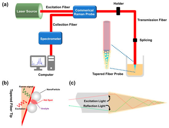
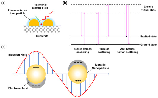
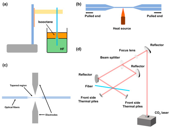
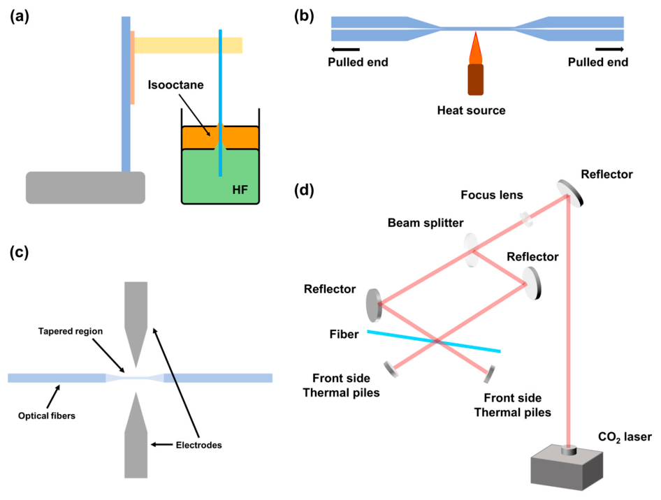
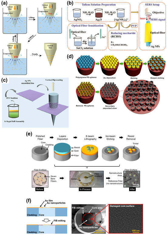
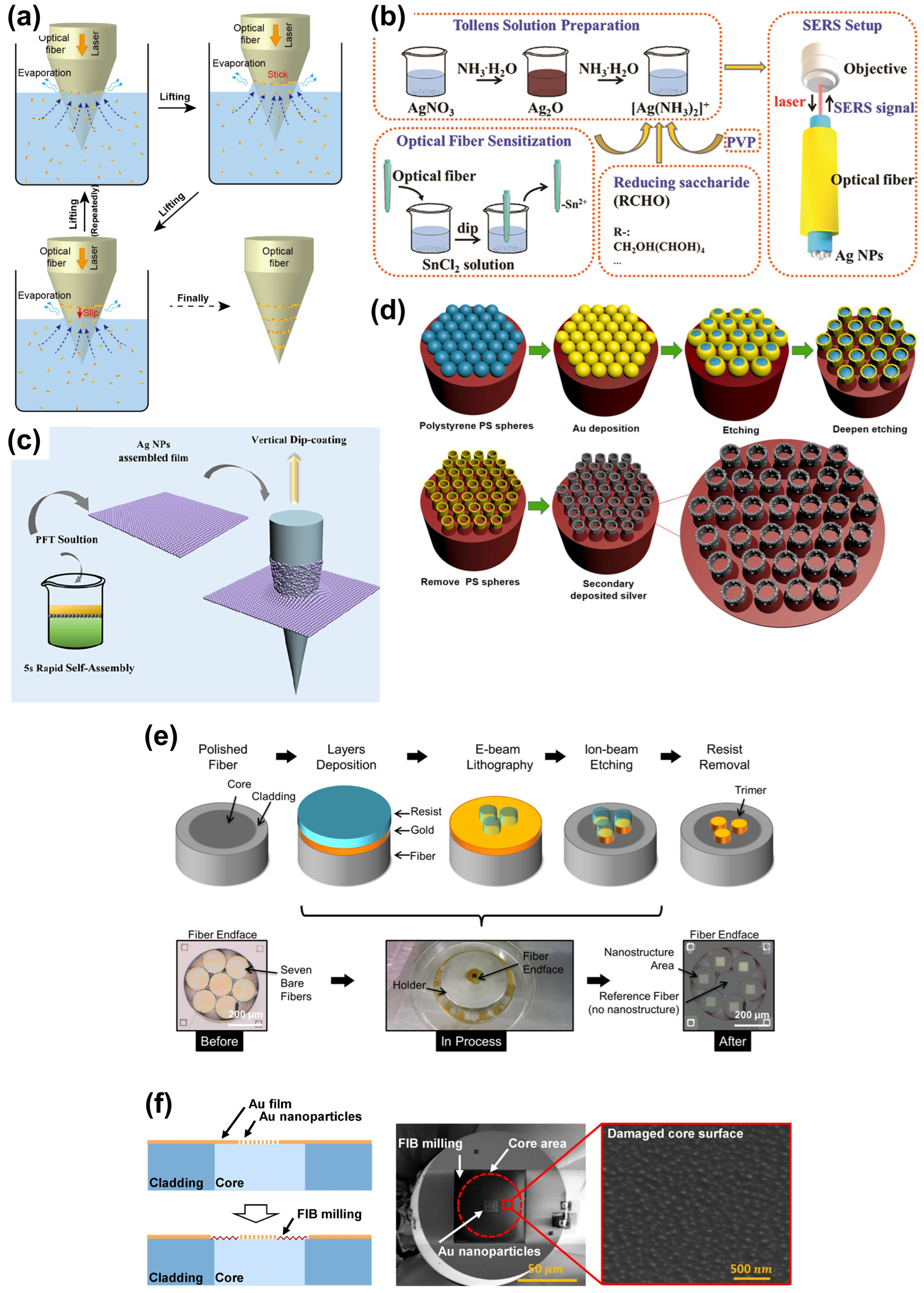
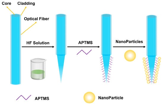
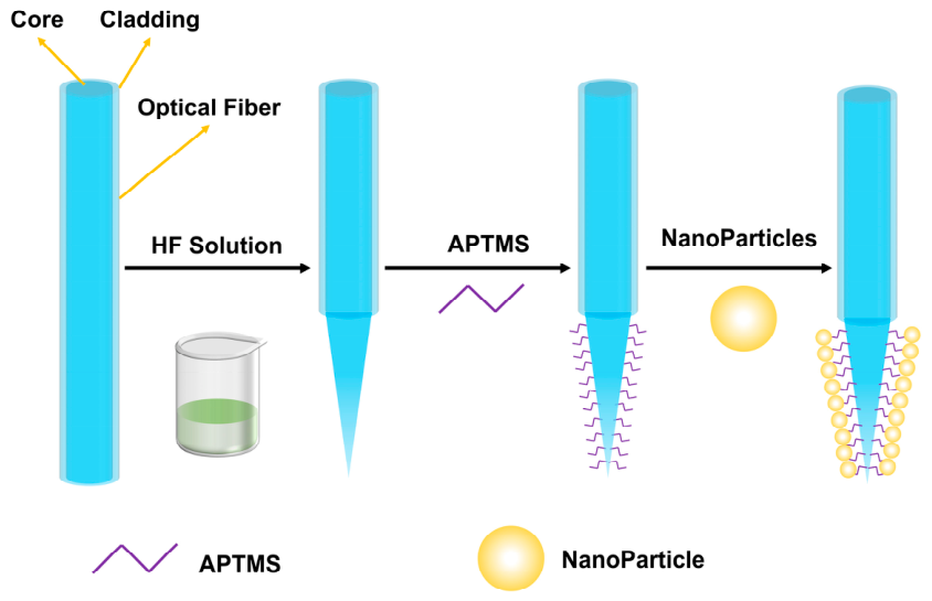

Disclaimer/Publisher’s Note: The statements, opinions and data contained in all publications are solely those of the individual author(s) and contributor(s) and not of MDPI and/or the editor(s). MDPI and/or the editor(s) disclaim responsibility for any injury to people or property resulting from any ideas, methods, instructions or products referred to in the content. |
© 2026 by the authors. Licensee MDPI, Basel, Switzerland. This article is an open access article distributed under the terms and conditions of the Creative Commons Attribution (CC BY) license.