


Abstract
In most ratiometric electrochemiluminescence (ECL) sensors, the utilization of different co-reactants for anodic and cathodic ECL luminophores, along with a broad potential scanning range, restricts their practical applications. Herein, we first reported dual-cathodic potential-resolved ECL from nitrogen/sulfur-codoped carbon dots (N,S-CDs) and mercaptopropionic acid-functionalized copper nanoclusters (MPA-Cu NCs) using their common co-reactant K2S2O8 within a potential range of 0 to −2 V, and developed a ratiometric ECL biosensor for miRNA-155 analysis. Initially, the ECL peak of MPA-Cu NCs at approximately −2 V on the electrode was quenched through resonance energy transfer (RET) by methylene blue. Subsequently, trace target miRNA-155 was converted into abundant output DNA via a DNA walker mechanism. In the presence of Pb2+, partial DNA was cleaved to remove methylene blue, thereby restoring the ECL intensity of MPA-Cu NCs. Furthermore, the cleaved DNA fragments sparked rolling circle amplification (RCA), which ultimately facilitated the loading of N,S-CDs onto the electrode surface, generating an ECL peak at approximately −1 V. As the concentration of miRNA-155 increased, both ECL signals rose simultaneously but with different magnitudes. The fabricated ratiometric ECL sensor achieved a linear detection range for miRNA-155 from 10 aM to 0.1 nM, with a limit of detection of 2.91 aM. Overall, this study offers a new strategy for constructing dual-cathodic ratiometric ECL biosensors and provides a promising approach for early disease diagnosis.
1. Introduction
Electrochemiluminescence (ECL), which integrates the advantages of chemiluminescence and electrochemistry, has found extensive applications in disease diagnosis [1,2], drug and food detection [3,4], and environmental assessment [5] owing to its simplicity, rapidity, low background, and high sensitivity. However, in single-signal ECL detection, factors such as analyte loading density, co-reactant concentration, and pH inevitably affect the accuracy of analytical results, making it difficult to completely eliminate false-positive or false-negative errors [6]. Inspired by fluorescence ratiometric methods, Chen’s group [7] employed CdS nanocrystals and luminol as cathodic emitters and anodic, respectively, and successfully constructed the first potential-resolved ECL sensor based on a dual-signal ratiometric strategy for biomolecule detection using potential-resolved ECL. Compared with conventional single-signal ECL measurements, the potential-resolved ratiometric strategy effectively eliminated errors induced by environmental variations, such as optical path changes, light scattering and background light interference, which significantly improved the sensitivity and accuracy of the sensor [8]. Thereafter, potential-resolved ratiometric ECL sensors attracted considerable attention in bioanalysis [9,10,11]. For instance, Xu’s team [12] utilized CdTe quantum dots (QDs) as the cathodic emitter, Ru(bpy)32+-encapsulated silica nanoparticles as the anodic emitter, and K2S2O8 and TPA as respective co-reactants to design an innovative ratiometric ECL biosensor for the analysis of cardiac-related miRNA-499. Nevertheless, traditional ratiometric ECL sensor designs typically relied on cathodic and anodic luminophores with large ECL peak potential differences [13,14] and required distinct co-reactants, which unavoidably led to interfering side reactions during wide potential scanning [15]. Therefore, the selection of potential-resolved luminophore pairs that emit within a narrow potential window (i.e., single cathodic or anodic scanning) and share the same co-reactant improved the reliability of analytical results. Zhang et al. [16] utilized a pair of cathodic emitters, g-C3N4 nanosheets and N,N’-dimethyl-3,4,9,10-perylenetetracarboxylic diimide (PDI-CH3), and K2S2O8 were used as the common co-reactants, to achieve a cathodic dual-potential resolved ratiometric biosensor for the quantitative analysis of carcinoembryonic antigen. However, PDI-CH3, as an organic dye, involved complex organic synthesis, was expensive, and potentially exhibited toxicity, thereby limiting its practical application in biological detection. Consequently, the use of low-cost luminophore pairs that emit from the same cathodic or anodic process and possess good biocompatibility holds significant research value and promising application prospects for ratiometric ECL sensors.
Nanomaterials have garnered substantial attention in various biosensing applications due to their excellent electrochemical, optical, and structural properties, for example, carbon dots (CDs) [17], polymer dots [18], QDs [19], and metal nanoclusters [20]. Among these, copper nanoclusters (Cu NCs) have been extensively investigated because of their small size, outstanding photostability, cost-effectiveness, and non-toxicity [21,22]. For example, Pan’s team [23] employed porous poly-L-cysteine (poly-L-Cys) as a template to generate controllable poly-L-Cys/Cu NCs via electrochemical reduction, and the confinement effect of poly-L-Cys enhanced the ECL efficiency and stability of Cu NCs, thereby enabling sensitive detection of alkaline phosphatase. Additionally, Wang’s group [24] developed an ECL biosensor employing MPA-Cu NCs for enrofloxacin analysis, where MPA-Cu NCs were obtained through a simple one-pot approach with 3-mercaptopropionic acid (MPA) serving as both the reducing and capping agent, offering straightforward preparation and low cost. Cu NCs typically produced an ECL emission peak at approximately −2 V (cathodic), so the identification of an ECL luminophore with a low potential difference at the cathode facilitated the narrowing of the potential scanning range. Carbon dots have received growing interest because of their outstanding biosafety, facile synthesis, and low production cost [25,26,27]. Recently, Guo et al. [28] developed a novel type of nitrogen- and sulfur-codoped CDs (N,S-NIR-CDs) exhibiting near-infrared ECL emission with low-potential excitation (around −1 V), representing a breakthrough in the low-potential emission of carbon dots. Meanwhile, both Cu NCs and CDs shared K2S2O8 as the co-reactant. Therefore, we developed MPA-Cu NCs and N,S-CDs as an ideal dual-cathodic ratiometric ECL emitter pair.
To enhance the efficiency of ECL sensors in biomolecule detection, specific signal amplification techniques involving cyclic amplification were commonly employed, such as DNA walkers [29], hybridization chain reaction (HCR) [30], polymerase chain reaction (PCR) [31], strand displacement amplification (SDA) [32], catalytic hairpin assembly (CHA) [33], and rolling circle amplification (RCA) [34], which have been widely applied in ECL biosensors. The DNA walker is a nanoscale molecular device composed of a walking arm and predefined track strands, capable of autonomously moving along a designated pathway and transducing signals during its locomotion. In recent years, DNA walkers have attracted tremendous attention due to their superior signal transduction capability [35,36]. Yuan’s group [37] developed a target-driven bipedal DNA walker-based biosensor as a sensitive platform for miRNA detection. The bipedal DNA walker overcame the trajectory limitations of monopod DNA walkers and improved amplification efficiency. Furthermore, polymerase-driven RCA enabled the continuous synthesis of long single-stranded DNA containing repeated sequences under isothermal conditions [38], providing substantial binding sites for ECL luminophores while amplifying signals [39]. This approach facilitated the spatial separation of dual luminophores in ratiometric ECL sensors, avoided energy transfer interference, and reduced the occurrence of side reactions. Therefore, we sought to integrate Cu NCs and N,S-CDs assembled with a DNA walker and RCA technologies to construct a dual-cathodic ratiometric ECL biosensor exhibiting excellent selectivity and an ultralow detection limit.
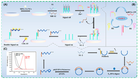
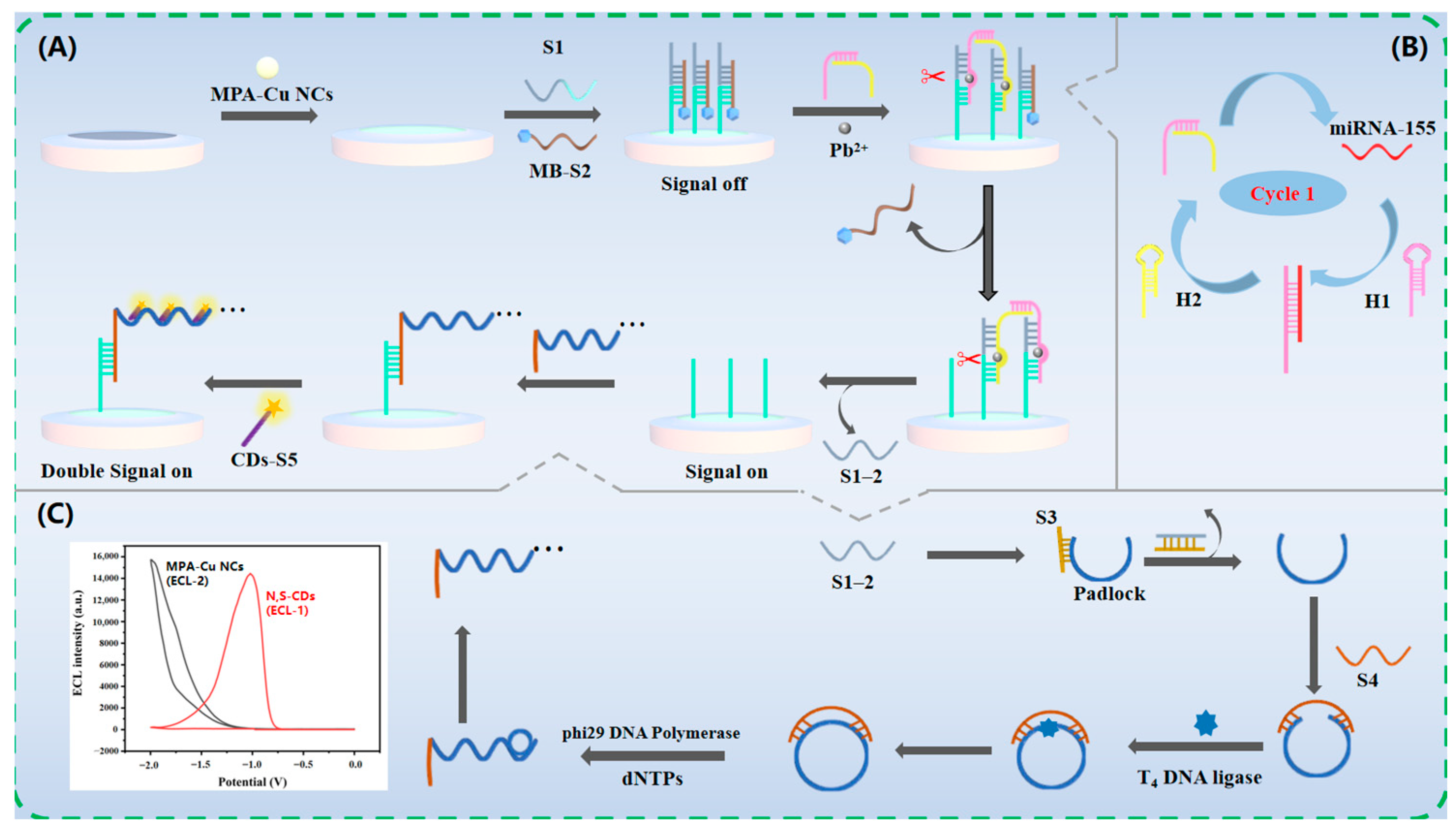
Inspired by the above developments, the present study proposed a novel dual-cathodic ECL ratiometric strategy for the sensitive quantification of miRNA-155. As illustrated in Scheme 1, MPA-Cu NCs were first modified onto the electrode surface, producing a strong ECL output at −2 V in the presence of K2S2O8 as the co-reactant. Subsequently, S1 hybrid was introduced onto the electrode along with methylene blue-labeled DNA S2 (MB-S2). Because of the superior quenching capability of MB, the ECL reached a “signal-off” status. Thereafter, a bipedal DNA walker, generated through target miRNA-155-catalyzed CHA, walked on the electrode in the presence of Pb2+. After multiple cycles, a large amount of S1–2 was cleaved by the bipedal DNA walker, releasing MB-S2 from the electrode interface and switching the ECL to a “signal-on” status. Meanwhile, the cleaved S1–2 reacted with S3 to release the padlock DNA, which was then repaired by T4 DNA ligase to form a circular template, triggering subsequent RCA. The RCA product, bearing numerous repeated DNA sequences, was immobilized on the electrode via specific hybridization with S1–1. Finally, the RCA product captured a large quantity of N,S-CDs-labeled CDs-S5, yielding another strong ECL signal at −1 V with K2S2O8 as the co-reactant. Quantitative detection of miRNA-155 was achieved by calculating the ratio of the two ECL signals (ECLN,S-CDs/ECLMPA-Cu NCs).
Scheme 1.
Preparation procedure of the dual-cathodic ratiometric ECL biosensor. (A) Electrode preparation; (B) DNA walker preparation; (C) RCA reaction.
2. Experimental
2.1. Synthesis of N,S-CDs
N,S-CDs were prepared following a study with minor modifications [40]. Briefly, 0.04 g of folic acid (nitrogen precursor) and 0.80 g of glutathione (GSH, sulfur precursor) were dispersed in 20 mL of formamide by ultrasonication for 10 min. After being transferred to a Teflon-lined reactor, the mixture was kept at 160 °C for 8 h within an electric blast drying oven. Following cooling to ambient temperature, the dark suspension was subjected to 4000 rpm for 10 min for the removal of large aggregates. The supernatant was collected and dialyzed (3500 Da MWCO) for 48 h, followed by membrane filtration (0.22 μm) to obtain the purified N,S-CDs solution. The final dark green N,S-CDs were freeze-dried and stored at 4 °C in the dark.
2.2. Synthesis of MPA-Cu NCs
MPA-Cu NCs were synthesized following a procedure reported in the literature [24] and underwent simple modifications. 20 mL of 20 mmol/L MPA and 20 mL of 1 mmol/L CuSO4·5H2O were mixed under nitrogen protection and stirred for 10 min. Subsequently, the mixture was neutralized to pH 7.4 with 1 mol/L NaOH and allowed to react at 37 °C for 20 min. During this process, the colorless solution gradually developed a white flocculent appearance. The mixed solution mentioned above was centrifuged at 8000 rpm for 20 min. The supernatant was discarded, and centrifuged three times with DMF. The aggregated MPA-Cu NCs were obtained by removing the supernatant. The obtained MPA-Cu NCs were dried with nitrogen, followed by storage at 4 °C until subsequent experiments.
2.3. Preparation of DNA Walker
The construction of the DNA walker was based on target-CHA. Prior to the experiments, all hairpin probes were heated to 95 °C for 10 min and then cooled down to ambient temperature over 30 min. Subsequently, 10 μL of miRNA-155 at varying concentrations was mixed thoroughly with 10 μL of 2 μM hairpin H1 and H2. The assembly of the DNA walker was promoted by incubating the mixture at 37 °C for 120 min.
2.4. RCA Reaction
The obtained solution containing the S1–2 chains was mixed with 5 μM S3-blocked padlock DNA and incubated at room temperature for 120 min. Subsequently, 5 μM S4 was added, and the mixture was further incubated at 37 °C for 2 h. Next, 10× T4 DNA ligase buffer and 5 U T4 DNA ligase were added, followed by standing at 37 °C for 2 h. Thereafter, the resulting mixture was combined with 10× phi29 buffer, 2 U phi29 DNA polymerase, and 2.5 mM dNTPs, and kept for 2 h at 37 °C. Finally, the mixture was kept at 65 °C for 15 min to inactivate the enzymes.
2.5. Assembly of the ECL Biosensor
MPA-Cu NCs were ultrasonically dispersed in 20 mL DMF, and the dispersion was deaerated with nitrogen for 5 min. Then, 10 μL of the dispersion was mixed with 0.02% chitosan (CS) and drop-cast onto a glassy carbon electrode (GCE). After nitrogen drying, the MPA-Cu NCs-modified electrode was obtained. For activation of the carboxyl groups on the MPA-Cu NCs layer, the modified GCE was soaked in a cross-linking solution containing 20 mM EDC and 10 mM NHS and incubated at 37 °C for 30 min. Next, 10 μL of a mixture containing 5 μM DNA strand S1 and MB-S2 was drop-cast onto the activated electrode surface and incubated overnight at 4 °C. The electrode surface was then blocked with 1.0 mM hexanethiol (HT) solution at 37 °C for 40 min to block nonspecific binding sites. Subsequently, 10 μL of the pre-assembled DNA walker (containing 8 μM Pb2+) was dropped onto the GCE surface and kept at room temperature for 120 min. The resulting solution containing S1–2 DNA was aspirated for use in the subsequent RCA reaction. Afterward, 10 μL of the RCA reaction product was applied to the electrode and incubated for 2 h at room temperature. The remaining active sites were blocked again with 1.0 mM HT solution for 40 min. Finally, 10 μL of N,S-CDs-modified CDs-S5 was dropped onto the electrode and kept at room temperature for 2 h. Following each modification step, the electrode surface was rinsed with deionized water.
2.6. ECL Measurement of the Biosensor
Prior to ECL measurement, the PBS solution (pH 7.4) containing 0.1 M K2S2O8 was purged with high-purity N2 gas for 15–20 min to remove dissolved oxygen. The assembled ECL biosensor was then placed into 5 mL of the deaerated PBS solution with 0.1 M K2S2O8. During testing, the photomultiplier tube was adjusted to 800 V, the scan potential window was set from 0 to −2.0 V, and the scanning rate was 100 mV/s. Throughout the measurement, a continuous N2 flow was maintained over the solution surface.
3. Results and Discussion
3.1. Characterization of MPA-Cu NCs
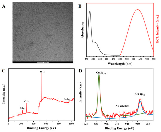
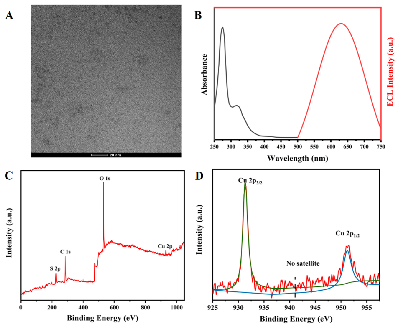
The TEM image in Figure 1A demonstrates that the obtained MPA-Cu NCs were uniformly spherical and monodisperse with an approximate size of 3 nm (high-magnification TEM image shown in Figure S1). Figure 1B (black line) illustrates the UV-vis absorption spectrum of MPA-Cu NCs, with prominent peaks at 275 nm corresponding to the transformation of the Cu-S hybrid bond orbital to the Cu-Cu [41]. The ECL emission spectrum of MPA-Cu NCs was measured in the presence of S2O82−, and the results showed an emission peak at 630 nm (Figure 1B, red curve). XPS was subsequently used to determine the elemental composition of the MPA-Cu NCs. Figure 1C exhibits the peaks at 161.2, 284.8, 531.1, and 931.3 eV, corresponding respectively to S 2p, C 1s, O 1s, and Cu 2p. Figure 1D displays two characteristic binding energy peaks of 931.3 eV (Cu 2p3/2) and 951.5 eV (Cu 2p1/2), indicating the oxidation state of copper between Cu(0) and Cu(I). Furthermore, the characteristic satellite peak of Cu(II) ions that was not observed at approximately 942 eV indicated that the synthesized MPA-Cu NCs contained no Cu(II) ions. Thus, the synthesized MPA-Cu NCs exhibited a valence state range of 0 to +1, which was in good agreement with previously reported findings [24].
Figure 1.
(A) TEM picture, (B) UV-vis absorption spectrogram (black curve) and ECL spectra (red curve), (C) XPS results, (D) Cu 2p high-resolution XPS spectrum of MPA-Cu NCs, Cu 2p3/2 characteristic peak (green curve) and Cu 2p1/2 characteristic peak (blue curve).
3.2. Characterization of N,S-CDs
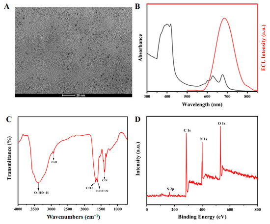
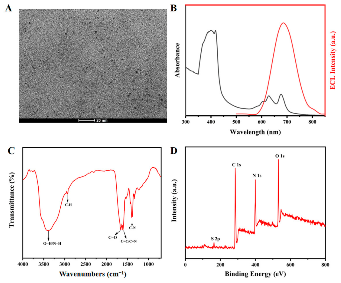
Figure 2A displays that the N,S-CDs were well-dispersed and spherical, with a mean size of about 2 nm (high-magnification TEM image shown in Figure S1). The characteristic UV-Vis absorption of the N,S-CDs (Figure 2B, black line) revealed two absorption bands at 350–430 nm and 600–670 nm, attributed to the π–π* and n–π* transitions of C=X (X = O, N, S) bonds within the aromatic structures of the CDs [40]. The ECL emission spectrum of the N,S-CDs was measured in the presence of S2O82−, showing an emission maximum at 686 nm (Figure 2B, red line). The FT-IR spectrum of the N,S-CDs (Figure 2C) showed a broad band at 3200–3450 cm−1 corresponding to O–H and N–H stretching, along with peaks at 2920, 1666, 1609, and 1397 cm−1, which were assigned to the stretching vibrations of C–H, C=O, C=N/C=C, and C–N bonds, respectively [42]. Those results suggested that the synthesized N,S-CDs were abundant in both amino and carboxyl groups. Additionally, surface elemental analysis by XPS (Figure 2D) confirmed the presence of S 2p, C 1s, N 1s, and O 1s peaks at 162.7, 284.8, 399.8, and 531.7 eV, respectively, demonstrating the successful integration of N and S elements into the CD framework.
Figure 2.
(A) TEM image, (B) UV-vis absorption spectrogram (black curve) and ECL spectra (red curve), (C) FT-IR spectra, (D) XPS results of N,S-CDs.
3.3. ECL Mechanism of Biosensor
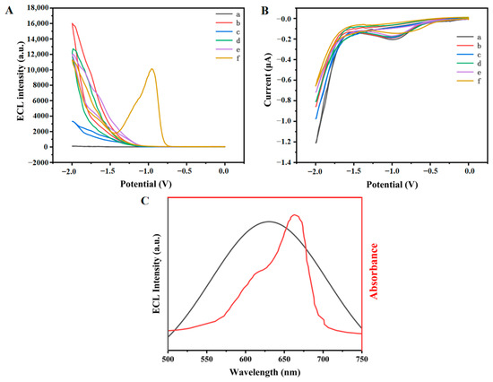
For verification of the combination procedure of the ECL biosensor, the ECL responses (Figure 3A) and current changes (Figure 3B) during stepwise modification were investigated in 0.1 M PBS (pH 7.4) with 0.1 M K2S2O8. In addition, electrochemical impedance spectroscopy (EIS, Figure S2A) and cyclic voltammetry (CV, Figure S2B) characterizations were performed. Initially, the bare GCE showed almost no ECL intensity (Figure 3A, curve a) due to the absence of ECL luminophores. When CS/MPA-Cu NCs were decorated on the electrode, a strong ECL emission peak was observed at −2.0 V (Figure 3A, curve b). However, due to the very poor electron conductivity of CS/MPA-Cu NCs, the peak current was significantly decreased (Figure 3B, curve b). The ECL mechanism of MPA-Cu NCs was triggered by K2S2O8 as the co-reactant to generate the excited state MPA-Cu NCs*. During electrochemical reduction, MPA-Cu NCs were transformed into negatively charged radical intermediates (MPA-Cu NCs•−). Simultaneously, S2O82− diffused within the solution underwent electrochemical reduction to the strong oxidant SO4•−. Subsequently, a reaction occurred between SO4•− and MPA-Cu NCs•−, producing the activated state MPA-Cu NCs*. Finally, MPA-Cu NCs* returned to the ground state by emitting photons along the radiative pathway, thereby generating the ECL signal. The ECL reactions were represented by Equations (1)–(4) [24]:
MPA-Cu NCs + e− → MPA-Cu NCs•−
S2O82− + e− → SO42− + SO4•−
MPA-Cu NCs•− + SO4•− → MPA-Cu NCs* + SO42−
MPA-Cu NCs* → MPA-Cu NCs + hv
Figure 3.
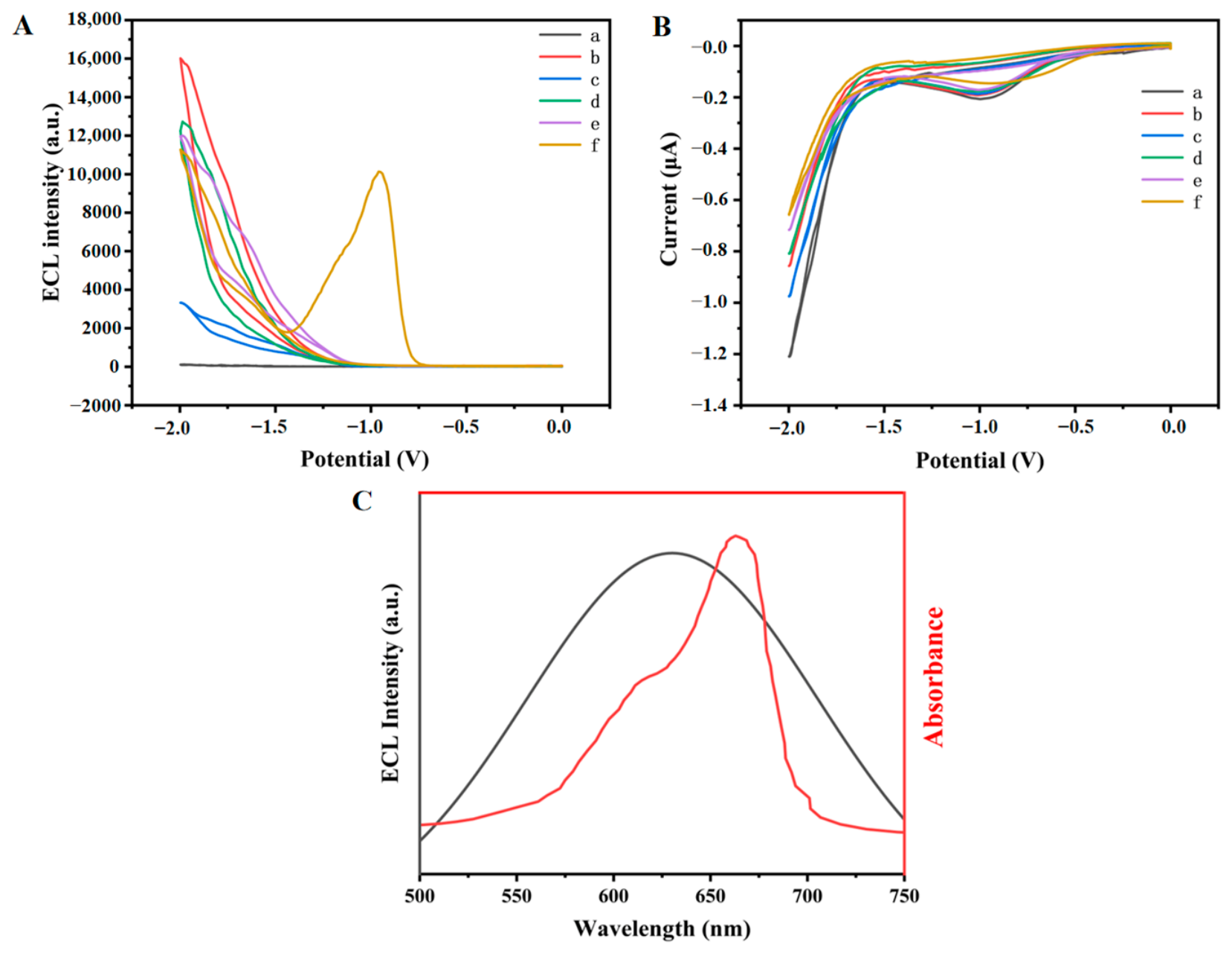
(A) ECL response curves and the (B) CV of the corresponding stepwise modified electrode of (a) bare GCE, (b) CS/MPA-Cu NCs/GCE, (c) S1 + S2/CS/MPA-Cu NCs/GCE, (d) DNA walker/S1 + S2/CS/MPA-Cu NCs/GCE, (e) RCA products/S1/CS/MPA-Cu NCs/GCE, (f) CDs-s5/RCA products/S1/CS/MPA-Cu NCs/GCE. in PBS (0.1 M pH 7.4) containing 0.1 mol/L K2S2O8 with a potential scan interval of −2.0–0 V and a scan rate of 100 mV/s. (C) The black curve is the ECL spectrum of MPA-Cu NCs and the red curve is the visible absorption spectrum of MB.
After the subsequent introduction of DNA S1 and MB-S2 onto the electrode, the ECL intensity exhibited a sharp decrease (Figure 3A, curve c). This phenomenon was attributed to both the electrochemiluminescence resonance energy transfer (ECL-RET) between MPA-Cu nanoclusters and MB, as well as the electron transfer capability of MB [43]. The results in Figure 3C showed significant overlap between the UV-vis absorption of MB (660 nm) and the ECL emission spectrum of MPA-Cu NCs (630 nm), indicating the presence of ECL-RET. However, the peak current increased (Figure 3B, curve c), which was ascribed to the efficient electron transfer capability of MB. Upon addition of the output DNA walker generated from the cycle, the MB-labeled DNA fragments were released, eliminating the ECL-RET effect, and the ECL signal recovered (Figure 3A, curve d), accompanied by a decrease in current intensity (Figure 3B, curve d). Afterwards, when the RCA product was introduced onto the electrode, the ECL signal and CV curve slightly decreased (Figure 3A,B, curve e), primarily because the negatively charged DNA impeded electron transfer. After incubation of CDs-S5 on the electrode, the N,S-CDs showed a strong ECL peak at −1 V, while the ECL peak of MPA-Cu NCs at −2 V decreased again (Figure 3A, curve f). Notably, although partial overlap existed between the UV absorption of N,S-CDs (628 nm) and the ECL emission spectrum of MPA-Cu NCs (630 nm), the distance between the two luminophores exceeded 10 nm, resulting in a negligible ECL-RET effect. The reduction in the ECL peak at −2 V and the current peak (Figure 3A, curve f) was attributed to the hindrance of electron transfer by the incorporated DNA. The ECL mechanism of the N,S-CDs/S2O82− system was similar to that of MPA-Cu NCs, following a “reduction-oxidation” co-reactant pathway. First, N,S-CDs and S2O82− on the electrode surface gained electrons and were reduced to N,S-CDs•− and SO4•−, respectively. Then, a reaction between N,S-CDs•− and SO4•− produced the unstable excited state species N,S-CDs*, which finally underwent radiative relaxation to emit light, as seen in Equations (5)–(8) [42]:
N,S-CDs + e− → N,S-CDs•−
S2O82− + e− → SO42− + SO4•−
N,S-CDs•− + SO4•− → N,S-CDs* + SO42−
N,S-CDs* → N,S-CDs + hv
3.4. Analytical Performance for miRNA-155 Detection
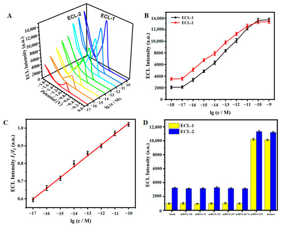
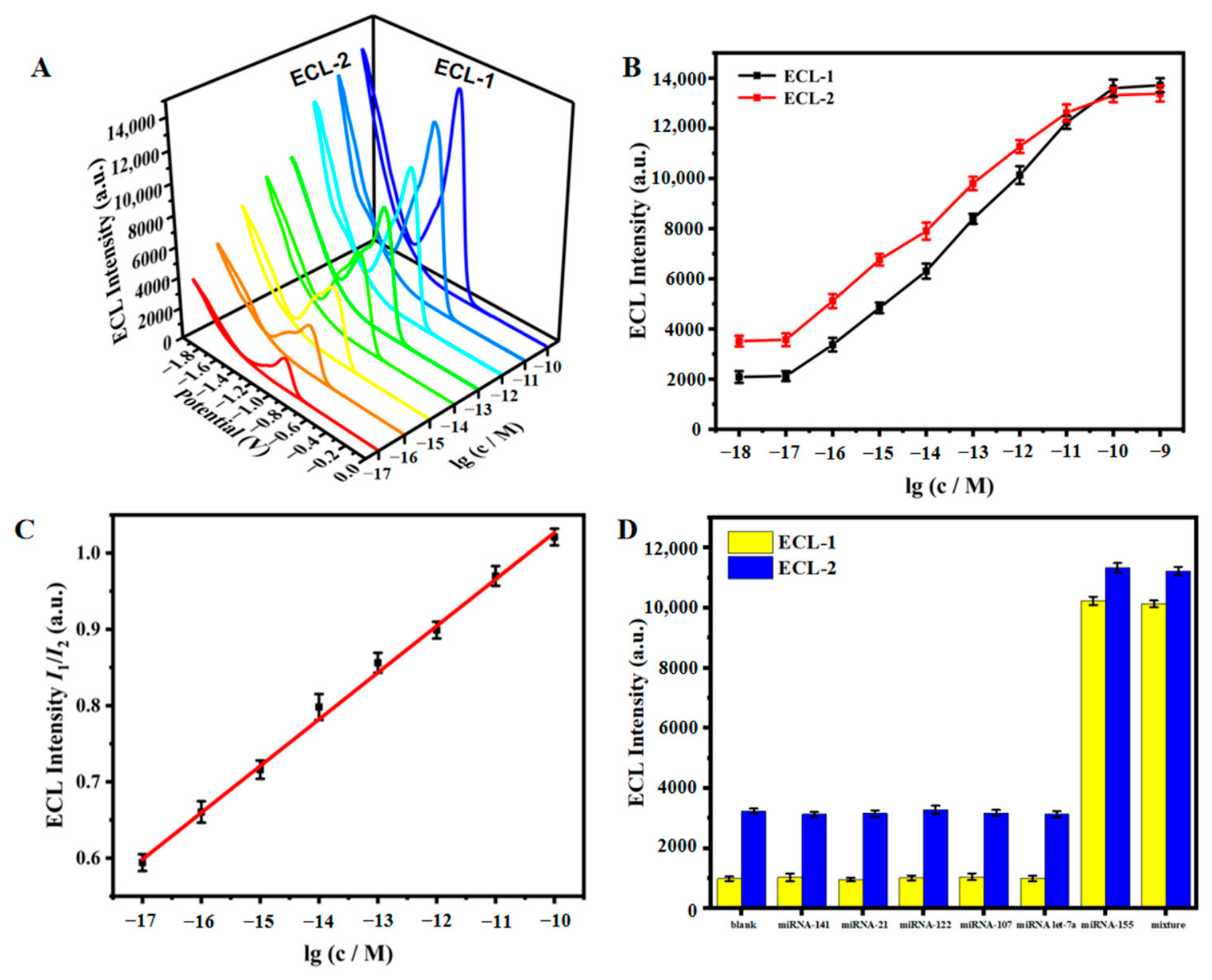
The analytical capability of the ECL biosensor was assessed by detecting various concentrations of miRNA-155 under the optimized conditions (Figure S3). Figure 4A displays that both the ECL signal of N,S-CDs at approximately −1 V (ECL-1) and that of MPA-Cu NCs at −2 V (ECL-2) gradually increased with increasing miRNA-155 concentration. Within the logarithmic concentration range of miRNA-155 from 10−17 M to 10−10 M, both ECL-1 and ECL-2 intensities increased simultaneously yet to different extents as the target concentration was increased (Figure 4B). Furthermore, a strong positive correlation existed between the logarithmic concentration of miRNA-155 and the ECL-1/ECL-2 ratio (Figure 4C). Consequently, quantitative detection of miRNA-155 was achieved by the ECL(I1/I2). The regression equation was ECL(I1/I2) = 0.06121 lg c + 1.63898 (R2 = 0.997), where I denotes ECL intensity and c corresponds to the miRNA-155 concentration. The detection limit (LOD) was 2.91 aM (S/N = 3). The constructed ECL biosensor exhibited advantages in terms of a lower LOD and a wider linear range for miRNA-155 detection when compared to previously reported methods (Table S2). This superior performance was primarily attributed to the ratiometric dual-signal strategy, which effectively eliminated background interference and substantially enhanced detection sensitivity.
Figure 4.
(A) ECL intensity of the miRNA-155 with concentrations ranging from 10−17 M to 10−10 M (from the red curve to the blue curve). (B) The relationship between ECL-1 and ECL-2 and the logarithmic concentration of the target miRNA-155. (C) Linear change diagram of miRNA-155 concentration logarithm versus ECL(I1/I2). (D) Selectivity of the fabricated ECL biosensor.
The specificity of the biosensor toward miRNA-155 was evaluated by using three interfering miRNAs (miRNA-141, miRNA-21, miRNA-122, miRNA-107 and miRNA let-7a). As illustrated in Figure 4D, the ECL intensities of these interfering miRNAs (10−11 M) were almost identical to that of the blank control group, even at a concentration 10-fold higher than that of miRNA-155 (10−12 M). Conversely, significant ECL signals were observed only when the interferents were mixed with miRNA-155 or when miRNA-155 was present alone. The results confirmed that the constructed ECL biosensor possessed high specificity for miRNA-155. To evaluate the stability, the biosensor was subjected to 10 consecutive scanning cycles (Figure S4), revealing that the ECL signals remained essentially stable, with relative standard deviations (RSD) of 2.87% for ECL-1 and 3.17% for ECL-2. Furthermore, the reproducibility was assessed by testing five ECL biosensors under identical conditions (Figure S5), yielding an RSD of 1.2% for the ratiometric signal. Long-term storage stability was also evaluated over 28 days (0, 7, 14, 28 days). As shown in Figure S6, the ratiometric ECL signal remained excellent throughout the 4-week period, with an RSD of 1.55%. These findings indicated that the developed ECL biosensor exhibited good stability and reproducibility, highlighting its potential for practical applications.
3.5. Real Application of the Biosensor
To further assess the detection efficiency of the developed biosensor in practical applications, recovery studies were carried out to identify the level of miRNA-155 in pretreated healthy human serum samples. Standard solutions at five different concentrations (1 × 10⁻¹⁷, 1 × 10⁻¹⁵, 1 × 10⁻¹³, 5 × 10⁻¹², and 1 × 10⁻¹⁰ M) were prepared using the standard addition approach. As listed in Table 1, the recovery rates for the samples varied from 93.8% to 105.1%, and RSD varied from 2.34% to 3.57%. These findings indicated that the designed ECL biosensor could be successfully utilized for the quantitative analysis of miRNA-155 in real biological samples.
Table 1.
Analytical results for miRNA-155 quantification in serum samples (n = 3).
4. Conclusions
In summary, we developed a highly sensitive and selective dual-cathodic ratiometric ECL biosensor for the detection of miRNA-155 by employing MPA-Cu NCs and N,S-CDs as a pair of cathodic potential-resolved luminophores on a single electrode. Varying concentrations of miRNA-155 first triggered a cyclic reaction to generate the DNA walker, thereby recovering the ECL signal of MPA-Cu NCs previously quenched by MB through ECL-RET. Concurrently, this process activated RCA, introducing N,S-CDs onto the electrode surface and achieving differential enhancement of the dual ECL signals. We found that the ECL-1/ECL-2 intensity ratio could be utilized for the quantitative determination of miRNA-155, spanning a linear response from 10−17 to 10−10 M with a LOD of 2.91 aM. Consequently, this novel dual-cathodic ratiometric ECL biosensor, which leveraged the high luminous efficiency of MPA-Cu NCs and N,S-CDs in combination with the dual amplification strategies of DNA walker and RCA, provided a new approach for miRNA detection and holds considerable potential for applications in ECL-based biomolecule analysis and early disease diagnosis.
Supplementary Materials
The following supporting information can be downloaded at https://www.mdpi.com/article/10.3390/chemosensors14060124/s1: The reagents and instruments used in the experiment. Cross-linking of N,S-CDs with S5 [44]. Figure S1: High-magnification TEM images of N,S-CDs and MPA-Cu NCs. Figure S2: EIS and CV graphs of the sensor. Figure S3: Optimization graphs of pH, potassium persulfate concentration, and reaction time for the ECL sensor. Figure S4: Stability test graph of the sensor. Figure S5: Reproducibility test graph of the sensor. Figure S6: Storage stability of the fabricated ECL biosensor. Table S1: Sequences of DNA and RNA used in this study. Table S2: Comparison with other miRNA-155 testing methods [45,46,47,48,49].
Author Contributions
Conceptualization: M.-Y.Z., Y.G. and J.L.; data planning: M.-Y.Z. and Y.G.; form analysis: M.-Y.Z., H.H., M.-L.L., M.-Z.D., C.-C.L. and H.-X.L.; methodology: M.-Y.Z., L.M., Z.X., F.Z., G.-S.C. and Y.-Z.W.; resources: G.-S.C. and Y.-Z.W.; Software: Y.G.; validation: M.-Y.Z. and J.L.; visualization: L.M.; investigation: Z.X., F.Z., G.-S.C. and Y.-Z.W.; writing—original manuscript preparation: M.-Y.Z. and Y.G.; writing—review and editing: M.-Y.Z., Y.G., G.-S.C. and Y.-Z.W.; funding acquisition: Y.-Z.W.; project management: Y.-Z.W.; supervision: Y.-Z.W. All authors have read and agreed to the published version of the manuscript.
Funding
National Natural Science Foundation of China (Grant No. 22476090, 21904050, 22104057, 22204079, 22578215), Natural Science Foundation of Jiangsu Province, China (BK20210638) and discipline fund, School of Food and Light Industry, Nanjing Tech University.
Institutional Review Board Statement
The study was conducted in accordance with the Declaration of Helsinki and approved by the Ethics Committee of Nanjing Drum Tower Hospital, Affiliated Hospital of Medical School, Nanjing University (protocol code: 2023-273-02, approved on 31 August 2023).
Informed Consent Statement
Informed consent was obtained from all subjects involved in the study.
Data Availability Statement
The original contributions presented in this study are included in the article. Further inquiries can be directed to the corresponding authors.
Conflicts of Interest
The authors declare no conflicts of interest.
References
- Giagu, G.; Fracassa, A.; Fiorani, A.; Villani, E.; Paolucci, F.; Valenti, G.; Zanut, A. From theory to practice: Understanding the challenges in the implementation of electrogenerated chemiluminescence for analytical applications. Microchim. Acta 2024, 191, 359. [Google Scholar] [CrossRef] [Scilit]
- Bak, Y.; Anjong, T.F.; Park, J.; Jeon, S.H.; Kim, J.; Lee, K.; Shin, I.; Kim, S. Monitoring disease-implicated hydrogen sulfide in blood using a facile deproteinization-based electrochemiluminescence chemosensor system. Sens. Actuators B Chem. 2023, 394, 134364. [Google Scholar] [CrossRef] [Scilit]
- Alboull, A.M.A.; Xiao, J.; Zheng, Y.; Zholudov, Y.T.; Snizhko, D.V.; Qi, L.; Niu, W.; Xu, G. Ru(bpy)32+ electrochemiluminescence of saccharides. Electrochim. Acta 2026, 569, 148899. [Google Scholar] [CrossRef] [Scilit]
- DPavão, P.E.; Botelho, C.N.; Fernandes, R.N.; Santos, C.C.D.; Damos, F.S.; De Cássia Silva Luz, R. A Simple, Cost-effective, and environmentally friendly method for determination of ciprofloxacin in drugs and urine samples based on electrogenerated chemiluminescence. Electroanalysis 2020, 32, 1498–1506. [Google Scholar]
- Mohan, B.; Najafov, B.; Sharma, P.K.; Pal, A.; Garazade, I.M.; Pombeiro, A.J.L.; Da Silva, M.F.C.G.; Sun, W.; Singjai, P. Electrochemiluminescent COFs and HOFs as porous material engineering systems for bioanalysis and environmental monitoring. Small 2026, 22, e14406. [Google Scholar] [CrossRef] [Scilit] [PubMed]
- Isildak, I.; Navaeipour, F.; Afsharan, H.; Kanberoglu, G.S.; Agir, I.; Ozer, T.; Annabi, N.; Totu, E.E.; Khalilzadeh, B. Electrochemiluminescence methods using CdS quantum dots in aptamer-based thrombin biosensors: A comparative study. Microchim. Acta 2020, 187, 25. [Google Scholar] [CrossRef] [Scilit]
- Zhang, H.-R.; Xu, J.-J.; Chen, H.-Y. Electrochemiluminescence ratiometry: A new approach to DNA biosensing. Anal. Chem. 2013, 85, 5321–5325. [Google Scholar] [CrossRef] [Scilit] [PubMed]
- Chen, G.; Hu, C.; Dai, W.; Luo, Z.; Zang, H.; Sun, S.; Zhen, S.; Zhan, L.; Huang, C.; Li, Y. Coreactant-free zirconium metal–organic framework with dual emission for ratiometric electrochemiluminescence detection of HIV DNA. Anal. Chem. 2024, 96, 10102–10110. [Google Scholar] [CrossRef] [Scilit] [PubMed]
- Huo, X.-L.; Lu, H.-J.; Xu, J.-J.; Zhou, H.; Chen, H.-Y. Recent advances of ratiometric electrochemiluminescence biosensors. J. Mater. Chem. B 2019, 7, 6469–6475. [Google Scholar] [CrossRef] [Scilit]
- He, Y.; Yang, G.; Zhao, J.; Tan, K.; Yuan, R.; Chen, S. Potentially tunable ratiometric electrochemiluminescence sensing based on conjugated polymer nanoparticle for organophosphorus pesticides detection. J. Hazard. Mater. 2022, 432, 128699. [Google Scholar] [CrossRef] [Scilit]
- Dong, X.; Du, Y.; Zhao, G.; Cao, W.; Fan, D.; Kuang, X.; Wei, Q.; Ju, H. Dual-signal electrochemiluminescence immunosensor for Neuron-specific enolase detection based on “dual-potential” emitter Ru(bpy)32+ functionalized zinc-based metal-organic frameworks. Biosens. Bioelectron. 2021, 192, 113505. [Google Scholar] [CrossRef] [Scilit]
- Sun, Y.; Fang, L.; Han, Y.; Feng, A.; Liu, S.; Zhang, K.; Xu, J.-J. Reversible Ratiometric Electrochemiluminescence Biosensor Based on DNAzyme Regulated Resonance Energy Transfer for Myocardial miRNA Detection. Anal. Chem. 2022, 94, 7035–7040. [Google Scholar] [CrossRef] [Scilit]
- Li, C.; Lu, J.; Guo, Y.; Song, Y.; Li, R.; Jiang, H.; Wang, W.; Han, P.; Ma, G.; Wang, J.; et al. Dual-potential ratiometric electrochemiluminescence platform based on PCN and Ti3CN@luminol for kanamycin detection in milk samples. Food Chem. 2025, 479, 143840. [Google Scholar] [CrossRef] [Scilit]
- Duan, G.; Gu, X.; Kan, X. Ratiometric electrochemiluminescence sensor for ultrasensitive and reliable detection of influenza A virus nucleoprotein. Sens. Actuators B Chem. 2025, 444, 138476. [Google Scholar] [CrossRef] [Scilit]
- Wang, S.; Zhu, S.; Kang, Z.; Chen, Y.; Liu, X.; Deng, Z.; Hu, K.; Wang, G.; Zhang, Y.; Zang, G. Recent advances and future prospects of the potential-resolved strategy in ratiometric, multiplex, and multicolor electrochemiluminescence analysis. Theranostics 2022, 12, 6779–6808. [Google Scholar] [CrossRef] [Scilit]
- Zhang, W.; Cui, X.; Li, F.; Yan, W.; Wang, Y.; Shang, L.; Ma, R.; Jia, L.; Li, C.; Wang, H. Perylene diimide and g-C3N4 nanosheet as potential-resolved cathode luminophores for ultrasensitive ratiometric electrochemiluminescence immunosensor. Sens. Actuators B Chem. 2022, 371, 132492. [Google Scholar] [CrossRef] [Scilit]
- Adsetts, J.R.; Zhang, R.; Yang, L.; Chu, K.; Wong, J.M.; Love, D.A.; Ding, Z. Efficient White electrochemiluminescent emission from carbon quantum dot films. Front. Chem. 2020, 8, 580022. [Google Scholar] [CrossRef] [Scilit] [PubMed]
- Liao, X.; Liu, K.; Li, F.; Wang, Y.; Hu, L.; Wu, D.; Ma, H.; Wei, Q. A novel ECL immunosensor based on the signal amplification effect of Ag@Cu2S nanocomposites on carbonized polymer dots for the sensitive detection of CEA. Sens. Actuators B Chem. 2026, 449, 139183. [Google Scholar] [CrossRef] [Scilit]
- Diez-Buitrago, B.; Saa, L.; Briz, N.; Pavlov, V. Development of portable CdS QDs screen-printed carbon electrode platform for electrochemiluminescence measurements and bioanalytical applications. Talanta 2021, 225, 122029. [Google Scholar] [CrossRef] [Scilit]
- Kim, J.H.; Choi, J.; Kim, J.; Kim, J. Enhanced near-infrared electrochemiluminescence of Au nanoclusters treated with piperidine. Bioelectrochemistry 2022, 147, 108192. [Google Scholar] [CrossRef] [Scilit] [PubMed]
- Wang, D.; Wang, P.; Liang, Z.; Li, Z.; Liu, N.; Ma, Q. Novel Cu nanocluster superlattice/MBene-induced ECL enhancement strategy for miRNA-221 detection. Chem. Eng. J. 2023, 478, 147512. [Google Scholar] [CrossRef] [Scilit]
- Lv, J.; Shan, L.; Xu, J.; Liang, J.; Fu, K.; Yu, H.; Wang, D.; Zhang, L.; Li, L.; Ji, W.; et al. Oxygen reduction reaction-regulated paper-based bipolar electrochemiluminescence biosensor for sensitive detection of carbendazim. Anal. Chem. 2025, 97, 13940–13948. [Google Scholar] [CrossRef] [Scilit] [PubMed]
- Pan, M.-C.; Lei, Y.-M.; Chai, Y.-Q.; Yuan, R.; Zhuo, Y. In situ controllable generation of copper nanoclusters confined in a poly-l-cysteine porous film with enhanced electrochemiluminescence for alkaline phosphatase detection. Anal. Chem. 2020, 92, 13581–13587. [Google Scholar] [CrossRef] [Scilit]
- Wang, D.; Jiang, S.; Liang, Y.; Wang, X.; Zhuang, X.; Tian, C.; Luan, F.; Chen, L. Selective detection of enrofloxacin in biological and environmental samples using a molecularly imprinted electrochemiluminescence sensor based on functionalized copper nanoclusters. Talanta 2022, 236, 122835. [Google Scholar] [CrossRef] [Scilit]
- Arcudi, F.; Ðorđević, L.; Rebeccani, S.; Cacioppo, M.; Zanut, A.; Valenti, G.; Paolucci, F.; Prato, M. Lighting up the electrochemiluminescence of carbon dots through pre- and post-Synthetic design. Adv. Sci. 2021, 8, 2100125. [Google Scholar] [CrossRef] [Scilit]
- Zhang, M.-Q.; Zhong, X.; Nan, N.; Xie, A.-L.; Lei, Y.-X.; Liu, Y.-K.; Guo, R.; Chen, F.-J.; Long, Y.-M.; Bao, L. Orientational assembly of carbon dots-enabled electrochemiluminescence sensors for ultrasensitive detection of halogenated phenols. Anal. Chem. 2025, 97, 9722–9730. [Google Scholar] [CrossRef] [Scilit]
- Yang, H.; Xue, X.; Zhang, Y.; Gao, W. Extraction-purified S,N-codoped carbon dots enable ternary synergistic electrochemiluminescence for ultrasensitive detection of copper ions. Anal. Chim. Acta 2025, 1374, 344505. [Google Scholar] [CrossRef] [Scilit] [PubMed]
- Guo, Y.-Z.; Liu, R.; Zeng, Y.; Liao, Y.-Y.; Liu, J.-L.; Chai, Y.-Q.; Yuan, R. The carbon dots with near-infrared electrochemiluminescence emission and low excitation potential for ultrasensitive analysis of microRNA-222. Chem. Eng. J. 2024, 488, 151057. [Google Scholar] [CrossRef] [Scilit]
- Peng, Z.; Li, J.; Song, C.; Yu, S.; Wang, F.; Huang, C.; Deng, S.; Lyv, Y.; Yang, D.; Yang, H.; et al. Orbital DNA walker-driven ECL biosensing based on Ag3PO4@PTCA for ultrasensitive detection of Pb2+. Biosens. Bioelectron. 2026, 293, 118121. [Google Scholar] [CrossRef] [Scilit]
- Sun, Y.; Li, H.; Wang, T.; Zhang, K.; Wang, X. Unveiling MOF-derived CuFe2O4 as a catalytic promoter of Ru(bpy)32+ ECL: A CRISPR/Cas13a-based strategy for miR-17 Detection. Sens. Actuators Rep. 2025, 10, 100406. [Google Scholar] [CrossRef] [Scilit]
- Liu, W.; Liu, Y.; Xiao, Z.; Jin, L.; Wu, M. Ultrasensitive electrochemiluminescence biosensor based on polymethylene blue nanoparticles and DNA network for Staphylococcus aureus detection. Food Chem. 2024, 442, 138471. [Google Scholar] [CrossRef] [Scilit]
- Wang, X.; Cui, J.; Gong, Q.; Zheng, L.; Liu, D.; Nie, G. A multiple signal amplification electrochemiluminescence sensor for Hg2+ detection based on Ce2Sn2O7/poly(5-formylindole) nanocomposites. Talanta 2025, 284, 127227. [Google Scholar] [CrossRef] [Scilit]
- Yang, Y.; Li, J.; Xiang, S.; Wang, F.; Yang, H.; Cai, R.; Tan, W. PdPt@SnS2 Nanosheets for a novel ultrasensitive electrochemiluminescence biosensor for miRNA-21 assay. Anal. Chem. 2024, 96, 9653–9658. [Google Scholar] [CrossRef] [Scilit] [PubMed]
- He, Y.; Zhang, Y.; Xiang, H.; Ren, K.; Yin, Y.; Gao, Y.; Yang, Y.; Zhang, W.; Liu, L.; Han, H.; et al. Magnetic bead-assisted one-pot RCA-activated CRISPR/Cas12a electrochemiluminescence biosensor for the detection of citrus Huanglongbing pathogen. Biosens. Bioelectron. 2025, 290, 117986. [Google Scholar] [CrossRef] [Scilit]
- Li, Y.-H.; Zhao, M.-L.; Zhang, P.; Yang, X.; Yuan, R.; Zhuo, Y. Target-driven annular DNA walker coupled with electrochemiluminescent silicon quantum dots for APE1 bioanalysis. Anal. Chem. 2023, 95, 18629–18634. [Google Scholar] [CrossRef] [Scilit]
- Yan, L.; Zhang, Y.; Guo, Q.; Tian, L.; Li, F.; Yang, Q.; Zhang, Y. Autoluminescent met-AuNCs mediated coreactant-free electrochemiluminescent biosensor based on DNA walker cascade signal amplification for Vibrio parahaemolyticus detection. Sens. Actuators B Chem. 2024, 408, 135536. [Google Scholar] [CrossRef] [Scilit]
- Sun, M.-F.; Liu, J.-L.; Chai, Y.-Q.; Zhang, J.; Tang, Y.; Yuan, R. Three-dimensional cadmium telluride quantum dots–DNA nanoreticulation as a highly efficient electrochemiluminescent emitter for ultrasensitive detection of MicroRNA from cancer cells. Anal. Chem. 2019, 91, 7765–7773. [Google Scholar] [CrossRef] [Scilit]
- Zheng, Z.; Qian, Z.; Huang, D.; Weng, Z.; Wang, J.; Lin, C.; Qiu, B.; Lin, Z.; Luo, F. Ultrasensitive homogeneous electrochemiluminescence biosensor for N-nitrosodimethylamine detection based on vertically-ordered mesoporous silica film-modified electrode and CRISPR/Cas12a-driven HRCA with triple signal amplification. Anal. Chem. 2025, 97, 5828–5835. [Google Scholar] [CrossRef] [Scilit] [PubMed]
- Zhao, J.; He, Y.; Tan, K.; Yang, J.; Chen, S.; Yuan, R. Novel ratiometric electrochemiluminescence biosensor based on BP-CdTe QDs with dual emission for detecting MicroRNA-126. Anal. Chem. 2021, 93, 12400–12408. [Google Scholar] [CrossRef] [Scilit]
- Liu, C.; Fan, W.; Cheng, W.; Gu, Y.; Chen, Y.; Zhou, W.; Yu, X.; Chen, M.; Zhu, M.; Fan, K.; et al. Red emissive carbon dot superoxide dismutase nanozyme for bioimaging and ameliorating acute lung injury. Adv. Funct. Mater. 2023, 33, 2213856. [Google Scholar] [CrossRef] [Scilit]
- Wang, D.; Wang, Z.; Wang, X.; Zhuang, X.; Tian, C.; Luan, F.; Fu, X. Functionalized copper nanoclusters-based fluorescent Probe with aggregation-induced emission property for selective detection of sulfide ions in food additives. J. Agric. Food Chem. 2020, 68, 11301–11308. [Google Scholar] [CrossRef] [Scilit] [PubMed]
- He, H.; Xie, B.; Li, S.; Gao, H.; Zhang, J.; Fu, H.; Liao, Y. Modular engineering of the DNAzyme-based electrochemiluminescence biosensor with the nitrogen and sulfur codoped carbon dots as near-infrared emitters for MicroRNA detection. Anal. Chem. 2025, 97, 9902–9910. [Google Scholar] [CrossRef] [Scilit]
- Luo, L.; Liu, X.; Bi, X.; Li, L.; You, T. Dual-quenching effects of methylene blue on the luminophore and co-reactant: Application for electrochemiluminescent-electrochemical ratiometric zearalenone detection. Biosens. Bioelectron. 2023, 222, 114991. [Google Scholar] [CrossRef] [Scilit]
- Feng, Q.-M.; Guo, Y.-H.; Xu, J.-J.; Chen, H.-Y. A surface-confined DNA assembly amplification strategy on DNA nanostructural scaffold for electrochemiluminescence biosensing. Biosens. Bioelectron. 2018, 100, 571–576. [Google Scholar] [CrossRef] [Scilit]
- Sun, W.; Hu, K.; Song, Z.; An, R.; Liang, X. One-pot detection of miRNA by dual rolling circle amplification at ambient temperature with high specificity and sensitivity. Biosensors 2025, 15, 317. [Google Scholar] [CrossRef] [Scilit]
- Liu, X.; Wang, Q.; Diao, Z.; Huo, D.; Hou, C. Label-free fluorescent biosensor based on AuNPs etching releasing signal for miRNA-155 detection. Talanta 2024, 278, 126481. [Google Scholar] [CrossRef] [Scilit]
- Sadrabadi, E.A.; Benvidi, A.; Dezfuli, A.S.; Asgharnejad, L.; Daneshpour, M.; Azimzadeh, M.; Khashayar, P. Sensitive nanobiosensor for miR-155 detection using a novel nanocomposite of carbon nanofiber, metal-organic framework, and two quantum dots. Microchem. J. 2023, 193, 109008. [Google Scholar] [CrossRef] [Scilit]
- Liang, Z.; Zhao, J.; Wang, P.; Nie, Y.; Xu, S.; Ma, Q. Gold nanorod vertical array-based electrochemiluminescence polarization assay for triple-negative breast cancer detection. Anal. Chem. 2022, 94, 1221–1229. [Google Scholar] [CrossRef] [Scilit] [PubMed]
- Luo, J.-H.; Li, Q.; Chen, S.-H.; Yuan, R. Coreactant-free dual amplified electrochemiluminescent biosensor based on conjugated polymer dots for the ultrasensitive detection of microRNA. ACS Appl. Mater. Interfaces 2019, 11, 27363–27370. [Google Scholar] [CrossRef] [Scilit]

Disclaimer/Publisher’s Note: The statements, opinions and data contained in all publications are solely those of the individual author(s) and contributor(s) and not of MDPI and/or the editor(s). MDPI and/or the editor(s) disclaim responsibility for any injury to people or property resulting from any ideas, methods, instructions or products referred to in the content. |
© 2026 by the authors. Licensee MDPI, Basel, Switzerland. This article is an open access article distributed under the terms and conditions of the Creative Commons Attribution (CC BY) license.