


Abstract
Brucellosis is a globally prevalent zoonosis that causes abortion and infertility in livestock, leading to substantial economic losses. Sensitive and reliable quantification of Brucella antibodies, particularly at trace levels, is critical for early diagnosis. In this work, an electrochemical immunosensor was developed by integrating electric field-assisted antigen immobilization with an electrode platform. The electrode was first electrochemically pretreated to improve interfacial reproducibility, and then sequentially modified with L-cysteine and glutaraldehyde to construct an antigen-coupling layer. During antigen immobilization, a custom-built electric field device was applied to regulate the interfacial arrangement of Brucella antigens. The fabrication process was characterized by scanning electron microscopy and cyclic voltammetry, and the analytical performance was evaluated by electrochemical impedance spectroscopy and voltammetric measurements. Under the optimized conditions, the proposed immunosensor exhibited a linear response to Brucella antibodies over the range of 1 × 10⁻⁶–10 IU/mL, with a correlation coefficient of 0.99 and a detection limit of 2.04 × 10⁻⁷ IU/mL. The sensor also showed acceptable specificity, repeatability, and short-term storage stability, with recoveries of 93.15–99.14% in spiked milk samples. These results indicate that electric field-assisted immobilization can serve as a useful interfacial regulation strategy for Brucella immunosensing and support the analytical feasibility of the proposed platform under controlled experimental conditions. Further validation in more complex biological matrices is still required.
1. Introduction
Brucellosis is a globally distributed zoonosis caused by Brucella. That affects livestock and humans and remains endemic in multiple regions with substantial veterinary and public-health burdens [1]. In livestock and poultry production systems, infection is often associated with reproductive disorders and reduced productivity. Humans can also become infected through contact with diseased animals or contaminated animal products. Therefore, sustained surveillance and early detection at the animal source are central to herd management and disease-control strategies [2,3].
Reliable diagnosis is essential for clinical management and for brucellosis control programs. Current laboratory approaches are commonly grouped into serological assays and direct pathogen detection. Serology is operationally attractive for large-scale screening, but accuracy can be influenced by cross-reactivity and by heterogeneity across hosts and infection stages. In one clinical setting, the febrile Brucella plate agglutination test (FBAT) showed poor agreement with RT-PCR, with an estimated sensitivity of 36.6% and specificity of 69.3% when RT-PCR was used as the reference [4]. In herd surveillance, rapid agglutination tests such as RBPT are often used for initial screening, followed by more specific confirmatory formats to improve reliability. Recent cross-sectional survey applied series testing using RBPT and c-ELISA in humans and multiple livestock species [5]. Pathogen detection by culture is informative but time-consuming and less amenable to decentralized testing. Nucleic-acid methods can improve analytical sensitivity, but field deployment may still be constrained by workflow complexity and instrumentation. Recent work therefore emphasizes portable molecular tests, including isothermal amplification and CRISPR-assisted schemes. For instance, a polymerase spiral reaction (PSR) assay targeting BruAb2_0168 was conducted at 65 °C and produced results after 60 min, with a detection limit of 1.33 fg [2]. Likewise, an RAA–CRISPR/Cas13a approach reported a fluorescence-method detection limit of 100 copies/µL and showed that non-target bacteria did not generate positive signals under the tested conditions [6]. Despite these advances, complementary platforms that are compact, low-cost, and readily deployable remain valuable for screening and surveillance where laboratory resources are limited. Rapid serological testing continues to play a central role in livestock programs, which sustains demand for improved field-deployable formats. Novak et al. developed a glycoprotein-based immunochromatographic strip test for bovine brucellosis that targets IgG against the O-polysaccharide and reported diagnostic sensitivity of 96.9% and specificity of 100%, with a 10 min assay time [3]. Accordingly, improved serological platforms remain important, especially when they offer higher sensitivity, good reproducibility, and compatibility with decentralized testing.
Electrochemical biosensors are well suited for decentralized screening because they can be miniaturized and paired with low-cost disposable screen-printed electrodes [7]. However, electrochemical immunosensor performance often hinges on the immobilized biointerface. Random adsorption or poorly controlled coupling can create heterogeneous recognition layers, lowering epitope accessibility and increasing the sensor variability. A practical way to address this bottleneck is to control interfacial assembly during immobilization. Recent work highlights that controlling the orientation of the recognition layer can improve immunosensor performance [8]. In addition, electrode potential and electric field effects can directly modulate protein surface interactions [9,10]. These findings indicate that interfacial regulation during immobilization is not merely a surface chemistry detail, but a factor that can directly influence recognition efficiency and signal consistency. Therefore, strategies that improve the organization of the immobilized antigen layer are of practical importance for electrochemical immunesensors.
Electrode surface modification is a key step in electrochemical biosensor design. It provides a controllable interface for bioreceptor immobilization. It can also tune interfacial electron-transfer behavior. For gold electrodes, organic thiols are widely used to fabricate SAMs, because they can form ordered monolayers by forming S-Au bonds via reactions between thiol anchoring groups and the gold surface [11]. In practice, thiol linkers typically combine an anchoring -SH group with a terminal functional group, which facilitates subsequent covalent coupling of bioreceptors and helps regulate receptor density while mitigating nonspecific adsorption. Thiol SAMs can provide more uniform and reproducible surfaces. This can improve the sensor consistency [12]. In electrochemical biosensing, thiol chemistry has been integrated into practical platforms. Chen et al. developed an electrochemical biosensor for Brucella detection in milk. Their platform used a gold electrode modified with 4-mercaptobenzoic acid and cysteamine-functionalized gold nanoparticles. This study provides a representative example of thiol-based surface engineering for Brucella assays [13]. Beyond thiol-based interfacial assembly, a variety of immobilization strategies have been developed to improve the controllability and functionality of biosensing interfaces. Boronic acid-based approaches are attractive because boronic acid groups can recognize cis-diol-containing biomolecules under mild conditions, thereby providing a feasible route for the immobilization of glycosylated targets and the construction of bioactive carriers [14]. Nitrilotriacetic acid (NTA)-metal complexes are also widely used for the site-directed capture of His-tagged proteins, as they can support relatively controlled orientation while helping preserve biological activity [15]. In addition, avidin–biotin interaction remains one of the most established affinity-based immobilization strategies because of its exceptionally high binding strength and specificity, and it has been successfully applied to convert homogeneous recognition into heterogeneous electrochemical analysis [16]. Magnetically assisted platforms provide another flexible option, as magnetic carriers enable rapid enrichment, efficient separation, and adaptable interfacial assembly, which can facilitate signal amplification and simplify assay procedures [17]. Rational design of the immobilization interface is a key determinant of biosensor performance. However, many of these approaches still rely on additional labels, affinity pairs, or magnetic components. Therefore, developing a strategy that can effectively regulate the interfacial state without requiring complex biorecognition engineering is of considerable practical value.
Applying a controlled electric field (EF) during bioreceptor immobilization provides a practical strategy to regulate interfacial organization, which is important because immunosensor performance is strongly influenced by the orientation and structural arrangement of the recognition layer [8,18]. EF-assisted immobilization has been reported to affect antibody orientation and improve antibody–antigen binding efficiency [19]. More broadly, interfacial studies and molecular simulations suggest that external electric fields can influence biomolecular orientation at electrode surfaces in ways that may favor electron transfer or molecular recognition [10,20]. In parallel, electrode polarization may alter adsorption energetics through changes in double-layer potential and hydration interactions. Negative polarization on Au was reported to increase the adsorption barrier of a model protein, indicating that electrical inputs can directly modulate protein surface interactions [9]. Applying an EF during antigen immobilization may help regulate interfacial organization and protein surface interactions at the electrode interface, which could in turn favor antigen accessibility, immunorecognition, and electrochemical signal transduction [8,10,21].
Although electrochemical immunosensors have made substantial progress in clinical diagnostics and environmental monitoring, controlling the organization of the immobilized antigen layer remains a key challenge for further improving analytical performance. Interfacial heterogeneity may reduce the accessibility of binding active sites, impair signal reproducibility, and consequently compromise sensing performance. To address this issue, the present study introduces a controlled EF-assisted interfacial regulation strategy into sensor fabrication to improve antigen immobilization on a disposable SPGE platform. The novelty of this work lies not in replacing established immobilization chemistries, but in integrating EF-guided interfacial organization with conventional covalent surface chemistry as a simple and practical approach for improving antigen layer uniformity.
On this basis, an electrochemical immunosensor for Brucella antibody detection was developed. The sensor was constructed on an electrochemically pretreated SPGE and sequentially modified with L-cysteine and glutaraldehyde to form an antigen coupling interface. During antigen loading, a controlled external electric field was applied to regulate the interfacial orientation and spatial distribution of charged or polar biomolecules. The fabrication process and electrochemical response characteristics were systematically characterized, and the analytical performance was evaluated in terms of linear range, limit of detection, specificity, stability, reproducibility, and recovery in spiked samples. The results indicate that electric field-assisted antigen immobilization could improve interfacial organization and analytical response on a electrochemical sensing platform.
2. Materials and Methods
2.1. Reagents
All chemicals and reagents used in this study were of analytical grade and were used as received. Potassium ferricyanide [K3Fe(CN)6], potassium ferrocyanide [K4Fe(CN)6], and potassium chloride (KCl) were purchased from Tianjin Beichen Fangzheng Reagent Factory (Tianjin, China). Brucella melitensis antigens (Brucella antigens) and a Brucella positive standard serum (Brucella antibodies) were obtained from Shengbo Biotechnology Co., Ltd. (Shijiazhuang, China). Escherichia coli DH5α rabbit polyclonal antibody (anti-DH5α pAbs) and Protein A rabbit polyclonal antibody (anti-Protein A pAbs) were supplied by Beijing Bioss Biotechnology Co., Ltd. (Beijing, China). L-cysteine (L-Cys) was purchased from Shanghai Macklin Biochemical Technology Co., Ltd. (Shanghai, China). A 25% glutaraldehyde (GA) solution was obtained from Sigma-Aldrich (Beijing, China), and bovine serum albumin (BSA) was acquired from Roche Diagnostics GmbH (Mannheim, Germany).
All aqueous solutions were freshly prepared before use, including phosphate-buffered saline (PBS, 10 mM, pH 7.4), GA solution (2.5% in PBS), L-Cys solution (10 mM in PBS), and blocking buffer (1% BSA in PBS). The electrolyte solution contained 2.5 mM K3Fe(CN)6, 2.5 mM K4Fe(CN)6, and 0.1 M KCl in PBS. In addition, a 0.5 M sulfuric acid solution was prepared by diluting concentrated H2SO4 (98%) with ultrapure water. Brucella antigens and antibodies were dissolved in PBS immediately before use. Brucella antibodies standards were prepared by serial dilution to final concentrations of 10−6 to 101 IU/mL (10−6, 10−5, 10−4, 10−3, 10−2, 10−1, 100, and 101 IU/mL). The Brucella antigens preparation was adjusted to 4 × 10⁹ CFU/mL. All handling steps involving biological materials were performed aseptically in an ultraclean workstation to minimize contamination.
2.2. Apparatus and Measurements
The surface morphology of the samples was characterized by scanning electron microscopy (SEM) (JSM-6490LV, JEOL, Tokyo, Japan). All electrochemical measurements were performed at 25 °C using a CHI660C electrochemical workstation (Shanghai Chenhua Instrument, Shanghai, China). A screen-printed gold electrode (SPGE) (X250BT, DropSens, Oviedo, Spain) served as the sensing platform and consisted of an integrated gold working electrode, platinum counter electrode, and silver reference electrode on a ceramic substrate (34 mm × 10 mm × 0.5 mm). A pH meter (PB-10, Sartorius, Göttingen, Germany) was used to measure solution pH. A voltage-current generator was purchased from Shenzhen Qihui Technology (Shenzhen, China). All aqueous solutions were prepared with ultrapure water produced by a Milli-Q system (Advantage A10, Millipore, Burlington, MA, USA).
Electrochemical measurements were conducted under the following conditions: Cyclic voltammetry (CV) was performed from 0.2 to 0.6 V with a step potential of 0.002 V and a scan rate of 0.05 V/s. Electrochemical impedance spectroscopy (EIS) was recorded over a frequency range of 0.1 Hz to 105 Hz using an AC amplitude of 0.005 V. Square wave voltammetry (SWV) was carried out from −0.5 to 0.6 V with a pulse amplitude of 0.025 V, a potential increment of 0.004 V, and a frequency of 15 Hz. CV was used for calibration analysis, EIS for selected interfacial characterization and selectivity/reproducibility assessment, and SWV for stability evaluation. The graphical illustrations and data plots were generated using Visio 2021 and Origin 2024.
2.3. Electrode Pretreatment
During immunosensor fabrication, electrode surfaces may contain native oxides and adsorbed impurities, which can impede electron transfer and compromise interfacial reproducibility. Therefore, electrochemical pretreatment is commonly applied to remove contaminants and activate the surface, thereby improving electrode performance [22,23]. The pretreatment procedure was performed as follows. First, the electrode was rinsed twice with ultrapure water to remove soluble residues and dust particles. Next, 10 μL of 0.5 M sulfuric acid was uniformly dispensed onto the electrode surface. The electrode was then connected to an electrochemical workstation, and CV was conducted from −0.1 V to +1.5 V at a scan rate of 0.1 V/s, until a stable voltammogram was obtained. Finally, the electrode was rinsed twice with ultrapure water to remove residual acid, yielding a clean and electrochemically active surface for subsequent functionalization.
2.4. Electric Field Generator (EFG)
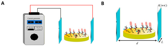
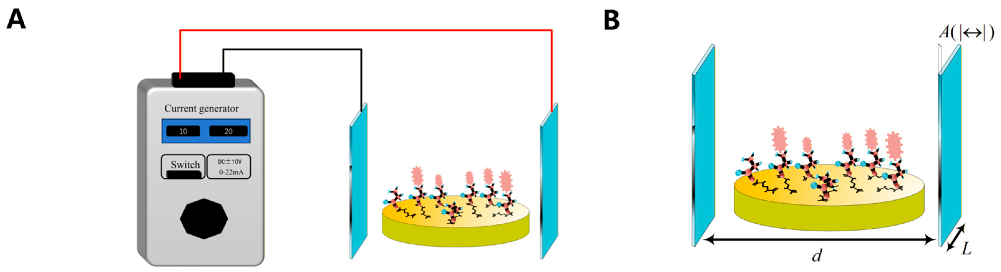
For oriented immobilization of Brucella antigens, a uniform electric field generator (EFG) system was constructed, as shown in Figure 1. The device consisted of two parallel indium tin oxide conductive glass plates (3 cm × 1 cm × 0.1 cm) mounted on an insulating substrate with an inter-plate gap of 1 cm and connected to an external voltage-current generator. By adjusting the generator output, a uniform electric field (EF) was established between the plates, and the functionalized electrode was positioned within the gap so that its surface was exposed to the controlled field.
Figure 1.
Configuration of the electric field generator (EFG) used for EF-assisted antigen immobilization. (A) Schematic illustration of the EFG setup. (B) Structural parameters and placement of the SPGE within the electric field region.
To describe the electrical operating conditions used for antigen immobilization, a nominal field-related parameter was estimated from the current-controlled device configuration within the same experiment setup. The electric field strength (E, µV/cm) generated in the medium was related to the output current (I, mA) as follows:
where is the resistivity of the medium (Ω·cm), L is the current path length (cm), A is the effective electrode area (cm2), and d is the distance between the parallel plates (cm). In this study, was fixed at 1 × 10⁻⁴ Ω·cm, while L, A, and d were set to 3 cm, 0.1 cm2, and 1 cm, respectively. Under these fixed conditions, E could be adjusted by varying I, thereby establishing a direct correspondence between the applied current and the derived field-related value.
It should be noted that, in this study, the electrical condition was controlled through the applied current of the device. Therefore, the field-related values calculated from Equation (1) were used as nominal operational parameters for internal comparison of immobilization conditions within the same configuration. Equation (1) provides a simplified operational estimate based on device geometry and medium properties.
Within the stable operating current range of the electrode, three current levels, 10, 20, and 30 mA, were selected. According to Equation (1), the corresponding field strengths were 30, 60, and 90 µV/cm, respectively. To optimize antigen immobilization, these three field levels were combined with three action durations (5, 10, and 15 s) in a full-factorial design.
2.5. Fabrication Immunosensor
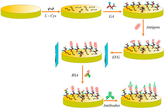
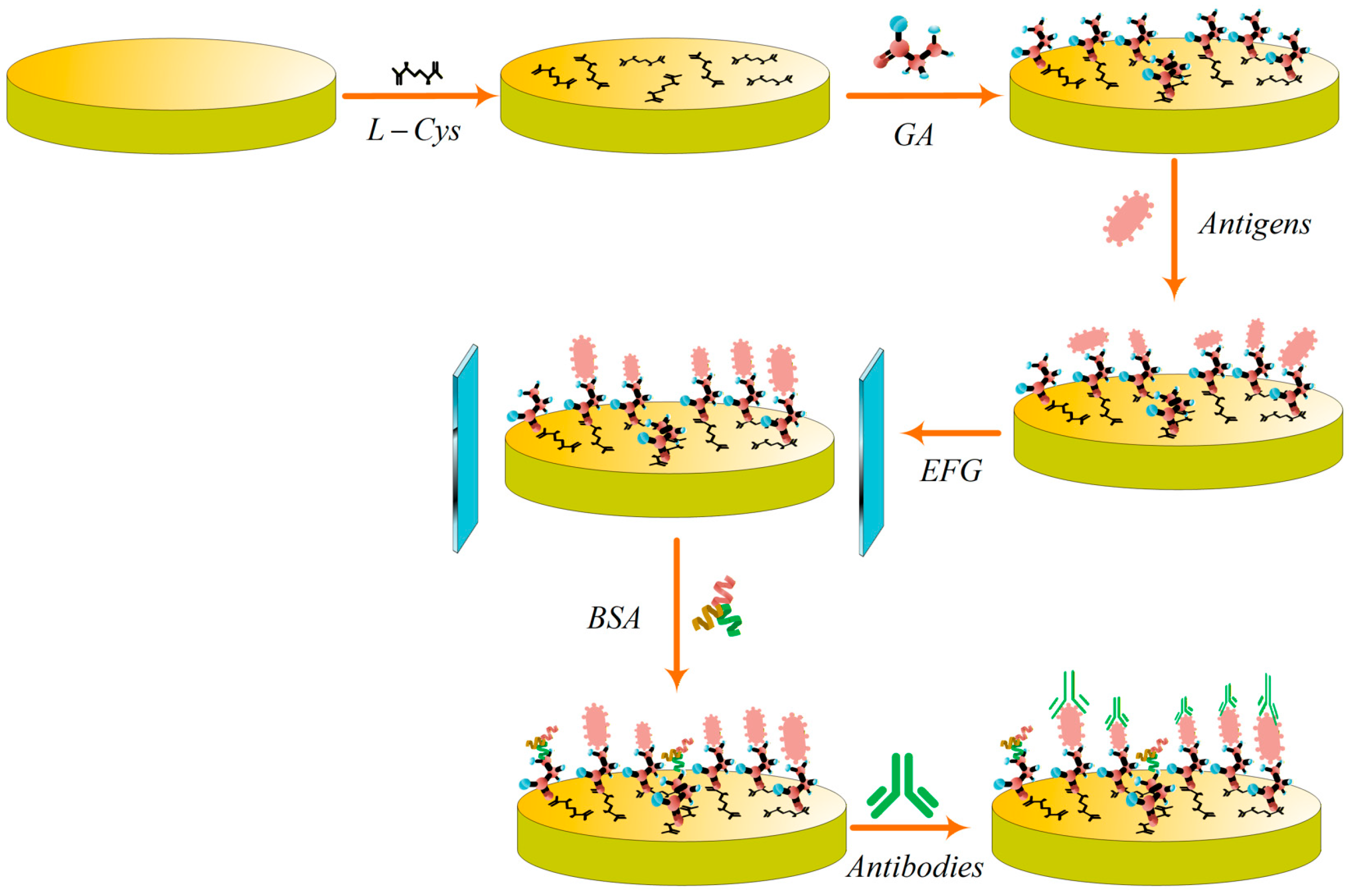
First, 10 μL of L-Cys solution was uniformly drop-cast onto the surface of a pretreated SPGE. The electrode was then incubated statically in a sealed environment at room temperature for 2 h, followed by rinsing twice with PBS and twice with ultrapure water. Next, 10 μL of GA solution was drop-cast onto the L-Cys modified SPGE and allowed to react in a humidity-controlled chamber at 37 °C for 1 h to cross-link GA with amino groups on the modified surface. After the reaction, the electrode was again rinsed twice with PBS and twice with ultrapure water. Subsequently, 10 μL of Brucella antigen solution was deposited onto the SPGE surface and subjected to EF-assisted oriented immobilization using an EFG. The electrode was then incubated in a humidity-controlled chamber at 37 °C for 2 h, and unbound antigens were removed by rinsing twice with ultrapure water. The incubation temperature of 37 °C was selected as a practical working condition based on common immunoreaction practice [24,25]. Finally, 1% BSA solution was drop-cast to block nonspecific binding sites, and the prepared immunosensor was stored at 4 °C until use.
For the detection assay, the immunosensor was incubated with Brucella antibody solution at different concentrations at 37 °C for 1 h. Electrochemical signals were then recorded by CV. As shown in Figure 2.
Figure 2.
Schematic illustration of the fabrication procedure and detection principle of the EF-assisted immunosensor for Brucella antibody detection.
3. Results and Discussion
3.1. Optimization of Process Parameters
3.1.1. Optimize Incubation Time
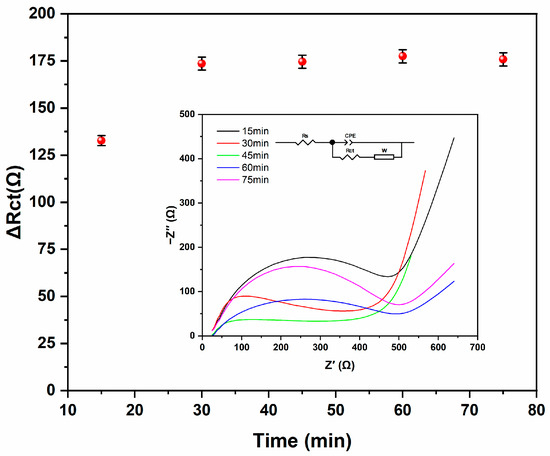
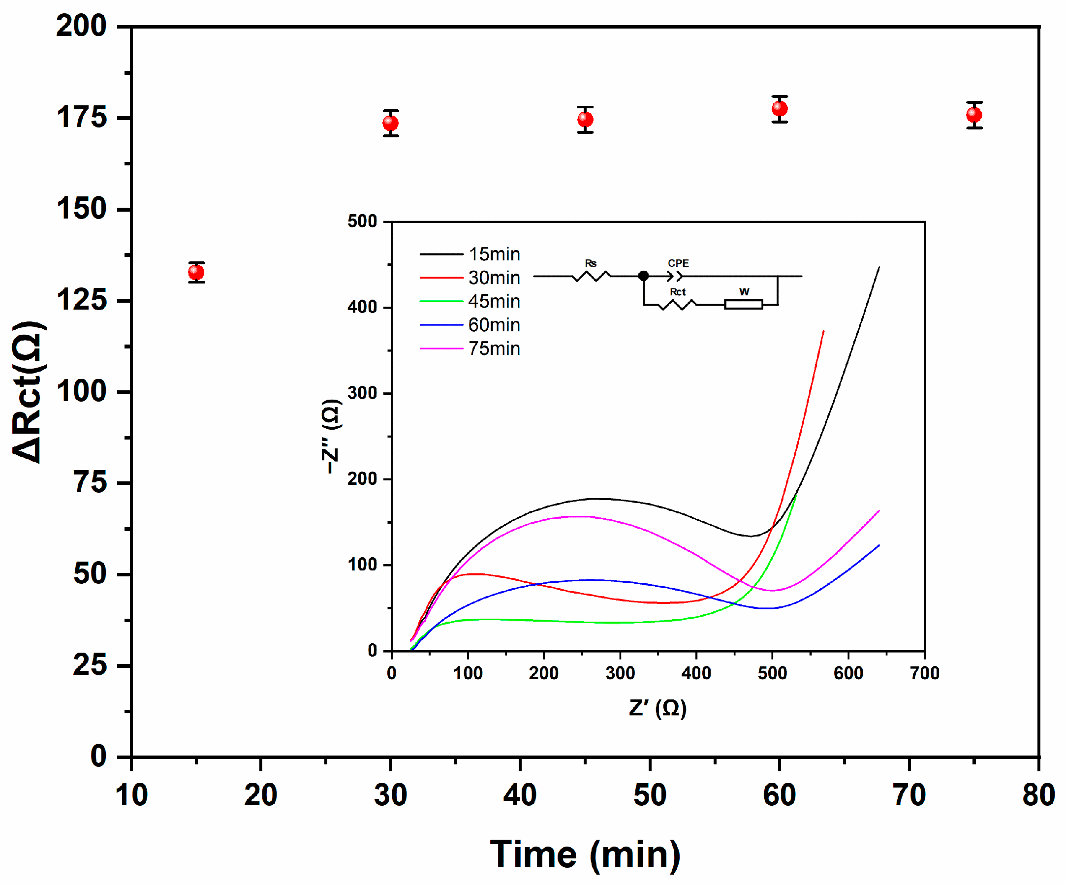
To optimize the incubation time for Brucella antibodies, a detection system was established using a Brucella antigen suspension (4 × 10⁹ CFU/mL) and a Brucella antibody solution (10 IU/mL). Five incubation durations (15, 30, 45, 60, and 75 min) were evaluated. After incubation under each condition, EIS was performed, and the resulting spectra were fitted using ZView to calculate the change in electron transfer resistance (ΔRct) [26,27]. The ΔRct value was used to assess the dependence of the EIS response on incubation time (n = 3). As shown in Figure 3, ΔRct increased from 132 Ω at 15 min to 173.5 Ω at 30 min (p < 0.05), indicating increased immune-complex formation on the electrode surface and a more complete antigen–antibody interaction. In contrast, further extending the incubation time to 45, 60, or 75 min did not yield a significant additional increase in ΔRct (p > 0.05). Therefore, considering both binding performance and experimental efficiency, 30 min was selected as the optimal antibody incubation time for subsequent experiments.
Figure 3.
Effect of antibody incubation time on the ΔRct response of the immunosensor.
3.1.2. Optimize EFG Parameters
To maximize the immobilization efficiency, the electric field (EF) parameters were systematically optimized under conditions of identical antigen loading and electrode configuration. This optimization was achieved by jointly varying the E and the action duration, with the resulting oxidation peak current evaluated by CV (Table 1, n = 3). A clear non-monotonic dependence on the EF parameters was observed, indicating the existence of an optimal operating window. Among the three current-controlled EF conditions examined, at 30 μV/cm, prolonging the action duration from 5 to 15 s led to a progressive decrease in current (from 42.6 ± 2.39 to 29.7 ± 2.05 μA). This suggests that low-field exposure is insufficient to improve interfacial assembly and may instead promote unfavorable surface rearrangement or the formation of an overly compact film, both of which impede charge transfer. In contrast, increasing the field to 60 μV/cm markedly enhanced the signal, with a maximum current achieved at 10 s (56.2 ± 1.35 μA), accompanied by the lowest relative standard deviation (RSD) of 2.4%. This result supports the notion that an appropriately tuned EF can facilitate the oriented immobilization of biomolecules, thereby improving interfacial electrochemical performance [19,28]. However, at 90 μV/cm, a prolonged treatment duration resulted in a gradual decrease in the current response (down to 34.2 ± 0.96 μA at 15 s). This phenomenon can be reasonably attributed to EF-induced conformational perturbation or partial loss of antigen integrity under excessive EF and cumulative exposure duration [29,30]. Consequently, a clear non-monotonic dependence on the electrical input condition was observed, indicating the existence of an optimal operating window. Under the intermediate current-controlled condition (60 μV/cm for 10 s), the sensor exhibited the highest current response and the lowest RSD, suggesting that an appropriately tuned electrically assisted immobilization process may improve interfacial organization and electrochemical performance in the present system.
Table 1.
Optimization of EF-assisted immobilization conditions as a function of electric field strength and action duration.
3.2. Characterization of Electrode
3.2.1. Characterization of Electrode Pretreatment
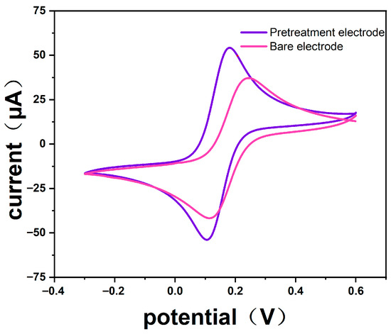
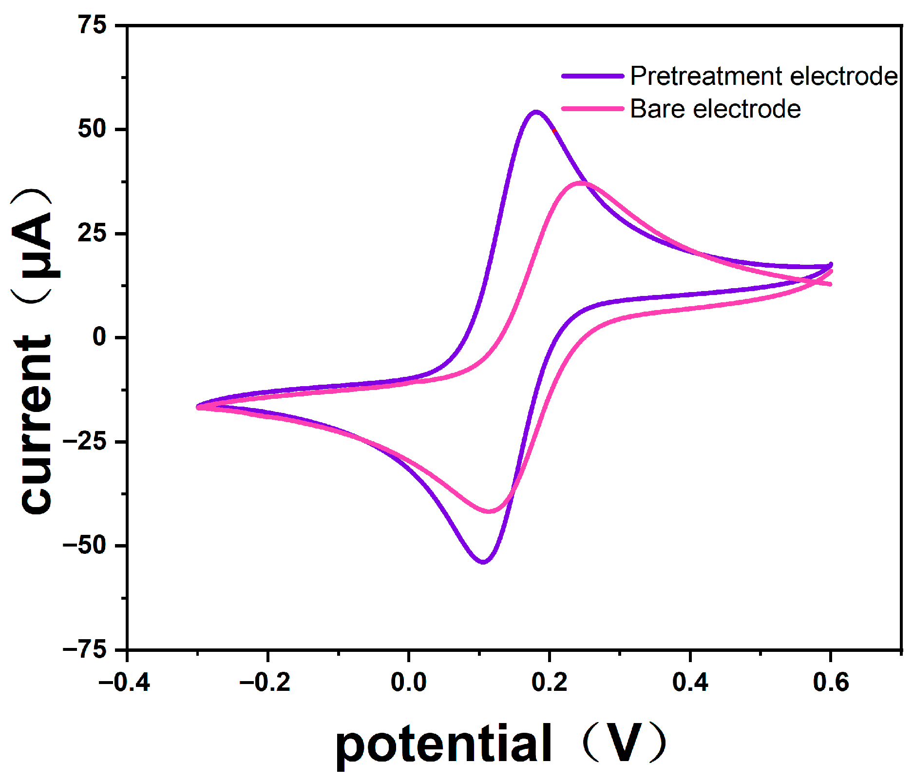
During the electrode pretreatment process, the oxidation peak current (Ipa), the absolute current ratio (|Ipa/Ipc|), and the peak-to-peak potential difference (ΔEp) were used as key metrics to characterize electrode performance. Accordingly, the pretreatment effect was assessed by comparing these parameters before and after pretreatment. The CV curve of the pretreated SPGE recorded in the electrolyte solution is shown in Figure 4. Compared to the untreated bare SPGE, the pretreated SPGE exhibited a significantly enhanced faradaic current in the potential range of 0.0–0.2 V. This increase indicates improved interfacial electron-transfer kinetics and a larger electrochemically active area, thereby facilitating charge transfer [31]. As shown in Table 2 (n = 3), the Ipa of the pretreated SPGE was markedly increased. This increase implies an improved electron-transfer capability, which is expected to enhance the signal responsiveness of the subsequent immunosensor. Meanwhile, the |Ipa/Ipc| decreased markedly toward unity, and the ΔEp was reduced. Together, these changes indicate enhanced electrochemical reversibility and more efficient electron transfer after pretreatment. Furthermore, the RSD value for the SPGE was substantially reduced, indicating that the pretreatment process effectively enhanced the electrochemical consistency and reproducibility of the electrode [32]. In summary, pretreatment produced a more stable and electrochemically favorable interface, providing a reliable foundation for subsequent antigen immobilization and antigen–antibody recognition [31].
Figure 4.
CV characterization of the SPGE before and after electrochemical pretreatment.
Table 2.
Electrochemical parameters of the SPGE before and after pretreatment.
3.2.2. Mechanism of the Immunosensor
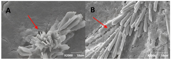
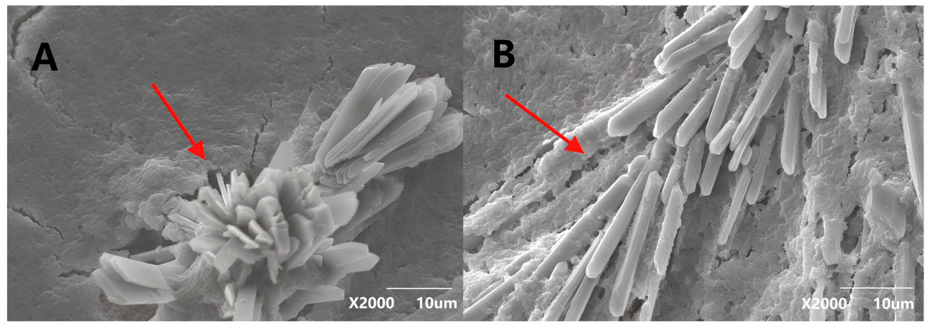
During the immunosensor preparation process, L-Cys was employed as a thiol-based molecular linker, its thiol (-SH) group forms strong Au-S bonds on the SPGE, thereby anchoring a cysteine-derived self-assembled monolayer (SAM). Concurrently, the terminal functional groups exposed by the L-Cys layer provide highly accessible reactive handles for subsequent coupling chemistry [33,34]. Subsequently, GA was introduced to impart aldehyde functionality for amine-directed coupling. Under aqueous conditions, GA could react with primary amines to form Schiff-base linkages and related crosslinked structures, thereby enabling covalent immobilization of biomolecules [35]. Oriented immobilization of Brucella antigens was characterized by SEM. As illustrated in Figure 5A, Brucella antigens immobilized on the electrode surface without EF treatment showed a disordered and random distribution. In sharp contrast, after applying a uniform external EF (Figure 5B), the antigens assembled uniformly and oriented regularly on the electrode surface. Such a more ordered interfacial arrangement is reasonably attributed to EF-induced bias in the orientation and local distribution of charged antigen molecules near the surface, rather than to a purely spontaneous random adsorption process. It should be noted that the EF mainly assists interfacial organization during antigen loading, whereas stable immobilization is provided by GA covalent coupling. Meanwhile, the free aldehyde groups on the GA cross-linking layer can covalently bind with amino groups on the antigen molecules, thus plausibly contributing to a more ordered and functionally favorable immobilization state. This interfacial organization is expected to improve epitope accessibility and recognition efficiency, which is consistent with the observed analytical enhancement. Upon antigen immobilization and subsequent antigen–antibody recognition, a progressively thicker and more insulating biolayer is formed at the electrode interface. This interfacial layer can increase steric hindrance and reduce the accessibility of the [Fe(CN)6]3−/4− redox probe to the electrode surface, thereby slowing interfacial charge transfer and decreasing the voltammetric current response. Accordingly, the decrease in the peak current detected by CV after incubation with Brucella antibodies was taken as the analytical response signal of the immunosensor. Because the present study did not include a complete set of component-removal controls, the mechanistic interpretation should be understood as being supported by comparative morphology and electrochemical performance.
Figure 5.
SEM characterization of Brucella antigen immobilization on the electrode surface. (A) Antigen layer formed without EF assistance. (B) Antigen layer formed under EF-assisted immobilization.
3.3. Calibration Curve of Immunosensor
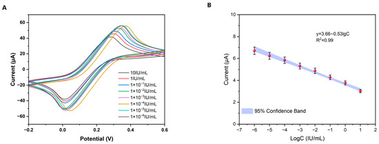
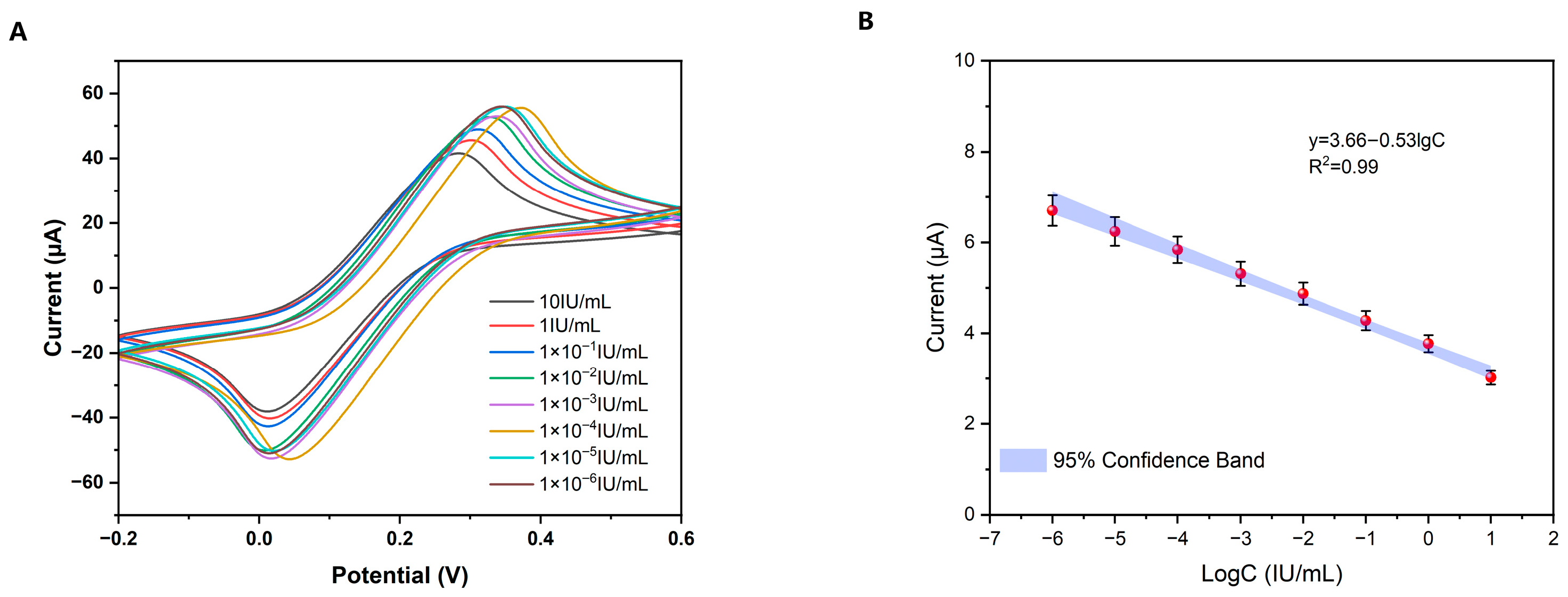
After the specific recognition of antigen and antibody, the protein film formed on the electrode surface will increase steric hindrance, thereby reducing the transfer of the [Fe(CN)6]3−/4− redox probe on the electrode surface and slowing down the charge transfer process. This leads to a decrease in the efficiency of interfacial electron transfer and a reduction in the peak current detected by voltammetry. To evaluate the analytical performance of the fabricated immunosensor, a quantitative calibration was established using the CV peak current change before and after immunoreaction as the analytical signal. Specifically, the peak current change was plotted against the logarithm of antibody concentration (lgC) and fitted by linear regression. As shown in Figure 6 (n = 3), the response signal was defined as ΔI = I0 − Ip, where I0 is the CV peak current of the freshly prepared immunosensor before antigen–antibody reaction, and Ip is the peak current recorded from the same sensor after incubation with Brucella antibody. Thus, the analytical signal is based on peak current attenuation caused by interfacial immunocomplex formation. The response exhibited a strong linear correlation over the concentration range from 10 to 1 × 10⁻⁶ IU/mL. The regression equation was y = 3.66 − 0.53lgC, with a correlation coefficient 0.99 (n = 3). The y is the ΔI response (μA), and the lgC is antibody concentration in IU/mL. Based on this calibration relationship, the limit of detection (LOD) was determined using the 3σ criterion according to the equation LOD = 3σ/S, where σ is the standard deviation of the blank response (0.02 μA) and S is the slope of the calibration curve (0.53). Accordingly, the LOD was calculated to be 2.04 × 10⁻⁷ IU/mL. Overall, these results demonstrate a broad linear dynamic range and high sensitivity for Brucella antibody quantification, supporting the analytical applicability of the proposed immunosensor.
Figure 6.
Analytical performance of the immunosensor for Brucella antibody detection. (A) CV responses recorded after incubation with different concentrations of Brucella antibodies. (B) Linear calibration plot of ΔI versus logarithmic Brucella antibody concentration (lgC). Concentrations from low to high: 1 × 10⁻⁵, 1 × 10⁻⁴, 1 × 10⁻³, 1 × 10⁻², 1 × 10⁻¹, 1, and 10 IU/mL.
3.4. Specificity, Stability, Reproducibility, and Recovery Rate Studies of Immunosensor
3.4.1. Specificity Studies of the Immunosensor
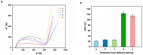
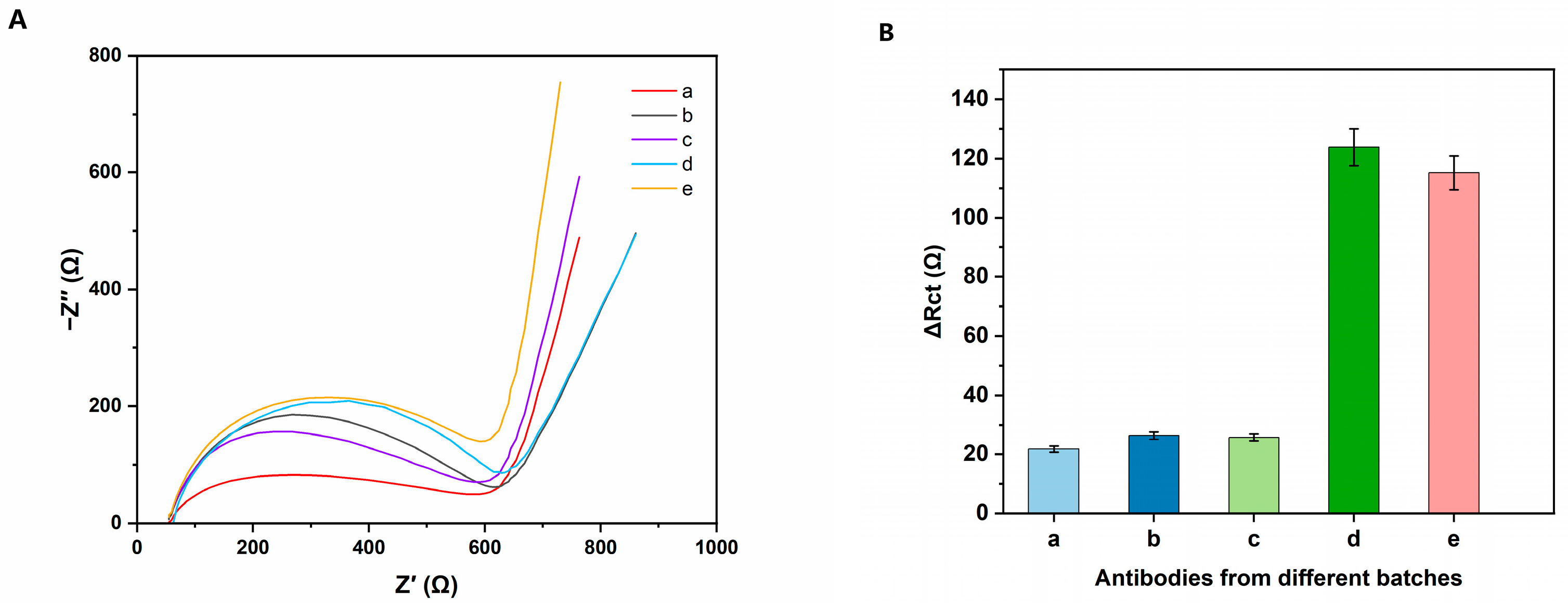
Cross-reactivity is an important indicator of immunosensor performance. To evaluate specificity, selectivity tests were performed using two common non-target antibodies (anti-DH5α pAbs and anti-Protein A pAbs) as potential interferents by EIS. Both interfering antibodies were tested at 10 IU/mL, identical to the concentration used for the target Brucella antibodies. As shown in Figure 7 (n = 3), the interfering antibody groups (b and c) produced responses comparable to the background control (a), and the corresponding ΔRct (Ω) values showed no significant differences (p > 0.05). By contrast, the target antibody group (d) exhibited a significantly higher signal than the background (a) (p < 0.05). Moreover, the mixed sample containing the target antibody plus interferents (e) remained significantly different from the background (a) (p < 0.05), indicating that interference did not mask target recognition. Together, these results confirm that the immunosensor selectively detects Brucella antibodies and maintains high specificity in the presence of non-target antibodies, supporting its applicability to complex matrices.
Figure 7.
Specificity evaluation of the immunosensor for Brucella antibody detection. (A) Nyquist plots obtained after incubation with different antibody samples. (B) Corresponding impedance response expressed as ΔRct. (a) Background signal; (b) anti-Protein A pAbs; (c) anti-DH5α pAbs; (d) Brucella antibodies; and (e) mixture of Brucella antibodies, anti-DH5α pAbs, and anti-Protein A pAbs. Error bars represent the standard deviation of three independent measurements (n = 3).
3.4.2. Stability Studies of the Immunosensor
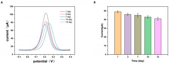
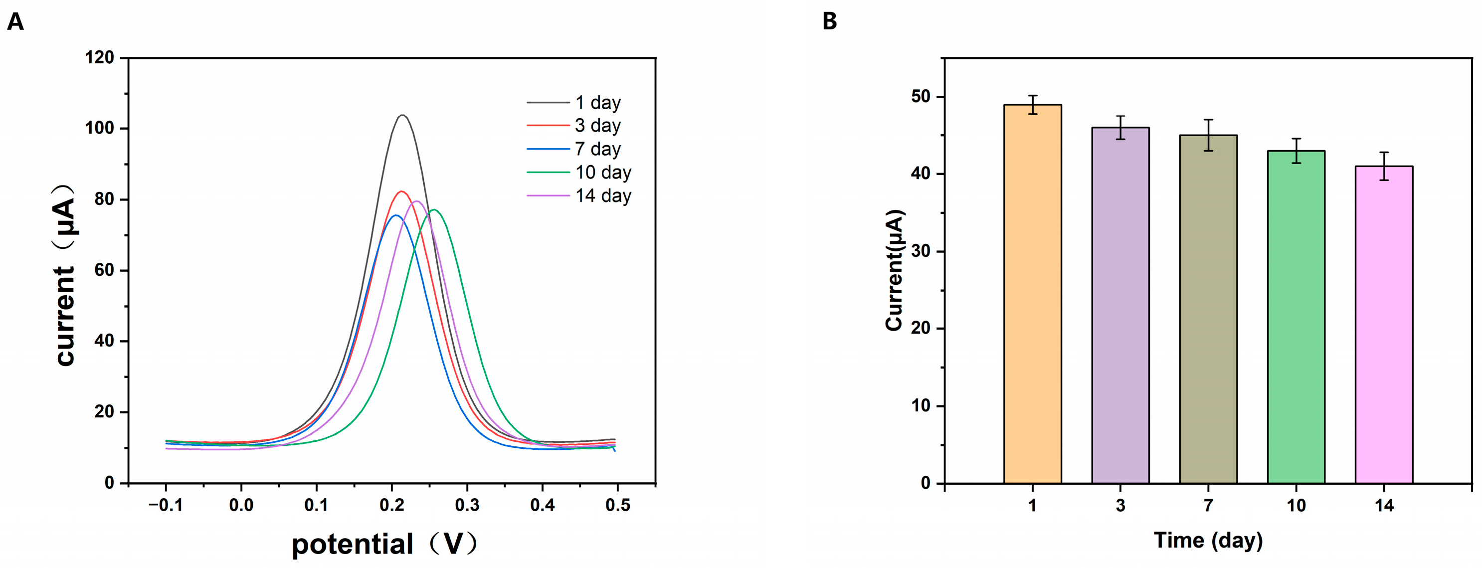
The storage stability of the immunosensor was assessed by SWV after storage at 4 °C for 1, 3, 7, 10, and 14 days, with measurements performed at each time point using a 10 IU/mL Brucella antibody solution. The signal variation was defined as ΔI (I0 − Ip), where I0 denotes the peak current obtained on day 1 and Ip represents the peak current recorded after the specified storage period. As shown in Figure 8 (n = 3), ΔI decreased monotonically with increasing storage time, indicating progressive loss of immunorecognition activity. To determine the practical shelf-life, an acceptance threshold based on initial-signal decay was applied: the lower limit was set to the day 1 mean minus two standard deviations (the day1 mean − 2 × RSD), corresponding to 44 μA. The mean ΔI values on days 3, 7, 10, and 14 were 46, 45, 43, and 41 μA, respectively. Because ΔI remained above the threshold through day 7 but dropped below it thereafter, the immunosensor was considered stable for approximately 7 days under the tested storage conditions. This result provides a practical foundation for recommending the storage period of the proposed biosensor.
Figure 8.
Storage stability of the immunosensor. (A) CV responses of the immunosensor recorded after storage for 1, 3, 7, 10, and 14 days. (B) Corresponding ΔI at different storage times. Error bars represent the standard deviation of three independent measurements (n = 3).
3.4.3. Reproducibility of the Immunosensor
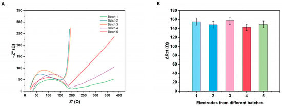
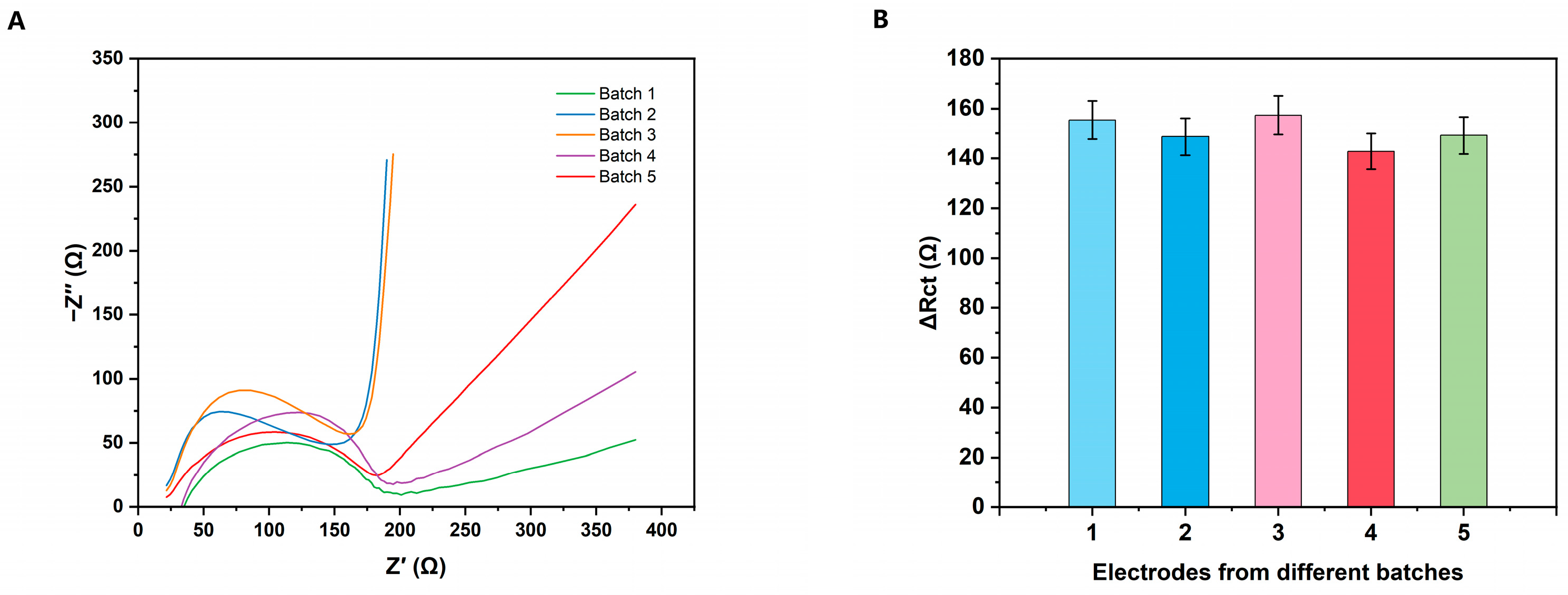
Reproducibility is a critical parameter for assessing immunosensor performance, as high reproducibility helps minimize measurement errors. In this study, five independently fabricated batches of immunosensors were evaluated by EIS using a Brucella antibody solution at 10 IU/mL. As shown in Figure 9, the ΔRct values ranged from 142.80 to 157.34 Ω, with a RSD of 4.12% (n = 3). These results indicate that the proposed fabrication method provides good reproducibility.
Figure 9.
Fabrication reproducibility of independently prepared immunosensors. (A) Nyquist plots recorded for five immunosensors fabricated in different batches. (B) Corresponding impedance responses expressed as ΔRct for electrodes from different batches. Error bars represent the standard deviation of three independent measurements (n = 3).
3.4.4. Recovery Rate of the Immunosensor
The practical applicability of the immunosensor was evaluated by detecting Brucella antibodies in milk samples using CV. Milk samples were spiked with antibodies at 10−6, 10−3, and 10 IU/mL, and then analyzed using the proposed method. As shown in Table 3 (n = 3), the recoveries ranged from 93.15% to 99.14% and RSD values were below 2%. The immunosensor maintained acceptable analytical performance in spiked milk samples, providing preliminary evidence for its applicability in simulated complex matrices. However, if extended to naturally infected or clinical samples, further validation in serum and naturally infected specimens would still be required.
Table 3.
Recovery results for Brucella antibody detection in spiked milk samples.
4. Conclusions
In this study, we developed a sensitive immunosensor for the quantitative detection of Brucella antibodies by employing EF-assisted antigen immobilization. Electrochemical pretreatment of the electrode surface enhanced electrochemical activity and improved interfacial uniformity, thereby establishing a reliable foundation for constructing the immunosensing interface. During antigen immobilization, a custom designed EFG was used to apply a uniform EF, which promoted a more homogeneous and oriented distribution of antigen molecules on the electrode. In addition, the proposed immunosensor demonstrated good repeatability, storage stability, and specificity, as well as satisfactory recoveries in spiked samples. Collectively, these results support the feasibility of the platform for sensitive Brucella antibody detection and indicate that EF-assisted oriented immobilization may be extended to other immunosensors. Accordingly, the current results support the analytical feasibility of the proposed immunosensor under controlled buffer conditions, whereas further validation in spiked serum and real samples is still required before broader claims on practical diagnostic applicability can be made.
Author Contributions
Conceptualization, W.Y. and J.M.; Methodology, J.M. and W.Y.; Software, J.M. and G.W.; Validation, J.M. and G.W.; Formal analysis, W.Y. and J.M.; Investigation, J.M. and G.W.; Resources, W.Y.; Data curation, J.M.; Writing—original draft preparation, W.Y. and J.M.; Writing—review and editing, W.Y. and G.W.; Visualization, J.M. and G.W.; Supervision, W.Y.; Project administration, W.Y.; Funding acquisition, W.Y. All authors have read and agreed to the published version of the manuscript.
Funding
The work was supported by Fundamental Research Program of Shanxi Province, China (Grant No. 20210302123389).
Institutional Review Board Statement
Not applicable.
Informed Consent Statement
Not applicable.
Data Availability Statement
All data are included within the manuscript.
Conflicts of Interest
The authors declare no conflicts of interest.
References
- Qureshi, K.A.; Parvez, A.; Fahmy, N.A.; Abdel Hady, B.H.; Kumar, S.; Ganguly, A.; Atiya, A.; Elhassan, G.O.; Alfadly, S.O.; Parkkila, S.; et al. Brucellosis: Epidemiology, Pathogenesis, Diagnosis and Treatment—A Comprehensive Review. Ann. Med. 2023, 55, 2295398. [Google Scholar] [CrossRef] [Scilit]
- Ashmi, M.; Kumar, B.; Agrawal, R.K.; Prakash, C.; Abhishek; Singh, K.P. Development of BruAb2_0168 Based Isothermal Polymerase Spiral Reaction Assay for Specific Detection of Brucella abortus in Clinical Samples. Mol. Cell. Probes 2021, 59, 101761. [Google Scholar] [CrossRef] [Scilit] [PubMed]
- Novak, A.; Melli, L.J.; Rey Serantes, D.A.; Caillava, A.J.; Comerci, D.J.; Ugalde, J.E.; Ciocchini, A.E. Development of a Novel Glycoprotein-based Immunochromatographic Test for the Rapid Serodiagnosis of Bovine Brucellosis. J. Appl. Microbiol. 2022, 132, 4277–4288. [Google Scholar] [CrossRef] [Scilit] [PubMed]
- Kiambi, S.G.; Fèvre, E.M.; Omolo, J.; Oundo, J.; De Glanville, W.A. Risk Factors for Acute Human Brucellosis in Ijara, North-Eastern Kenya. PLoS Negl. Trop. Dis. 2020, 14, e0008108. [Google Scholar] [CrossRef] [Scilit] [PubMed]
- Lita, E.P.; Ochi, E.B.; Misinzo, G.; Van Heerden, H.; Katani, R.; Godfroid, J.; Mathew, C. Seroprevalence and Risk Factors of Brucellosis in Pastoralists and Their Livestock in Central Equatoria State, South Sudan. PLoS Negl. Trop. Dis. 2024, 18, e0012144. [Google Scholar] [CrossRef] [Scilit]
- Liu, H.; Xu, L.; Xiu, Y.; Ta, N.; Xu, Q.; Fan, Y.; Li, K.; Zhao, H.; Piao, D.; Ren, F.; et al. A CRISPR/Cas13a-Assisted Precise and Portable Test for Brucella Nucleic Acid Detection. Front. Cell. Infect. Microbiol. 2025, 15, 1545953. [Google Scholar] [CrossRef] [Scilit]
- Liang, G.; He, Z.; Zhen, J.; Tian, H.; Ai, L.; Pan, L.; Gong, W. Development of the Screen-Printed Electrodes: A Mini Review on the Application for Pesticide Detection. Environ. Technol. Innov. 2022, 28, 102922. [Google Scholar] [CrossRef] [Scilit]
- Zhang, Y.; Ma, M.; Aisa, H.A.; Chen, L. Oriented Immobilization of IgG for Immunosensor Development. Chemosensors 2025, 13, 50. [Google Scholar] [CrossRef] [Scilit]
- Fritz, P.A.; Bera, B.; Van Den Berg, J.; Visser, I.; Kleijn, J.M.; Boom, R.M.; Schroën, C.G.P.H. Electrode Surface Potential-Driven Protein Adsorption and Desorption through Modulation of Electrostatic, van Der Waals, and Hydration Interactions. Langmuir 2021, 37, 6549–6555. [Google Scholar] [CrossRef] [Scilit]
- Vacek, J.; Zatloukalová, M.; Dorčák, V.; Cifra, M.; Futera, Z.; Ostatná, V. Electrochemistry in Sensing of Molecular Interactions of Proteins and Their Behavior in an Electric Field. Microchim. Acta 2023, 190, 442. [Google Scholar] [CrossRef] [Scilit]
- Han, S.; Han, J.W.; Seong, S.; Son, Y.J.; Kaizu, R.; Latag, G.V.; Hayashi, T.; Noh, J. Formation and Structure of Highly Ordered Self-Assembled Monolayers on Au(111) via Vapor Deposition of Dioctyl Diselenides. Int. J. Mol. Sci. 2025, 26, 9192. [Google Scholar] [CrossRef] [Scilit]
- Son, Y.J.; Han, J.W.; Kang, H.; Seong, S.; Han, S.; Maeda, S.; Chikami, S.; Hayashi, T.; Hara, M.; Noh, J. Formation and Thermal Stability of Ordered Self-Assembled Monolayers by the Adsorption of Amide-Containing Alkanethiols on Au(111). Int. J. Mol. Sci. 2023, 24, 3241. [Google Scholar] [CrossRef] [Scilit]
- Chen, H.; Liu, H.; Cui, C.; Zhang, X.; Yang, W.; Zuo, Y. Highly Sensitive Detection of Brucella in Milk by Cysteamine Functionalized Nanogold/4-Mercaptobenzoic Acid Electrochemical Biosensor. J. Food Meas. Charact. 2022, 16, 3501–3511. [Google Scholar] [CrossRef] [Scilit]
- Sun, T.; Yi, X.; Liu, L.; Zhao, F. Colorimetric Immunoassays with Boronic Acid-Decorated, Peroxidase-like Metal-Organic Frameworks as the Carriers of Antibodies and Enzymes. Molecules 2024, 29, 3000. [Google Scholar] [CrossRef] [Scilit] [PubMed]
- Zhu, L.; Chang, Y.; Li, Y.; Qiao, M.; Liu, L. Biosensors Based on the Binding Events of Nitrilotriacetic Acid–Metal Complexes. Biosensors 2023, 13, 507. [Google Scholar] [CrossRef] [Scilit] [PubMed]
- Chang, Y.; Ma, X.; Sun, T.; Liu, L.; Hao, Y. Electrochemical Detection of Kinase by Converting Homogeneous Analysis into Heterogeneous Assay through Avidin-Biotin Interaction. Talanta 2021, 234, 122649. [Google Scholar] [CrossRef] [Scilit]
- Chang, Y.; Wang, Y.; Zhang, J.; Xing, Y.; Li, G.; Deng, D.; Liu, L. Overview on the Design of Magnetically Assisted Electrochemical Biosensors. Biosensors 2022, 12, 954. [Google Scholar] [CrossRef] [Scilit]
- Lu, Z.-Y.; Chan, Y.-H. The Importance of Antibody Orientation for Enhancing Sensitivity and Selectivity in Lateral Flow Immunoassays. Sens. Diagn. 2024, 3, 1613–1634. [Google Scholar] [CrossRef] [Scilit]
- Kim, H.J.; Park, D.; Park, Y.; Kim, D.-H.; Kim, J. Electric-Field-Mediated In-Sensor Alignment of Antibody’s Orientation to Enhance the Antibody–Antigen Binding for Ultrahigh Sensitivity Sensors. Nano Lett. 2022, 22, 6537–6544. [Google Scholar] [CrossRef] [Scilit]
- Yoon, T.; Park, W.; Kim, Y.; Na, S. Electric Field-Mediated Regulation of Enzyme Orientation for Efficient Electron Transfer at the Bioelectrode Surface: A Molecular Dynamics Study. Appl. Surf. Sci. 2023, 608, 155124. [Google Scholar] [CrossRef] [Scilit]
- Long, Z.; Meng, J.; Weddle, L.R.; Videla, P.E.; Menzel, J.P.; Cabral, D.G.A.; Liu, J.; Qiu, T.; Palasz, J.M.; Bhattacharyya, D.; et al. The Impact of Electric Fields on Processes at Electrode Interfaces. Chem. Rev. 2025, 125, 1604–1628. [Google Scholar] [CrossRef] [Scilit] [PubMed]
- García García, D.; Espinosa Garcia, L.; Madrigal-Santillán, E.O.; Morales-González, J.A.; Madrigal-Bujaidar, E.; Álvarez-González, I.; Damian-Matsumura, P.; Jiménez-Salazar, J.E.; Batina, N.; Garcia-Melo, L.F. Impact of Cleaning Procedures on Screen-Printed Gold Electrodes Performance for Mutation Detection. J. Appl. Electrochem. 2025, 55, 1937–1946. [Google Scholar] [CrossRef] [Scilit]
- Jiménez-Pérez, R.; González-Sánchez, M.I.; Gomis-Berenguer, A.; Iniesta, J.; Valero, E. Enhanced Surface Properties and Electrochemical Performance of Carbon-Based Screen-Printed Electrodes via Hydrogen Peroxide Activation. Electrochim. Acta 2025, 536, 146721. [Google Scholar] [CrossRef] [Scilit]
- Johnstone, R.W.; Andrew, S.M.; Hogarth, M.P.; Pietersz, G.A.; McKenzie, I.F.C. The Effect of Temperature on the Binding Kinetics and Equilibrium Constants of Monoclonal Antibodies to Cell Surface Antigens. Mol. Immunol. 1990, 27, 327–333. [Google Scholar] [CrossRef] [Scilit] [PubMed]
- Wang, Z.; Wang, M.; Yu, Y.; Lu, Y.; Xia, Q.; Wei, P. Integrating Thermodynamics and Molecular Kinetics into Western Blotting Teaching. Front. Mol. Biosci. 2025, 12, 1640740. [Google Scholar] [CrossRef] [Scilit]
- Wang, N.; Pan, G.; Guan, S.; Rong, S.; Wang, D.; Gao, Z.; Tian, P.; Li, Q. A Broad-Range Disposable Electrochemical Biosensor Based on Screen-Printed Carbon Electrodes for Detection of Human Noroviruses. Front. Bioeng. Biotechnol. 2022, 10, 845660. [Google Scholar] [CrossRef] [Scilit]
- Zhu, Q.; Sun, X.; Yang, D.; Ma, J.; Kang, X.; Zheng, L.; Zhang, J.; Wu, Z.; Han, B. Carbon Dioxide Electroreduction to C2 Products over Copper-Cuprous Oxide Derived from Electrosynthesized Copper Complex. Nat. Commun. 2019, 10, 3851. [Google Scholar] [CrossRef] [Scilit]
- Emaminejad, S.; Javanmard, M.; Gupta, C.; Chang, S.; Davis, R.W.; Howe, R.T. Tunable Control of Antibody Immobilization Using Electric Field. Proc. Natl. Acad. Sci. USA 2015, 112, 1995–1999. [Google Scholar] [CrossRef] [Scilit]
- Jiang, Z.; You, L.; Dou, W.; Sun, T.; Xu, P. Effects of an Electric Field on the Conformational Transition of the Protein: A Molecular Dynamics Simulation Study. Polymers 2019, 11, 282. [Google Scholar] [CrossRef] [Scilit]
- Wang, X.; Li, Y.; He, X.; Chen, S.; Zhang, J.Z.H. Effect of Strong Electric Field on the Conformational Integrity of Insulin. J. Phys. Chem. A 2014, 118, 8942–8952. [Google Scholar] [CrossRef] [Scilit]
- Khor, C.K.; Denker, S.; Ignaszak, A. Surface Analysis of Metrohm BT220 Screen-Printed Electrodes through Electrochemical Techniques: Importance of Pretreatment. Front. Chem. 2025, 13, 1602365. [Google Scholar] [CrossRef] [Scilit]
- Whittingham, M.J.; Hurst, N.J.; Crapnell, R.D.; Garcia-Miranda Ferrari, A.; Blanco, E.; Davies, T.J.; Banks, C.E. Electrochemical Improvements Can Be Realized via Shortening the Length of Screen-Printed Electrochemical Platforms. Anal. Chem. 2021, 93, 16481–16488. [Google Scholar] [CrossRef] [Scilit]
- Love, J.C.; Estroff, L.A.; Kriebel, J.K.; Nuzzo, R.G.; Whitesides, G.M. Self-Assembled Monolayers of Thiolates on Metals as a Form of Nanotechnology. Chem. Rev. 2005, 105, 1103–1170. [Google Scholar] [CrossRef] [Scilit]
- Xu, Q.-M.; Wan, L.-J.; Wang, C.; Bai, C.-L.; Wang, Z.-Y.; Nozawa, T. New Structure of l -Cysteine Self-Assembled Monolayer on Au(111): Studies by In Situ Scanning Tunneling Microscopy. Langmuir 2001, 17, 6203–6206. [Google Scholar] [CrossRef] [Scilit]
- Migneault, I.; Dartiguenave, C.; Bertrand, M.J.; Waldron, K.C. Glutaraldehyde: Behavior in Aqueous Solution, Reaction with Proteins, and Application to Enzyme Crosslinking. BioTechniques 2004, 37, 790–802. [Google Scholar] [CrossRef] [Scilit]

Disclaimer/Publisher’s Note: The statements, opinions and data contained in all publications are solely those of the individual author(s) and contributor(s) and not of MDPI and/or the editor(s). MDPI and/or the editor(s) disclaim responsibility for any injury to people or property resulting from any ideas, methods, instructions or products referred to in the content. |
© 2026 by the authors. Licensee MDPI, Basel, Switzerland. This article is an open access article distributed under the terms and conditions of the Creative Commons Attribution (CC BY) license.