


Abstract
Human milk proteins exhibit extensive molecular heterogeneity arising from genetic variation, transcript diversity, post-translational modifications, and proteolytic processing. This review integrates evidence on major human milk caseins and selected whey proteins within a genome–transcript–proteoform–peptidoform framework, incorporating comparative mammalian data where relevant. Particular emphasis is placed on distinguishing predicted molecular consequences, relationships supported by indirect evidence, and experimentally demonstrated molecular entities and transitions. Genomic variation and alternative RNA processing provide potential sources of primary-sequence diversity, whereas phosphorylation, glycosylation, and proteolysis further expand protein and peptide heterogeneity. Human β-casein provides a particularly informative example, with experimentally characterized phosphorylation- and glycosylation-defined proteoforms and corresponding modification-resolved peptidoforms. However, evidence remains uneven across molecular levels, and direct tracing from maternal genotype to expressed transcripts, secreted proteoforms, and downstream peptidoforms within the same individual remains limited. By identifying experimentally established relationships and unresolved molecular connections, this review provides an evidence-based framework for interpreting human milk lactoprotein heterogeneity and guiding future integrated proteogenomic investigations.
1. Introduction
Human milk is a dynamic biological fluid that provides nutrients and bioactive components involved in infant growth, mineral homeostasis, immune protection, and gastrointestinal development [1,2]. Its protein fraction includes three major caseins (CNs), namely αS1-casein (αS1-CN), β-casein (β-CN), and κ-casein (κ-CN), alongside whey proteins such as α-lactalbumin (α-LA), lactoferrin (LF), secretory immunoglobulin A (sIgA), and lysozyme (LZ) [1]. Although their abundance changes during lactation, human milk lactoprotein heterogeneity extends beyond quantitative variation and can be examined across four interconnected molecular levels: (a) genomic variation, (b) transcript diversity, (c) protein/proteoform heterogeneity, and (d) peptide/peptidoform diversity [3,4,5].
(a) At the genomic level, coding variants may alter the primary amino acid sequences of lactoproteins, whereas regulatory variants may influence the expression of their encoding genes. The functional human casein repertoire is encoded by CSN1S1, CSN2, and CSN3, whereas the αS2-casein-related loci are annotated as pseudogenes, unlike their functional counterparts in dairy ruminants [6,7,8]. Comparative genomic investigations reveal both conserved and species-specific characteristics of the casein gene cluster, emphasizing the importance of distinguishing shared molecular mechanisms from lineage-specific features [6,9].
(b) Transcript-level heterogeneity reflects developmental regulation, differences among mammary epithelial cell populations, and alternative RNA processing [10,11,12]. Maternal genetic variation can also influence gene expression in the lactating mammary environment [13]. However, transcript identification or differential expression alone does not establish the translation of corresponding protein products or their secretion into milk [14,15].
(c) At the protein level, genetic variation, alternative RNA processing, and post-translational modifications (PTMs) represent potential sources of proteoform diversity [5]. Phosphorylation and glycosylation contribute substantially to the molecular heterogeneity of human milk CNs and whey proteins, with experimentally characterized differences among protein forms [16,17,18]. The effects of phosphorylation on calcium binding and micellar association have been experimentally demonstrated for human β-CN [19,20]. However, the functional consequences of other modifications, including their potential effects on proteolytic susceptibility, require evaluation at the level of individual proteoforms.
(d) At the peptide level, endogenous milk proteases generate peptides before ingestion, whereas gastrointestinal digestion produces additional peptide populations [21,22]. Differences in peptide sequences, cleavage boundaries, and retained modifications generate distinct peptidoforms [23]. Integrated proteomic and peptidomic investigations have revealed substantial molecular diversity and complex relationships between milk proteins and their derived peptides [4,24]. Nevertheless, identification of individual peptide species does not, by itself, demonstrate biological activity or physiological benefits in infants [25].
Previous reviews have characterized the composition and biological functions of the human milk proteome [1] and the generation and potential activities of milk-derived bioactive peptides [25]. Although recent studies have integrated proteomic and peptidomic evidence [24], the molecular relationships connecting successive levels of lactoprotein heterogeneity remain incompletely established. The present review addresses this gap by systematically distinguishing predicted molecular consequences, relationships supported by indirect evidence, and experimentally demonstrated molecular entities or transitions. Selected mammalian comparisons provide additional mechanistic and evolutionary context. By identifying established molecular relationships and unresolved gaps in cross-omic evidence, this review provides a framework for interpreting human milk lactoprotein heterogeneity and guiding future integrated proteogenomic investigations. The conceptual framework is summarized in Figure 1.
Figure 1.
Conceptual framework for human milk lactoprotein heterogeneity across four interconnected molecular levels: (a) DNA/genomic variation; (b) RNA/transcript diversity and gene expression; (c) protein/proteoform heterogeneity arising from primary-sequence differences and post-translational modifications; and (d) peptide/peptidoform diversity generated through endogenous and gastrointestinal proteolysis. β-Casein is presented schematically as an illustrative parent protein. Gray arrows indicate the conceptual progression between molecular levels, while the blue arrow in panel (b) represents transcription and RNA processing. Colors distinguish genomic regions, transcript elements, amino acid segments, and peptide products without representing a fixed biochemical classification. In panel (c), colored circles schematically represent amino acid residues, yellow circles labeled “P” indicate phosphorylation, and branched structures illustrate glycosylation. In panel (d), colored peptide fragments represent proteolytic products and their potential post-translational modifications. The chromosome ideogram in panel (a) indicates the approximate location of the casein gene cluster on chromosome 4q13.3.
2. Materials and Methods
2.1. Review Design and Literature Search
This study was designed as an integrative review examining human milk lactoprotein heterogeneity across four interconnected molecular levels: (a) genome, (b) transcript, (c) proteoform, and (d) peptidoform. The primary objective was to critically evaluate the available evidence and determine the extent to which relationships between successive molecular levels have been experimentally established.
The review focused on the three functional human CNs encoded by CSN1S1, CSN2, and CSN3, together with α-LA, LF, LZ, and sIgA. Additional proteins, particularly osteopontin (OPN) and the polymeric immunoglobulin receptor (pIgR)-derived secretory component, were included selectively when relevant to protein assembly, PTMs, or peptide generation.
Literature searches were conducted iteratively by the author team using MEDLINE (PubMed), Web of Science Core Collection, and Scopus. The initial literature synthesis covered publications available through 29 August 2026, with additional relevant sources considered during manuscript revision. No restrictions were imposed on publication date or language. Search terms combined human milk or breast milk with individual lactoprotein names and terms related to genetic variation, alleles, haplotypes, alternative splicing, transcriptomics, proteomics, PTMs, proteoforms, proteolysis, peptidomics, and bioactive peptides.
Original experimental studies providing direct genomic, transcriptomic, proteomic, or peptidomic evidence were prioritized. Historical biochemical and protein-sequencing investigations were retained when they provided foundational molecular information, whereas review articles were used primarily for contextualization and identification of relevant primary sources. Studies involving non-human mammals were included selectively when they contributed mechanistic, structural, or evolutionary information relevant to human lactoproteins. Publications lacking sufficient relevance to the molecular questions investigated were not prioritized for detailed analysis.
Literature identification and selection followed an iterative integrative approach rather than a predefined systematic-review screening protocol. Studies were evaluated according to their relevance, methodological characteristics, and contribution to the molecular evidence considered. Because the literature search was conducted iteratively and no consolidated search log or prospective screening register was maintained, the numbers of records initially identified, screened, and excluded cannot be reliably reconstructed retrospectively. The final reference list therefore reflects the sources incorporated into this integrative review rather than the outcome of a predefined systematic screening process.
2.2. Genomic and Population-Genomic Resources
Genomic organization, chromosomal localization, transcriptional orientation, exon–intron structure, and reference transcripts were evaluated using the human GRCh38.p14 reference assembly (Genome Reference Consortium human build 38, patch 14) (GCF_000001405.40), primarily through the National Center for Biotechnology Information (NCBI) Gene and Reference Sequence (RefSeq) databases. Ensembl/GENCODE annotations were consulted as complementary resources.
Matched Annotation from NCBI and EMBL-EBI (MANE) Select transcripts were preferred as representative reference sequences [26]. For the characterization of selected coding variants, the MANE Select RefSeq transcripts NM_001890.2 (CSN1S1), NM_001891.4 (CSN2), and NM_001394997.1 (CSN3) were used. For CSN3, NM_001394997.1 corresponds to ENST00000304954.4 (MANE Select), whereas NM_005212.3 is a separately validated RefSeq transcript encoding the same κ-CN precursor. Alternative transcripts were considered when relevant to RNA-processing diversity or historical variant annotations. Versioned accession numbers and Human Genome Variation Society (HGVS) nomenclature were used to ensure consistent variant descriptions [27].
Sequence variation was investigated using NCBI Gene, RefSeq, the Single Nucleotide Polymorphism Database (dbSNP), ClinVar, Ensembl, and the Genome Aggregation Database (gnomAD). Variants were selected as illustrative examples of different molecular mechanisms rather than as an exhaustive catalog. Selection emphasized coding substitutions affecting amino-acid physicochemical properties, variants potentially modifying disulfide interactions or PTM sites, splice-associated variants, and regulatory alterations. Historically characterized molecular variants were also considered when relevant. Computationally predicted consequences were distinguished from experimentally demonstrated effects on transcript structure or protein expression [7,8].
Population-genomic data were retrieved from gnomAD (GRCh38) at different stages of the study, with additional data obtained from gnomAD v4.1.2 on 21 October 2026. The current release was v4.1.2; however, variant-frequency data and filtering status remained unchanged from v4.1.1, as the v4.1.2 update affected only gene-constraint metrics [28]. The 1000 Genomes Project Phase 3 was considered as a complementary resource when phased genomic information was relevant [29]. Allele frequency (AF), allele count (AC), allele number (AN), and homozygote counts were evaluated where available. Genetic ancestry groups were interpreted according to the nomenclature of the corresponding database release.
Allele frequencies were treated as dataset-specific genomic observations rather than estimates of lactoprotein phenotype prevalence. Aggregated frequency data were not used to infer allelic phase or reconstruct haplotypes. Physical linkage, linkage disequilibrium, allelic phase, and haplotypes were therefore considered distinct genomic characteristics.
2.3. Transcriptomic, Proteoform, and Peptidoform Evidence
Transcriptomic evidence was obtained from curated RefSeq and Ensembl/GENCODE annotations and experimental investigations employing bulk RNA sequencing, milk-fat-layer transcriptomics, single-cell RNA sequencing, and related approaches. Gene expression, transcript-isoform abundance, allele-specific expression (ASE), and expression quantitative trait locus (eQTL) associations were evaluated separately. Transcript annotation or differential expression was not considered direct evidence of translation or secretion of the corresponding protein products.
Protein-level evidence included biochemical characterization, direct amino-acid sequencing, electrophoresis, bottom-up and top-down proteomics, intact-protein mass spectrometry, and targeted analyses of PTMs. Molecular forms arising from genetically encoded sequence variation, alternative RNA processing, or covalent modifications were interpreted within the proteoform framework [5,30]. Identification of individual modification sites was distinguished from experimental characterization of complete intact-protein proteoforms.
Unless otherwise specified, amino-acid positions were referenced to canonical precursor sequences, including signal peptides, using the corresponding versioned UniProt Knowledgebase (UniProtKB) or RefSeq accessions. Positions originally reported for mature proteins were reconciled with precursor numbering, accounting for signal-peptide cleavage and differences between historical sequences and current reference annotations.
Proteolytically generated peptides were evaluated separately from intact proteoforms. Peptides differing in amino-acid sequence, cleavage boundaries, or retained modifications were considered distinct peptidoforms [23]. Endogenous milk peptides were distinguished from gastrointestinal digestion products when supported by the experimental design. Peptide identification or sequence-based prediction of biological activity was not considered sufficient evidence of physiological effects.
Pre-analytical and methodological factors, including sample collection, storage conditions, freeze–thaw exposure, processing procedures, endogenous proteolytic activity, protein fractionation, and analytical resolution, were considered when evaluating the comparability of molecular findings across studies.
2.4. Cross-Omic Evidence Integration
Molecular evidence was evaluated across four interconnected levels: (a) genomic variation, (b) transcript structure and expression, (c) intact proteins and proteoforms, and (d) peptides and peptidoforms. To distinguish directly observed molecular characteristics from inferred relationships, three operational evidence categories were applied:
(i) Predicted: A molecular entity or downstream consequence inferred from sequence annotation, computational analysis, or established biochemical principles without direct experimental confirmation at the molecular level concerned. (ii) Supported: A molecular relationship supported by concordant observations at different biological levels or complementary experimental evidence, but lacking direct demonstration of the specific transition under consideration. (iii) Experimentally demonstrated: A molecular entity directly identified using an appropriate analytical method or a relationship between molecular levels established through direct experimental evidence.
Evidence classification was applied separately to individual molecular entities and to relationships connecting successive molecular levels. Thus, independent identification of a genomic variant and a corresponding protein sequence was not considered sufficient to establish their direct relationship. Similarly, identification of an alternatively processed transcript did not establish the translation and secretion of its predicted protein product.
Comparative mammalian evidence was interpreted within the same framework. Sequence conservation, structural similarity, and analogous biochemical mechanisms were considered informative for mechanistic interpretation but were not treated as direct evidence of equivalent molecular forms or physiological effects in human milk.
2.5. Preparation of Figures and Tables
All six scientific figures are original illustrations generated with the assistance of OpenAI ChatGPT (GPT-5.6 Sol), based on scientific concepts, detailed content specifications, and instructions provided by the authors. The resulting illustrations were carefully reviewed and refined by the authors to ensure scientific accuracy, including the verification of terminology, labels, reference accessions, genomic coordinates, and residue numbering. All six scientific tables were developed based on a synthesis of the cited scientific literature and publicly available database records. None of the figures or tables was reproduced from previously published sources. The authors take full responsibility for the originality and scientific accuracy of the final graphical and tabular content.
3. The Human Milk Lactoprotein Landscape: Composition and Molecular Heterogeneity
Human milk contains a complex and dynamic protein fraction whose composition varies throughout lactation and among individuals. Its proteins are conventionally classified into casein, whey, and milk-fat-globule-membrane-associated fractions, whose relative contributions change with lactational stage [2]. Beyond quantitative variation, lactoproteins exhibit molecular heterogeneity arising from primary-sequence variation, alternative RNA processing, PTMs, and proteolytic processing [4,17].
Human lactation is commonly divided into colostral, transitional, and mature stages. For the purposes of this review, colostrum refers to milk produced during approximately the first 72 h postpartum, transitional milk corresponds broadly to days 4–15, and mature milk begins around day 16. These boundaries are operational and may vary among studies [1,31].
Classical investigations reported whey-to-casein ratios of approximately 90:10 during early lactation, 60:40 in mature milk, and 50:50 during later lactation [32]. In contrast, Liao et al. [3], using liquid chromatography–tandem mass spectrometry (LC–MS/MS) with intensity-based absolute quantification (iBAQ), reported whey-to-casein ratios ranging from 45:55 to 97:3 across 88 human milk samples collected between postpartum days 1 and 360. While the former estimates describe broad lactational patterns, the latter illustrate substantial variability among individual samples. Differences between studies may also reflect analytical methodology, sampling design, and protein quantification strategies, emphasizing the need for caution when comparing reported protein proportions.
3.1. The Human Casein Fraction
The functional human casein fraction comprises αS1-CN, β-CN, and κ-CN, with β-CN generally representing the predominant component. Unlike major dairy ruminants, humans lack an established functional αS2-casein counterpart [6]. Human CNs exhibit substantial conformational flexibility and intrinsic disorder, facilitating intermolecular association, mineral interactions, and proteolytic accessibility [33]. Human and bovine casein micelles differ in composition and physicochemical organization. Comparative literature reports mean micelle diameters of approximately 64–80 nm in human milk and 150–182 nm in bovine milk [2]. In fractionated human milk micelles, κ-CN enrichment increases as micelle size decreases [34].
3.1.1. αS1-CN: Abundance, Structural Characteristics, and Molecular Heterogeneity
Human αS1-CN is a relatively minor component of the casein fraction, exhibiting considerable interindividual variability. Using targeted LC–MS/MS, Altendorfer et al. [35] reported concentrations ranging from 0.003 to 0.540 g/L in 20 milk samples collected during the first postpartum week.
The canonical human αS1-CN precursor comprises 185 amino acids, including a 15-residue signal peptide, yielding a mature protein of 170 residues. Unlike several ruminant orthologues, human αS1-CN contains three cysteine residues [36], which can participate in intermolecular disulfide bonding, as demonstrated by the formation of αS1-CN–κ-CN complexes [37].
Phosphorylation is a major source of αS1-CN proteoform heterogeneity. Sørensen et al. [38] characterized nonphosphorylated, monophosphorylated (Ser18), and diphosphorylated (Ser18/Ser26) forms using mature-protein numbering (corresponding to Ser33 and Ser41 in the canonical precursor). Subsequent mass-spectrometric investigations identified additional phosphosites and proteoforms [39,40,41].
3.1.2. β-CN: Abundance, Molecular Architecture, and Heterogeneity
β-CN is the predominant human CN, although its reported abundance and lactational profile vary across studies. Using LC–MS/MS, Liao et al. [3] reported concentrations ranging from 0.04 to 4.42 g/L (mean, approximately 1.21 g/L; n = 88), with an overall decline during the first postpartum year. Earlier immunochemical investigations yielded higher concentration estimates and different lactational patterns [42,43]. These discrepancies may partly reflect methodological differences, including the detection of immunoreactive β-CN degradation products.
The canonical human β-CN precursor comprises 226 amino acids, including a 15-residue signal peptide, yielding a mature protein of 211 residues (UniProtKB P05814). Earlier protein sequencing described a 212-residue mature sequence and identified amino-acid heterogeneity at selected positions [44]. The contemporary mature reference sequence contains 39 proline residues (approximately 18.5%), contributing to its characteristic conformational flexibility.
Phosphorylation is a major source of β-CN proteoform heterogeneity, with experimentally characterized molecular species containing zero to five phosphate groups (0P–5P), predominantly within the N-terminal region [17,44]. Experimental investigations have demonstrated phosphorylation-dependent differences in Ca2+ binding, intermolecular association, and partitioning between micellar and soluble fractions [19,20]. O-glycosylation and proteolytic processing contribute additional molecular diversity, with β-CN representing an important precursor of endogenous human milk peptides [45,46].
3.1.3. αS2-Casein-Related Loci: Absence of a Functional Human Counterpart
Human milk lacks an established functional αS2-casein (αS2-CN), in contrast to the milk of cattle, goats, and sheep, where this protein is encoded by CSN1S2. Nevertheless, the human casein gene cluster retains two evolutionarily related loci, CSN1S2AP and CSN1S2BP, both annotated as pseudogenes [6,7,8]. Although pseudogene status does not necessarily preclude transcriptional activity, transcript annotation or detection alone does not demonstrate the production of a corresponding protein. To date, no secreted human αS2-CN protein has been experimentally established.
3.1.4. κ-CN: Abundance, Structural Characteristics, and Micellar Stabilization
Human κ-CN plays a distinct structural role within the casein fraction by contributing to the colloidal stabilization of calcium-sensitive CNs. Its abundance varies during lactation, with Cuillière et al. [43] reporting a modest increase from approximately 1.2 g/L in colostrum to 1.3 g/L in transitional milk, followed by a decline to 0.9 g/L after the eighth postpartum week. An early increase was also reported by Goonatilleke et al. [16], whereas Liao et al. [3] observed concentrations ranging from 0.10 to 1.72 g/L, with an overall decline during the first postpartum year.
Given its structural association with β-CN, the relative abundance of these two CNs is also relevant. Cuillière et al. [43] reported mean sample-level κ-CN/β-CN concentration ratios of 0.61 ± 0.49 in colostrum, 0.32 ± 0.12 in transitional milk, and 0.30 ± 0.15 in mature milk. These values were calculated from individual milk samples and should not be equated with ratios calculated from the separately reported mean protein concentrations.
The canonical human κ-CN precursor comprises 182 amino acids, including a 20-residue signal peptide, yielding a mature protein of 162 residues (UniProtKB P07498). Earlier protein sequencing identified a shorter, 158-residue sequence [47], a difference that must be considered when comparing historical and contemporary residue annotations.
Extensive O-glycosylation is a defining molecular characteristic of human κ-CN. Early biochemical investigations reported carbohydrate contents of approximately 40–55% by mass in purified preparations [48,49], while a subsequent glycoproteomic study identified additional site-specific O-glycosylation outside the principal C-terminal glycosylation region [18].
The contribution of κ-CN to micellar stability has been demonstrated experimentally. Dev et al. [49] showed that more than 95% of tetraphosphorylated human β-CN remained in suspension at 10 mM Ca2+ when the molar κ-CN/β-CN ratio exceeded approximately 0.15. Subsequent micelle-fractionation studies revealed greater κ-CN enrichment in smaller micellar populations, consistent with its role in stabilizing the micellar interface [34].
3.2. Major Whey Proteins and Osteopontin
The major whey proteins considered in this review include α-LA, LF, sIgA, and LZ, which perform distinct biological functions. α-LA participates in lactose synthesis and contributes to amino-acid supply, LF is involved in iron sequestration and antimicrobial defense, sIgA provides mucosal immune protection, and LZ exerts antibacterial activity through peptidoglycan hydrolysis [1,2].
A notable compositional distinction from bovine milk is the absence of endogenous β-lactoglobulin from the human milk whey proteome [2]. These proteins also exhibit distinct patterns of molecular heterogeneity, involving different combinations of PTMs, higher-order molecular assembly, and proteolytic processing.
3.2.1. α-LA: Abundance, Molecular Structure, and Functional Specialization
α-LA, encoded by LALBA, is a major human milk whey protein whose abundance varies during lactation. Cuillière et al. [50] reported higher concentrations in transitional than mature milk, while Goonatilleke et al. [16] also documented lactation-dependent variation. In a multinational study involving 452 mature-milk samples, Jackson et al. [51] reported a mean concentration of 2.44 ± 0.64 g/L.
The canonical human α-LA precursor comprises 142 amino acids, including a 19-residue signal peptide, yielding a mature protein of 123 residues and approximately 14 kDa. Its compact structure contains a Ca2+-binding site and is stabilized by four intramolecular disulfide bonds [52]. Functionally, α-LA acts as a regulatory component of the lactose synthase complex by interacting with β-1,4-galactosyltransferase 1 (B4GALT1), thereby promoting lactose synthesis [53]. It also provides indispensable amino acids, particularly tryptophan and branched-chain amino acids [54].
Compared with human CNs and LF, α-LA exhibits relatively limited experimentally characterized proteoform heterogeneity. Although alternative LALBA transcripts have been annotated, the curated transcript variants considered in this review encode the same protein precursor and therefore do not establish the existence of sequence-distinct secreted proteoforms [7,8,55].
3.2.2. LF: Abundance, Molecular Architecture, and Heterogeneity
LF, encoded by LTF, is an iron-binding glycoprotein of the transferrin family and an important component of human milk antimicrobial defense [56,57]. Its concentration generally declines during lactation. In a systematic review of 52 studies, Rai et al. [57] reported unweighted means of study means of 4.91 g/L in early milk and 2.10 g/L in mature milk, with considerable variability among studies.
The canonical human LF precursor comprises 710 amino acids, including a 19-residue signal peptide, yielding a mature protein of 691 residues. The unglycosylated polypeptide has a calculated molecular mass of approximately 76 kDa, whereas native glycosylated LF is commonly described as an approximately 80 kDa protein [56,58]. Structurally, LF consists of homologous N- and C-terminal lobes, each capable of binding one Fe3+ ion in association with a carbonate anion. Iron binding induces conformational changes that influence protein stability and molecular behavior [59].
Human LF exhibits substantial glycosylation heterogeneity, predominantly involving N-linked glycans with variable fucosylation, sialylation, and site occupancy [58,60]. O-linked glycosylation has also been experimentally identified [18]. Furthermore, native mass spectrometry has revealed distinct apo-, monoferric-, and diferric LF populations, representing reversible iron-loading states rather than separate covalent proteoforms [58].
Beyond iron sequestration, LF exhibits antimicrobial properties through additional mechanisms. Proteolytic cleavage can generate biologically active fragments, including lactoferricin-related peptides with experimentally demonstrated antibacterial activity [61].
3.2.3. sIgA: Abundance, Molecular Assembly, and Heterogeneity
sIgA is the predominant immunoglobulin form in human milk. Unlike other major milk proteins, sIgA is a multichain antibody complex comprising polymeric immunoglobulin A (IgA), a joining chain (J chain), and the secretory component (SC), derived from the extracellular domain of the pIgR. The J chain contributes to polymeric assembly and receptor recognition, whereas SC participates in epithelial transport and stabilization of the secretory complex [62].
Goldman et al. [63] reported that approximately 90% of total milk IgA occurred in secretory form during the first postpartum year. In a longitudinal study of Gambian mothers involving 1590 milk samples from 65 women, Weaver et al. [64] reported a median total IgA concentration of 0.708 g/L. This value represents total IgA rather than an sIgA-specific measurement.
The molecular heterogeneity of sIgA reflects antibody sequence and clonotype diversity, variation between immunoglobulin A subclasses 1 and 2 (IgA1 and IgA2), oligomeric assembly, and extensive glycosylation. Mass spectrometry-based clonal profiling has characterized IgA1 antibody repertoires, whereas mass photometry of purified total milk IgA (IgA1 and IgA2) revealed a dominant dimeric population and higher-mass species interpreted as probable trimeric and tetrameric secretory assemblies [65]. Site-specific glycoproteomic analyses have further characterized heterogeneous N-glycosylation of the IgA heavy chains, J chain, and SC, together with O-glycosylation of the IgA1 hinge region [66]. Glycosylation-defined molecular forms and differences in oligomeric assembly represent distinct levels of structural heterogeneity.
Functionally, sIgA contributes to mucosal immune protection through antigen recognition, microbial agglutination, and interference with pathogen adhesion. Glycan-mediated interactions may additionally contribute to host–microbial interactions [67].
3.2.4. LZ: Abundance, Molecular Structure, and Physical Partitioning
LZ, encoded by LYZ, is a small antimicrobial enzyme whose abundance varies during human lactation. In a study of 636 milk samples, Montagne et al. [68] reported an initial decline in concentration from 0.36 g/L in colostrum to 0.30 g/L in transitional milk, followed by an increase to 0.83 g/L at postpartum days 57–84.
The canonical human LZ precursor comprises 148 amino acids, including an 18-residue signal peptide, yielding a mature protein of 130 residues and approximately 14.7 kDa. Its compact structure is stabilized by four intramolecular disulfide bonds [69,70]. LZ exerts antibacterial activity by hydrolyzing β-(1 → 4) glycosidic bonds in bacterial peptidoglycan. Experimental studies have also demonstrated its cooperative antibacterial activity with LF against selected Gram-negative bacteria [71].
Although conventionally classified as a whey protein, LZ also associates with casein micelles. Jaeser et al. [72] reported that approximately 75% of LZ was associated with the micellar fraction in milk samples from ten donors, with comparable enzymatic activity in the micelle-associated and soluble fractions.
3.2.5. OPN: A Highly Modified Protein and Source of Endogenous Peptides
OPN, encoded by SPP1, is an intrinsically disordered phosphoglycoprotein characterized by extensive PTMs and susceptibility to proteolytic processing [73]. In human milk, Schack et al. [74] reported a mean OPN concentration of 138 ± 79 mg/L in 29 samples collected between postpartum days 6 and 58.
OPN participates in cell adhesion and integrin-mediated interactions, with its molecular properties influenced by phosphorylation, glycosylation, and proteolytic cleavage [73]. Christensen et al. [75] identified seven distinct N-terminal OPN fragments in human milk and demonstrated proteolytic cleavage near integrin-binding motifs by enzymes including plasmin and cathepsin D. Additional OPN-derived peptides have been identified through human milk peptidomic investigations [46].
3.3. Lactation-Dependent and Interindividual Remodeling of the Human Milk Protein Landscape
Lactation stage is a major determinant of human milk protein composition. A systematic review and meta-analysis encompassing 71 studies and 113,829 milk samples identified declining concentrations of most investigated protein-related components during lactation, whereas OPN exhibited an increasing trend in the pooled analysis [76]. However, individual lactoproteins follow distinct temporal patterns. α-LA, β-CN, and κ-CN may increase during the transition from colostrum to transitional milk, while LF generally declines and LZ tends to increase during later lactation [16,57,68].
These lactation-dependent changes are accompanied by interindividual variability, potentially associated with maternal and infant characteristics. However, evidence for such associations remains inconsistent. For LF, Villavicencio et al. [77] identified lactation stage as the most consistently associated factor, whereas other investigated determinants showed less reproducible relationships. Differences in sampling protocols, storage conditions, protein fractionation, and analytical methods further complicate comparisons across studies [2].
Importantly, changes in total protein abundance do not necessarily parallel changes in molecular-form distributions. Glycosylation patterns and modification-site occupancy may vary independently of parent-protein concentrations. For example, lactation-dependent changes in LF and sIgA glycan profiles have been experimentally documented even when the concentrations of the corresponding proteins declined [16,60].
4. Genomic Origins of Lactoprotein Heterogeneity
Genomic variation contributes to human milk lactoprotein heterogeneity through changes in protein-coding sequences, transcriptional regulation, and RNA processing. Coding variants may alter protein primary structure, whereas regulatory and splice-associated variants can influence gene expression or transcript composition without necessarily modifying the encoded amino-acid sequence [78].
The genomic architecture of human lactoprotein genes provides an additional level of diversity, reflecting differences in chromosomal organization, evolutionary history, and protein-coding capacity. These characteristics are particularly evident in the casein gene cluster, whose organization has been shaped by duplication, sequence divergence, and pseudogenization [6,79].
4.1. Genomic Architecture of Major Human Lactoprotein Genes
The three functional human casein genes, CSN1S1, CSN2, and CSN3, are physically linked within the secretory calcium-binding phosphoprotein (SCPP) region of chromosome 4, together with two αS2-casein-related pseudogenes [6,7,8]. In contrast, LALBA, LTF, and LYZ occupy separate genomic loci, while sIgA is assembled from components encoded by multiple genes, including somatically rearranged immunoglobulin loci.
4.1.1. The Human Casein Gene Cluster
The human casein gene cluster is located within the SCPP region at chromosome 4q13.3 and comprises three functional casein genes (CSN1S1, CSN2, and CSN3) and two αS2-casein-related pseudogenes (CSN1S2AP and CSN1S2BP) [6,7,8]. In the GRCh38.p14 reference assembly, these loci extend across approximately 320 kb, from 69.93 to 70.25 Mb on chromosome 4. Their arrangement, according to increasing genomic coordinates, is as follows:
CSN1S1 (+) → CSN2 (−) → CSN1S2AP (+) → CSN1S2BP (+) → CSN3 (+)
The positive and negative symbols indicate transcriptional orientation relative to the reference chromosome, whereas the arrows between loci indicate their order along the chromosome. The physical linkage of the functional casein genes and the opposite transcriptional orientation of CSN2 were initially established by Fujiwara et al. [80].
According to current NCBI Gene/RefSeq annotation, CSN1S1 spans positions 69,931,068–69,946,574 (approximately 15.5 kb) and contains 16 gene-level exons, whereas CSN2 extends from 69,955,256 to 69,965,728 (approximately 10.5 kb), with eight exons. These genes are separated by approximately 8.7 kb at their nearest genomic boundaries. The distal functional casein gene, CSN3, occupies positions 70,238,381–70,251,432 (approximately 13.1 kb) and contains six annotated gene-level exons. This contemporary annotation differs from the original five-exon structural characterization of CSN3 [7,8,81].
The two αS2-casein-related pseudogenes are located between CSN2 and CSN3. CSN1S2AP extends from positions 70,067,386 to 70,085,273 (approximately 17.9 kb), whereas CSN1S2BP occupies positions 70,133,604–70,146,704 (approximately 13.1 kb) [7,8]. Both are located on the positive genomic strand and lack established conventional protein-coding function. Their transcript-based exon structures are not directly comparable with the gene-level exon counts assigned to functional casein genes. Nevertheless, Lemay et al. [31] identified CSN1S2AP among the most abundant transcripts in the human milk-fat-layer transcriptome during transitional and mature lactation, without evidence of a corresponding secreted αS2-CN protein. The genomic coordinates, transcriptional orientation, and exon organization of the five casein-related loci are illustrated in Figure 2.
Figure 2.
Genomic organization of the human casein/SCPP region at chromosome 4q13.3 in the GRCh38.p14 reference assembly. Genomic coordinates, gene structures, and intergenic distances are based on NCBI Gene/RefSeq annotations [7,8]. The historical estimates of approximately 10 kb between CSN1S1 and CSN2 and approximately 300 kb between CSN1S1 and CSN3, as well as the transcriptional orientations of these genes, were reported by Fujiwara et al. [80].
The five casein-related loci are not directly contiguous, as additional SCPP genes occur within the intervening genomic regions. Specifically, the genes encoding statherin (STATH), histatin 3 (HTN3), and histatin 1 (HTN1) are located between CSN2 and CSN1S2AP, whereas proline-rich 27 (PRR27), odontogenic ameloblast-associated protein (ODAM), and follicular dendritic cell secreted protein (FDCSP) are located between CSN1S2BP and CSN3 [6,7,8,79].
CSN1S1 (+) → CSN2 (−) → STATH (+) → HTN3 (+) → HTN1 (+) → CSN1S2AP (+) → CSN1S2BP (+) → PRR27 (+) → ODAM (+) → FDCSP (+) → CSN3 (+)
4.1.2. Dispersed Whey-Protein Genes
Unlike the physically linked casein genes, the principal non-immunoglobulin human whey-protein genes occupy distinct genomic loci. LALBA, encoding α-LA, is located at chromosome 12q13.11, whereas LTF, encoding LF, resides at 3p21.31, and LYZ, encoding lysozyme C, is positioned at 12q15 [7,8].
In the GRCh38.p14 reference assembly, LALBA spans positions 48,567,684–48,571,845 on chromosome 12 (NC_000012.12), corresponding to approximately 4.2 kb, and contains five annotated gene-level exons. LYZ occupies positions 69,348,381–69,354,234 on the same chromosome (approximately 5.9 kb), with four exons. LALBA is transcribed from the negative strand, whereas LYZ is oriented on the positive strand. Despite their shared chromosomal location, the two genes are separated by approximately 20.8 Mb and do not constitute a compact genomic cluster [7,8].
The LTF gene extends from positions 46,435,645 to 46,485,234 on chromosome 3 (NC_000003.12), spanning approximately 49.6 kb on the negative strand. Current NCBI gene-level annotation identifies 22 exons, reflecting a more extensive genomic structure than the approximately 24.5 kb, 17-exon organization described in earlier investigations [7,8,82]. These differences should be interpreted in relation to the annotation framework and transcript structures considered, rather than as evidence of changes in the underlying genomic locus.
Despite their genomic separation, LALBA and LYZ share an evolutionary relationship. Comparative sequence and structural investigations support the origin of α-LA through duplication and functional divergence of an ancestral lysozyme-related gene. This evolutionary divergence was accompanied by specialization of α-LA in lactose synthesis and retention of peptidoglycan-hydrolyzing activity by lysozyme C [83,84].
The human genome also contains the evolutionarily related PAEP locus, encoding glycodelin, and the nearby PAEPP1 pseudogene. However, neither locus encodes an established endogenous human milk β-lactoglobulin (β-LG) [7,8]. Reports of bovine β-LG-derived material in human milk require separate consideration, as immunochemical cross-reactivity and the detection of bovine-derived peptides do not necessarily demonstrate the presence of intact β-LG [85]. The chromosomal locations, transcriptional orientations, and exon–intron organization of LALBA, LTF, and LYZ are illustrated in Figure 3.
Figure 3.
Chromosomal location and genomic architecture of the principal non-immunoglobulin human whey-protein genes. (a) LALBA (α-lactalbumin), located on chromosome 12q13.11; (b) LTF (lactoferrin), located on chromosome 3p21.31; and (c) LYZ (lysozyme C), located on chromosome 12q15. Each panel illustrates the chromosomal location, genomic coordinates, transcriptional orientation, and exon organization of the corresponding gene based on the GRCh38.p14 reference assembly. The lower diagram provides a comparative representation of the positions of LALBA and LYZ on chromosome 12, including their approximate genomic separation.
4.1.3. Multi-Locus Genomic Architecture of Human sIgA
Unlike single-gene lactoproteins, sIgA is assembled from components encoded by multiple genomic loci. Its molecular organization involves immunoglobulin heavy and light chains, a joining chain (J chain), and the secretory component (SC), with distinct mechanisms contributing to their genetic diversity and expression [62].
The immunoglobulin heavy-chain locus (IGH) is located at chromosome 14q32.33 and extends from positions 105,586,437 to 106,879,844 (NC_000014.9) in the GRCh38.p14 reference assembly. This region contains variable (V), diversity (D), joining (J), and constant-region gene segments. Somatic V(D)J recombination generates heavy-chain variable-region diversity, whereas class-switch recombination enables expression of different immunoglobulin isotypes. Within this locus, IGHA1 (105,707,168–105,708,664) and IGHA2 (105,586,937–105,588,394) encode the α1 and α2 heavy-chain constant regions, respectively, defining the IgA1 and IgA2 subclasses. Both genes are integral components of the broader IGH locus rather than independent immunoglobulin loci [7,8].
Immunoglobulin light chains originate from either the immunoglobulin κ locus (IGK) at chromosome 2p11.2 or the immunoglobulin λ locus (IGL) at chromosome 22q11.22. In GRCh38.p14, IGK occupies positions 88,857,361–90,235,368 (NC_000002.12), whereas IGL extends from 22,026,076 to 22,922,913 (NC_000022.11). Both loci contain variable and joining gene segments that undergo somatic V–J recombination during B-cell development, contributing to antibody repertoire diversity [7,8].
In contrast, the accessory components of sIgA are encoded by independent protein-coding genes that do not undergo V(D)J recombination. The joining chain is encoded by JCHAIN, located at chromosome 4q13.3 (NC_000004.12:70,655,541–70,666,508), and contributes to polymeric IgA assembly and recognition by the pIgR. The receptor itself is encoded by PIGR, located at chromosome 1q32.1 (NC_000001.11:206,928,522–206,946,466). During epithelial transcytosis, pIgR binds J-chain-containing polymeric IgA at the basolateral surface. Following transport to the apical membrane, proteolytic cleavage releases the antibody complex, with the receptor-derived extracellular domain remaining associated as the secretory component [86,87,88].
All reported genomic coordinates correspond to the unrearranged germline loci represented in GRCh38.p14. Because IGH, IGK, and IGL undergo somatic rearrangement during B-cell differentiation, the genomic configurations encoding expressed immunoglobulin chains differ from those of their respective reference loci [7,8].
4.2. Sequence Variation and Genetically Encoded Lactoprotein Heterogeneity
Inherited sequence variation may contribute to human milk lactoprotein heterogeneity through alterations in protein-coding regions, splice-associated sequences, and cis-regulatory elements. Missense variants can modify protein primary structure, whereas nonsense, frameshift, and splice-associated variants may affect protein production or transcript architecture. Regulatory variants may additionally influence gene expression, while combinations of variants occurring on the same chromosome can form haplotypes with distinct molecular consequences [27,89].
Ponzone et al. [90] reported a β-casein variant designated β-E in a Kenyan population, whereas Ponzone and Voglino [91] described electrophoretic α- and β-casein polymorphisms in Italian cohorts. Such historical electrophoretic patterns should not automatically be assigned to contemporary sequence-defined alleles. Nagasawa et al. [92] demonstrated that the human β-casein fractions historically designated A–E differed in phosphorylation and could be dephosphorylated. Accordingly, the β-E variant reported in genetic studies should not be equated with the phosphorylation-defined β-casein E fraction or assigned to a modern CSN2 allele without direct molecular evidence.
4.2.1. CSN1S1: Sequence Variation and Potential αS1-CN Heterogeneity
Human CSN1S1 exhibits coding-sequence variation with the potential to generate distinct αS1-CN primary structures. Five representative missense variants, described relative to the MANE Select transcript NM_001890.2, are presented in Table 1, together with their HGVS descriptions, dbSNP identifiers, predicted amino-acid substitutions, and molecular interpretations [93,94]. These substitutions involve changes in amino-acid charge, polarity, and side-chain chemistry. Notably, p.Asp166Tyr and p.Glu177Gln eliminate negatively charged residues, whereas p.Thr67Ala removes a hydroxyl-containing side chain. The remaining substitutions, p.Val181Ile and p.Met182Val, involve hydrophobic residues with different structural characteristics. Although these variants may generate distinct αS1-CN primary sequences, their effects on phosphorylation, intermolecular disulfide interactions, and protein secretion have not been experimentally established. Moreover, direct allele-resolved evidence demonstrating the corresponding secreted proteoforms in human milk remains unavailable in the literature examined.
Table 1.
Selected missense variants in human casein genes, their molecular consequences, and available protein-level evidence.
4.2.2. CSN2: Coding Variation and Multiallelic Sites in Human β-CN
Coding-sequence variation within human CSN2 represents a potential source of β-CN primary-structure heterogeneity, distinct from the phosphorylation-defined proteoforms described previously. Early complementary DNA (cDNA) investigations by Menon and Ham [97] identified sequence differences relative to previously available β-CN sequence information (GenBank X13766). However, these historical observations cannot be assigned to contemporary sequence-defined alleles without direct molecular reconciliation.
Six representative missense alleles, described relative to the MANE Select transcript NM_001891.4, are summarized in Table 1, together with their HGVS descriptions, dbSNP identifiers, and predicted molecular consequences [93,94]. These variants involve substitutions affecting amino-acid charge, polarity, and side-chain chemistry. Of particular interest is the multiallelic position rs140189537, where c.365C>G predicts p.Thr122Arg, whereas c.365C>T predicts p.Thr122Met. The former introduces a positively charged arginine residue, while the latter introduces a hydrophobic, sulfur-containing methionine residue. Consequently, these alternative alleles represent distinct molecular variants and must be considered separately when interpreting their potential consequences and population frequencies.
Although genetically distinct β-CN sequences may undergo additional PTMs, direct experimental evidence linking the selected CSN2 alleles to specific secreted human milk β-CN proteoforms has not been established in the studies reviewed.
4.2.3. αS2-CN-Related Pseudogenes: Sequence Variation Without an Established Secreted Protein Product
Unlike functional human casein genes, CSN1S2AP and CSN1S2BP are classified as αS2-CN-related pseudogenes (Section 4.1.1). Although nucleotide variation within these loci may contribute to genomic heterogeneity, conventional missense-variant terminology and predictions of protein-level consequences are not directly applicable in the absence of an established protein-coding product.
Transcriptional activity has been experimentally documented for CSN1S2AP in human milk-fat-layer RNA [31], but the production of a corresponding secreted αS2-CN protein has not been demonstrated. Accordingly, neither pseudogene is included in Table 1, which summarizes selected coding variants of functional human casein genes. Their potential contribution to transcriptomic heterogeneity is addressed in Section 5.
4.2.4. CSN3: Coding Variation and Potential Effects on κ-CN Molecular Heterogeneity
Coding-sequence variation within human CSN3 may contribute to κ-CN molecular heterogeneity by altering amino-acid residues involved in protein conformation, intermolecular interactions, or PTMs. Three representative missense variants, described relative to the MANE Select transcript NM_001394997.1, are presented in Table 1, together with their HGVS descriptions, dbSNP identifiers, predicted amino-acid substitutions, and molecular interpretations [93,94].
The p.Pro52Ser substitution may affect local structural properties, whereas p.Arg65Cys introduces an additional cysteine residue that could alter thiol availability and disulfide-mediated association. Such effects remain hypothetical for this variant: alternative disulfide pairing or altered assembly of the corresponding human milk κ-CN proteoform has not been experimentally demonstrated [37].
The p.Thr151Ala substitution affects Thr151 of human κ-CN (UniProtKB P07498), a residue annotated as an O-linked glycosylation site in that database. Because O-GalNAc attachment requires a serine or threonine hydroxyl group [96], replacement by alanine removes this potential acceptor site. However, whether this allele changes glycosylation at other sites or alters the distribution of secreted human milk κ-CN glycoproteoforms remains experimentally uncharacterized.
4.3. Population-Genomic Distribution and Allele Frequencies of Human Lactoprotein Variants
Population-genomic analyses reveal substantial differences in the frequency and ancestry-specific distribution of sequence variants within human casein genes. The seven representative missense alleles investigated in CSN1S1, CSN2, and CSN3 exhibit distinct frequency patterns in gnomAD v4.1.2 (GRCh38), as summarized in Table 2 [28,98].
Table 2.
Population-genomic distribution of selected coding variants in human casein genes based on gnomAD v4.1.2 (GRCh38).
4.3.1. Population Distribution of CSN1S1 Variants
The two investigated CSN1S1 variants exhibited markedly different population distributions. The p.Asp166Tyr substitution was more frequently represented and occurred across several ancestry groups, whereas p.Thr67Ala was considerably rarer and was observed exclusively in the Admixed American group within the analyzed dataset. Although p.Asp166Tyr exhibited its highest ancestry-specific frequency in the Middle Eastern group, this observation was based on a relatively small number of alternate alleles and a limited number of callable chromosomes. Consequently, the apparent differences among ancestry groups should be interpreted in relation to sampling variability and unequal population representation.
4.3.2. Population Distribution of CSN2 Variants
Among the seven investigated alleles, CSN2 p.Thr122Met exhibited the highest aggregate frequency and was distributed across several ancestry groups, particularly non-Finnish Europeans. In contrast, p.Thr122Arg, encoded by an alternative substitution at the same multiallelic position (rs140189537), was substantially less frequent. The marked difference between these two alleles illustrates the importance of distinguishing alternative nucleotide substitutions at multiallelic positions rather than assigning a single frequency to the corresponding dbSNP identifier. The third investigated variant, p.Ser225Thr, exhibited an intermediate frequency relative to the other two CSN2 alleles, with most observations originating from the South Asian group. However, this distribution cannot independently establish population-specific enrichment without accounting for differences in cohort composition and sampling.
4.3.3. Population Distribution of CSN3 Variants
Both investigated CSN3 variants were rare, although their population representation differed substantially. The p.Pro52Ser substitution was identified as a singleton, whereas p.Thr151Ala was represented by multiple alternate alleles, predominantly in the exome dataset. Both variants were observed within the non-Finnish European ancestry group. The higher frequency of p.Thr151Ala does not establish greater functional significance relative to p.Pro52Ser. Although p.Thr151Ala affects an annotated O-glycosylation acceptor residue, its population distribution provides no direct information regarding the occurrence or abundance of the corresponding secreted κ-CN glycoproteoforms.
The allele counts, frequencies, and ancestry-specific distributions of the seven investigated CSN1S1, CSN2, and CSN3 variants are summarized in Table 2.
Although the investigated alleles exhibited distinct population-frequency patterns, their distribution cannot be used to infer the overall genetic diversity of CSN1S1, CSN2, and CSN3. Furthermore, allele-frequency data do not establish the functional significance of the corresponding protein variants or their chromosome-specific combinations. The latter requires haplotype-level analysis, as discussed in Section 4.4. A further limitation is the unequal representation of genetic ancestry groups in gnomAD v4, with European-ancestry samples predominating and African/African American and South Asian cohorts represented by smaller and geographically heterogeneous samples [28]. These aggregated categories cannot capture the full genetic diversity within African or South Asian populations; in particular, the historical Kenyan β-casein observation discussed in Section 4.2 cannot be confirmed or generalized from the present frequency analysis [90]. Additional population-specific and donor-matched studies are therefore needed before interpreting geographic differences in casein variation.
4.4. Linkage and Allelic Phase in the Human Casein Gene Cluster
The physical clustering of the functional human casein genes CSN1S1, CSN2, and CSN3 on chromosome 4 provides the genomic framework for multilocus haplotypes. As described in Section 4.1.1, these genes occur within the same casein-related region, together with the αS2-CN-related pseudogenes CSN1S2AP and CSN1S2BP [6,7,8,80]. However, physical proximity does not necessarily imply strong linkage disequilibrium (LD), as allelic associations are influenced by recombination and population history [89,99].
Physical linkage, allelic phase, LD, and haplotype structure describe related but distinct properties of genomic variation. Physical linkage refers to the location of loci on the same chromosome, whereas allelic phase specifies the chromosome-specific arrangement of variants. Alleles occurring on the same homologous chromosome are in cis, whereas those distributed between opposite homologs are in trans. A haplotype represents a combination of alleles inherited along a single chromosomal segment, while LD describes their non-random association within a population [89,99]. Thus, physical linkage alone does not determine haplotype frequencies or the strength of allelic associations.
Population-scale genomic resources provide opportunities to investigate the haplotype structure of the human casein region. Phase 3 of the 1000 Genomes Project generated phased genomic data for 2504 individuals representing 26 populations, enabling chromosome-specific analysis of variants within CSN1S1, CSN2, and CSN3 [29]. More recently, the harmonized Human Genome Diversity Project (HGDP) and 1000 Genomes dataset provided phased genomic information from 4094 individuals across 80 populations [100]. Nevertheless, the reliability of statistical phasing depends on allele frequency, local LD, sequencing information, and reference-panel representation, particularly for rare variants [99,101]. These resources complement the gnomAD v4.1.2 allele-frequency data examined in Section 4.3. Whereas aggregate frequency datasets characterize the population distribution of individual variants, phased individual-level genotypes enable the identification of chromosome-specific allelic combinations. The HGDP + 1000 Genomes callset therefore provides an appropriate resource for investigating human casein haplotypes [100], although such an analysis was not undertaken in the present review.
Together, these approaches address two complementary dimensions of human casein genetic heterogeneity: sequence variation within individual genes and multilocus allelic combinations across the linked casein region. The relationship between physical linkage, allelic phase, and possible chromosome-specific configurations of heterozygous casein variants is illustrated schematically in Figure 4. Further integration of phased maternal genotypes with allele-specific mammary expression and milk proteomic data could clarify the contribution of inherited multilocus variation to human lactoprotein heterogeneity.
Figure 4.
Physical organization of the human casein-related genomic region and schematic representation of allelic phase. (a) Simplified organization of the chromosome 4 casein-related region containing the functional genes CSN1S1, CSN2, and CSN3, together with the αS2-casein-related pseudogenes CSN1S2AP and CSN1S2BP. (b) Schematic examples of alternative phased configurations involving heterozygous variants across the three functional casein genes.
4.5. Comparative Mammalian Perspectives on Milk Protein Gene Evolution
Comparative genomic investigations indicate that mammalian milk protein genes originated from evolutionarily conserved gene families that subsequently underwent lineage-specific duplication, sequence divergence, pseudogenization, and functional diversification. The casein genes, which belong to the SCPP family, retain broadly conserved genomic organization despite substantial interspecific differences in coding capacity and protein structure [102,103,104]. However, evolutionary conservation differs considerably among individual casein genes, while the principal whey-protein families have followed distinct evolutionary trajectories. These differences provide an important context for understanding the genomic architecture, primary-sequence variation, and molecular heterogeneity of human milk lactoproteins. The comparative organization of the major casein genes and selected whey-protein characteristics across representative mammalian species is summarized in Table 3.
Table 3.
Comparative genomic localization, organization, functional status, and mature casein protein lengths across representative mammalian species.
4.5.1. Comparative Genomic Organization of the Casein Gene Cluster
Comparative genomic analyses reveal substantial conservation of casein gene organization and synteny across mammals, despite considerable divergence in coding sequences and lineage-specific differences in gene content [102,103]. In humans, the casein gene cluster is located at chromosome 4q13.3, within the broader SCPP region, and contains three functional casein genes (CSN1S1, CSN2, and CSN3) together with two αS2-CN-related pseudogenes (CSN1S2AP and CSN1S2BP) [6,7,8]. Homologous loci occupy different chromosomal positions in other mammals. For example, CSN2 was mapped to chromosome 3p13–p12 in chimpanzees, compared with chromosome 4q13–q21 in humans, consistent with the pericentric inversion distinguishing these chromosomes [106]. Such chromosomal differences do not necessarily disrupt the conserved genomic linkage of casein-related genes.
The genomic extent of casein-containing regions also varies among mammalian lineages. The principal human casein-related loci extend across approximately 320 kb, whereas corresponding clusters in cattle and goats are commonly described as spanning approximately 250 kb [102,128]. A more compact cluster of approximately 190 kb has been characterized in camelids [120]. In mice, the extended casein-containing locus has been reported to span approximately 330–400 kb, depending on the genomic interval considered [121,122]. However, these estimates are not strictly comparable because their boundaries may differ in the inclusion of intervening SCPP-family genes and associated regulatory regions. Consequently, variation in cluster size should not be interpreted as a direct indicator of functional casein gene number or protein diversity, which also reflects lineage-specific duplication, pseudogenization, and sequence divergence.
4.5.2. Evolutionary Diversification of CSN1S1 and αS1-CN
The CSN1S1 gene provides a particularly informative example of interspecific differences in casein sequence conservation and protein length. The mature human αS1-CN reference sequence contains 170 amino acids, compared with 199 residues in the selected complete reference forms of cattle, goats, sheep, and water buffaloes [105,108]. More pronounced differences occur in other mammalian lineages. In dromedary camels, the UniProtKB reference sequence O97943 corresponds to a 215-residue mature protein, whereas Kappeler et al. [118] characterized a sequence of 207 residues. The canonical mouse αS1-CN sequence contains 298 residues (UniProtKB P19228). These differences illustrate substantial evolutionary diversification in αS1-CN length, although comparisons must account for reference-sequence selection and variation among molecular forms [109].
Primary-sequence divergence is also pronounced. Maryniak et al. [129] reported approximately 33% amino-acid identity between human and bovine αS1-CN, compared with 88% between bovine αS1-CN and its caprine or ovine counterparts. Thus, similar protein lengths among domestic ruminants are accompanied by substantially greater sequence conservation than that observed between humans and cattle.
Within primates, a markedly different evolutionary pattern is evident. Saenger et al. [107] reported sequence identities of 97%, 95%, and 92% between human αS1-CN and the corresponding proteins of chimpanzees (Pan troglodytes), western lowland gorillas (Gorilla gorilla gorilla), and Sumatran orangutans (Pongo abelii), respectively. Sequence identity decreased to 82% in green monkeys (Chlorocebus sabaeus) and was substantially lower in cattle (33%) and dromedary camels (39%). These findings demonstrate pronounced conservation within hominids, contrasting with the greater sequence divergence observed across more distant mammalian lineages.
Saenger et al. [107] additionally predicted a coiled-coil-forming region at corresponding positions in the αS1-CN sequences of the investigated hominids. This finding suggests conservation of selected structural characteristics, although predicted structural similarity does not independently establish functional equivalence among species.
4.5.3. Evolutionary Conservation and Divergence of CSN2 and β-CN
Compared with αS1-CN, β-CN exhibits relatively limited variation in mature protein length among the investigated placental mammals. The human reference protein contains 211 amino acids, compared with 209 residues in cattle and water buffaloes, 207 in goats and sheep, 217 in dromedary camels, and 216 in mice [105,109,110]. Nevertheless, conservation of protein length does not imply a corresponding degree of amino-acid sequence identity.
Maryniak et al. [129] reported approximately 55% sequence identity between human and bovine β-CN, compared with 91% between bovine β-CN and its caprine or ovine orthologs. Human and camel β-CN exhibited approximately 60% identity in the same analysis. These findings indicate that β-CN has retained broadly comparable polypeptide lengths while accumulating substantial sequence divergence across mammalian lineages.
The relatively conserved length of β-CN is consistent with the retention of general molecular characteristics, including proline-rich regions and phosphorylation-associated sequence motifs. However, the distribution and composition of these features differ among species, and their conservation should be evaluated at the level of defined molecular regions rather than inferred solely from overall protein length [103,109].
Additional evolutionary diversification is observed in monotremes. An additional β-CN-related gene, CSN2B, had been identified in earlier monotreme studies and was also reported by Zhou et al. [126]. This duplication represents a distinct evolutionary mechanism from the sequence diversification observed among placental mammalian β-CN orthologs. Although the biological significance of the additional gene remains incompletely characterized, its presence demonstrates that β-CN evolution has involved both coding-sequence divergence and lineage-specific expansion of the casein gene repertoire.
4.5.4. Lineage-Specific Evolution of CSN1S2-Related Genes
The αS2-CN gene family exhibits particularly pronounced interspecific differences in functional gene retention, copy number, and molecular organization. Whereas cattle, goats, sheep, water buffaloes, horses, and camelids possess functional CSN1S2 loci, humans retain two αS2-CN-related pseudogenes, CSN1S2AP and CSN1S2BP. Consequently, the human casein repertoire lacks an established functional αS2-CN counterpart to the protein synthesized by many other placental mammals [6,103,104].
Comparative genomic reconstructions support separate pseudogenization events rather than a single simultaneous loss of protein-coding capacity at the two loci. On the basis of sequence-disruption patterns across primate lineages, Pajic et al. [104] inferred that CSN1S2B lost protein-coding capacity earlier in Old World monkeys, whereas CSN1S2A was inactivated later in the hominin lineage, before the common ancestor of modern humans and Neanderthals. These timings represent phylogenetic inferences rather than directly observed evolutionary events.
A contrasting evolutionary outcome is observed in mice, which retain two protein-coding αS2-CN-related paralogs, Csn1s2a and Csn1s2b. Experimental investigations have demonstrated pronounced differences in their regulation during lactation. Lee et al. [121] reported that Csn1s2b expression increased more than 250-fold between postpartum days 1 and 10, compared with an approximately 6-fold increase in Csn1s2a expression. Subsequent functional analyses identified distinct regulatory elements controlling casein gene expression within the extended mammary/salivary locus [122]. These findings demonstrate that duplicated casein genes may undergo substantial regulatory differentiation in addition to coding-sequence divergence.
Structural diversification also occurs within individual CSN1S2 loci. In horses, Brinkmann et al. [114] characterized two αS2-CN variants differing by 17 amino acids as a consequence of an approximately 1.3 kb genomic deletion involving two coding exons, one associated with an equid-specific duplication. Both forms were experimentally confirmed through genomic sequencing, transcript analysis, and protein-level characterization. This example provides direct evidence of a genomic alteration associated with distinct expressed protein products, illustrating a molecular relationship extending beyond sequence-based prediction.
Comparative investigations of camelid casein genes have additionally revealed lineage-specific differences in genomic structure and protein length. Pauciullo et al. [120] documented conserved features of CSN1S2 organization within camelids, alongside genomic differences relative to ruminants. At the protein level, the mature dromedary αS2-CN sequence contains 178 amino acids, compared with approximately 207–208 residues in the representative bovine, caprine, ovine, and water buffalo sequences [105,110,118]. Collectively, these findings demonstrate that αS2-CN evolution has involved gene duplication, pseudogenization, regulatory diversification, and changes in protein structure.
4.5.5. Evolutionary Diversification of CSN3 and κ-CN
The evolutionary history of CSN3 differs from that of the calcium-sensitive casein genes. Comparative analyses of SCPP-family members suggest that κ-CN evolved through a pathway associated with an ancestral FDCSP-related gene, whereas the calcium-sensitive CNs are more closely related to the SCPPPQ1 lineage [103]. These proposed relationships indicate that the modern casein repertoire originated through diversification of ancestral SCPP genes rather than through a single uniform evolutionary pathway.
Despite this distinction, CSN3 is conserved across the principal mammalian lineages investigated. The mature human κ-CN reference sequence contains 162 amino acids, compared with 169 residues in cattle and water buffaloes, 171 in goats and sheep, and 162 in dromedary camels [105,108,109]. Thus, κ-CN exhibits relatively limited differences in protein length compared with αS1-CN. However, primary-sequence conservation is considerably lower than suggested by these length similarities. Maryniak et al. [129] reported approximately 52% amino-acid identity between human and bovine κ-CN, compared with 85% between bovine κ-CN and its caprine or ovine orthologs. Human and camel κ-CN exhibited approximately 60% identity. These observations indicate substantial sequence divergence despite the retention of broadly comparable polypeptide lengths.
The interpretation of κ-CN evolution is particularly relevant because its contribution to casein micelle stabilization depends on molecular characteristics extending beyond primary-sequence length, including the organization of hydrophilic regions, glycosylation, and susceptibility to proteolytic processing. Consequently, the functional significance of interspecific sequence differences must be evaluated in relation to defined structural regions and experimentally characterized physicochemical properties rather than inferred from sequence divergence alone [103,109].
Further evolutionary diversification has been documented in monotremes. Zhou et al. [126] identified an additional κ-CN-related gene, CSN3B, in the platypus genome. Together with the additional CSN2B locus, this finding demonstrates that monotreme casein evolution involved lineage-specific gene duplication, providing another example of the evolutionary diversification of the mammalian casein repertoire.
4.5.6. Comparative Evolution of LALBA and α-LA
The evolutionary origin of α-LA differs fundamentally from that of the CNs. α-LA belongs to the lysozyme-related protein family and originated through duplication and subsequent functional divergence of an ancestral c-type lysozyme-related gene. This evolutionary process was accompanied by the specialization of α-LA as a regulatory component of lactose synthase, while conventional lysozymes retained their peptidoglycan-hydrolyzing activity [83].
In contrast to the pronounced variation in αS1-CN length, mature α-LA proteins exhibit substantial conservation of polypeptide length across many placental mammals. The characterized mature proteins of humans, cattle, goats, sheep, and horses commonly comprise 123 amino acids. Nevertheless, differences in primary sequence remain evident. Maryniak et al. [129] reported approximately 74% sequence identity between human and bovine α-LA, compared with 95% between bovine α-LA and its caprine or ovine orthologs.
The relatively conserved length and structural organization of α-LA are consistent with the maintenance of molecular features required for its interaction with B4GALT1 and its role in lactose synthesis. However, conservation of sequence or structural characteristics alone does not establish complete functional equivalence across species. Comparative interpretation should therefore distinguish evolutionary conservation of the protein framework from experimentally demonstrated conservation of molecular interactions and biochemical activity [83,129].
4.5.7. Comparative Evolution of LTF and LF
LF represents another evolutionarily conserved whey protein whose molecular characteristics differ among mammalian species. Unlike the CNs, LF belongs to the transferrin family and exhibits a bilobal architecture comprising homologous N- and C-terminal iron-binding domains. This structural organization reflects an evolutionary history distinct from that of SCPP-derived CNs and is associated with the conservation of iron-binding and antimicrobial functions [56].
Comparative sequence analyses demonstrate substantial conservation among mammalian LF orthologs. Maryniak et al. [129] reported approximately 70% amino-acid identity between human and bovine LF and approximately 74% between human and camel LF. The corresponding identities between bovine LF and its caprine or ovine orthologs were approximately 92%. These findings indicate greater sequence conservation within domestic ruminants than between more evolutionarily distant mammalian lineages. However, conservation of the LF coding sequence does not establish equivalent milk abundance or molecular-form distributions across species. In a comparative LC–MS/MS analysis based on normalized spectral counts, Beck et al. [130] found that LF exhibited an approximately 2.5-fold higher relative abundance in human than in rhesus macaque (Macaca mulatta) milk (log2 fold change = 1.311; Benjamini–Hochberg-adjusted p = 0.04092). The analysis included milk from three women and five macaques, sampled at approximately 180 and 90 days postpartum, respectively; thus, differences in lactation stage may contribute to the observed contrast. The result does not represent an absolute LF concentration or establish whether the difference arises from transcriptional regulation, secretion, protein turnover, or proteoform composition. LF glycosylation and iron loading constitute further levels of heterogeneity not resolved by this comparative approach.
4.5.8. Evolutionary Relationships Involving LYZ, PAEP, and β-LG
The evolutionary relationship between α-LA and c-type lysozyme illustrates the diversification of homologous proteins through gene duplication and functional specialization. Whereas α-LA participates in lactose synthesis through its interaction with B4GALT1, conventional c-type lysozyme retains enzymatic activity associated with antibacterial defense. These proteins therefore provide an example of how evolutionarily related structural frameworks can acquire distinct biochemical functions [83].
A different evolutionary relationship characterizes β-LG and glycodelin. β-LG is a prominent whey protein in the milk of cattle, goats, sheep, horses, and several other mammals, but it is not an established endogenous constituent of human milk. Nevertheless, the human PAEP gene encodes glycodelin, a structurally related member of the lipocalin family primarily associated with reproductive functions. Although β-LG and glycodelin share evolutionary and structural characteristics, their tissue-specific expression, glycosylation patterns, and biological functions differ substantially [131].
The distribution of β-LG-related proteins is not uniform across mammalian lineages. Azuma and Yamauchi [132] identified a β-LG-like component in the milk of cynomolgus monkeys (Macaca fascicularis), indicating that related whey proteins may occur in nonhuman primates despite the absence of an established endogenous human milk β-LG counterpart. Similarly, β-LG has been identified in marsupial milk, including that of the tammar wallaby, through comparative protein characterization [123,131].
These findings emphasize that evolutionary relationships among milk proteins must be interpreted at several distinct levels. The presence of homologous genes does not necessarily imply equivalent mammary expression, while similarities in amino-acid sequence or protein structure do not establish identical physiological functions. Accordingly, comparative analyses should distinguish genomic conservation, experimentally demonstrated milk-protein expression, and functional characterization.
4.5.9. Evolutionary Implications for Human Milk Lactoprotein Heterogeneity
Comparative genomic and protein-level evidence demonstrates that the principal human lactoprotein families have followed distinct evolutionary trajectories. The retention of three functional casein genes alongside two αS2-CN-related pseudogenes distinguishes the human casein repertoire from that of many domestic mammals [6,7,8]. In contrast, the retention of two functional αS2-related paralogs in mice and additional β- and κ-CN genes in monotremes illustrates lineage-specific diversification through gene duplication and differential retention [103,104,121,126]. At the protein level, the pronounced divergence of αS1-CN contrasts with the relatively conserved length of β-CN and the greater structural conservation of α-LA and LF. These observations demonstrate that gene copy number, protein length, amino-acid sequence identity, and functional specialization represent related but distinct dimensions of mammalian lactoprotein evolution.
5. Transcriptomic Origins of Lactoprotein Heterogeneity
The lactating mammary transcriptome represents an intermediate molecular layer between maternal genomic variation and milk protein synthesis. Transcriptomic heterogeneity arises from differences in gene-expression levels and RNA processing, including alternative promoter utilization, transcription initiation, exon selection, splicing, and transcript termination [78,133]. These mechanisms may generate transcripts encoding distinct polypeptide precursors or alternative RNA species that preserve the same protein-coding sequence. Their contribution to milk protein heterogeneity depends on transcript abundance, coding potential, translation, and secretion. Accordingly, alternative RNA species and their predicted protein products must be distinguished from experimentally identified lactoprotein isoforms [7,8,134].
5.1. Curated Transcript Architecture and Alternative RNA Processing
Curated RefSeq annotations reveal three principal patterns of transcriptomic variation among human lactoprotein genes: (a) coding-altering transcript diversity, (b) coding-preserving RNA variation, and (c) restricted representation of alternative coding transcripts. These patterns differ in their potential contribution to protein primary-sequence heterogeneity [7,8]. The corresponding transcript architectures and molecular consequences are summarized in Table 4.
Table 4.
Curated transcript architecture of major human lactoprotein genes and implications for protein-coding heterogeneity.
(a) Coding-altering transcript diversity. Among the casein genes, CSN1S1 and CSN2 exhibit alternative transcripts encoding sequence-distinct precursors. Alternative exon utilization in CSN1S1 was experimentally demonstrated by Johnsen et al. [36], who identified three distinct messenger RNA (mRNA) species. An additional evolutionary characteristic of human CSN2 involves the omission of a 27-nucleotide exon-like sequence retained in several other mammalian β-CN transcripts [135,136]. This species-specific difference is distinct from the alternative coding transcripts represented in current human RefSeq annotations. The LTF locus also exhibits coding-altering transcript variation. An alternative 5′ exon in delta-lactoferrin (ΔLF) mRNA was experimentally characterized, whereas transcription from a distinct promoter was proposed from the upstream genomic sequence rather than established by a direct promoter-function assay in that study [137]. Subsequent investigations have characterized the delta-lactoferrin (ΔLf) protein as an intracellular isoform, without establishing its secretion into human milk [138].
(b) Coding-preserving RNA variation. In contrast, the curated transcript variants of CSN3 and LALBA preserve their respective protein-coding sequences. Thus, alternative transcript architectures do not necessarily generate distinct polypeptide precursors [7,8].
(c) Restricted representation of alternative coding transcripts. The LYZ locus is represented by a single independently maintained coding RefSeq transcript, indicating limited coding-transcript diversity within the current curated annotation [7,8]. Transcriptional activity has also been detected at the αS2-CN-related pseudogene CSN1S2AP in the human milk-fat-layer transcriptome [31]. However, neither pseudogene transcription nor the annotation of alternative coding transcripts independently establishes the presence of corresponding protein products in milk.
5.2. Lactation-Dependent Remodeling of the Mammary Transcriptome
Mammary gene expression undergoes substantial remodeling during lactation, involving changes in lactoprotein transcription and broader molecular pathways associated with secretory differentiation. Using RNA isolated from the human milk fat layer, Lemay et al. [31] identified distinct transcriptional profiles during colostral, transitional, and mature lactation. These profiles corresponded more closely to the milk sodium-to-potassium (Na/K) ratio than to postpartum day, indicating that chronological sampling does not necessarily reflect the physiological progression of secretory activation. The transition toward mature lactation was accompanied by increased transcriptional representation of pathways involved in protein synthesis, cellular processing, lactose metabolism, and lipid biosynthesis, whereas immune-associated programs were more prominent during the colostral stage. During mature lactation, CSN2 and LALBA collectively accounted for approximately 45% of total mRNA in the milk-fat-layer transcriptome [31].
In a subsequent population-scale analysis, Johnson et al. [13] examined milk-cell transcriptomes from 316 women approximately one month postpartum. The three most highly expressed protein-coding genes, CSN2, LALBA, and CSN3, collectively represented 34.5% of protein-coding transcripts. Although both studies identified marked transcriptional predominance of major lactoprotein genes, their quantitative estimates are not directly comparable because they were derived from different milk fractions, populations, and transcript denominators.
5.3. Single-Cell Heterogeneity of the Lactating Mammary Epithelium
Single-cell RNA sequencing has revealed transcriptionally distinct populations of secretory mammary epithelial cells, indicating that bulk transcriptomic measurements reflect both gene-expression differences within cells and changes in cellular composition [10,11].
Twigger et al. [10] analyzed 110,744 breast-derived cells, including 56,030 recovered from human milk, and identified two major secretory luminal populations, designated LC1 and LC2. Both populations expressed major lactoprotein genes, including LALBA, CSN2, and CSN3, but differed in their transcriptional programs. LC2 showed greater enrichment of pathways associated with lipid synthesis and milk-component biosynthesis, whereas LC1 exhibited stronger representation of transcriptional regulation, immune-associated processes, and cellular-stress responses.
Longitudinal analysis by Nyquist et al. [11], involving 48,478 cells from 50 milk samples collected from 15 donors between 3 and 632 days postpartum, demonstrated changes in lactocyte composition and gene expression throughout lactation. The relative representation of LC1 increased with postpartum time, while CSN2 expression within this population declined alongside changes in STAT5-associated regulatory activity. Conversely, LYZ expression increased across epithelial populations.
Additional cellular diversity was documented by Gleeson et al. [12], who analyzed 26,922 maternal milk cells and identified six transcriptionally distinct lactocyte subpopulations. Despite differences in their molecular signatures, LALBA and CSN2 collectively accounted for more than 40% of detected gene counts. Thus, the marked abundance of major lactoprotein transcripts occurs within a heterogeneous cellular environment rather than a transcriptionally uniform epithelial population.
The cellular origin of particular milk proteins also differs. sIgA, for example, depends on immunoglobulin synthesis by plasma cells and epithelial transport mediated by the pIgR. Its abundance therefore involves transcriptional and secretory processes distributed across distinct cellular compartments [86,87].
An additional methodological consideration is that milk-derived cells may not fully represent the cellular composition of mammary tissue in situ. Cell release, viability, sampling, and processing can influence the populations recovered for single-cell analysis. Consequently, transcriptional differences observed in expressed milk should not automatically be extrapolated to the relative abundance or activity of individual cell populations within intact mammary tissue [11].
5.4. Genetic Regulation of Lactoprotein Expression: eQTLs, Allele-Specific Expression, and Allele-Dependent RNA Processing
Maternal genetic variation may influence lactoprotein transcription through changes in total gene expression, allelic transcript abundance, or RNA-processing patterns. These regulatory effects can be investigated through expression quantitative trait locus analysis, allele-specific expression measurements, and transcript-resolved sequencing.
5.4.1. Expression Quantitative Trait Loci
eQTLs identify associations between genetic variants and gene-expression levels across individuals. Although these associations provide evidence of genetic regulation, they do not necessarily establish the causal nucleotide or the molecular mechanism responsible for altered transcription [13,78].
Johnson et al. [13] investigated the genetic regulation of the human milk transcriptome using paired maternal genomic and RNA-sequencing data. Local eQTL mapping in 230 unrelated women identified significant regulatory associations for 2790 of 17,302 genes tested. Comparison with eQTL profiles from 45 tissues represented in the Genotype-Tissue Expression (GTEx) project classified 482 genes as exhibiting milk-specific regulatory associations, whereas 2308 showed associations shared with at least one other tissue. Milk-specific eQTLs were identified for several genes directly involved in lactoprotein synthesis, including CSN1S1, CSN3, and LALBA, as well as LYZ, which contributes to antimicrobial defense [13]. The relatively limited sharing of these regulatory associations with non-lactating breast tissue indicates that the genetic regulation of mammary gene expression is influenced by the physiological state of lactation. However, a gene-level eQTL association does not establish whether its effect is mediated through promoter activity, transcript stability, RNA processing, or another regulatory mechanism. Nor does it demonstrate a corresponding change in secreted protein abundance. Furthermore, the genetic analysis was conducted predominantly in women of European genetic ancestry, using milk collected approximately one month postpartum, which limits extrapolation to other populations and lactational stages.
5.4.2. Allele-Specific Expression
ASE provides a complementary approach to identifying genetically regulated transcription by comparing RNA derived from the two alleles of a gene within heterozygous individuals. Because both alleles are exposed to the same cellular environment, differences in their relative transcript abundance may indicate cis-regulatory effects. Nevertheless, allelic imbalance may also reflect differences in RNA processing, transcript stability, or technical mapping bias [78].
For human lactoprotein genes, ASE analysis could establish whether particular maternal variants or haplotypes are associated with preferential transcription from one allele. Such investigations would complement the population-level allele frequencies presented in Section 4.3 and the haplotype relationships discussed in Section 4.4. The milk-specific eQTL associations identified by Johnson et al. [13] should not, however, be interpreted as direct demonstrations of ASE at individual lactoprotein loci. Allele-resolved expression requires informative heterozygous variants and RNA measurements capable of distinguishing the corresponding allelic transcripts.
5.4.3. Allele-Dependent RNA Processing and Long-Read Transcript Analysis
Genetic variation may influence transcript architecture independently of total gene-expression levels through allele-dependent promoter utilization, transcription initiation, exon inclusion, splice-site selection, or transcript termination [78]. Such effects can be investigated by integrating phased maternal genotypes with transcript-level RNA sequencing. For example, at the CSN2 locus, a variant affecting total transcript abundance would represent a different regulatory phenotype from one influencing the relative expression of specific coding isoforms, reflecting the distinction between gene-expression regulation and transcript-usage regulation [78,139].
Long-read RNA sequencing provides an analytical framework for resolving complete exon combinations, alternative transcript termini, and splice junctions within individual RNA molecules. When informative heterozygous variants are captured in the same transcript, reads may also support allele- or haplotype-specific assignment [78,133]. This approach is particularly relevant to CSN1S1 and CSN2, which exhibit coding-altering transcript variation, and to the αS2-CN-related pseudogene CSN1S2AP, whose transcription has been detected in the human milk-fat-layer transcriptome [7,8,31,36]. For CSN1S2AP, transcript-resolved sequencing could clarify RNA structure and alternative processing, whereas complementary ribosome-profiling and sequence-specific proteomic analyses would be required to investigate potential translation and protein-level consequences.
5.5. Comparative Mammalian Evidence for Transcript Processing and Mammary Gene Regulation
Comparative studies of mammalian lactoproteins provide experimentally characterized examples of RNA-processing mechanisms that contribute to milk protein heterogeneity. In goats (Capra hircus), Ferranti et al. [140] identified multiple mature αS1-CN forms and demonstrated their association with alternative exon skipping and cryptic splice-site utilization. The corresponding deletions were established through analysis of mammary-derived transcripts and structural characterization of milk proteins. Unlike the alternative CSN1S1 transcripts documented in humans, for which the complete repertoire of secreted protein isoforms remains insufficiently characterized, the caprine findings establish a direct relationship between RNA processing and distinct milk-protein products.
Additional evidence has been obtained in dromedary camels (Camelus dromedarius). Ryskaliyeva et al. [141] identified alternative CSN1S2 transcripts encoding αS2-CN isoforms containing additional nine- or ten-residue segments. These molecular forms were characterized through complementary cDNA sequencing and mass spectrometric analyses, demonstrating that alternative splice-site utilization can generate sequence-distinct proteins detectable in milk. The corresponding isoforms also exhibited different phosphorylation patterns, indicating that transcript-derived sequence variation may coexist with additional proteoform heterogeneity.
Experimental investigations in mice (Mus musculus) have provided complementary evidence concerning the regulation of mammary casein gene expression. Lee et al. [121] demonstrated differential lactation-associated regulation of the duplicated Csn1s2a and Csn1s2b genes. Subsequent targeted deletion of regulatory elements within the casein locus revealed gene-specific dependence on local enhancers and a shared super-enhancer [122]. These experimentally established regulatory relationships differ from the human milk eQTL associations described by Johnson et al. [13], which identify genotype–expression relationships but do not independently establish the causal regulatory elements.
6. Post-Translational Modifications and Proteoform Heterogeneity of Human Milk Lactoproteins
PTMs expand the molecular diversity of human milk proteins beyond their genetically encoded amino-acid sequences. Among these, phosphorylation and glycosylation represent the most extensively characterized sources of lactoprotein proteoform heterogeneity. Differences in phosphorylation state, glycosylation-site occupancy, and glycan composition can generate multiple molecular species from the same polypeptide backbone [17,58,142]. Disulfide bonds contribute primarily to protein folding and covalent assembly, whereas variation in disulfide connectivity or intermolecular association may generate additional molecular forms [37,143]. Other covalent modifications, including oxidation, deamidation, and nonenzymatic glycation, may further contribute to protein heterogeneity, although their occurrence in native human milk lactoproteins must be distinguished from modifications introduced during sample handling, storage, or processing [144,145,146].
The principal mechanisms illustrated in Figure 5 include phosphorylation, glycosylation, and disulfide-bond formation, together with a schematic representation of the combined contribution of phosphorylation and O-glycosylation to β-CN proteoform diversity.
Figure 5.
Selected post-translational mechanisms contributing to human milk lactoprotein heterogeneity. (a) Serine/threonine phosphorylation; (b) N-linked and O-linked glycosylation; (c) disulfide-bond formation; and (d) schematic representation of β-casein proteoform diversity arising from variable phosphorylation (0P–5P) and O-glycosylation. Black arrows indicate the direction of the illustrated molecular processes or transitions between modification states. In panel (d), the red arrow indicates increasing phosphorylation (0P–5P), whereas the purple arrow indicates the addition of O-linked glycosylation. Modification positions and combinations are illustrative and do not necessarily represent individually characterized intact proteoforms.
6.1. Phosphorylation and the Generation of Casein Phosphoproteoforms
Phosphorylation represents a major source of molecular heterogeneity in human milk CNs. The covalent addition of phosphate groups, predominantly to serine and occasionally threonine residues (hydroxyl-containing amino-acid side chains), modifies protein charge and calcium-binding properties without altering the genetically encoded amino-acid sequence. Experimental studies identify FAM20C as a major secretory-pathway kinase that phosphorylates caseins and other secreted proteins, preferentially at acidic sequence motifs such as S-x-E/pS [147,148]. These findings support a mechanistic role for FAM20C in milk-protein phosphorylation but do not assign every phosphosite detected in native human milk caseins to this enzyme.
Human β-CN provides one of the most extensively characterized examples of phosphorylation-dependent proteoform heterogeneity. Biochemical and mass-spectrometric analyses have identified six phosphorylation-defined molecular forms containing zero to five phosphate groups (0P–5P) [40,44]. Dingess et al. [17] confirmed the presence of all six forms through intact-protein mass spectrometry, with 2P and 4P species predominating in the investigated samples. Five phosphorylation sites, namely Thr18, Ser21, Ser23, Ser24, and Ser25, were identified within the N-terminal region of the canonical β-CN precursor. Longitudinal analysis revealed changes in β-CN phosphorylation during the first 16 weeks of lactation, including declining occupancy of selected N-terminal phosphosites. However, these observations were derived from two donors and demonstrated interindividual differences in phosphorylation dynamics [17]. Differences were also identified between the phosphorylation profiles of β-CN proteins and their endogenous peptide products, with the latter generally exhibiting higher relative phosphorylation. These findings indicate that the modification patterns detected in the peptidome do not necessarily reproduce those of the corresponding intact protein population.
The degree of β-CN phosphorylation influences its electrostatic properties and interactions with calcium and other CNs. Experimental studies have demonstrated differences in calcium-dependent association among phosphorylation-defined β-CN fractions [19,149,150]. Nevertheless, the functional effects of individual phosphorylation patterns cannot be inferred solely from the number of attached phosphate groups, because their positions and combinations may also influence molecular interactions.
Human αS1-CN exhibits a more complex phosphorylation repertoire than initially recognized. Early biochemical investigations identified relatively weakly phosphorylated forms, whereas Poth et al. [40] resolved nine αS1-CN molecular species, including previously uncharacterized phosphoforms containing up to eight phosphate groups. Phosphorylation was concentrated within two serine-rich regions, demonstrating that the detectable diversity extends beyond the unphosphorylated, monophosphorylated, and diphosphorylated forms described in earlier studies [38,39,40]. Targeted tandem mass spectrometry subsequently confirmed phosphorylation at Ser33 and Ser41 and identified Ser71 as an additional phosphosite in human milk αS1-CN. Phosphorylation at Ser71 was detected reproducibly but at substantially lower intensity than at the two established sites. Evidence for phosphorylation at other positions, including Ser89, was less consistent across samples [41].
Phosphorylation is also a major contributor to the molecular heterogeneity of human milk OPN. Christensen et al. [142] identified 36 phosphorylation sites in native human milk OPN, comprising 34 serine and two threonine residues. Of these, 29 were located within mammary gland casein kinase-associated recognition motifs, six within casein kinase II-associated motifs, and one outside either motif category. The identified phosphorylation sites were predominantly clustered within defined regions of the OPN sequence. However, their characterization does not establish simultaneous occupancy of all 36 positions within individual protein molecules. Site-specific variation was observed, indicating that the total repertoire of experimentally identified phosphosites must be distinguished from the phosphorylation patterns of individual OPN proteoforms [142].
6.2. Glycosylation: Site Occupancy, Glycan Microheterogeneity, and Glycoproteoforms
Glycosylation contributes substantially to human lactoprotein heterogeneity through variation in glycosylation-site occupancy (macroheterogeneity), glycan composition and structure at individual sites (microheterogeneity), and glycosylation patterns across multiple sites within the same protein (meta-heterogeneity) [151]. Differences in monosaccharide composition, branching, glycosidic linkages, fucosylation, and sialylation generate diverse glycoforms from the same polypeptide sequence. Unlike protein primary structure, glycan assembly is not directly template-driven but depends on coordinated enzymatic processing within the secretory pathway [152].
N-linked glycosylation occurs predominantly at asparagine residues within Asn-X-Ser/Thr motifs, where X is generally not proline, whereas mucin-type O-linked glycosylation involves glycan attachment to serine or threonine residues without an equivalent obligatory consensus sequence [96,153]. Identical monosaccharide compositions may correspond to structurally distinct glycans because of differences in linkage configuration, branching, or positional isomerism [151].
6.2.1. N-Glycosylation of LF and sIgA
Human LF exhibits extensive N-glycosylation heterogeneity, with three well-characterized sites at Asn156, Asn497, and Asn642 of the canonical precursor (UniProtKB P02788). Asn156 and Asn497 are predominantly occupied by complex-type glycans exhibiting variable fucosylation and sialylation, whereas Asn642 displays substantially lower occupancy [16,58,60]. Using site-specific glycoproteomics, Zhang et al. [154] reported five N-glycosylation sites and 50 distinct N-glycan compositions in human milk LF, extending the previously characterized repertoire. However, the identification of additional glycosylation sites must be distinguished from quantitative site occupancy and the characterization of complete intact glycoproteoforms.
More recently, Zhu et al. [58] combined bottom-up glycoproteomics with native mass spectrometry to characterize LF glycosylation and iron-loading states. Within the three-site framework examined, most LF molecules carried glycans at two sites, most likely Asn156 and Asn497, whereas a smaller population exhibited occupancy at all three. Site-specific analyses revealed differences in glycan composition and occupancy, while native mass spectrometry distinguished glycosylation-defined molecular forms from those differing in iron-loading state. No clear association was established between the principal glycoform and ferroform distributions.
LF glycosylation also varies during lactation. Barboza et al. [60], analyzing milk from five donors over the first ten postpartum weeks, documented a decrease in overall glycosylation during the second week, followed by increased glycosylation and higher-order fucosylation. Subsequent analysis demonstrated site-specific changes in glycan distributions, including increased representation of selected fucosylated structures, which did not necessarily parallel changes in total LF abundance [16]. Zhu et al. [58] further documented longitudinal variation in site-specific glycan heterogeneity.
sIgA exhibits additional glycosylation complexity because its mature secretory complex comprises several glycosylated polypeptide chains. Site-specific analysis of human colostral sIgA by Huang et al. [66] identified N-glycosylation at seven sites of the secretory component, one site of the J chain, and five sites of the IgA heavy chains. Approximately 30 glycan compositions were associated with the secretory component, six with the J chain, and 16 with the heavy chains. Additionally, 54 O-linked glycan compositions were assigned to the IgA1 hinge region through comparison with a theoretical glycopeptide library. The identified glycans included complex structures differing in branching, fucosylation, sialylation, and terminal carbohydrate residues [66]. Longitudinal glycoproteomic analyses subsequently demonstrated changes in the relative abundance of specific glycan compositions associated with individual sIgA components during lactation, including changes occurring independently of parent-protein concentration [16]. However, site-specific glycopeptide identification does not necessarily establish which glycan combinations coexist within individual intact secretory complexes.
6.2.2. O-Glycosylation of Human CNs and OPN
O-linked glycosylation contributes substantially to human CN heterogeneity, particularly in κ-CN and β-CN. Human κ-CN exhibits extensive glycan microheterogeneity within its C-terminal caseinoglycomacropeptide region, where classical biochemical investigations identified multiple carbohydrate attachments and considerable variation in glycan composition [155].
The current UniProtKB annotation for human κ-CN (P07498) includes O-glycosylation at Thr133, Thr143, Thr148, Thr151, Thr157, Thr167, and Thr169 (canonical precursor numbering). Historical biochemical analysis by Fiat et al. [155] demonstrated multiple carbohydrate attachments and marked glycan heterogeneity in the human caseinoglycomacropeptide; these historical observations should be distinguished from current residue-level annotations and do not quantify occupancy at each listed site. Thesbjerg et al. [18] reported O-glycosylation at Ser102 and Thr106 outside the principal C-terminal glycosylation region. Their longitudinal investigation of 184 milk samples from 66 donors detected HexNAc(1)Hex(1) and HexNAc(2)Hex(2) glycan compositions (HexNAc, N-acetylhexosamine; Hex, hexose; Fuc, fucose) at these positions, without significant changes in the corresponding glycan–site combinations during the investigated lactational stages.
The relationship between genetic variation and O-glycosylation is particularly relevant to the CSN3 c.451A>G variant described in Section 4.2.4 [93,94]. The predicted p.Thr151Ala substitution replaces Thr151, a residue annotated as O-glycosylated in UniProtKB P07498. This substitution would eliminate the threonine acceptor at that position, but its effects on site occupancy, compensatory glycosylation, and the distribution of secreted κ-CN glycoproteoforms have not been demonstrated experimentally.
Human β-CN exhibits combined phosphorylation and O-glycosylation, with the two modifications predominantly distributed toward the N-terminal and C-terminal regions, respectively. Dingess et al. [17] demonstrated O-glycosylation at Thr207 and Thr214 of the canonical precursor, identifying HexNAc(1)Hex(1) as the predominant glycan composition. Differences in glycan distributions and site occupancy were observed between the proteomic fraction and endogenous β-CN-derived peptides. Thesbjerg et al. [18] subsequently identified a broader distribution of β-CN O-glycosylation. Five positions were detected in the proteomic fraction (Ser120, Thr122, Thr207, Thr214, and Ser225), whereas five were identified in the endogenous peptidome (Thr18, Ser21, Ser24, Ser28, and Thr214). Across both fractions, nine distinct positions and 18 glycan compositions were reported, with Thr214 representing the only shared site. Several glycan–site combinations displayed significant lactation-associated changes, including increased HexNAc(1)Hex(1) at Ser120 and HexNAc(2)Hex(2)Fuc(1) at Thr207 and Thr214, despite the relative stability of overall O-linked glycan abundance.
O-glycosylation has also been reported for human αS1-CN. Thesbjerg et al. [18] identified an O-linked Hex(2) glycan assignment at Thr61, without significant variation across the investigated lactational stages. However, the available evidence regarding αS1-CN glycan diversity and the distribution of its glycosylated molecular forms remains more limited than that for κ-CN and β-CN.
Human milk OPN exhibits extensive phosphorylation and O-glycosylation. Christensen et al. [142] identified five O-glycosylated threonine residues in native human milk OPN, corresponding to Thr134, Thr138, Thr143, Thr147, and Thr152 of the canonical precursor (UniProtKB P10451). These sites form a localized threonine-rich cluster, whereas the experimentally identified phosphorylation sites are distributed predominantly across other regions of the protein. The same investigation examined two potential N-glycosylation sites, corresponding to precursor Asn79 and Asn106, but found no evidence of their occupancy in the analyzed human milk OPN. Although extensive phosphorylation and O-glycosylation have been demonstrated, their complete combinations within individual intact OPN molecules remain insufficiently characterized [142].
6.3. Disulfide-Bond Formation and Higher-Order Protein Assembly
Disulfide bonds contribute to the structural stability and molecular assembly of human milk proteins through covalent linkage of cysteine residues. Their formation occurs predominantly during oxidative folding within the endoplasmic reticulum, involving protein disulfide isomerases and associated redox systems [143]. Unlike phosphorylation and glycosylation, conserved native disulfide bonds contribute to protein structure but do not, by themselves, imply the existence of multiple proteoforms [30]. Molecular heterogeneity may arise from differences in cysteine oxidation states or disulfide connectivity, whereas intermolecular disulfide bonds can generate distinct covalent protein assemblies, as demonstrated for human αS1-CN–κ-CN heteropolymers [37].
Human α-LA and LZ each contain four native intramolecular disulfide bonds, whereas LF contains multiple disulfide bridges across its two lobes. Studies of engineered human LZ variants have shown that individual bonds contribute differently to folding and stability [69,156]. However, these experiments do not establish alternative disulfide-defined LZ proteoforms in native human milk.
Intermolecular disulfide bonding contributes directly to the higher-order organization of human CNs. Rasmussen et al. [37] demonstrated that human αS1-CN occurs in high-molecular-mass complexes that dissociate under reducing conditions. Biochemical characterization identified disulfide-linked heteropolymers containing αS1-CN and κ-CN, establishing covalent association between distinct casein chains. Such assemblies differ from molecular forms generated by modifications within individual polypeptides and from noncovalent protein associations.
Disulfide-mediated assembly is also relevant to sIgA. The secretory complex contains covalently associated immunoglobulin chains and a J chain, while its secretory component can form an intermolecular disulfide bond with an IgA heavy chain. Analysis of human colostral sIgA established a linkage between Cys311 of an IgA α-chain and Cys467 of the secretory component, using the sequence-numbering convention of the original study [157]. This covalent interaction contributes to the stability of the multichain secretory complex.
Genetically encoded cysteine substitutions may provide additional opportunities for altered disulfide connectivity. The CSN3 p.Arg65Cys variant discussed in Section 4.2.4 introduces an additional cysteine residue into the κ-CN precursor, potentially modifying its capacity for intra- or intermolecular thiol interactions. However, alternative disulfide pairing or changes in covalent assembly have not been experimentally demonstrated for the corresponding sequence-defined human milk κ-CN variant.
6.4. Additional Covalent Modifications: Oxidation, Deamidation, and Nonenzymatic Glycation
Oxidation, deamidation, and nonenzymatic glycation may contribute to covalent heterogeneity within human milk protein populations. However, their occurrence and site-specific distribution in native lactoproteins are less extensively characterized than phosphorylation and glycosylation, and their interpretation requires differentiation between biologically occurring modifications and those introduced during sample handling or analysis [144,145,146].
Oxidation affects several amino-acid residues, particularly methionine and cysteine, and may influence protein conformation, stability, and molecular interactions. Methionine oxidation generates methionine sulfoxide, a reversible modification that changes the physicochemical properties of the affected residue without altering the genetically encoded amino-acid sequence [158]. In human milk proteomic investigations, Milkovska-Stamenova et al. [145] evaluated methionine oxidation alongside other analytical parameters when comparing sample-preparation strategies. However, methionine oxidation may also occur during protein extraction, digestion, and mass-spectrometric analysis, complicating the determination of its physiological origin [159]. Consequently, identification of oxidized lactoprotein-derived peptides does not independently establish the abundance of corresponding oxidized proteoforms in freshly secreted milk.
Deamidation involves the conversion of asparagine and glutamine residues into acidic derivatives, potentially altering protein charge and local structural properties. Spontaneous asparagine deamidation may generate aspartate and isoaspartate residues, with reaction rates influenced by sequence context, temperature, pH, and incubation time [144]. Importantly, nonenzymatic deamidation may occur during proteomic sample preparation, particularly under mildly alkaline conditions and prolonged enzymatic digestion. Moreover, peptide-N-glycosidase F-mediated removal of N-linked glycans converts glycosylated asparagine residues into aspartate, introducing an additional source of modification that must be distinguished from spontaneous deamidation [144]. Therefore, detection of deamidated peptides does not, without appropriate experimental controls, establish the occurrence of corresponding naturally deamidated lactoprotein proteoforms.
Nonenzymatic glycation differs from enzyme-mediated glycosylation and involves reactions between reducing sugars or reactive carbonyl compounds and susceptible protein functional groups, particularly lysine amino groups. Early glycation products may undergo subsequent chemical transformations, generating advanced glycation end products (AGEs) with distinct molecular properties [160]. Direct evidence of glycation-associated modifications in human milk was provided by Arena et al. [146], who identified protein-bound and free glycation compounds in raw human milk. Protein-bound lysine modifications were detected, including Nε-carboxymethyllysine, demonstrating that nonenzymatic protein modification is not restricted to industrially processed milk products. Nevertheless, the identification of these compounds does not establish the complete molecular identities, site occupancies, or relative abundances of glycation-defined proteoforms associated with individual human lactoproteins [146].
6.5. Combinatorial PTM Patterns and Proteoform-Level Evidence
Multiple PTMs may coexist within the same polypeptide, generating molecular species that differ in modification number, identity, and localization. Although the number of theoretically possible combinations increases with the number of modifiable residues, the repertoire occurring in vivo is constrained by enzyme specificity, local sequence environment, residue accessibility, and biosynthetic conditions [161,162].
Human β-CN provides experimental evidence of combinatorial PTM heterogeneity. Intact-protein mass spectrometry has resolved phosphorylation-defined molecular species containing zero to five phosphate groups (0P–5P), together with additional O-glycosylated forms [17]. However, the identification of individual phosphosites and glycosylation sites does not establish every possible combination within intact β-CN molecules. Human milk OPN presents a more extensive modification repertoire, comprising 36 experimentally identified phosphorylation sites and five O-glycosylated residues, with variable phosphorylation-site occupancy [142].
Human LF exhibits a different pattern of molecular heterogeneity. Native mass-spectrometric analysis identified predominant glycoproteoforms carrying glycans at two N-glycosylation sites and less abundant forms with three occupied sites [58]. These covalent differences coexist with variation in iron-loading states, although iron binding represents reversible noncovalent ligand occupancy rather than a PTM. Zhu et al. [58] found no clear association between the principal glycosylation profiles and iron-loading states.
Experimental characterization of combinatorial proteoforms requires preservation of molecular connectivity between modifications. Bottom-up glycoproteomic and phosphoproteomic analyses provide site-level information but generally cannot establish which distant modifications coexist within individual protein molecules. Intact-protein mass spectrometry can resolve species differing in modification load, whereas top-down fragmentation may additionally localize multiple modifications within the same molecular species. Nevertheless, positional phosphoisomers and structurally distinct glycans with identical compositions may remain unresolved, depending on the analytical approach [5,162].
6.6. Comparative Mammalian Perspectives on Lactoprotein Post-Translational Modifications
Comparative investigations of mammalian milk proteins have revealed species-associated differences in phosphorylation patterns, glycosylation-site utilization, glycan composition, and disulfide-mediated molecular assembly. These differences are particularly well documented for CNs, LF, and OPN.
CN phosphorylation exhibits considerable interspecific variation. Early investigations identified human αS1-CN forms containing zero to two phosphate groups, compared with more extensively phosphorylated counterparts in bovine (Bos taurus), caprine (Capra hircus), and ovine (Ovis aries) milk [38]. Subsequent mass-spectrometric analysis, however, identified nine human αS1-CN phosphoforms, with up to eight phosphate groups per molecule [40]. In bovine milk, Fang et al. [163] characterized αS1-CN-8P and αS1-CN-9P, together with αS2-CN forms containing 9–15 phosphate groups. The least and most extensively phosphorylated αS2-CN forms occurred in trace amounts and were detected only in a subset of the 529 investigated cows. Ryskaliyeva et al. [119] additionally identified αS2-CN forms carrying up to twelve phosphate groups in camel milk. Such αS2-CN phosphoproteoforms have no direct human counterpart because humans lack a functional αS2-CN gene [6,7,8].
O-glycosylation of κ-CN also exhibits interspecific differences. Pisano et al. [164] identified six glycosylation sites in bovine κ-CN, including Thr136, which was associated specifically with the A genetic variant. Human κ-CN exhibits extensive glycosylation within its C-terminal caseinoglycomacropeptide region, while additional sites outside this domain have been identified more recently [18,155].
LF N-glycosylation provides further evidence of species-associated molecular diversity. Zhang et al. [154] reported five N-glycosylation sites in human LF, three in bovine LF, four in caprine LF, and five in ovine LF, associated with 50, 23, 34, and 57 distinct N-glycan compositions, respectively. Differences between the five human LF sites reported by Zhang et al. [154] and the three sites examined by J. Zhu et al. [58] further emphasize the importance of analytical methodology in assessing glycosylation-site utilization.
The influence of the mammary expression system was demonstrated by Le Parc et al. [165], who compared native human LF, bovine LF, and recombinant human LF produced in transgenic cattle. Despite retaining the human amino-acid sequence, recombinant LF shared 16 N-glycan compositions with bovine LF but only nine with native human LF. This finding indicates that the cellular environment of protein synthesis contributes to glycan heterogeneity independently of the genetically encoded polypeptide sequence.
OPN exhibits extensive phosphorylation and O-glycosylation in both human and bovine milk. Christensen et al. [142] identified 36 phosphorylation sites and five O-glycosylated threonine residues in human milk OPN, whereas Sørensen et al. [166] characterized 28 phosphorylation sites and three O-glycosylated residues in bovine OPN. Although both proteins exhibit clustered phosphorylation patterns, the experimentally characterized modification repertoires differ.
Disulfide-mediated assembly provides an additional point of comparison. Rasmussen et al. [37] demonstrated the occurrence of disulfide-linked αS1-CN–κ-CN heteropolymers in human milk. In bovine milk, naturally occurring αS2-CN–κ-CN complexes, αS2-CN interchain disulfide bonds, and κ-CN multimeric disulfide patterns have been characterized experimentally [167,168,169].
Oxidative modifications and nonenzymatic glycation have also been investigated comparatively, although evidence at the intact-proteoform level remains limited. Sokolova et al. [170] compared human milk with ultrapasteurized bovine milk and reported greater protein carbonyl formation in the bovine samples following experimentally induced metal-catalyzed oxidation, despite the absence of significant differences in baseline oxidation. Zhu et al. [171] reported more pronounced protein oxidation and aggregation in raw goat milk than in raw cow milk during refrigerated storage at 4 °C for seven days. Regarding glycation, Arena et al. [146] identified protein-bound glycation compounds in raw human milk and compared their abundance with those in minimally processed infant formula and pasteurized bovine milk. However, differences in processing and storage conditions limit the extent to which these findings can be attributed to intrinsic interspecific variation.
7. From Proteoforms to Peptidoforms: Proteolysis and Bioactivity
Proteolytic processing constitutes an additional mechanism of human milk protein diversification, generating peptide populations that differ in amino-acid sequence, cleavage boundaries, and retained PTMs. Endogenous peptide formation depends on the molecular properties of parent proteins and the activity and specificity of milk-associated proteases [17,46]. However, identification of an endogenous peptide or its correspondence to a previously characterized bioactive sequence does not independently establish physiological activity, which requires functional evidence for the specific molecular species [25,45].
7.1. The Endogenous Human Milk Peptidome: Selective Proteolysis of Parent Proteoforms
Human milk contains numerous endogenous peptides detectable before infant gastrointestinal digestion. Their distribution reflects selective proteolytic processing rather than random protein degradation or a direct relationship with parent-protein abundance [45,46].
Dallas et al. [45] identified 328 endogenous peptide sequences derived from 37 parent proteins, with β-CN representing the predominant peptide source. In contrast, no fragments originating from several abundant whey proteins, including LF, α-LA, and sIgA, were detected under the analytical conditions employed. Guerrero et al. [46] subsequently identified nearly 700 peptides originating from 30 proteins in mature milk from 15 mothers. Approximately 95% of these peptides were derived from only four proteins: β-CN, αS1-CN, OPN, and the pIgR. The substantial contribution of OPN and pIgR, despite their comparatively lower abundance, further demonstrates that endogenous peptide generation is not determined solely by parent-protein concentration.
Peptide mapping revealed protein-specific cleavage patterns. Whereas β-CN-derived peptides covered extensive regions of the parent sequence, several other proteins yielded fragments concentrated within more restricted regions. These differences were associated with substrate accessibility, local structural disorder, and the distribution of protease-compatible cleavage sites [45,46].
Initial endoproteolytic cleavage may be followed by additional internal cleavage or exopeptidase-mediated terminal trimming, generating families of overlapping peptides with related sequences but distinct N- or C-terminal boundaries. Integrated proteome–peptidome network analysis of 297 human milk samples provided further evidence consistent with coordinated proteolytic and aminopeptidase-associated processing [24]. Nevertheless, peptide overlap and statistical network associations do not, by themselves, establish the enzymes responsible for individual cleavage events.
7.2. Proteolytic Systems Generating Endogenous Human Milk Peptides
The plasmin/plasminogen system is experimentally documented in human milk, whereas the contributions of other milk-associated proteases, including cathepsin D, elastase, and exopeptidases, have frequently been inferred from endogenous peptide cleavage patterns [172,173]. Enzyme abundance, activation state, substrate specificity, and protease–inhibitor balance can influence peptide generation. However, detection of an enzyme or a compatible cleavage motif does not, on its own, establish which protease generated a particular endogenous peptide.
The plasmin/plasminogen system is among the best-characterized proteolytic pathways in human milk. Heegaard et al. [174] demonstrated that casein micelles bind plasminogen and tissue-type plasminogen activator (t-PA), whereas urokinase-type plasminogen activator (u-PA) is associated with the milk-cell fraction through its receptor. Human casein also enhanced t-PA-mediated plasminogen activation, suggesting a mechanism through which proteolytic activity may be concentrated near susceptible casein substrates.
Direct evidence of plasmin-mediated CN processing was provided by Greenberg and Groves [175], who demonstrated cleavage of isolated human β-CN and identified a corresponding plasmin-generated fragment in human milk. Ferranti et al. [176] subsequently characterized casein-derived peptide families consistent with initial plasmin-like cleavage at specific lysine residues, followed by additional endoproteolytic and exoproteolytic processing. These findings provide complementary biochemical and peptidomic evidence for sequential casein degradation.
Other proteolytic systems may contribute to endogenous peptide diversity. Cleavage-site analyses have identified peptide termini compatible with elastase, cathepsin D, and additional protease specificities [46,172]. Nielsen et al. [177], comparing foremilk and hindmilk from four mothers, reported a higher number of unique peptides in hindmilk, with cleavage patterns suggesting contributions from plasmin, elastase, and terminal trimming. Nevertheless, the largely overlapping peptide repertoires and the reliance on computational enzyme assignments limit conclusions about the precise enzymes responsible for individual cleavage events.
Human milk OPN provides a particularly informative example of experimentally characterized proteolysis. Christensen et al. [75] identified seven naturally occurring N-terminal fragments generated by cleavage near the integrin-binding region. Using canonical precursor numbering (UniProtKB P10451), the corresponding cleavage sites were Leu167–Arg168, Arg168–Ser169, Ser169–Lys170, Lys170–Ser171, Ser171–Lys172, Lys172–Lys173, and Phe174–Arg175. In vitro experiments demonstrated preferential plasmin cleavage at Lys170–Ser171 and cathepsin D cleavage at Leu167–Arg168; plasmin also cleaved the thrombin-compatible Arg168–Ser169 bond. Importantly, plasmin-mediated cleavage enhanced OPN-dependent cellular adhesion through αVβ3 and α5β1 integrins in the experimental system, whereas comparable effects were not observed following cathepsin D cleavage [75]. These findings demonstrate that distinct proteolytic events can generate OPN fragments with different experimentally measurable functional properties.
7.3. Endogenous Milk Proteolysis vs. Infant Gastrointestinal Digestion
Human milk proteins undergo proteolytic processing both before and after ingestion. Endogenous peptides are already present in expressed milk, whereas infant gastrointestinal digestion generates additional molecular species through gastric and intestinal proteolysis. Distinguishing these peptide populations is essential because identification of a peptide after digestion does not establish its occurrence in milk before ingestion [21].
7.3.1. Peptide Remodeling During Infant Gastric Digestion
Dallas et al. [21] investigated maternal milk and gastric aspirates from three mother–infant dyads, identifying 198 unique peptides in milk and 649 in gastric samples, with only 64 sequences shared between the two compartments. Although β-CN remained the predominant peptide source, gastric digestion substantially increased the representation of peptides derived from LF, α-LA, κ-CN, lactadherin, and serum albumin. These findings demonstrate that the endogenous milk peptidome and the peptide population generated during infant gastric digestion differ markedly in both sequence composition and parent-protein representation.
Post-translationally modified peptides were detected in both compartments. Dallas et al. [21] identified 42 phosphorylated sequences across the combined dataset, including 28 detected in milk and 19 in gastric aspirates, with some occurring in both. Sixteen peptides carried two phosphate groups, and seven contained three, demonstrating that multiple phosphorylation sites can be retained within proteolytic fragments. Although these modifications may influence peptide charge and mineral-binding properties, their detection does not establish enhanced mineral absorption or other physiological effects.
The investigators also identified 98 milk peptides and 205 gastric peptides that partially or completely overlapped previously characterized bioactive sequences [21]. However, sequence correspondence indicates potential functional relevance rather than experimentally demonstrated activity of the individual peptides.
7.3.2. Endogenous Occurrence of Human β-Casomorphins
Human β-CN contains a casomorphin-associated sequence region that can generate several related peptides through proteolytic processing. Human β-casomorphin-7 (βhCM-7; YPFVEPI) corresponds to residues 66–72 of the canonical precursor (UniProtKB P05814; mature residues 51–57), whereas human β-casomorphin-8 (βhCM-8; YPFVEPIP) extends through precursor residue 73.
Direct evidence for the endogenous occurrence of βhCM-8 was provided by Renlund et al. [178], who isolated the peptide from milk obtained from one lactating woman and confirmed its identity through amino-acid sequencing and mass spectrometry. Subsequently, Enjapoori et al. [179] identified naturally occurring βhCM-8, -9, -10, and -11, together with related precursor and truncated peptides, in breast milk collected at different lactational stages.
These findings establish that casomorphin-related peptides can be generated through endogenous proteolysis before infant gastrointestinal digestion. However, their identification does not independently establish opioid or other physiological effects at the concentrations occurring in human milk.
7.4. From Peptide Sequence to Bioactive Peptidoform
The biological properties of milk-derived peptides depend on their molecular identity, including amino-acid sequence, cleavage boundaries, and retained PTMs. Consequently, sequence identification or similarity to a previously characterized bioactive peptide does not independently establish biological activity [25,45].
Dallas et al. [45] identified 59 endogenous human milk peptides sharing sequence similarity with previously characterized functional peptides, including 41 related to antibacterial sequences and 18 to immunomodulatory sequences. The investigators also demonstrated antibacterial activity of an isolated endogenous milk peptide fraction against Escherichia coli and Staphylococcus aureus using radial diffusion assays. However, inhibition was observed at peptide concentrations of 6 and 8 μg/μL, respectively, compared with an estimated total endogenous peptide concentration of approximately 0.3 μg/μL in the analyzed milk. Moreover, antibacterial activity was demonstrated for the peptide mixture rather than attributed to individual molecularly identified peptides [45].
The distinction between experimentally generated bioactive peptides and naturally occurring endogenous peptidoforms is illustrated by human lactoferricin. Bellamy et al. [61] isolated an antimicrobial peptide from human lactoferrin following experimental pepsin digestion and demonstrated antibacterial activity of the isolated peptide and corresponding synthetic analogues. These findings establish the antimicrobial potential of human lactoferrin-derived sequences under the investigated experimental conditions. However, they do not demonstrate that the same peptide occurs naturally in human milk samples at concentrations sufficient to exert comparable biological effects.
PTMs introduce an additional dimension of peptidoform heterogeneity. Dingess et al. [17], examining longitudinal milk samples from two donors, demonstrated the retention of phosphorylation and O-glycosylation in endogenous β-CN-derived peptides. The investigators identified overlapping peptide families containing modified and unmodified molecular species, with differences in their relative distributions across lactation. Although these observations establish modification-resolved peptidoform diversity, the biological activities of the individual phosphorylated and glycosylated peptides were not directly evaluated.
Functional interpretation therefore requires differentiation between sequence-based predictions, experimentally demonstrated activity of peptide mixtures, and direct characterization of individual peptidoforms. Establishing physiological relevance additionally requires information on peptide concentrations, stability during gastrointestinal digestion, and activity under conditions representative of the infant intestinal environment [25,45].
7.5. Lactation- and Donor-Dependent Remodeling of the Human Milk Peptidome
The endogenous human milk peptidome varies throughout lactation and between donors, reflecting differences in parent-protein abundance, proteolytic processing, and peptide modification patterns. Zhu et al. [4] longitudinally analyzed milk from two donors at nine time points during the first 16 postpartum weeks, obtaining quantitative measurements for 1320 proteins and 2096 endogenous peptides. Although both donors exhibited temporal changes, their proteomic and peptidomic profiles showed substantial interindividual differences. An isolated perturbation at week 6 in one donor involved increased abundance of neutrophil- and complement-associated proteins and peptides, consistent with a possible inflammatory or infectious response, although its cause was not established. Importantly, changes in endogenous peptide abundance did not consistently parallel changes in the corresponding parent proteins [4].
Modification-resolved analysis further demonstrated variation in β-CN-derived peptidoforms. Using longitudinal samples from two donors, Dingess et al. [17] identified changes in phosphorylation and O-glycosylation patterns within both the protein and endogenous peptide fractions. The relative distributions of modified species differed between these fractions, while individual glycosylation patterns also varied across lactation and between donors. These findings indicate that variation in parent-protein abundance, proteolytic cleavage, and modification-specific peptide recovery may contribute differently to the observed peptidoform composition.
Gestational age at delivery represents another source of peptidomic variation. Dallas et al. [180] compared 28 milk samples from 14 mothers who delivered prematurely with 32 samples from eight mothers who delivered at term. Preterm milk contained significantly more identified peptides (359 ± 8.33 versus 268 ± 9.78) and higher mean total peptide concentrations (17.1 ± 1.31 versus 11.0 ± 0.95 μg/mL). Cleavage-site analysis suggested greater plasmin-associated activity in preterm milk, together with potential contributions from exopeptidases. However, these enzyme assignments were inferred from peptide patterns rather than established through direct measurement of each corresponding proteolytic activity [180].
Pre-analytical conditions must also be considered when interpreting endogenous peptide profiles. Howland et al. [181] identified 3587 peptides originating from 212 proteins while examining the influence of storage conditions on human donor milk. Peptidomic variability increased with storage temperature and duration, with the largest changes observed after storage at room temperature for 24 h. Smaller but significant differences occurred after storage at 4 °C for 6 h, whereas samples maintained at −20 °C for 120 h were more similar to those frozen immediately at −80 °C. These findings indicate that proteolysis occurring after milk expression may alter the measured peptide repertoire and must be distinguished from biological variation present at collection. Sampling, handling and processing may additionally introduce oxidation, non-enzymatic deamination, or glycation/lactosylation (Section 6.4), potentially complicating modification-resolved peptide identification; these chemical changes should be distinguished from those present in freshly expressed milk [144,145,146,159].
Donor-milk processing may further alter measured lactoprotein profiles. Holder pasteurization (HoP; 62.5 °C for 30 min) can reduce detectable immunoglobulin A (IgA), LF, and LZ, with retention depending partly on the temperature profile and analytical assay. In 24 donor-milk samples, Buffin et al. [182] found higher retention with an optimized pasteurizer than with a standard pasteurizer: 71.3% versus 53.7% for IgA, 21.6% versus 16.0% for LF, and 84.2% versus 74.4% for LZ. Using pooled milk from four donors, Kontopodi et al. [183] reported better preservation of several heat-sensitive bioactive proteins with high-temperature short-time processing than with HoP. These studies document differences in measured protein abundance or activity, rather than identifying specific covalent proteoforms or endogenous peptidoforms. Milk-processing history should therefore be reported when comparing molecular profiles.
The principal human milk proteins contributing to endogenous peptide diversity, together with the available evidence for PTM-defined peptidoforms and biological activity, are summarized in Table 5.
Table 5.
Principal human milk proteins and related loci: endogenous peptide generation, PTM-resolved peptidoform evidence, and functional characterization.
7.6. Comparative Mammalian Perspectives on Milk Proteolysis and Peptidoform Diversity
Comparative peptidomic studies have identified species-associated differences in endogenous milk peptide repertoires, reflecting variation in parent-protein composition, primary sequences, and proteolytic processing. Dallas et al. [185] identified 159 endogenous peptides in milk from six Holstein cows, predominantly derived from αS1-CN, β-CN, and αS2-CN, with approximately 73% of the sequences detected in all six animals. Human milk investigations have similarly identified β-CN as a major endogenous peptide precursor, alongside contributions from αS1-CN, OPN, and the pIgR [45,46]. The occurrence of αS2-CN-derived peptides in bovine milk reflects an additional protein substrate absent from the functional human casein repertoire. Nevertheless, quantitative comparisons between independently conducted studies remain limited by differences in lactational stage, sample preparation, and analytical methodology.
Direct interspecific investigations have further characterized this diversity. Ning et al. [186] compared the endogenous peptidomes of human, bovine, and donkey colostrum, identifying differences in peptide composition and parent-protein contributions. Zhang et al. [187] extended comparative analysis to human, ovine, bovine, caprine, equine, and camel milk, identifying 392, 302, 401, 607, 482, and 440 endogenous oligopeptides, respectively. Although β-CN represented a major peptide precursor across these species, peptide sequence distributions and molecular characteristics differed. The reported bioinformatics-based functional similarity between caprine and human milk oligopeptides does not establish equivalent biological activities.
β-Casomorphins provide a particularly informative example of sequence-dependent differences in proteolytic peptide generation. Bovine β-casomorphin-7 (βbCM-7; YPFPGPI) corresponds to residues 75–81 of the canonical bovine β-CN precursor (UniProtKB P02666; mature residues 60–66). The bovine A1 and A2 variants differ at mature residue 67 (precursor position 82), containing histidine and proline, respectively. Experimental proteolysis studies have demonstrated that the Ile66–His67 bond in A1 β-CN is more susceptible to cleavage than the corresponding Ile66–Pro67 bond in A2 β-CN under specific enzymatic conditions [188]. However, βbCM-7 release is not determined exclusively by this substitution. Cattaneo et al. [189] detected βbCM-7 following simulated gastrointestinal digestion of both A1- and A2-containing dairy products, generally observing greater release from A1 samples. Daniloski et al. [190] subsequently reported comparable βbCM-7 concentrations in digests of A1/A1 and A2/A2 milk, whereas larger differences were observed in fermented products. Under the investigated semi-dynamic digestion conditions, A1/A1 Cheddar cheese released nearly ten-fold more βbCM-7 than its A2/A2 counterpart. These findings indicate that the effects of primary-sequence variation on peptide release depend substantially on the food matrix and proteolytic conditions.
Human β-CN contains a distinct β-casomorphin-7 sequence (βhCM-7; YPFVEPI), while the endogenous occurrence of longer human β-casomorphins has been experimentally demonstrated in human milk [178,179]. Consequently, differences in parent-protein sequence and proteolytic processing limit direct extrapolation of bovine β-casomorphin release patterns to human milk. Furthermore, experimentally demonstrated peptide generation does not establish equivalent biological activity or physiological effects across species. The comparative sequence context of bovine β-CN variants A1 and A2 and human β-CN, together with the corresponding β-casomorphin regions and their proteolytic susceptibility, is illustrated in Figure 6.
Figure 6.
Comparative sequence context and proteolytic generation of bovine and human β-casomorphins. (a) Bovine β-CN variants A1 and A2 differ at mature residue 67 (precursor position 82), containing histidine and proline, respectively. This substitution influences proteolytic cleavage at Ile66–His67 in A1 β-CN and Ile66–Pro67 in A2 β-CN, immediately downstream of βbCM-7 (YPFPGPI; mature residues 60–66). βbCM-7 release depends on the genetic variant and digestion conditions [188,189,190]. (b) Human β-CN contains a distinct casomorphin sequence region, including βhCM-7 (YPFVEPI; mature residues 51–57; precursor residues 66–72) and longer peptides. The endogenous occurrence of βhCM-8–11 in human milk samples has been experimentally demonstrated [178,179]. The illustrated differences in protein sequence and proteolytic susceptibility do not establish equivalent peptide release or physiological activity between species.
8. Cross-Omic Integration: Evidence, Gaps, and Future Perspectives
Human milk lactoprotein heterogeneity results from interconnected genomic, transcriptomic, proteomic, and peptidomic processes. Although substantial molecular diversity has been characterized at each level, the experimental evidence linking successive levels remains uneven. Cross-omic integration is therefore essential for distinguishing independently documented molecular features from relationships demonstrated within the same biological system.
8.1. Cross-Omic Evidence and Remaining Molecular Gaps
Evidence remains uneven across the genome–transcript–proteoform–peptidoform continuum. Three CSN1S1 mRNA types have been characterized experimentally, whereas the omission of a genomic exon from human CSN2 mRNA has been documented without demonstrating co-expression of corresponding exon-included and exon-skipped human transcripts [36,135,136]. Selected coding variants are cataloged for casein genes [93,94], but protein-level expression cannot be inferred from their database annotation. Independent investigations have identified protein PTMs and endogenous milk peptides [17,18,45,46]. Johnson et al. [13] reported milk-specific eQTL associations involving CSN1S1, CSN3, LALBA, and LYZ; these establish associations between maternal genotype and milk-derived RNA abundance, not corresponding protein-level effects.
Human β-CN provides a particularly informative example of partial cross-level integration. In longitudinal samples from two donors, Dingess et al. [17] annotated 16 intact β-CN proteoforms, comprising six phosphorylation states (0P–5P) and ten glycoproteoforms, and identified endogenous β-CN peptidoforms bearing phosphorylation and O-glycosylation. These findings demonstrate modified molecular populations at the protein and peptide levels but do not assign each peptide to a unique intact parental proteoform or connect the identified species to phased maternal variants and corresponding transcripts. Accordingly, Table 6 distinguishes directly established molecular entities from supported but incompletely resolved relationships and gaps in continuous genome-to-peptidoform tracing.
Table 6.
Cross-omic evidence matrix for major human milk lactoproteins and related loci.
8.2. Future Directions for Integrated Molecular Tracing
Addressing these gaps will require donor-matched genomic, transcriptomic, proteomic, and peptidomic analyses. Phased maternal genotypes, coupled with long-read RNA sequencing of milk-derived mammary cells or milk-fat-layer material, could resolve allele-associated transcript structures and expression patterns, provided that coverage and cellular heterogeneity are adequately addressed [10,11,12,13,31,78]. For sIgA, clonotype-resolved approaches would be needed to investigate somatically diversified antibody sequences [65].
Customized protein databases incorporating donor-specific coding variants and experimentally supported splice junctions could assist mass-spectrometric identification of sequence-distinct milk peptides [191]. However, variant-specific assignments require attention to peptide uniqueness, possible confounding by chemical modifications, and false-discovery control for novel peptides. Intact-protein and top-down mass spectrometry could then investigate co-occurring sequence changes and PTMs within secreted proteoforms, subject to analytical coverage and resolution [5,191].
For the transcribed αS2-casein-related pseudogene CSN1S2AP [31], ribosome profiling could investigate candidate translation, but homologous-sequence mapping and ribosome occupancy cannot establish the production or secretion of a stable protein. Locus-specific protein evidence would be required [192]. Modification-resolved peptidomics, potentially combined with controlled proteolysis of characterized precursors, could test specific proteoform-to-peptidoform relationships [17,23,46].
Longitudinal sampling and standardized collection, storage, and protein/peptide extraction are essential to distinguish biological differences from ex vivo proteolysis and analytical variation [17,181]. The caprine CSN1S1 F allele illustrates allele-associated alternative splicing, but the proposed cis-acting mutations and equivalent mechanisms in human caseins should not be regarded as experimentally established [193].
8.3. Functional and Translational Implications
The functional implications of human milk lactoprotein heterogeneity require evaluation of defined molecular species rather than extrapolation from total protein abundance. Experiments with isolated human β-CN have demonstrated calcium binding and phosphorylation-associated differences in micellar dissociation under controlled physicochemical conditions [19,20]. These findings establish molecular effects but do not demonstrate changes in infant calcium absorption or clinical outcomes. Similarly, the functional consequences of individual glycosylated proteoforms or modified peptidoforms cannot be inferred solely from their detection [17,25].
Human and bovine β-CN differ in primary sequence and phosphorylation-site context, while sequence-dependent proteolysis may generate different digestion-derived peptide repertoires [17,44,188,189,190]. When selecting and evaluating casein ingredients for infant-formula design, these differences justify examining calcium binding, micellar behavior, and proteolytic susceptibility under relevant formulation and digestion conditions rather than assuming equivalence with human β-CN [2,19,20,189,190]. These physicochemical and in vitro considerations alone do not demonstrate improved calcium bioavailability, gastrointestinal tolerance, or clinical outcomes in infants.
Infant-formula research illustrates translational investigation at the formulation level rather than at the level of individual proteoforms. In a randomized trial, low-protein formulas containing α-LA-enriched or casein glycomacropeptide-reduced whey supported adequate growth and showed selected protein-metabolism measures closer to those of a breastfed reference group [194]. Because the interventions modified both total protein content and whey composition, these outcomes cannot be attributed to specific human milk proteoforms or peptidoforms, nor do they establish nutritional equivalence to breastfeeding.
Proteoform- and peptidoform-resolved profiles may eventually support milk characterization and processing assessment. Lactation-associated changes in protein glycosylation and β-CN modifications have been measured [16,17], while storage alters endogenous peptide patterns and pasteurization conditions influence retention of selected milk proteins [181,182,183]. These observations do not establish validated molecular biomarkers. Translational applications would require standardized collection and analysis, independent validation across donors and settings, and prespecified associations with relevant milk-quality or health outcomes.
Finally, peptide identification, sequence-based activity prediction, and in vitro bioactivity must remain distinct from demonstrated physiological effects in infants. Analyses of infant gastric contents document further milk-protein proteolysis after ingestion but do not establish the biological activity of each detected peptide [21,25,184]. Functional evaluation should therefore use well-characterized molecular species, physiologically relevant exposure conditions, and appropriate infant-relevant models before clinical claims are made.
9. Conclusions
Human milk lactoprotein heterogeneity reflects genomic variation, transcript diversity, post-translational modifications, and proteolytic processing. Evidence is uneven across proteins and molecular levels: variants and alternative transcripts have been documented for selected genes, while distinct secreted proteoforms and endogenous peptidoforms have been experimentally characterized for particular milk proteins. Comparative mammalian findings provide evolutionary and mechanistic context but cannot substitute for human-specific molecular evidence.
Associations between maternal genotype and milk-derived gene expression have been demonstrated for selected lactoprotein loci, but they do not establish effects on secreted protein forms. Human β-CN provides particularly strong protein- and peptide-level evidence, including phosphorylation- and O-glycosylation-defined proteoforms and modified endogenous peptidoforms. Nevertheless, direct tracing from phased maternal variants through expressed transcripts to specific secreted proteoforms and their peptide products remains insufficiently demonstrated in donor-matched human milk samples. Independently characterized molecular entities should therefore not be interpreted as proof of an uninterrupted biosynthetic or proteolytic relationship.
Future longitudinal, donor-matched investigations integrating phased genomics, allele- and isoform-resolved transcriptomics, personalized proteogenomics, intact-protein analysis, and modification-resolved peptidomics could address these gaps. Establishing functional relevance will additionally require experiments with defined molecular species under physiologically relevant conditions; molecular identification or predicted bioactivity alone does not demonstrate effects on infant health.
Author Contributions
Conceptualization, A.B.S., A.C.G., S.P. and D.C.D.; methodology, A.B.S. and A.C.G.; investigation, A.B.S. and A.C.G.; resources, A.B.S. and A.C.G.; data curation, A.B.S. and A.C.G.; writing—original draft preparation, A.B.S. and A.C.G.; writing—review and editing, A.B.S., A.C.G., S.P. and D.C.D.; supervision, A.C.G., S.P. and D.C.D. All authors have read and agreed to the published version of the manuscript.
Funding
This research was funded by The Health Programme (PS) 2021–2027, Policy Objective 4, Priority 3, Project title ‘Support for Enhancing Performance and Innovation in Excellent Medical Doctoral Research’, acronym INNODOC-MED, SMIS code 351058.
Institutional Review Board Statement
Not applicable.
Informed Consent Statement
Not applicable.
Data Availability Statement
No new experimental data were generated in this review. Population-genomic data analyzed for the selected casein variants were obtained from the publicly accessible Genome Aggregation Database (gnomAD; GRCh38, v4.1.2; https://gnomad.broadinstitute.org/; accessed on 21 October 2026). The allele counts, allele numbers, and frequencies extracted for these variants are reported in Table 2. Reference sequence and variant annotations were consulted through the public resources described in Section 2.2. No original sequencing, transcriptomic, proteomic, or peptidomic datasets were generated.
Acknowledgments
During preparation of this manuscript, OpenAI ChatGPT (GPT-5.6 Sol) was used for language editing and for generating the six scientific figures from original concepts and detailed instructions provided by the authors. All AI-assisted illustrations were reviewed and verified by the authors, corrected where necessary, and approved for inclusion in the manuscript. The authors take full responsibility for the final scientific content.
Conflicts of Interest
The authors declare no conflicts of interest.
Abbreviations
The following abbreviations are used in this manuscript:
| αS1-CN | αS1-casein |
| αS2-CN | αS2-casein |
| α-LA | α-lactalbumin |
| β-CN | β-casein |
| β-LG | β-lactoglobulin |
| βbCM-7 | bovine β-casomorphin-7 |
| βhCM-7 | human β-casomorphin-7 |
| βhCM-8 | human β-casomorphin-8 |
| κ-CN | κ-casein |
| ΔLF | alternative lactoferrin transcript (ΔLF mRNA) |
| ΔLf | intracellular delta-lactoferrin protein isoform |
| AC | allele count |
| AF | allele frequency |
| AFR | African/African American (gnomAD ancestry group) |
| AMR | Admixed American (gnomAD ancestry group) |
| AN | allele number |
| ASE | allele-specific expression |
| CCDS | Consensus Coding Sequence |
| cDNA | complementary DNA |
| CN | casein |
| CNs | caseins |
| D | experimentally demonstrated (Table 6 evidence category) |
| dbSNP | Single Nucleotide Polymorphism database |
| DNA | deoxyribonucleic acid |
| eQTL | expression quantitative trait locus |
| FIN | Finnish European (gnomAD ancestry group) |
| Fuc | fucose (glycan composition shorthand) |
| GalNAc | N-acetylgalactosamine |
| gnomAD | Genome Aggregation Database |
| GRCh38 | Genome Reference Consortium Human Build 38 |
| GTEx | Genotype–Tissue Expression project |
| Hex | hexose (glycan composition shorthand) |
| HexNAc | N-acetylhexosamine (glycan composition shorthand) |
| HGDP | Human Genome Diversity Project |
| HGVS | Human Genome Variation Society |
| HoP | Holder pasteurization |
| iBAQ | intensity-based absolute quantification |
| IgA | immunoglobulin A |
| IgA1 | immunoglobulin A1 (subclass) |
| IgA2 | immunoglobulin A2 (subclass) |
| LC1 | secretory luminal cell population 1 (study-defined label) |
| LC2 | secretory luminal cell population 2 (study-defined label) |
| LC–MS/MS | liquid chromatography–tandem mass spectrometry |
| LD | linkage disequilibrium |
| LF | lactoferrin |
| LZ | lysozyme |
| MANE | Matched Annotation from NCBI and EMBL-EBI |
| MID | Middle Eastern (gnomAD ancestry group) |
| mRNA | messenger RNA |
| MS | mass spectrometry |
| NCBI | National Center for Biotechnology Information |
| ND | mature protein length not established for the selected reference sequence |
| NE | not established or not systematically evaluated (Table 6 evidence category) |
| NFE | non-Finnish European (gnomAD ancestry group) |
| NR | genomic information not reported on a comparable basis |
| OPN | osteopontin |
| P | predicted (Table 6 evidence category) |
| pIgR | polymeric immunoglobulin receptor |
| PTM | post-translational modification |
| RefSeq | Reference Sequence database |
| REM | Remaining (gnomAD ancestry group) |
| RNA | ribonucleic acid |
| S | supported (Table 6 evidence category) |
| SAS | South Asian (gnomAD ancestry group) |
| SC | secretory component |
| SCPP | secretory calcium-binding phosphoprotein |
| sIgA | secretory immunoglobulin A |
| SNP | single-nucleotide polymorphism |
| UniProtKB | UniProt Knowledgebase |
| UTR | untranslated region |
| V(D)J | variable (V), diversity (D), and joining (J) gene-segment recombination |
References
- Zhu, J.; Dingess, K.A. The functional power of the human milk proteome. Nutrients 2019, 11, 1834. [Google Scholar] [CrossRef] [Scilit] [PubMed]
- Meng, F.; Uniacke-Lowe, T.; Ryan, A.C.; Kelly, A.L. The composition and physico-chemical properties of human milk: A review. Trends Food Sci. Technol. 2021, 112, 608–621. [Google Scholar] [CrossRef] [Scilit]
- Liao, Y.; Weber, D.; Xu, W.; Durbin-Johnson, B.P.; Phinney, B.S.; Lönnerdal, B. Absolute quantification of human milk caseins and the whey/casein ratio during the first year of lactation. J. Proteome Res. 2017, 16, 4113–4121. [Google Scholar] [CrossRef] [Scilit] [PubMed]
- Zhu, J.; Dingess, K.A.; Mank, M.; Stahl, B.; Heck, A.J.R. Personalized profiling reveals donor- and lactation-specific trends in the human milk proteome and peptidome. J. Nutr. 2021, 151, 826–839. [Google Scholar] [CrossRef] [Scilit] [PubMed]
- Roberts, D.S.; Loo, J.A.; Tsybin, Y.O.; Liu, X.; Wu, S.; Chamot-Rooke, J.; Agar, J.N.; Paša-Tolić, L.; Smith, L.M.; Ge, Y. Top-down proteomics. Nat. Rev. Methods Primers 2024, 4, 38. [Google Scholar] [CrossRef] [Scilit] [PubMed]
- Rijnkels, M.; Elnitski, L.; Miller, W.; Rosen, J.M. Multispecies comparative analysis of a mammalian-specific genomic domain encoding secretory proteins. Genomics 2003, 82, 417–432. [Google Scholar] [CrossRef] [Scilit] [PubMed]
- National Center for Biotechnology Information (NCBI). NCBI Gene Databases. Available online: https://www.ncbi.nlm.nih.gov/gene/ (accessed on 16 September 2026).
- National Center for Biotechnology Information (NCBI). NCBI RefSeq Database. Available online: https://www.ncbi.nlm.nih.gov/refseq/ (accessed on 16 September 2026).
- Malewski, T.; Kamiński, S.; Śmiełowski, J.; Oleński, K.; Bogdanowicz, W. Molecular diversity of the casein gene cluster in Bovidae: Insights from SNP microarray analysis. Animals 2024, 14, 3034. [Google Scholar] [CrossRef] [Scilit] [PubMed]
- Twigger, A.-J.; Engelbrecht, L.K.; Bach, K.; Schultz-Pernice, I.; Pensa, S.; Stenning, J.; Petricca, S.; Scheel, C.H.; Khaled, W.T. Transcriptional changes in the mammary gland during lactation revealed by single cell sequencing of cells from human milk. Nat. Commun. 2022, 13, 562. [Google Scholar] [CrossRef] [Scilit] [PubMed]
- Nyquist, S.K.; Gao, P.; Haining, T.K.J.; Retchin, M.R.; Golan, Y.; Drake, R.S.; Kolb, K.; Mead, B.E.; Ahituv, N.; Martinez, M.E.; et al. Cellular and transcriptional diversity over the course of human lactation. Proc. Natl. Acad. Sci. USA 2022, 119, e2121720119. [Google Scholar] [CrossRef] [Scilit] [PubMed]
- Gleeson, J.P.; Chaudhary, N.; Fein, K.C.; Doerfler, R.; Hredzak-Showalter, P.; Whitehead, K.A. Profiling of mature-stage human breast milk cells identifies six unique lactocyte subpopulations. Sci. Adv. 2022, 8, eabm6865. [Google Scholar] [CrossRef] [Scilit]
- Johnson, K.E.; Heisel, T.; Allert, M.; Fürst, A.; Yerabandi, N.; Knights, D.; Jacobs, K.M.; Lock, E.F.; Bode, L.; Fields, D.A.; et al. Human milk variation is shaped by maternal genetics and impacts the infant gut microbiome. Cell Genom. 2024, 4, 100638. [Google Scholar] [CrossRef] [Scilit] [PubMed]
- Ponomarenko, E.A.; Krasnov, G.S.; Kiseleva, O.I.; Kryukova, P.A.; Arzumanian, V.A.; Dolgalev, G.V.; Ilgisonis, E.V.; Lisitsa, A.V.; Poverennaya, E.V. Workability of mRNA sequencing for predicting protein abundance. Genes 2023, 14, 2065. [Google Scholar] [CrossRef] [Scilit] [PubMed]
- Lehe, M.D.; Almofeez, R.; Jeffery, E.D.; Sheynkman, G.M. Advances in mass spectrometry instrumentation and methodology for analysis of alternative protein isoforms. J. Mass Spectrom. 2026, 61, e70024. [Google Scholar] [CrossRef] [Scilit] [PubMed]
- Goonatilleke, E.; Huang, J.; Xu, G.; Wu, L.; Smilowitz, J.T.; German, J.B.; Lebrilla, C.B. Human milk proteins and their glycosylation exhibit quantitative dynamic variations during lactation. J. Nutr. 2019, 149, 1317–1325. [Google Scholar] [CrossRef] [Scilit] [PubMed]
- Dingess, K.A.; Gazi, I.; van den Toorn, H.W.P.; Mank, M.; Stahl, B.; Reiding, K.R.; Heck, A.J.R. Monitoring human milk β-casein phosphorylation and O-glycosylation over lactation reveals distinct differences between the proteome and endogenous peptidome. Int. J. Mol. Sci. 2021, 22, 8140. [Google Scholar] [CrossRef] [Scilit] [PubMed]
- Thesbjerg, M.N.; Poulsen, K.O.; Astono, J.; Poulsen, N.A.; Larsen, L.B.; Nielsen, S.D.-H.; Stensballe, A.; Sundekilde, U.K. O-linked glycosylations in human milk casein and major whey proteins during lactation. Int. J. Biol. Macromol. 2024, 267, 131613. [Google Scholar] [CrossRef] [Scilit] [PubMed]
- Sood, S.M.; Chang, P.; Slattery, C.W. Interaction properties of doubly phosphorylated β-casein, a major component of the human milk caseins. J. Dairy Sci. 1992, 75, 2937–2945. [Google Scholar] [CrossRef] [Scilit] [PubMed]
- Sood, S.M.; Herbert, P.J.; Slattery, C.W. Structural studies on casein micelles of human milk: Dissociation of β-casein of different phosphorylation levels induced by cooling and ethylenediaminetetraacetate. J. Dairy Sci. 1997, 80, 628–633. [Google Scholar] [CrossRef] [Scilit] [PubMed]
- Dallas, D.C.; Guerrero, A.; Khaldi, N.; Borghese, R.; Bhandari, A.; Underwood, M.A.; Lebrilla, C.B.; German, J.B.; Barile, D. A peptidomic analysis of human milk digestion in the infant stomach reveals protein-specific degradation patterns. J. Nutr. 2014, 144, 815–820. [Google Scholar] [CrossRef] [Scilit] [PubMed]
- Nielsen, S.D.; Beverly, R.L.; Dallas, D.C. Milk proteins are predigested within the human mammary gland. J. Mammary Gland Biol. Neoplasia 2017, 22, 251–261. [Google Scholar] [CrossRef] [Scilit] [PubMed]
- LeDuc, R.D.; Deutsch, E.W.; Binz, P.-A.; Fellers, R.T.; Cesnik, A.J.; Klein, J.A.; Van Den Bossche, T.; Gabriels, R.; Yalavarthi, A.; Perez-Riverol, Y.; et al. Proteomics Standards Initiative’s ProForma 2.0: Unifying the encoding of proteoforms and peptidoforms. J. Proteome Res. 2022, 21, 1189–1195. [Google Scholar] [CrossRef] [Scilit] [PubMed]
- Dekker, P.M.; Boeren, S.; Saccenti, E.; Hettinga, K.A. Network analysis of the proteome and peptidome sheds light on human milk as a biological system. Sci. Rep. 2024, 14, 7569. [Google Scholar] [CrossRef] [Scilit] [PubMed]
- Wada, Y.; Lönnerdal, B. Bioactive peptides derived from human milk proteins: An update. Curr. Opin. Clin. Nutr. Metab. Care 2020, 23, 217–222. [Google Scholar] [CrossRef] [Scilit] [PubMed]
- Morales, J.; Pujar, S.; Loveland, J.E.; Astashyn, A.; Bennett, R.; Berry, A.; Cox, E.; Davidson, C.; Ermolaeva, O.; Farrell, C.M.; et al. A joint NCBI and EMBL-EBI transcript set for clinical genomics and research. Nature 2022, 604, 310–315. [Google Scholar] [CrossRef] [Scilit] [PubMed]
- den Dunnen, J.T.; Dalgleish, R.; Maglott, D.R.; Hart, R.K.; Greenblatt, M.S.; McGowan-Jordan, J.; Roux, A.-F.; Smith, T.; Antonarakis, S.E.; Taschner, P.E.M. HGVS recommendations for the description of sequence variants: 2016 update. Hum. Mutat. 2016, 37, 564–569. [Google Scholar] [CrossRef] [Scilit] [PubMed]
- Genome Aggregation Database (gnomAD). gnomAD v4.1.2. Broad Institute. 2026. Available online: https://gnomad.broadinstitute.org/news/2026-09-gnomad-v4-1-2/ (accessed on 21 October 2026).
- The 1000 Genomes Project Consortium. A global reference for human genetic variation. Nature 2015, 526, 68–74. [Google Scholar] [CrossRef] [Scilit] [PubMed]
- Smith, L.M.; Kelleher, N.L. The Consortium for Top Down Proteomics. Proteoform: A single term describing protein complexity. Nat. Methods 2013, 10, 186–187. [Google Scholar] [CrossRef] [Scilit] [PubMed]
- Lemay, D.G.; Ballard, O.A.; Hughes, M.A.; Morrow, A.L.; Horseman, N.D.; Nommsen-Rivers, L.A. RNA sequencing of the human milk fat layer transcriptome reveals distinct gene expression profiles at three stages of lactation. PLoS ONE 2013, 8, e67531. [Google Scholar] [CrossRef] [Scilit] [PubMed]
- Kunz, C.; Lönnerdal, B. Re-evaluation of the whey protein/casein ratio of human milk. Acta Paediatr. 1992, 81, 107–112. [Google Scholar] [CrossRef] [Scilit] [PubMed]
- Redwan, E.M.; Xue, B.; Almehdar, H.A.; Uversky, V.N. Disorder in milk proteins: Caseins, intrinsically disordered colloids. Curr. Protein Pept. Sci. 2015, 16, 228–242. [Google Scholar] [CrossRef] [Scilit] [PubMed]
- Dev, B.C.; Sood, S.M.; DeWind, S.; Slattery, C.W. κ-Casein and β-caseins in human milk micelles: Structural studies. Arch. Biochem. Biophys. 1994, 314, 329–336. [Google Scholar] [CrossRef] [Scilit] [PubMed]
- Altendorfer, I.; König, S.; Braukmann, A.; Saenger, T.; Bleck, E.; Vordenbäumen, S.; Kubiak, A.; Schneider, M.; Jose, J. Quantification of αS1-casein in breast milk using a targeted mass spectrometry-based approach. J. Pharm. Biomed. Anal. 2015, 103, 52–58. [Google Scholar] [CrossRef] [Scilit] [PubMed]
- Johnsen, L.B.; Rasmussen, L.K.; Petersen, T.E.; Berglund, L. Characterization of three types of human alpha s1-casein mRNA transcripts. Biochem. J. 1995, 309, 237–242. [Google Scholar] [CrossRef] [Scilit] [PubMed]
- Rasmussen, L.K.; Due, H.A.; Petersen, T.E. Human αs1-casein: Purification and characterization. Comp. Biochem. Physiol. B Biochem. Mol. Biol. 1995, 111, 75–81. [Google Scholar] [CrossRef] [Scilit] [PubMed]
- Sørensen, E.S.; Møller, L.; Vinther, M.; Petersen, T.E.; Rasmussen, L.K. The phosphorylation pattern of human αS1-casein is markedly different from the ruminant species. Eur. J. Biochem. 2003, 270, 3651–3655. [Google Scholar] [CrossRef] [Scilit] [PubMed]
- Kjeldsen, F.; Savitski, M.M.; Nielsen, M.L.; Shi, L.; Zubarev, R.A. On studying protein phosphorylation patterns using bottom-up LC–MS/MS: The case of human α-casein. Analyst 2007, 132, 768–776. [Google Scholar] [CrossRef] [Scilit] [PubMed]
- Poth, A.G.; Deeth, H.C.; Alewood, P.F.; Holland, J.W. Analysis of the human casein phosphoproteome by 2-D electrophoresis and MALDI-TOF/TOF MS reveals new phosphoforms. J. Proteome Res. 2008, 7, 5017–5027. [Google Scholar] [CrossRef] [Scilit] [PubMed]
- König, S.; Altendorfer, I.; Saenger, T.; Bleck, E.; Vordenbäumen, S.; Schneider, M.; Jose, J. Ser71 of αS1-casein is phosphorylated in breast milk—Evidence from targeted mass analysis. Mol. Nutr. Food Res. 2017, 61, 1700496. [Google Scholar] [CrossRef] [Scilit] [PubMed]
- Chtourou, A.; Brignon, G.; Ribadeau-Dumas, B. Quantification of β-casein in human milk. J. Dairy Res. 1985, 52, 239–247. [Google Scholar] [CrossRef] [Scilit] [PubMed]
- Cuillière, M.L.; Trégoat, V.; Béné, M.C.; Faure, G.; Montagne, P. Changes in the κ-casein and β-casein concentrations in human milk during lactation. J. Clin. Lab. Anal. 1999, 13, 213–218. [Google Scholar] [CrossRef]
- Greenberg, R.; Groves, M.L.; Dower, H.J. Human β-casein. Amino acid sequence and identification of phosphorylation sites. J. Biol. Chem. 1984, 259, 5132–5138. [Google Scholar] [CrossRef] [Scilit]
- Dallas, D.C.; Guerrero, A.; Khaldi, N.; Castillo, P.A.; Martin, W.F.; Smilowitz, J.T.; Bevins, C.L.; Barile, D.; German, J.B.; Lebrilla, C.B. Extensive in vivo human milk peptidomics reveals specific proteolysis yielding protective antimicrobial peptides. J. Proteome Res. 2013, 12, 2295–2304. [Google Scholar] [CrossRef] [Scilit] [PubMed]
- Guerrero, A.; Dallas, D.C.; Contreras, S.; Chee, S.; Parker, E.A.; Sun, X.; Dimapasoc, L.; Barile, D.; German, J.B.; Lebrilla, C.B. Mechanistic peptidomics: Factors that dictate specificity in the formation of endogenous peptides in human milk. Mol. Cell. Proteom. 2014, 13, 3343–3351. [Google Scholar] [CrossRef] [Scilit] [PubMed]
- Brignon, G.; Chtourou, A.; Ribadeau-Dumas, B. Preparation and amino acid sequence of human κ-casein. FEBS Lett. 1985, 188, 48–54. [Google Scholar] [CrossRef] [Scilit] [PubMed]
- Yamauchi, K.; Azuma, N.; Kobayashi, H.; Kaminogawa, S. Isolation and properties of human κ-casein. J. Biochem. 1981, 90, 1005–1012. [Google Scholar] [CrossRef] [Scilit] [PubMed]
- Dev, B.C.; Sood, S.M.; DeWind, S.; Slattery, C.W. Characterization of human κ-casein purified by FPLC. Prep. Biochem. 1993, 23, 389–407. [Google Scholar] [CrossRef] [Scilit] [PubMed]
- Cuillière, M.L.; Abbadi, M.; Molé, C.; Montagne, P.; Béné, M.C.; Faure, G. Microparticle-enhanced nephelometric immunoassay of alpha-lactalbumin in human milk. J. Immunoass. 1997, 18, 97–109. [Google Scholar] [CrossRef] [Scilit] [PubMed]
- Jackson, J.G.; Janszen, D.B.; Lönnerdal, B.; Lien, E.L.; Pramuk, K.P.; Kuhlman, C.F. A multinational study of alpha-lactalbumin concentrations in human milk. J. Nutr. Biochem. 2004, 15, 517–521. [Google Scholar] [CrossRef] [Scilit] [PubMed]
- Acharya, K.R.; Ren, J.S.; Stuart, D.I.; Phillips, D.C.; Fenna, R.E. Crystal structure of human α-lactalbumin at 1.7 Å resolution. J. Mol. Biol. 1991, 221, 571–581. [Google Scholar] [CrossRef] [Scilit] [PubMed]
- Ramakrishnan, B.; Qasba, P.K. Crystal structure of lactose synthase reveals a large conformational change in its catalytic component, the β1,4-galactosyltransferase-I. J. Mol. Biol. 2001, 310, 205–218. [Google Scholar] [CrossRef] [Scilit] [PubMed]
- Layman, D.K.; Lönnerdal, B.; Fernstrom, J.D. Applications for α-lactalbumin in human nutrition. Nutr. Rev. 2018, 76, 444–460. [Google Scholar] [CrossRef] [Scilit] [PubMed]
- National Center for Biotechnology Information (NCBI). Consensus Coding Sequence (CCDS) Database; CCDS3538.1 (CSN3), CCDS8765.1 (LALBA). Available online: https://www.ncbi.nlm.nih.gov/CCDS/CcdsBrowse.cgi (accessed on 16 September 2026).
- Baker, E.N.; Baker, H.M. Molecular structure, binding properties and dynamics of lactoferrin. Cell. Mol. Life Sci. 2005, 62, 2531–2539. [Google Scholar] [CrossRef] [Scilit] [PubMed]
- Rai, D.; Adelman, A.S.; Zhuang, W.; Rai, G.P.; Boettcher, J.; Lönnerdal, B. Longitudinal changes in lactoferrin concentrations in human milk: A global systematic review. Crit. Rev. Food Sci. Nutr. 2014, 54, 1539–1547. [Google Scholar] [CrossRef] [Scilit] [PubMed]
- Zhu, J.; Bauzá-Martinez, J.; Gouw, J.W.; Zhao, X.; Heck, A.J.R.; Stahl, B.; Reiding, K.R.; Dingess, K.A. The ferroform and glycoform landscape of human milk lactoferrin dissected through hybrid mass spectrometric approaches. Front. Nutr. 2025, 12, 1697081. [Google Scholar] [CrossRef] [Scilit] [PubMed]
- Anderson, B.F.; Baker, H.M.; Norris, G.E.; Rice, D.W.; Baker, E.N. Structure of human lactoferrin: Crystallographic structure analysis and refinement at 2.8 Å resolution. J. Mol. Biol. 1989, 209, 711–734. [Google Scholar] [CrossRef] [Scilit] [PubMed]
- Barboza, M.; Pinzon, J.; Wickramasinghe, S.; Froehlich, J.W.; Moeller, I.; Smilowitz, J.T.; Ruhaak, L.R.; Huang, J.; Lönnerdal, B.; German, J.B.; et al. Glycosylation of human milk lactoferrin exhibits dynamic changes during early lactation enhancing its role in pathogenic bacteria–host interactions. Mol. Cell. Proteom. 2012, 11, M111.015248. [Google Scholar] [CrossRef] [Scilit] [PubMed]
- Bellamy, W.; Takase, M.; Yamauchi, K.; Wakabayashi, H.; Kawase, K.; Tomita, M. Identification of the bactericidal domain of lactoferrin. Biochim. Biophys. Acta 1992, 1121, 130–136. [Google Scholar] [CrossRef] [Scilit] [PubMed]
- Kumar, N.; Arthur, C.P.; Ciferri, C.; Matsumoto, M.L. Structure of the secretory immunoglobulin A core. Science 2020, 367, 1008–1014. [Google Scholar] [CrossRef] [Scilit] [PubMed]
- Goldman, A.S.; Garza, C.; Nichols, B.L.; Goldblum, R.M. Immunologic factors in human milk during the first year of lactation. J. Pediatr. 1982, 100, 563–567. [Google Scholar] [CrossRef] [Scilit] [PubMed]
- Weaver, L.T.; Arthur, H.M.; Bunn, J.E.; Thomas, J.E. Human milk IgA concentrations during the first year of lactation. Arch. Dis. Child. 1998, 78, 235–239. [Google Scholar] [CrossRef] [Scilit] [PubMed]
- Dingess, K.A.; Hoek, M.; van Rijswijk, D.M.H.; Tamara, S.; den Boer, M.A.; Veth, T.; Damen, M.J.A.; Barendregt, A.; Romijn, M.; Juncker, H.G.; et al. Identification of common and distinct origins of human serum and breastmilk IgA1 by mass spectrometry-based clonal profiling. Cell. Mol. Immunol. 2023, 20, 26–37. [Google Scholar] [CrossRef] [Scilit] [PubMed]
- Huang, J.; Guerrero, A.; Parker, E.; Strum, J.S.; Smilowitz, J.T.; German, J.B.; Lebrilla, C.B. Site-specific glycosylation of secretory immunoglobulin A from human colostrum. J. Proteome Res. 2015, 14, 1335–1349. [Google Scholar] [CrossRef] [Scilit] [PubMed]
- Donald, K.; Petersen, C.; Turvey, S.E.; Finlay, B.B.; Azad, M.B. Secretory IgA: Linking microbes, maternal health, and infant health through human milk. Cell Host Microbe 2022, 30, 650–659. [Google Scholar] [CrossRef] [Scilit] [PubMed]
- Montagne, P.; Cuillière, M.L.; Molé, C.; Béné, M.C.; Faure, G. Microparticle-enhanced nephelometric immunoassay of lysozyme in milk and other human body fluids. Clin. Chem. 1998, 44, 1610–1615. [Google Scholar] [CrossRef] [Scilit]
- Taniyama, Y.; Yamamoto, Y.; Kuroki, R.; Kikuchi, M. Evidence for difference in the roles of two cysteine residues involved in disulfide bond formation in the folding of human lysozyme. J. Biol. Chem. 1990, 265, 7570–7575. [Google Scholar] [CrossRef] [Scilit]
- Durek, T.; Torbeev, V.Y.; Kent, S.B.H. Convergent chemical synthesis and high-resolution X-ray structure of human lysozyme. Proc. Natl. Acad. Sci. USA 2007, 104, 4846–4851. [Google Scholar] [CrossRef] [Scilit] [PubMed]
- Ellison, R.T., III; Giehl, T.J. Killing of Gram-negative bacteria by lactoferrin and lysozyme. J. Clin. Investig. 1991, 88, 1080–1091. [Google Scholar] [CrossRef] [Scilit] [PubMed]
- Jaeser, M.; Moeckel, U.; Weigel, K.; Henle, T. Natural association of lysozyme and casein micelles in human milk. J. Agric. Food Chem. 2022, 70, 1652–1658. [Google Scholar] [CrossRef] [Scilit] [PubMed]
- Sørensen, E.S.; Christensen, B. Milk osteopontin and human health. Nutrients 2023, 15, 2423. [Google Scholar] [CrossRef] [Scilit] [PubMed]
- Schack, L.; Lange, A.; Kelsen, J.; Agnholt, J.; Christensen, B.; Petersen, T.E.; Sørensen, E.S. Considerable variation in the concentration of osteopontin in human milk, bovine milk, and infant formulas. J. Dairy Sci. 2009, 92, 5378–5385. [Google Scholar] [CrossRef] [Scilit] [PubMed]
- Christensen, B.; Schack, L.; Kläning, E.; Sørensen, E.S. Osteopontin is cleaved at multiple sites close to its integrin-binding motifs in milk and is a novel substrate for plasmin and cathepsin D. J. Biol. Chem. 2010, 285, 7929–7937. [Google Scholar] [CrossRef] [Scilit] [PubMed]
- Li, N.; Li, T.; Fang, Q.; Deng, Z.; He, J.; Duan, S.; Zheng, L.; Li, J. Dynamic profiles of total protein, amino acids, and representative bioactive proteins in human milk across lactation stages: A systematic review and meta-analysis. J. Agric. Food Chem. 2026, 74, 17714–17727. [Google Scholar] [CrossRef] [Scilit] [PubMed]
- Villavicencio, A.; Rueda, M.S.; Turin, C.G.; Ochoa, T.J. Factors affecting lactoferrin concentration in human milk: How much do we know? Biochem. Cell Biol. 2017, 95, 12–21. [Google Scholar] [CrossRef] [Scilit] [PubMed]
- Glinos, D.A.; Garborcauskas, G.; Hoffman, P.; Ehsan, N.; Jiang, L.; Gokden, A.; Dai, X.; Aguet, F.; Brown, K.L.; Garimella, K.; et al. Transcriptome variation in human tissues revealed by long-read sequencing. Nature 2022, 608, 353–359. [Google Scholar] [CrossRef] [Scilit] [PubMed]
- Kawasaki, K.; Weiss, K.M. Mineralized tissue and vertebrate evolution: The secretory calcium-binding phosphoprotein gene cluster. Proc. Natl. Acad. Sci. USA 2003, 100, 4060–4065. [Google Scholar] [CrossRef] [Scilit] [PubMed]
- Fujiwara, Y.; Miwa, M.; Nogami, M.; Okumura, K.; Nobori, T.; Suzuki, T.; Ueda, M. Genomic organization and chromosomal localization of the human casein gene family. Hum. Genet. 1997, 99, 368–373. [Google Scholar] [CrossRef] [Scilit] [PubMed]
- Edlund, A.; Johansson, T.; Leidvik, B.; Hansson, L. Structure of the human kappa-casein gene. Gene 1996, 174, 65–69. [Google Scholar] [CrossRef] [Scilit] [PubMed]
- Kim, S.J.; Yu, D.Y.; Pak, K.W.; Jeong, S.; Kim, S.W.; Lee, K.K. Structure of the human lactoferrin gene and its chromosomal localization. Mol. Cells 1998, 8, 663–668. [Google Scholar] [CrossRef] [Scilit]
- Prager, E.M.; Wilson, A.C. Ancient origin of lactalbumin from lysozyme: Analysis of DNA and amino acid sequences. J. Mol. Evol. 1988, 27, 326–335. [Google Scholar] [CrossRef] [Scilit] [PubMed]
- Nitta, K.; Sugai, S. The evolution of lysozyme and alpha-lactalbumin. Eur. J. Biochem. 1989, 182, 111–118. [Google Scholar] [CrossRef] [Scilit] [PubMed]
- Picariello, G.; De Cicco, M.; Nocerino, R.; Paparo, L.; Mamone, G.; Addeo, F.; Berni Canani, R. Excretion of dietary cow’s milk derived peptides into breast milk. Front. Nutr. 2019, 6, 25. [Google Scholar] [CrossRef] [Scilit] [PubMed]
- Johansen, F.-E.; Braathen, R.; Brandtzaeg, P. Role of J chain in secretory immunoglobulin formation. Scand. J. Immunol. 2000, 52, 240–248. [Google Scholar] [CrossRef] [Scilit] [PubMed]
- Kumar Bharathkar, S.; Parker, B.W.; Malyutin, A.G.; Haloi, N.; Huey-Tubman, K.E.; Tajkhorshid, E.; Stadtmueller, B.M. The structures of secretory and dimeric immunoglobulin A. eLife 2020, 9, e56098. [Google Scholar] [CrossRef] [Scilit] [PubMed]
- Kumar Bharathkar, S.; Stadtmueller, B.M. Structural and Biochemical Requirements for Secretory Component Interactions with Dimeric IgA. J. Immunol. 2024, 213, 226–234. [Google Scholar] [CrossRef] [Scilit] [PubMed]
- Slatkin, M. Linkage disequilibrium—Understanding the evolutionary past and mapping the medical future. Nat. Rev. Genet. 2008, 9, 477–485. [Google Scholar] [CrossRef] [Scilit] [PubMed]
- Ponzone, A.; Voglino, G.F.; Tognolo, A. Milk casein polymorphism in the Kikuyu population. Ann. Genet. 1975, 18, 203–205. [Google Scholar] [PubMed]
- Ponzone, A.; Voglino, G.F. Milk casein polymorphism in man. Helv. Paediatr. Acta 1976, 31, 47–52. [Google Scholar] [PubMed]
- Nagasawa, T.; Kiyosawa, I.; Kuwahara, K. Human casein. III. DEAE-cellulose-urea chromatography of human casein and dephosphorylation of casein fractions. J. Dairy Sci. 1971, 54, 987–993. [Google Scholar] [CrossRef] [Scilit] [PubMed]
- National Center for Biotechnology Information (NCBI). DbSNP Databases. Available online: https://www.ncbi.nlm.nih.gov/snp/ (accessed on 21 October 2026).
- National Center for Biotechnology Information (NCBI). ClinVar Database. Available online: https://www.ncbi.nlm.nih.gov/clinvar/ (accessed on 21 October 2026).
- MacArthur, M.W.; Thornton, J.M. Influence of proline residues on protein conformation. J. Mol. Biol. 1991, 218, 397–412. [Google Scholar] [CrossRef] [Scilit] [PubMed]
- Brockhausen, I.; Wandall, H.H.; Ten Hagen, K.G.; Stanley, P. O-GalNAc glycans. In Essentials of Glycobiology, 4th ed.; Varki, A., Cummings, R.D., Esko, J.D., Stanley, P., Hart, G.W., Aebi, M., Mohnen, D., Kinoshita, T., Packer, N.H., Prestegard, J.H., et al., Eds.; Cold Spring Harbor Laboratory Press: Cold Spring Harbor, NY, USA, 2022; Chapter 10. [Google Scholar]
- Menon, R.S.; Ham, R.G. Human beta-casein: Partial cDNA sequence and apparent polymorphism. Nucleic Acids Res. 1989, 17, 2869. [Google Scholar] [CrossRef] [Scilit] [PubMed]
- Karczewski, K.J.; Francioli, L.C.; Tiao, G.; Cummings, B.B.; Alföldi, J.; Wang, Q.; Collins, R.L.; Laricchia, K.M.; Ganna, A.; Birnbaum, D.P.; et al. The mutational constraint spectrum quantified from variation in 141,456 humans. Nature 2020, 581, 434–443. [Google Scholar] [CrossRef] [Scilit] [PubMed]
- Browning, S.R.; Browning, B.L. Haplotype phasing: Existing methods and new developments. Nat. Rev. Genet. 2011, 12, 703–714. [Google Scholar] [CrossRef] [Scilit] [PubMed]
- Koenig, Z.; Yohannes, M.T.; Nkambule, L.L.; Zhao, X.; Goodrich, J.K.; Kim, H.A.; Wilson, M.W.; Tiao, G.; Hao, S.P.; Sahakian, N.; et al. A harmonized public resource of deeply sequenced diverse human genomes. Genome Res. 2024, 34, 796–809. [Google Scholar] [CrossRef] [Scilit] [PubMed]
- Delaneau, O.; Marchini, J. The 1000 Genomes Project Consortium. Integrating sequence and array data to create an improved 1000 Genomes Project haplotype reference panel. Nat. Commun. 2014, 5, 3934. [Google Scholar] [CrossRef] [Scilit] [PubMed]
- Rijnkels, M. Multispecies comparison of the casein gene loci and evolution of casein gene family. J. Mammary Gland Biol. Neoplasia 2002, 7, 327–345. [Google Scholar] [CrossRef] [Scilit] [PubMed]
- Kawasaki, K.; Lafont, A.-G.; Sire, J.-Y. The evolution of milk casein genes from tooth genes before the origin of mammals. Mol. Biol. Evol. 2011, 28, 2053–2061. [Google Scholar] [CrossRef] [Scilit] [PubMed]
- Pajic, P.; Landau, L.; Gokcumen, O.; Ruhl, S. Saliva Protein Genes in Humans were Shaped During Primate Evolution. Genome Biol. Evol. 2025, 17, evaf165. [Google Scholar] [CrossRef] [Scilit] [PubMed]
- The UniProt Consortium. UniProt Knowledgebase (UniProtKB), Protein Entry Records and Accession Identifiers. Available online: https://www.uniprot.org/ (accessed on 26 October 2026).
- McConkey, E.H.; Menon, R.; Williams, G.; Baker, E.; Sutherland, G.R. Assignment of the gene for beta-casein (CSN2) to 4q13–q21 in humans and 3p13–p12 in chimpanzees. Cytogenet. Cell Genet. 1996, 72, 60–62. [Google Scholar] [CrossRef] [Scilit] [PubMed]
- Saenger, T.; Schulte, M.F.; Vordenbäumen, S.; Herrmann, F.C.; Bertelsbeck, J.; Meier, K.; Bleck, E.; Schneider, M.; Jose, J. Structural Analysis of Breast-Milk αS1-Casein: An α-Helical Conformation Is Required for TLR4-Stimulation. Int. J. Mol. Sci. 2024, 25, 1743. [Google Scholar] [CrossRef] [Scilit] [PubMed]
- Caroli, A.M.; Chessa, S.; Erhardt, G.J. Invited review: Milk protein polymorphisms in cattle: Effect on animal breeding and human nutrition. J. Dairy Sci. 2009, 92, 5335–5352. [Google Scholar] [CrossRef] [Scilit]
- Runthala, A.; Mbye, M.; Ayyash, M.; Xu, Y.; Kamal-Eldin, A. Caseins: Versatility of Their Micellar Organization in Relation to the Functional and Nutritional Properties of Milk. Molecules 2023, 28, 2023. [Google Scholar] [CrossRef] [Scilit]
- Hassanin, A.A.; Osman, A.; Atallah, O.O.; El-Saadony, M.T.; Abdelnour, S.A.; Taha, H.S.A.; Awad, M.F.; Elkashef, H.; Ahmed, A.E.; Abd El-Rahim, I.; et al. Phylogenetic comparative analysis: Chemical and biological features of caseins (alpha-S-1, alpha-S-2, beta- and kappa-) in domestic dairy animals. Front. Vet. Sci. 2022, 9, 952319. [Google Scholar] [CrossRef] [Scilit] [PubMed]
- Rehman, S.U.; Feng, T.; Wu, S.; Luo, X.; Lei, A.; Luobu, B.; Hassan, F.-U.; Liu, Q. Comparative Genomics, Evolutionary and Gene Regulatory Regions Analysis of Casein Gene Family in Bubalus bubalis. Front. Genet. 2021, 12, 662609. [Google Scholar] [CrossRef] [Scilit] [PubMed]
- Milenkovic, D.; Martin, P.; Guérin, G.; Leroux, C. A specific pattern of splicing for the horse αS1-casein mRNA and partial genomic characterization of the relevant locus. Genet. Sel. Evol. 2002, 34, 509–519. [Google Scholar] [CrossRef] [Scilit] [PubMed]
- Lenasi, T.; Rogelj, I.; Dovč, P. Characterization of equine cDNA sequences for αS1-, β- and κ-casein. J. Dairy Res. 2003, 70, 29–36. [Google Scholar] [CrossRef] [Scilit] [PubMed]
- Brinkmann, J.; Koudelka, T.; Keppler, J.K.; Tholey, A.; Schwarz, K.; Thaller, G.; Tetens, J. Characterization of an equine α-S2-casein variant due to a 1.3 kb deletion spanning two coding exons. PLoS ONE 2015, 10, e0139700. [Google Scholar] [CrossRef] [Scilit] [PubMed]
- Brinkmann, J.; Jagannathan, V.; Drögemüller, C.; Rieder, S.; Leeb, T.; Thaller, G.; Tetens, J. Genetic variability of the equine casein genes. J. Dairy Sci. 2016, 99, 5486–5497. [Google Scholar] [CrossRef] [Scilit] [PubMed]
- Bell, K.; McKenzie, H.A.; Shaw, D.C. Porcine β-lactoglobulin A and C. Occurrence, isolation and chemical properties. Mol. Cell. Biochem. 1981, 35, 103–111. [Google Scholar] [CrossRef] [Scilit] [PubMed]
- Alexander, L.J.; Beattie, C.W. Sequence of porcine beta-lactoglobulin cDNA. Anim. Genet. 1992, 23, 263–265. [Google Scholar] [CrossRef] [Scilit] [PubMed]
- Kappeler, S.; Farah, Z.; Puhan, Z. Sequence analysis of Camelus dromedarius milk caseins. J. Dairy Res. 1998, 65, 209–222. [Google Scholar] [CrossRef] [Scilit] [PubMed]
- Ryskaliyeva, A.; Henry, C.; Miranda, G.; Faye, B.; Konuspayeva, G.; Martin, P. Combining different proteomic approaches to resolve complexity of the milk protein fraction of dromedary, Bactrian camels and hybrids, from different regions of Kazakhstan. PLoS ONE 2018, 13, e0197026. [Google Scholar] [CrossRef] [Scilit] [PubMed]
- Pauciullo, A.; Shuiep, E.T.; Ogah, M.D.; Cosenza, G.; Di Stasio, L.; Erhardt, G. Casein gene cluster in camelids: Comparative genome analysis and new findings on haplotype variability and physical mapping. Front. Genet. 2019, 10, 748. [Google Scholar] [CrossRef] [Scilit] [PubMed]
- Lee, H.K.; Willi, M.; Kuhns, T.; Liu, C.; Hennighausen, L. Redundant and non-redundant cytokine-activated enhancers control Csn1s2b expression in the lactating mouse mammary gland. Nat. Commun. 2021, 12, 2239. [Google Scholar] [CrossRef] [Scilit] [PubMed]
- Lee, H.K.; Willi, M.; Liu, C.; Hennighausen, L. Cell-specific and shared regulatory elements control a multigene locus active in mammary and salivary glands. Nat. Commun. 2023, 14, 4992. [Google Scholar] [CrossRef] [Scilit] [PubMed]
- Molloy, M.P.; Herbert, B.R.; Yan, J.X.; Williams, K.L.; Gooley, A.A. Identification of wallaby milk whey proteins separated by two-dimensional electrophoresis, using amino acid analysis and sequence tagging. Electrophoresis 1997, 18, 1073–1078. [Google Scholar] [CrossRef] [Scilit] [PubMed]
- Renfree, M.B.; Papenfuss, A.T.; Deakin, J.E.; Lindsay, J.; Heider, T.; Belov, K.; Rens, W.; Waters, P.D.; Pharo, E.A.; Shaw, G.; et al. Genome sequence of an Australian kangaroo, Macropus eugenii, provides insight into the evolution of mammalian reproduction and development. Genome Biol. 2011, 12, R81. [Google Scholar] [CrossRef] [Scilit] [PubMed]
- Lefèvre, C.M.; Sharp, J.A.; Nicholas, K.R. Characterisation of monotreme caseins reveals lineage-specific expansion of an ancestral casein locus in mammals. Reprod. Fertil. Dev. 2009, 21, 1015–1027. [Google Scholar] [CrossRef] [Scilit] [PubMed]
- Zhou, Y.; Shearwin-Whyatt, L.; Li, J.; Song, Z.; Hayakawa, T.; Stevens, D.; Fenelon, J.C.; Peel, E.; Cheng, Y.; Pajpach, F.; et al. Platypus and echidna genomes reveal mammalian biology and evolution. Nature 2021, 592, 756–762. [Google Scholar] [CrossRef] [Scilit] [PubMed]
- Alexander, L.J.; Das Gupta, N.A.; Beattie, C.W. The sequence of porcine αs2-casein cDNA. Anim. Genet. 1992, 23, 365–367. [Google Scholar] [CrossRef] [Scilit]
- Rahmatalla, S.A.; Arends, D.; Brockmann, G.A. Review: Genetic and protein variants of milk caseins in goats. Front. Genet. 2022, 13, 995349. [Google Scholar] [CrossRef] [Scilit] [PubMed]
- Maryniak, N.Z.; Hansen, E.B.; Ballegaard, A.-S.R.; Sancho, A.I.; Bøgh, K.L. Comparison of the Allergenicity and Immunogenicity of Camel and Cow’s Milk—A Study in Brown Norway Rats. Nutrients 2018, 10, 1903. [Google Scholar] [CrossRef] [Scilit] [PubMed]
- Beck, K.L.; Weber, D.; Phinney, B.S.; Smilowitz, J.T.; Hinde, K.; Lönnerdal, B.; Korf, I.; Lemay, D.G. Comparative Proteomics of Human and Macaque Milk Reveals Species-Specific Nutrition during Postnatal Development. J. Proteome Res. 2015, 14, 2143–2157. [Google Scholar] [CrossRef] [Scilit] [PubMed]
- Sawyer, L. β-Lactoglobulin and glycodelin: Two sides of the same coin? Front. Physiol. 2021, 12, 678080. [Google Scholar] [CrossRef] [Scilit] [PubMed]
- Azuma, N.; Yamauchi, K. Identification of alpha-lactalbumin and beta-lactoglobulin in cynomolgus monkey (Macaca fascicularis) milk. Comp. Biochem. Physiol. B 1991, 99, 917–921. [Google Scholar] [CrossRef] [Scilit] [PubMed]
- Monzó, C.; Liu, T.; Conesa, A. Transcriptomics in the era of long-read sequencing. Nat. Rev. Genet. 2025, 26, 681–701. [Google Scholar] [CrossRef] [Scilit] [PubMed]
- Tress, M.L.; Abascal, F.; Valencia, A. Alternative splicing may not be the key to proteome complexity. Trends Biochem. Sci. 2017, 42, 98–110. [Google Scholar] [CrossRef] [Scilit] [PubMed]
- Menon, R.S.; Chang, Y.F.; Jeffers, K.F.; Ham, R.G. Exon skipping in human beta-casein. Genomics 1992, 12, 13–17. [Google Scholar] [CrossRef] [Scilit] [PubMed]
- Martin, P.; Leroux, C. Exon-skipping is responsible for the 9 amino acid residue deletion occurring near the N-terminal of human beta-casein. Biochem. Biophys. Res. Commun. 1992, 183, 750–757. [Google Scholar] [CrossRef] [Scilit] [PubMed]
- Siebert, P.D.; Huang, B.C.B. Identification of an alternative form of human lactoferrin mRNA that is expressed differentially in normal tissues and tumor-derived cell lines. Proc. Natl. Acad. Sci. USA 1997, 94, 2198–2203. [Google Scholar] [CrossRef] [Scilit] [PubMed]
- Mariller, C.; Hardivillé, S.; Hoedt, E.; Huvent, I.; Pina-Canseco, S.; Pierce, A. Delta-lactoferrin, an intracellular lactoferrin isoform that acts as a transcription factor. Biochem. Cell Biol. 2012, 90, 307–319. [Google Scholar] [CrossRef] [Scilit] [PubMed]
- Qi, T.; Wu, Y.; Fang, H.; Zhang, F.; Liu, S.; Zeng, J.; Yang, J. Genetic control of RNA splicing and its distinct role in complex trait variation. Nat. Genet. 2022, 54, 1355–1363. [Google Scholar] [CrossRef] [Scilit] [PubMed]
- Ferranti, P.; Addeo, F.; Malorni, A.; Chianese, L.; Leroux, C.; Martin, P. Differential splicing of pre-messenger RNA produces multiple forms of mature caprine αs1-casein. Eur. J. Biochem. 1997, 249, 1–7. [Google Scholar] [CrossRef] [Scilit] [PubMed]
- Ryskaliyeva, A.; Henry, C.; Miranda, G.; Faye, B.; Konuspayeva, G.; Martin, P. Alternative splicing events expand molecular diversity of camel CSN1S2 increasing its ability to generate potentially bioactive peptides. Sci. Rep. 2019, 9, 5243. [Google Scholar] [CrossRef] [Scilit] [PubMed]
- Christensen, B.; Nielsen, M.S.; Haselmann, K.F.; Petersen, T.E.; Sørensen, E.S. Post-translationally modified residues of native human osteopontin are located in clusters: Identification of 36 phosphorylation and five O-glycosylation sites and their biological implications. Biochem. J. 2005, 390, 285–292. [Google Scholar] [CrossRef] [Scilit] [PubMed]
- Wang, L.; Wang, C.-C. Oxidative protein folding fidelity and redoxtasis in the endoplasmic reticulum. Trends Biochem. Sci. 2023, 48, 40–52. [Google Scholar] [CrossRef] [Scilit] [PubMed]
- Hao, P.; Ren, Y.; Alpert, A.J.; Sze, S.K. Detection, evaluation and minimization of nonenzymatic deamidation in proteomic sample preparation. Mol. Cell. Proteom. 2011, 10, O111.009381. [Google Scholar] [CrossRef] [Scilit] [PubMed]
- Milkovska-Stamenova, S.; Wölk, M.; Hoffmann, R. Evaluation of Sample Preparation Strategies for Human Milk and Plasma Proteomics. Molecules 2021, 26, 6816. [Google Scholar] [CrossRef] [Scilit] [PubMed]
- Arena, S.; De Pascale, S.; Ciaravolo, V.; Mejia Monroy, M.; Gouw, J.W.; Stahl, B.; Bäuerl, C.; Collado, M.C.; De Filippo, C.; Scaloni, A.; et al. Protein-bound and free glycation compounds in human milk: A comparative study with minimally processed infant formula and pasteurized bovine milk. Food Chem. 2025, 463, 141265. [Google Scholar] [CrossRef] [Scilit] [PubMed]
- Tagliabracci, V.S.; Engel, J.L.; Wen, J.; Wiley, S.E.; Worby, C.A.; Kinch, L.N.; Xiao, J.; Grishin, N.V.; Dixon, J.E. Secreted kinase phosphorylates extracellular proteins that regulate biomineralization. Science 2012, 336, 1150–1153. [Google Scholar] [CrossRef] [Scilit] [PubMed]
- Tagliabracci, V.S.; Wiley, S.E.; Guo, X.; Kinch, L.N.; Durrant, E.; Wen, J.; Xiao, J.; Cui, J.; Nguyen, K.B.; Engel, J.L.; et al. A single kinase generates the majority of the secreted phosphoproteome. Cell 2015, 161, 1619–1632. [Google Scholar] [CrossRef] [Scilit] [PubMed]
- Sood, S.M.; Chang, P.; Slattery, C.W. Interactions in human casein systems: Self-association of fully phosphorylated human beta-casein. Arch. Biochem. Biophys. 1985, 242, 355–364. [Google Scholar] [CrossRef] [Scilit] [PubMed]
- Sood, S.M.; Erickson, G.; Slattery, C.W. The formation of casein micelles reconstituted with Ca2+ and added inorganic phosphate is influenced by the non-phosphorylated form of human beta-casein. Protein J. 2005, 24, 227–232. [Google Scholar] [CrossRef] [Scilit] [PubMed]
- Čaval, T.; Heck, A.J.R.; Reiding, K.R. Meta-heterogeneity: Evaluating and describing the diversity in glycosylation between sites on the same glycoprotein. Mol. Cell. Proteom. 2021, 20, 100010. [Google Scholar] [CrossRef] [Scilit] [PubMed]
- Yang, Y.; Franc, V.; Heck, A.J.R. Glycoproteomics: A balance between high-throughput and in-depth analysis. Trends Biotechnol. 2017, 35, 598–609. [Google Scholar] [CrossRef] [Scilit] [PubMed]
- Stanley, P.; Moremen, K.W.; Lewis, N.E.; Taniguchi, N.; Aebi, M. N-Glycans. In Essentials of Glycobiology, 4th ed.; Cold Spring Harbor Laboratory Press: Cold Spring Harbor, NY, USA, 2022; Chapter 9. [Google Scholar]
- Zhang, Y.; Zhang, L.; Xia, S.; Hou, Y.; Wu, T.; Zhou, P. Site-specific glycoproteomic analysis of purified lactoferrin from human and animal milk. Int. J. Biol. Macromol. 2024, 254, 127766. [Google Scholar] [CrossRef] [Scilit] [PubMed]
- Fiat, A.-M.; Jollès, J.; Aubert, J.-P.; Loucheux-Lefebvre, M.-H.; Jollès, P. Localisation and importance of the sugar part of human casein. Eur. J. Biochem. 1980, 111, 333–339. [Google Scholar] [CrossRef] [Scilit] [PubMed]
- Taniyama, Y.; Yamamoto, Y.; Nakao, M.; Kikuchi, M.; Ikehara, M. Role of disulfide bonds in folding and secretion of human lysozyme in Saccharomyces cerevisiae. Biochem. Biophys. Res. Commun. 1988, 152, 962–967. [Google Scholar] [CrossRef] [Scilit] [PubMed]
- Fallgreen-Gebauer, E.; Gebauer, W.; Bastian, A.; Kratzin, H.D.; Eiffert, H.; Zimmermann, B.; Karas, M.; Hilschmann, N. The covalent linkage of secretory component to IgA. Structure of sIgA. Biol. Chem. Hoppe Seyler 1993, 374, 1023–1028. [Google Scholar] [CrossRef] [Scilit] [PubMed]
- Levine, R.L.; Moskovitz, J.; Stadtman, E.R. Oxidation of methionine in proteins: Roles in antioxidant defense and cellular regulation. IUBMB Life 2000, 50, 301–307. [Google Scholar] [CrossRef] [Scilit] [PubMed]
- Bettinger, J.Q.; Welle, K.A.; Hryhorenko, J.R.; Ghaemmaghami, S. Quantitative analysis of in vivo methionine oxidation of the human proteome. J. Proteome Res. 2020, 19, 624–633. [Google Scholar] [CrossRef] [Scilit] [PubMed]
- Rabbani, N.; Thornalley, P.J. Protein glycation—Biomarkers of metabolic dysfunction and early-stage decline in health in the era of precision medicine. Redox Biol. 2021, 42, 101920. [Google Scholar] [CrossRef] [Scilit] [PubMed]
- Aebersold, R.; Agar, J.N.; Amster, I.J.; Baker, M.S.; Bertozzi, C.R.; Boja, E.S.; Costello, C.E.; Cravatt, B.F.; Fenselau, C.; Garcia, B.A.; et al. How many human proteoforms are there? Nat. Chem. Biol. 2018, 14, 206–214. [Google Scholar] [CrossRef] [Scilit] [PubMed]
- Brodbelt, J.S. Deciphering combinatorial post-translational modifications by top-down mass spectrometry. Curr. Opin. Chem. Biol. 2022, 70, 102180. [Google Scholar] [CrossRef] [Scilit] [PubMed]
- Fang, Z.H.; Visker, M.H.P.W.; Miranda, G.; Delacroix-Buchet, A.; Bovenhuis, H.; Martin, P. The relationships among bovine αS-casein phosphorylation isoforms suggest different phosphorylation pathways. J. Dairy Sci. 2016, 99, 8168–8177. [Google Scholar] [CrossRef] [Scilit] [PubMed]
- Pisano, A.; Packer, N.H.; Redmond, J.W.; Williams, K.L.; Gooley, A.A. Characterization of O-linked glycosylation motifs in the glycopeptide domain of bovine kappa-casein. Glycobiology 1994, 4, 837–844. [Google Scholar] [CrossRef] [Scilit] [PubMed]
- Le Parc, A.; Karav, S.; Rouquié, C.; Maga, E.A.; Bunyatratchata, A.; Barile, D. Characterization of recombinant human lactoferrin N-glycans expressed in the milk of transgenic cows. PLoS ONE 2017, 12, e0171477. [Google Scholar] [CrossRef] [Scilit] [PubMed]
- Sørensen, E.S.; Højrup, P.; Petersen, T.E. Posttranslational modifications of bovine osteopontin: Identification of twenty-eight phosphorylation and three O-glycosylation sites. Protein Sci. 1995, 4, 2040–2049. [Google Scholar] [CrossRef] [Scilit] [PubMed]
- Rasmussen, L.K.; Petersen, T.E. Purification of disulphide-linked αs2- and κ-casein from bovine milk. J. Dairy Res. 1991, 58, 187–193. [Google Scholar] [CrossRef] [Scilit] [PubMed]
- Rasmussen, L.K.; Højrup, P.; Petersen, T.E. Localization of two interchain disulfide bridges in dimers of bovine αs2-casein. Parallel and antiparallel alignments of the polypeptide chains. Eur. J. Biochem. 1992, 203, 381–386. [Google Scholar] [CrossRef] [Scilit] [PubMed]
- Rasmussen, L.K.; Højrup, P.; Petersen, T.E. The multimeric structure and disulfide-bonding pattern of bovine κ-casein. Eur. J. Biochem. 1992, 207, 215–222. [Google Scholar] [CrossRef] [Scilit] [PubMed]
- Sokolova, M.A.; Vysokogorskiy, V.E.; Rosenfeld, Y.G.; Antonov, O.V.; Komarova, A.A.; Podolnikova, Y.A. Intensity of processes of oxidative modification of proteins in women’s and cow’s milk. Vopr. Pitan. 2022, 91, 83–89. (In Russian) [Google Scholar] [CrossRef] [Scilit] [PubMed]
- Zhu, L.; Fan, Z.; Li, W.; Shan, Y. Goat Milk Exhibits a Higher Degree of Protein Oxidation and Aggregation than Cow Milk During Cold Storage. Foods 2025, 14, 852. [Google Scholar] [CrossRef] [Scilit] [PubMed]
- Khaldi, N.; Vijayakumar, V.; Dallas, D.C.; Guerrero, A.; Wickramasinghe, S.; Smilowitz, J.T.; Medrano, J.F.; Lebrilla, C.B.; Shields, D.C.; German, J.B. Predicting the important enzymes in human breast milk digestion. J. Agric. Food Chem. 2014, 62, 7225–7232. [Google Scholar] [CrossRef] [Scilit] [PubMed]
- Demers-Mathieu, V.; Nielsen, S.D.; Underwood, M.A.; Borghese, R.; Dallas, D.C. Analysis of milk from mothers who delivered prematurely reveals few changes in proteases and protease inhibitors across gestational age at birth and infant postnatal age. J. Nutr. 2017, 147, 1152–1159. [Google Scholar] [CrossRef] [Scilit] [PubMed]
- Heegaard, C.W.; Larsen, L.B.; Rasmussen, L.K.; Højberg, K.-E.; Petersen, T.E.; Andreasen, P.A. Plasminogen activation system in human milk. J. Pediatr. Gastroenterol. Nutr. 1997, 25, 159–166. [Google Scholar] [CrossRef] [Scilit] [PubMed]
- Greenberg, R.; Groves, M.L. Plasmin cleaves human β-casein. Biochem. Biophys. Res. Commun. 1984, 125, 463–468. [Google Scholar] [CrossRef] [Scilit] [PubMed]
- Ferranti, P.; Traisci, M.V.; Picariello, G.; Nasi, A.; Boschi, V.; Siervo, M.; Falconi, C.; Chianese, L.; Addeo, F. Casein proteolysis in human milk: Tracing the pattern of casein breakdown and the formation of potential bioactive peptides. J. Dairy Res. 2004, 71, 74–87. [Google Scholar] [CrossRef] [Scilit] [PubMed]
- Nielsen, S.D.; Beverly, R.L.; Dallas, D.C. Peptides released from foremilk and hindmilk proteins by breast milk proteases are highly similar. Front. Nutr. 2017, 4, 54. [Google Scholar] [CrossRef] [Scilit] [PubMed]
- Renlund, S.; Erlandsson, I.; Hellman, U.; Silberring, J.; Wernstedt, C.; Lindström, L.; Nyberg, F. Micropurification and amino acid sequence of beta-casomorphin-8 in milk from a woman with postpartum psychosis. Peptides 1993, 14, 1125–1132. [Google Scholar] [CrossRef] [Scilit] [PubMed]
- Enjapoori, A.K.; Kukuljan, S.; Dwyer, K.M.; Sharp, J.A. In vivo endogenous proteolysis yielding beta-casein derived bioactive beta-casomorphin peptides in human breast milk for infant nutrition. Nutrition 2019, 57, 259–267. [Google Scholar] [CrossRef] [Scilit] [PubMed]
- Dallas, D.C.; Smink, C.J.; Robinson, R.C.; Tian, T.; Guerrero, A.; Parker, E.A.; Smilowitz, J.T.; Hettinga, K.A.; Underwood, M.A.; Lebrilla, C.B.; et al. Endogenous human milk peptide release is greater after preterm birth than term birth. J. Nutr. 2015, 145, 425–433. [Google Scholar] [CrossRef] [Scilit] [PubMed]
- Howland, V.; Klaedtke, M.; Ruhnau, J.; Dhople, V.M.; Grabe, H.J.; Völker, U.; Heckmann, M.; Hammer, E. Impact of storage conditions on the breast milk peptidome. Nutrients 2020, 12, 2733. [Google Scholar] [CrossRef] [Scilit] [PubMed]
- Buffin, R.; Hays, S.; Drai, J.; Sarda, M.-N.; Picaud, J.-C. Better control of Holder pasteurization results in higher retention of human milk lactoferrin, IgA, and lysozyme. Front. Pediatr. 2018, 6, 381. [Google Scholar] [CrossRef] [Scilit] [PubMed]
- Kontopodi, E.; Boeren, S.; Stahl, B.; van Goudoever, J.B.; van Elburg, R.M.; Hettinga, K. High-temperature short-time preserves human milk’s bioactive proteins and their function better than pasteurization techniques with long processing times. Front. Pediatr. 2022, 9, 798609. [Google Scholar] [CrossRef] [Scilit] [PubMed]
- Wada, Y.; Lönnerdal, B. Bioactive peptides derived from human milk proteins—Mechanisms of action. J. Nutr. Biochem. 2014, 25, 503–514. [Google Scholar] [CrossRef] [Scilit] [PubMed]
- Dallas, D.C.; Guerrero, A.; Parker, E.A.; Garay, L.A.; Bhandari, A.; Lebrilla, C.B.; Barile, D.; German, J.B. Peptidomic profile of milk of Holstein cows at peak lactation. J. Agric. Food Chem. 2014, 62, 58–65. [Google Scholar] [CrossRef] [Scilit] [PubMed]
- Ning, J.; Chen, J.; Zhu, Q.; Shi, M.; Chen, J.; Liu, X.; Luo, X.; Yue, X. Peptidome profiling of human, bovine, and donkey colostrum through label-free quantitative analysis reveals proteolysis of milk proteins. Food Funct. 2024, 15, 7161–7173. [Google Scholar] [CrossRef] [Scilit] [PubMed]
- Zhang, M.; Deng, Z.; Song, H.; Zhao, C.; Zou, Y.; Li, Y.; Zheng, L. Whey protein and endogenous oligopeptide profiles: A comparative study of human and major domestic animal milk. Food Biosci. 2025, 66, 106309. [Google Scholar] [CrossRef] [Scilit]
- Jinsmaa, Y.; Yoshikawa, M. Enzymatic release of neocasomorphin and β-casomorphin from bovine β-casein. Peptides 1999, 20, 957–962. [Google Scholar] [CrossRef] [Scilit] [PubMed]
- Cattaneo, S.; Masotti, F.; Stuknytė, M.; De Noni, I. Impact of in vitro static digestion method on the release of beta-casomorphin-7 from bovine milk and cheeses with A1 or A2 beta-casein phenotypes. Food Chem. 2023, 404, 134617. [Google Scholar] [CrossRef] [Scilit] [PubMed]
- Daniloski, D.; McCarthy, N.A.; Vasiljevic, T.; Sharma, N.; Fitzpatrick, C.J.; Brodkorb, A.; Le, T.T.; Knudsen, L.J.; Larsen, L.B.; Poulsen, N.A. Release of β-casomorphin-7 in A1/A1 and A2/A2 dairy systems: Insights from simulated semi-dynamic gastrointestinal digestion. Food Chem. 2026, 506, 148157. [Google Scholar] [CrossRef] [Scilit] [PubMed]
- Nesvizhskii, A.I. Proteogenomics: Concepts, applications and computational strategies. Nat. Methods 2014, 11, 1114–1125. [Google Scholar] [CrossRef] [Scilit] [PubMed]
- Vasylieva, V.; Arefiev, I.; Bourassa, F.; Trifiro, F.-A.; Brunet, M.A. Proteomics Can Rise to the Challenge of Pseudogenes’ Coding Nature. J. Proteome Res. 2024, 23, 5233–5249. [Google Scholar] [CrossRef] [Scilit] [PubMed]
- Leroux, C.; Mazure, N.; Martin, P. Mutations away from splice site recognition sequences might cis-modulate alternative splicing of goat alpha s1-casein transcripts. Structural organization of the relevant gene. J. Biol. Chem. 1992, 267, 6147–6157. [Google Scholar] [CrossRef] [Scilit]
- Tinghäll Nilsson, U.; Hernell, O.; Lönnerdal, B.; Lindberg Hartvigsen, M.; Neergaard Jacobsen, L.; Staudt Kvistgaard, A.; Karlsland Åkeson, P. Low-Protein Formulas with Alpha-Lactalbumin-Enriched or Glycomacropeptide-Reduced Whey: Effects on Growth, Nutrient Intake and Protein Metabolism during Early Infancy: A Randomized, Double-Blinded Controlled Trial. Nutrients 2023, 15, 1010. [Google Scholar] [CrossRef] [Scilit] [PubMed]
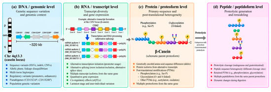
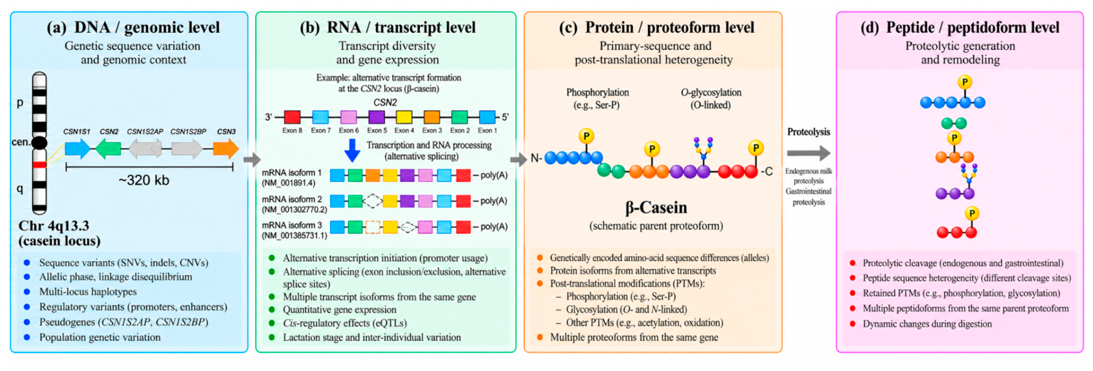
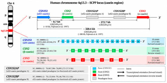
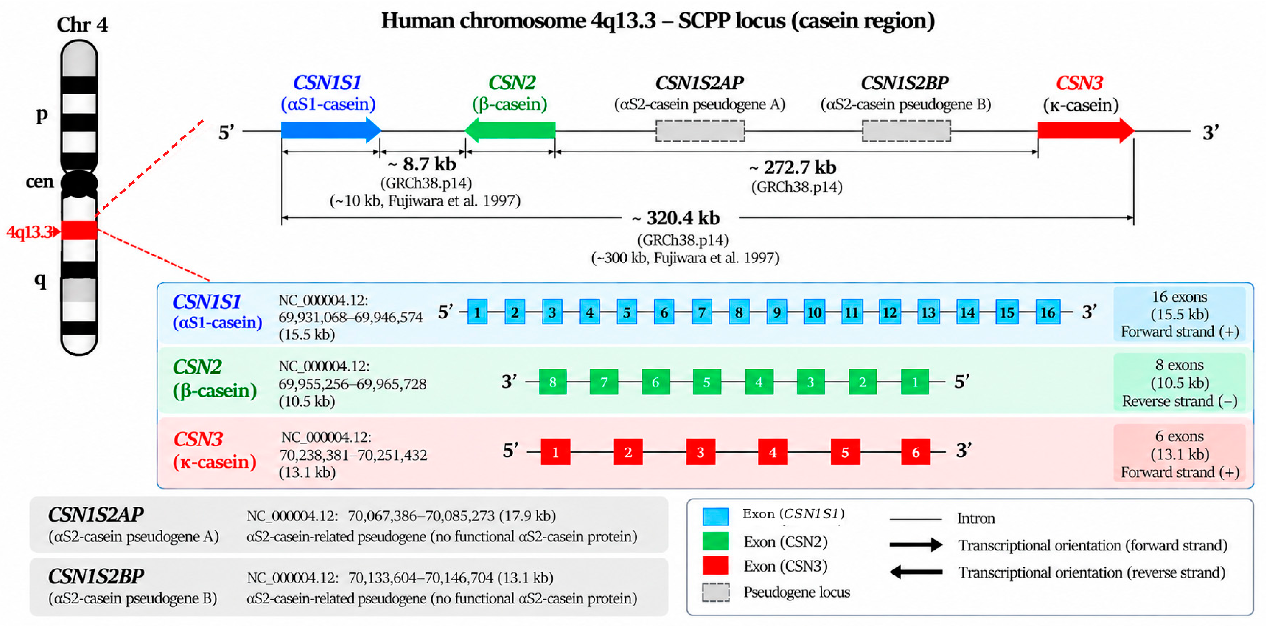
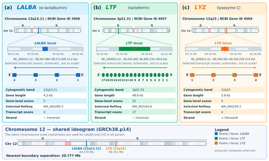
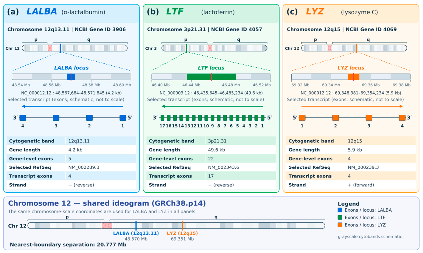
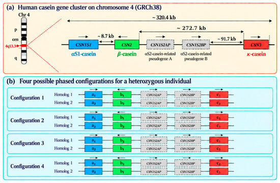
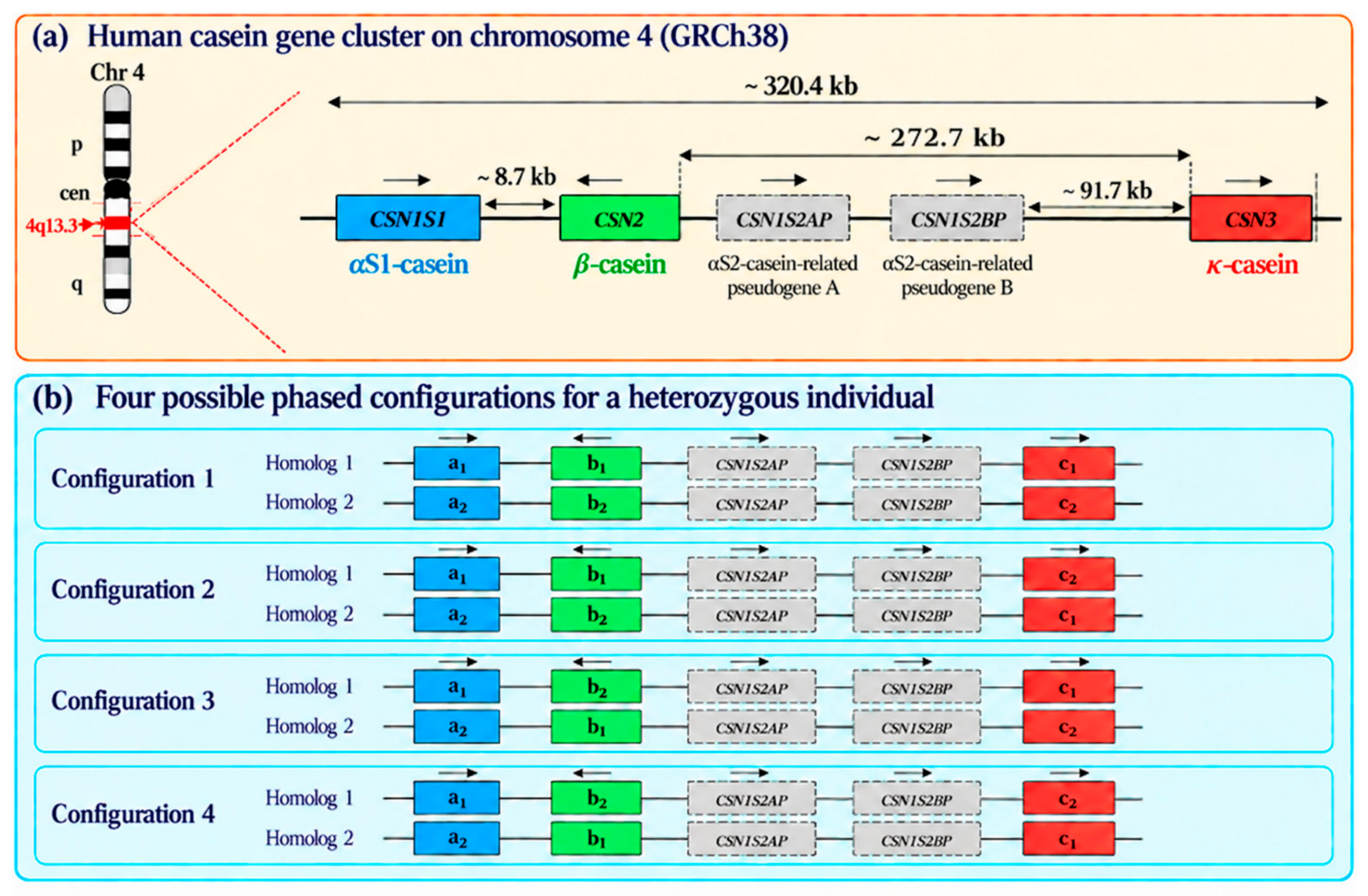
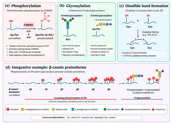
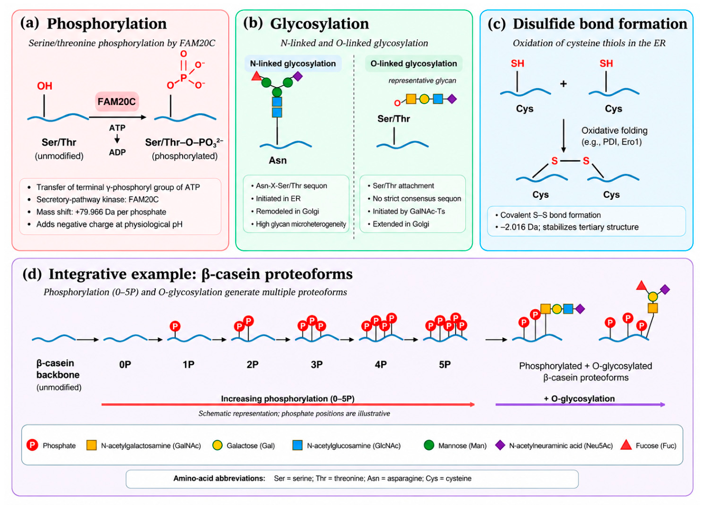
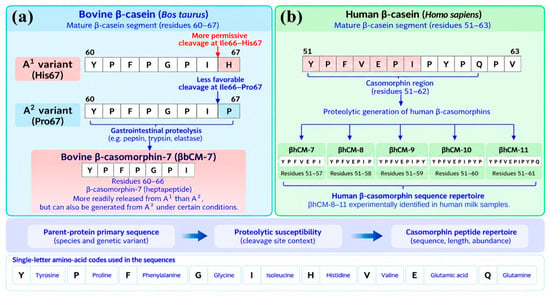
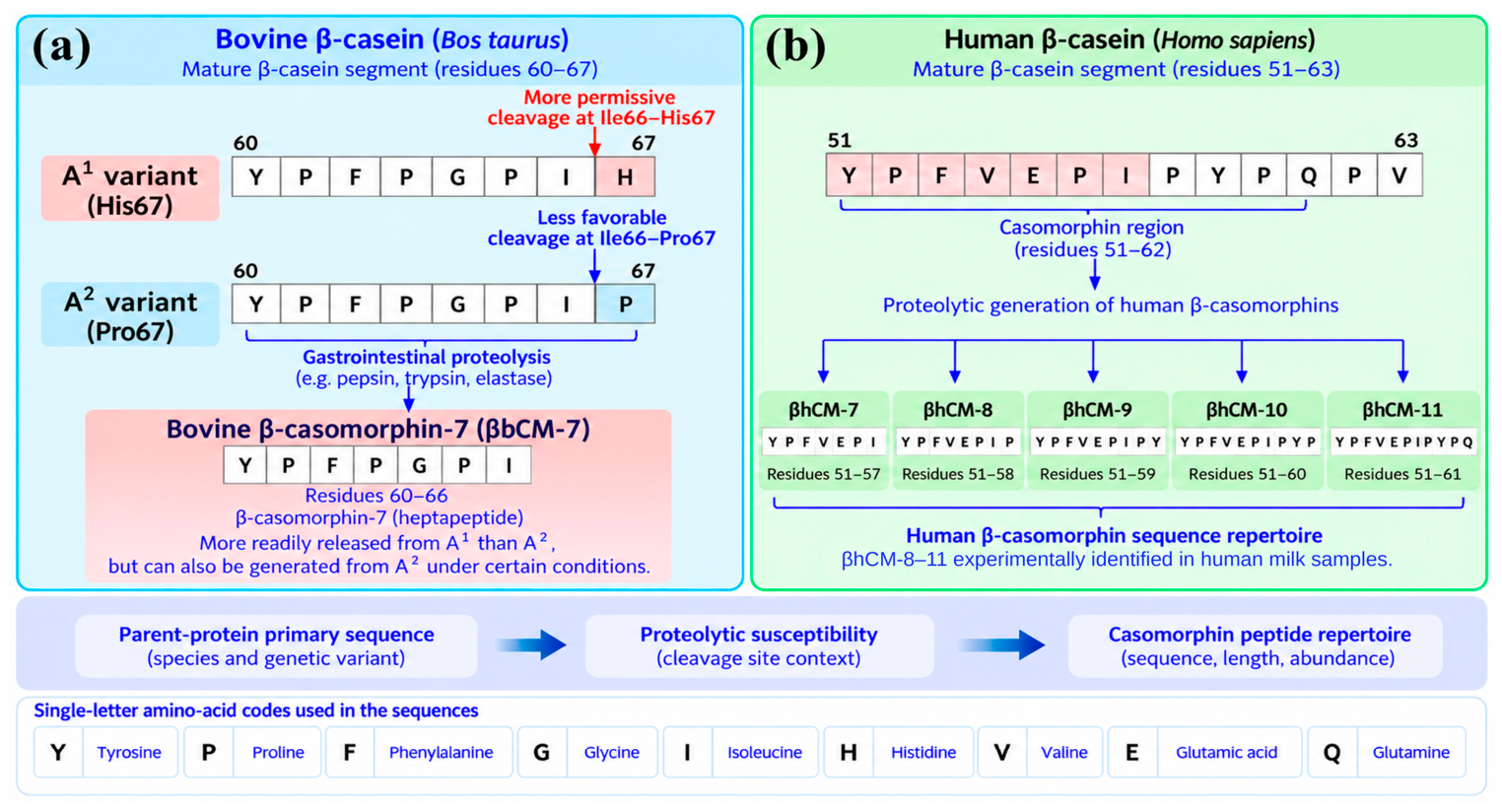

Disclaimer/Publisher’s Note: The statements, opinions and data contained in all publications are solely those of the individual author(s) and contributor(s) and not of MDPI and/or the editor(s). MDPI and/or the editor(s) disclaim responsibility for any injury to people or property resulting from any ideas, methods, instructions or products referred to in the content. |
© 2026 by the authors. Licensee MDPI, Basel, Switzerland. This article is an open access article distributed under the terms and conditions of the Creative Commons Attribution (CC BY) license.