


Abstract
Bisphenol A (BPA) surrounds us nowadays and this exposure is known to cause numerous health problems, cancer included. Since BPA is structurally similar to natural Estradiol-17β, it acts as a xenoestrogen overstimulating estrogen receptors. This triggers multiple pathways, mainly via inflammatory responses, that may lead to the progression of cancer. For hormone-sensitive cancers such as type 1 endometrial cancer, this is especially true. This article examined the most common pathways cited that involve inflammation leading to the formation, maintenance and aggressiveness of cancer stem cells, with the inflammation linked to BPA exposure. Other pathways were also examined where BPA may be involved in mutagenesis. The results and detailed discussions of the pathways are presented, along with clarifying tables. Cancer stem cell formation and maintenance are affected by chronic BPA exposure leading to type 1 endometrial cancer, but with some evidence gaps that still need to be resolved. Reactive Oxygen Species (ROS) formation is a key step, directly damaging DNA, causing oncogene formation, disabling proofreading genes, and triggering tumorigenesis. The known key pathways relevant to the formation and progression of endometrial cancer, and mechanisms concerning how BPA may trigger the process are discussed.
1. Introduction
Bisphenol A (BPA) is a monomer used to manufacture plastic that is extremely common in our modern environment. Recently, BPA has been found to be an endocrine-disrupting compound (EDC) with a molecular structure which mimics estrogen in vivo. BPA has been used for several decades as an epoxy resin and polycarbonate plastic in the manufacture of plastic polymers, but, in the last two decades, it has been shown to leak its monomers, especially into food and beverages stored in plastic containers. Its main use has been in products that are commercially available, which include the lining inside of beverages and canned foods, baby formula packaging, baby bottles, household food containers and food packaging. Often, it is also used for dental prosthetics, and is commonly found in thermal paper such as sales receipts [1,2]. The European Union banned BPA for use in materials that come into contact with food at the end of 2024. However, this EDC (BPA) has been found in urine and blood samples across the globe; despite whether people live in a rural developing region or live in a highly developed advanced area. The health effects range from reproductive problems and cancers in adults to abnormal childhood development [3]. Typically, bisphenol binds to membrane-bound estrogen receptors (ERs), as well as nuclear receptors, and could theoretically also interact with many other endocrine pathways. For example, it has been proposed that BPA could disrupt thyroid hormone receptors, androgen receptors and other endocrine-signaling pathways. Quite a few studies have shown that the xenoestrogen BPA has significant effects at nanogram concentrations, although traditional toxic levels are in the microgram range, because of its disruption of the normal hormonal signaling pathways [4]. Since BPA mimics estrogen, the theory for at least a decade has been that it may be correlated with hormone-sensitive cancers, including type 1 endometrial cancer (EC).
Describing cancers can often be challenging. There is a complex naming system based on the location of the cancer or staging, the type of histology, grade, cell marker type or organ of origin. Some types of cancer present multiple accepted names. This adds to the intricacy of cancer research. For this article, the primary focus will be on type 1 endometrial cancer (EC), which is known to be hormone-dependent, with some discussion of type 2 EC, which is generally considered not to be hormone-dependent [5]. EC may also be referred to as uterine corpus endometrial cancer (UCEC), as opposed to cervical cancer [6,7]. As the name suggests, EC originates from the inner lining of the corpus (main body) of the uterus, known as the endometrial lining. Type 1 EC is the most common form of this carcinoma, and accounts for eighty to ninety percent of all EC cases. Type 1 EC is hormone-sensitive and has been shown to be driven by excess estrogen. Type 1 EC is typically considered to be less aggressive and often has a more favorable prognosis. Type 2 EC is not linked with excess estrogen and is far less common. However, type 2 EC is a fast-growing, aggressive carcinoma with an often-unfavorable prognosis [8]. Given these facts, one may think that type 1 EC is almost benign, but it is not. Research has shown that the stage of the cancer is far more important than the type of EC, and type 1 EC can be just as dangerous as type 2 EC. Often, the slower-growing type 1 EC may go undetected for much longer than the faster-growing type 2 EC [9].
Traditionally, both types of EC were classified histologically. These histological subtypes were usually only found in type 1 EC or in type 2 EC, with little or no crossover [6]. Subtypes found in Type 1 EC include grades 1 and 2 endometrioid adenocarcinoma, mucinous adenocarcinoma, and adenocarcinoma with squamous differentiation. As the names imply, all are adenocarcinomas, with endometrioid type (grades 1 and 2) being the most common, and grade 1 has better differentiated cells than grade 2. All of these adenocarcinomas depend on estrogen for growth. The more aggressive type 2 EC histological subtypes include the following: serous carcinoma, clear cell carcinoma, grade 3 endometrioid adenocarcinoma, carcinosarcoma and undifferentiated carcinomas [7,8]. Unfortunately, microscopic histological subtypes give little or no information about their characteristics at the molecular level. While they may be useful clinically, the histology will not provide researchers with deeper insights into new therapies or ways of preventing EC.
In the last two decades, a new molecular classification system has arisen that gives insight into the growing process at the molecular level. There are currently four main molecular subtypes that have been classified by the The Cancer Genome Atlas (TCGA). These are the following: DNA polymerase epsilon (POLE)-mutated, Mismatch repair-deficient (predominant in type 1), No specific molecular profile (predominant in type 1), and P53 abnormal [10]. The No specific molecular profile (NSMP) subtype is by far the most commonly found in type 1 EC, and is often referred to as “Copy Number Low (NSMP)” due to its relatively low number of variations [11]. The second most common molecular subtype found in type 1 EC is Mismatch repair-deficient (MMRd), also known as microsatellite unstable (MSI/hypermutated), which is driven by a mismatch repair deficiency [12]. While the two subtypes above are the most common in type 1 EC, the other two less frequently do appear in type 1 EC. The third most common subtype is DNA polymerase epsilon (POLE)-mutated, also known simply as POLE-mutated. In this subtype, DNA damage to the POLE gene causes a loss of DNA proofreading ability as cells divide. These errors continually add up, giving this subtype the additional name of “hypermutated” or “ultramutated” [13]. Despite the frightening image of hypermutated DNA, this subtype actually has the most favorable prognosis [14]. By contrast, the least common subtype, with the highest morbidity, is the P53 abnormal form, also known as “Copy number high” (serous-like). This subtype is highly aggressive with P53 mutations that actually stimulate cancer growth [15,16]. Therefore, the NSMP and MMRd subtypes are by far the most common in type 1 EC, but the POLE-mutated and P53abn subtypes do appear as well, albeit with much less frequency. Since type 1 EC is by definition hormone-sensitive and BPA is a well-established powerful EDC, it reasonable to extrapolate that BPA is playing a role in type 1 EC, even in the rarer subtypes found in type 1 EC. Table 1 summarizes the characteristics and prognoses the four molecular subtypes.
Table 1.
Characteristics of the four molecular subtypes.
Discussed in this review will be the mechanisms behind the molecular subtypes published in various journals, and, then, this review will give an overview of how BPA may be contributing to EC via carcinogenesis and/or the maintenance of EC through several biochemical mechanisms. The overstimulation of estrogen receptors by BPA within the uterine lining leads to inflammation, which then activates various biochemical pathways [17,18]. This may lead to the formation of cancer stem cells (CSCs), as well as decreased cellular adhesion. The term “cancer stem cell” can be misleading. While a very low number of actual stem cells present may be mutated and form a carcinoma, the fact is that a mutated proto-oncogene in a somatic cell can (and usually does) take on the characteristics of a stem cell, meaning that it displays the characteristics of a traditional stem cell. Therefore, an oncogene mutation in a somatic cell can, and often does, give it the functional characteristics of a traditional stem cell: undifferentiated and capable of self-renewal. This ability defines malignant cancer cells at least in part; therefore, this article will refer to malignant cancer cells as CSCs [17,18].
Therefore, it has been established that BPA disrupts the function of ERs, and this leads to inflammation. Unfortunately, further research needs to be done in order to fully understand the exact mechanism of how inflammation becomes initiated after chronic ER overstimulation. Other various pathways related to inflammation may also alter the tumor microenvironment, which allows the newly formed CSCs to be maintained. Furthermore, there are other inflammatory pathways that cause oxidative stress and the formation of reactive oxygen species (ROS). These ROS cause DNA damage that leads to oncogene formation as well as epigenetic modifications. Those epigenetic modifications often allow EC tumor cells to evade the immune system. This article will also discuss positive feedback loops that are caused by ROS and inflammation.
Finally, the role of mutated P53 caused by oxidative stress and DNA damage will be considered.
2. Mechanisms by Which Hormones Are Affected by Bisphenol
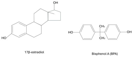
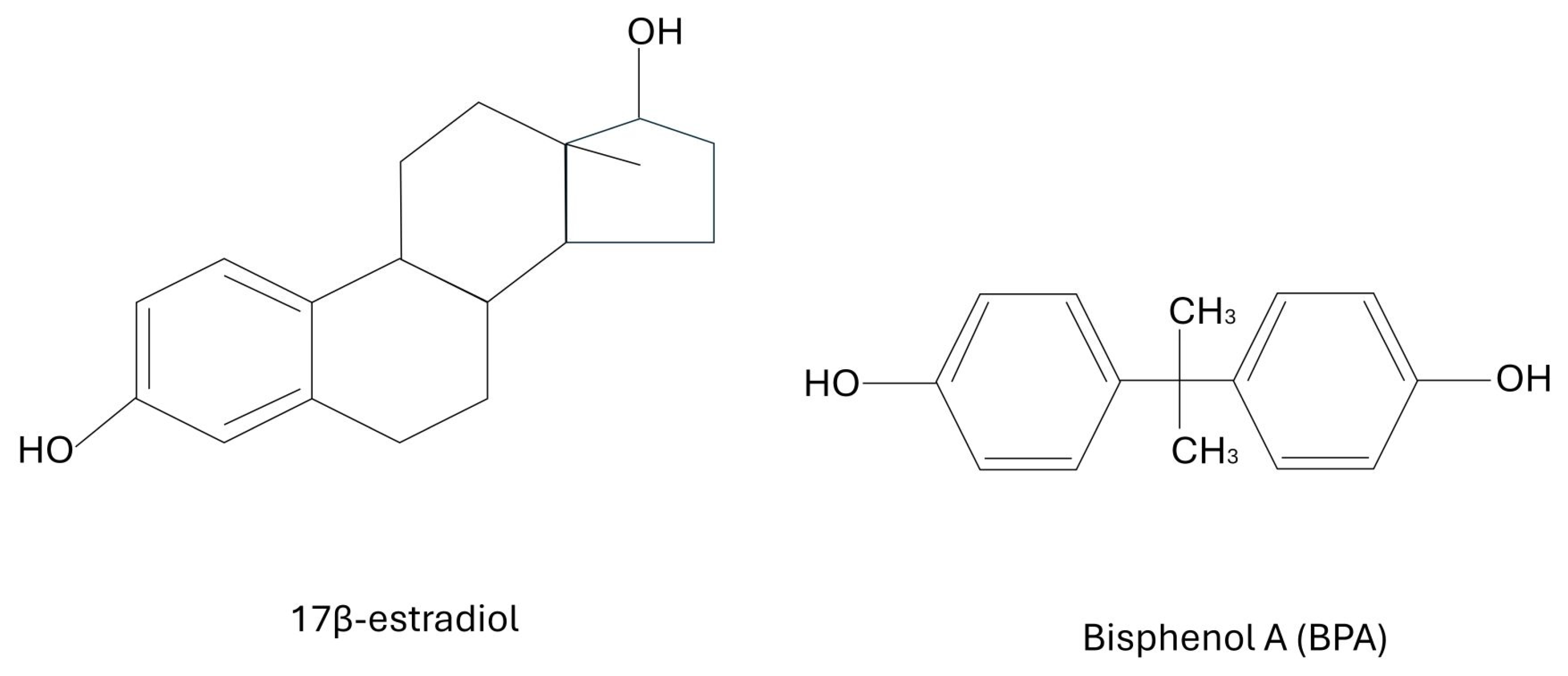
As mentioned above, BPA and other bisphenols can mimic estrogen. Estrogen is a key hormone regulating various bodily functions, which includes the reproductive system. The structural similarity between BPA and naturally occurring 17β-estradiol is the key. 17β-estradiol is a very potent form of naturally occurring estrogen and is not always present. It is produced for specific endocrine signaling as needed; however, during chronic BPA exposure, the ERs are chronically binding to BPA and are being continuously activated. The structural similarity of BPA to 17β-estradiol causes endocrine-signaling disruption and activates inflammatory pathways, which should be effectively deactivated. 17β-estradiol and BPA have similar chemical backbones that enable BPA to interfere with the body’s natural estrogenic activity. Although there are other EDCs in our environment that may mimic hormones as well, such as phthalates and certain pesticides, bisphenols maintain a stronger binding affinity for ERs compared to other compounds, which give BPA a stronger disruptive hormonal effect. Other EDCs include flame-retardants and heavy metals, but, overall, bisphenols such as BPA are the most pathogenic strictly because of their structural similarity to 17β-estradiol [2,4]. Figure 1 below shows the structures of BPA and 17β-estradiol.
Figure 1.
The structures of BPA and 17β-estradiol.
3. Recent Studies Have Examined the Relation Between Chronic BPA Exposure and Type 1 EC
Several studies have shown that BPA may be attributable to the development of type 1 EC, which is known to be hormone-sensitive [18,19]. There are many articles suggesting a direct cause-and-effect relationship between type 1 EC and BPA. Several studies have documented BPA concentrations in blood and serum that correlate to a higher incidence of both EC and breast cancer. For example, a clinical study from Poland in 2025 of 84 female patients showed that breast cancer was associated with a roughly 200% increased level of serum BPA, while EC was associated with a nearly 400% higher level of serum BPA [20]. The same study also used questionnaires and found that the women with elevated BPA levels tended to consume foods that had more contact with BPA-style plastics than patients who did not [20]. Another study showed similar results, with female breast cancer patients in Bangladesh showing similar patterns [21].
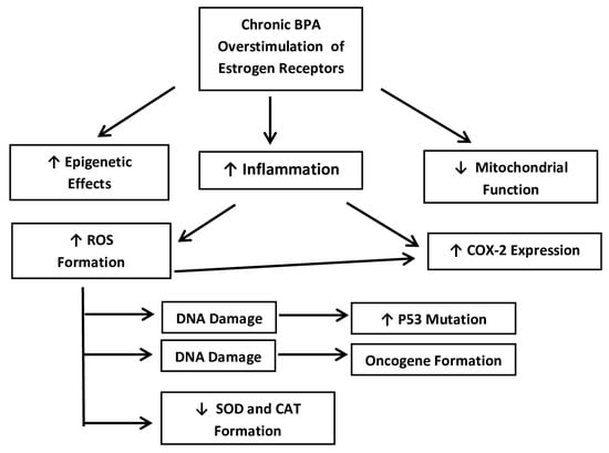
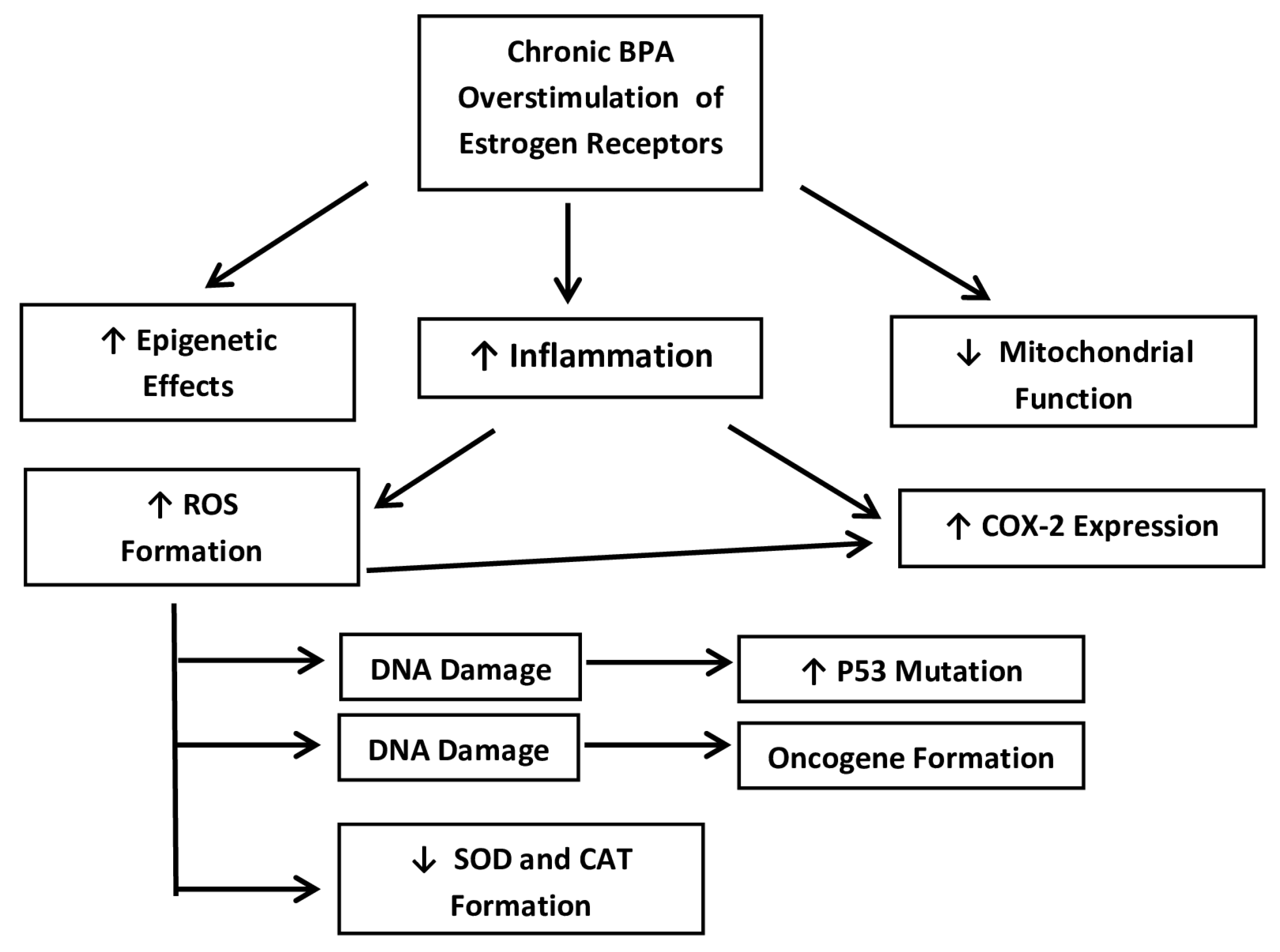
It has been proposed that BPA may trigger and promote hormone-sensitive cancers at the molecular level. In essence, chronic BPA exposure causes the overstimulation of ERs, which triggers many pathways discussed below [17,18]. The ER+ cells affected are all epithelial cells lining the endometrium. At concentrations found in patients with elevated BPA serum and/or urine concentrations, some of the pathways could include cancer stem cell activation by way of the cyclo-oxygenase-2 (COX2) pathway. Other pathways may decrease cell adhesion, which could lead to the formation of CSCs. Another major pathway includes the creation of ROS, as well as decreased CAT and SOD enzymes, thus creating a positive feedback loop. Meanwhile, other research has shown the p53 tumor suppressor having increased transcription activity. Nearly all the pathways that will be examined here start with inflammation, and that often creates ROS. That then may activate several enzymatic cascades, which lead to the formation of EC [22]. In the rest of this article, the details of the relevant pathways will be explained and the molecular mechanisms of how BPA may promote these adenocarcinomas will be explained in detail (Figure 2).
Figure 2.
A simplified flow-chart of the main mechanistic pathways that are presented below. The pathways occur in ER+ epithelial cells lining the endometrium. Source: Own data.
3.1. The COX-2 Pathway Activates Cancer Stem Cells with Decreased Cell Adhesion
Significantly, the COX-2 pathway maintains and activates CSCs in type 1 EC. Not only does it contribute to the aggressiveness of the tumor, but it may also have a role in treatment resistance. One future idea for therapy that has been proposed is to use various treatments, such as nonsteroidal anti-inflammatory drugs (NSAIDs), in order to block the COX-2 pathway [23,24]. The conversion of arachidonic acid to prostaglandins is catalyzed by the COX-2 enzyme. Those above-mentioned lipids play a significant role in inflammation, which leads to cell proliferation. With regard to EC, CSCs are activated by the COX-2 pathway. This population of self-renewing tumor cells is able to differentiate into multiple cell types that then form the adenocarcinoma. In general, CSCs are believed to cause tumor initiation, progression, metastasis, and resistance to conventional therapies in several types of cancer, not just in EC [25,26].
Elevated levels of COX-2 expression have been associated clinically with a poor prognosis and reduced survival in general. Due to this, some studies have put their focus into mainly targeting the COX-2 pathway as a strategy. Altering the tumor microenvironment may be how the COX-2 pathway affects carcinomas. As inflammation increases, the COX2 expression is upregulated, which then increases prostaglandin production. The increased prostaglandin production then supports the expansion and maintenance of CSCs. Prostaglandin E2 (PGE2) is the main prostaglandin formed. PGE2, in particular, has been shown to promote resistance to apoptosis and self-renewal in CSCs. Various signaling pathways can also be activated by PGE2, and those include the PI3K/Akt, Wnt/β-catenin and STAT3 pathways that have been shown to have an involvement in stem cell regulation [27,28]. Due to the above-listed pathways, COX-2 and its effectors found downstream can trigger the epithelial–mesenchymal transition (EMT). EMT is an important part of the metastatic process that causes epithelial cells to obtain increased motility and, therefore, invasiveness. This decreased cell adhesion causes the increased cell motility, and, thereby, invasiveness, which causes the metastasis found in late-stage EC [29,30]. This is where type 1 EC can be just as dangerous as the more aggressive type 2 EC. Since type 1 EC tends to grow more slowly, it may be overlooked, as the symptoms are attributed to other causes. Therefore, the metastasized cells may quietly spread into other organ systems, making the prognosis far more bleak [9]. A niche for CSCs may also be formed by the COX-2 pathway. This unique specialized microenvironment thereby supports the survival of CSCs and may shield CSCs from the immune system.
There are several ways in which BPA can stimulate the COX-2 pathway. The first pathway involves BPA and the inflammatory response. BPA has been repeatedly shown to cause inflammation, which may cause an increase in COX-2 expression. This, in turn, causes the increased production of prostaglandins, most importantly, PGE2 [25,31]. In turn, the potential for EC increases, and the weakened cell adhesion leads to a metastasized form of type 1 EC. The estrogenic activity of BPA could further modulate the COX-2 pathway, as studies have shown [22,26]. Meanwhile, even in the non-metastasized forms of type 1 EC, the COX-2 pathway is responsible for the formation of CSCs, as well as their proliferation and maintenance.
The particular mechanism of exactly how the chronic overstimulation of ERs causes inflammation is still not well-understood. However, there are several studies showing that BPA does activate ERs, and it has been shown that the chronic overstimulation of ERs causes an inflammatory response. A point of further research would be to investigate which inflammatory cytokines might be involved in the overstimulation of ERs. The discussion section will cover this gap in the knowledge in further detail.
These main mechanisms thought to correlate BPA exposure to COX-2 upregulation are presented below in Table 2. Worth mentioning, but not presented below, are some other factors that could be contributing to the situation. This includes oxidative stress caused by inflammation that could upregulate COX-2 expression [22]. Furthermore, epigenetic modifications caused by inflammation could also upregulate the COX-2 pathway. Unregulated DNA methylation has been shown to be caused by BPA, and BPA can also modify histones [32,33,34]. Some studies have theorized that those epigenetic changes may affect several processes including inflammation and the subsequent expression of COX-2 [22].
Table 2.
Summary of the effects of the COX-2 pathway in promoting endometrial cancer.
3.2. Cancer Stem Cell Activation via Reactive Oxygen Species (ROS) and Epigenetic Changes
Chronic overstimulation of estrogen receptors by BPA is commonly found in the literature. That overstimulation caused by the xenoestrogen then initiates an inflammatory response. That inflammatory response has been documented through several mechanistic pathways to cause oxidative stress and the formation of ROS [35,36,37]. As documented above, that inflammation can upregulate the COX-2 pathway alone; however, the ROS created through the inflammatory response can also directly damage DNA, leading to the formation of oncogenes, thereby creating CSCs [22].
It has been shown that ROS may cause extensive damage throughout any biological system. An older term for this was “free radicals”, meaning they contained an unpaired or “free” electron that could cause damage. However, now, a more accurate term, ROS, is applied. Not only may an unpaired electron be involved, but any oxygen-containing species (with or without free electrons) may pull electrons via various redox reactions [38]. ROS causes tissue and cell damage by overwhelming the cellular antioxidant defenses. This leads to oxidative stress and may affect many biomolecules including lipids, proteins and nucleic acids [39,40]. For example, ROS may cause lipid peroxidation that can not only destroy cellular membrane integrity via hydroxyl radical and peroxyl radical formation, but may also create mutagenic aldehydes from these lipids, such as malondialdehyde (MDA) and 4-hydroxynonenal (4-HNE) [41]. Furthermore, ROS directly reacts with pyrimidine and purine bases or the sugar-phosphate backbone of DNA. This frequently causes strand breaks (both single- and double-stranded) and base modifications that can lead to oncogenesis. The most commonly cited mutation contributing to cancer of all types is a P53 mutation [42], which is discussed in Section 3.4. Therefore, BPA causes inflammation, which increases ROS formation, which then contributes to the pathways discussed here.
Furthermore, some research shows that BPA may be responsible for epigenetic effects. The most common of these post-transcriptional modifications deal with DNA methylation and the modification of histones [32,33,34]. After DNA replication, the DNA is proofread and each CG dimer is methylated on the cysteine residue. This gives the molecular evidence that that the new DNA strand was copied precisely [33,34]. Thus, if the DNA is methylated epigenetically under the influence of BPA when it has not been accurately duplicated, it can cause a serious mutation to pass through the proofreading stage, causing nearly all of the mutations found in the four molecular subtypes of EC to be copied with each cellular division. This would also allow the carcinoma to go undetected by the immune system [43]. This information is summarized in Table 3.
Table 3.
The formation of CSCs due to the effects of ROS and epigenetic changes.
3.3. Decreased SOD and CAT Activate Cancer Stem Cells
Both superoxide dismutase (SOD) and catalase (CAT) are important enzymes that reduce the amount of ROS within a cellular system. SOD will create less reactive hydrogen peroxide (H2O2) and oxygen (O2) molecules from the extremely damaging superoxide radicals (O2−). Meanwhile, CAT is the next step of the process as it degrades H2O2 into water (H2O) and oxygen (O2). This process is vital in order to prevent an accumulation of too much ROS, which could cause damage [44]. This means that a decrease in SOD and CAT activity would increase the levels of ROS and lead to inflammation and oxidative stress, which may trigger the CSCs referred to above. In an odd twist, a positive feedback loop could also be true. The inflammation could also directly increase ROS. BPA causes the inflammation, which creates ROS. This downregulates the SOD and CAT enzymes, causing ROS levels to increase in a positive feedback loop, causing increasing amounts of cellular and DNA damage.
Another way that BPA may be increasing ROS formation is by interfering with mitochondrial function. The disruption of the electron transport chain (ETC) causes increased electron leakage and increases the production of ROS. Dysfunction in the mitochondria will decrease the synthesis of SOD2 (which is the mitochondrial version of SOD) as well as other antioxidants. This would also contribute to oxidative stress [45].
There are also other pathways that can be linked to CSC activation that involve SOD and CAT. If SOD and CAT have been decreased, then excess ROS may allow for the activation of alternative signaling pathways. These include Nuclear factor kappa B (NF-κB), Hypoxia-inducible factor 1-alpha (HIF-1α), and the Wnt/β-catenin pathway. Studies have shown that these pathways promote CSCs [27,46,47,48]. NF-κB’s general function is immunological and provides resistance against pathogens as a master regulator. It is therefore also involved in the immunological inflammation response [46,47]. Through this mechanism, it may enter the COX2 pathway that was previously described. HIF-1α regulates oxygen. If any cell becomes hypoxic, HIF-1α gets expressed and delivers the necessary oxygen [48]. Unfortunately, studies have shown that ROS can stabilize HIF-1α when oxygen concentrations are in the proper range; therefore, HIF-1α will deliver excess oxygen without hypoxia, contributing to oxidative stress [31]. Finally, if the Wnt/β-catenin pathway becomes dysregulated, it may cause the formation of tumors [27]. Once any of these pathways become dysregulated, they usually trigger tumorigenesis, malignant transformations and metastasis. The dysfunction of these enzymes may be caused by DNA damage after ROS exposure and is believed to be linked with disruptions in the immune checkpoints during mitosis [36].
BPA may be triggering the increased inflammation, which then lowers the SOD and CAT levels, and causes increased levels of ROS. Once more, since all four molecular subtypes of EC are due to genetic mutations of some sort, it is reasonable to assume that one or more of these pathways is involved. This information is summarized in Table 4.
Table 4.
Summary of the effects of decreased SOD and CAT and the formation of cancer stem cells after BPA.
3.4. The Loss or Mutation of p53 Promotes Tumorigenesis and Increased CSCs
A mutated or missing p53 is a common cause of CSC formation in all forms of cancers, with more than half of all studied tumors worldwide showing a mutated form of P53 [43]. Although the P53 mutation is less likely in type 1 EC, it does occur. During mitosis, P53 is an extremely important tumor suppressor and works to block cell division if there are errors during DNA replication [49]. This process effectively stops tumor formation by not allowing the cell to divide further. Since more than half of all cancer cells contain a mutated form of p53, it shows that blocking mitosis is vitally important. However, it is even worse than not being able to stop mitosis. It has been found that mutated p53 often inhibits differentiation, which contributes to the maintenance of CSC populations [50,51,52]. Therefore, the CSCs will continue to multiply as cancer cells.
Non-functional (often dysfunctional) P53 is nearly always attributed to a mutation in the DNA. How the mutation occurs could be random chance, a virus, radiation or chemical exposure. However, in both forms of EC, no exact cause of P53 mutation has been established [51]. However, ROS formation caused by BPA inducing an inflammatory response is quite possible, and ROS formation has been shown repeatedly to cause DNA damage [22,35]. Therefore, although the P53 mutation in type 1 EC is less common, when it does occur, it is most likely due to a DNA mutation. Table 4 summarizes the role of mutated P53 in type 1 EC. This information is summarized in Table 5.
Table 5.
Summary of the effects of mutated P53 and the formation of cancer stem cells after BPA.
4. Discussion
The EDC known as BPA is so structurally similar to the biologically active form of estrogen (estradiol-17β) that it overstimulates ERs and is most likely contributing to EC, especially type 1 EC [1,18]. Evidence shows that BPA definitely binds to ERs and causes an inflammatory response when there should be no endocrine signal to the ER [18,19]. Afterwards, these signals lead to several well-documented cascades. Unfortunately, the exact mechanism of how the chronic binding of BPA to ERs triggers inflammation at the molecular level remains unexplained. This is a problem that needs further research. It has been theorized that inflammatory genes such as IL6, TNFA, and NFKB, as well as some proliferation-related genes such as PCNA and CMYC, may be involved. However, the final data has not been published yet. Despite this gap in the knowledge, it is already clear that BPA does trigger inflammation and that inflammation leads to several pathways that promote carcinomas in EC.
The recently proposed four molecular subtypes of EC described by TCGA have been helpful in explaining EC at a biochemical level. As previously mentioned, the two most common subtypes found in type 1 EC are the Mismatch repair-deficient subtype and the No specific molecular profile subtype. More common in type 2 EC is the POLE-mutated subtype and the abnormal P53 subtype, the latter being the most dangerous form of EC with a very negative prognosis [10]. While type 1 EC tends to be less aggressive, it is still just as dangerous as type 2 EC. The main difference is that type 1 EC is hormone-sensitive, while type 2 is not. However, all four molecular subtypes may appear in type 1 EC, so all four should be considered.
Generally, BPA overstimulates ERs, which leads to inflammation. The inflammatory response often triggers the COX-2 pathway, which leads to the proliferation, maintenance and invasiveness of CSCs [21]. Several studies have focused upon these pathways as one of the most likely future treatments of cancers in general [23,24]. Furthermore, that same BPA-triggered inflammation can lead to ROS formation, which can also lead to a further upregulation of the COX-2 pathway, along with direct DNA damage as well as epigenetic effects, both of which are typical of CSC formation and maintenance [25]. That same inflammation may also decrease SOD2 and CAT formation, which then leads to further ROS formation and a positive feedback loop. Also of interest is that the overstimulated ERs can also decrease mitochondrial function and interfere with the electron transport chain, causing it to leak electrons, which further creates ROS [35,36,37]. Once more, ROS can and often does directly damage DNA. This may directly cause oncogene formation, and most likely also contributes to mutated P53, which causes the most aggressive and dangerous form of EC.
5. Conclusions
There is evidence that BPA may be promoting, maintaining and contributing to the invasiveness of the hormone-sensitive type 1 EC. The general biochemical mechanisms are triggered by the overstimulation of ERs, causing inflammation. The two main over-arching pathways involve COX-2 and ROS formation caused by the inflammatory response with many downstream effects that may promote tumorigenesis, a favorable microenvironment for CSCs and increased cancer cell invasiveness. However, the overstimulation of ERs may also directly trigger epigenetic effects that help hide cancer cells from the immune system as well as directly interfere with mitochondrial function, causing the electron transport chain to leak electrons further creating ROS. Notably, the formation of ROS can directly damage DNA, leading to not only oncogene formation but also mutated forms of the P53 enzyme, causing an extremely dangerous form of EC.
More research is needed as it pertains to the precise nature of which inflammatory genes are being triggered by BPA’s overstimulation of ERs in not only EC, but in breast cancer as well. For now, there is a hypothesis as to which inflammatory enzymes are being expressed, and how they might be controlled in order to reduce the effects of these pathways on the promotion of several types of hormone-sensitive cancers.
Author Contributions
Conceptualization, M.T. and J.W.; methodology, M.T.; validation, J.W.; formal analysis, M.T. and J.W.; investigation, M.T.; resources, J.W., K.Z.-W. and W.G.; writing—original draft preparation, M.T.; writing—review and editing, M.T., M.J., W.G., K.Z.-W. and J.W.; supervision, J.W.; project administration, J.W.; funding acquisition, J.W. and M.J. All authors have read and agreed to the published version of the manuscript.
Funding
This research received no external funding.
Institutional Review Board Statement
Not applicable.
Informed Consent Statement
Not applicable.
Data Availability Statement
Further inquiries can be directed to the corresponding authors.
Acknowledgments
The authors would like to acknowledge the use of Canva.com for graphic design in the preparation of this manuscript. Canva.com provided a user-friendly platform for creating high-quality visual elements, contributing to the overall clarity and presentation of the research findings.
Conflicts of Interest
The authors declare no conflicts of interest.
Abbreviations
The following abbreviations are used in this manuscript:
| BPA | Bisphenol A, 2,2-Bis(4-hydroxyphenyl) propane |
| CAT | Catalase (CAT) or Chloramphenicol acetyltransferase |
| COX-2 | Cyclo-oxygenase-2 |
| CSC | Cancer Stem Cell |
| POLE | DNA polymerase epsilon (POLE)-mutated |
| EC | Endometrial Cancer |
| EDC | Endocrine Disrupting Compound |
| EMT | Epithelial–Mesenchymal Transition |
| ER | Estrogen Receptor |
| ETC | Electron Transport Chain |
| HIF-1α | Hypoxia-inducible factor 1-alpha |
| MMRd | Mismatch repair-deficient |
| MSI | Microsatellite unstable |
| NF-κB | Nuclear factor kappa B |
| NSAID | Non-Steroidal Anti-Inflammatory Drug |
| NSMP | No specific molecular profile |
| PGE2 | Prostaglandin E2 |
| ROS | Reactive Oxygen Species |
| SOD | Superoxide dismutase |
| TCGA | The Cancer Genome Atlas |
| UCEC | Uterine corpus endometrial cancer |
| Wnt/β-catenin | Pathway leading to uncontrolled cell growth |
References
- Murata, M.; Kang, J.H. Bisphenol A (BPA) and cell signaling pathways. Biotechnol. Adv. 2018, 36, 311–327. [Google Scholar] [CrossRef] [Scilit] [PubMed]
- Tsai, W.T. Human health risk on environmental exposure to Bisphenol-A: A review. J. Environ. Sci. Health Part C 2006, 24, 225–255. [Google Scholar] [CrossRef] [Scilit] [PubMed]
- Ucheana, I.A.; Ihedioha, J.N.; Njoku, J.B.C.; Abugu, H.O.; Ekere, N.R. Migration of Bisphenol A from Epoxy-Can Malt Drink under Various Storage Conditions and Evaluation of Its Health Risk. Int. J. Anal. Chem. 2024, 104, 3936–3955. [Google Scholar] [CrossRef] [Scilit]
- European Food Safety Authority (EFSA) Panel on Food Contact Materials, Enzymes, Flavourings and Processing Aids. Scientific Opinion on the risks to public health related to the presence of bisphenol A (BPA) in foodstuffs: Executive summary. EFSA J. 2015, 13, 3978. Available online: https://efsa.onlinelibrary.wiley.com/doi/epdf/10.2903/j.efsa.2015.3978 (accessed on 24 April 2025). [CrossRef] [Scilit]
- Yang, H.P.; Wentzensen, N.; Trabert, B.; Gierach, G.L.; Felix, A.S.; Gunter, M.J.; Hollenbeck, A.; Park, Y.; Sherman, M.E.; Brinton, L.A. Endometrial cancer risk factors by 2 main histologic subtypes: The NIH-AARP Diet and Health Study. Am. J. Epidemiol. 2013, 177, 142–151. [Google Scholar] [CrossRef] [Scilit] [PubMed]
- Santoro, A.; Angelico, G.; Travaglino, A.; Inzani, F.; Arciuolo, D.; d’Amati, A.; D’Alessandris, N.; Scaglione, G.; Valente, M.; Urtueta, B.P.; et al. Beyond the WHO 2020 Classification of Female Genital Tumors: Types of Endometrial Cancer: A Pathological and Molecular Focus on Challenging Rare Variants. Int. J. Mol. Sci. 2024, 25, 10320. [Google Scholar] [CrossRef] [Scilit] [PubMed]
- Pu, H.; Wen, X.; Luo, D.; Guo, Z. Regulation of Progesterone Receptor Expression in Endometriosis, Endometrial Cancer, and Breast Cancer by Estrogen, Polymorphisms, Transcription Factors, Epigenetic Alterations, and Ubiquitin-Proteasome System. J. Steroid Biochem. Mol. Biol. 2023, 227, 106199. [Google Scholar] [CrossRef] [Scilit] [PubMed]
- Galant, N.; Krawczyk, P.; Monist, M.; Obara, A.; Gajek, Ł.; Grenda, A.; Nicoś, M.; Kalinka, E.; Milanowski, J. Molecular Classification of Endometrial Cancer and Its Impact on Therapy Selection. Int. J. Mol. Sci. 2024, 25, 5893. [Google Scholar] [CrossRef] [Scilit] [PubMed]
- Noer, M.C.; Antonsen, S.L.; Ottesen, B.; Christensen, I.J.; Høgdall, C. Type I Versus Type II Endometrial Cancer: Differential Impact of Comorbidity. Int. J. Gynecol. Cancer 2018, 28, 586–593. [Google Scholar] [CrossRef] [Scilit] [PubMed]
- Arciuolo, D.; Travaglino, A.; Raffone, A.; Raimondo, D.; Santoro, A.; Russo, D.; Varricchio, S.; Casadio, P.; Inzani, F.; Seracchioli, R.; et al. TCGA Molecular Prognostic Groups of Endometrial Carcinoma: Current Knowledge and Future Perspectives. Int. J. Mol. Sci. 2022, 23, 11684. [Google Scholar] [CrossRef] [Scilit] [PubMed]
- Momeni-Boroujeni, A.; Nguyen, B.; Vanderbilt, C.M.; Ladanyi, M.; Abu-Rustum, N.R.; Aghajanian, C.; Ellenson, L.H.; Weigelt, B.; Soslow, R.A. Genomic landscape of endometrial carcinomas of no specific molecular profile. Mod. Pathol. 2022, 35, 1269–1278. [Google Scholar] [CrossRef] [Scilit] [PubMed]
- Almperi, E.A.; Margioula-Siarkou, C.; Almperis, A.; Margioula-Siarkou, G.; Flindris, S.; Daponte, A.I.; Papamitsou, T.; Dinas, K.; Petousis, S. Mismatch Repair Deficiency Profiling and Its Impact on Management and Prognosis in Endometrial Cancer Patients: A Comprehensive Update. Cureus 2025, 17, e99364. [Google Scholar] [CrossRef] [Scilit] [PubMed]
- Vemuri, A.; Tjota, M.Y.; Wanjari, P.; Fleming, G.F.; Wang, P.; Bennett, J.A. A Deeper Dive into POLE -Mutated Endometrial Carcinomas: The Contributions and Consequences of Tumor Mutational Burden. Am. J. Surg. Pathol. 2026, 50, 301–312. [Google Scholar] [CrossRef] [Scilit] [PubMed]
- Fanale, D.; Corsini, L.R.; Piraino, P.; Pedone, E.; Brando, C.; Bazan Russo, T.D.; Ferraro, P.; Simone, A.; Contino, S.; Prestifilippo, O.; et al. POLE-mutated endometrial cancer: New perspectives on the horizon? Front. Oncol. 2025, 15, 1633260. [Google Scholar] [CrossRef] [Scilit] [PubMed]
- Chang, Y.W.; Kuo, H.L.; Chen, T.C.; Chen, J.; Lim, L.; Wang, K.L.; Chen, J.R. Abnormal p53 expression is associated with poor outcomes in grade I or II, stage I, endometrioid carcinoma: A retrospective single-institute study. J. Gynecol. Oncol. 2024, 35, e78. [Google Scholar] [CrossRef] [Scilit] [PubMed]
- Vaziri Fard, E.; Imboden, S.; Rau, T.; Epstein, E.; Petta, T.B.; Walia, S.; Carlson, J.W. p53 Abnormal (Copy Number High) Endometrioid Endometrial Carcinoma Has a Prognosis Indistinguishable From Serous Carcinoma. Int. J. Gynecol. Pathol. 2024, 43, 515–526. [Google Scholar] [CrossRef] [Scilit] [PubMed]
- Caravia, L.G.; Mitranovici, M.I.; Oala, I.E.; Tiron, A.T.; Simionescu, A.A.; Borcan, A.M.; Craina, M. The Importance of Cancer Stem Cells and Their Pathways in Endometrial Cancer: A Narrative Review. Cells 2025, 14, 594. [Google Scholar] [CrossRef] [Scilit] [PubMed]
- Winz, C.; Suh, N. Understanding the Mechanistic Link between Bisphenol A and Cancer Stem Cells: A Cancer Prevention Perspective. J. Cancer Prev. 2021, 26, 18–24. [Google Scholar] [CrossRef] [Scilit] [PubMed]
- Stillwater, B.J.; Bull, A.C.; Romagnolo, D.F.; Neumayer, L.A.; Donovan, M.G.; Selmin, O.I. Bisphenols and Risk of Breast Cancer: A Narrative Review of the Impact of Diet and Bioactive Food Components. Front. Nutr. 2020, 7, 581388. [Google Scholar] [CrossRef] [Scilit] [PubMed]
- Derkaczew, M.; Zglejc-Waszak, K.; Dabrowski, L.; Kocik, J.; Zdaniukiewicz, A.; Thoene, M.; Jozwik, M.; Gonkowski, S.; Wojtkiewicz, J. An Analysis of the Role of Bisphenol A in Breast and Reproductive-System Cancers. J. Clin. Med. 2025, 14, 4706. [Google Scholar] [CrossRef] [Scilit] [PubMed]
- Saha, H.R.; Hossain, P.; Uddin, M.J.; Karim, M.S.; Kabir, T.; Uddin, N.; Islam, M.S.; Sarkar, B.C. BPA (Bisphenol A) Exposure As An Environmental Risk Factor—A Cross Sectional Study on Breast Cancer Among the Bangladeshi Female Population. J. Environ. Pollut. Hum. Health 2024, 12, 47–52. [Google Scholar]
- Song, H.; Park, J.; Bui, P.T.C.; Choi, K.; Gye, M.C.; Hong, Y.C.; Kim, J.H.; Lee, Y.J. Bisphenol A induces COX-2 through the mitogen-activated protein kinase pathway and is associated with levels of inflammation-related markers in elderly populations. Environ. Res. 2017, 158, 490–498. [Google Scholar] [CrossRef] [Scilit] [PubMed]
- Cha, Y.I.; DuBois, R.N. NSAIDs and cancer prevention: Targets downstream of COX-2. Annu. Rev. Med. 2007, 58, 239–252. [Google Scholar] [CrossRef] [Scilit] [PubMed]
- Rüegg, C.; Zaric, J.; Stupp, R. Non steroidal anti-inflammatory drugs and COX-2 inhibitors as anti-cancer therapeutics: Hypes, hopes and reality. Ann. Med. 2003, 35, 476–487. [Google Scholar] [CrossRef] [Scilit] [PubMed]
- Pang, L.Y.; Hurst, E.A.; Argyle, D.J. Cyclooxygenase-2: A Role in Cancer Stem Cell Survival and Repopulation of Cancer Cells during Therapy. Stem Cells Int. 2016, 2016, 2048731. [Google Scholar] [CrossRef] [Scilit] [PubMed]
- Wu, M.; Guan, J.; Li, C.; Gunter, S.; Nusrat, L.; Ng, S.; Dhand, K.; Morshead, C.; Kim, A.; Das, S. Aberrantly activated Cox-2 and Wnt signaling interact to maintain cancer stem cells in glioblastoma. Oncotarget 2017, 8, 82217–82230. [Google Scholar] [CrossRef] [Scilit] [PubMed]
- Liu, J.; Xiao, Q.; Xiao, J.; Niu, C.; Li, Y.; Zhang, X.; Zhou, Z.; Shu, G.; Yin, G. Wnt/β-catenin signalling: Function, biological mechanisms, and therapeutic opportunities. Signal Transduct. Target. Ther. 2022, 7, 3. [Google Scholar] [CrossRef] [Scilit] [PubMed]
- Gorowska-Wojtowicz, E.; Duliban, M.; Kotula-Balak, M.; Bilinska, B. Modulatory Effects of Estradiol and Its Mixtures with Ligands of GPER and PPAR on MAPK and PI3K/Akt Signaling Pathways and Tumorigenic Factors in Mouse Testis Explants and Mouse Tumor Leydig Cells. Biomedicines 2022, 10, 1390. [Google Scholar] [CrossRef] [Scilit] [PubMed]
- Debnath, P.; Huirem, R.S.; Dutta, P.; Palchaudhuri, S. Epithelial-mesenchymal transition and its transcription factors. Biosci. Rep. 2022, 42, BSR20211754. [Google Scholar] [CrossRef] [Scilit] [PubMed]
- Zhang, C.X.; Huang, R.Y.; Sheng, G.; Thiery, J.P. Epithelial-mesenchymal transition. Cell 2025, 188, 5436–5486. [Google Scholar] [CrossRef] [Scilit] [PubMed]
- Harris, R.E.; Casto, B.C.; Harris, Z.M. Cyclooxygenase-2 and the inflammogenesis of breast cancer. World J. Clin. Oncol. 2014, 5, 677–692. [Google Scholar] [CrossRef] [Scilit] [PubMed]
- Singh, S.; Li, S.S. Epigenetic effects of environmental chemicals bisphenol A and phthalates. Int. J. Mol. Sci. 2012, 13, 10143–10153. [Google Scholar] [CrossRef] [Scilit] [PubMed]
- Qin, T.; Zhang, X.; Guo, T.; Yang, T.; Gao, Y.; Hao, W.; Xiao, X. Epigenetic Alteration Shaped by the Environmental Chemical Bisphenol A. Front. Genet. 2021, 11, 618966. [Google Scholar] [CrossRef] [Scilit] [PubMed]
- Senyildiz, M.; Karaman, E.F.; Bas, S.S.; Pirincci, P.A.; Ozden, S. Effects of BPA on global DNA methylation and global histone 3 lysine modifications in SH-SY5Y cells: An epigenetic mechanism linking the regulation of chromatin modifiying genes. Toxicol. In Vitro 2017, 44, 313–321. [Google Scholar] [CrossRef] [Scilit] [PubMed]
- Srinivas, U.S.; Tan, B.W.Q.; Vellayappan, B.A.; Jeyasekharan, A.D. ROS and the DNA damage response in cancer. Redox Biol. 2019, 25, 101084. [Google Scholar] [CrossRef] [Scilit] [PubMed]
- Anderson, M.T.; Staal, F.J.; Gitler, C.; Herzenberg, L.A.; Herzenberg, L.A. Separation of oxidant-initiated and redox-regulated steps in the NF-kappa B signal transduction pathway. Proc. Natl. Acad. Sci. USA 1994, 91, 11527–11531. [Google Scholar] [CrossRef] [Scilit] [PubMed]
- Biswas, S.K. Does the Interdependence between Oxidative Stress and Inflammation Explain the Antioxidant Paradox? Oxidative Med. Cell. Longev. 2016, 2016, 5698931. [Google Scholar] [CrossRef] [Scilit] [PubMed]
- Chandimali, N.; Bak, S.G.; Park, E.H.; Lim, H.J.; Won, Y.S.; Kim, E.K.; Park, S.I.; Lee, S.J. Free radicals and their impact on health and antioxidant defenses: A review. Cell Death Discov. 2025, 11, 19. [Google Scholar] [CrossRef] [Scilit] [PubMed]
- Liu, S.; Liu, J.; Wang, Y.; Deng, F.; Deng, Z. Oxidative Stress: Signaling Pathways, Biological Functions, and Disease. MedComm 2025, 6, e70268. [Google Scholar] [CrossRef] [Scilit] [PubMed]
- Juan, C.A.; Pérez de la Lastra, J.M.; Plou, F.J.; Pérez-Lebeña, E. The Chemistry of Reactive Oxygen Species (ROS) Revisited: Outlining Their Role in Biological Macromolecules (DNA, Lipids and Proteins) and Induced Pathologies. Int. J. Mol. Sci. 2021, 22, 4642. [Google Scholar] [CrossRef] [Scilit] [PubMed]
- Li, S.; Cao, T.; Zhang, Q. Oxidative stress as a driver of organelle cascade damage in neurological diseases. Front. Aging Neurosci. 2026, 18, 1892923. [Google Scholar] [CrossRef] [Scilit] [PubMed]
- An, X.; Yu, W.; Liu, J.; Tang, D.; Yang, L.; Chen, X. Oxidative cell death in cancer: Mechanisms and therapeutic opportunities. Cell Death Dis. 2024, 15, 556. [Google Scholar] [CrossRef] [Scilit] [PubMed]
- Rivlin, N.; Brosh, R.; Oren, M.; Rotter, V. Mutations in the p53 Tumor Suppressor Gene: Important Milestones at the Various Steps of Tumorigenesis. Genes Cancer 2011, 2, 466–474. [Google Scholar] [CrossRef] [Scilit] [PubMed]
- Zhu, B.; Song, L.; Li, M.; Cheng, M.; Tang, W.; Miao, L. Comparison of the effects between catalase and superoxide dismutase on regulating macrophage inflammatory response and protecting osteogenic function of periodontal ligament cells. Biochem. Biophys. Res. Commun. 2025, 56, 151523. [Google Scholar] [CrossRef] [Scilit] [PubMed]
- Khan, S.; Beigh, S.; Chaudhari, B.P.; Sharma, S.; Aliul Hasan Abdi, S.; Ahmad, S.; Ahmad, F.; Parvez, S.; Raisuddin, S. Mitochondrial dysfunction induced by Bisphenol A is a factor of its hepatotoxicity in rats. Environ. Toxicol. 2016, 31, 1922–1934. [Google Scholar] [CrossRef] [Scilit] [PubMed]
- Albensi, B.C. What Is Nuclear Factor Kappa B (NF-κB) Doing in and to the Mitochondrion? Front. Cell Dev. Biol. 2019, 7, 154. [Google Scholar] [CrossRef] [Scilit] [PubMed]
- Xia, Y.; Shen, S.; Verma, I.M. NF-κB, an active player in human cancers. Cancer Immunol. Res. 2014, 2, 823–830. [Google Scholar] [CrossRef] [Scilit] [PubMed]
- Lee, J.W.; Bae, S.H.; Jeong, J.W.; Kim, S.H.; Kim, K.W. Hypoxia-inducible factor (HIF-1)alpha: Its protein stability and biological functions. Exp. Mol. Med. 2004, 36, 1–12. [Google Scholar] [CrossRef] [Scilit] [PubMed]
- Arizti, P.; Fang, L.; Park, I.; Yin, Y.; Solomon, E.; Ouchi, T.; Aaronson, S.A.; Lee, S.W. Tumor suppressor p53 is required to modulate BRCA1 expression. Mol. Cell. Biol. 2000, 20, 7450–7459. [Google Scholar] [CrossRef] [Scilit] [PubMed]
- Gasco, M.; Shami, S.; Crook, T. The p53 pathway in breast cancer. Breast Cancer Res. 2002, 4, 70–76. [Google Scholar] [CrossRef] [Scilit] [PubMed]
- Battista, S.; Martelli, M.L.; Fedele, M.; Chiappetta, G.; Trapasso, F.; De Vita, G.; Battaglia, C.; Santoro, M.; Viglietto, G.; Fagin, J.A.; et al. A mutated p53 gene alters thyroid cell differentiation. Oncogene 1995, 11, 2029–2037. [Google Scholar] [PubMed]
- Yan, W.; Liu, G.; Scoumanne, A.; Chen, X. Suppression of inhibitor of differentiation 2, a target of mutant p53, is required for gain-of-function mutations. Cancer Res. 2008, 68, 6789–6796. [Google Scholar] [CrossRef] [PubMed]

Disclaimer/Publisher’s Note: The statements, opinions and data contained in all publications are solely those of the individual author(s) and contributor(s) and not of MDPI and/or the editor(s). MDPI and/or the editor(s) disclaim responsibility for any injury to people or property resulting from any ideas, methods, instructions or products referred to in the content. |
© 2026 by the authors. Licensee MDPI, Basel, Switzerland. This article is an open access article distributed under the terms and conditions of the Creative Commons Attribution (CC BY) license.