


Abstract
Contemporary research on facial aging is undergoing a profound paradigm shift, transitioning from macroscopic descriptive phenotyping to refined cellular-molecular mechanistic dissection. This review reveals these frontiers across multi-omics and structural dimensions, highlighting single-cell transcriptomic alterations (e.g., KLF6 and HES1 dysregulation), proteomic hubs (e.g., KNG1), epigenetic causal chains, and microbiome community succession within the cutaneous microenvironment. Concurrently, we map the progressive biomechanical failure of deeper tissues, including sequential adipose atrophy, SMAS-muscle complex degeneration, and sex-specific skeletal remodeling. Within the evolving landscape of regenerative aesthetics, calcium hydroxylapatite (CaHA) has emerged not merely as a passive space-occupying volumizer but as a dynamic biostimulatory material. We examine how CaHA’s morphology and rheological properties govern its biological behavior. Human histological studies provide evidence that CaHA is associated with fibroblast activity and temporal extracellular-matrix remodeling, particularly changes in type III and type I collagen expression. Changes in elastin- and angiogenesis-related markers have also been reported, whereas the mechanotransduction and immune-regulatory pathways proposed to underlie these responses remain supported predominantly by in vitro, ex vivo, and preclinical evidence and have not been fully validated in human facial tissue. Finally, we translate these material-driven interactions into advanced clinical strategies, including rheologically programmed dilution, layer-specific vectoring, and synergistic therapies. By bridging intricate aging biology with material-guided regeneration, this review provides a robust conceptual framework for individualized and precise rejuvenation.
1. Introduction
With advances in science and technology, rising aesthetic expectations, and increasing life expectancy, facial rejuvenation has become one of the fastest-growing and most rapidly evolving evidence-based fields in aesthetic medicine. Facial rejuvenation aims to restore histologic alterations driven by intrinsic aging and photo exposure. Its scope includes improving skin texture, lifting or excising lax tissues, and replenishing volume deficits to achieve a holistic optimization of facial contour and dynamic expression [1]. The global aesthetic medicine market has shown substantial scale and growth potential: it was valued at USD 63.4 billion in 2021 and is projected to expand at a compound annual growth rate (CAGR) of 9.6% from 2022 to 2030 [2]. According to the latest data from The Aesthetic Society, facelift procedures increased by 37.3% in 2023, representing one of the highest growth rates among surgical interventions [3].
To meet this growing clinical demand with precision, our understanding of facial aging must transcend superficial symptom management. While traditional hypotheses primarily attributed aging to gravity-induced ptosis and volume deflation, contemporary research provides a diverging and profound perspective, conceptualizing facial aging as an active, “inside-out” biological cascade. Progressive skeletal resorption and remodeling undermine soft-tissue support, which is followed by atrophy and descent of deep fat compartments and a decline in the mechanical integrity of the SMAS–muscle complex. Superficial fat compartments then sag and deform. At the cutaneous level, aging manifests as extracellular matrix (ECM) degradation, a chronic inflammatory microenvironment, and pigmentary dysregulation [4].
In recent years, accumulating evidence has further uncovered a more refined cellular and molecular basis for these phenotypes. Recent single-cell transcriptomic analyses have provided a high-resolution molecular atlas of human skin aging and identified age-associated, cell-type-specific regulatory candidates, including KLF6 in epidermal basal cells and HES1 in dermal fibroblasts [5]. Functional perturbation experiments further support roles for these factors in cellular senescence-related phenotypes, although their contribution to facial aging at the tissue and clinical levels remains incompletely defined. Similarly, quantitative 4D proteomic analyses identified KNG1 as an age-associated molecular hub, with experimental data linking KNG1-related pathways to collagen degradation, elastic-fiber remodeling, and oxidative stress [6]. However, much of the mechanistic evidence derives from experimental models, and its causal relevance to human facial aging requires further validation.
Epigenetic profiling has also opened a new window into the biological mechanisms of aging. Using an epigenome-wide Mendelian randomization (EWMR) approach, researchers have systematically investigated—under a causal-inference framework—the association between blood DNA methylation and perceived facial age [7]. Meanwhile, research on facial musculature is shifting toward an integrated “skin–fascia–muscle” biomechanical system, combining surface electromyography, skin elastometry, and 3D displacement analysis to achieve synchronized quantification of muscular electrical activity, skin physical properties, and soft-tissue displacement [8]. Collectively, these advances reflect a broader frontier trend: aging research is transitioning from descriptive phenotyping to mechanistic dissection and targeted intervention.
At present, surgical facial rejuvenation is characterized by substantial invasiveness, prolonged recovery, and potentially higher risks [9]. Consequently, calcium hydroxylapatite (CaHA) has assumed a pivotal role in facial rejuvenation owing to its distinctive biostimulatory properties and durable clinical outcomes [10,11]. A market survey by Global Info Research estimated that global CaHA revenue was approximately USD 6723 million in 2024 and is expected to reach USD 11,467 million by 2031 [12]. Radiesse, a widely used CaHA product, contains 30% CaHA microspheres suspended in a 70% carboxymethylcellulose (CMC) gel carrier, thereby providing immediate volumization together with longer-term biostimulatory effects. Human and preclinical studies support CaHA-associated collagen remodeling, while changes in elastin- and angiogenesis-related markers have also been reported [13].
Notably, CaHA research has progressed substantially in materials science, mechanistic elucidation, and clinical innovation. With the development of characterization technologies such as scanning electron microscopy (SEM) and energy-dispersive X-ray spectroscopy (EDX), associations between CaHA microstructural characteristics and biological responses are increasingly being investigated. Particle size, geometry, surface topography, and carrier composition may influence particle–tissue interactions, inflammatory responses, fibroblast behavior, and ECM remodeling; however, the causal relationships between these material properties and specific immune-regulatory pathways remain incompletely established, particularly in human facial tissue [14]. Meanwhile, rheological programmability enables order-of-magnitude modulation of the storage modulus, allowing CaHA to transition continuously and precisely along a spectrum from cohesive volumizer to diffusible biostimulatory agent, thus covering clinical scenarios ranging from deep structural support to mid-plane filling and superficial tightening. The role of the carrier is also evolving from a passive delivery medium to an active modulator of the local microenvironment. From classic CMC to emerging levan-based thermoresponsive hydrogels, carrier systems—through degradation kinetics, pore architecture, and interactions with active components—profoundly affect the release timing and spatial distribution of CaHA microspheres. At the mechanistic level, emerging technologies, including single-cell analytics, spatial transcriptomics, and advanced in vivo imaging, provide opportunities to investigate the cellular pathways potentially involved in CaHA–tissue interactions. Proposed mechanisms include mechanotransduction, immune–microenvironment modulation, and gene-program–associated remodeling; however, most remain incompletely characterized in CaHA-treated human facial tissue. Clinically, biomechanics-informed vector-lifting techniques based on facial retaining ligaments, multi-plane and multi-dilution sequential injection strategies, combination regimens with hyaluronic acid and microfocused ultrasound, and emerging approaches such as microneedle delivery are gaining momentum. Looking ahead, frontier directions include developing intelligent composite materials capable of sensing and responding to dynamic microenvironments; resolving mechanosensors and downstream signaling cascades at single-cell resolution; and ultimately enabling individualized, cell-type–targeted interventions guided by patient-specific aging maps.
Thus, the objective of this review is to integrate two major themes—multilayer mechanisms of facial aging and CaHA-driven tissue regeneration—to systematically construct a panoramic framework spanning aging mechanisms, material properties, regenerative pathways, and application innovation. Through this synthesis, we aim to lay a robust theoretical foundation for the next generation of precise and efficient regenerative rejuvenation therapies.
2. Literature Search Strategy
2.1. Search Strategy
This article was designed as a narrative review supported by a structured literature search. A comprehensive search of PubMed, Web of Science, and Scopus was conducted to identify relevant literature. The literature search covered publications available from database inception to 26 August 2026. To reflect the multidisciplinary scope of this review, the search strategy was organized into five major domains: (1) mechanisms of facial aging; (2) physicochemical and material properties of CaHA; (3) regenerative and biological mechanisms of CaHA; (4) clinical applications, efficacy, safety, and complications associated with CaHA-based treatments; and (5) comparisons with other injectable fillers and biostimulatory materials. Search terms were combined using Boolean operators (“AND” and “OR”) and included relevant keywords and Medical Subject Headings (MeSH), where applicable.
For facial aging, search terms included combinations of “facial aging”, “skin aging”, “photoaging”, “facial fat aging”, “adipose tissue aging”, “SMAS”, “facial muscle aging”, “facial skeletal aging”, “bone remodeling”, “extracellular matrix”, “fibroblast”, “cellular senescence”, “oxidative stress”, “microbiome”, “epigenetics”, and “microRNA”. For CaHA-related evidence, terms included “calcium hydroxylapatite”, “calcium hydroxyapatite”, “CaHA”, “biostimulatory filler”, “collagen”, “collagen neogenesis”, “fibroblast”, “elastin”, “angiogenesis”, “neovascularization”, “macrophage”, “immune modulation”, “regeneration”, “facial rejuvenation”, “clinical efficacy”, “adverse events”, “complications”, and “safety”. Priority was given to studies published from 2020 to 2026, while older foundational studies were retained when necessary for established key anatomical, biological, or material-science concepts.
2.2. Inclusion and Exclusion Criteria
Peer-reviewed original studies and relevant reviews were included if they provided mechanistic, histological, biomaterial, or clinical evidence relevant to multilayer facial aging or CaHA-based facial rejuvenation. Eligible evidence included in vitro cellular and biomaterial studies, animal studies, human histological investigations, observational clinical studies, clinical trials, and systematic or narrative review articles relevant to the predefined topics. Studies were excluded if they were unrelated to facial aging or aesthetic applications of CaHA, focused on non-facial or non-aesthetic uses without relevant mechanistic implications, lacked substantive biological, material, histological, or clinical evidence, or consisted solely of conference abstracts, editorials, letters, or duplicate publications.
Titles and abstracts were initially screened for relevance, followed by full-text assessment of potentially eligible articles. A total of 1121 records were identified through the structured database searches. After removal of 247 duplicate records, 874 unique records underwent title and abstract screening. Of these, 221 articles were retained for full-text assessment, and 118 publications met the predefined eligibility criteria and were included through the database search. To avoid excluding historically important evidence, the reference lists of included studies and relevant reviews were additionally screened, and citation tracking was performed to identify earlier foundational publications of clear anatomical, biological, histological, or clinical relevance. Fourteen additional publications were identified through this supplementary process. During subsequent manuscript revision, additional targeted literature was identified to address reviewer-requested topics, particularly long-term clinical safety, comparative evidence involving HA, PLLA, and PCL, and recent clinical studies of CaHA-based treatments. These newly incorporated publications were evaluated using the same eligibility and evidence-appraisal principles described above. Accordingly, the final narrative synthesis was updated to include both the original structured-search evidence base and the additional relevant studies identified during revision. Non-peer-reviewed market or professional-society reports were used only for contextual background and not to support mechanistic, efficacy, or safety conclusions.
2.3. Evidence Appraisal and Interpretation
Because this review aimed to integrate evidence across multidisciplinary research domains—including molecular, cellular, and biomechanical aging mechanisms, biomaterials science, and clinical aesthetic practice—and the included studies varied substantially in study design, experimental models, and outcome assessment, the evidence was synthesized narratively rather than through formal meta-analysis.
Human clinical studies were considered the most directly relevant evidence for treatment efficacy and safety, whereas human histological studies provided direct evidence of tissue-level remodeling. Animal studies were regarded as supportive preclinical evidence, while in vitro cellular and biomaterial studies were interpreted primarily as mechanistic or hypothesis-generating evidence. Particular caution was applied when interpreting isolated in vitro experiments, high-concentration or non-physiological exposure models, indirect molecular or histological markers, material-characterization studies without biological validation, or studies lacking direct human evidence. Importantly, evidence was not ranked solely according to study design; its relevance was judged in relation to the specific claim being evaluated. For example, a human biopsy study may provide stronger evidence of tissue remodeling than a clinical rating scale, but cannot by itself establish the molecular pathway responsible for that remodeling. Conversely, a randomized clinical trial may establish efficacy without explaining the biological mechanism.
3. Biological Basis and Cellular–Molecular Mechanisms of Facial Aging
Facial aging arises from the combined effects of genetic predisposition and environmental exposures, leading to progressive cellular dysfunction and structural alterations in facial tissues (Figure 1). A detailed understanding of the cellular and molecular mechanisms across facial layers is fundamental to achieving precise, mechanism-guided facial rejuvenation.
Figure 1.
Multiscale anatomical, cellular, and molecular framework of facial aging across five hierarchical layers. This figure compares the structural and biological characteristics of young (left) and aged (right) facial tissues across five distinct anatomical layers. Original figure created by the authors. Skin Layer: Youthful skin maintains structural homeostasis via a balanced extracellular matrix (ECM), including collagen, proteoglycan and elastin, alongside intact immune surveillance. In contrast, aging skin is characterized by the senescent melanocytes dysfunction, ECM degradation, cumulative reactive oxygen species (ROS), and suppressed immune surveillance (e.g., via the HLA-E/NKG2A axis). Subcutaneous Adipose: Young adipose tissue is characterized by the mature adipocytes secreting anti-inflammatory factors and a functional adipose-derived stromal vascular fraction (SVF). Aging is associated with pathological adipocyte hypertrophy, a pro-inflammatory microenvironment, reduced paracrine support from adipose-derived stem cells (ADSCs), and declining vascular density and function. SMAS-Muscle: Age-related structural and functional decline includes reduced vascularization, decreased contractility of facial mimetic muscles (e.g., zygomaticus major and orbicularis oculi muscles), and attenuated attachment of the orbicularis oculi muscle (OOM) to the inferior orbital rim, contributing to tissue ptosis and orbital fat herniation. Deep Facial Spaces: Progressive volume loss and redistribution of buccal and midfacial fat pads, together with connective tissue laxity, result in facial hollowing and deepening of nasolabial folds. Skeletal Layer: Bone remodeling imbalance characterized by enhanced osteoclast-mediated resorption leads to orbital enlargement, maxillary retrusion, and clockwise rotation of midfacial skeletal architecture. Collectively, bidirectional mechanical and biochemical interactions among these layers amplify aging progression, emphasizing its nature as a dynamic, interconnected, multilevel biological process rather than a linear degenerative cascade. Black arrows indicate the relationship between structural changes and their clinical manifestations, whereas red arrows indicate the direction of skeletal remodeling and rotation.
3.1. Skin Layer
Skin aging can be broadly categorized into intrinsic aging, driven primarily by genetic factors, and extrinsic aging, driven predominantly by ultraviolet (UV) radiation and also termed photoaging. Both processes converge on a shared endpoint—disruption of the cutaneous microenvironment—characterized by reduced numbers of keratinocytes, melanocytes, Langerhans cells, and fibroblasts, alongside a relative increase in certain immune-cell populations, including eosinophils, lymphocytes, and mast cells. A central pathological event is the accelerated degradation of the extracellular matrix (ECM) coupled with diminished synthesis of collagen and elastin. Clinically, these changes manifest as skin thinning, xerosis, wrinkle formation, reduced sweating, hyperpigmentation, and telangiectasia [15].
For example, exposure to 450-nm blue light has been shown to decrease the viability of human keratinocytes (NCTC 2544) in a dose-dependent manner, while significantly increasing intracellular reactive oxygen species (ROS) and the DNA damage marker 8-hydroxy-2′-deoxyguanosine (8-OHdG). This exposure also upregulates matrix metalloproteinases (MMPs), including MMP-1 and MMP-12, thereby promoting degradation of dermal elastin and collagen [16,17]. Collectively, these observations suggest that skin aging is driven by interconnected molecular networks (Figure 2).
Figure 2.
Overview of the mechanisms underlying intrinsic and extrinsic skin aging within the epidermal–dermal microenvironment. Skin aging is a systems-level and multiscale process driven by coordinated molecular and cellular alterations, including transcriptional reprogramming, proteomic remodeling, epigenetic regulation, and microRNA-mediated post-transcriptional control. These changes are further influenced by immune dysregulation, alterations in skin microbiome composition, pigment metabolic imbalance, and progressive telomere attrition, collectively contributing to structural and functional decline of the epidermal–dermal unit. Dashed lines indicate reported protein–protein interactions among KNG1, MME, MMPs, and EPHX2; dashed connections between melanocytes indicate their clustered spatial distribution in aged skin. Arrows indicate the direction of age-associated changes or regulatory relationships: gray curved arrows represent progressive age-related transitions, purple arrows indicate transcription factor-associated regulatory effects, black arrows indicate regulatory direction, and downward arrows indicate age-associated decreases in the indicated factors or processes. Original figure created by the authors.
3.1.1. Core Transcription Factors and Protein Hubs
Single-cell transcriptomics provides a high-resolution atlas of cellular heterogeneity in skin aging. Based on specific markers, eleven major skin cell types were identified using uniform manifold approximation and projection (UMAP), including basal cells (KRT14+), spinous cells (KRT10+), melanocytes (TYR+ and DCT+), fibroblasts (PDGFRA+), and immune cells (PTPRC+). Although the overall proportions of these cell types showed little variation across age groups, thousands of differentially expressed genes revealed two core hallmarks of aging: upregulation of apoptosis- and cytokine-mediated pathways, and downregulation of pathways involved in cell proliferation, differentiation, and ECM synthesis. Further analysis pinpointed cell-type-specific key transcription factors. In basal cells, KLF6 expression was downregulated during aging. Functionally, shRNA-mediated knockdown of KLF6 accelerated senescence, suppressed proliferation, and promoted interleukin-6 (IL-6) secretion, consistent with the epidermal thinning, barrier impairment, and inflammatory status observed in aged skin. In fibroblasts, the Notch signaling downstream transcription factor HES1 emerged as a central regulatory hub during aging. The JAG1–NOTCH3 signaling between fibroblasts and basal cells weakened over time, illustrating how the dermal microenvironment influences epidermal homeostasis. Functional assays confirmed that HES1 knockdown induced fibroblast senescence and upregulated inflammatory and p53 pathways. Conversely, overexpression or CRISPR-dCas9-mediated activation of HES1 delayed senescence and promoted proliferation [5].
Beyond transcriptional regulators, recent 4D proteomics has identified kininogen-1 (KNG1) as another pivotal hub in skin aging. Compared with 3-month-old C57BL/6J mice, KNG1 expression was significantly upregulated in the dorsal skin of 9- and 18-month-old mice (Figure 3A–D). Analysis of human sun-protected (buttock) skin samples from the dataset GSE106940 also revealed significantly higher KNG1 mRNA levels in aged skin. Immunohistochemical staining corroborated the age-related upregulation of KNG1 protein (Figure 3E,F) [6]. Protein–protein interaction analysis indicated that KNG1 interacts with membrane metallo-endopeptidase (MME), MMP-1/9, and epoxide hydrolase 2 (EPHX2), thereby promoting the degradation of elastic and collagen fibers and increasing oxidative stress. Together, these findings identify KNG1 as an age-associated molecular candidate linking ECM degradation and oxidative-stress pathways. The combination of murine experimental data and human expression evidence strengthens its biological plausibility; however, the current evidence does not establish that KNG1 is a clinically actionable driver of human facial aging. In particular, longitudinal human data and intervention studies demonstrating that modulation of KNG1 alters facial-aging phenotypes are lacking. KNG1 should therefore be considered a promising mechanistic and biomarker candidate rather than an established therapeutic target.
Figure 3.
Age-associated changes in KNG1 expression in murine and human skin. Reprinted with permission from Ref. [6], under the terms of the Creative Commons Attribution-NonCommercial-NoDerivatives 4.0 International License (CC BY-NC-ND 4.0). (A) Immunohistochemical (IHC) staining of KNG1 in the dorsal skin of mice aged 3, 9, and 18 months. Brown staining indicates KNG1-positive immunoreactivity, whereas blue-purple staining represents hematoxylin-counterstained nuclei. Scale bar = 25 μm. (B) Quantification of relative KNG1 levels corresponding to the murine IHC samples in (A). (C) Western blotting of KNG1 protein expression levels in mouse skin at 3, 9, and 18 months. (D) Quantification of relative KNG1 expression from the Western blot in (C). (E) Representative image of IHC staining for KNG1 in the eyelid skin of human females aged 23 and 50 years, with a scale bar representing 25 μm. (F) Quantification of relative KNG1 levels in young and aged human skin corresponding to the IHC samples in (E). Statistical analysis: Data are presented as the mean ± SEM (n = 5 per group) in (B) and the mean ± SEM (n = 3 per group) in (D), and as the mean ± SD in (F). Statistical significance is indicated as follows: * p < 0.05, ** p < 0.01, and *** p < 0.001.
3.1.2. Microbiome Community Succession
The role of the skin microbiome in facial aging has increasingly become a focus of research. A multicenter, deep metagenome-assembled genome (MAG) study by Sun et al. (2024) first proposed two distinct skin microbial community types, or “cutotypes”: the C-cutotype, dominated by Cutibacterium, which is typically observed in younger individuals with higher sebum levels, and the S-cutotype, dominated by Streptococcus, which is enriched in older individuals with lower sebum levels [18]. Functional analysis further uncovered a potential causal link between these cutotypes and aging phenotypes. In the microbiome of older individuals, pro-aging-associated pathways (AG-pathways) were found to be upregulated. For example, the O-antigen synthesis pathway, which generates lipopolysaccharide (LPS), can trigger skin inflammatory responses. The fatty acid β-oxidation pathway consumes the coenzyme NAD+ and generates ROS, exacerbating oxidative stress and mitochondrial damage. In contrast, the microbiomes of younger individuals exhibited greater activity in anti-aging-associated pathways (anti-AG-pathways). Notably, the L-arginine biosynthesis pathway is involved in collagen synthesis, while the de novo adenosine nucleotide biosynthesis pathway contributes to glutathione synthesis and oxidative stress defense. Collectively, these findings suggest that locally modulating microbiome metabolic functions—for example, by supplementing NAD+ precursors or specific amino acids—may offer a novel strategy for combating skin aging.
However, these findings should be interpreted primarily as associative and hypothesis-generating rather than as evidence that microbiome succession causally drives facial aging. The identified metabolic pathways were inferred from metagenomic profiles, and their functional activity and downstream effects on aging phenotypes were not directly validated through longitudinal or interventional studies. Moreover, age-associated differences in sebum production, lifestyle, environment, and skincare practices may themselves shape microbial composition, making directionality difficult to establish. Accordingly, microbiome-targeted strategies remain biologically plausible but clinically unproven.
3.1.3. Epigenetic Associations and Causal-Inference Evidence
Conventional “epigenetic clocks” are primarily constructed based on their correlation with age, which limits their ability to clarify whether CpG sites, as epigenetic biomarkers, are drivers of or merely responders to the aging process. To identify causal epigenetic events that actively drive aging, a study by Vladimir et al., published in Clinical Epigenetics, employed an epigenome-wide mendelian randomization (EWMR) approach. This method used methylation quantitative trait loci (meQTLs) from whole blood samples as the exposure and data from a genome-wide association study (GWAS) on perceived facial aging in the UK Biobank as the outcome. The analysis identified 1299 CpG sites with potential causal effects and clearly categorized them into disruptive sites that accelerate aging and protective sites that delay aging. Functional enrichment analysis revealed that these sites were significantly associated with pathways involved in pigmentation (e.g., IRF4, BNC2, ASIP, EXOC2, MC1R), extracellular matrix regulation (e.g., ELN, LOX, COL8A1, MMP3), fatty acid metabolism (e.g., FADS1, FADS2), and oxidative stress (e.g., GPX1, NAT2), thereby delineating a molecular chain linking “genetic variants-DNA methylation changes-gene expression-facial aging phenotypes” [7]. These findings provide causal-inference evidence linking selected methylation loci to perceived facial aging and identify biologically relevant pathways involving pigmentation, ECM regulation, lipid metabolism, and oxidative stress. However, the analysis was based on blood-derived meQTLs and a population-level perceived-aging phenotype rather than direct measurements in facial skin. Mendelian randomization also depends on assumptions regarding instrument validity and pleiotropy. Therefore, these CpG sites should currently be regarded as candidate causal regulators rather than experimentally validated tissue-specific drivers of facial aging, and their therapeutic relevance remains to be established.
3.1.4. Immune Surveillance and Evasion
The skin immune microenvironment becomes dysregulated during aging. As a key immune sentinel in the epidermis, Langerhans cells (LCs)—members of the mononuclear phagocyte system—directly interact with T cells via their dendritic processes upon recognizing pathogens or damage signals, thereby initiating local immune responses. During skin aging, the number of LCs decreases, accompanied by diminished migratory capacity, reduced expression of antimicrobial peptides, and impaired function. For example, diminished secretion of interleukin-1β (IL-1β) attenuates the responsiveness of LCs to chemokines, affecting their migration. A decline in human β-defensin-3 weakens the clearance capacity against pathogens such as Staphylococcus aureus. Furthermore, their antigen-presenting function declines, and the expression of co-stimulatory molecules decreases, compromising their ability to initiate T-cell responses [19]. In addition to the functional decline of LCs, fibroblasts contribute to immune dysregulation through immune evasion. Specifically, they can express the non-classical MHC molecule HLA-E, which activates the inhibitory receptor NKG2A on natural killer (NK) cells and CD8+ T cells, thereby suppressing immune surveillance [20].
3.1.5. Dysregulated Pigment Metabolism
Melanocytes, which produce melanin, function to protect keratinocyte nuclei from chromosomal damage caused by radiation. Their dysfunction is a key driver of skin aging. A recent study utilizing the senescence-specific gene set SenSkin™ revealed that CDKN1A serves as an identifying marker for senescent melanocytes. Spatial transcriptomic analysis showed that cells highly expressing CDKN1A form clustered aggregations in photoexposed skin. This spatial distribution pattern provides a mechanistic explanation for the patchy, clustered appearance of age spots and hyperpigmentation in aged skin, rather than a uniform distribution. Furthermore, senescent melanocytes exhibit upregulation of the MC1R gene, a key receptor for eumelanin synthesis. While eumelanin effectively dissipates ultraviolet radiation and provides photoprotection, its uncontrolled overproduction, driven by the aberrantly activated synthesis pathway in senescent melanocytes, leads to senile pigmentation, age spots, and uneven skin tone. This explains the pigmentary disorders commonly associated with photoaged skin [21]. Additionally, senescent melanocytes amplify the aging effect through paracrine signaling. They express various inflammatory markers and senescence-associated secretory phenotype (SASP) factors, such as CXCL3. These secreted factors can induce mitochondrial dysfunction and elevate ROS levels in neighboring keratinocytes, thereby suppressing their proliferation [22].
3.1.6. MicroRNA Regulatory Networks
MicroRNAs (miRNAs) are short non-coding RNA molecules that regulate gene expression post-transcriptionally. They also function as mediators of intercellular communication and can be packaged within vesicles such as exosomes to transmit senescence signals between cells. During skin aging, miRNAs regulate several convergent biological processes, particularly ECM homeostasis, cellular senescence, oxidative stress, and inflammation. Among the best-characterized examples, miR-29 suppresses collagen synthesis and is increased in UV-exposed and aged fibroblasts, contributing to reduced dermal matrix production [5]. Other aging-associated miRNAs, including miR-23a-3p, miR-92a-3p, and members of the miR-34 family, impair profibrotic signaling or promote cell-cycle arrest and senescence [23,24,25]. UV-responsive miR-21-5p further links inflammatory and autophagy-related signaling to increased MMP activity [26]. Collectively, these findings indicate that age-associated miRNA dysregulation converges on three clinically relevant outcomes—reduced collagen synthesis, enhanced ECM degradation, and persistence of a pro-senescent inflammatory microenvironment.
3.1.7. Telomere Shortening
Telomeres are repetitive DNA–protein structures that protect chromosome ends and preserve genomic stability. Progressive telomere shortening during aging activates a persistent DNA-damage response, ultimately driving irreversible cell-cycle arrest and cellular senescence through canonical senescence pathways. Senescent fibroblasts subsequently acquire a pro-inflammatory and matrix-degrading secretory phenotype, thereby promoting chronic inflammation, ECM deterioration, and impaired tissue regeneration. Clinically, these changes contribute to wrinkle formation, skin laxity, and diminished regenerative capacity [15].
3.2. Subcutaneous Adipose Layer
Facial fat aging is not simply a matter of volume loss. Rather, it is a complex three-dimensional and dynamic process that involves compartment-specific atrophy and displacement, functional deterioration of adipocytes, and progressive biomechanical imbalance of the face [27].
3.2.1. Sequential Atrophy and Pseudoptosis
Rohrich et al. proposed the theory of sequential atrophy and pseudoptosis [27]. Preferential volume loss in the deep fat compartments weakens their support for the overlying superficial fat pads. Consequently, the superficial fat pads collapse, accumulate, and deform under the influence of gravity, presenting clinically as soft tissue ptosis (e.g., malar pad descent, deepening of the nasolabial fold). The sequential atrophy theory provides a predictable temporal map for facial aging. Research by Wan et al. found that the average adipocyte area in the nasolabial fat compartment was significantly larger than that in the deep medial cheek fat pad. A smaller cell area typically indicates higher metabolic activity and faster lipolysis rates. This suggests that deep fat adipocytes may possess higher metabolic activity and turnover, leading to earlier apoptosis and volume loss during aging [28].
Facial aging is a complex three-dimensional change involving specific fat compartment atrophy, displacement, and herniation. A longitudinal MRI study following 63 Asian patients over an average of 81.6 months showed no significant change in the thickness of the subcutaneous temporal fat. However, the superficial temporal fat compartment underwent significant atrophy, inferior displacement, elongation, and even herniation onto the zygomatic surface, which was identified as the primary cause of temporal hollowing. The buccal fat pad predominantly exhibited inferior displacement, elongation, and superficial herniation below the zygomatic arch, with a rate strongly correlated with time (0.89 mm/year). This inferior displacement and herniation were key factors exacerbating the formation of the labiomandibular groove (marionette lines) [29]. Consistently, Lambros compared youthful and aged photographs from 130 patients using stable skeletal landmarks, including interpupillary distance, medial canthal tendon distance, and the columellar base. In 96.4% of individuals, the lid–cheek junction remained stable over decades without true vertical descent. The apparent aging change was attributed primarily to anterior orbital fat protrusion that casts a deeper shadow above the lid–cheek junction, creating an optical effect driven by anteroposterior volume redistribution rather than gravity-related vertical drooping. Other midface markers, such as nevi and orbicularis oculi rhytids, also remained spatially stable over time, supporting the view that aging is driven mainly by loss of deep support and three-dimensional compartmental shifts rather than absolute skin ptosis [30].
3.2.2. Cellular Functional Decline
The functional state of adipose tissue plays a crucial role in maintaining a youthful facial appearance. The “Facial Fat Fitness” model proposed by Galanis posits that in young, healthy fat, adipocyte precursor cells, regulated by the transcription factor peroxisome proliferator-activated receptor gamma (PPARγ), differentiate into small, mature adipocytes. These cells secrete anti-inflammatory factors such as adiponectin and leptin, which promote fibroblast synthesis of collagen, elastic fibers, and hyaluronic acid, thereby maintaining a favorable metabolic microenvironment. Aging, excessive nutrition (e.g., high-sugar, high-fat diets), and UV exposure lead to attenuated PPARγ signaling, impaired adipogenesis, and pathological hypertrophy of existing adipocytes. These hypertrophic cells release large quantities of pro-inflammatory factors (e.g., leptin, TNF-α) and free fatty acids. This cascade inhibits the synthetic function of fibroblasts and induces them to secrete MMPs, accelerating ECM degradation and resulting in skin thinning and laxity [31]. Furthermore, adipose-derived stem cells (ADSCs) within the dermal white adipose tissue (dWAT) secrete growth factors, cytokines, and peptide hormones. A decline in the secretion of various factors, including vascular endothelial growth factor (VEGF), keratinocyte growth factor (KGF), transforming growth factor-β1 (TGF-β1), IL-1, and IL-6, leads to a gradual reduction in collagen in the papillary and reticular dermis. Concurrently, increased MMP-1 activity contributes to collagen disorganization [32]. Additionally, ADSCs and their conditioned medium have demonstrated potent antioxidant properties. The functional decline of ADSCs impairs their ability to effectively protect fibroblasts from oxidative stress damage induced by chemicals and UVB radiation [33]. Adipocytes are terminally differentiated cells that rely on the surrounding vascular network for nutrients. The stromal vascular fraction (SVF), which includes endothelial cells, pericytes, smooth muscle cells, and immune cells, along with ADSCs, migrates in a coordinated manner to generate new vascular networks. This process promotes toxin clearance and improves nutrient delivery to recipient sites, thereby decelerating facial aging [34]. Anatomically, dWAT is closely adjacent to sebaceous glands and hair follicles. Through paracrine signaling, dWAT can regulate sebaceous gland activity and the hair follicle growth cycle. Dysfunction of dWAT can thus lead to decreased skin hydration, hair-related issues, and a perceived older age [35].
3.2.3. Biomechanical Imbalance
Recent research has shifted from static histologic descriptions of facial aging to dynamic biomechanical processes. Maintenance of a youthful facial contour depends on a precise mechanical equilibrium system composed of the skin, ligaments, fascia, and fat compartments. Facial fat is partitioned by fibrous septa into discrete compartments (for example, nasolabial and middle temporal compartments). These fibrous septa originate from the deep dermis or the superficial musculoaponeurotic system (SMAS) and insert into deeper fascial layers or periosteum, forming a complex reticular suspension system. When this mechanical balance is disrupted, due to deep fat atrophy and ligamentous laxity, the superficial fat loses its support. Under gravity, it slides inferiorly along the SMAS, adopting a teardrop morphology that manifests clinically as deepened nasolabial folds, malar bags, and jowling along the mandibular border [36]. A longitudinal computed tomography (CT) study of 262 patients provided compelling imaging evidence for this teardrop change. The volume and thickness of upper and middle zone fat compartments (e.g., pre-zygomatic fat) significantly decreased, while the volume and width of lower zone compartments (e.g., nasolabial fat, submental fat) significantly increased. Concurrently, the overall height of the superficial fat layer increased, suggesting tissue elongation [37]. Thus, modern strategies for facial rejuvenation have evolved from simple volume augmentation to structural volume repositioning. Achieving truly natural and harmonious results requires a profound understanding of the specific aging patterns of each fat compartment, enabling targeted filling of atrophic areas and repositioning displaced tissues [38,39].
3.3. SMAS–Muscle Layer
3.3.1. Functional Remodeling of Mimetic Muscles
Facial muscle aging exhibits a pattern distinct from somatic skeletal muscles, described as “global stability with local dysregulation.” In a pioneering study, Cotofana et al. systematically measured the electromyographic activity of nine facial muscle groups. Their results indicated no statistically significant differences between younger and older groups in overall muscle electrical activity signals, baseline noise, or signal-to-noise ratios. Specific analysis, however, revealed a significant reduction in both the signal and baseline noise of the zygomaticus major muscle. This suggests a weakening in both the contractile capability and the resting-state electrical activity of this muscle, providing a potential electrophysiological mechanism for the clinically observed decline in smile expressiveness and sagging morphology in the midface. Conversely, the procerus muscle and corrugator supercilii muscles demonstrated a significant enhancement in signal-to-noise ratio, indicating an improved ability to activate effectively from a resting state. The long-term, repetitive over-contraction of these muscles leads to micro-damage in the skin and soft tissues, directly driving the irreversible transition from dynamic wrinkles to static glabellar lines [40]. Facial appearance at rest and during expression is determined by the antagonistic interplay between levator and depressor muscle groups. When levator strength declines, depressors—such as the depressor anguli oris and the platysma—may exert a net downward vector on the soft tissues. Clinically, this presents as downturned mouth corners (often perceived as sadness or anger), blurring of the mandibular border (caused by combined platysma overactivity and laxity), and an overall “sagging” facial configuration [41]. Another study, utilizing high-resolution CT imaging, was the first to report the age-related weakening of the attachment between the orbicularis oculi muscle (OOM) and the inferior orbital rim in a Japanese cohort. In older individuals, only the nasal portion of the OOM remained attached to the bone, while most of the mid and lateral segments were detached. This disengagement of the OOM from its bony anchor elucidates key mechanisms underlying the infraorbital rim show, orbital fat herniation, lower eyelid “bags”, and the transient canthal descent during postural changes (e.g., from lying to sitting) [42].
Furthermore, Frank et al. pioneered the concept of the decoupling between muscle electrical activity and mechanical function. They proposed that muscle function does not decline linearly with electrical activity but follows a “functional threshold” model. Their study employed a multi-modal, multi-layered quantification of the midface structure using Cutometry skin elasticity testing, 3D displacement analysis, and surface-derived electromyography (sEMG). The data revealed that while the electrical signal intensity of the zygomaticus major, levator labii superioris alaeque nasi, and depressor anguli oris muscles decreased with age, the amplitude of soft tissue displacement they drove showed no statistically significant change. This suggests the existence of a functional threshold for facial muscles; during early aging, the electrophysiological decline may not yet reach this threshold, allowing mechanical function to be compensated [8]. This insight points to a potential window for clinical intervention.
3.3.2. Deterioration of SMAS Mechanical Properties and Microvascular Nutritional Support
The SMAS functions as a mechanical and nutritional conduction network between the superficial and deep layers of the face, with direct connections to numerous muscles and fasciae (e.g., zygomaticus major, orbicularis oculi, platysma, parotid fascia). These connection points serve as key mechanical anchors. Using 320-row spiral CT (Aquilion ONE, Canon Medical Systems, Tochigi, Japan), the SMAS can be clearly visualized as a linear membranous structure within the subcutaneous fat, with density similar to muscle and connective tissue. Composed of muscle fibers, collagen, and elastic fibers, the SMAS was identifiable in all 18 subjects (aged 21–82), confirming it is a universally present and stable structure. Degeneration and breakage of collagen fibers lead to a decrease in the tensile strength of the SMAS. Degradation of elastic fibers results in a loss of their recoil capacity. Aging is associated with a decline in the density and strength of Type III collagen, a phenomenon particularly pronounced in the nasolabial fold region [43].
Beyond transmitting muscle contraction forces and maintaining facial contour, the SMAS also supports neurovascular bundles. Its blood supply primarily originates from branches of the facial and ophthalmic arteries, forming a complex vascular network that ensures the nutrition and metabolism of the skin and subcutaneous tissues. A recent study, for the first time, utilized 3.0T magnetic resonance angiography (MRA), micro-CT, histology, and immunohistochemistry to investigate the microvascular network within the SMAS. The decreased expression of ICAM-2, an endothelial cell marker, suggests impaired endothelial function, affecting angiogenesis and leukocyte migration [44]. These observations support an association between aging and structural as well as microvascular alterations within the SMAS. However, the available evidence remains largely anatomical, imaging-based, and histological, and does not establish that age-related microvascular decline is a primary causal driver of SMAS degeneration. Likewise, whether restoring local vascularity can reverse collagen loss or improve clinical facial-aging outcomes has not been demonstrated. Thus, vascular remodeling represents a biologically relevant research direction rather than an established therapeutic target in facial rejuvenation.
In summary, SMAS aging is a multidimensional, multi-layered process involving the comprehensive degeneration of the vascular, collagen, adipose, and muscular systems. Future facial rejuvenation strategies should place greater emphasis on the holistic restoration of the SMAS microenvironment, particularly the reconstruction of the vascular network and the regeneration of collagen structure.
3.4. Deep Facial Spaces
The deep facial spaces are functional compartments containing deep fat pads, ligaments, vasculature, nerves, and loose connective tissue. Their aging is primarily characterized by atrophy of the deep fat pads and attenuation of supportive strength in ligaments and connective tissue, leading to mid- and lower facial laxity and contour changes.
The buccal space is the most representative deep facial space, housing the buccal fat pad, facial arteries and veins, the parotid duct, branches of the facial nerve, and lymphatic vessels. Age-related changes in the buccal fat pad are central to the mechanism of midface aging [45]. The buccal fat pad is a specialized adipose structure, comprising a main body and four extensions: the buccal extension (which is the most superficial), the pterygoid extension, the superficial temporal extension, and the deep temporal extension. Its volume increases to approximately 8000 mm3 between the ages of 10 and 20 years, and then progressively decreases over the next 30 years to about 7000 mm3. This reduction weakens its support for overlying soft tissues, contributing to the formation of a crescent-shaped hollow inferior to the orbicularis oculi muscle and the deepening of the nasolabial fold [46]. Consequently, buccal fat pad resection performed for facial slimming requires careful evaluation, as it may prematurely remove a deep support structure that naturally diminishes with age, potentially accelerating the appearance of midface aging.
The aging mechanism within the deep spaces of the mandibular region is highly complex. A 2025 study by Inoue et al. provided the first evidence that the bucco-mandibular space is composed of two functionally distinct compartments. The adipo-neurovascular part (ANVP), located laterally, predominantly consists of adipose tissue and houses critical structures, including the facial vessels, the marginal mandibular branch of the facial nerve, and the mental nerve, rendering it a high-risk zone for surgical and injectable procedures. The loose connective tissue part (LCTP), situated medially and adjacent to the periosteum, consists of loose connective tissue that provides a gliding plane for facial muscle movement. The study also explained the mechanism underlying jowl formation. The LCTP lacks robust ligamentous support, and its connective tissue becomes increasingly susceptible to attenuation with age. This creates a space into which the overlying superficial fat can descend and redistribute gravitationally. This process, together with the relaxation of the premasseteric space, contributes to the deepening of the prejowl sulcus and the formation of jowls [47].
3.5. Skeletal Layer
3.5.1. Skeletal Remodeling and Clockwise Rotation of the Midface
A landmark longitudinal study by Fourgeot et al. provided rigorous quantitative evidence for the “clockwise rotation” theory of facial skeletal aging [48]. The study performed facial CT scans on 56 subjects. The results revealed a complex remodeling pattern in the midfacial bones, characterized by concurrent angular reduction and increased linear distances. The glabellar, orbital, piriform, and maxillary angles all showed significant decreases. The loss of infraorbital rim support explains the emergence of eye bags and tear trough deformities. The reduction in maxillary anterior projection leads to a loss of support for the malar fat pad, contributing to its apparent descent. Concurrently with the angular reductions, the linear distances from a vertical line descending from the frontonasal suture to various skeletal landmarks (e.g., the orbital floor and piriform aperture) significantly increased. This confirms the bone resorption and posterior retraction of the midfacial skeleton.
3.5.2. Sex-Specific Changes in the Orbital and Midfacial Regions
The orbit, as the bony container for the globe and periorbital fat, exhibits unique region-specific aging characteristics. A CT-based imaging study by Okuda et al. reported that orbital dimensions in a Japanese population show no significant changes before the age of 60, whereas after this threshold, the orbit exhibits significant enlargement and progressive ovalization. This reduces osseous support for the globe and orbital contents, providing a structural basis for aging phenotypes such as enophthalmos, upper eyelid hollowing, and tear trough deformity [42].
Another study revealed that skeletal aging follows distinct patterns between males and females. Primary changes in males occur in the orbital region, whereas in females, they are predominant in the midface. This finding has direct implications for sex-specific rejuvenation strategies.
Notable sexual dimorphism exists in these age-related orbital changes. In males, both the orbital diagonal diameter and orbital width exhibit a significant decrease with advancing age, particularly during the transition from young to middle adulthood. This finding suggests that bone formation may occur in the male orbit, leading to a relative horizontal narrowing of the orbital aperture. In contrast, females demonstrate a slight increase in orbital dimensions, indicative of age-related orbital bone resorption. Similarly, sex-specific differences are evident in midfacial changes. A significant age-related decrease in the maxillary angle was observed only in females, indicating the posterior retrusion of the anterior maxillary wall in Asian females. Furthermore, piriform aperture width significantly increased in females, while none of the piriform parameters changed significantly in men. This suggests a tendency for lateral widening at the nasal base in women, which may contribute to the deepening of the nasolabial fold [49]. The authors proposed that mechanical stress and hormonal levels underlie these sex-specific skeletal changes. The postmenopausal decline in estrogen levels in females shifts bone metabolism toward resorption, which may account for the more pronounced resorptive changes in the maxillary angle and piriform aperture. Bone remodels in response to mechanical stress from overlying soft tissues. The stronger masticatory and facial muscle attachments in males exert greater traction forces on the bone, potentially stimulating bone growth. This could explain the apparent inward, growth-like changes in the orbital region.
4. CaHA in Mechanism-Guided Facial Rejuvenation
Biostimulatory agents are injectable materials that exert their effects by eliciting host biological responses rather than by functioning solely as space-occupying volumizers. Their central mechanism is to trigger a controlled foreign-body response, which recruits fibroblasts and activates their biosynthetic function, ultimately driving ECM deposition—particularly type I and III collagen—and tissue remodeling. By fundamentally improving skin texture and strengthening soft-tissue support, biostimulatory fillers can achieve natural and durable facial rejuvenation outcomes, serving as an effective complement to surgery and conventional fillers [50,51]. Radiesse® (Merz Pharma, Frankfurt, Germany) is a biostimulatory filler composed of 30% CaHA microspheres (25–45 μm) suspended in a 70% CMC gel carrier. Its clinical performance integrates both immediate and long-term effects: the CMC gel provides instant volumization, whereas the CaHA microspheres function as a scaffold that guides collagen ingrowth. Human and preclinical studies support CaHA-associated fibroblast activity and collagen remodeling, while changes in elastin- and angiogenesis-related markers have also been reported. However, the strength of evidence differs across these outcomes, with collagen remodeling supported by relatively more direct human histological data than the proposed mechanisms underlying elastin and vascular responses (Figure 4).
Figure 4.
Material characteristics, proposed immunobiological and mechanotransduction pathways, and observed matrix remodeling associated with calcium hydroxylapatite (CaHA). This figure illustrates multiscale biological responses following CaHA injection, from initial immune responses to long-term tissue regeneration. Original figure created by the authors. (A) Physicochemical Properties: CaHA microspheres are suspended in a carboxymethylcellulose (CMC) carrier. Particle size and shape critically determine host response: an ideal particle size (20–150 μm) prevents phagocytosis, whereas irregular fragments trigger macrophage migration and inflammation. The overlapping inset provides a magnified schematic representation of the microstructure of CaHA microspheres. (B) Proposed Immunobiological Response: Protein adsorption and innate immune-cell interactions form part of the general host response to particulate biomaterials. Macrophage polarization and Treg-associated regulatory pathways have been proposed as potential contributors to regenerative tissue responses; however, CaHA-specific induction of M2 polarization, Treg recruitment, and downstream anti-fibrotic signaling has not been directly demonstrated in human tissue. (C) Proposed Mechanotransduction and Observed Matrix Remodeling: Direct CaHA–fibroblast contact has been associated with increased collagen expression in vitro. Mechanical stretching and signaling through integrins, FAK, ROCK, and YAP/TAZ represent plausible mechanotransduction pathways, but their specific involvement in CaHA-treated human tissue has not been directly established. Human histological studies support temporal collagen remodeling and changes in elastin- and angiogenesis-associated markers, whereas the molecular pathways responsible for these responses remain incompletely defined. In clinical practice, advanced aesthetic strategies—including dilution, layered injection, and combination approaches (e.g., hyaluronic acid fillers or energy-based devices such as MFU-V)—modulate these regenerative pathways to optimize spatial distribution, tissue integration, and neocollagenesis. Together, these coordinated immune and mechanical processes position CaHA as a bioactive stimulator rather than a passive volumizing filler. The upward arrows indicate proposed increases in proteoglycan and elastin levels associated with CaHA-induced extracellular matrix remodeling.
4.1. Material Characteristics and Biological Responses
The biostimulatory efficacy and clinical safety of CaHA are fundamentally determined by its physicochemical properties—including particle morphology, size, and rheology—as well as its carrier system. These microscopic parameters modulate inflammatory responses, phagocytic clearance, collagen synthesis kinetics, and ultimately, the quality of tissue remodeling.
4.1.1. Morphological Characteristics: Determinants of Biocompatibility and Mechanism of Action
The morphological features of CaHA, including particle size, shape, and surface architecture, markedly influence phagocytosis, particle migration, and the formation of nodules and granulomas [52]. The ideal particle size range is often considered to be 20–150 μm to balance biodegradability while minimizing macrophage-mediated uptake and migration risk [53].
Based on this premise, Nobre et al. employed a rigorous centrifugation protocol to remove the organic gel carrier and thereby reveal the intrinsic morphology and composition of purified CaHA particles. Using scanning electron microscopy (SEM), they characterized five commercial CaHA fillers (Radiesse, Rennova Diamond, HarmonyCa, Victalab, and Optimus). Only Radiesse and HarmonyCa contained particles predominantly within the ideal 20–150 μm range, effectively mitigating the risk of phagocytosis and migration by macrophages. In contrast, Rennova Diamond, Victalab, and Optimus contained a significant proportion of particles smaller than 20 µm. Because particles below this threshold can be phagocytosed and subsequently migrate to regional lymph nodes, such distributions may increase the long-term risk of foreign-body reactions. Notably, Radiesse exhibited a relatively uniform spherical morphology with a textured surface, whereas the other products showed irregular morphologies, including hollow-sphere fragments, fractured spheres, and sharp edges, which may provoke a more pronounced early inflammatory response [54].
Another study, which did not remove the gel carrier, comparatively analyzed three CaHA fillers. The results suggested that even with similar average particle sizes (HarmonyCa: 32.54 µm, Radiesse: 32.51 µm, and Hydroxyfill: 34.68 µm), smooth, spherical particles (e.g., HarmonyCa) integrate more readily with surrounding tissue, elicit a milder and more localized inflammatory response, and favor tissue regeneration and long-term stability. Irregular, rough, and fragmented particles (e.g., Radiesse and Hydroxyfill) provide anchor points for immune cells and are more likely to trigger a robust foreign-body reaction, potentially increasing the risk of adverse events such as nodules and granulomas [55]. Mechanistically, these differences may partially stem from the carrier formulations: HarmonyCa uses a cross-linked hyaluronic acid (HA) gel, while Radiesse and Hydroxyfill employ CMC gel. This study also indicated that the same product could present differing morphologies under different processing methods (air-drying versus centrifugation to remove the carrier), suggesting that the carrier and preparation process itself may influence particle integrity and contribute to clinical variation.
Furthermore, morphology elucidates the mechanistic differences across biostimulatory fillers. McCarthy compared the particle systems of CaHA (Radiesse) and poly-L-lactic acid (PLLA; Sculptra). CaHA particles presented as smooth, relatively uniform spheres with a narrow diameter distribution (25–45 μm), whereas PLLA exhibited irregular, fragmented, flake-like particles spanning a broader size range (2–150 μm). Quantitatively, CaHA showed higher circularity (85% vs. 58%) and roundness (89% versus 73%). Because particles smaller than 20 μm are more readily phagocytosed, it is notable that CaHA contained approximately 8% phagocytosable particles, compared with 46% in PLLA. These differences likely underpin distinct biological pathways: CaHA is proposed to promote neocollagenesis primarily through mechanotransduction and a more balanced synthesis of type I and III collagen, whereas irregular PLLA particles may act via immune-mediated mechanisms, including the release of proinflammatory cytokines (e.g., IL-1β, IL-8, and TNF-α) and macrophage polarization toward an M2-like phenotype, resulting in greater type III collagen encapsulation around particles [56].
4.1.2. Rheological Tunability: A Broad Application Spectrum
Rheological tunability is a core advantage of CaHA–CMC. By diluting with normal saline, clinicians can shift Radiesse along a continuum from a volumizing filler to a predominantly biostimulatory agent, or to intermediate states.
McCarthy et al. evaluated CaHA rheology, including storage modulus (G′), loss modulus (G″), complex viscosity (η*), loss factor (tan δ), cohesivity, and extrusion force. Following a 1:1 dilution, G′ decreased from 1617 Pa to 31 Pa, and after a 1:3 dilution, it fell further to 1.25 Pa, indicating a rapid loss of immediate lift and projection with increasing dilution. Beyond this 1:1 dilution, its volumizing effect relies primarily on subsequent collagen stimulation rather than on intrinsic gel stiffness. Dilution also markedly reduced cohesivity. After 1:1 dilution, the product exhibited droplet-like rather than string-like features. Lower cohesivity may facilitate intratissue spread and integration, increasing contact between CaHA microspheres and fibroblasts, and thereby maximizing biostimulatory effects. In parallel, tan δ increased from 0.4 (undiluted) to 2.2 (1:3 dilution), consistent with a transition from a more solid-like gel to a more liquid-like sol [57]. Accordingly, adjusting dilution enables tailored control of stiffness, viscosity, and spreadability for different clinical objectives (e.g., deep volumization vs. skin tightening), and may also reduce the risk of vascular occlusion, offering a potentially safer approach for high-risk regions such as the periorbital area and the nasolabial fold.
A key question is whether precise dilution can provide a sufficiently wide rheological range for CaHA–CMC to emulate diverse HA fillers and thereby enable “one product, multiple indications.” In a comparison with rheological data from 23 HA fillers and PLLA, the elastic modulus of CaHA–CMC spanned from 0.1 Pa at 1:3 dilution to 962 Pa when undiluted, whereas HA fillers generally ranged from 10 to 545 Pa. The investigators even reported the exact saline volume that should be added to 1.5 mL of CaHA–CMC to match the G′ of specific HA fillers [58]. Thus, simple saline dilution may allow CaHA to approximate a broad spectrum of HA products, enabling a controlled transition from deep structural shaping to superficial skin-quality improvement.
4.1.3. Carrier System: Stable Delivery and Synergistic Effects
The carrier system is not merely a vehicle for CaHA microspheres; it also co-determines in vivo residence, degradation kinetics, and biological activity.
One study incorporated hydroxyapatite (HAp) into a levan hydrogel (LevH) to form a LevH/HAp composite. Gel formation depended on hydrophobic association, hydrogen bonding, and micelle packing. The composite retained thermoresponsive behavior, remaining injectable as a liquid at 4 °C and forming a supportive gel at 37 °C. As the HAp concentration increased, the hydrogel developed more micropores, potentially facilitating cell migration and nutrient exchange. Interestingly, HAp accelerated degradation in vitro but substantially delayed degradation in vivo: pure LevH fully dissolved by day 5 in vitro, whereas LevH containing 1 wt% HAp dissolved within 3 days; in vivo, pure LevH was completely degraded by week 4, while composites containing 0.5% and 1% HAp retained approximately 20–30% at week 8. The authors proposed that HAp may alter in vivo degradation pathways by promoting fibroblast migration and collagen deposition, thereby integrating the hydrogel with surrounding tissue to form a more stable composite structure. In CCK-8 assays, HAp significantly promoted fibroblast proliferation in a concentration-dependent manner (1% > 0.5% > 0.1% > 0% HAp). Hydroxyproline measurements likewise showed higher collagen content in the 0.5% and 1% HAp composites than in pure levan, with the 1% HAp group peaking at 8 weeks, supporting a sustained biostimulatory effect [59]. Notably, the newly developed levan-based carrier achieved stronger and longer-lasting collagen stimulation with only 1% HAp, and H&E staining and macrophage immunofluorescence suggested no overt inflammatory response.
The CaHA filler Radiesse uses Na-CMC as its carrier gel. However, pure Na-CMC has drawbacks such as insufficient mechanical strength and a tendency for rapid erosion within tissues due to its excessive hydrophilicity. Research by Ali et al. systematically optimized the physicochemical properties of Na-CMC films by adding different concentrations (0.05–0.5%) of the surfactant Tween 80. Tween 80 is an amphiphilic molecule with both hydrophilic and hydrophobic ends. It acts like a molecular wedge, inserting itself between Na-CMC polymer chains and thereby rearranging their alignment and interactions. A formulation containing 0.3% (w/w) Tween 80 achieved the best balance of adsorption capacity, mechanical strength, erosion resistance, and microstructure, and was identified as an optimized composition for skin-regeneration applications [60]. Collectively, these findings suggest that next-generation biostimulatory fillers should be designed by co-optimizing the active particles and the carrier system as an integrated platform.
4.2. Mechanisms Underlying CaHA-Induced Neocollagenesis and Angiogenesis
The effects of CaHA extend well beyond physical volumization. The biological effects attributed to CaHA are supported by evidence of unequal strength. Human histological studies provide the most direct evidence for collagen remodeling and selected changes in elastin- and angiogenesis-associated markers. In contrast, the cellular pathways proposed to underlie these responses—including mechanotransduction and immune regulation—are derived largely from in vitro, ex vivo, animal, and broader biomaterial studies and remain incompletely validated in human facial tissue. The following sections therefore distinguish directly observed tissue responses from mechanistic interpretations and hypothesis-generating pathways [61].
4.2.1. Mechanical Stimulation and Mechanotransduction: Direct Contact-Triggered Programmed Neocolagenesis
In vitro evidence indicates that direct contact between CaHA microspheres and dermal fibroblasts is an important determinant of CaHA-associated neocollagenesis. In a study combining ex vivo human tissue analysis with in vitro fibroblast experiments, only fibroblasts in direct contact with CaHA microspheres showed increased COLIII expression, whereas non-contact fibroblasts exhibited expression levels comparable to unstimulated controls. COLIII expression increased within 24 h of CaHA exposure, while COLI synthesis increased by 72 h, suggesting a temporally ordered collagen response. However, this short-term in vitro pattern should not be interpreted as direct proof of physiological collagen replacement or exclusion of fibrotic remodeling in vivo [62]. The mechanism by which direct particle–cell contact activates fibroblasts remains incompletely defined; mechanical stretching and mechanoreceptor-mediated signaling have been proposed, but specific pathways such as integrin–FAK, ROCK, or YAP/TAZ signaling have not yet been directly validated in this experimental context.
Dose–response data further support the notion that the magnitude of CaHA’s effect is primarily determined by the number of activated cells. Across concentrations of 1.5, 3.0, and 4.5 mg/mL, the proportion of activated cells (% COLIII-high–expressing cells) increased in proportion to the CaHA dose. In contrast, within the 1.5–4.5 mg/mL range, COLIII expression per activated cell did not differ significantly, remaining at approximately 185% of the control level [62]. CaHA concentration increased the proportion of COLIII-high-expressing fibroblasts, whereas COLIII expression per activated cell remained essentially unchanged. These findings suggest that the increase in collagen output is driven primarily by recruitment of a greater number of responsive fibroblasts rather than by progressively increasing COLIII expression within individual cells. This observation also raises the hypothesis that microsphere distribution may influence the spatial extent of tissue biostimulation, although this relationship has not been directly established in vivo.
Ex vivo human soft-tissue studies further suggest that the spatial distribution of biostimulatory products differs substantially according to material properties and post-injection manipulation. Using real-time ultrasound imaging of post-abdominoplasty soft-tissue flaps, Casabona et al. found that Radiesse exhibited significantly greater vertical and horizontal spread after standardized massage, whereas PLLA-based and H-HA products showed reduced post-massage distribution [63]. These findings indicate product-specific differences in tissue dispersion and integration behavior and suggest broader tissue exposure to CaHA after massage. However, ultrasound-based spread does not directly demonstrate increased microsphere–cell contact, fibroblast activation, collagen production, or long-term clinical efficacy. The authors further proposed that uneven distribution or aggregation may influence efficacy and adverse-event risk, including nodules or delayed inflammatory responses, but these clinical consequences were not directly assessed in the ex vivo model.
An important consideration is how this contact-dependent response relates to the broader tissue effects sought with diluted or hyperdiluted CaHA. Greater dilution increases the spatial dispersion of CaHA microspheres but simultaneously reduces particle density per unit tissue. In the experimental study described above, increasing CaHA concentration increased the proportion of COLIII-high fibroblasts rather than the magnitude of COLIII expression within each activated cell, suggesting that the number and spatial distribution of particle–cell contacts may be important determinants of the overall collagen response [62]. Consistently, ex vivo distribution studies have emphasized that greater product spread may enhance tissue exposure while reducing the number of particles available to stimulate fibroblasts within a given area, indicating a trade-off between regional coverage and local particle density [63].
This dilution-dependent relationship is also supported by preclinical histological evidence. In a porcine model, fibroblast counts decreased significantly as CaHA was diluted from 1:3 to 1:19 when injection volume was held constant, although counts remained above those of untreated controls. Collagen density was lower with the 1:19 dilution than with concentrated CaHA, whereas no significant differences in collagen density were detected among groups when the absolute amount of CaHA was held constant [64]. These findings suggest that increasing dilution may broaden the spatial distribution of stimulation without necessarily maintaining the same local intensity of fibroblast activation. Therefore, the optimal dilution likely represents a balance between maximizing tissue coverage and preserving sufficient particle–cell interaction.
4.2.2. Immunomodulation: Mild Foreign Body Reaction-Driven Regeneration
Corduff et al. proposed the concept of aesthetic regenerative scaffolds (ARS) as an immunological framework for understanding how injectable biomaterials may influence tissue-repair pathways beyond direct fibroblast stimulation. Within this model, material properties such as surface chemistry, hydrophilicity, topography, and stiffness may alter early protein adsorption and subsequent immune-cell responses, potentially shifting healing toward regenerative rather than chronically fibrotic outcomes. However, the ARS concept remains primarily a mechanistic framework derived from the broader biomaterials and wound-healing literature rather than a CaHA-specific pathway established by direct clinical evidence [65].
More direct experimental evidence comes from an in vitro study comparing early macrophage cytokine responses to CaHA and PLLA. Pre-polarized human M1 and M2 macrophages were exposed to commercially available CaHA or PLLA for 24 h, followed by screening of 40 inflammatory cytokines and confirmatory ELISA for selected mediators. In M1 macrophages, PLLA significantly increased CCL1, sTNFR2, MIP-1α, and IL-8 relative to CaHA and/or unstimulated controls, whereas MIP-1α and MIP-1β were increased in M2 macrophages exposed to PLLA. In contrast, none of the 40 screened cytokines was significantly upregulated by CaHA relative to unstimulated controls in either macrophage phenotype [13]. These findings provide in vitro evidence that CaHA elicits a comparatively attenuated early inflammatory cytokine profile relative to PLLA. However, because the macrophages were already polarized before exposure, the study does not demonstrate that CaHA itself induces M2 polarization, nor does it establish downstream Treg recruitment or anti-fibrotic effects in vivo.
Supportive evidence from the broader hydroxyapatite biomaterials literature further suggests that macrophage phenotype may represent an important interface between material properties and regenerative signaling. In vitro studies using aesthetic CaHA have demonstrated a comparatively attenuated early inflammatory cytokine response relative to PLLA, but do not establish that CaHA actively induces M2 polarization. Supportive preclinical evidence from hydroxyapatite-based biomaterial models indicates that M2-associated macrophage responses and IL-10 signaling can promote a pro-regenerative microenvironment; however, these findings cannot be directly extrapolated to injectable aesthetic CaHA [66]. Thus, reduced early inflammatory signaling is supported experimentally, whereas active immune polarization, Treg recruitment, and downstream anti-fibrotic or regenerative effects remain proposed mechanisms requiring in vivo and clinical validation.
4.2.3. Molecular Pathways and Omics Evidence
Waibel et al. employed bulk RNA sequencing (bulk RNA-seq) to compare late-stage gene expression differences between PLLA-SCA (Sculptra) and CaHA (Radiesse). CaHA activated an IL-6–mediated regulatory network, with key nodes including SPP1 (involved in biomineralization) and CHIT1 (a marker of macrophage activation). This network was enriched for pathways related to cellular stress, cellular senescence, and ECM organization. In contrast, PLLA significantly upregulated genes involved in fatty acid metabolism (FABP4), adipogenic differentiation (PLIN1), and leptin (LEP), among others [67]. This pattern aligns with clinical observations: PLLA may restore volume and improve skin quality by inducing healthy adipose regeneration, whereas CaHA may act more directly on ECM remodeling through its calcium salt component. However, because sampling was performed using a 3-mm punch biopsy that did not include the subcutaneous fat layer—the principal site of adipogenesis and metabolic activity—and because only limited dermal white adipose tissue is present within the dermis, this approach may substantially underestimate the full adipose-related mechanism of PLLA. Furthermore, it may not capture the potential indirect effects of CaHA on deeper tissues.
Experimental studies have also implicated signaling networks associated with fibroblast activation and ECM synthesis in the early response to CaHA. In animal models, increased expression of Smad2 and EGFR has been observed during the early post-injection period, together with changes in procollagen, elastin, and fibrillin expression. Rather than indicating a single linear pathway, these findings suggest transient activation of interconnected profibrotic and growth-factor signaling that supports fibroblast proliferation and matrix production. Importantly, these molecular changes were most evident during the first four weeks and were no longer significantly different by week 12, suggesting that CaHA may act primarily as an early biological stimulus, after which ECM remodeling and maturation continue over a longer period [68].
4.2.4. ECM Remodeling: Ordered Regeneration of Collagen, Elastic Fibers, and Proteoglycans
In a prospective split-face comparative study involving serial human punch biopsies, Yutskovskaya et al. directly compared CaHA (Radiesse®) with HA (Juvéderm® VOLUMA®; Allergan, Pringy, France) after supraperiosteal injection. At 4 months, COLIII staining was significantly greater with CaHA, whereas by 9 months COLI staining was significantly higher and COLIII staining lower than with HA. Elastin, Ki-67, and angiogenesis-associated staining were also greater with CaHA at both time points. These findings provide direct human histological evidence for a temporally ordered pattern of ECM remodeling, characterized by early COLIII predominance followed by increased COLI deposition. However, the biological significance of this transition cannot be determined from collagen subtype expression alone, and the underlying mechanisms were not directly investigated [69].
Beyond supraperiosteal applications, diluted CaHA has also been evaluated in thin or lax skin settings. In a prospective human study of skin laxity in the neck and décolletage, serial peri-auricular biopsies demonstrated a temporal remodeling pattern after diluted CaHA injection: COLIII expression increased significantly at 4 months, declined by 7 months while remaining above baseline, and was accompanied by sustained increases in COLI expression at both 4 and 7 months. Elastin- and angiogenesis-associated staining also increased significantly at both follow-up time points. In parallel, ultrasound and cutometry showed increased dermal thickness and improvements in skin elasticity and pliability. These findings provide direct human histological and biophysical evidence that diluted CaHA is associated with sustained extracellular matrix remodeling in thin-skin indications; however, the transition from early COLIII elevation toward later COLI predominance should be interpreted as a temporal remodeling pattern rather than direct proof of molecular collagen replacement or restoration of native dermal architecture [70].
To determine whether the filler-induced COLI/III ratio differs from the physiological ratio, in an in vivo rat model, Masson trichrome staining, ImageJ-based quantification, and immunohistochemical H-scores showed that both COLI and COLIII contents were significantly higher in the CaHA group than in controls, while the COLI/COLIII ratio did not differ significantly between groups. These findings provide preclinical evidence that CaHA-associated collagen deposition can occur without a detectable shift in the relative balance between COLI and COLIII in this model. However, preservation of the COLI/COLIII ratio alone does not establish equivalence to physiological remodeling or exclude fibrotic remodeling, and confirmation in human tissue remains necessary [71].
Importantly, the biological significance of this temporal shift from relatively greater COLIII expression at earlier stages toward COLI predominance at later stages remains unresolved. Such a transition is not specific to regenerative restoration of the native dermis, because an increasing COLI/COLIII ratio also occurs during the remodeling phase of conventional cutaneous wound healing and scar maturation. In reparative healing, mature scar tissue is composed predominantly of type I collagen, whereas regenerative healing is more closely associated with restoration of a basket-weave collagen architecture resembling uninjured skin [72].
A recent review comparing normal skin with pathological scars emphasized that scar tissue differs from unwounded dermis not only in collagen abundance but also in fibril structure, fiber organization, and network arrangement. Pathological scars show altered fibril dimensions, fiber orientation, packing, and cross-linking; the relative proportion of type III to type I collagen is also reduced compared with unscarred adult dermis [73]. These observations further indicate that changes in COLI/COLIII abundance should not be interpreted independently of collagen ultrastructure and spatial organization when assessing whether CaHA-induced remodeling resembles native dermis or scar-like tissue. Objective histopathological analysis has further demonstrated that collagen in normotrophic, hypertrophic, and keloid scars is organized more parallelly than in normal skin, while keloid scars also exhibit thicker collagen bundles [74]. Therefore, even a mature and apparently organized collagen matrix should not automatically be equated with restoration of normal dermal architecture.
Accordingly, the COLIII-to-COLI transition observed after CaHA treatment is most appropriately interpreted as evidence of collagen remodeling and matrix maturation, but it cannot currently distinguish restoration of native dermal architecture from the formation of an organized scar-like matrix. Resolving this distinction will require analyses extending beyond collagen subtype abundance. Future studies should incorporate quantitative assessment of collagen fiber orientation and three-dimensional architecture, ultrastructural evaluation of fibril organization, fibrosis-related molecular markers, and biomechanical comparison with untreated normal dermis. Until such evidence becomes available, whether CaHA-induced collagen remodeling represents regenerative reconstruction of native dermis or a highly organized reparative matrix should remain an open mechanistic question.
Proteoglycan remodeling has also been explored in human skin following CaHA treatment. In a single-arm before–after histological study, González and Goldberg obtained paired infra-auricular punch biopsies before and 6 months after CaHA injection and quantified proteoglycan content using Alcian blue staining. Although several subjects showed marked increases in proteoglycan staining, substantial inter-individual variability was observed, and the group-level change did not reach statistical significance. Changes in elastin and elastic-fiber staining were similarly heterogeneous and were not statistically significant across the cohort. These findings therefore provide preliminary human histological evidence of CaHA-associated alterations in non-collagenous ECM components rather than definitive evidence of increased proteoglycan or elastin synthesis. Given the water-binding and structural functions of proteoglycans, such changes could potentially contribute to tissue hydration and mechanical support [75]. However, a direct relationship between histological proteoglycan changes and clinically perceived improvements in hydration, radiance, or fullness has not been established.
In an in vivo rat subcutaneous implantation study, Karatas et al. evaluated a newly developed CaHA/CMC formulation at 30, 45, 60, 90, and 120 days. Histological and histomorphometric analyses demonstrated time-dependent increases in dermal thickness, collagen density, fibroblast density, and elastic-fiber density compared with controls. Elastic-fiber density became significantly elevated from day 45 onward, while collagen density showed a progressive increase over the observation period [76]. These findings provide preclinical evidence for sustained matrix remodeling after CaHA/CMC implantation; however, they were obtained in a rat dorsal subcutaneous model using a specific experimental formulation and therefore cannot be directly extrapolated to human facial tissue or to all commercially available CaHA products.
4.2.5. Angiogenesis: Promoting Tissue Integration and Metabolic Support
Neovascularization is a key indicator for assessing filler bioactivity and tissue integration. In an in vivo murine study, Hu et al. observed CD31-positive neovascularization within the interstitial spaces surrounding CaHA microspheres at 4 weeks after subcutaneous implantation, with more prominent vessel formation by week 16. Vascular and connective-tissue ingrowth was also observed around the microspheres, suggesting that CaHA may provide a local scaffold permissive to vascular integration. In comparison, only limited neovascularization was observed in the HA group, whereas vascular formation was minimal around the ECM material. These findings provide preclinical in vivo evidence for CaHA-associated neovascularization; however, whether this structural vascular response translates into sustained improvements in tissue perfusion or metabolic support in human facial tissue remains to be established [77].
In a retrospective clinical study of 40 patients treated with 1:2 hyperdiluted CaHA for mid- and lower-face rejuvenation, Rovatti et al. reported significant clinical improvement together with exploratory non-invasive imaging findings. In a subset of three patients, dynamic optical coherence tomography (D-OCT) demonstrated increased vessel density at 4 months after treatment, while reflectance confocal microscopy revealed concurrent changes in collagen morphology. These observations provide preliminary human imaging evidence supporting CaHA-associated vascular remodeling; however, the imaging analysis was limited to a very small subset of the cohort and did not directly assess vascular perfusion, endothelial function, or histological neovascularization. Accordingly, these findings should be interpreted as supportive rather than definitive evidence of angiogenesis in human facial tissue [78].
The available evidence regarding the biological and regenerative effects of CaHA, together with the corresponding evidence types, key findings, and current limitations, is summarized in Table 1.
Table 1.
Evidence Appraisal and Limitations of the Biological and Regenerative Effects of Calcium Hydroxylapatite.
4.2.6. Bridging Aging Mechanisms to CaHA Bioactivity: An Individualized Facial Rejuvenation Framework
Facial aging is a heterogeneous, multilayer process, and the biological relevance of individual aging mechanisms to CaHA treatment is not equivalent. Therefore, individualized rejuvenation should not begin with selection of a predefined injection protocol, but with identification of the patient’s dominant aging phenotype and the anatomical or biological mechanisms responsible for that phenotype. Precision treatment subsequently requires determination of whether the identified deficit is biologically responsive to CaHA, only indirectly or biomechanically compensable by the material, or not currently supported as a CaHA-responsive target. On this basis, the biological and rheological properties of CaHA can then be matched to the therapeutic objective through selection of dilution, injection plane, spatial distribution, and, when necessary, combination treatment.
To critically connect the multilayer mechanisms of facial aging with CaHA-mediated effects, we classify aging-related deficits according to four levels of correspondence with CaHA: direct biological correspondence, plausible but incompletely established biological correspondence, biomechanical correspondence, and aging mechanisms without established CaHA responsiveness. This classification avoids implying that all mechanisms involved in facial aging can be reversed by CaHA.
Direct biological correspondence is strongest for selected components of dermal aging. Aging skin is characterized by fibroblast dysfunction, reduced collagen and elastin synthesis, accelerated matrix degradation, and impaired microvascular support. In contrast, the most consistently described CaHA-associated effects involve fibroblast activation, collagen remodeling, elastin-associated matrix restoration, and angiogenic responses [62,69,70,76]. These findings provide a biological rationale for using diluted or hyperdiluted CaHA when the dominant patient profile is characterized by reduced skin elasticity, fine wrinkling, dermal thinning, or mild laxity. In such patients, greater material dispersion and superficial or subdermal distribution may shift the therapeutic emphasis toward broader ECM remodeling. However, these effects should be interpreted as evidence of tissue remodeling rather than proof of complete restoration of native youthful dermal architecture. Also, CaHA primarily addresses selected downstream consequences of cutaneous aging, particularly impaired ECM homeostasis and reduced tissue support, rather than directly reversing the upstream molecular alterations identified through transcriptomic, proteomic, epigenetic, or microbiome studies. For example, although KLF6, HES1, KNG1, epigenetic alterations, and microbiome succession contribute to the biological context of skin aging, direct evidence that CaHA modulates these upstream regulators remains limited.
A second group comprises aging processes for which biological correspondence with CaHA is plausible but remains incompletely established. Immune dysregulation represents another potential point of convergence between facial aging and CaHA-mediated regeneration. Aging skin is characterized by a persistent, low-grade inflammatory microenvironment, impaired immune surveillance, including functional decline of Langerhans cells and fibroblast-mediated immune evasion through the HLA-E/NKG2A axis [20]. Experimental evidence suggests that immune–biomaterial interactions may contribute to the regenerative response associated with CaHA. In vitro studies using aesthetic CaHA have demonstrated a relatively limited early pro-inflammatory cytokine response in both M1- and M2-polarized human macrophages compared with PLLA, supporting a comparatively low-inflammatory tissue response. Evidence from hydroxyapatite-based biomaterial models further indicates that hydroxyapatite can favor M2-associated macrophage responses and anti-inflammatory mediators such as IL-10, suggesting a potential immunoregulatory contribution to tissue regeneration [79]. However, these findings should not be interpreted as direct evidence that aesthetic CaHA induces M2 polarization or Treg recruitment in aged human facial skin. Whether such immune modulation attenuates inflammaging or reverses aging-associated immune-evasion mechanisms, including the HLA-E/NKG2A axis, remains unknown.
The relationship between CaHA and age-related adipose dysfunction also appears to be indirect. Facial adipose aging involves not only compartment-specific volume loss and redistribution but also impaired adipocyte function, reduced paracrine support from ADSCs, and deterioration of the surrounding vascular microenvironment [33]. Current evidence does not establish that CaHA directly restores adipocyte function, promotes adipogenesis, or reverses age-related ADSC dysfunction. Nevertheless, CaHA-associated neovascularization may represent a potential point of biological convergence. Histological studies have demonstrated CD31-positive neovascularization surrounding CaHA microspheres, while dynamic optical coherence tomography has shown increased dermal vascular density following treatment [77]. Such vascular responses could improve local perfusion and metabolic support and may thereby create a tissue environment more permissive to residual adipocyte and ADSC function. However, this proposed link remains indirect, and no current evidence demonstrates that CaHA-induced vascular remodeling translates into functional restoration or regeneration of aged facial adipose tissue. Dedicated studies incorporating adipose-specific cellular, vascular, and functional endpoints are therefore required to clarify this relationship.
At deeper anatomical levels, the relationship between facial aging and CaHA becomes increasingly biomechanical rather than purely regenerative. Age-related weakening of the SMAS–muscle complex, attenuation of connective-tissue support, and skeletal remodeling collectively reduce facial projection and mechanical stability. CaHA may partially compensate for these changes through immediate volumization, connective-tissue remodeling, and structural support, particularly when material rheology and injection depth are matched to the mechanical requirements of the target plane. However, current evidence does not demonstrate that CaHA reverses facial muscle electrophysiological changes, re-establishes altered muscle–bone attachments, stimulates bone formation or corrects craniofacial skeletal resorption. In these settings, the principal therapeutic value of the material derives from its mechanical properties, including stiffness, cohesivity, projection, and tissue support [80]. Undiluted or minimally diluted CaHA placed in deeper subcutaneous or supraperiosteal planes may therefore compensate for selected structural deficits and restore contour or projection, but this should not be interpreted as regeneration of lost adipose tissue, reversal of skeletal resorption, or restoration of age-related SMAS or muscular degeneration. This distinction between biological regeneration and biomechanical compensation is central to individualized treatment planning.
Finally, several mechanisms discussed in the biological section of this review—including microbiome alterations, epigenetic changes, telomere attrition, pigmentary dysregulation, and selected transcriptomic or proteomic signatures—are important for understanding the heterogeneity and upstream biology of facial aging but are not currently established as direct therapeutic targets of CaHA. Notably, the skin microbiome represents a largely unexplored interface between facial aging and CaHA-mediated regeneration. Age-related shifts in microbial community structure and pro-aging metabolic pathways may contribute to inflammation, oxidative stress, and ECM dysfunction [18]; however, the CaHA literature provides no direct evidence that treatment alters microbial composition, microbial enzymatic activity, or microbiome-derived metabolic signaling. Whether CaHA affects the cutaneous microbiome directly, indirectly through changes in the local tissue microenvironment, or not at all therefore remains unknown. Integrating microbiome profiling with metabolomic, inflammatory, and tissue-remodeling endpoints in future studies may clarify whether microbiome-related pathways contribute to the biological response to CaHA. Similarly, advanced ptosis, marked ligamentous or SMAS deterioration, substantial fat-compartment loss, and pronounced skeletal deficiency may exceed the corrective capacity of CaHA alone. When such deficits dominate the clinical phenotype, alternative or combination approaches should be considered rather than extending the indication of CaHA beyond the available evidence.
Accordingly, the proposed framework defines individualization as identification of the patient’s dominant phenotype and underlying anatomical or biological mechanisms, whereas precision refers to matching that deficit with the evidence-supported biological and biomechanical capabilities of CaHA through appropriate selection of material behavior, dilution, injection plane, spatial distribution, and combination strategy. Table 2 summarizes this mechanism-to-practice framework.
Table 2.
Framework Linking Facial Aging Phenotypes to CaHA Responsiveness and Clinical Strategy.
Future research should move beyond generalized descriptions of CaHA-mediated biostimulation and systematically define its layer-specific effects across the aging face. By integrating single-cell and spatial multi-omics, quantitative histology, biomaterial–tissue interaction studies, and clinically relevant outcome measures, it may become possible to identify more precise CaHA-responsive pathways. Establishing this multiscale target map would not only clarify the mechanistic boundaries of CaHA-mediated regeneration but also provide a stronger biological rationale and more intervenable therapeutic target for individualized, material-property-guided, and precise treatment strategies.
4.3. Translating CaHA Biology into Practice: Clinical Protocols and Evolving Techniques
CaHA/CMC exhibits excellent biocompatibility, as the CaHA component is identical to the inorganic constituent of human bones and teeth. Human histological studies support CaHA-associated fibroblast activity and temporal remodeling of type I and III collagen, while changes in elastin- and angiogenesis-associated markers have also been reported. However, the evidence supporting these outcomes is not equivalent: collagen remodeling is supported by relatively more direct human tissue data, whereas elastin and vascular responses remain less consistently characterized and their underlying molecular mechanisms are incompletely established. Upon injection, the body recognizes CaHA microspheres as minute scaffolds, recruiting fibroblasts and stimulating them to produce substantial amounts of native collagen (primarily types I and III), elastin, and new blood vessels. The CMC component, a water-soluble gel derived from plant cellulose constituting approximately 70% of the product volume, acts as a carrier gel, providing immediate volumetric filling upon injection. A recent systematic review indicates that, according to the Oxford Centre for Evidence-Based Medicine criteria, CaHA/CMC holds Level 1a evidence from randomized controlled trials for mandibular contouring and nasolabial fold correction, and Level 1b evidence for cheek augmentation and the treatment of HIV-associated facial lipoatrophy [84].
4.3.1. Dilution and Layered Injection: A Biomechanics-Based Strategy
As the contemporary understanding of facial aging has shifted toward a multilayered, interdependent process, injection strategies grounded in biomechanics have emerged as a major focus. Amaral et al. pioneered the “vector lift technique (V-Lift),” which is based on the biomechanical principles of facial ligaments. This approach uses sequential injections of CaHA at different concentrations, targeting ligament origins and insertion sites. Undiluted CaHA is placed in the deep supraperiosteal plane to induce “redensification” of underlying tissues, thereby converting the proximal ligament segment into a biomechanical anchoring point. Diluted CaHA is then delivered into the superficial subcutaneous plane to promote neocollagenesis and enhance vector-based lifting of superficial ligamentous structures. This concept can be interpreted using the “torch” theory: facial retaining ligaments can be considered lever arms that originate from the periosteum or deep facial fascia and insert into the dermis, traversing multiple facial layers to provide fixation; the zygomatic arch functions as a key supporting fulcrum (Figure 5A). At 90 days post-treatment, significant improvements were observed in midface lifting, restoration of the zygomatic arch contour, and correction of the nasolabial fold and mandibular border [80].
Building on the V-Lift framework, Hernandez et al. proposed using the line of ligaments as the treatment boundary and emphasized that repositioning of the lateral face can exert a transmitted rejuvenating effect on the mid-to-lower face. The line of ligaments demarcates an anatomic and functional boundary between the medial and lateral face. The lateral facial fascial layers are arranged largely in parallel, with less deep fat and fewer mimetic muscles, making them more susceptible to gravitational descent. In contrast, the medial face features obliquely oriented fascia, muscle, and fat, with more active facial expression muscles. Accordingly, injections in the lateral face primarily produce lifting effects, whereas injections medially are more focused on volume restoration. Their protocol comprised two steps. First, undiluted CaHA was injected in the supraperiosteal plane at the zygomatic arch, mandibular angle, and prejowl area to reconstruct support at key skeletal landmarks. Second, diluted CaHA was injected in the subdermal plane at the anterior temporal area, posterior temporal area, and premasseteric area to achieve global biostimulation (Figure 5B) [85]. In this context, however, “global biostimulation” should be understood as a clinical concept describing broad regional treatment coverage rather than demonstrated microscopically uniform fibroblast activation. Hernandez et al. distributed diluted CaHA through multiple subdermal vectors across the anterior temporal, posterior temporal, and premasseteric regions, thereby treating a broad lateral facial field; however, the study did not include skin biopsies or spatially resolved assessment of fibroblast activation. More recent clinical guidance similarly describes hyperdilution as a means of reducing immediate volumization while enhancing product spread and fibroblast interaction across larger treatment fields [86]. Importantly, the concept of broad regional biostimulation should not be equated with microscopically uniform stimulation. Expert guidance on hyperdiluted CaHA emphasizes wide superficial distribution and specifically highlights the importance of achieving even product dispersion across the treatment field. This implies that the spatial extent of biostimulation is at least partly technique-dependent. If microspheres are unevenly distributed, localized differences in particle–cell contact may theoretically generate focal or heterogeneous regenerative responses. Accordingly, hyperdilution is better understood as increasing the spatial coverage of multiple stimulatory microdomains rather than creating a completely homogeneous biological field throughout the treated region [87].
Figure 5.
Clinical application and standardized injection strategies of CaHA for facial rejuvenation. Reproduced from Ref. [85], under the terms of the Creative Commons Attribution (CC BY) license. (A) Schematic illustration of the injection planes, vectors, and directions for different CaHA formulations. Global facial repositioning is achieved through vectorization of facial ligaments and redensification of the sub-SMAS layer, with the zygomatic arch serving as a pivotal skeletal support point. The blue areas represent CaHA placement. Repositioning the cutaneous ligaments generates a suspension effect, contributing to the restoration of facial contour and attenuation of laxity following the V-Lift procedure. Reprinted with permission from Ref. [80]. (B) Supraperiosteal and subdermal injection techniques. Step 1: Focal biostimulation. The line of ligaments is indicated by black dots and a dashed black line, while white structures represent facial ligaments, adhesions, and septa. Supraperiosteal boluses of CaHA(+) are administered (denoted by blue dots). If additional volume is required, the number of boluses can be increased while reducing the volume per injection. For enhanced mandibular definition, subdermal CaHA(+) injections can be applied along the mandibular line and ramus (blue line). Step 2: Global biostimulation. Through an entry point located at the transition between the body and frontal process of the zygomatic bone (blue circle), diluted CaHA (1:1) is administered subdermally. The injection is distributed across the anterior and posterior temporal areas, with the remainder placed in the premasseteric region. If additional access for the premasseteric approach is necessary, a second entry point may be established in the jowl. Reprinted with permission from Ref. [85]. (C) Illustration of the CaHA injection technique across three distinct anatomical regions. (i) Temporal region: The cannula entry point (black dots) is in the anterior zygomatic area, with retrograde injections (solid lines) administered above the superficial temporal fascia. (ii) Zygomatic and zygoma-malar transition area: Supraperiosteal microbolus injections (yellow dots) are administered in this region. (iii) Mandibular and platysma-auricular fascia area: Retrograde injections are performed along the mandibular contour and within the platysma-auricular fascia area. Reprinted with permission from Ref. [88]. (D) Flowchart of the CaHA dilution and preparation protocol. Dilution 1 (1.5 mL CaHA + 0.5 mL lidocaine) is prepared for the zygomatic area; Dilution 2 (1 mL of Dilution 1 supplemented with 1 mL saline) is applied to the temporal area; and Dilution 3 (1.5 mL CaHA + 1.5 mL lidocaine) is utilized for the mandibular contour. Targeted injection zones and corresponding volumes are specified for each hemiface. Reprinted with permission from Ref. [88].
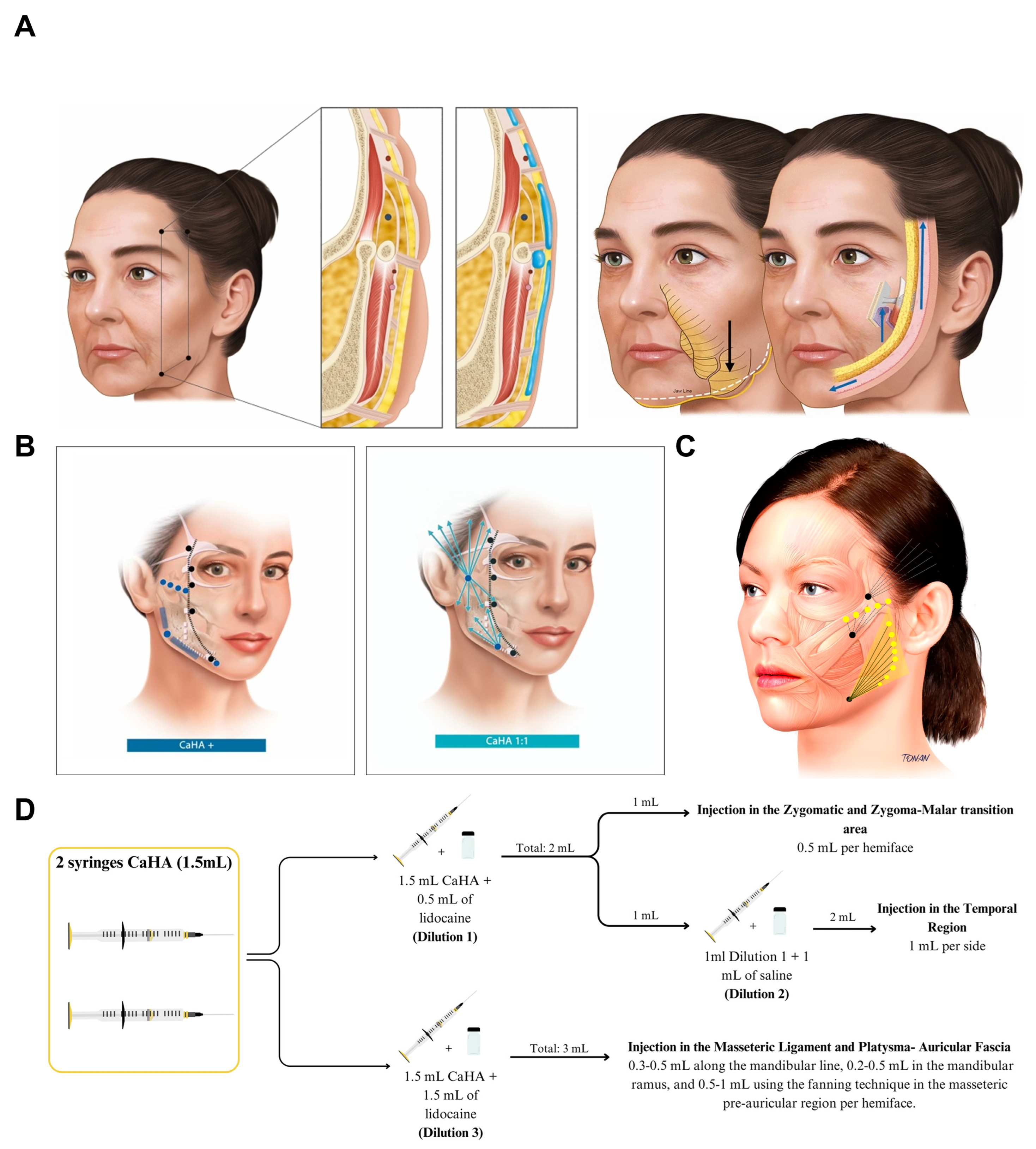
Global Aesthetic Improvement Scale (GAIS) outcomes indicated improvement in the upper face in 66% of cases and in the mid-to-lower face in 83% [85]. However, the study was limited by a very small sample size (n = 6) and the absence of a control group, substantially reducing statistical power; larger, multicenter studies integrating histology and imaging are warranted.
Subsequently, Saavedra refined three dilution ratios to match region-specific goals: lower dilution for immediate support in the zygomatic and zygomaticomalar regions, higher dilution for ligament repositioning in the temporal region, and intermediate dilution for mandibular contouring and regenerative needs (Figure 5C,D). Evaluations using a 5-point Likert scale, FACE-Q, GAIS, standardized 2D photography, and 3D imaging demonstrated durable and clinically meaningful aesthetic improvements over 12 months, high patient satisfaction, and a favorable safety profile [88].
Notably, injection strategies for the mandibular region vary according to clinical objectives. Hernandez et al. prioritized the restoration of bony support at the mandibular angle and therefore used focal supraperiosteal injections of undiluted CaHA. In contrast, Saavedra et al. aimed to optimize the global mandibular contour and long-term improvement; accordingly, they used a 1:1 dilution at an intermediate concentration and performed linear injections, leveraging diffusion to enhance collagen-stimulatory effects across the region.
Moreover, Hong and Park reported that even undiluted CaHA injected into the subdermal plane can significantly improve skin quality. CaHA not only achieved sustained midface volume restoration within 24 weeks but also produced instrument-confirmed improvements in barrier function, hydration, and biomechanical elasticity [89].
4.3.2. Combined Therapy with Hyaluronic Acid and CaHA
In recent years, hyaluronic acid (HA) and CaHA have attracted increasing interest as a combination approach for facial rejuvenation because of their complementary mechanisms [90]. HA provides volumization through its strong water-binding capacity, thereby correcting contour deficits, whereas CaHA induces fibroblast-mediated synthesis of collagen and elastin, leading to longer-term improvements in skin quality and tissue firmness. Clinical practice has evolved from early exploratory manual premixing to standardized, commercially integrated products (e.g., HArmonyCa™), indicating that HA–CaHA combination therapy is now a mature and promising treatment paradigm. A systematic review suggested that combined use may upregulate pro-regenerative pathways, including VEGF, EGFR, and TGF-β/Smad, thereby enhancing the synthesis of COLI, COLIII, and elastin beyond that achieved by either component alone, while eliciting a lower inflammatory response [91].
A multicenter, retrospective cohort study evaluated the lower-face contouring effects of manually premixed CaHA (Radiesse) and CPM-HA (Belotero Volume) in 41 patients. The mean clinician-rated Merz Aesthetics Scale® for the jawline (CR-MASJ) improved from 2.12 at baseline to 0.68 at 3 months and 1.27 at 12 months. At 3 months, 100% of patients achieved an improvement of ≥1 point, with 43.9% exhibiting a 2-point improvement. At 12 months, 85% maintained an improvement of ≥1 point [92]. While these findings provide supportive clinical evidence, manual premixing is technically demanding, and the rheological properties of the filler may change after mixing, raising concerns regarding consistency and stability.
Building on the clinical rationale established by manual premixing, an integrated commercial formulation (HArmonyCa™; Allergan Aesthetics, an AbbVie company) was developed by combining HA and CaHA with 0.3% lidocaine in a single 1.25-mL syringe. In a prospective, non-randomized interventional study of 15 healthy women treated in the preauricular region, bilateral volume increased significantly over 180 days, while facial tension vectors increased by approximately 2 mm and remained stable during follow-up [93]. Ultrasound demonstrated temporal changes in product distribution, and elastography showed increased tissue viscoelasticity from Day 60 onward. These findings support measurable volumetric, tightening, and tissue-property changes after treatment. However, the imaging findings should not be interpreted as direct histological proof of neocollagenesis. Elastography reflects changes in tissue mechanical properties, and the reported ultrasound patterns were indirect imaging surrogates rather than biopsy-confirmed collagen deposition. Moreover, the study was small, included only women, lacked a control or comparator group, and followed patients for only 6 months, limiting both causal attribution to the individual HA or CaHA components and generalizability. The observed late increase in volume may therefore be consistent with progressive tissue remodeling, but cannot by itself establish CaHA-driven collagen regeneration as the sole mechanism underlying the clinical effect.
Subsequently, Proietti et al. reported a larger retrospective real-world study involving 129 patients with chronoaging, photoaging, and concomitant skin conditions, including oily and acne-prone skin, rosacea, and scarring [94]. At 9 months, both physician- and patient-reported GAIS assessments indicated substantial aesthetic improvement, and standardized 3D imaging documented changes at rest and during facial movement, including smiling, chin depression, and pinch testing. These findings are clinically relevant because they suggest that HArmonyCa™ may provide visible improvement across a heterogeneous real-world population and may retain aesthetic effects during dynamic facial expression. However, the evidence should be interpreted cautiously. The study was retrospective and uncontrolled, treatment sites and injected volumes varied substantially, and many patients had undergone other aesthetic procedures within the surrounding treatment period, making causal attribution to HArmonyCa™ alone difficult. Moreover, the main efficacy assessments relied on an adapted GAIS and photographic evaluation rather than blinded or quantitative biomechanical endpoints. Therefore, the study supports real-world feasibility and hypothesis-generating evidence for dynamic facial improvement, but does not establish comparative efficacy or justify broadening the indication to specific dermatologic subgroups without prospective controlled confirmation.
Barone et al. reported a single illustrative case in which HArmonyCa™ was combined with a high-G′ HA filler (Voluma™; Allergan Aesthetics, an AbbVie company) in a 56-year-old woman with skin laxity, reduced skin thickness and turgor, a negative eyelid vector, and bilateral malar hollowing. Each side was treated with 2.5 mL of HArmonyCa™ and 1 mL of Voluma™, and standardized photographs at 6 months showed restoration of malar fullness, with subjectively improved skin elasticity and high patient satisfaction [95]. This report illustrates the feasibility of combining a hybrid HA/CaHA filler with a structurally supportive HA product to address different components of facial aging within the same treatment plan. However, as a single Level V case report without a control group, quantitative outcome measures, or separate evaluation of the two products, it cannot determine the individual contribution of HArmonyCa™ or Voluma™ to the observed improvement, nor can it establish superiority of the combination over either product alone. The findings should therefore be regarded as hypothesis-generating support for a complementary multimaterial strategy rather than evidence of comparative efficacy.
4.3.3. Combined Strategy of CaHA and MFU-V
A prospective, randomized pilot study compared two sequencing strategies for combined microfocused ultrasound with visualization (MFU-V) and hyperdiluted CaHA-CMC in 12 women with mild-to-moderate lower facial and submental laxity [96]. Patients received either MFU-V followed approximately six weeks later by CaHA-CMC or the reverse sequence. Both groups demonstrated aesthetic improvement, whereas the MFU-V-first group showed greater increases in elastin staining intensity and coverage and generally more favorable GAIS and Patient-Perceived Age outcomes. However, not all clinical endpoints consistently favored this sequence; by Day 120, FACE-Q Satisfaction with Lower Face and Jawline improvements were similar between groups. Given the pilot design, very small sample size (n = 6 per group), short follow-up, and multiple clinical and histological endpoints, these findings should be interpreted as preliminary evidence favoring MFU-V-first sequencing rather than definitive proof of an optimal treatment order.
The biological explanation for this sequence effect also remains hypothetical. The authors proposed that MFU-V-induced wound healing may increase HSP47 expression and fibroblast recruitment, thereby creating a larger pool of cells available for subsequent CaHA–cell interactions, whereas applying MFU-V after CaHA could transiently disrupt newly formed matrix through thermal injury [96]. However, HSP47 expression, fibroblast recruitment, and CaHA-specific mechanotransduction were not directly measured in the trial. Therefore, the proposed synergy between MFU-V and CaHA should be regarded as a mechanistically plausible interpretation of the observed histological pattern rather than a demonstrated causal pathway.
A subsequent secondary exploratory analysis of the same 12-patient cohort examined whether age influenced elastin remodeling after combined treatment [97]. No significant correlation was detected between age and changes in elastin staining intensity or area across patients aged 39–62 years. However, because the analysis was underpowered and excluded both younger and older populations, the absence of a significant association should not be interpreted as evidence that age has no effect on biostimulatory responsiveness. Rather, the findings suggest that a strong age-dependent effect was not detectable within this limited cohort and require confirmation in larger, prospectively powered studies covering a broader age range.
4.3.4. Combined Strategy of CaHA and Absorbable Threads
The BIO3 protocol has been evaluated using a combined in vitro and retrospective clinical design [98]. In a full-thickness human skin-equivalent model, combined exposure to CaHA and P(LA/CL) thread fragments produced greater upregulation of COL1A1, COL3A1, and ELN than either treatment alone, supporting the biological plausibility of complementary ECM stimulation. However, these findings reflect gene-expression changes in an in vitro model and should not be interpreted as direct evidence of synergistic tissue regeneration in vivo.
Clinically, the retrospective analysis included 11 women treated either with threads alone (n = 6) or with CaHA approximately 60 days before thread placement (n = 5). Both groups showed aesthetic improvement, while the CaHA-pretreated group demonstrated numerically greater increases in dermal and subcutaneous thickness on ultrasound and a larger proportion of high-category GAIS responses at Day 120. However, these findings were not uniformly supported across outcomes: between-group differences in WSRS were not significant, and facial laxity scores at Day 120 favored the threads-only group. In addition, most ultrasound differences did not reach statistical significance, and GAIS results were reported descriptively because of the small sample size.
The authors proposed that CaHA-induced dermal densification may improve thread anchorage and tissue support, but this mechanism was not directly tested histologically or biomechanically. Therefore, the available data support the feasibility and biological plausibility of sequential CaHA–thread treatment, but do not yet establish clinical synergy, superior efficacy, or enhanced thread anchorage. Larger prospective controlled studies are required to determine whether the apparent imaging and aesthetic differences translate into a reproducible clinical advantage.
Overall, these findings support a layered regenerative paradigm in which biochemical stimulation and mechanical reinforcement act in concert. Such combination strategies may provide a more comprehensive approach to facial rejuvenation by addressing both tissue quality and structural repositioning, representing an emerging direction in minimally invasive aesthetic medicine.
4.3.5. Microneedle-Based Delivery
Microneedling-assisted transepidermal CaHA delivery has recently been explored as an experimental alternative to conventional injection. In a 2025 proof-of-concept study, microneedling was evaluated using ex vivo porcine skin, while the biological effects of CaHA were examined separately in an immortalized human keratinocyte (HaCaT) model [99]. In the ex vivo experiments, high-intensity microneedling produced greater dermal CaHA retention but was accompanied by substantial tissue disruption, whereas low-intensity microneedling resulted in limited deposition that was partially enhanced by hyaluronidase pretreatment. Separately, exposure of HaCaT cells to CaHA nanoparticles increased COL3A1 and COL4A1 mRNA expression without significant changes in COL1A1 or MMP1.
These findings provide proof-of-concept evidence that microneedling can facilitate transepidermal CaHA deposition and that CaHA exposure may influence selected collagen-related transcripts. However, several translational limitations are important. The delivery experiments were performed in ex vivo porcine skin rather than living human tissue, and the molecular findings were obtained from an immortalized keratinocyte culture rather than from microneedle-treated skin. Moreover, increased COL3A1 and COL4A1 mRNA expression does not establish corresponding protein synthesis, organized collagen deposition, basement-membrane restoration, or clinical rejuvenation. The study also employed nanoscale CaHA dispersed in PVP rather than a conventional commercial CaHA-CMC microsphere filler, further limiting direct extrapolation to established aesthetic formulations. Therefore, microneedling-assisted CaHA delivery should currently be regarded as an experimental translational strategy requiring in vivo human validation of formulation, dosing, tissue response, long-term safety, and clinical efficacy before it can be considered an alternative to conventional injectable CaHA.
4.3.6. Mechanical Removal Technique
Although the overall safety profile of CaHA is favorable, its non-dissolvable nature remains a major clinical concern. In particular, when complications such as nodule formation, overcorrection, or vascular compression occur, management is substantially more challenging than with HA fillers. Conventional approaches—including local massage, intralesional corticosteroids or 5-fluorouracil injections, and laser-based treatments—have unpredictable efficacy and rarely achieve definitive resolution.
Mechanical removal has been proposed as a rescue option for selected localized CaHA complications. Cohen et al. described a preliminary four-patient case series using a 1-mm grater-type microliposuction cannula under negative pressure to debulk or remove palpable CaHA excess and nodules [100]. In all four cases, the targeted material was reduced or removed without reported short-term complications, and no repeat treatment was required. Ultrasound was used in selected cases to confirm localization and removal.
However, this evidence remains preliminary. The study involved only four patients, lacked a comparator, relied largely on clinical palpation and visual assessment, and provided limited follow-up, so long-term recurrence, reproducibility, and comparative effectiveness are unknown. Importantly, the technique is intended for localized excess material or nodules and is not applicable to inadvertent intravascular injection or embolic vascular occlusion. Mechanical aspiration should therefore be regarded as a technically feasible option for carefully selected CaHA complications rather than an established or broadly validated reversal strategy.
4.4. Safety, Complications, Contraindications, and Limitations of Current Clinical Evidence
4.4.1. Overall Safety Profile and Common Adverse Events
The safety profile of CaHA is best interpreted by distinguishing expected, transient injection-site reactions from adverse events that are clinically significant because of their severity, persistence, or need for intervention, rather than by considering reported event rates in isolation. Across the available clinical literature, pain, erythema, edema, bruising or ecchymosis, swelling, induration, and transient palpable irregularities are among the most frequently reported treatment-related reactions. Most are mild and self-limiting; however, their reported incidence varies markedly across studies [101,102,103]. This heterogeneity is itself important when interpreting the evidence, as observed adverse-event rates are influenced not only by the treatment and injection technique, but also by how events are defined, actively assessed, attributed to the procedure or product, and monitored over time. Accordingly, differences in reported adverse-event frequencies should not automatically be interpreted as differences in the underlying safety of CaHA.
The 2024 systematic review by Zago Sá Fortes et al. provides the broadest quantitative estimate currently available. Among 5032 CaHA-treated patients, early adverse events were reported in 21.8%, compared with 0.85% for delayed events and 0.27% for severe events [102]. Edema, bruising, hematoma, and pain predominated among early reactions. However, the frequency of early events ranged from 0% to 100% across individual studies, and the authors specifically identified the absence of standardized adverse-event definitions and assessment methods as a major source of heterogeneity. This point substantially qualifies interpretation of the pooled 21.8% estimate: it should not be regarded as a universal incidence of early reactions after CaHA treatment, but rather as an aggregate of studies that differed markedly in anatomical site, injection protocol, product formulation, follow-up, and adverse-event ascertainment. Indeed, some studies systematically captured minor expected post-injection changes, whereas others appeared to record primarily events considered clinically meaningful. Thus, differences in reported incidence may partly reflect differences in surveillance intensity rather than true differences in biological tolerability.
This distinction is illustrated by recent facial studies in which active surveillance yielded considerably higher frequencies of minor reactions without corresponding increases in clinical severity. Rovatti et al. retrospectively evaluated 40 patients receiving 1:2 hyperdiluted CaHA for mid- and lower-face rejuvenation [78]. When patients were specifically examined for local reactions, erythema and edema were identified in all patients and persisted for approximately 2–3 weeks, while ecchymosis occurred in 25%. Nevertheless, the mean pain score was only 1.7 on a 10-point scale, all reactions resolved without additional treatment, and no major adverse event occurred. Similarly, in the 2025 prospective split-face study by Bravo et al., systematic 30-day patient diaries identified pain in 80% of participants, together with swelling, induration, redness, palpable lumps, bruising or hematoma, and transient color change; all had resolved by day 30 [103]. These findings demonstrate why the frequency of an injection-site reaction should not be used interchangeably with its clinical significance. A study that actively captures erythema, edema, or discomfort may report a high “adverse-event rate” despite an otherwise uncomplicated recovery.
Conversely, the long-term multicenter trial by Green et al. provides a useful framework for separating the contribution of the procedure itself from that of the implanted material [101]. Among 175 patients treated with CaHA-CMC(+) for jawline contouring and followed for up to 60 weeks, 26.3% experienced at least one treatment-emergent adverse event considered related to the procedure, the product, or both. Events attributed only to the injection procedure—including bruising, erythema, edema, hemorrhage, pain, rash, and swelling—were all mild, with 92.9% resolving within 15 days without sequelae. Product-attributed events included induration, inflammation, local masses, nodules, and product-distribution abnormalities and were likewise predominantly mild, although their resolution was generally less rapid. Three serious adverse events—cholecystitis, erysipelas, and hypertension—were reported during follow-up, but none was considered related to either CaHA-CMC(+) or the injection procedure. This distinction is important when interpreting safety. Transient bruising resulting from tissue trauma during needle or cannula passage, for example, primarily reflects the invasiveness of the injection procedure, whereas a persistent nodule attributed to the implanted material may indicate a product-related tissue response, inappropriate product deposition, or distribution abnormality. Although both may be recorded under the broad category of treatment-emergent adverse events, they should not be interpreted equivalently when assessing the intrinsic safety profile of CaHA. Accordingly, safety evaluation should consider not only the number of reported adverse events, but also their attribution, severity, duration, and requirement for clinical intervention.
Recent prospective controlled evidence further supports the generally favorable short- to intermediate-term tolerability of CaHA-based fillers. In a multicenter, randomized, double-blind trial involving 210 Chinese participants with nasolabial folds, 188 completed 12 months of follow-up. The most frequently reported reactions were redness, swelling, procedural pain, and bruising; all were mild or moderate, resolved within one week without treatment, and no participant discontinued because of an adverse event. However, these findings remain formulation- and indication-specific and do not exclude rare or delayed complications that would require substantially larger cohorts and longer surveillance [104].
Taken together, the available evidence supports a generally acceptable short- to intermediate-term tolerability profile for appropriately administered CaHA, but also highlights important limitations in how this conclusion should be interpreted. First, minor local reactions may be substantially under- or over-represented depending on whether studies use spontaneous reporting, investigator examination, or structured patient diaries. Second, adverse-event rates derived from one anatomical region or injection technique cannot be assumed to apply uniformly across the face. Third, even relatively large prospective trials remain inadequately powered to define the incidence of very rare complications. Finally, studies with short follow-up may provide reassurance regarding early tolerability while being inherently unable to exclude delayed inflammatory or foreign-body reactions; for example, the 2025 prospective comparative study followed patients for only four months, a limitation explicitly acknowledged by the investigators [103]. Therefore, the most defensible interpretation of the current evidence is not that CaHA is “free of significant adverse effects,” but that common treatment-associated reactions are predominantly transient and manageable, whereas the true risk profile must also incorporate less frequent, delayed, and potentially severe complications that require separate consideration.
4.4.2. Severe Complications and Their Management
Acute vascular complications and visual loss. Although vascular complications following CaHA injection are rare, their potential consequences are disproportionately severe. Inadvertent intra-arterial injection may result in tissue ischemia, necrosis, permanent scarring, visual impairment, or blindness. A recent systematic review identified at least 11 published cases of CaHA-associated vascular occlusion with visual impairment, most following injection to the nasal dorsum and several involving the glabellar region. The proposed mechanism involves inadvertent intra-arterial injection followed by retrograde embolization toward the ophthalmic circulation and subsequent obstruction of retinal or ciliary arterial branches. Importantly, the clinical significance of these events is amplified by the limited reversibility of CaHA. Unlike HA fillers, CaHA cannot be enzymatically dissolved with hyaluronidase, and no reliably effective reversal strategy has been established for CaHA-associated retinal vascular occlusion. Current management approaches are therefore largely extrapolated from experience with HA-related vascular events and remain supported predominantly by case reports, expert opinion, and other low-level evidence [105]. Consequently, the rarity of reported visual complications should not be interpreted as evidence of negligible clinical importance: even an exceptionally low-frequency event warrants substantial attention when the potential outcome is permanent and available rescue strategies are uncertain.
The same principle applies to non-ocular vascular compromise. Expert consensus emphasizes that vascular injury may occur despite the use of blunt cannulas or aspiration, neither of which completely eliminates the possibility of intravascular placement [106]. Once vascular compromise is suspected, prompt recognition is critical, particularly when unexpected pain, blanching, livedoid discoloration, or other signs of ischemia develop.
However, available evidence suggests that prognosis and management should not be considered equivalent for ocular and non-ocular vascular events. In a CaHA-specific systematic review of 43 reported vascular complications, Lindgren and Welsh identified 15 vision-related and 23 non-vision-related human cases, together with five experimental studies evaluating potential treatments. Among cases with reported outcomes, complete improvement was documented in 83% of non-visual complications, whereas only 40% of vision-related cases achieved complete or near-complete recovery [107]. Although these proportions derive from heterogeneous case reports rather than denominator-based clinical cohorts and therefore cannot provide comparative recovery probabilities, they illustrate the markedly poorer reversibility of vascular injury once the ophthalmic circulation is involved.
Delayed nodules and foreign-body granulomas represent another clinically relevant complication of CaHA-based biostimulation. A 2025 systematic review identified 40 studies comprising 117 patients with filler-associated granulomatous reactions, of whom 32 cases (27.35%) were associated with CaHA [108]. Importantly, the reported 27.35% represents the proportion of CaHA-associated cases among published granuloma reports and not the incidence of granuloma among all CaHA-treated patients; it therefore cannot be interpreted as a population-level risk estimate. This distinction is essential because complication-focused reviews and case reports are intrinsically enriched for adverse outcomes and do not provide an appropriate denominator for estimating absolute risk. The temporal pattern is nevertheless clinically informative. CaHA-associated granulomatous reactions in the review developed after a median interval of approximately 7.35 months, indicating that clinically relevant inflammatory complications may arise well beyond the follow-up periods used in many efficacy studies. This raises an important limitation in the interpretation of short-term safety data: absence of delayed adverse events during several weeks or months of observation does not exclude later foreign-body or inflammatory reactions. The review further emphasized that granulomatous reactions may be influenced by material characteristics, injection depth, particle aggregation, excessive dosing, inadequate product preparation, and host immune status, although the relative contribution of each remains incompletely defined [108].
Importantly, a post-treatment “nodule” should be regarded as a clinical phenotype rather than a single pathological diagnosis. McCarthy et al. specifically distinguished non-inflammatory focal accumulations of CaHA microspheres from inflammatory nodules and true foreign-body granulomas. Non-inflammatory accumulations are generally attributed to technical factors such as excessive product deposition, superficial placement, inadequate dispersion, rapid injection, or placement in mechanically unfavorable locations; histologically, they lack the inflammatory-cell infiltrate expected in granulomatous lesions. By contrast, delayed inflammatory nodules may be associated with infection, biofilm formation, hypersensitivity, or granulomatous inflammation and may present with erythema, tenderness, edema, or warmth [109]. This distinction has direct therapeutic implications: mechanically redistributing a focal accumulation and suppressing an inflammatory or infectious process address fundamentally different mechanisms and should therefore not be used interchangeably.
McCarthy et al. proposed a stepwise management framework for CaHA focal accumulations based on invasiveness, cost, and potential risk [109]. Small, non-visible lesions may initially be observed for spontaneous degradation, whereas symptomatic or cosmetically evident focal accumulations may be approached with mechanical dispersion and in situ dilution before escalation to pharmacological, device-based, or invasive interventions. Sodium thiosulfate (STS), a calcium-chelating agent proposed as a potential CaHA reversal strategy, has received particular attention; however, the experimental evidence remains inconsistent. The authors further cautioned against treating STS as an established CaHA “reversal agent”; experimental data suggested that its apparent effect may largely reflect particle dispersion rather than true dissolution, while tissue necrosis and hemorrhage have been reported in experimental models. Importantly, this algorithm was developed from a concise literature review and expert synthesis (Level of Evidence 4) rather than comparative clinical trials. It should therefore be interpreted as a pragmatic management framework rather than a validated treatment hierarchy. Nevertheless, its most clinically useful contribution is the principle that treatment selection should follow identification of the underlying nodule phenotype rather than the presence of a palpable lesion alone.
Accurate diagnosis is particularly important because a palpable post-treatment lesion does not necessarily represent the same pathological process. Product accumulation, maldistribution, inflammatory nodules, granulomatous reactions, abscess formation, and vascular complications may overlap clinically but require different management strategies. The distinction between these entities therefore provides the clinical rationale for imaging rather than making ultrasound merely an adjunctive technical investigation. High-frequency ultrasound is increasingly used as an adjunctive diagnostic tool. A 2026 literature review reported that CaHA typically appears as hyperechoic deposits with posterior acoustic shadowing, with the sonographic appearance varying according to dilution and formulation. In suspected complications, color Doppler may assist in evaluating vascular flow, while granulomatous reactions may appear as hypoechoic nodules with variable vascularity; ultrasound can also help identify filler migration, focal overfilling, and abscess formation [110]. Although imaging does not replace clinical assessment and, when necessary, histopathological confirmation, it may improve anatomical localization and help distinguish product-related abnormalities from inflammatory or vascular pathology, particularly in cosmetically sensitive facial regions where biopsy may be undesirable. Accordingly, management of CaHA-associated nodules is best viewed as a diagnosis-directed process: observation or mechanical redistribution may be appropriate for selected non-inflammatory accumulations, whereas inflammatory, infectious, granulomatous, or vascular pathology requires a different therapeutic strategy.
An international expert consensus identified the glabella as contraindicated for CaHA and emphasized heightened caution in vascular high-risk regions, including the nose, nasolabial folds, and periorbital area, where arterial anastomoses may permit embolic propagation toward the ophthalmic circulation. Importantly, neither blunt cannula use nor aspiration completely eliminates intravascular risk, as cannulas may still penetrate arteries and aspiration with CaHA is considered unreliable. Risk reduction therefore depends primarily on detailed vascular-anatomy knowledge, appropriate injection-plane selection, slow low-pressure delivery, avoidance of large boluses, and immediate cessation when unexpected pain or blanching occurs [106].
4.4.3. Contraindications and High-Risk Situations
CaHA treatment should be avoided or postponed in certain anatomical and clinical situations in which the risk of complications is increased. These situations include clearly contraindicated treatment areas, anatomically high-risk regions, previously operated or scarred tissues with potentially altered vascular anatomy, and active local or systemic conditions that may increase procedural risk. Importantly, not all of these situations constitute absolute contraindications. Some require complete avoidance, whereas others warrant temporary deferral or additional precautions before treatment.
From an anatomical perspective, vascular risk is particularly relevant because CaHA is not readily reversible after inadvertent intravascular placement. A CaHA-specific expert consensus identified the glabella as a contraindicated treatment area and emphasized increased vascular risk in the nose and nasolabial folds, where arterial connections may permit embolization toward the ophthalmic circulation [106]. The nasal tip and alar region were also classified as not recommended treatment sites in the consensus framework. This recommendation is consistent with their relatively limited collateral blood supply, such that occlusion of terminal arterial branches may more readily result in tissue ischemia and necrosis. A “high-risk area” should not be interpreted simply as any region containing major vessels. Risk is increased in regions with limited collateral circulation, where arterial occlusion is more likely to result in tissue ischemia and necrosis. It is also elevated in areas with extensive arterial anastomoses, which may permit retrograde embolization to reach functionally critical end organs such as the retina. Accordingly, the glabella, nasal region, and other areas with potential ophthalmic arterial connections warrant substantially greater caution than routine facial augmentation sites.
The infraorbital region represents a specific anatomical limitation for direct CaHA injection. In the Pan-Asian consensus, CaHA treatment of the anteromedial cheek was considered acceptable, whereas direct injection into the infraorbital area was specifically discouraged for safety reasons; the panel recommended HA fillers when correction of this region was required [111]. This distinction is anatomically plausible because the infraorbital region has a thin dermis, minimal subcutaneous fat, and high aesthetic visibility, leaving little soft-tissue coverage to conceal even small irregularities in filler placement. A recent two-case report further emphasized that relatively superficial or excessive product deposition in this region can produce clinically apparent nodules because minor volume irregularities are readily visible through the thin tissue envelope [112].
Previous surgery or scarring may create a high-risk setting by altering the normal vascular anatomy of the treatment area. In patients with a history of rhinoplasty, facial vessels may be displaced from their usual anatomical course, while the operated tissues may also have a more tenuous blood supply. As a result, injection based solely on expected anatomical landmarks may be less reliable, and vascular injury or ischemic complications may occur more readily. Scar tissue may further increase risk by fixing vessels within the surrounding tissue, reducing their ability to move away from an advancing needle or cannula and thereby making them more susceptible to direct penetration [106].
Recent ultrasound studies provide direct imaging evidence that nasal vascular anatomy may be less predictable than conventional anatomical landmarks suggest. In a Doppler ultrasound study reviewed in 2026, approximately half of the examined subjects had at least one vessel within the deep midline of the nose, despite this plane traditionally being regarded as relatively safe for filler injection. This finding indicates that a presumed “safe” plane cannot be assumed to be avascular in every patient and that blind injection may therefore underestimate individual vascular risk. The same review also highlighted that previous rhinoplasty can alter nasal vascularization, making vessel location less predictable during subsequent filler correction. In such post-surgical cases, pre-procedural ultrasound may help identify the actual vascular course before injection and thereby reduce reliance on potentially misleading surface or textbook anatomical landmarks [113].
A separate group of high-risk situations relates to active local or systemic conditions that may increase the likelihood of infection or complicate post-treatment recovery. In broader filler consensus guidance, injection is discouraged in areas affected by active inflammatory or infectious processes, such as active acne or herpes, and treatment should be postponed in patients who are acutely unwell, including those with fever, an upper respiratory infection, or influenza. Pretreatment assessment should also identify conditions associated with increased infection risk, including immunocompromised states, recurrent skin disease, poor periodontal hygiene, previous filler-related complications, multiple prior filler treatments, and poorly controlled type 2 diabetes [114]. Importantly, the consensus does not regard stable autoimmune disease as an absolute contraindication; patients with preserved wound healing may be considered individually. These recommendations highlight a clinically relevant distinction between conditions that require temporary deferral because of active infection or systemic illness and background comorbidities that warrant individualized risk assessment rather than automatic exclusion.
Prospective CaHA trials have commonly adopted conservative exclusion criteria, including active local inflammation or infection, unhealed wounds, known hypersensitivity to filler components, pregnancy or lactation, keloid tendency, inflammatory autoimmune disease, recent facial procedures, and severe systemic illness [104]. These criteria help define the populations in which safety has been prospectively evaluated but should not automatically be interpreted as universally established absolute contraindications. Clinical decision-making should therefore distinguish evidence-based contraindications from precautionary trial exclusions and individualized risk factors.
Taken together, contraindications and high-risk situations for CaHA should be evaluated according to the mechanism and reversibility of the anticipated risk rather than treated as a single category. Some situations, such as injection into explicitly contraindicated anatomical sites, warrant complete avoidance, whereas active infection or acute systemic illness generally calls for temporary treatment deferral. Other circumstances, including surgically altered anatomy or uncertain vascular pathways, require heightened caution and may justify additional imaging, modification of the treatment plan, or selection of an alternative modality. This risk-stratified approach is particularly important for CaHA because misplaced or intravascular product cannot be readily reversed. At the same time, many of these recommendations are derived from expert consensus, anatomical studies, or limited clinical evidence rather than high-level comparative trials; therefore, they should be interpreted as precautionary clinical guidance rather than uniformly validated contraindications.
4.4.4. Treatment Limitations, Inter-Patient Variability and Patient Selection
Despite its ability to improve contour definition, structural support, and localized volume, CaHA has an inherent therapeutic ceiling when the dominant aging changes extend beyond volume deficiency or loss of skeletal support. In CaHA(+)-specific guidance for jawline rejuvenation, patients with marked jowl formation, substantial lower-face skin laxity, or excess cervical adiposity were considered unlikely to achieve optimal results with filler treatment alone. In these patients, the principal deformity reflects tissue redundancy, descent, or excess adipose tissue rather than an isolated volumetric deficit that can be corrected by augmentation. Accordingly, skin-tightening procedures, fat-reduction strategies, or other complementary modalities may be required [115]. These observations define an important limitation of CaHA-based rejuvenation: although the material can restore contour, projection, and support, it cannot directly remove redundant skin, reduce excess adipose tissue, or anatomically reposition markedly descended soft tissues. Therefore, in advanced lower-face aging, CaHA should be regarded as a supportive or adjunctive intervention rather than a substitute for procedures that directly address tissue laxity, excess volume, or structural descent.
Beyond indication-related limitations, CaHA also has important material- and technique-dependent constraints. Its relative firmness and limited reversibility reduce the margin for error when precise placement cannot be ensured, and inappropriate deposition or overcorrection cannot be rapidly reversed enzymatically as with HA fillers. Outcomes are also influenced by injection plane, dilution, injected volume, anatomical site, and operator technique. Real-world evidence from an HA–CaHA hybrid formulation further showed that non-inflammatory nodules were frequently attributed to focal product accumulation and were associated with procedural factors such as bolus injection, superficial deposition, excessive local volume, and treatment of highly mobile areas [116]. Although these findings derive from a hybrid formulation rather than isolated CaHA, they reinforce the broader principle that the clinical safety and performance of CaHA-based treatment cannot be separated from injection technique and anatomical context.
Among patients for whom CaHA is considered appropriate, treatment still cannot be standardized across individuals because baseline facial morphology, skin characteristics, and desired facial shape and contour vary substantially. The Pan-Asian CaHA consensus specifically identified differences from Caucasian and Latin American populations in acne-scarring tendency, skin thickness, pigmentation, pore size, and sebum secretion. Importantly, Asian patients should not be considered a morphologically homogeneous population. The consensus noted substantial variation in facial shape and aesthetic preferences even among Asian ethnicities; for example, Patients in Hong Kong and Taiwan commonly present with relatively broad, euryprosopic faces but may seek a more elongated, leptoprosopic contour, whereas preferences for facial length differ among Singaporean, Korean, and Malaysian populations [111]. These differences are clinically relevant because the amount and location of structural augmentation required to narrow, elongate, or redefine facial contours cannot be standardized across populations. Accordingly, CaHA treatment planning should account not only for skin characteristics but also for baseline facial morphology and the specific proportional change sought by the individual patient, rather than applying a uniform contouring strategy to all Asian patients.
Skin thickness and collagen-producing capacity are two practical determinants of how hyperdilute CaHA should be used. Lorenc et al. recommended progressively greater dilution as the skin becomes thinner: approximately 1:2–1:3 for normal skin, 1:4 for thin skin, and up to 1:6 for atrophic skin, because a more dilute preparation reduces the risk of visible or palpable product in tissues with limited soft-tissue coverage. Treatment frequency should likewise be adjusted to the patient rather than fixed in advance. The authors noted that appreciable collagen-related improvement generally develops after approximately 4–6 weeks and that two to three sessions, usually spaced about six weeks apart, are commonly required for an optimal response. Patients with more severe laxity or lower collagen-producing capacity may require additional sessions, and more mature patients may need complementary treatments to achieve comparable improvement [117]. Thus, both dilution and treatment frequency should be selected according to measurable tissue characteristics and the anticipated regenerative capacity of the patient, rather than applied as a uniform protocol.
Inter-patient variability may also extend beyond anatomy to the host biological response to CaHA. Verma et al. reported a 43-year-old woman who developed persistent diffuse edema and induration three weeks after CaHA injection despite having previously tolerated CaHA, PLLA, and HA fillers without complications. The inflammatory reaction was subsequently associated with previously undiagnosed Hashimoto’s thyroiditis and resolved rapidly after thyroid hormone replacement [118]. The authors proposed an autoimmune-mediated foreign-body inflammatory mechanism involving macrophage and T-cell activation. This observation raises the possibility that underlying immune or endocrine conditions may modify tissue responses to CaHA even in patients with previous uneventful filler exposure. However, because this evidence is derived from a single case report, it should be regarded as hypothesis-generating rather than proof that autoimmune disease uniformly predicts an adverse CaHA response or constitutes a general contraindication.
Pre-existing comorbidities may also influence the suitability of CaHA by altering the options available if a complication occurs. In a reported case, a patient with hemophilia A and HIV-associated facial lipoatrophy had undergone more than ten CaHA and PLLA treatments over approximately 20 years, with factor VIII replacement administered before injections under hematologic guidance. After developing a CaHA-associated vascular occlusion in the infraorbital region, standard management options were constrained because antiplatelet and anticoagulant therapies were considered inappropriate in the setting of his bleeding disorder [119]. The authors further speculated that repeated long-term filler treatment may have contributed to local vessel compression through residual volume and collagen stimulation, although this mechanism was not directly demonstrated. This case therefore highlights a specific aspect of patient selection: comorbidities should be evaluated not only for their effect on procedural risk, but also for whether they may restrict the management of a potential adverse event. Given that this evidence is based on a single case, it should not be interpreted as establishing hemophilia or repeated filler exposure as general contraindications to CaHA.
Patient selection is essential when considering CaHA-based facial augmentation. Although CaHA has low immunogenicity and does not routinely require pre-treatment allergy testing, injectable correction may be inappropriate when the underlying deformity exceeds what soft-tissue augmentation can reasonably address. In particular, filler-based treatment has limited efficacy in patients with severe chin retrusion, for whom surgical options such as alloplastic implantation or genioplasty may provide more predictable structural correction. In addition, CaHA should be used cautiously in situations in which precise placement cannot be ensured, because its relatively firm, incompressible characteristics and non-reversibility increase the consequences of overcorrection or inappropriate deposition [120]. Accordingly, contraindication and patient-selection decisions should consider not only material hypersensitivity but also anatomical severity, treatment goals, and whether the defect is fundamentally amenable to injectable correction.
4.4.5. Limitations of Current Clinical Evidence
The current clinical evidence base should nevertheless be interpreted with caution. Although multiple clinical studies support the efficacy and overall safety of CaHA, substantial methodological heterogeneity remains. A recent systematic review of facial CaHA and PLLA studies identified considerable variation in injection techniques, patient-selection criteria, treatment protocols, and follow-up durations, which complicates direct comparison between studies. In addition, many studies relied on subjective assessment tools such as the Global Aesthetic Improvement Scale (GAIS), rather than objective quantitative endpoints, and the reviewed populations were predominantly Caucasian, limiting generalizability across different skin types and ethnic backgrounds. Risk-of-bias assessment further showed that several included studies had concerns or high risk related to randomization, outcome measurement, and selective reporting, indicating that the apparent consistency of favorable clinical outcomes should not be interpreted as equivalent to uniformly high-quality evidence [121].
Evidence gaps are also apparent outside established facial indications. For example, the systematic review of Radiesse® body applications concluded that although diluted and hyperdiluted CaHA/CMC techniques are widely used and generally associated with favorable safety outcomes, many of these newer applications remain insufficiently supported by randomized controlled trials [122].
Evidence regarding CaHA-associated foreign-body granulomas remains particularly limited, as the published literature is dominated by case reports and retrospective observations, frequently with incomplete histological confirmation, heterogeneous diagnostic definitions, and insufficient reporting of injection technique and dose; consequently, the true incidence and comparative risk of this complication cannot currently be reliably determined [108]. The current imaging evidence for filler-related complications is also highly heterogeneous, with much of the available literature consisting of case reports and small case series using inconsistent terminology; consequently, standardized ultrasonographic diagnostic criteria have yet to be fully established [110].
Long-term safety assessment is further constrained by variation in follow-up duration and incomplete synthesis of adverse-event data. A 2026 systematic review protocol designed to quantify adverse events associated with major facial collagen biostimulators noted that no comprehensive synthesis had yet characterized their overall frequency and spectrum, and proposed standardized classification into immediate (<24 h), early (24 h–4 weeks), and delayed (>4 weeks) events. This temporal distinction is particularly important for CaHA, because foreign-body granulomas, persistent edema, biofilm-associated infection, and late inflammatory nodules may not become apparent during short-term follow-up [123]. Therefore, favorable short-term tolerability should not automatically be interpreted as evidence of long-term safety.
Long-term clinical safety requires separate consideration from short-term tolerability and treatment durability. One of the longest CaHA-specific follow-up studies was an extension of the pivotal Radiesse trial, in which 102 patients with nasolabial folds entered long-term follow-up and 99 were followed for approximately 3 years. No nodules, granulomas, necrosis, erosion, infections, or other delayed adverse events were reported at the 24- and 36-month evaluations, with follow-up extending to 39 months after the last CaHA injection [124]. These findings provide reassuring long-term CaHA-specific evidence; however, a cohort of approximately 100 patients remains insufficient to exclude very rare events, and adverse-event ascertainment relied on clinical examination and patient reporting.
More recent studies extend the safety evidence but also illustrate its limitations. A 2025 study of diluted STIIM CaHA reported only mild, self-limited bruising, tenderness, and edema and no serious adverse events during a 360-day observation period; however, the study included only 32 participants, lacked a control group, and only 16 patients were available for the 360-day assessment [125]. In a larger real-world multicenter analysis of 403 patients treated with an HA–CaHA hybrid filler, 243 had at least 12 months of exposure, with a median interval of 15.4 months from first treatment to chart review. Most documented treatment-emergent events were mild, and no treatment-emergent serious adverse events were reported; nevertheless, this evidence cannot be directly extrapolated to isolated CaHA because the product contained both HA and CaHA, and the retrospective design, concomitant aesthetic treatments, and reliance on medical records may have underestimated event rates [116].
Taken together, the available evidence is more robust for common short-term injection-related reactions than for rare, delayed, or cumulative complications. Importantly, evidence that aesthetic benefit can persist for 12–39 months should not be conflated with evidence that long-term safety has been definitively established. Large denominator-based prospective cohorts with standardized adverse-event surveillance, extended follow-up, and documentation of repeated treatments are still needed to define the true incidence of delayed inflammatory, granulomatous, infectious, and vascular complications.
Accordingly, larger multicenter prospective studies using standardized treatment protocols, objective outcome measures, longer follow-up, more diverse patient populations, and systematic adverse-event reporting are still required to better define the long-term benefit–risk profile of CaHA.
The study designs, treatment strategies, clinical outcomes, and reported adverse events of representative clinical studies evaluating CaHA-based facial rejuvenation are summarized in Table 3.
Table 3.
Summary of Representative Clinical Studies on the Efficacy and Safety of Calcium Hydroxylapatite in Facial Rejuvenation.
4.5. Comparative Clinical Positioning of CaHA Among Injectable Fillers and Biostimulators
Although HA, CaHA, PLLA, and PCL are often discussed within the same broad landscape of injectable facial rejuvenation, their clinical roles are not interchangeable. A more useful comparison is not simply whether a material functions as a “filler” or a “biostimulator,” but how its effects are distributed across immediate correction, mechanical support, progressive tissue remodeling, durability, and safety. From this perspective, CaHA occupies a distinctive position because its initial structural behavior and subsequent biological activity can both contribute to the clinical outcome, while dilution further allows the balance between these two functions to be modified according to the therapeutic objective.
4.5.1. Biological Mechanisms and Regenerative Profiles
Although CaHA, PLLA, and PCL are all classified as biostimulatory injectables, their regenerative pathways are not identical. PCL is mechanistically closest to CaHA in that both rely on persistent particulate scaffolds that support collagen deposition; however, the surrounding tissue response appears to differ. Human biopsy studies of PCL have demonstrated fibroblastic and histiocytic infiltration around persistent microspheres, with eosinophils also reported in some specimens, whereas comparable eosinophilic infiltration has not been consistently described after CaHA treatment. PCL particles may also remain detectable for several years, suggesting a more prolonged material-associated stimulus than CaHA. Haddad et al. therefore considered PCL and CaHA broadly similar in their capacity to induce neocollagenesis, while emphasizing differences in particle persistence and the character of the local tissue response [127].
PLLA differs more clearly from CaHA in the pathway by which fibroblast activation is initiated. Smith et al. describe PLLA degradation as a macrophage-mediated process that triggers cytokine signaling and indirectly stimulates fibroblasts, whereas CaHA is characterized more prominently by scaffold-associated fibroblast activation and ECM remodeling. This difference provides a plausible biological basis for the slower and more variable development of PLLA-associated clinical effects. Importantly, however, the mechanistic evidence for PCL remains much less robust: only two PCL studies were included in the 2025 systematic review, and PCL could not be incorporated into the meta-analysis because of insufficient data [128]. Thus, CaHA, PLLA, and PCL should not be regarded as mechanistically interchangeable simply because all three ultimately promote collagen formation; they differ in the cellular route of stimulation, inflammatory contribution, and persistence of the material-associated remodeling stimulus.
HA occupies a different position because its principal effect is not mediated by a persistent particulate scaffold. Its primary action is hydrophilic volume replacement, although cross-linked HA can secondarily stimulate collagen synthesis through restoration of mechanical tension within the dermal ECM. Human studies summarized by Haddad et al. reported increased procollagen expression and mechanically stretched fibroblasts after HA injection, but the magnitude and durability of this response remain incompletely quantified [127]. Accordingly, HA-associated remodeling is better regarded as a secondary mechanobiological effect rather than a regenerative pathway directly equivalent to the particle-mediated biostimulation of CaHA, PLLA, or PCL.
Recent molecular evidence suggests that the distinction between CaHA and PLLA may extend beyond differences in treatment kinetics to the biological compartments they influence. In a randomized comparative study of 21 subjects with nasolabial-fold deficiency, bulk RNA sequencing of treated tissue demonstrated distinct gene-expression profiles 90 days after PLLA and CaHA injection. PLLA was uniquely associated with the regulation of genes related to adipocyte function and regeneration, whereas a comparable adipocyte-related signature was not identified after CaHA treatment. These findings raise the possibility that PLLA may influence the aging soft-tissue environment through mechanisms extending beyond fibroblast-mediated collagen remodeling, potentially including adipocyte-associated regenerative pathways. By contrast, the currently characterized biological effects of CaHA remain more strongly centered on fibroblast–particle interactions, ECM remodeling, and material-dependent structural support [67]. Thus, the distinction between the two materials may involve not only the speed of biostimulation but also the tissue compartment in which regenerative signaling predominates. However, these transcriptomic findings remain mechanistic and hypothesis-generating and should not be interpreted as direct evidence of clinically meaningful adipose regeneration after PLLA treatment.
Viewed together, these materials should therefore not be treated as interchangeable “collagen stimulators.” Their common endpoint of increased ECM deposition is reached through biologically distinct routes. HA primarily restores volume through hydrophilic expansion and rheology-dependent mechanical support, while any subsequent collagen response appears to represent a secondary mechanobiological effect. CaHA combines immediate material-based support with microsphere-associated fibroblast activation and ECM remodeling. PLLA relies more strongly on particle degradation, macrophage recruitment, cytokine signaling, and subsequent fibroblast activation, whereas PCL couples a persistent microsphere scaffold with a comparatively prolonged foreign-body-associated remodeling response. These distinctions are clinically relevant because the mechanism of stimulation influences not only collagen production, but also onset of effect, need for repeated treatment, persistence of the material, and the phenotype of delayed complications. However, mechanistic evidence is not equally mature across materials: PCL remains comparatively understudied, and some PLLA datasets are confounded by concomitant energy-based treatments, limiting attribution of observed remodeling to the injectable alone [128,129].
4.5.2. Clinical Efficacy and Functional Positioning
A clinically relevant distinction between CaHA and HA lies in the balance between structural support and soft-tissue volumization. In a prospective, randomized single-center study comparing equal 3-mL volumes of CaHA(+) and VYC-20L (Juvéderm Voluma®, a highly cross-linked HA filler) for midface volume loss, both treatments produced sustained aesthetic improvement over 1 year, but a greater proportion of CaHA-treated patients remained at least “very improved” at 90, 180, and 360 days. CaHA was also associated with lower cost per point of patient-reported aesthetic improvement in this specific practice setting. Importantly, the authors emphasized that these differences should be interpreted in the context of distinct rheological properties and mechanisms of action rather than as evidence of universal superiority. CaHA exhibited substantially greater stiffness and complex viscosity than VYC-20L, supporting its use when tissue elevation and foundational support are required, whereas VYC-20L was considered better suited to reproducing soft-tissue volume in patients without substantial tissue descent [126]. Thus, the comparison illustrates that individualized filler selection should be guided by the dominant anatomical deficit—structural support versus volume replacement—as well as reversibility, durability, and patient goals, rather than by a simple hierarchy of filler efficacy.
The comparison between CaHA and PCL is more revealing when viewed through the concept of mechanical phenotype rather than longevity alone. In a retrospective study of 147 patients undergoing T-zone rejuvenation, both materials produced clinical improvement, but CaHA was associated with greater tactile hardness, lower dynamic nasion displacement, higher nasofrontal-angle target achievement, and better maintenance of radix height and chin projection. The reported mean duration was also longer with CaHA, while overall adverse-event rates were comparable. Rather than simply indicating that CaHA “performs better,” these findings suggest that the two materials occupy different points along a rigidity–elasticity continuum: CaHA may be better suited to patients requiring stronger projection and resistance to dynamic deformation, whereas PCL may be more appropriate when a softer tactile profile and more gradual correction are preferred. The authors themselves proposed material selection according to bony contour requirements, soft-tissue characteristics, and tolerance for gradual change [130]. This study therefore supports a clinically useful principle: regenerative fillers should be selected not only by durability, but by the type of mechanical behavior required in the target anatomy.
The clinical overlap between CaHA and PLLA is substantial. A recent systematic review including 14 facial studies found that both materials improved clinically relevant aging outcomes, including skin elasticity, wrinkle severity, and facial volume. However, the reported CaHA studies more frequently emphasized contour restoration and focal volumetric correction, particularly in the jawline and nasolabial folds, whereas PLLA studies more commonly assessed progressive improvements in skin quality, elasticity, and diffuse volume restoration. Treatment protocols also differed considerably: CaHA was often administered in a single session with an optional touch-up, whereas PLLA protocols extended to as many as six treatment sessions in some studies. These findings suggest that the two materials have overlapping efficacy but are not functionally identical in practice; CaHA is particularly suited to indications in which immediate contour correction or structural augmentation is required, whereas PLLA is more commonly used when gradual, distributed tissue restoration is the principal objective. Importantly, this positioning should not be interpreted as evidence of comparative superiority, because the included studies differed substantially in anatomical indication, injection volume, number of sessions, and outcome measures, and several had methodological concerns or high risk of bias [121].
A broader systematic review of controlled clinical trials further illustrates both the efficacy and the limits of the current comparative evidence for CaHA. Amiri et al. identified 13 controlled trials involving 1171 participants, including eight studies of facial treatment; facial comparators included HA fillers, human collagen, PLLA, cross-linked polyacrylamide, and autologous fat. In nasolabial-fold studies, CaHA produced greater global aesthetic improvement than nonanimal-stabilized HA in one split-face trial, with evaluators rating the CaHA-treated side as superior in 47% of participants and inferior in only 5%. Another study found greater wrinkle-severity improvement with CaHA than HA in 31% of treated folds, whereas a separate trial comparing CaHA with several HA formulations found no significant between-product difference on the wrinkle-severity scale [131]. These apparently divergent findings are clinically important because they argue against treating filler efficacy as a fixed material property independent of indication, formulation, and endpoint. Rather, the available controlled evidence supports CaHA as an effective option for facial contouring and wrinkle correction, but does not establish consistent superiority over alternative fillers across indications. The substantial heterogeneity of the available evidence limits direct efficacy comparisons. Most comparator materials were represented by only one or two studies, outcome measures varied considerably, and all included trials had at least one domain rated as high risk of bias. Therefore, the current evidence is insufficient to establish a definitive efficacy hierarchy and is better interpreted in terms of the distinct clinical roles of individual materials.
These findings suggest that comparative efficacy is better conceptualized as “deficit–material matching” rather than as a fixed hierarchy of filler performance. HA remains particularly adaptable when the dominant objective is soft-volume replacement, contour blending, or treatment in mobile regions where rheological flexibility and reversibility are valued. CaHA is clinically distinct when immediate projection or tissue support is desired together with subsequent matrix remodeling, particularly in structurally deficient regions such as the jawline or chin. PLLA is more naturally positioned for patients willing to accept a gradual response and repeated treatment sessions in exchange for diffuse, progressive tissue restoration, whereas PCL combines immediate carrier-mediated correction with longer-lasting scaffold-associated augmentation. Importantly, these roles overlap rather than define rigid indications, and individual products within each material class may differ substantially in rheology, particle properties, injection protocol, and clinical behavior. Accordingly, efficacy comparisons that ignore anatomical indication, treatment volume, number of sessions, and endpoint definition risk attributing to the material what may actually reflect differences in protocol or patient selection [129].
The emergence of combined and hybrid HA–CaHA formulations further illustrates why filler classes should not be treated as mutually exclusive categories. Conceptually, HA can provide immediate hydration and volumetric integration while CaHA contributes structural behavior and delayed tissue remodeling, potentially combining complementary temporal and mechanical profiles. However, the current evidence does not establish that combining the two materials necessarily produces biological synergy rather than additive effects. A recent systematic review identified substantial heterogeneity in HA–CaHA products, mixing ratios, injection sequence, timing, treated sites, and outcome measures, with generally short follow-up and limited mechanistic validation. Accordingly, hybrid formulations should presently be viewed as an evolving strategy for combining material properties rather than proof that the biological limitations of either component have been overcome [91].
4.5.3. Onset, Duration and Longevity of Clinical Effects
The temporal profile of clinical response differs substantially among HA, CaHA, PLLA, and PCL and should not be reduced to a single nominal “duration” for each material. CaHA provides immediate contour correction through the carrier gel and mechanical properties of the injected product, followed by a more prolonged tissue-remodeling phase. Long-term prospective data support this biphasic clinical course. In a randomized multicenter study of CaHA-CMC(+) for jawline treatment, 77.9% and 78.7% of patients remained ≥1-point responders on the Merz Jawline Assessment Scale at 12 and 24 weeks, respectively, and 62.9% remained responders at 48 weeks. Among patients who did not undergo retreatment at week 48, 43.5% still met the responder criterion at week 60, while 56.5% continued to report subjective aesthetic improvement [101]. These data indicate that clinically meaningful correction can persist beyond one year in a substantial proportion of patients, but they also demonstrate a progressive decline in response rather than a fixed duration of effect.
A systematic review of 14 facial studies reported clinical effects persisting for up to approximately 25 months in some PLLA studies, compared with commonly reported durations of 12–18 months for CaHA [121]. This difference, however, should not be interpreted as a simple shorter-versus-longer hierarchy. CaHA combines an early structural effect with subsequent tissue remodeling, whereas PLLA follows a more gradual course in which clinical improvement depends predominantly on progressive collagen remodeling and is often built through repeated treatment sessions. A 2024 systematic review of 11 randomized controlled trials found that several studies reported sustained improvements in dermal thickness, facial lipoatrophy severity, and aesthetic scores for at least 25 months. However, the treatments underlying these long-term outcomes were heterogeneous and frequently involved repeated sessions; for example, contemporary trials permitted up to four injections at approximately 4-week intervals [132]. Thus, the prolonged effect of PLLA should be interpreted as the cumulative result of serial treatment and induced collagen remodeling rather than as persistence of the initially injected material itself. Direct longevity comparisons with CaHA remain limited because the available studies differ substantially in product formulation, number of treatment sessions, dose, anatomical indication, reconstitution protocol, and outcome definition. In addition, five of the 11 PLLA trials were judged to be at high risk of bias, further limiting the strength of any definitive head-to-head durability ranking.
PCL combines an immediate volumizing phase with a prolonged material-associated biostimulatory effect. In a prospective European multicenter study of 90 patients treated with a single PCL injection for nasolabial folds and no subsequent touch-up, 92% remained WSRS responders at 6 months, 84% at 12 months, and 64% at 18 months; investigator-rated global aesthetic improvement was still present in approximately 80% of patients at month 18 [133]. Because no additional treatment was allowed during follow-up, these results provide relatively direct evidence of sustained efficacy after a single treatment session.
Objective volumetric data provide complementary evidence for PCL persistence. In a retrospective case series using three-dimensional stereophotogrammetry, all nine patients treated with PCL-1 in the midface retained volume gains at 2 years, with measured increases of approximately 50–150% relative to the initially injected volume. This finding is consistent with a sustained tissue-augmentation effect after dissipation of the carrier gel and suggests that the long-term clinical correction is not explained solely by persistence of the initial injected volume [134]. Nevertheless, the study involved only nine patients, and the authors themselves emphasized the need for larger prospective studies; therefore, the magnitude of the reported 2-year volumetric increase should not be generalized to PCL-treated patients as a whole.
HA longevity is particularly formulation-dependent and should not be represented by a single class-wide duration. Recent data with resilient HA illustrate this point. In a 2025 study of RHA4 used for multilayer midface treatment, short-term clinical improvement was evident within 2 weeks, while an illustrative long-term case showed maintained correction of midface sagging, the lid–cheek junction, nasolabial folds, and jowls over 2 years without additional touch-up or apparent migration [135]. This observation demonstrates that selected contemporary HA formulations can retain clinically visible effects considerably longer than the traditionally cited duration of conventional HA fillers. However, the 2-year observation was based on a single case rather than a controlled long-term cohort and therefore should be interpreted as proof of possibility rather than an estimate of expected durability for HA fillers in general.
Longevity also requires conceptual clarification because material persistence, duration of biological remodeling, and duration of visible clinical correction are not synonymous. For HA, persistence is strongly influenced by cross-linking, rheology, anatomical site, and enzymatic degradation. In CaHA, the CMC carrier contributes to early correction, whereas subsequent collagen remodeling may sustain clinical benefit after the carrier phase has diminished. PLLA provides an instructive contrast: clinical improvement has been reported for up to approximately 25 months, although the injected polymer itself is progressively hydrolyzed and may be substantially eliminated earlier; thus, its apparent longevity reflects cumulative host-tissue remodeling rather than continued volumetric occupation by the original suspension. PCL differs again because its microspheres may remain detectable for considerably longer than the typical duration of clinically reported correction. Therefore, a nominal duration in months should not be interpreted as an intrinsic material constant, and cross-material comparisons should distinguish product residence time from tissue remodeling and patient-perceived aesthetic persistence [129,132].
Treatment burden further complicates durability comparisons. A single-session CaHA study and a multi-session PLLA protocol do not provide directly comparable estimates of “longevity,” because the latter may represent the cumulative effect of serial stimulation. Likewise, maintenance treatment, optional touch-up, anatomical site, baseline severity, and outcome threshold can all extend or shorten the reported duration. Although systematic reviews report longer maximum follow-up effects for PLLA than for CaHA, the substantial heterogeneity in dosing schedules and study design precludes interpreting these values as evidence that one material is intrinsically more durable than another.
4.5.4. Safety, Reversibility, and Complication Profiles
Safety represents another clinically important dimension of material selection, although direct comparisons should be interpreted cautiously because adverse-event estimates vary with product formulation, injection site and technique, treatment volume, follow-up duration, and reporting definitions. For HA fillers, a systematic review of 48 high-level evidence studies found that most reported events were transient and mild to moderate, with swelling, erythema, bruising, pain, tenderness, and localized masses being the most frequent reactions; these generally resolved spontaneously within days to weeks. Product- and technique-related events, including nodules, induration, edema, and contour irregularities, were less common but occasionally required medical intervention. Severe adverse events were rare, although persistent edema, angioedema, infection, and other serious complications were reported [136]. An additional practical characteristic of HA is its enzymatic reversibility with hyaluronidase, which provides an important management option when correction is undesirable or certain complications occur. In contrast, CaHA, PLLA, and PCL cannot be rapidly reversed enzymatically, increasing the importance of appropriate patient selection, anatomical knowledge, injection technique, and avoidance of overcorrection.
Safety differences are likewise more informative when interpreted by complication phenotype, latency, and manageability rather than crude event frequency. Mild procedure-related reactions—pain, edema, erythema, bruising, and tenderness—occur across all injectable classes and are generally self-limited. More meaningful distinctions emerge in delayed or severe events. HA may be complicated by persistent edema, inflammatory nodules, infection, migration, and rare vascular occlusion, although hyaluronidase offers a material-specific rescue option. CaHA shares the potential for nodules, granulomatous reactions, and severe vascular complications but lacks rapid enzymatic reversal. PLLA has a particularly technique- and protocol-sensitive delayed adverse-event profile, with papules, induration, and subcutaneous nodules linked in part to particle aggregation, reconstitution, injection technique, and post-treatment care. PCL similarly carries the potential for delayed foreign-body nodules or granulomatous reactions, while the prolonged persistence of its microspheres may extend the period over which material-associated responses remain biologically relevant [128,132].
Importantly, available safety data do not support a simple cross-material ranking. A recent meta-analysis of biostimulants reported high statistical heterogeneity across pooled adverse-event estimates, and PCL could not be included in the quantitative synthesis because of insufficient evidence. The PLLA literature also remains affected by protocol heterogeneity and substantial risk of bias, while comparative CaHA studies differ in anatomical site, dilution, dose, and adverse-event ascertainment [128]. Therefore, clinical safety should be judged not only by how often an event is reported, but also by its severity, timing, reversibility, anatomical consequence, and the quality of evidence used to estimate its frequency.
Reversibility should be considered a clinically independent property rather than simply another component of safety. HA is unique among these four major material classes because enzymatic degradation with hyaluronidase provides a practical means of reducing or removing product in cases of overcorrection, contour irregularity, misplaced material, or selected complications. This advantage should not be overstated, because dissolution of intravascular HA does not guarantee tissue or visual recovery once ischemic injury has occurred. By contrast, CaHA, PLLA, and PCL lack a rapid and predictable enzymatic dissolution strategy. Once these materials are misplaced or excessive, management may depend on observation, massage, corticosteroids or 5-fluorouracil for inflammatory nodules, ultrasound-guided procedures, or mechanical removal in selected cases. Limited reversibility therefore changes the risk calculus before injection: conservative dosing, precise anatomical placement, and avoidance of overcorrection become more consequential when a material cannot be readily removed after treatment [129].
For PLLA, available randomized evidence suggests an acceptable but technique-sensitive safety profile. A 2024 systematic review of 11 RCTs found that most reported adverse events were mild to moderate, including bruising, bleeding, tenderness, firmness, hematoma, erythema, pain, edema, pruritus, and transient neurologic or vasovagal symptoms. A clinically distinctive concern is the development of papules, induration, and subcutaneous nodules, which may persist and were still observed at 2-year follow-up in one trial. The review further noted that particle agglomeration after reconstitution may contribute to nodule formation, while post-injection massage has been recommended as a preventive strategy. Importantly, many adverse events appeared related to injection procedures and post-treatment care rather than PLLA itself [132]. Nevertheless, confidence in these findings remains limited: five of the 11 RCTs had high risk of bias, and substantial heterogeneity in reconstitution and administration protocols precluded quantitative synthesis. Thus, PLLA appears reasonably safe, but its safety remains protocol-dependent and incompletely characterized.
A systematic review of 79 studies provides a more nuanced perspective on the safety of CaHA and PCL. Among 5032 CaHA-treated patients included in quantitative analyses, early, delayed, and severe adverse events occurred in 21.8%, 0.85%, and 0.27%, respectively, whereas corresponding rates among 1119 PCL-treated patients were 83.3%, 5.6%, and 0%. However, these numerical differences should not be interpreted as a direct safety ranking. The PCL evidence base was substantially smaller, and nearly 70% of its pooled sample derived from a single study that classified all participants as having early adverse events, while definitions of early reactions varied across studies. More clinically informative are the distinct temporal and severity profiles: rare severe CaHA complications included vascular occlusion and tissue necrosis, whereas PCL case reports documented nodules and biopsy-confirmed foreign-body granulomas emerging 14–730 days after treatment [102]. Thus, comparative safety should be judged by event severity, latency, manageability, and evidence quality rather than crude incidence alone.
A clinically important limitation of non-HA biostimulators is that delayed nodular complications are not only non-reversible but may also be diagnostically heterogeneous. In a 2024 series of 55 ultrasound-confirmed complications after PLLA, CaHA, or PCL injection, nodules accounted for 89.1% of presentations and 60% occurred more than one month after treatment. The authors emphasized that these nodules may represent either localized product accumulation or a foreign-body granulomatous reaction, two entities that cannot be reliably distinguished on clinical examination alone. High-frequency ultrasound can help differentiate retained filler from ill-defined granulomatous tissue and identify injection depth, migration, inflammation, or vascular involvement. This distinction has direct therapeutic relevance because, unlike HA-related overcorrection, there is no universally effective dissolution strategy for isolated CaHA, PLLA, or PCL. In the reported series, complete resolution was achieved in only 9% of cases despite multiple treatment approaches, and hyaluronidase appeared useful only when an HA-containing CaHA hybrid product was involved [137]. These findings therefore highlight not a higher incidence of complications with biostimulators, but a greater challenge in diagnosing and managing delayed nodular reactions once they occur. The same series also suggests that the clinical phenotype of complications may differ across materials. Nodules predominated among collagen biostimulators, whereas previous large complication series cited by the authors reported edema more frequently with CaHA and nodules more frequently with PLLA. In the present cohort, most biostimulator-associated nodules lacked overt inflammatory signs, and proposed contributors included local product accumulation, particle characteristics, excessive product deposition, injection into highly mobile regions, and inappropriate dilution, although the exact pathogenesis remains unresolved.
Rare vascular events deserve separate consideration because their consequences may be disproportionate to their frequency. A 2026 review of visual loss associated specifically with biostimulatory injectables identified at least 11 published cases of CaHA-related vascular occlusion with visual impairment, most involving the nasal dorsum, and two confirmed cases after PLLA injection; no published case of blindness was identified for PCL, although a facial artery embolism has been reported [105]. The mechanism is analogous to that described for other fillers—unintentional intra-arterial injection followed by retrograde embolization toward the ophthalmic circulation—but management is more problematic for particulate biostimulators because they cannot be enzymatically dissolved. High-risk anatomical regions include the glabella, nasal dorsum, periorbital area, and forehead, while high-pressure bolus injection and altered vascular anatomy after previous surgery may further increase risk. These reports do not establish comparative incidence because the evidence consists largely of case reports and case series, but they demonstrate that the absence of hyaluronidase reversibility becomes most clinically important when a vascular event occurs.
Accordingly, material safety cannot be judged solely by the frequency of adverse events. For HA, reversibility with hyaluronidase provides a unique rescue option; for CaHA, PLLA, and PCL, safety depends more heavily on prevention because delayed nodules, granulomatous reactions, and vascular embolic events lack an equivalent rapid dissolution strategy. Clinical selection should therefore consider not only how often an adverse event occurs, but also whether it is delayed, reversible, and realistically manageable once established.
Taken together, these comparisons suggest that injectable rejuvenation materials are better distinguished by their functional profiles than by a simple filler-versus-biostimulator classification. HA is characterized primarily by precise and adjustable soft-tissue volumization, PCL by a comparatively softer mechanical profile combined with progressive collagen-mediated correction, and PLLA by delayed and cumulative biostimulation that may involve biological compartments beyond fibroblast-mediated ECM remodeling. CaHA, in contrast, occupies a particularly tunable position by combining immediate structural support with subsequent ECM remodeling, with dilution further shifting its clinical behavior from focal projection toward broader biostimulation. Importantly, these distinctions do not establish a universal hierarchy of efficacy. Rather, they support a deficit–material matching approach in which the choice of injectable is guided by the patient’s dominant anatomical deficit, desired onset and evolution of correction, required mechanical behavior, expected durability, reversibility, adverse-event profile, and tolerance for repeat treatment. This comparative perspective therefore complements the individualized rejuvenation framework proposed above and positions CaHA as one option within a broader, mechanism- and material-guided therapeutic spectrum rather than as a universally preferable filler.
5. Conclusions and Future Perspectives
Taken together, facial aging is a process in which a progressive decline of the biomechanical support system—from deep structures (bone) to superficial tissues (fat compartments)—serves as the initiating driver, and is subsequently amplified by interacting mechanisms including the microbiome, epigenetic regulation, immune modulation, pigmentary dysregulation, and telomere attrition.
In this context, CaHA can be regarded as a mechanical scaffold with biologically active tissue interactions, although the strength of evidence differs across the proposed pathways. Direct CaHA–fibroblast contact and temporal collagen remodeling are supported by experimental and human histological observations, whereas the specific mechanotransduction pathways linking particle contact to gene expression remain unresolved. Similarly, in vitro data indicate a relatively limited early pro-inflammatory cytokine response to CaHA, but active M2 polarization, Treg recruitment, and downstream anti-fibrotic effects have not been directly demonstrated in human tissue. Human biopsy studies support temporal changes in COLIII and COLI expression, but these findings cannot currently distinguish restoration of native dermal architecture from an organized reparative matrix. Finally, preclinical studies demonstrate CaHA-associated neovascularization, while preliminary human imaging suggests increased vascular density; whether these vascular changes improve tissue perfusion or metabolic function remains unknown.
From a translational standpoint, two implications follow. First, injection strategies should be optimized with rheological principles in mind: by precisely adjusting dilution ratios to modulate the storage modulus (G′), clinicians can tailor CaHA to distinct objectives, including deep structural support, mid-plane volumization, and superficial tightening. Second, combination regimens (e.g., with HA or MFU-V) offer mechanistic complementarity, but sequencing is critical. Combination regimens, including MFU-V and CaHA, may provide complementary effects, but the optimal treatment sequence remains uncertain. A small randomized pilot study suggested that MFU-V followed by CaHA approximately six weeks later may produce more favorable histological and selected clinical outcomes than the reverse sequence. However, the sample size was very limited, and the proposed explanation—that MFU-V-induced wound healing increases fibroblast availability for subsequent CaHA–cell interactions—was not directly tested. Accordingly, this sequencing strategy should be regarded as preliminary and hypothesis-generating rather than as an established synergistic protocol.
Despite these advances, three key challenges remain for the future development of CaHA. First, at the level of cell-type–specific regulation, existing studies have largely focused on fibroblasts and macrophages; interactions between CaHA and other aging-relevant cell types (e.g., keratinocytes, adipocytes, and stem cells), as well as potential regulation of core aging-driving transcription factors (e.g., KLF6, HES1, and KNG1), remain insufficiently understood. Integrating single-cell omics and related approaches may help map these interactions. Second, in mechanotransduction, the full pathway from the material–tissue interface to nuclear gene expression—including the recognition mechanisms of mechanosensors (Piezo/TRPV4 channels and integrin complexes), spatiotemporal activation patterns of intracellular mechanotransduction pathways (e.g., YAP/TAZ), and the ultimate program that induces regeneration-specific transcription—still lacks systematic delineation. Future work should integrate materials, mechanical characterization, mechanosensor activation/inhibition experiments, and quantitative assays of key pathway proteins. Finally, smart biomaterial development represents a promising future research direction rather than an established clinical pathway. This includes developing targeted surface functionalization (e.g., coupling growth factors or senolytic drugs that selectively induce apoptosis of senescent cells) to enable precise localization and controlled release of therapeutic molecules, designing smart gels responsive to the local microenvironment (e.g., pH and MMP concentrations), and integrating multimodal composite systems (photothermal, magnetic, etc.) to achieve on-demand dynamic regulation of material behavior.
Overall, CaHA should be regarded as a promising and increasingly evidence-supported biomaterial for facial rejuvenation rather than as a fully established regenerative therapy. Current human clinical and histological evidence supports its structural effects, fibroblast-associated activity, and temporal extracellular-matrix remodeling, whereas several proposed regenerative pathways—including specific mechanotransduction cascades, immune-regulatory effects, functional angiogenesis, and cell-type–specific responses—remain incompletely validated in human facial tissue. In addition, the clinical literature remains limited by heterogeneity in treatment protocols, dilution strategies, anatomical indications, outcome measures, follow-up duration, and adverse-event ascertainment, while high-quality long-term and comparative evidence remains relatively limited. Accordingly, future progress should focus not only on expanding clinical applications, but also on validating mechanisms in human tissue, standardizing treatment protocols, defining long-term benefit–risk profiles, and identifying the patient phenotypes most likely to benefit from specific material and injection strategies.
Author Contributions
Conceptualization, Y.J. and Y.X.; writing—original draft preparation, Y.J.; writing—review and editing, Y.X.; visualization, Y.J.; supervision, Y.X. All authors have read and agreed to the published version of the manuscript.
Funding
This research received no external funding.
Institutional Review Board Statement
Not applicable. This is a review article and does not involve human or animal subjects.
Informed Consent Statement
Not applicable.
Data Availability Statement
No new data were created or analyzed in this study. Data sharing is not applicable to this article.
Acknowledgments
The authors would like to express their sincere gratitude to the clinical team at the Plastic Surgery Hospital, Chinese Academy of Medical Sciences, and Peking Union Medical College. Their exceptional clinical insights and extensive practical experience provided a profound foundation for translating the basic mechanisms of facial aging into the material-property-guided clinical strategies discussed in this review. The schematic illustrations and mechanistic figures in this manuscript were created using Adobe Illustrator 2026. The authors have reviewed and edited the output and take full responsibility for the content of this publication.
Conflicts of Interest
The authors declare no conflicts of interest.
Abbreviations
The following abbreviations are used in this manuscript:
| 3′UTRs | 3′ untranslated regions |
| 8-OHdG | 8-hydroxy-2′-deoxyguanosine |
| ADSCs | Adipose-derived stem cells |
| AG-pathways | Pro-aging-associated pathways |
| ANVP | Adipo-neurovascular part |
| ARS | Aesthetic regenerative scaffolds |
| BC | Basal cell |
| bulk RNA-seq | Bulk RNA sequencing |
| CAGR | Compound annual growth rate |
| CaHA | Calcium hydroxylapatite |
| CMC | Carboxymethylcellulose |
| COLI | Type I collagen |
| COLIII | Type III collagen |
| COLIV | Type IV collagen |
| D-OCT | Dynamic optical coherence tomography |
| dWAT | Dermal white adipose tissue |
| EC | Endothelial cell |
| ECM | Extracellular matrix |
| EDX | Energy-dispersive X-ray spectroscopy |
| ELISA | Enzyme-linked immunosorbent assay |
| EPHX2 | Epoxide hydrolase 2 |
| EWMR | Epigenome-wide Mendelian randomization |
| FB | Fibroblast |
| GAIS | Global Aesthetic Improvement Scale |
| GC | Granular cell |
| GWAS | Genome-wide association study |
| HA | Hyaluronic acid |
| HAp | Hydroxyapatite |
| IC | Immune cell |
| IL | Interleukin |
| KGF | Keratinocyte growth factor |
| KNG1 | Kininogen-1 |
| LCs | Langerhans cells |
| LCTP | Loose connective tissue part |
| LevH | Levan hydrogel |
| LPS | Lipopolysaccharide |
| MAG | Metagenome-assembled genome |
| MC | Mitotic cell |
| ME | Melanocyte |
| meQTLs | Methylation quantitative trait loci |
| MFU-V | Microfocused ultrasound with visualization |
| miRNAs | MicroRNAs |
| MME | Membrane metallo-endopeptidase |
| MMP | Matrix metalloproteinase |
| NK | Natural killer |
| OOM | Orbicularis oculi muscle |
| PLLA | Poly-L-lactic acid |
| PPARγ | Peroxisome proliferator-activated receptor gamma |
| PT | Pericyte |
| RCM | Reflectance confocal microscopy |
| ROS | Reactive oxygen species |
| SA-β-gal | Senescence-Associated β-galactosidase |
| SASP | Senescence-associated secretory phenotype |
| SEM | Scanning electron microscopy |
| sEMG | Surface-derived electromyography |
| SMAS | Superficial musculoaponeurotic system |
| SN | Sella–nasion |
| SVF | Stromal vascular fraction |
| TGF-β | Transforming growth factor-β |
| UMAP | Uniform manifold approximation and projection |
| UV | Ultraviolet |
| VEGF | Vascular endothelial growth factor |
| VHF | Vellus hair follicle |
References
- Liu, Y.; Mao, R.; Xiao, M.; Zhu, W.; Liu, Y.; Xiao, H. Facial rejuvenation: A global trend of dermatological procedures in the last decade. Plast. Reconstr. Surg. Glob. Open 2024, 12, e5801. [Google Scholar] [CrossRef] [Scilit]
- Budiman, S. The Rise of Plastic Surgery and Aesthetics in Private Equity Healthcare. SCALE Healthcare Community. 2023. Available online: https://www.scale-community.com/insights/the-rise-of-plastic-surgery-and-aesthetics-in-private-equity-healthcare/ (accessed on 17 June 2026).
- Aesthetic Society. Aesthetic plastic surgery national databank statistics 2023. Aesthet. Surg. J. 2024, 44, 1–25. [Google Scholar] [CrossRef] [Scilit]
- Swift, A.; Liew, S.; Weinkle, S.; Garcia, J.K.; Silberberg, M.B. The facial aging process from the “inside out”. Aesthet. Surg. J. 2021, 41, 1107–1119. [Google Scholar] [CrossRef] [Scilit]
- Zou, Z.; Long, X.; Zhao, Q.; Zheng, Y.; Song, M.; Ma, S.; Jing, Y.; Wang, S.; He, Y.; Esteban, C.R.; et al. A single-cell transcriptomic atlas of human skin aging. Dev. Cell 2021, 56, 383–397.e8. [Google Scholar] [CrossRef] [Scilit]
- Zhang, X.; Yang, X.; Liu, X.; Huang, J.; Zhang, Y.; Xu, X.; Chen, Q.; Zhao, S.; Huang, T.; Zhang, M.; et al. Decoding skin aging: The role of KNG1 in collagen and elastic fibre degradation. npj Aging 2025, 11, 80. [Google Scholar] [CrossRef] [Scilit]
- Vladimir, K.; Perišić, M.M.; Štorga, M.; Mostashari, A.; Khanin, R. Epigenetics insights from perceived facial aging. Clin. Epigenet. 2023, 15, 176. [Google Scholar] [CrossRef] [Scilit]
- Frank, K.; Moellhoff, N.; Engerer, N.; Kaye, K.O.; Alfertshofer, M.; Gotkin, R.H.; Kassirer, S.; Weinmann, C.; Casabona, G.; Cotofana, S. Understanding the interplay between skin, fascia, and muscles of the midface in facial aging. Aesthet. Plast. Surg. 2024, 48, 3568–3576. [Google Scholar] [CrossRef] [Scilit]
- Bay, C.C. A 16-year analysis of aesthetic surgery volume and its association with US economic performance. Aesthet. Surg. J. Open Forum 2024, 6, ojae019. [Google Scholar] [CrossRef] [Scilit]
- Van Loghem, J. Calcium hydroxylapatite in regenerative aesthetics: Mechanistic insights and mode of action. Aesthet. Surg. J. 2025, 45, 393–403. [Google Scholar] [CrossRef] [Scilit]
- Huang, Y.; Ma, X.; Guo, J. Calcium hydroxylapatite for facial rejuvenation: Properties, mechanisms, and clinical applications. Tissue Eng. Part B Rev. 2026, 19373368261451012. [Google Scholar] [CrossRef] [Scilit]
- Global Calcium Hydroxylapatite (CaHA) Fillers Market 2025 by Manufacturers, Regions, Type and Application, Forecast to 2031—GlobalInfoResearch. Available online: https://www.globalinforesearch.com/reports/2806346/calcium-hydroxylapatite--caha--fillers (accessed on 17 June 2026).
- Nowag, B.; Schäfer, D.; Hengl, T.; Corduff, N.; Goldie, K. Biostimulating fillers and induction of inflammatory pathways: A preclinical investigation of macrophage response to calcium hydroxylapatite and poly-L lactic acid. J. Cosmet. Dermatol. 2024, 23, 99–106. [Google Scholar] [CrossRef] [Scilit]
- Col, V.D.; Matte, B.F. Calcium hydroxyapatite biostimulators: A comparative study of biological response and particle morphology. Biomedicines 2026, 14, 1447. [Google Scholar] [CrossRef] [Scilit]
- Lee, H.; Hong, Y.; Kim, M. Structural and functional changes and possible molecular mechanisms in aged skin. Int. J. Mol. Sci. 2021, 22, 12489. [Google Scholar] [CrossRef] [Scilit]
- Gu, Y.; Han, J.; Jiang, C.; Zhang, Y. Biomarkers, oxidative stress and autophagy in skin aging. Ageing Res. Rev. 2020, 59, 101036. [Google Scholar] [CrossRef] [Scilit]
- Avola, R.; Graziano, A.C.E.; Pannuzzo, G.; Bonina, F.; Cardile, V. Hydroxytyrosol from olive fruits prevents blue-light-induced damage in human keratinocytes and fibroblasts. J. Cell. Physiol. 2019, 234, 9065–9076. [Google Scholar] [CrossRef] [Scilit]
- Sun, C.; Hu, G.; Yi, L.; Ge, W.; Yang, Q.; Yang, X.; He, Y.; Liu, Z.; Chen, W.-H. Integrated analysis of facial microbiome and skin physio-optical properties unveils cutotype-dependent aging effects. Microbiome 2024, 12, 163. [Google Scholar] [CrossRef] [Scilit]
- Chambers, E.S.; Vukmanovic-Stejic, M. Skin barrier immunity and ageing. Immunology 2020, 160, 116–125. [Google Scholar] [CrossRef] [Scilit]
- Gruber, F.; Kremslehner, C.; Eckhart, L.; Tschachler, E. Cell aging and cellular senescence in skin aging—Recent advances in fibroblast and keratinocyte biology. Exp. Gerontol. 2020, 130, 110780. [Google Scholar] [CrossRef] [Scilit]
- Yu, G.T.; Ganier, C.; Allison, D.B.; Tchkonia, T.; Khosla, S.; Kirkland, J.L.; Lynch, M.D.; Wyles, S.P. Mapping epidermal and dermal cellular senescence in human skin aging. Aging Cell 2025, 24, e14358. [Google Scholar] [CrossRef] [Scilit]
- Kim, J.C.; Park, T.J.; Kang, H.Y. Skin-aging pigmentation: Who is the real enemy? Cells 2022, 11, 2541. [Google Scholar] [CrossRef] [Scilit]
- Gerasymchuk, M.; Cherkasova, V.; Kovalchuk, O.; Kovalchuk, I. The role of microRNAs in organismal and skin aging. Int. J. Mol. Sci. 2020, 21, 5281. [Google Scholar] [CrossRef] [Scilit]
- Long, B.; Pan, W.; Wu, S.; Nong, Q.; Li, W.; Chen, S.; Guo, H. Advances in the application of multi-omics analysis in skin aging. Front. Aging 2025, 6, 1596050. [Google Scholar] [CrossRef] [Scilit]
- Liang, X.; Zhang, C.; Shen, L.; Ding, L.; Guo, H. Role of non-coding RNAs in UV-induced radiation effects (review). Exp. Ther. Med. 2024, 27, 262. [Google Scholar] [CrossRef] [Scilit]
- Soheilifar, M.H.; Masoudi-Khoram, N.; Shirkavand, A.; Ghorbanifar, S. Non-coding RNAs in photoaging-related mechanisms: A new paradigm in skin health. Biogerontology 2022, 23, 289–306. [Google Scholar] [CrossRef] [Scilit]
- Rohrich, R.J.; Avashia, Y.J.; Savetsky, I.L. Prediction of facial aging using the facial fat compartments. Plast. Reconstr. Surg. 2021, 147, 38S–42S. [Google Scholar] [CrossRef] [Scilit]
- Wan, D.; Amirlak, B.; Giessler, P.; Rasko, Y.; Rohrich, R.J.; Yuan, C.; Lysikowski, J.; Delgado, I.; Davis, K. The differing adipocyte morphologies of deep versus superficial midfacial fat compartments: A cadaveric study. Plast. Reconstr. Surg. 2014, 133, 615e–622e, Correction in Plast. Reconstr. Surg. 2017, 140, 1321. https://doi.org/10.1097/PRS.0000000000004085. [Google Scholar] [CrossRef] [Scilit]
- Yang, C.-S.; Huang, Y.-L.; Chen, C.-B.; Deng, C.-Y.; Liu, Y.-T.; Huang, P.-H.P.; Chang, K.-C. Aging process of lateral facial fat compartments: A retrospective study. Aesthet. Surg. J. 2021, 41, NP247–NP254. [Google Scholar] [CrossRef] [Scilit]
- Lambros, V. Observations on periorbital and midface aging. Plast. Reconstr. Surg. 2007, 120, 1367–1376. [Google Scholar] [CrossRef] [Scilit]
- Galanin, I.; Nicu, C.; Tower, J.I. Facial fat fitness: A new paradigm to understand facial aging and aesthetics. Aesthet. Plast. Surg. 2021, 45, 151–163. [Google Scholar] [CrossRef] [Scilit]
- Ridha, Z.; Fabi, S.G.; Zubair, R.; Dayan, S.H. Decoding the implications of glucagon-like peptide-1 receptor agonists on accelerated facial and skin aging. Aesthet. Surg. J. 2024, 44, NP809–NP818, Correction in Aesthet. Surg. J. 2025, 45, 754. https://doi.org/10.1093/asj/sjaf039. [Google Scholar] [CrossRef] [Scilit]
- Kim, W.-S.; Park, B.-S.; Kim, H.-K.; Park, J.-S.; Kim, K.-J.; Choi, J.-S.; Chung, S.-J.; Kim, D.-D.; Sung, J.-H. Evidence supporting antioxidant action of adipose-derived stem cells: Protection of human dermal fibroblasts from oxidative stress. J. Dermatol. Sci. 2008, 49, 133–142. [Google Scholar] [CrossRef] [Scilit]
- Strong, A.L.; Rohrich, R.J.; Tonnard, P.L.; Vargo, J.D.; Cederna, P.S. Technical precision with autologous fat grafting for facial rejuvenation: A review of the evolving science. Plast. Reconstr. Surg. 2024, 153, 360–377. [Google Scholar] [CrossRef] [Scilit]
- Chen, J.; Fan, Z.-X.; Zhu, D.-C.; Guo, Y.-L.; Ye, K.; Dai, D.; Guo, Z.; Hu, Z.-Q.; Miao, Y.; Qu, Q. Emerging role of dermal white adipose tissue in modulating hair follicle development during aging. Front. Cell Dev. Biol. 2021, 9, 728188. [Google Scholar] [CrossRef] [Scilit]
- Freytag, L.; Alfertshofer, M.G.; Frank, K.; Moellhoff, N.; Helm, S.; Redaelli, A.; Voropai, D.; Hernandez, C.A.; Green, J.B.; Cotofana, S. Understanding facial aging through facial biomechanics. Facial Plast. Surg. Clin. N. Am. 2022, 30, 125–133. [Google Scholar] [CrossRef] [Scilit]
- Sarigul Guduk, S.; Cevik Cenkeri, H.; Derin Cicek, E.; Kus, S. Evaluation of aging changes of the superficial fat compartments of the midface over time: A computed tomography study. J. Cosmet. Dermatol. 2022, 21, 1430–1435. [Google Scholar] [CrossRef] [Scilit]
- Hong, G.; Kim, S.; Park, Y.; Park, S.Y.; Chan, L.K.W.; Lee, K.W.A.; Sydorchuk, O.; Wan, J.; Yi, K. Anatomical considerations for thread lifting procedure. J. Cosmet. Dermatol. 2025, 24, e16618. [Google Scholar] [CrossRef] [Scilit]
- Eid, L.; Mao, X.; Zhao, B.; Mao, J.; Qian, S.; Zhang, Y.; Zhang, Y.; Zhang, L.; Cheng, L.; Zhang, Y.; et al. Fat compartment gliding theory—A novel technique for the repositioning of superficial fat compartments for facial rejuvenation. Clin. Cosmet. Investig. Dermatol. 2023, 16, 3077–3090. [Google Scholar] [CrossRef] [Scilit]
- Cotofana, S.; Assemi-Kabir, S.; Mardini, S.; Giunta, R.E.; Gotkin, R.H.; Moellhoff, N.; Avelar, L.E.T.; Mercado-Perez, A.; Lorenc, Z.P.; Frank, K. Understanding facial muscle aging: A surface electromyography study. Aesthet. Surg. J. 2021, 41, NP1208–NP1217. [Google Scholar] [CrossRef] [Scilit]
- Kapoor, K.M.; Saputra, D.I.; Porter, C.E.; Colucci, L.; Stone, C.; Brenninkmeijer, E.E.A.; Sloane, J.; Sayed, K.; Winaya, K.K.; Bertossi, D. Treating aging changes of facial anatomical layers with hyaluronic acid fillers. Clin. Cosmet. Investig. Dermatol. 2021, 14, 1105–1118. [Google Scholar] [CrossRef] [Scilit]
- Okuda, I.; Akita, K.; Komemushi, T.; Imaizumi, K.; Jinzaki, M.; Ohjimi, H. Basic consideration for facial aging: Age-related changes of the bony orbit and orbicularis oculi muscle in east asians. Aesthet. Surg. J. 2023, 43, 408–419. [Google Scholar] [CrossRef] [Scilit]
- Okuda, I.; Akita, K.; Komemushi, T.; Irimoto, M.; Nakajima, Y. Basic consideration for facial aging: Analyses of the superficial musculoaponeurotic system based on anatomy. Aesthet. Surg. J. 2021, 41, NP113–NP123. [Google Scholar] [CrossRef] [Scilit]
- Hînganu, D.; Hînganu, M.V.; Tamaș, C.; Costan, V.V.; Hristian, L.; Negru, D.; Calistru, A.E.; Cucu, R.P.; Lozneanu, L. Vascular perspectives of the midfacial superficial musculoaponeurotic system. Diagnostics 2024, 14, 2294. [Google Scholar] [CrossRef] [Scilit]
- Kim, C.Y.; Takeshita, Y.; Kugimoto, T.; Harada, H.; Park, J.S.; Tubbs, R.S.; Iwanaga, J. Anatomy of the buccal space: Surgical and radiological perspectives. J. Craniofac. Surg. 2024, 35, 1972–1976. [Google Scholar] [CrossRef] [Scilit]
- Rohrich, R.J.; Stuzin, J.M.; Savetsky, I.L.; Avashia, Y.J.; Agrawal, N.A.; Prada, M. The role of the buccal fat pad in facial aesthetic surgery. Plast. Reconstr. Surg. 2021, 148, 334–338. [Google Scholar] [CrossRef] [Scilit]
- Inoue, E.; Iwanaga, J.; Han, A.; Tabira, Y.; Haikata, Y.; Kikuchi, K.; Saga, T.; Watanabe, K. New anatomical findings in the mandibular body region: Two parts that make up bucco-mandibular space. Clin. Anat. 2025, 38, 523–531. [Google Scholar] [CrossRef] [Scilit]
- Fourgeot, E.; Graillon, N.; Savoldelli, C.; Dessi, P.; Adalian, P.; Michel, J.; Radulesco, T. Intra-individual aging of the facial skeleton. Aesthet. Surg. J. 2021, 41, NP1907–NP1915. [Google Scholar] [CrossRef] [Scilit]
- Feng, W.-J.; Li, F.-W.; Zhan, W.-F.; Lin, F.-C.; Luo, S.-K. Three-dimensional analysis of age-related orbital and midfacial bone remodeling in asians. Dermatol. Surg. 2020, 46, e139–e145. [Google Scholar] [CrossRef] [Scilit]
- Yi, K.; Jitaree, B.; Yoon, S.E.; Rosaline, I.; Chua, D.; Wan, J.; Pamela, R.; Park, H.; Kim, S. Facial ligament thickening using poly-D,L-lactic acid injection. J. Cosmet. Dermatol. 2025, 24, e70300. [Google Scholar] [CrossRef] [Scilit]
- Fisher, S.M.; Borab, Z.; Weir, D.; Rohrich, R.J. The emerging role of biostimulators as an adjunct in facial rejuvenation: A systematic review. J. Plast. Reconstr. Aesthet. Surg. 2024, 92, 118–129. [Google Scholar] [CrossRef] [Scilit]
- Kadouch, J.A. Calcium hydroxylapatite: A review on safety and complications. J. Cosmet. Dermatol. 2017, 16, 152–161. [Google Scholar] [CrossRef] [Scilit]
- Berlin, A.L.; Hussain, M.; Goldberg, D.J. Calcium hydroxylapatite filler for facial rejuvenation: A histologic and immunohistochemical analysis. Dermatol. Surg. 2008, 34, S64–S67. [Google Scholar] [CrossRef] [Scilit]
- de Moraes Nobre, M.; Fraga, M.A.A.; Dal Coll, V.; Brito, C.A.; de Menezes, L.R.; Thebit, M.M.; Correr, A.B. Characterization of particles from collagen bio-stimulating materials used in facial filling procedures. Microsc. Res. Tech. 2025, 88, 1935–1944. [Google Scholar] [CrossRef] [Scilit]
- Sanchez Rico, G.A.; Canto, S.B.A. Three calcium hydroxylapatite-based dermal fillers marketed in mexico: Comparison of particle size and shape using electron microscopy. J. Cosmet. Dermatol. 2025, 24, e70100. [Google Scholar] [CrossRef] [Scilit]
- McCarthy, A.D.; Hartmann, C.; Durkin, A.; Shahriar, S.; Khalifian, S.; Xie, J. A morphological analysis of calcium hydroxylapatite and poly-L-lactic acid biostimulator particles. Ski. Res. Technol. 2024, 30, e13764. [Google Scholar] [CrossRef] [Scilit]
- McCarthy, A.D.; Soares, D.J.; Chandawarkar, A.; El-Banna, R.; Hagedorn, N. Dilutional rheology of radiesse: Implications for regeneration and vascular safety. J. Cosmet. Dermatol. 2024, 23, 1973–1984. [Google Scholar] [CrossRef] [Scilit]
- McCarthy, A.D.; Soares, D.J.; Chandawarkar, A.; El-Banna, R.; de Lima Faria, G.E.; Hagedorn, N. Comparative rheology of hyaluronic acid fillers, poly-L-lactic acid, and varying dilutions of calcium hydroxylapatite. Plast. Reconstr. Surg. Glob. Open 2024, 12, e6068. [Google Scholar] [CrossRef] [Scilit]
- Hwang, Y.; Lee, J.S.; An, H.; Oh, H.; Sung, D.; Tae, G.; Choi, W.I. Hydroxyapatite-embedded levan composite hydrogel as an injectable dermal filler for considerable enhancement of biological efficacy. J. Ind. Eng. Chem. 2021, 104, 491–499. [Google Scholar] [CrossRef] [Scilit]
- Ali, M.; Khan, N.R.; Basit, H.M.; Mahmood, S. Physico-chemical based mechanistic insight into surfactant modulated sodium carboxymethylcellulose film for skin tissue regeneration applications. J. Polym. Res. 2020, 27, 20. [Google Scholar] [CrossRef] [Scilit]
- Amiri, M.; Meçani, R.; Niehot, C.D.; Phillips, T.; Kolb, J.; Daughtry, H.; Muka, T. Skin regeneration-related mechanisms of calcium hydroxylapatite (CaHA): A systematic review. Front. Med. 2023, 10, 1195934. [Google Scholar] [CrossRef] [Scilit]
- Nowag, B.; Casabona, G.; Kippenberger, S.; Zöller, N.; Hengl, T. Calcium hydroxylapatite microspheres activate fibroblasts through direct contact to stimulate neocollagenesis. J. Cosmet. Dermatol. 2023, 22, 426–432. [Google Scholar] [CrossRef] [Scilit]
- Casabona, G.; Alfertshofer, M.; Kaye, K.; Frank, K.; Moellhoff, N.; Davidovic, K.; Cotofana, S. Ex vivo product distribution of injectable biostimulator substances. Aesthet. Surg. J. 2023, 43, NP348–NP356. [Google Scholar] [CrossRef] [Scilit]
- Botsali, A.; Erbil, H.; Eşme, P.; Gamsızkan, M.; Aksoy, A.O.; Caliskan, E. The comparative dermal stimulation potential of constant-volume and constant-amount diluted calcium hydroxylapatite injections versus the concentrated form. Dermatol. Surg. 2023, 49, 871–876. [Google Scholar] [CrossRef] [Scilit]
- Corduff, N. Introducing aesthetic regenerative scaffolds: An immunological perspective. J. Cosmet. Dermatol. 2023, 22, 8–14. [Google Scholar] [CrossRef] [Scilit]
- Mahon, O.R.; Browe, D.C.; Gonzalez-Fernandez, T.; Pitacco, P.; Whelan, I.T.; Von Euw, S.; Hobbs, C.; Nicolosi, V.; Cunningham, K.T.; Mills, K.H.G.; et al. Nano-particle mediated M2 macrophage polarization enhances bone formation and MSC osteogenesis in an IL-10 dependent manner. Biomaterials 2020, 239, 119833. [Google Scholar] [CrossRef] [Scilit]
- Waibel, J.; Ziegler, M.; Nguyen, T.Q.; Le, J.H.T.D.; Qureshi, A.; Widgerow, A.; Meckfessel, M. Comparative bulk RNA-seq analysis of poly-L-lactic acid versus calcium hydroxylapatite reveals a novel, adipocyte-mediated regenerative mechanism of action unique to PLLA. Dermatol. Surg. 2024, 50, S166–S171. [Google Scholar] [CrossRef] [Scilit]
- Fan, Y.; Choi, T.-H.; Chung, J.-H.; Jeon, Y.-K.; Kim, S. Hyaluronic acid-cross-linked filler stimulates collagen type 1 and elastic fiber synthesis in skin through the TGF-β/smad signaling pathway in a nude mouse model. J. Plast. Reconstr. Aesthet. Surg. 2019, 72, 1355–1362. [Google Scholar] [CrossRef] [Scilit]
- Yutskovskaya, Y.; Kogan, E.; Leshunov, E. A randomized, split-face, histomorphologic study comparing a volumetric calcium hydroxylapatite and a hyaluronic acid-based dermal filler. J. Drugs Dermatol. 2014, 13, 1047–1052. [Google Scholar]
- Yutskovskaya, Y.A.; Kogan, E.A. Improved neocollagenesis and skin mechanical properties after injection of diluted calcium hydroxylapatite in the neck and décolletage:A pilot study. J. Drugs Dermatol. 2017, 16, 68–74. [Google Scholar]
- Yanatma, I.; Sarac, G.; Gul, M.; Gul, S.; Kapicioglu, Y. Comparison of polycaprolactone and calcium hydroxylapatite dermal fillers in a rat model. Dermatol. Ther. 2021, 34, e14716. [Google Scholar] [CrossRef] [Scilit]
- Singer, A.J. Healing mechanisms in cutaneous wounds: Tipping the balance. Tissue Eng. Part B Rev. 2022, 28, 1151–1167. [Google Scholar] [CrossRef] [Scilit]
- Zhou, C.J.; Guo, Y. Mini review on collagens in normal skin and pathological scars: Current understanding and future perspective. Front. Med. 2024, 11, 1449597. [Google Scholar] [CrossRef] [Scilit]
- Verhaegen, P.D.H.M.; Van Zuijlen, P.P.M.; Pennings, N.M.; Van Marle, J.; Niessen, F.B.; Van Der Horst, C.M.A.M.; Middelkoop, E. Differences in collagen architecture between keloid, hypertrophic scar, normotrophic scar, and normal skin: An objective histopathological analysis. Wound Repair Regen. 2009, 17, 649–656. [Google Scholar] [CrossRef] [Scilit]
- González, N.; Goldberg, D.J. Evaluating the effects of injected calcium hydroxylapatite on changes in human skin elastin and proteoglycan formation. Dermatol. Surg. 2019, 45, 547–551. [Google Scholar] [CrossRef] [Scilit]
- Karatas, E.; Koc, K.; Yilmaz, M.; Aydin, H.M. Characterization and comparative investigation of hydroxyapatite/carboxymethyl cellulose (CaHA/CMC) matrix for soft tissue augmentation in a rat model. ACS Omega 2024, 9, 31586–31600. [Google Scholar] [CrossRef] [Scilit]
- Hu, Y.; Lu, H.; Yuan, X.; Yang, Z.; Gao, Q.; Qi, Z. The histologic reaction and permanence of hyaluronic acid gel, calcium hydroxylapatite microspheres, and extracellular matrix bio gel. J. Cosmet. Dermatol. 2023, 22, 2685–2691. [Google Scholar] [CrossRef] [Scilit]
- Rovatti, P.P.; Pellacani, G.; Guida, S. Hyperdiluted calcium hydroxylapatite 1:2 for mid and lower facial skin rejuvenation: Efficacy and safety. Dermatol. Surg. 2020, 46, e112–e117. [Google Scholar] [CrossRef] [Scilit]
- Nguyen, L.; Sorbe, C.; Seeber, N.; Schneider, S.W.; Herberger, K. Laser and energy-based devices for treating rosacea—A systematic review and network meta-analysis. J. Dtsch. Dermatol. Ges. J. Ger. Soc. Dermatol. 2026, 24, 24–32. [Google Scholar] [CrossRef] [Scilit]
- Amaral, V.M.; Ramos, H.H.A.; Cavallieri, F.A.; Muniz, M.; Muzy, G.; De Almeida, A.T. An innovative treatment using calcium hydroxyapatite for non-surgical facial rejuvenation: The vectorial-lift technique. Aesthet. Plast. Surg. 2024, 48, 3206–3215, Correction in Aesthet. Plast. Surg. 2024, 48, 3058. https://doi.org/10.1007/s00266-024-04167-y. [Google Scholar] [CrossRef] [Scilit]
- Shomorony, A.; Melley, L.E.; Deane, E.C.; Bloom, J.D. Autologous fat grafting of the periorbital and midface areas. Facial Plast. Surg. Clin. N. Am. 2026, 34, 55–62. [Google Scholar] [CrossRef] [Scilit]
- Rivas, S.; Pandya, A.G. Treatment of melasma with topical agents, peels and lasers: An evidence-based review. Am. J. Clin. Dermatol. 2013, 14, 359–376. [Google Scholar] [CrossRef] [Scilit]
- Perper, M.; Eber, A.E.; Fayne, R.; Verne, S.H.; Magno, R.J.; Cervantes, J.; ALharbi, M.; ALOmair, I.; Alfuraih, A.; Nouri, K. Tranexamic acid in the treatment of melasma: A review of the literature. Am. J. Clin. Dermatol. 2017, 18, 373–381. [Google Scholar] [CrossRef] [Scilit]
- Guida, S.; Galadari, H. A systematic review of radiesse/calcium hydroxylapatite and carboxymethylcellulose: Evidence and recommendations for treatment of the face. Int. J. Dermatol. 2024, 63, 150–160. [Google Scholar] [CrossRef] [Scilit]
- Hernandez, C.; Viscomi, B.; Faria, G.; Vasconcelos, R.; Schneider, C.; Moreno, J.; Muniz, M. Multilayer technique using calcium hydroxylapatite biostimulation with different dilutions in the lateral face. Aesthet. Surg. J. Open Forum 2024, 6, ojae049. [Google Scholar] [CrossRef] [Scilit]
- Soza, G.M. Hyperdilute calcium hydroxylapatite for skin quality and contour: Clinical applications, technique, and practical considerations. Dermatol. Clin. 2026, 44, 443–451. [Google Scholar] [CrossRef] [Scilit]
- Goldie, K.; Peeters, W.; Alghoul, M.; Butterwick, K.; Casabona, G.; Chao, Y.Y.Y.; Costa, J.; Eviatar, J.; Fabi, S.G.; Lupo, M.; et al. Global consensus guidelines for the injection of diluted and hyperdiluted calcium hydroxylapatite for skin tightening. Dermatol. Surg. 2018, 44, S32–S41, Correction in Dermatol. Surg. 2019, 45, 327. https://doi.org/10.1097/DSS.0000000000001782. [Google Scholar] [CrossRef] [Scilit]
- Saavedra Deus, B.; Viana, R. Minimally invasive facial lifting using the versatility of calcium hydroxyapatite (STIIM). J. Cosmet. Dermatol. 2025, 24, e70427. [Google Scholar] [CrossRef] [Scilit]
- Hong, J.Y.; Park, K.Y. Dual benefits of calcium hydroxyapatite filler: A prospective study on midface volume restoration and skin quality enhancement. J. Cosmet. Dermatol. 2025, 24, e70265. [Google Scholar] [CrossRef] [Scilit]
- Chang, N.W.M.; Chan, E.W.S. Preliminary investigation and safety profile of a novel hybrid filler (hyaluronic acid–calcium hydroxylapatite) in Asian facial rejuvenation. Plast. Reconstr. Surg. Glob. Open 2026, 14, e7414. [Google Scholar] [CrossRef] [Scilit]
- Meçani, R.; Amiri, M.; Kadouch, J.; Sajic, D.; Lin, F.; Cheung, J.; Barrera, D.; Haroon, O.; Sil-Zavaleta, S.; Chao, Y.; et al. Combined and hybrid treatments of hyaluronic acid (HA) and calcium hydroxylapatite (CaHA): A systematic review of mechanisms of action, aesthetic effectiveness, satisfaction, and safety profile. Aesthet. Plast. Surg. 2025, 49, 5292–5313. [Google Scholar] [CrossRef] [Scilit]
- Fakih-Gomez, N.; Kadouch, J. Combining calcium hydroxylapatite and hyaluronic acid fillers for aesthetic indications: Efficacy of an innovative hybrid filler. Aesthet. Plast. Surg. 2022, 46, 373–381. [Google Scholar] [CrossRef] [Scilit]
- Urdiales-Gálvez, F.; Braz, A.; Cavallini, M. Facial rejuvenation with the new hybrid filler HArmonyCaTM: Clinical and aesthetic outcomes assessed by 2D and 3D photographs, ultrasound, and elastography. J. Cosmet. Dermatol. 2023, 22, 2186–2197. [Google Scholar] [CrossRef] [Scilit]
- Proietti, I.; Spagnoli, A.; Favaroni, A.; Gritti, A.; Dal Canton, M.; Quartucci, S.; Sciuto, C.; Bertossi, D.; Patalano, M.; Cavallini, M.; et al. HArmonyCaTM hybrid filler to restore connective tissue: An Italian real-life retrospective study. J. Cosmet. Dermatol. 2024, 23, 3883–3892. [Google Scholar] [CrossRef] [Scilit]
- Barone, M.; Salzillo, R.; Persichetti, P. Minimally invasive facial rejuvenation with the hybrid filler HArmonyCaTM. Aesthet. Plast. Surg. 2024, 48, 5251–5253. [Google Scholar] [CrossRef] [Scilit]
- Doyle, A.; Looi, I.; Chu, P. Microfocused ultrasound with visualization (MFU-V) and hyperdilute calcium hydroxylapatite (CaHA-CMC) of the lower face and submentum to treat skin laxity: A pilot study demonstrating superiority of MFU-V first followed by hyperdilute CaHA-CMC. Aesthet. Surg. J. 2025, 45, 305–312. [Google Scholar] [CrossRef] [Scilit]
- Doyle, A. Exploratory analysis of age-related trends in biostimulatory response to combined calcium hydroxylapatite-carboxymethylcellulose and microfocused ultrasound with visualization treatments. Aesthet. Surg. J. Open Forum 2025, 7, ojaf023. [Google Scholar] [CrossRef] [Scilit]
- Tonon, L.; Alves, M.O.; Viana, R. BIO3 protocol: Clinical and In vitro evidence of triple biostimulation with CaHA and P (LA/CL) threads. J. Cosmet. Dermatol. 2026, 25, e70803. [Google Scholar] [CrossRef] [Scilit]
- Costa, A.L.O.; Barcellos, F.V.C.; Mariano, M.T.S.; Santos, E.M.; Queiroz, L.D.R.P.; Santos, E.P.; De Melo, V.R.O.; Santos, S.H.S.; Farias, L.C.; De Paula, A.M.B.; et al. Transepidermal delivery of calcium hydroxyapatite (CaHA) microneedling: A novel approach for inducing collagen types III and IV. Biomedicines 2025, 13, 2463. [Google Scholar] [CrossRef] [Scilit]
- Cohen, S.R.; Patton, S.; Wesson, J.; Tiryaki, K.T.; Mora, A. Radiesse rescue: A preliminary study for a simple and effective technique for the removal of calcium hydroxyapatite–based fillers. Aesthet. Surg. J. 2023, 43, 365–369. [Google Scholar] [CrossRef] [Scilit]
- Green, J.B.; Del Campo, R.; Durkin, A.J.; Funt, D.K.; Nasrallah, N.; Martinez, K.; Moradi, A. Long-term duration and safety of radiesse (+) for the treatment of jawline. J. Cosmet. Dermatol. 2024, 23, 3202–3209. [Google Scholar] [CrossRef] [Scilit]
- Zago Sá Fortes, R.; Cassol Spanemberg, J.; Cherubini, K.; Salum, F.G. Adverse events and satisfaction outcomes with calcium hydroxylapatite and polycaprolactone fillers in facial aesthetics: A systematic review. Cosmetics 2024, 11, 165. [Google Scholar] [CrossRef] [Scilit]
- Bravo, B.S.F.; Bravo, L.G.; Gouvea, B.F.; Neves, M.R.B.; Nobre, C.S.; Silva, C.D.S.; Do Nascimento, C.M.O.L. Calcium Hydroxylapatite-Based Fillers in Facial Rejuvenation: A Prospective, Single-Center, Unblinded Comparative Outcome Study of Radiesse® vs. Rennova® Diamond Intense. J. Clin. Med. 2025, 14, 4072. [Google Scholar] [CrossRef] [Scilit]
- Pan, Y.; Luo, Z.; Chen, S.; Lu, N.; Zhang, Y.; Yang, Y.; Chen, C.; Liu, J.; Zhang, R.; Ge, Y.; et al. A multicenter, randomized, double-blind, parallel-grouped, positive-controlled, non-inferiority clinical study to evaluate the efficacy and safety of injectable calcium hydroxylapatite microsphere hydrogel fillers in the correction of nasolabial fold in chinese subjects. Aesthet. Plast. Surg. 2025, 49, 1661–1668. [Google Scholar] [CrossRef] [Scilit]
- Young, S.M.; Goh, A.S.C.; Jeyabal, P.; Ooi, A.S.H. Visual loss in biostimulator injectables: A review of incidence, risk factors, etiology and management proposal. Aesthet. Plast. Surg. 2026, 50, 1994–2005. [Google Scholar] [CrossRef] [Scilit]
- Van Loghem, J.; Funt, D.; Pavicic, T.; Goldie, K.; Yutskovskaya, Y.; Fabi, S.; Siebenga, P.; Thuis, J.; Hkeik, J.; Kadouch, J.; et al. Managing intravascular complications following treatment with calcium hydroxylapatite: An expert consensus. J. Cosmet. Dermatol. 2020, 19, 2845–2858. [Google Scholar] [CrossRef] [Scilit]
- Lindgren, A.L.; Welsh, K.M. Management of vascular complications following calcium hydroxylapatite filler injections: A systemic review of cases and experimental studies. Plast. Aesthet. Res. 2022, 9, 50–75. [Google Scholar] [CrossRef] [Scilit]
- Wang, H.; Wu, D.; Lo, C.; Liu, S.; Ji, Q.; Reem, A.-A.; Qiu, H. Foreign body granulomas reaction related to collagen stimulatory cosmetic fillers: A systematic review. J. Cosmet. Dermatol. 2025, 24, e70459. [Google Scholar] [CrossRef] [Scilit]
- McCarthy, A.D.; Van Loghem, J.; Martinez, K.A.; Aguilera, S.B.; Funt, D. A structured approach for treating calcium hydroxylapatite focal accumulations. Aesthet. Surg. J. 2024, 44, 869–879. [Google Scholar] [CrossRef] [Scilit]
- Khorasanizadeh, F.; Momeni, A.; Daneshvar, A.; Ghannadzadeh, R.; Etesami, I.; Wortsman, X. Ultrasonographic evaluation of cosmetic fillers: Patterns and frequent complications—A literature review. Clin. Imaging 2026, 131, 110708. [Google Scholar] [CrossRef] [Scilit]
- Corduff, N.; Chen, J.F.; Chen, Y.H.; Choi, H.S.; Goldie, K.; Lam, Y.; Lesthari, N.I.; Lim, T.S.; Luo, S.; Quiambao, A.; et al. Pan-asian consensus on calcium hydroxyapatite for skin biostimulation, contouring, and combination treatments. J. Clin. Aesthet. Dermatol. 2021, 14, E76–E85. [Google Scholar]
- Lau, E.; Ming, S.Y.; Yi, K. Early clinical experience in the management of nodular complications following cross-linked porcine collagen injection for tear trough augmentation: A two-case report. Cureus 2026, 18, e104457. [Google Scholar] [CrossRef] [Scilit]
- Arrigoni, F.; Belletti, S.; Caiazzo, C.; Cavallini, M.; Cordovana, A.; De Santis, A.; Lodigiani, A.F.; Lazzari, R.; Mariotti, M.; Merenda, V.; et al. Ultrasound imaging in aesthetic medicine: Safety and precision in injectable procedures. J. Ultrasound 2026, 29, 289–297. [Google Scholar] [CrossRef] [Scilit]
- Goodman, G.J.; Liew, S.; Callan, P.; Hart, S. Facial aesthetic injections in clinical practice: Pretreatment and posttreatment consensus recommendations to minimise adverse outcomes. Australas. J. Dermatol. 2020, 61, 217–225. [Google Scholar] [CrossRef] [Scilit]
- Moradi, A.; Green, J.B.; Kwok, G.P.; Nichols, K.; Rivkin, A. Guidelines for optimal patient outcomes using calcium hydroxylapatite for jawline contour. Aesthet. Surg. J. Open Forum 2023, 5, ojad019. [Google Scholar] [CrossRef] [Scilit]
- Braz, A.; Colucci, L.; Macedo De Oliveira, L.; Monteiro, G.; Ormiga, P.; Wanick, F.; Cazerta, C.; Kerson, G.; Musumeci, M.; Silberberg, M. A retrospective analysis of safety in participants treated with a hybrid hyaluronic acid and calcium hydroxyapatite filler. Plast. Reconstr. Surg. Glob. Open 2024, 12, e5622. [Google Scholar] [CrossRef] [Scilit]
- Lorenc, Z.P.; Black, J.M.; Cheung, J.S.; Chiu, A.; Del Campo, R.; Durkin, A.J.; Graivier, M.; Green, J.B.; Kwok, G.P.; Marcus, K.; et al. Skin tightening with hyperdilute CaHA: Dilution practices and practical guidance for clinical practice. Aesthet. Surg. J. 2022, 42, NP29–NP37. [Google Scholar] [CrossRef] [Scilit]
- Verma, K.K.; Edwards, K.A.; Friedmann, D.P. Calcium hydroxylapatite-induced inflammatory facial edema and induration due to hashimoto’s thyroiditis. Cureus 2023, 15, e40947. [Google Scholar] [CrossRef] [Scilit]
- Williams, M.N.; Burgess, C. Management of calcium hydroxyapatite vascular occlusion in a hemophiliac with HIV-associated facial lipoatrophy. Dermatol. Surg. 2021, 47, 1173–1174. [Google Scholar] [CrossRef] [Scilit]
- Go, B.C.; Frost, A.S.; Friedman, O. Using injectable fillers for chin and jawline rejuvenation. World J. Otorhinolaryngol. Head Neck Surg. 2023, 9, 131–137. [Google Scholar] [CrossRef] [Scilit]
- Ferreira, A.C.M.; Silva, L.R.; Espasandin, I.; Sant’Anna, J.F.; Mourão, C.A.; Tedesco, A.D.; Vieira, M.G.; Colaço, A.R.A.; Varela, R.M.; De Paula Barbosa, A. Efficacy, durability, and safety of collagen biostimulators based on poly-L-lactic acid (PLLA) and calcium hydroxyapatite (CaHA) in the face: A systematic review. Aesthet. Plast. Surg. 2026, 50, 1291–1300. [Google Scholar] [CrossRef] [Scilit]
- Galadari, H.; Guida, S. A systematic review of radiesse® (calcium hydroxylapatite): Evidence and recommendations for the body. Int. J. Dermatol. 2024, 63, 881–889. [Google Scholar] [CrossRef] [Scilit]
- Honnef, L.R.; Coelho, M.S.; Dias De Oliveira, J.M.; Polmann, H.; Gonçalves, T.M.S.V.; Pauletto, P.; Stefani, C.M.; Munoz-Lora, V.R.M.; De Luca Canto, G. Proportion of adverse events of injectable collagen biostimulators after facial aesthetic treatment: A systematic review protocol. J. Clin. Med. 2026, 15, 3182. [Google Scholar] [CrossRef] [Scilit]
- Bass, L.S.; Smith, S.; Busso, M.; McClaren, M. Calcium hydroxylapatite (radiesse) for treatment of nasolabial folds: Long-term safety and efficacy results. Aesthet. Surg. J. 2010, 30, 235–238. [Google Scholar] [CrossRef] [Scilit]
- Petrone, G.; Leão, C.; Morais, D.; Ducati, E.; Kasai, M.; Viana, R. Clinical evaluation of an innovative CaHA bio-stimulator in facial rejuvenation: 1 year follow-up. J. Cosmet. Dermatol. 2025, 24, e70210. [Google Scholar] [CrossRef] [Scilit]
- Durkin, A.; Lackey, A.; Tranchilla, A.; Poling, M.; Glassman, G.; Woltjen, N. Single-center, prospective comparison of calcium hydroxylapatite and vycross-20L in midface rejuvenation: Efficacy and patient-perceived value. J. Cosmet. Dermatol. 2021, 20, 442–450. [Google Scholar] [CrossRef] [Scilit]
- Haddad, S.; Galadari, H.; Patil, A.; Goldust, M.; Al Salam, S.; Guida, S. Evaluation of the biostimulatory effects and the level of neocollagenesis of dermal fillers: A review. Int. J. Dermatol. 2022, 61, 1284–1288. [Google Scholar] [CrossRef] [Scilit]
- Smith, L. Biostimulants in aesthetic medicine: A systematic review and meta-analysis of efficacy, safety, and patient satisfaction. Aesthet. Surg. J. 2025, 45, 1278–1284. [Google Scholar] [CrossRef] [Scilit]
- DeVries, A.; Chaudry, A.; Lam, W.; Lawson, A.; Nestor, M.S. Injectable fillers and collagen stimulators for facial aesthetics: Products, indications, techniques, and complications. J. Craniofac. Surg. 2026. Epub ahead of printing. [Google Scholar] [CrossRef] [Scilit]
- Gao, C.; Wang, X. Polycaprolactone versus calcium hydroxylapatite injectable treatments in the T-zone for facial rejuvenation: A clinical comparative study. Am. J. Transl. Res. 2026, 18, 4557–4566. [Google Scholar] [CrossRef] [Scilit]
- Amiri, M.; Meçani, R.; Llanaj, E.; Niehot, C.D.; Phillips, T.L.; Goldie, K.; Kolb, J.; Muka, T.; Daughtry, H. Calcium hydroxylapatite (CaHA) and aesthetic outcomes: A systematic review of controlled clinical trials. J. Clin. Med. 2024, 13, 1686. [Google Scholar] [CrossRef] [Scilit]
- Signori, R.; Barbosa, A.D.P.; Cezar-dos-Santos, F.; Carbone, A.C.; Ventura, S.; Nobre, B.B.D.S.; Neves, M.L.B.B.; Câmara-Souza, M.B.; Poluha, R.L.; De La Torre Canales, G. Efficacy and safety of poly-L-lactic acid in facial aesthetics: A systematic review. Polymers 2024, 16, 2564. [Google Scholar] [CrossRef] [Scilit]
- Moers-Carpi, M.; Christen, M.-O.; Delmar, H.; Brun, P.; Bodokh, I.; Kestemont, P. European multicenter prospective study evaluating long-term safety and efficacy of the polycaprolactone-based dermal filler in nasolabial fold correction. Dermatol. Surg. 2021, 47, 960–965. [Google Scholar] [CrossRef] [Scilit]
- Angelo-Khattar, M. Objective assessment of the long-term volumizing action of a polycaprolactone-based filler. Clin. Cosmet. Investig. Dermatol. 2022, 15, 2895–2901. [Google Scholar] [CrossRef] [Scilit]
- Flégeau, K.; Ballarini, S.; Brusini, R.; Vantou, C.; Liao, Z.; Hirt-Burri, N.; Bourdon, F.; Tseng, F.-W.; Faivre, J. Safety and performance of RHA4 in the midface using the multilayering technique: Preclinical and clinical evidence. Plast. Reconstr. Surg. Glob. Open 2025, 13, e6560. [Google Scholar] [CrossRef] [Scilit]
- Kyriazidis, I.; Spyropoulou, G.-A.; Zambacos, G.; Tagka, A.; Rakhorst, H.A.; Gasteratos, K.; Berner, J.E.; Mandrekas, A. Adverse events associated with hyaluronic acid filler injection for non-surgical facial aesthetics: A systematic review of high level of evidence studies. Aesthet. Plast. Surg. 2024, 48, 719–741. [Google Scholar] [CrossRef] [Scilit]
- Ianhez, M.; de Goés e Silva Freire, G.; Sigrist, R.M.S.; Colpas, P.T.; de Faria, I.A.; Parada, M.O.A.B.; Miot, H.A. Complications of collagen biostimulators in Brazil: Description of products, treatments, and evolution of 55 cases. J. Cosmet. Dermatol. 2024, 23, 2829–2835. [Google Scholar] [CrossRef] [Scilit]
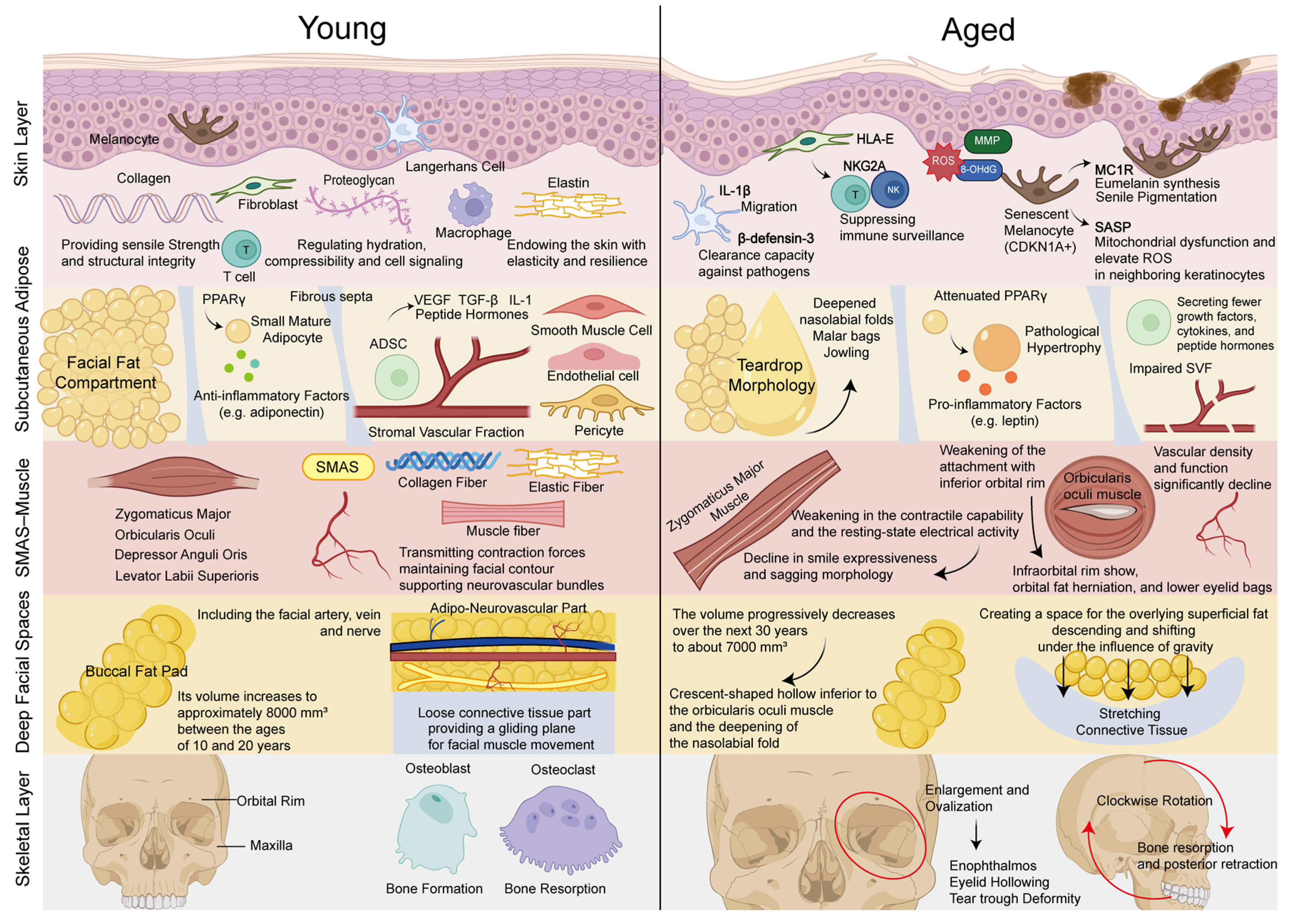
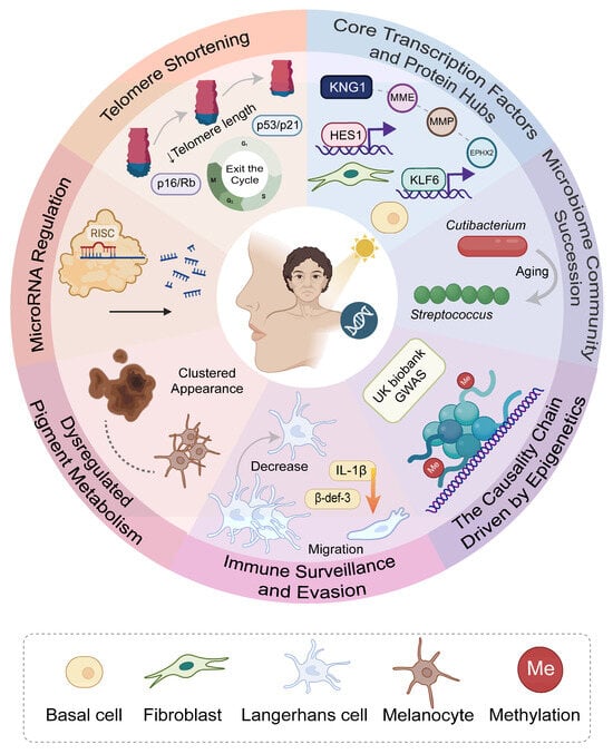
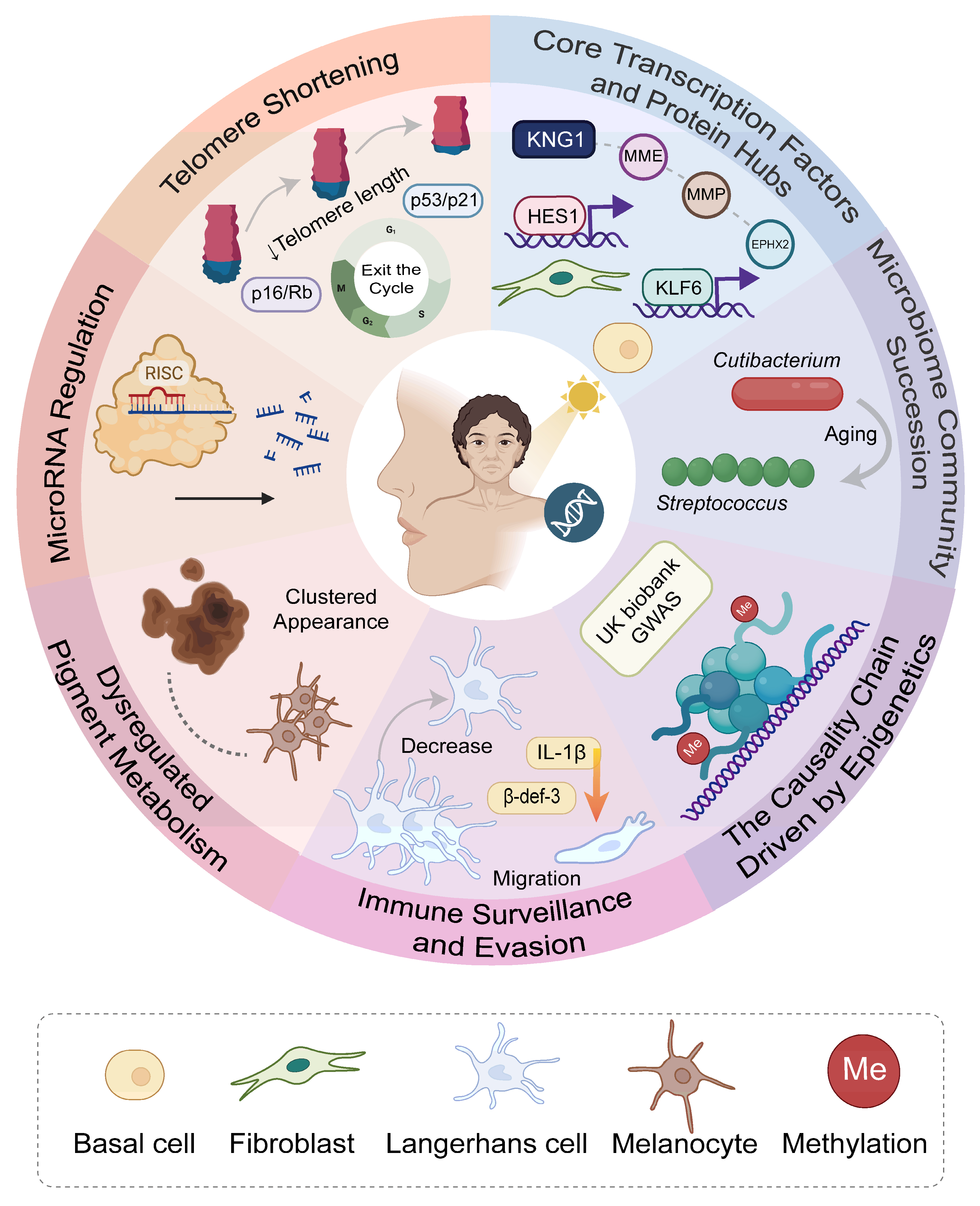
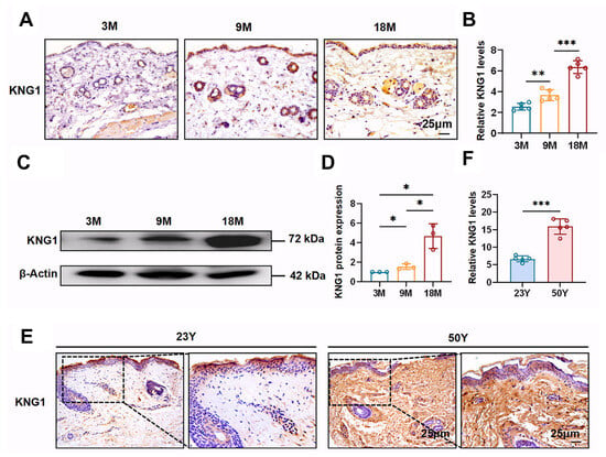
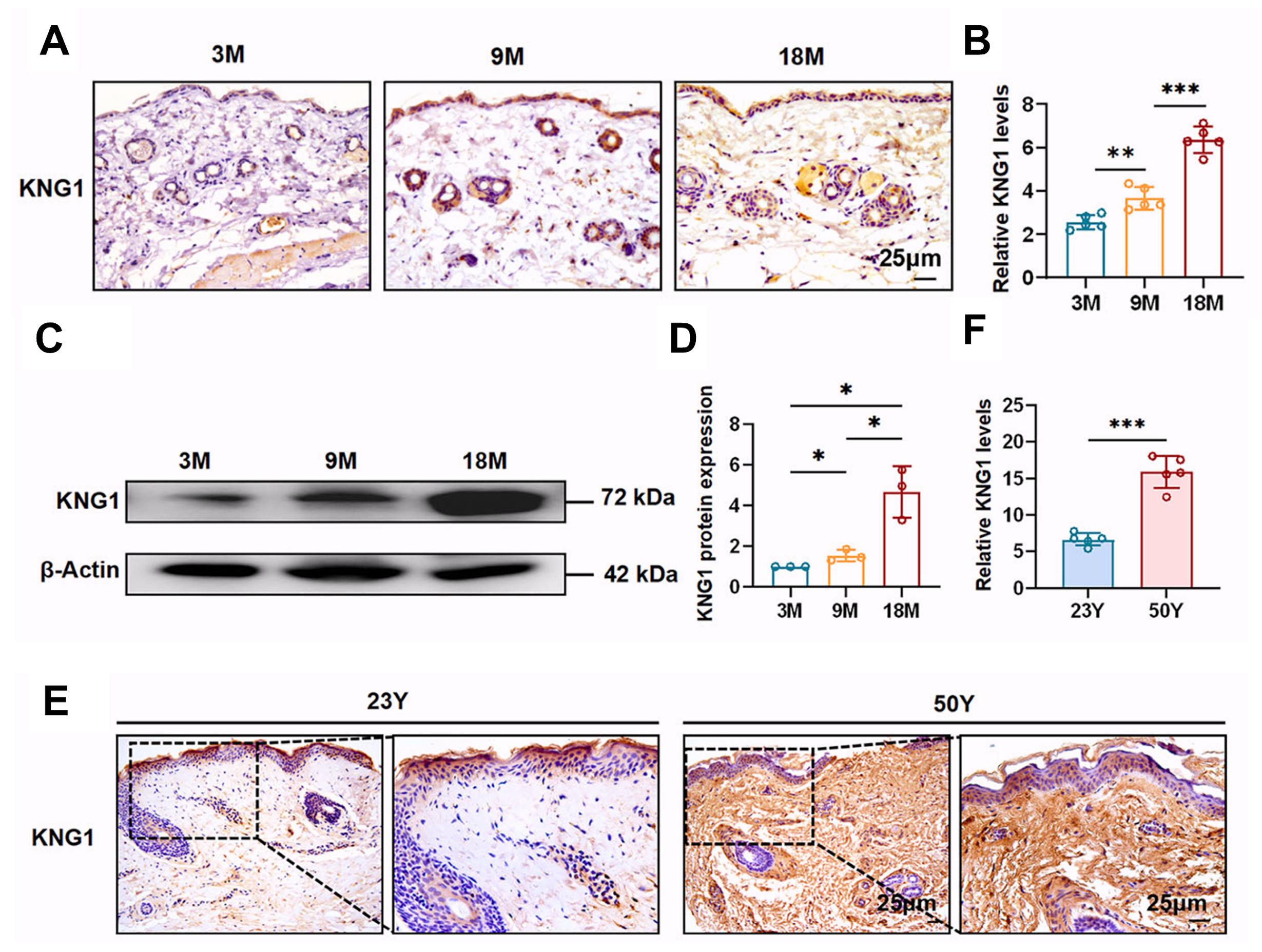
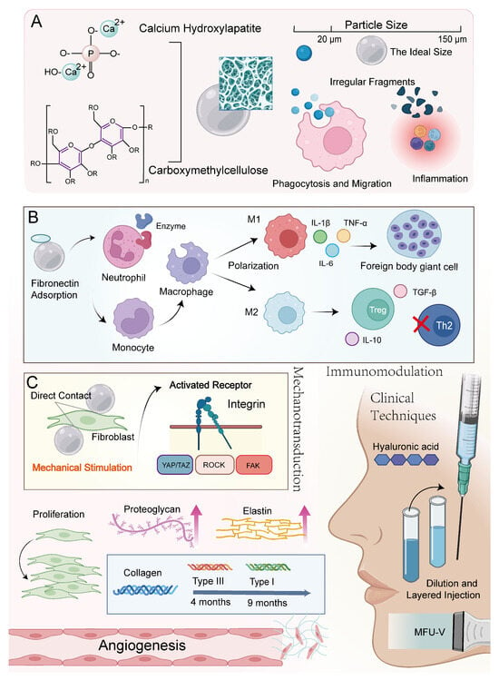
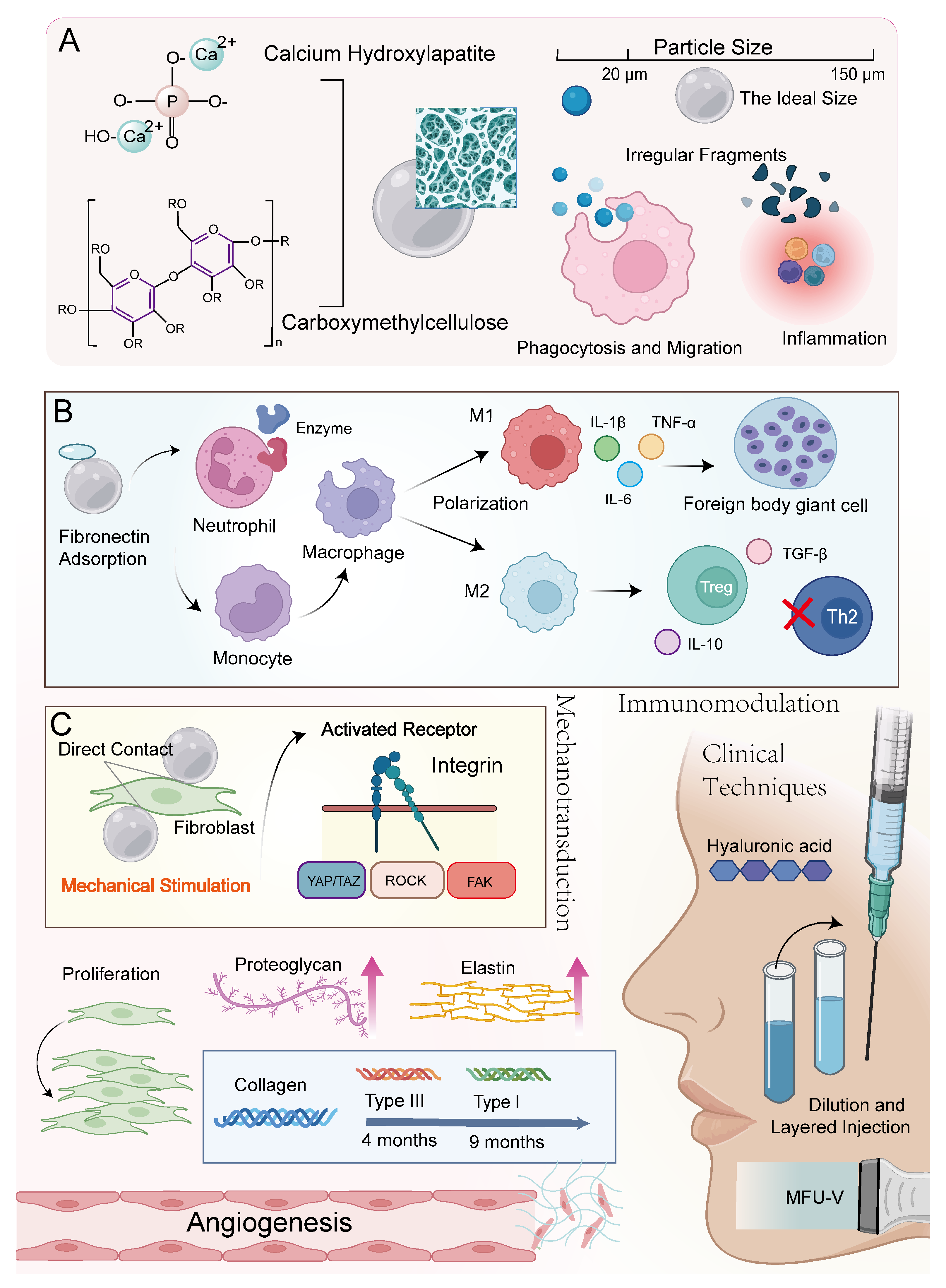

Disclaimer/Publisher’s Note: The statements, opinions and data contained in all publications are solely those of the individual author(s) and contributor(s) and not of MDPI and/or the editor(s). MDPI and/or the editor(s) disclaim responsibility for any injury to people or property resulting from any ideas, methods, instructions or products referred to in the content. |
© 2026 by the authors. Licensee MDPI, Basel, Switzerland. This article is an open access article distributed under the terms and conditions of the Creative Commons Attribution (CC BY) license.