


Abstract
The development of effective cosmetic formulations containing L-ascorbic acid is hindered by its rapid degradation and poor skin permeation. To address these limitations, this study identified an optimal synergistic antioxidant combination to enhance L-ascorbic acid efficacy and encapsulated it within a niosomes to improve stability and skin retention. Among various ratios evaluated via DPPH assays, a 1:1:2:2 combination of L-ascorbic acid, alpha-tocopherol, ferulic acid, and ergothioneine (Mix D) exhibited the highest synergism. Subsequent FRAP, anti-tyrosinase, and anti-aging inhibition assays confirmed that Mix D contributed to skin-brightening and anti-aging efficacy. Consequently, Mix D was encapsulated into niosomes (NI3-3A), initially showing a vesicle size of 130.67 nm and a PDI of 0.11, and an entrapment efficiency of 70.63%. NI3-3A provided exceptional chemical protection, limiting L-ascorbic acid degradation to 25.06% and maintaining an entrapment efficiency of 63.93% over 90 days due to its robust bilayer structure. Furthermore, it exhibited remarkable physicochemical stability, with a vesicle size of 140.27 nm and PDI of 0.09 after 90 days, while achieving a skin retention of 56.91% significantly higher than the unencapsulated solution (11.32%). In conclusion, this niosomal formulation successfully overcomes the stability limitations of L-ascorbic acid, offering a promising active ingredient for the development of advanced skin-brightening and anti-aging topical products.
1. Introduction
Skin is constantly exposed to external stressors, such as ultraviolet (UV) radiation, particulate matter, and tobacco smoke. These environmental factors promote the accumulation of reactive oxygen species (ROS), leading to oxidative damage and accelerated extrinsic skin aging [1]. At the molecular level, ROS degrade extracellular matrix components, including collagen and elastin fibers, thereby compromising skin integrity and contributing to wrinkles formation and skin laxity [2]. Furthermore, oxidative stress can disrupt melanogenesis and induce inflammation, resulting in hyperpigmentation and the overall deterioration of skin quality [1,2]. Antioxidants play a crucial role in scavenging free radicals and maintaining skin homeostasis. However, the endogenous antioxidant defense system may be insufficient to counteract the excessive ROS generated from daily environmental exposure; therefore, the topical application of antioxidants has become an effective strategy for protecting the skin against oxidative damage and premature aging [3].
L-ascorbic acid (LAA), commonly known as vitamin C, is a potent antioxidant with multiple skincare benefits. It acts as an essential cofactor for enzymes involved in collagen synthesis and inhibits tyrosinase, a key enzyme in melanin production, thereby reducing dark spots, freckles, and hyperpigmentation [4]. Despite its extensive benefits, LAA is highly unstable and susceptible to oxidative degradation, particularly in aqueous formulations, which significantly limits its formulation stability and long-term efficacy [5].
To overcome the instability of LAA and enhance its photoprotective effects, previous studies have explored its combination with other antioxidants, such as alpha-tocopherol (VE) and ferulic acid (FA). A formulation containing 15% LAA, 1% VE, and 0.5% FA has been reported to significantly improve photoprotection and prevent photo carcinogenesis more effectively than the individual antioxidants alone [6,7]. FA, a phenolic antioxidant, acts synergistically with LAA and VE to enhance UV protection and improve formulation stability [6]. Furthermore, ergothioneine (EGT), a potent antioxidant with distinct protective mechanisms [8], has demonstrated synergistic effects when combined with FA and glutathione in preventing UVB-induced cellular damage [9]. Therefore, it is hypothesized that incorporating EGT into the LAA, VE, and FA combination in appropriate ratio may further enhance the synergistic antioxidant and photoprotective effects.
To mitigate these stability issues and enhance the skin penetration of bioactive compounds, the development of an efficient carrier system is essential. Niosomes are non-ionic surfactant-base vesicles structurally analogous to liposomes that offer superior chemical stability, enhance transdermal penetration, and a favorable low-irritation profile [10]. Furthermore, it is more cost-effective and easier to handle than liposomes, making them a commercially attractive delivery system for cosmetic application. Encapsulation of LAA and other antioxidants within niosomes may improve formulation stability and therapeutic efficacy while potentially allowing the use of lower concentration of active compounds.
Therefore, this study aimed to determine the optimal ratio of LAA, VE, FA, and EGT for achieving synergistic antioxidant effects. The optimized antioxidant combination was subsequently evaluated for its anti-tyrosinase and anti-aging activities, followed by the development and characterization of a niosomal delivery system to enhance formulation stability and skin permeation.
2. Materials and Methods
2.1. Materials
L-ascorbic acid (LAA), alpha-tocopherol (VE), ferulic acid (FA), ergothioneine (EGT), cholesterol, phospholipids (hydrogenated lecithin), ethylhexyl palmitate, phenoxyethanol, polysorbate 80, and sorbitan oleate were purchased from Chanjao Longevity (Bangkok, Thailand). Next, 1,1-diphenyl-2-picrylhydrazyl (DPPH), 2,4,6-Tris (2-pyridyl)-s-triazinee (TPTZ), collagenase enzyme, elastase enzyme, N-(3-[2-Furyl] acryloyl)-Leu-Gly-Pro-Ala (FALGPA), hyaluronidase enzyme, L-DOPA, L-tyrosine, n-Succinyl-Ala-Ala-Ala-p-nitro anilide (AAAPVN), and tyrosinase enzyme were purchased from Sigma-Aldrich (St. Louis, MO, USA). Water, ethanol, and methanol were purchased from RCI Labscan (Bangkok, Thailand).
2.2. Determination of Antioxidant Activity
2.2.1. DPPH Free Radical Scavenging Assay
DPPH radical scavenging assay was used to evaluate the free radical scavenging activity of LAA, VE, FA, and EGT, and their combinations [11]. Briefly, a 0.1 mM DPPH solution was prepared in absolute ethanol. Each sample was dissolved in an appropriate solvent at various concentrations and mixed with the DPPH solution in a 96-well microplate. Following 30 min incubation in the dark, the absorbance was measured at 520 nm using a microplate reader (BMG Labtech, SPECTROstar Nano, Ortenberg, Germany). All determinations were performed in triplicate. The percentage of DPPH radical inhibition is calculated using the following equation.
where Control represents the absorbance of the control, and Sample represents the absorbance of the sample. The results were expressed as the IC50 value, which was determined from the inhibition curve.
% inhibition = ((Control − Sample)/Control) × 100
2.2.2. Ferric Reducing Antioxidant Power (FRAP) Assay
FRAP assay was utilized to investigate the reducing capacity of LAA, VE, FA, and EGT as well as their combinations [12]. The working FRAP regent was prepared by mixing a sodium acetate buffer, a TPTZ solution, and a ferric chloride solution. Each sample was dissolved in an appropriate solvent at various concentrations. Briefly, each sample was mixed with the FRAP regent in 96-well microplate. The reaction mixtures were incubated at an ambient temperature for 10 min. Subsequently, the absorbance was measured at 593 nm using a microplate reader (BMG Labtech, SPECTROstar Nano, Ortenberg, Germany). All assays were performed in triplicate, and the mean results were used to calculate the Equivalent Concentration 1 (EC1) values. The EC1 values represent the antioxidant concentration required to achieve a ferric reducing capacity equivalent to that of a 1 mM ferrous salt solution.
2.2.3. Evaluation of Synergistic Effect of Antioxidant Combination
To evaluate potential interactions among the antioxidant combinations, the ratios were initially designed based on the previously determined IC50 value of the individual compounds obtained from the DPPH assay. Briefly, LAA and VE were fixed at a ratio of 1:1, while the concentrations of FA and EGT were systematically varied to prepare four combinations: Mix A (1:1:1:1), Mix B (1:1:2:1), Mix C (1:1:1:2), and Mix D (1:1:2:2). The antioxidant activities of these combinations were subsequently evaluated using both DPPH and FRAP assay under the same experimental conditions applied to the individual compounds. Finally, the Combination Index (CI) was calculated based on the obtained antioxidant capacities (IC50 and EC1 value) as shown in the following equation to determine the presence of synergistic, additive, or antagonistic effects among the combinations [13].
where a, b, c, and d represent IC50 or EC1 of each antioxidant in the combination while A, B, C, and D represent IC50 or EC1 of individual antioxidant. When CI < 1; synergism, CI = 1; additive, CI > 1; antagonism.
CI = ((a/A) + (b/B) + (c/C) + (d/D))
2.3. Determination of Anti-Tyrosinase Activity
A modified dopachrome method using L-tyrosine and L-DOPA as substrates was employed to evaluate the anti-tyrosinase activity of LAA, VE, FA, EGT, and their selected combinations [14]. The samples, prepared at various concentrations in appropriate solvents, were assayed in a 96-well microplate. For the reaction mixture, 70 μL of the test sample, 70 μL of phosphate buffer, and 70 μL of tyrosinase enzyme solution (50 units/mL in pH 6.5 phosphate buffer) were combined and pre-incubated at 25 °C for 10 min. The reaction was subsequently triggered by the addition of 70 μL of the substrate (2.5 mM L-tyrosine or L-DOPA in phosphate buffer). After a 20-min incubation at 25 °C, the absorbance of the formed dopachrome was measured at 450 nm using a microplate reader (BMG Labtech, SPECTROstar Nano, Ortenberg, Germany). All determinations were performed in triplicate. The percentage of tyrosinase inhibition was calculated using the following equation:
where Aa is an absorbance of control, Ab is an absorbance of the sample.
Tyrosinase inhibition activity (%) = [(Aa − Ab)/(Aa)] × 100
2.4. Determination of Anti-Aging Activity
2.4.1. Hyaluronidase Inhibitory Activity
The hyaluronidase inhibitory activity of LAA, VE, FA, EGT, and their selected combinations were evaluated according to an established method [15], with minor modifications. In this assay, hyaluronidase inhibition was determined by quantifying the hydrolyzed hyaluronic acid, where the turbidity of the final reaction served as an indicator of residual, undigested hyaluronic acid. Briefly, 50 μL of the test sample was combined with 100 μL of hyaluronidase enzyme solution (2 mg/mL). This mixture was pre-incubated at 37 °C for 10 min. Subsequently, the enzymatic reaction was initiated by adding 100 μL of the substrate solution (0.03% w/v hyaluronic acid in PBS), followed by further incubation at 37 °C for 45 min. To terminate the reaction and precipitate any undigested substrate, 1 mL of acidic bovine serum albumin solution was added. The final mixture was allowed to stand at room temperature for 10 min. Finally, the turbidity was quantified by measuring the absorbance at 600 nm using a microplate reader (BMG Labtech, SPECTROstar Nano, Ortenberg, Germany). All measurements were performed in triplicate, and the percentage of hyaluronidase inhibition was calculated using the following equation:
where Asample is the absorbance of the reaction with samples, hyaluronidase, hyaluronic acid solution, and acetic albumin acid solution. Acontrol is the absorbance of the reaction with deionized water, hyaluronic acid solution, and acetic albumin acid solution.
Hyaluronidase inhibition activity (%) = (Asample/Acontrol) × 100
2.4.2. Collagenase Inhibitory Activity
The collagenase inhibitory of LAA, VE, FA, EGT, and their selected combinations were evaluated according to an established method [15], with minor modifications. The collagenase enzyme solution was reconstituted in a 50 mM tricine buffer (pH 7.5) supplemented with 400 mM sodium chloride and 10 mM calcium chloride. The synthetic peptide FALGPA, which mimics the native structure of collagen, served as the substrate and was dissolved in the same tricine buffer to achieve a final concentration of 2 mM. For the assay, 10 μL of the test sample was mixed with 40 μL of the enzyme solution and pre-incubated at room temperature for 15 min. Subsequently, the enzymatic hydrolysis was initiated by the addition of 50 μL of the FALGPA substrate. The decrease in absorbance was immediately monitored at 340 nm in kinetic mode using a microplate reader (BMG Labtech, SPECTROstar Nano, Ortenberg, Germany). All assays were performed in triplicate, and the percentage of collagenase inhibition was calculated using the following equation:
where Slopcontrol is the rate of absorbance change of the control reaction. Slopsample is the rate of absorbance change of the sample reaction.
Collagenase inhibition activity (%) = [(Slopcontrol − Slopsample)/(Slopcontrol)] × 100
2.4.3. Elastase Inhibitory Activity
The elastase inhibitory activity of LAA, VE, FA, EGT, and their selected combinations were evaluated according to an established method [15], with minor modifications. The inhibitory potential against elastase was assessed using AAAPVN as the specific substrate. Briefly, 50 μL of the test sample was pre-incubated with 25 μL of elastase enzyme solution (2 mg/mL, prepared in 100 mM Tris-HCl buffer, pH 8.0) at room temperature for 20 min. Subsequently, the enzymatic reaction was initiated by the addition of 25 μL of a 4.4 mM AAAPVN solution. The change in absorbance was immediately monitored at 410 nm in kinetic mode using a microplate reader (BMG Labtech, SPECTROstar Nano, Ortenberg, Germany). All assays were performed in triplicate, and the percentage of elastase inhibition was calculated using the following equation:
where Slopcontrol is the rate of absorbance change of the control reaction. Slopsample is the rate of absorbance change of the sample reaction.
Elastase inhibition activity (%) = [(Slopcontrol − Slopsample)/(Slopcontrol)] × 100
2.5. Development of Niosomes
An adapted sonication method [16] was employed for the preparation of the niosomes. To determine the optimal composition for stability and physicochemical characteristics, the concentrations of surfactants and phospholipids were systematically varied (Table 1). Initially, the total concentration of the non-ionic surfactant mixture (sorbitan oleate and polysorbate 80 at a weight ratio of 1:1 (w/w)) was investigated at 5%, 10%, and 15% (w/w), while the concentration of cholesterol and ethylhexyl palmitate were kept constant. Based on the particle size, polydispersity index (PDI), and zeta potential, the formulation containing 5% (w/w) surfactant mixture was selected for further optimization. To further improve vesicle integrity and stability, phospholipids were subsequently incorporated at concentrations of 0.5%, 1.0%, 1.5%, and 2.0% (w/w), while maintaining the surfactant mixture at 5% (w/w). Briefly, the lipid phase—consisting of phospholipids, cholesterol, ethylhexyl palmitate, sorbitan oleate, and phenoxyethanol—was weighed and gently stirred at 60 °C for 15 min until completely dissolved. Simultaneously, the aqueous phase was prepared by dissolving polysorbate 80 in DI water under constant agitation at 63–65 °C for 15 min. The aqueous phase was then gradually added to the lipid phase and homogenized using a high-shear homogenizer at 10,000 rpm for 15 min to obtain a coarse particle. Subsequently, the emulsion was subjected to probe sonication at 65% intensity for 15 min (1 s on/off pulse interval). Throughout the sonication process, the temperature was maintained below 20 °C to prevent overheating. The resulting formulations were characterized in terms of particle size, PDI, and zeta potential to identify the optimal formulation composition.
Table 1.
Composition of niosome with varying surfactant and phospholipid contents.
Preparation of Antioxidant-Loaded Niosomes
Based on the physical stability profiles obtained from the previous optimization studies, three blank niosome formulations exhibiting the most desirable characteristics—particularly an appropriate particle size, PDI, and zeta potential—were selected as candidates for antioxidants loading. The previously optimized combination of vitamin C and antioxidants, comprising the optimal ratio Mix D (1:1:2:2; consisting of 150 mg of LAA, 150 mg of VE, 300 mg of FA, and 300 mg of EGT), was successfully incorporated into selected blank niosomes. During the preparation process, VE and FA were dissolved directly in the lipid phase. Meanwhile, LAA and EGT were introduced into the aqueous phase prior to niosome formation.
2.6. Characterization of Antioxidant-Loaded Niosomes
2.6.1. Particle Size, Polydispersity Index (PDI), and Zeta Potential
The mean particle size, PDI, and zeta potential of the prepared niosomes were measured using a Zetasizer Nano ZS (Malvern Instruments, Malvern, UK) [17]. Prior to analysis, each sample was diluted 100-fold with DI water to achieve a suitable scattering intensity. The dynamic light scattering (DLS) measurements were conducted at a fixed scattering angle of 173° and a constant temperature of 25 °C. All measurements were performed in triplicate, and the results were reported as the mean values.
2.6.2. Chemical Marker Analysis Using High-Performance Liquid Chromatography
For the physicochemical characterization of the combination of LAA and antioxidant-loaded niosomes, the quantitative determination of LAA content in the formulations was performed using HPLC (Shimadzu, Kyoto, Japan) [18]. To extract the active marker, the niosome formulations were disrupted by mixing with methanol, followed by vortex mixing and centrifugation until a clear supernatant was obtained. The resulting supernatant was then collected for HPLC analysis. A stock solution of LAA was initially prepared by dissolving 40 mg of LAA in 100 mL of DI water. Subsequently, ten working standard solutions were prepared by serial dilution to achieve concentrations ranging from 0.0781 to 40 mg/L. The mobile phase consisted of deionized water adjusted to pH 2.2 with sulfuric acid and methanol (80:20, v/v). Chromatographic analysis was performed using an LC-2050C 3D HPLC system equipped with a Eurospher II 100-5 C18 column (250 mm × 4.5 mm; KNAUER Wissenschaftliche Geräte GmbH, Berlin, Germany). The detection wavelength was set at 243 nm. The injection volume was 20 μL. Elution was carried out under isocratic conditions at room temperature with a constant flow rate of 1.0 mL/min.
2.6.3. Determination of Entrapment Efficiency (EE)
The entrapment efficiency (EE) of the combination of LAA and antioxidant-loaded niosomes was determined using an indirect method [17]. To determine the total amount of LAA, 500 μL of the niosomes was mixed with 500 μL of methanol, vortexed, and sonicated to completely disrupt the vesicles. To determine the amount of unentrapped LAA, another 500 μL of the niosomes was transferred into an Amicon® Ultra centrifugal filter (0.5 mL, 50 kDa MWCO, Merck, Darmstadt, Germany) with a pore size of 10 to 100 nm, and centrifuged at 10,000 rpm for 30 min. The filtrate containing the free LAA was collected from the bottom of the tube. Both the total and free LAA concentrations were subsequently analyzed using the previously described HPLC method. All measurements were performed in triplicate, and the percentage of EE was calculated using the following equation:
%EE = [(Total compound − Free compound)/Total compound] × 100
2.6.4. Morphological Evaluation via Transmission Electron Microscopy (TEM)
The morphology of the combination of LAA and antioxidant-loaded niosomes was visualized using a JEM-2100 Plus transmission electron microscope (JEOL, Tokyo, Japan) [17]. Briefly, the sample was diluted 1:100 with DI water, dropped onto a 300-mesh copper grid, and air-dried for 15 min. The vesicles were then negatively stained with 3% (w/v) phosphotungstic acid in DI water. Excess staining solution was gently removed using filter paper prior to analysis. The prepared sample was observed using the TEM at an accelerating voltage of 100 kV with magnifications ranging from 10,000× to 20,000×.
2.7. Skin Retention and Permeation Studies
In vitro skin retention and permeation studies were conducted using a Franz diffusion cell apparatus (V9-CA, PermeGear Inc., Hellertown, PA, USA) equipped with a Strat-M® membrane (Strat-M® EMD Millipore, Burlington, MA, USA) [11]. The receptor compartment (13 mL) was filled with PBS (pH 7.4) and maintained at 37 ± 0.5 °C throughout the experiment. Briefly, 1 mL of the test formulation was applied to the donor compartment. At specific time points (1, 2, 4, 6, 8, and 12 h), 1 mL aliquots were withdrawn from the receptor compartment and immediately replaced with an equal volume of a fresh medium to maintain sink conditions. The collected samples were then quantified by HPLC. To evaluate the membrane retention, the Strat-M® membrane was removed after 12 h, gently washed with DI water to eliminate residual formulation, and cut into small pieces. The retained LAA was extracted with methanol using sonication for 10 min prior to HPLC analysis. All procedures were carried out in triplicate.
2.8. Stability Tests
2.8.1. Photostability
The photostability of the combination of LAA and antioxidant-loaded niosomes was investigated and compared with intact solution containing LAA and antioxidants. Both formulations were stored at room temperature (RT) and exposed to ambient natural light for a continuous period of 4 weeks. Following the exposure period, the remaining concentration of LAA in the samples was quantified utilizing an HPLC analysis. All analyses were performed in triplicate, and the percentage of remaining LAA was calculated to determine the degradation profile of each formulation.
2.8.2. Short-Term Stability
The short-term stability of the combination of LAA and antioxidant-loaded niosomes was evaluated by storing the formulations in tight amber glass bottles under two conditions: 4 ± 0.5 °C, 30 ± 0.5 °C (RT) for 3 months [14]. At the end of the specified storage periods, the physicochemical stability of the formulations was assessed.
2.9. Statistical Analysis
The experimental results are expressed as the mean ± standard deviation (SD) derived from three independent measurements. Statistical analysis for comparisons between two groups was performed using an independent t-test. Conversely, comparisons among multiple formulations were analyzed using a one-way analysis of variance (ANOVA), followed by Fisher’s Least Significant Difference (LSD) post hoc test. Statistically significant difference was declared at p < 0.05.
2.10. Artificial Intelligence
During the preparation of this manuscript, the authors used Gemini, developed by Google. The system was accessed and utilized in July–August 2026 (powered by the Gemini 1.5 Pro model) to assist in the creation of the graphical abstract, and to translate and refine the English language from the authors’ original Thai drafts. After using this tool, the authors carefully reviewed and edited all outputs, and take full responsibility for the content of this publication. All scientific concepts, experimental designs, data analyses, interpretations, and methodological procedures were conceived and performed solely by the authors. No scientific conclusions or interpretations were generated by artificial intelligence.
3. Results and Discussion
3.1. Antioxidant Activity of Individual Compounds
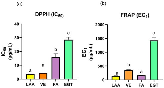
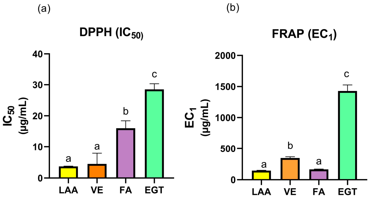
The antioxidant activity of individual compounds (LAA, VE, FA, and EGT) were evaluated using DPPH radical scavenging and FRAP assays. Oxidative stress induced by environmental factors is a primary driver of premature skin aging, leading to cellular damage and the degradation of structural biomolecules [19]. In the DPPH assay, a lower IC50 value signifies a stronger capacity to scavenge free radicals through hydrogen or electron donation mechanisms [20]. As shown in Figure 1a, among the individual compounds, LAA and VE exhibited the most potent antioxidant activities with the lowest IC50 values of 3.7 ± 0.11 μg/mL and 4.5 ± 3.5 μg/mL, respectively, and no significant difference between them (p > 0.05). These values were significantly lower than FA (16 ± 2.4 μg/mL) and EGT (28.5 ± 1.8 μg/mL) (p < 0.05), indicating that LAA and VE possess superior radical scavenging activity. The FRAP assay reflects the reducing power of antioxidants based on their ability to donate electrons and reduce ferric iron (Fe3+) complex to its ferrous (Fe2+) form. In this assay, a lower EC1 value indicates a superior reducing capacity [21]. Among the individual compounds, LAA demonstrated the lowest EC1 value (150 ± 6.17 μg/mL), followed by FA (170 ± 4.69 μg/mL), with no significant difference observed between them (p > 0.05). Both LAA and FA showed significantly higher reducing power than VE (350 ± 20.9 μg/mL) and EGT (1430 ± 95.86 μg/mL) (p < 0.05) (Figure 1b).
Figure 1.
Antioxidant activity of LAA, VE, FA, and EGT using the DPPH (a) and FRAP (b) assays. Different superscript letters (a–c) indicate significant differences among samples as determined by one-way ANOVA with Tukey’s post hoc test (p < 0.05).
The results of this study showed that LAA, VE, FA, and EGT each possessed distinct antioxidant capacities, as reflected by their varying performance in the DPPH and FRAP assays. While LAA and VE exhibited superior free radical scavenging activity, LAA and FA demonstrated the highest reducing powder. Since no individual compound could fully address the diverse mechanisms of oxidative damage, a combined formulation was developed to integrate these compounds and leverage their complementary mechanisms to achieve a synergistic antioxidant effect superior to that of the individual compounds.
3.2. Antioxidant Synergism
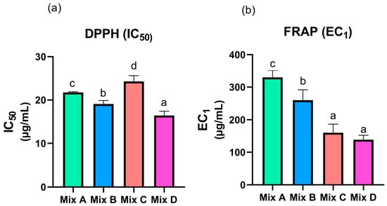
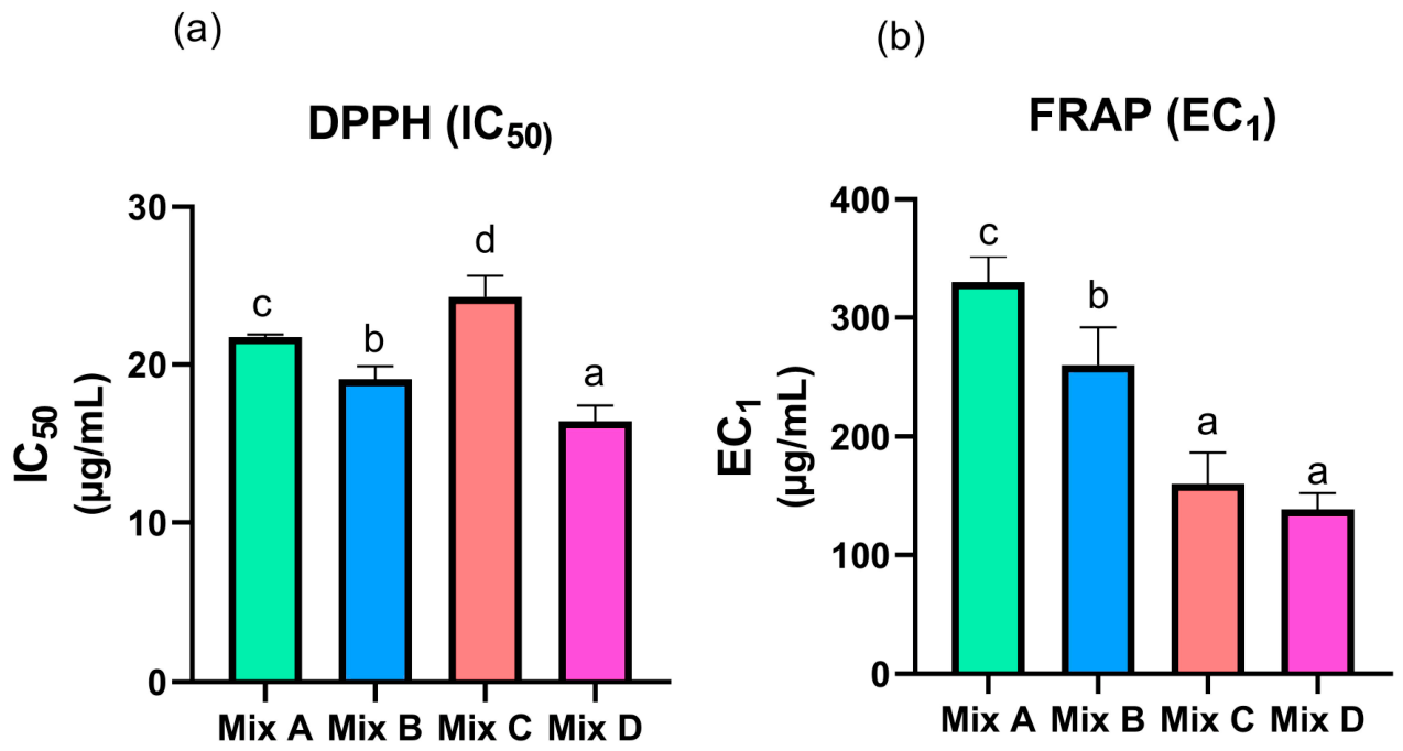
To evaluate the interactions among the combinations of LAA, VE, FA, and EGT, the Combination Index (CI) values of Mix A (1:1:1:1), Mix B (1:1:2:1), Mix C (1:1:1:2), and Mix D (1:1:2:2) were determined using both DPPH and FRAP assays. Regarding the DPPH assay as shown in Figure 2a, Mix D (16.40 ± 1.01 μg/mL) exhibited the most potent antioxidant activity, followed by Mix B (19.07 ± 0.81 μg/mL), Mix A (21.73 ± 0.15 μg/mL), and Mix C (24.27 ± 1.32 μg/mL). Statistical analysis confirmed that all combinations differed significantly from one another (p < 0.05). Similarly, in the FRAP assay as shown in Figure 2b, Mix D (138.43 ± 14.39 μg/mL) and Mix C (160.62 ± 26.35 μg/mL) demonstrated the highest reducing efficacy, with no significant difference observed between them (p > 0.05). These were followed by Mix B (260.19 ± 31.78 μg/mL) and Mix A (329.99 ± 21.06 μg/mL), which showed the lowest reducing power. Notably, Mix D in the FRAP assay outperformed individual LAA, the most potent individual compound.
Figure 2.
Antioxidant activity of the combinations of LAA and antioxidants using the DPPH (a) and FRAP (b) assays. Different superscript letters (a–d) indicate significant differences among samples as determined by a one-way ANOVA with Tukey’s post hoc test (p < 0.05).
The CI analysis revealed that the antioxidant combinations exhibited varying synergistic and antagonistic interactions depending on the formulation ratio and the specific antioxidant mechanism involved. Such synergistic interactions are highly advantageous for cosmetic applications, as they may provide greater protective efficacy than individual compounds while reducing the required concentration of each antioxidant [22]. This approach may also help minimize potential skin irritation and formulation costs.
Regarding the DPPH assay, all combinations demonstrated synergistic effects (CI < 1), as shown in Table 2. Among these, Mix D exhibited the strongest synergism, with the lowest CI value of 0.58. This was followed by Mix B (CI = 0.74), Mix A (CI = 0.85), and Mix C (CI = 0.96), respectively. In contrast, the FRAP assay revealed different interaction profiles depending on the antioxidant ratio. Both Mix A and Mix B exhibited antagonistic effects, with CI values of 1.40 and 1.16, respectively (CI > 1). Interestingly, increasing the proportion of EGT in the combinations promoted synergistic interactions. Specifically, Mix C and Mix D demonstrated synergistic effects, with CI values of 0.53 and 0.54 (CI < 1), respectively.
Table 2.
Combination index (CI) values of the combination of LAA and antioxidants using DPPH and FRAP assays.
Overall, Mix D was the only combination that consistently achieved strong synergistic interactions in both assays. Previous studies have demonstrated that the combination of L-ascorbic acid (LAA) and vitamin E (VE) forms an effective antioxidant network in which oxidized α-tocopherol can be regenerated by ascorbic acid, resulting in enhanced antioxidant activity and photoprotection. The combination of these two antioxidants has been shown to provide superior protection against UV-induced erythema and oxidative damage compared with either antioxidant alone [7]. The incorporation of ferulic acid (FA) further enhances this antioxidant system by improving the stability of LAA and VE and increasing their synergistic photoprotective efficacy. Previous studies demonstrated that the addition of ferulic acid to a vitamin C/vitamin E formulation doubled the photoprotective effect and provided greater protection against oxidative stress and UV-induced damage. Moreover, ferulic acid readily forms a resonance-stabilized phenoxy radical, and exhibits synergistic interactions with ascorbic acid, thereby contributing to the improved antioxidant performance of the system [6]. The addition of EGT in Mix D further enhanced this performance. This synergistic effect can be attributed to the unique sulfur-containing structure of EGT, which allows it to act as a versatile and stable scavenger, capable of neutralizing a wider range of oxidative stressors than individual antioxidants [8]. Moreover, EGT functions as a terminal antioxidant and an effective metal chelator; it captures metal ions that would otherwise accelerate the oxidative degradation of LAA and VE [23]. Similar to ascorbic acid, ergothioneine can undergo one-electron oxidation to generate resonance-stabilized radical intermediates, potentially facilitating electron transfer and antioxidant recycling within the antioxidant network. These properties may explain why Mix D, which contained higher proportions of both FA and EGT, exhibited the strongest synergistic effects among all formulations. Interestingly, the interaction patterns differed between the DPPH and FRAP assays, indicating that the contribution of EGT depends on the reaction mechanism. The DPPH assay reflects both hydrogen atom transfer (HAT) and single-electron transfer (SET) mechanisms, whereas the FRAP assay exclusively measures ferric ion reduction through SET reactions. Therefore, the tautomeric structure and redox behavior of EGT may influence its antioxidant contribution under different assay conditions [24]. Consequently, the integration of EGT into the LAA, VE, and FA system creates a multi-target protective network, explaining why Mix D exhibited superior and consistent synergistic efficacy. Given these findings, Mix D was selected as the optimal formulation for subsequent evaluation of anti-tyrosinase and anti-aging activities, as well as for the development of antioxidant-loaded niosomes.
3.3. Anti-Tyrosinase Activity of Individual Compounds and Antioxidant Combination
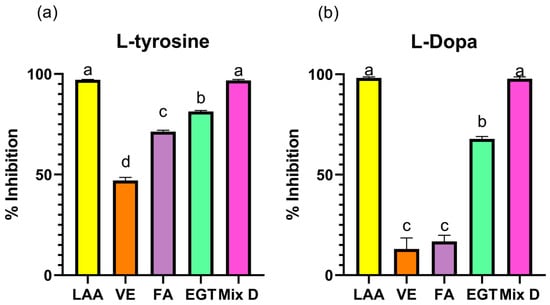
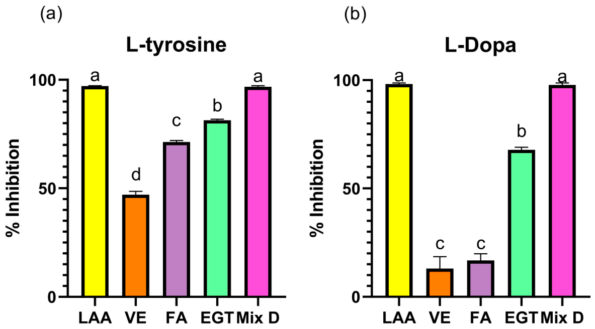
Tyrosinase is a key enzyme involved in melanogenesis by regulating two major rate-limiting steps: the hydroxylation of L-tyrosine to L-DOPA, and the subsequent oxidation of L-DOPA to dopaquinone [25]. Due to its central role in skin pigmentation, tyrosinase inhibition is widely recognized as an effective strategy for developing skin-whitening and brightening cosmetic ingredients. In this study, the tyrosinase-inhibiting potential of individual compounds (LAA, VE, FA, and EGT) and the selected combination (Mix D), were evaluated using both L-tyrosine and L-DOPA as substrates. The results are shown in Figure 3.
Figure 3.
Anti-tyrosinase activity of individual compounds and the selected antioxidant combination (concentration of 1 mg/mL) when L-tyrosine (a) and L-DOPA (b) were used as the substrate. Different superscript letters (a–d) indicate significant differences among samples as determined by a one-way ANOVA with Tukey’s post hoc test (p < 0.05).
When L-tyrosine was employed as the substrate, LAA and Mix D exhibited the highest anti-tyrosinase activity (97.10 ± 0.01% and 96.77± 0.03%, respectively), showing no statistically significant difference between them (p > 0.05). Both samples showed significantly higher inhibitory activities than EGT (81.37 ± 0.12%), which in turn was significantly higher than FA (71.35 ± 0.30%), followed by VE (47.03 ± 4.16%) as the lowest inhibitor. A similar trend was observed when L-DOPA was used as the substrate. LAA and Mix D exhibited the strongest anti-tyrosinase activity (98.19 ± 0.66% and 97.80 ± 0.72%, respectively), with no statistically significant difference between them. These were followed by EGT (67.85 ± 0.40%), which showed significantly higher activity than FA (16.80 ± 0.36%), and VE (13.01 ± 0.59%). Notably, no statistically significant difference was observed between FA and VE (p > 0.05).
The potent anti-tyrosinase activity of Mix D suggests a multi-modal inhibitory mechanism. LAA serves as the primary inhibitor, acting as a non-competitive or mixed-type inhibitor that reduces dopaquinone back to L-DOPA [26]. FA shares a profound structural similarity with L-tyrosine due to its biosynthetic origin, allowing it to act as a competitive inhibitor by binding directly to the active site of tyrosinase [27]. EGT has been demonstrated to inhibit tyrosinase in a non-competitive manner, interacting with the active center via hydrogen bonding and salt bridges, which induces structural changes that block the catalytic site [28]. Although VE acts primarily as a versatile antioxidant to enhance formulation stability, recent studies indicate that specific tocopherol isomers can directly inhibit tyrosinase activity and suppress melanin synthesis [29]. While VE may not exhibit the same potency as competitive inhibitors like FA, its presence contributes to the overall inhibitory activity of Mix D. This indirect and direct inhibitory effect, combined with its role in maintaining the redox balance, reinforces the synergistic efficacy of Mix D.
Collectively, the combination of LAA, VE, FA, and EGT provides additional antioxidant and skin-protective benefits, supporting the development of multifunctional cosmetic formulations for managing hyperpigmentation and promoting overall skin health.
3.4. Anti-Aging Activity of Individual Compounds and Antioxidant Combination
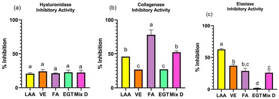
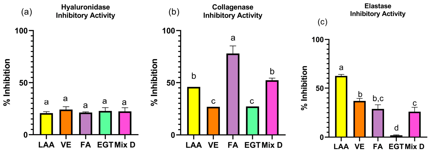
The anti-aging potential of individual compounds (LAA, VE, FA, and EGT) and the selected antioxidant combination (Mix D) were evaluated by assessing their inhibitory effects against three key enzymes involved in extracellular matrix (ECM) degradation, namely hyaluronidase, collagenase, and elastase. ECM plays a crucial role in maintaining skin hydration, structural integrity, and elasticity. Hyaluronic acid is a critical hydrating polysaccharide that maintains skin moisture and volume, but it is broken down by hyaluronidase, leading to dehydration and increased tissue permeability. In the dermal layer, collagen and elastin are the primary structural proteins responsible for mechanical strength, structural integrity, firmness, and elasticity. Their degradation by collagenase and elastase directly causes skin fragmentation, sagging, and wrinkle formation [30]. Therefore, the inhibition of these enzymes represents a major strategy for developing anti-aging cosmetic ingredients. The anti-aging activities of all evaluated samples at a concentration of 10 mg/mL are summarized in Figure 4.
Figure 4.
Anti-aging activity of individual compounds and the selected antioxidant combination (concentration of 10 mg/mL). Hyaluronidase inhibition (a), collagenase inhibition (b) and elastase inhibition (c). Different superscript letters (a–d) indicate significant differences among samples as determined by a one-way ANOVA with Tukey’s post hoc test (p < 0.05).
Regarding hyaluronidase inhibition (Figure 4a), all samples exhibited comparable activity, including LAA (20.64 ± 1.43%), VE (24.02 ± 2.73%), FA (21.23 ± 0.65%), EGT (22.72 ± 3.16%), and Mix D (22.32 ± 3.48%). No statistically significant differences were observed among the tested samples (p > 0.05), indicating that all compounds and their combination possessed similar inhibitory effects against hyaluronidase under the experimental conditions. In contrast, significant differences were observed in the collagenase inhibition among the tested samples (Figure 4b, p < 0.05). FA exhibited the highest collagenase inhibitory activity (77.85 ± 7.49%). Mix D (52.28 ± 2.12%) and LAA (45.93 ± 0.0%) showed moderate inhibitory activities with no significant difference between them, whereas VE (26.71 ± 0.1%) and EGT (27.04 ± 0.0%) showed the lowest inhibitory activities against collagenase. For elastase inhibition (Figure 4c), significant differences were observed among the tested samples (p < 0.05). LAA demonstrated the highest inhibitory activity (62.5 ± 1.5%) which was significantly higher than all other samples. VE (37.00 ± 2.5%) and FA (28.83 ± 4.31%) exhibited moderate activity, followed by Mix D (26.00 ± 4.58%). EGT showed the lowest inhibitory activity (1.83 ± 0.58%).
The collagenase inhibitory activity of FA (77.85 ± 7.49%) aligns with previous research indicating that FA acts as a potent inhibitor of collagen degradation [31,32]. Previous studies have demonstrated that FA protects collagen by binding to telopeptide regions without disrupting its triple-helical structure, effectively terminating collagen fibril progression [31]. These findings highlight the potential of FA as a key ingredient for maintaining structural integrity and mitigating ECM fragmentation in cosmetic applications.
The potent elastase inhibition by LAA (62.5 ± 1.5%) suggests that LAA plays a regulatory role in ECM degradation pathways beyond its established function in collagen synthesis [33]. Previous research indicates that LAA modulates the expression of remodeling enzymes, including those involved in elastin and collagen metabolism, potentially through the regulation of metabolic sensors like HIF-1 [33]. These findings underscore the multi-functional capacity of LAA in maintaining dermal integrity by mitigating the enzymatic breakdown of elastin.
While EGT and VE exhibited limited direct enzyme inhibition, their anti-aging potential primarily lies in scavenging ROS, thereby preventing oxidative stress-induced signaling pathways that upregulate ECM-degrading enzymes. The moderate inhibitory activity of Mix D suggests a synergistic benefit, offering a balanced approach to target multiple pathways of ECM degradation. Overall, these results demonstrate that while FA and LAA were the most potent inhibitors of collagenase and elastase, respectively, the antioxidant combination (Mix D) provides broad-spectrum protection. Consequently, Mix D emerges as a promising multifunctional system for cosmetic applications aimed at addressing the complex, multi-pathway nature of skin aging.
3.5. Development of Unloaded Niosomes
The physicochemical properties of the developed unloaded niosomes are summarized in Table 3. Generally, an ideal lipid-based nanocarrier system for topical delivery requires a small particle size (<200 nm) to facilitate effective skin penetration through the stratum corneum, a narrow polydispersity index (PDI ≤ 0.2) to ensure vesicle homogeneity [34], and a sufficient surface charge zeta potential magnitude greater then ±30 mV to provide adequate electrostatic repulsion and prevent particle aggregation [35].
Table 3.
Particle size, PDI, and zeta potential of unloaded niosomes.
Among the initial formulations (NI1, NI2, and NI3), NI3 were selected for further development. Despite NI3 shows slightly larger particle size (102.13 ± 0.31 nm) than other formulations, it exhibited the lowest PDI (0.13 ± 0.01) and the highest zeta potential (−28.77 ± 2.14 mV), indicating physically stable colloidal system. The relatively low zeta potential of these initial formulations is attributed to the predominant use of non-ionic surfactants.
Surfactant concentration significantly influences niosomal properties. While higher levels generally reduce vesicle size, excessive concentration can disrupt bilayer packing, leading to membrane instability or the formation of mixed micelles instead of stable vesicles [36,37,38]. Furthermore, high surfactant levels often reduce zeta potential magnitude because niosome structure specifically bilayer rigidity is highly dependent on the surfactant-to-lipid ratio. Non-ionic surfactants primarily rely on steric rather than electrostatic stabilization [38].
To enhance physical stability and minimize particle aggregation, phospholipids were incorporated into the subsequent formulations (NI3-1 to NI3-4). The incorporation of phospholipids successfully increased the negative surface charge, resulting in zeta potential values ranging from −42.5 ± 2.52 mV to −68.33 ± 1.29 mV. The incorporation of phospholipids contributes to the stability of the system through the ionization of their polar head groups. For instance, phospholipids (hydrogenated lecithin) provide a significant negative charge in an aqueous medium, which increases the zeta potential magnitude. This enhanced surface charge strengthens the electrostatic repulsion between vesicles, effectively preventing aggregation and ensuring long-term colloidal stability [39]. Furthermore, phospholipids may contribute to improved membrane flexibility and vesicle structural integrity, which are desirable characteristics for topical delivery systems.
Following phospholipid incorporation, the resulting NI3 sub-series successfully maintained an average particle size within the nanometer range (96.90 ± 0.33 nm to 122.70 ± 2.30 nm), which is optimal scale for enhancing cosmetic ingredients to penetrate through the stratum corneum. Regarding formulations uniformity, NI3-1, NI3-2, and NI3-3 demonstrated excellent homogeneity, with PDI values ranging from 0.14 ± 0.01 to 0.17 ± 0.01. Conversely, formulation NI3-4 exhibited a significantly higher PDI value (0.41 ± 0.02), indicating a highly polydisperse and non-uniform vesicle size distribution.
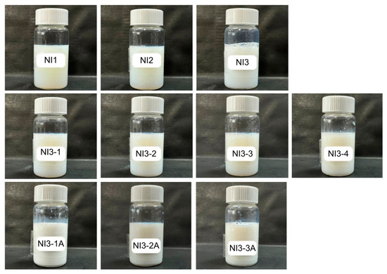
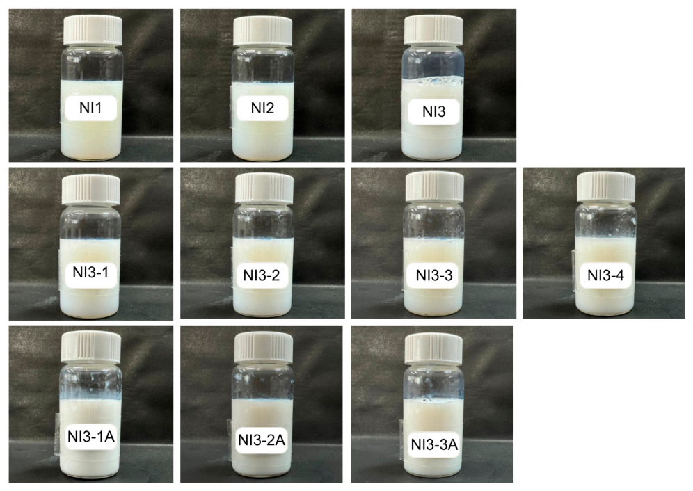
Interestingly, despite possessing the highest negative zeta potential, NI3-4 exhibited physical instability with phase separation occurring within 3 days of storage. This finding suggests that a high zeta potential alone is insufficient to guarantee formulation stability. The elevated PDI value observed for NI3-4 indicates a broad and heterogeneous vesicle size distribution, which may have contributed to physical instability. Heterogeneity can promote vesicle fusion, aggregation, or other destabilization processes during storage, ultimately leading to phase separation [34]. Consequently, NI3-1, NI3-2, and NI3-3 were selected as the optimal carriers for subsequent antioxidants loading, as they exhibited an appropriate nano-size, low PDI values, high negative zeta potentials, and satisfactory physical stability (Figure 5).
Figure 5.
Physical appearance of the developed niosomal formulations, including the unloaded niosomes (NI1 to NI3, NI3-1 to NI3-4), LAA and antioxidant-loaded niosomes (NI3-1A to NI3-3A).
3.6. Preparation, Characterization, and Stability of LAA and Antioxidant-Loaded Niosomes
The antioxidant combination (Mix D) at a concentration of 150 mg of LAA, 150 mg of VE, 300 mg of FA, and 300 mg of EGT was successfully prepared by loading into selected niosomes (NI3-1, NI3-2, and NI3-3). The concentrations and ratios of these antioxidants were determined based on the optimal synergistic combinations previously evaluated [9]. All formulations exhibited physical properties consistent with the characteristics of well-defined nano-vesicular delivery systems, including nanoscale particle sizes, low PDI values, and high negative zeta potentials [40], as summarized in Table 4. However, despite these promising initial characteristics, physical instability of NI3-1A was observed during the two-week preliminary storage study at room temperature (RT). This instability may be attributed to an insufficient balance between surfactant and lipid components, which could have compromised vesicle membrane integrity and reduced the formulation’s ability to accommodate the antioxidant payload during storage [10]. Consequently, NI3-1A was excluded from the short-term stability study (Figure 5).
Table 4.
Particle size, PDI, and zeta potential of LAA and antioxidant-loaded niosomes.
The stability study of the NI3-2A formulation revealed that storage temperature significantly influences its physicochemical parameters (Table 5). Regarding particle size, samples stored at room temperature (RT) exhibited a statistically significant increase throughout the 90-day storage period (p < 0.05), whereas at 4 °C, significant changes were only detected from 60-day onwards, indicating that 4 °C storage more effectively delayed particle growth. In terms of PDI, values remained stable at RT over 90 days (p > 0.05), while at 4 °C, significant fluctuations occurred at 60 day and 90 day (p < 0.05), although all values remained within an acceptable range. Finally, a significant decrease in zeta potential magnitude was observed from 60 day in both storage conditions (p < 0.05), the surface charge in both environments remained sufficiently high to maintain colloidal stability throughout the study.
Table 5.
Short-term stability of LAA and antioxidant-loaded niosomes at RT (room temperature) and 4 °C.
The NI3-3A formulation demonstrated superior stability compared to NI3-2A throughout the 90-day study. While NI3-3A exhibited a significant increase in particle size starting from 60 day (p < 0.05), its PDI and zeta potential values remained statistically unchanged, indicating a more robust colloidal system. Furthermore, NI3-3A stored at 4 °C displayed the highest stability among all tested formulations. Under refrigerated conditions, this formulation maintained excellent homogeneity and surface charge stability, confirming its suitability for long-term storage.
The three-months stability revealed that both formulations maintained favorable colloidal characteristics throughout the storage period, characterized by consistently low PDI values (PDI ~0.1) and stable zeta potential lower than −35 mV under both refrigerated (4 °C) and room temperature (RT) conditions (Table 5). Interestingly, the only distinct variation between the two was their particle size. NI3-3A exhibited superior structural resilience, successfully maintaining a tightly controlled vesicle size under both RT and 4 °C. Conversely, NI3-2A exhibited a noticeable increase in particle size during storage at room temperature, suggesting greater susceptibility to temperature-induced vesicle growth or aggregation. Based on its superior particle size stability while maintaining comparable PDI and zeta potential values, NI3-3A was selected for further characterization. Subsequent evaluations included entrapment efficiency (%EE), morphology evaluation using transmission electron microscopy (TEM), skin retention and permeation studies, and photostability study.
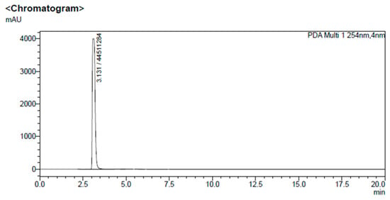
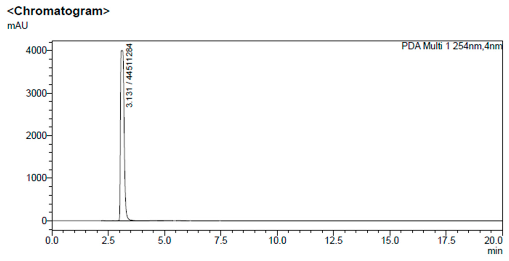
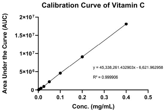
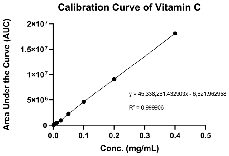
High-Performance Liquid Chromatography (HPLC) was employed to establish a quantitative method for analyzing the LAA content in the niosomes [41]. The successful identification of LAA was confirmed via the chromatographic profile as illustrated in Figure 6. LAA exhibited a sharp and well-resolved peak at a retention time of 3.131 min. The selected LAA and antioxidant-loaded niosome (NI3-3A) exhibited a high initial entrapment efficiency (%EE) of 70.63% on Day 0, which slightly decreased to 63.93% after 90 days of storage at 4 °C, indicating that most of the encapsulated LAA remained retained within the niosomal vesicles throughout the storage period. Regarding chemical stability, the total LAA content decreased by 25.06% over the 90 days period. This value represents the overall degradation of LAA in the entire formulation, including both the encapsulated and unencapsulated LAA. In contrast, the encapsulation efficiency (EE) specifically reflects the proportion of LAA retained within the niosomes. Because unencapsulated LAA is more susceptible to degradation than encapsulated LAA during storage, the overall loss of total LAA is expected to be greater than the decrease in entrapment efficiency. These findings suggest that the niosomal system effectively protected the encapsulated LAA and improved its stability during storage. This sustained encapsulation and chemical stability are attributed to the structural composition of the mixed lipid–surfactant bilayer, which forms a highly ordered and rigid vesicular membrane that effectively prevents chemical leakage and protects the entrapped LAA from rapid oxidation. These outcomes align well with previous literature on related lipid–surfactant vesicle systems, which similarly demonstrated that the integration of phospholipids, surfactants, and cholesterol can enhance both the entrapment efficiency and chemical stability of labile compounds such as LAA [42]. Quantification was validated against a standard calibration curve of LAA, as shown in Figure 7.
Figure 6.
HPLC chromatogram of LAA.
Figure 7.
Calibration curve of vitamin C.
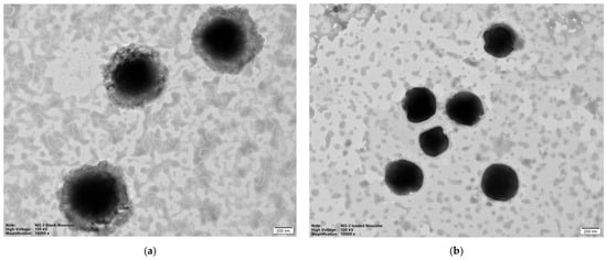
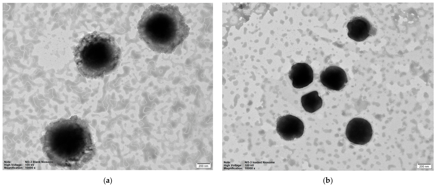
The morphology of the unloaded (NI3-3) and LAA and antioxidant-loaded niosomes (NI3-3A) is presented in Figure 8. TEM micrographs revealed that both formulations were spherical and homogeneously distributed, showing no signs of aggregation. The encapsulation of the active compounds did not alter the core spherical structure of the vesicles. A comparative observation between NI3-3 and NI3-3A indicated subtle differences in internal density and surface texture; the loaded niosomes exhibited a slightly denser appearance, which can be attributed to the incorporation of the compounds within the vesicular structure. Based on the physicochemical properties of the actives, hydrophilic components (such as L-ascorbic acid and ergothioneine) are typically associated with the aqueous core or polar head groups, whereas lipophilic antioxidants (such as alpha-tocopherol and ferulic acid) are integrated within the lipid bilayer [34,40]. Furthermore, the particle sizes observed within the TEM fields confirmed the nanostructure of the delivery system, with vesicle diameters remaining consistently below 200 nm, supporting the stability and structural integrity of the niosomal formulation.
Figure 8.
Morphology of unloaded niosomes (a), and LAA and antioxidant-loaded niosomes (b) observed by TEM.
3.7. Photostability of LAA and Antioxidant-Loaded Niosomes
Initially, photostability test was conducted by exposing both the solution containing LAA and antioxidants and NI3-3A. Therefore, a 28-day stability study was conducted under ambient room light and natural diurnal cycles to better simulate real-world storage conditions.
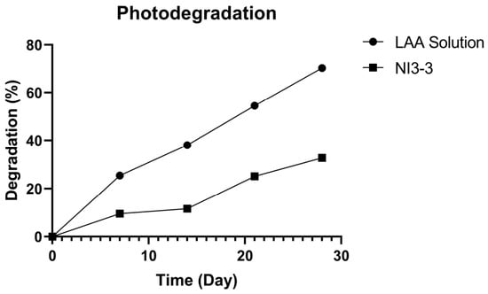
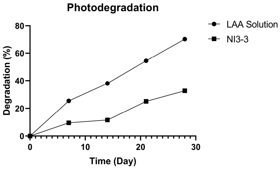
Quantitative analysis confirmed that the NI3-3A can enhance the chemical stability of LAA during the 28-day photostability study (Table 6 and Figure 9). The NI3-3A effectively retarded photodegradation of LAA. The formulation exhibited strong protective capabilities during the first two weeks, with a minor loss of LAA content of 9.55% and 11.6% on Day 7 and 14, respectively. At Day 28, it retained 67.32% of LAA content. Remarkably, the percentage of remaining LAA in NI3-3A was 2.2-fold higher than that observed in the solution at the end of the study. The enhanced photostability of NI3-3A may be attributed to the niosomal vesicular structure, which likely reduced the exposure of encapsulated LAA to environmental degradative factors, including light and oxygen. Furthermore, the presence of phospholipids, surfactants, and cholesterol in the bilayer serves as an effective physical barrier that shields the encapsulated LAA within its aqueous core from direct photo-oxidation [43]. In contrast, the solution degraded rapid degradation upon storage, losing 25.42% of its initial LAA content by Day 7 and 54.52% by Day 21. By Day 28, the degradation had reached to 70.26. These findings highlight the susceptibility of unencapsulated LAA to degradation during prolonged exposure to ambient light and oxygen.
Table 6.
Degradation (%) of LAA in solution and LAA in antioxidant-loaded niosomes (NI3-3A) during photostability test for 28 days.
Figure 9.
Photodegradation profiles (%) of the solution, and LAA and antioxidant-loaded niosomes (NI3-3A) during photostability test for 28 days.
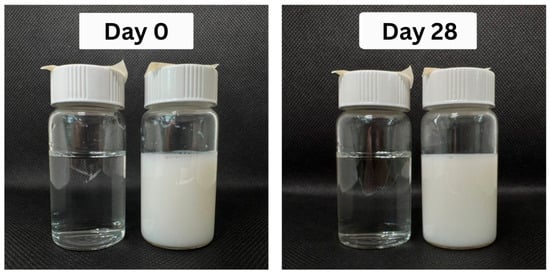
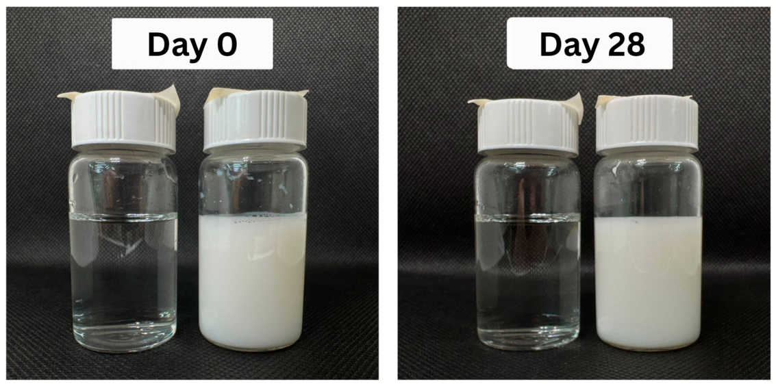
Physical appearance observations (Figure 10) showed that both formulations remained macroscopically stable throughout the 28 days. The solution stayed clear and transparent, while NI3-3A maintained a homogenous, milky-white appearance without visible precipitation, sedimentation, or phase separation. Although LAA degradation was observed in the solution during storage, no visible browning or discoloration occurred. In conclusion, both formulations maintained acceptable physical appearance throughout the study period. However, the chemical stability results demonstrated that NI3-3A provides significantly greater protection against LAA degradation than the solution. These findings suggest that niosomal encapsulation may be an effective strategy for improving the storage stability of LAA in cosmetic formulations.
Figure 10.
Visual appearance of solution (left), and LAA and antioxidant-loaded niosomes (NI3-3A) (right) at Day 0 and after 28 days of photostability test.
3.8. Skin Retention and Permeation Studies of LAA and Antioxidant-Loaded Niosomes
The in vitro skin permeation study revealed that LAA was not detected in the receptor chamber under the experimental conditions throughout the 12 h experimental period (1, 2, 4, 6, 8, and 12 h). This finding suggests that LAA was not able to permeate completely through the Strat-M® membrane under the experimental conditions employed. For topical cosmetic applications, retention of active compounds within the upper skin layers rather than systemic permeation is generally desirable, as the intended effects are localized to the skin surface, epidermis, and dermis [44]. Conversely, substantial retention of LAA was observed within the membrane layer after 12 h of exposure. The NI3-3A achieved a remarkable skin retention of 56.91%, which was significantly higher than the solution containing LAA and antioxidants (11.32%).
This enhanced deposition can be attributed to the physicochemical properties of the niosomes. Composed of non-ionic surfactants and cholesterol, the flexible lipid–surfactant bilayer of NI3-3A shares structural similarities with the lipid matrix of the stratum corneum, promoting high affinity and interaction with the membrane lipids. Additionally, the non-ionic surfactant components can interact with the tightly packed lipid arrangement, facilitating the close deposition and localization of the vesicles. Consequently, the niosomal system effectively improves the topical retention of the entrapped hydrophilic LAA and antioxidants within the target skin layers.
3.9. Limitations of the Study
This study has some limitations that should be considered. First, the anti-tyrosinase and anti-aging activities were evaluated at only one concentration. Therefore, dose–response relationships and IC50 values were not determined, and the synergistic effects of the antioxidant combination on these activities were not evaluated. Further studies using a range of concentrations are needed to determine IC50 values and assess possible synergistic interactions among the antioxidants. Second, the limit of detection (LOD) and limit of quantification (LOQ) of LAA in the receptor medium were not determined. Thus, very low concentrations of LAA in the receptor chamber may have been below the sensitivity of the analytical method. Therefore, determination of the LOD and LOQ in the receptor medium should be included in future studies to better evaluate LAA permeation. Finally, EE was not determined for all formulations during the screening and stability studies because the initial selection of formulations was based mainly on their physicochemical properties and physical stability. As a result, possible leakage of the encapsulated compounds during storage could not be directly compared among all formulations. Future studies should determine EE for the candidate formulations during storage to better understand vesicle integrity and the retention of the encapsulated compounds.
4. Conclusions
In conclusion, this study successfully developed a multifunctional niosomal delivery system for enhancing the chemical instability and skin retention of labile antioxidants combination. The selected antioxidants combination, designated as Mix D (L-ascorbic acid, alpha-tocopherol, ferulic acid, and ergothioneine at a ratio of 1:1:2:2), demonstrated synergistic antioxidant interactions in both DPPH and FRAP assays and exhibited anti-tyrosinase and anti-aging activities against hyaluronidase, collagenase, and elastase. These findings suggest that the combination may provide multifunctional benefits for cosmetic applications targeting oxidative stress, hyperpigmentation, and skin aging. The selected niosomal formulation (NI3-3A) was successfully developed with nanoscale particle size, low polydispersity index, and good physical stability during storage. Furthermore, niosomal encapsulation significantly improved the stability of LAA, maintaining a high entrapment efficiency after 90 days of storage and markedly reducing LAA degradation during photostability testing. Notably, skin retention study highlighted the formulation’s exceptional ability to localize active delivery, achieving a remarkable skin retention. Consequently, the results demonstrate the potential of niosomal encapsulation as an effective strategy for improving the stability and topical delivery of antioxidant combinations. This formulation may serve as a promising platform for the development of anti-aging and skin-brightening cosmetic products. Further studies using biological skin models and human subjects are warranted to confirm its long-term efficacy and safety.
Author Contributions
Conceptualization, K.K. and P.W.; methodology, K.K., P.P. and P.W.; validation, K.K. and P.P.; formal analysis, K.K., P.W. and P.P.; investigation, K.K. and P.W.; resources, K.K. and P.W.; data curation, P.W., K.K. and P.P.; writing—original draft preparation, P.W.; writing—review and editing, P.W., P.P. and K.K.; supervision, K.K.; project administration, K.K. All authors have read and agreed to the published version of the manuscript.
Funding
This research received no external funding.
Institutional Review Board Statement
Not applicable.
Informed Consent Statement
Not applicable.
Data Availability Statement
The data presented in this study are available from the corresponding author upon reasonable request.
Acknowledgments
The authors thank Chiang Mai University for its financial support, and are grateful to the Faculty of Pharmacy, Chiang Mai University, for providing the necessary facilities and resources. During the preparation of this manuscript, the authors used Gemini (Google) to assist in the creation of the graphical abstract and to translate and refine the English language from the authors’ original Thai drafts. After using this tool, the authors carefully reviewed and edited all outputs and take full responsibility for the content of this publication.
Conflicts of Interest
The authors declare no conflicts of interest.
References
- Martic, I.; Jansen-Dürr, P.; Cavinato, M. Effects of Air Pollution on Cellular Senescence and Skin Aging. Cells 2022, 11, 2220. [Google Scholar] [CrossRef] [Scilit] [PubMed]
- Rinnerthaler, M.; Bischof, J.; Streubel, M.K.; Trost, A.; Richter, K. Oxidative Stress in Aging Human Skin. Biomolecules 2015, 5, 545–589. [Google Scholar] [CrossRef] [Scilit] [PubMed]
- Dreher, F.; Maibach, H. Protective Effects of Topical Antioxidants in Humans. Curr. Probl. Dermatol. 2001, 29, 157–164. [Google Scholar] [CrossRef] [Scilit] [PubMed]
- Al-Niaimi, F.; Chiang, N.Y.Z. Topical Vitamin C and the Skin: Mechanisms of Action and Clinical Applications. J. Clin. Aesthetic Dermatol. 2017, 10, 14–17. [Google Scholar]
- Yang, L.-F.; Zeng, C.-M. The Degradation Products of Ascorbic Acid Inhibit Amyloid Fibrillation of Insulin and Destabilize Preformed Fibrils. Molecules 2018, 23, 3122. [Google Scholar] [CrossRef] [Scilit] [PubMed]
- Lin, F.; Lin, J.-Y.; Gupta, R.D.; Tournas, J.A.; Burch, J.A.; Selim, M.A.; Monteiro-Riviere, N.A.; Grichnik, J.M.; Zielinski, J.; Pinnell, S.R. Ferulic Acid Stabilizes a Solution of Vitamins C and E and Doubles Its Photoprotection of Skin. J. Investig. Dermatol. 2005, 125, 826–832. [Google Scholar] [CrossRef] [Scilit] [PubMed]
- Lin, J.-Y.; Selim, M.A.; Shea, C.R.; Grichnik, J.M.; Omar, M.M.; Monteiro-Riviere, N.A.; Pinnell, S.R. UV Photoprotection by Combination Topical Antioxidants Vitamin C and Vitamin E. J. Am. Acad. Dermatol. 2003, 48, 866–874. [Google Scholar] [CrossRef] [Scilit] [PubMed]
- Liu, H.; Tang, W.; Wang, X.; Jiang, J.; Zhang, W.; Wang, W. Safe and Effective Antioxidant: The Biological Mechanism and Potential Pathways of Ergothioneine in the Skin. Molecules 2023, 28, 1648. [Google Scholar] [CrossRef] [Scilit] [PubMed]
- Tsay, G.J.; Lin, S.-Y.; Li, C.-Y.; Mau, J.-L.; Tsai, S.-Y. Comparison of Single and Combined Use of Ergothioneine, Ferulic Acid, and Glutathione as Antioxidants for the Prevention of Ultraviolet B Radiation-Induced Photoaging Damage in Human Skin Fibroblasts. Processes 2021, 9, 1204. [Google Scholar] [CrossRef] [Scilit]
- Bautista-Solano, A.A.; Dávila-Ortiz, G.; Perea-Flores, M.d.J.; Martínez-Ayala, A.L. A Comprehensive Review of Niosomes: Composition, Structure, Formation, Characterization, and Applications in Bioactive Molecule Delivery Systems. Molecules 2025, 30, 3467. [Google Scholar] [CrossRef] [Scilit] [PubMed]
- Nitthikan, N.; Leelapornpisid, P.; Natakankitkul, S.; Chaiyana, W.; Mueller, M.; Viernstein, H.; Kiattisin, K. Improvement of Stability and Transdermal Delivery of Bioactive Compounds in Green Robusta Coffee Beans Extract Loaded Nanostructured Lipid Carriers. J. Nanotechnol. 2018, 2018, 7865024. [Google Scholar] [CrossRef] [Scilit]
- Nitthikan, N.; Preedalikit, W.; Supadej, K.; Chaichit, S.; Leelapornpisid, P.; Kiattisin, K. Exploring the Wound Healing Potential of a Cuscuta chinensis Extract-Loaded Nanoemulsion-Based Gel. Pharmaceutics 2024, 16, 573. [Google Scholar] [CrossRef] [Scilit] [PubMed]
- Duarte, D.; Vale, N. Evaluation of Synergism in Drug Combinations and Reference Models for Future Orientations in Oncology. Curr. Res. Pharmacol. Drug Discov. 2022, 3, 100110. [Google Scholar] [CrossRef] [Scilit] [PubMed]
- Kiattisin, K.; Nantarat, T.; Leelapornpisid, P. Evaluation of Antioxidant and Antityrosinase Activities as Well as Stability of Green and Roasted Coffee Bean Extracts from Coffea arabica and Coffea canephora Grown in Thailand. J. Pharmacogn. Phytother. 2016, 8, 182–192. [Google Scholar] [CrossRef] [Scilit]
- Preedalikit, W.; Chittasupho, C.; Leelapornpisid, P.; Potprommanee, S.; Kiattisin, K. Comparison of Biological Activities and Protective Effects on PAH-Induced Oxidative Damage of Different Coffee Cherry Pulp Extracts. Foods 2023, 12, 4292. [Google Scholar] [CrossRef] [Scilit] [PubMed]
- Chantadee, T.; Chaichit, S.; Kiattisin, K.; Poomanee, W.; Okonogi, S.; Phumat, P. Emulsification-Based Liposomal Formulation of Gallic Acid and Curcumin as Potent Topical Antioxidants. Drug Discov. Ther. 2025, 19, 38–48. [Google Scholar] [CrossRef] [Scilit] [PubMed]
- Jandang, W.; Ampasavate, C.; Kiattisin, K. Natural Stabilizers and Nanostructured Lipid Carrier Entrapment for Photosensitive Compounds, Curcumin and Capsaicin. Pharmaceutics 2024, 16, 412. [Google Scholar] [CrossRef] [Scilit] [PubMed]
- Ahmida, M.H.S. Determination of Ascorbic Acid in Vitamin C (Tablets) by High-Performance Liquid Chromatography. Asian J. Chem. 2009, 21, 6463–6467. [Google Scholar]
- Naidoo, K.; Birch-Machin, M.A. Oxidative Stress and Ageing: The Influence of Environmental Pollution, Sunlight and Diet on Skin. Cosmetics 2017, 4, 4. [Google Scholar] [CrossRef] [Scilit]
- Gulcin, İ.; Alwasel, S.H. DPPH Radical Scavenging Assay. Processes 2023, 11, 2248. [Google Scholar] [CrossRef] [Scilit]
- Gulcin, İ.; Alwasel, S.H. Fe3+ Reducing Power as the Most Common Assay for Understanding the Biological Functions of Antioxidants. Processes 2025, 13, 1296. [Google Scholar] [CrossRef] [Scilit]
- Chou, T.-C. Theoretical Basis, Experimental Design, and Computerized Simulation of Synergism and Antagonism in Drug Combination Studies. Pharmacol. Rev. 2006, 58, 621–681. [Google Scholar] [CrossRef] [Scilit] [PubMed]
- Cheah, I.K.; Halliwell, B. Ergothioneine; antioxidant potential, physiological function and role in disease. Biochim. Biophys. Acta (BBA)-Mol. Basis Dis. 2012, 1822, 784–793. [Google Scholar] [CrossRef] [Scilit] [PubMed]
- Hondal, R.J. Selenium vitaminology: The connection between selenium, vitamin C, vitamin E, and ergothioneine. Curr. Opin. Chem. Biol. 2023, 75, 102328. [Google Scholar] [CrossRef] [Scilit] [PubMed]
- Chang, T.-S. An Updated Review of Tyrosinase Inhibitors. Int. J. Mol. Sci. 2009, 10, 2440–2475. [Google Scholar] [CrossRef] [Scilit] [PubMed]
- Choi, J.Y.; Ha, J.W.; Boo, Y.C. Multifaceted Effects of L-Cysteine, L-Ascorbic Acid, and Their Derivatives on the Viability and Melanin Synthesis of B16/F10 Cells under Different Conditions. Antioxidants 2024, 13, 330. [Google Scholar] [CrossRef] [Scilit] [PubMed]
- Graf, E. Antioxidant potential of ferulic acid. Free Radic. Biol. Med. 1992, 13, 435–448. [Google Scholar] [CrossRef] [Scilit] [PubMed]
- Liu, H.-M.; Tang, W.; Wang, X.-Y.; Jiang, J.-J.; Zhang, Y.; Liu, Q.-L.; Wang, W. Experimental and theoretical studies on inhibition against tyrosinase activity and melanin biosynthesis by antioxidant ergothioneine. Biochem. Biophys. Res. Commun. 2023, 682, 163–173. [Google Scholar] [CrossRef] [Scilit] [PubMed]
- Wang, J.; Wang, H.; Guo, K.; Zhang, X.; Zhang, X. The synergistic effect of α-tocopherol and phloretin-loaded nanoemulsions on improvement of the stability, antioxidant, and tyrosinase inhibitory potentiality. J. Food Sci. 2024, 89, 8398–8409. [Google Scholar] [CrossRef] [Scilit] [PubMed]
- Lee, H.; Hong, Y.; Kim, M. Structural and functional changes and possible molecular mechanisms in aged skin. Int. J. Mol. Sci. 2021, 22, 12489. [Google Scholar] [CrossRef] [Scilit] [PubMed]
- Jayamani, J.; Naisini, A.; Madhan, B.; Shanmugam, G. Ferulic acid, a natural phenolic compound, as a potential inhibitor for collagen fibril formation and its propagation. Int. J. Biol. Macromol. 2018, 113, 277–284. [Google Scholar] [CrossRef] [Scilit] [PubMed]
- Purushothaman, J.R.; Rizwanullah, M. Ferulic Acid: A Comprehensive Review. Cureus 2024, 16, e68063. [Google Scholar] [CrossRef] [Scilit] [PubMed]
- Pullar, J.M.; Carr, A.C.; Vissers, M.C.M. The Roles of Vitamin C in Skin Health. Nutrients 2017, 9, 866. [Google Scholar] [CrossRef] [Scilit] [PubMed]
- Danaei, M.; Dehghankhold, M.; Ataei, S.; Hasanzadeh Davarani, F.; Javanmard, R.; Dokhani, A.; Khorasani, S.; Mozafari, M.R. Impact of Particle Size and Polydispersity Index on the Clinical Applications of Lipidic Nanocarrier Systems. Pharmaceutics 2018, 10, 57. [Google Scholar] [CrossRef] [Scilit] [PubMed]
- Solano, A.A.B.; Dávila-Ortiz, G.; de Jesús Perea-Flores, M.; Martínez-Ayala, A.L. Optimization and Synthesis of Nano-Niosomes for Encapsulation of Triacontanol by Box–Behnken Design. Molecules 2024, 29, 4421. [Google Scholar] [CrossRef] [Scilit] [PubMed]
- Mawazi, S.M.; Ge, Y.; Widodo, R.T. Niosome Preparation Techniques and Structure—An Illustrated Review. Pharmaceutics 2025, 17, 67. [Google Scholar] [CrossRef] [Scilit] [PubMed]
- Huanbutta, K.; Suwanpitak, K.; Ponlakorn, P.; Sonkongdang, P.; Kongklai, I.; Luangpraditkun, K.; Semangoen, T.; Sangnim, T. Development and evaluation of drug-loaded niosomes fabricated by flow chemistry: A novel vortex tube reactor approach. OpenNano 2025, 23, 100243. [Google Scholar] [CrossRef] [Scilit]
- Yaghoobian, M.; Haeri, A.; Bolourchian, N.; Shahhosseni, S.; Dadashzadeh, S. The Impact of Surfactant Composition and Surface Charge of Niosomes on the Oral Absorption of Repaglinide as a BCS II Model Drug. Int. J. Nanomed. 2020, 15, 8767–8781. [Google Scholar] [CrossRef] [Scilit] [PubMed]
- Bae, D.-H.; Shin, J.-S.; Shin, G.-S.; Jin, F.-L.; Park, S.-J. Effect of Lecithin on Dermal Safety of Nanoemulsion Prepared from Hydrogenated Lecithin and Silicone Oil. Bull. Korean Chem. Soc. 2009, 30, 821–825. [Google Scholar] [CrossRef] [Scilit][Green Version]
- Lens, M. Niosomes as Vesicular Nanocarriers in Cosmetics: Characterisation, Development and Efficacy. Pharmaceutics 2025, 17, 287. [Google Scholar] [CrossRef] [Scilit] [PubMed]
- Ullah, S.; Hussain, A.; Ali, J.; Urrehman, K.; Ullah, A. A Simple and Rapid HPLC Method for Analysis of Vitamin-C in Local Packed Juices of Pakistan. Middle-East J. Sci. Res. 2012, 12, 1085–1091. [Google Scholar] [CrossRef]
- Wang, R.; Ma, C.; Yan, H.; Zhao, H.; Wang, P.; Zhang, S.; Ju, J.; Yu, S.; Yin, Z. Lecithin/Cholesterol/Tween 80 Liposomes for Co-Encapsulation of Vitamin C and Xanthoxylin. ACS Appl. Nano Mater. 2024, 7, 5982–5995. [Google Scholar] [CrossRef] [Scilit]
- Tissera, C.E.; Barnetche, M.E.; Silva, O.F.; Fernández, M.A. Increasing the Stability of Ascorbic Acid Through Encapsulation in Food-Grade Vesicles: An Approach for Nutritional Improvement. Int. J. Food Sci. Technol. 2025, 60, vvae027. [Google Scholar] [CrossRef] [Scilit]
- Jadach, B.; Bielawna, Z. Smarter Skin Delivery: Nanosomes and Advanced Nanocarriers in Cutting-Edge Cosmetics. Molecules 2026, 31, 1312. [Google Scholar] [CrossRef] [Scilit] [PubMed]

Disclaimer/Publisher’s Note: The statements, opinions and data contained in all publications are solely those of the individual author(s) and contributor(s) and not of MDPI and/or the editor(s). MDPI and/or the editor(s) disclaim responsibility for any injury to people or property resulting from any ideas, methods, instructions or products referred to in the content. |
© 2026 by the authors. Licensee MDPI, Basel, Switzerland. This article is an open access article distributed under the terms and conditions of the Creative Commons Attribution (CC BY) license.