


Abstract
Cod liver oil is a rich source of polyunsaturated fatty acids (PUFAs) but is highly susceptible to oxidative degradation, limiting its use in topical formulations. This study aimed to develop stable cod liver oil nanoemulsions for topical application and to evaluated the influence of surfactant ratio (lecithin/PEG-15 hydroxystearate: 2.5:1 and 1:1, w/w), emulsification method (ultrasonication or high-pressure homogenization), and vitamin E acetate supplementation on their physicochemical properties and oxidative stability. Eight nanoemulsions were characterized in terms of droplet size, polydispersity, ζ-potential, vitamin E acetate encapsulation efficiency, oxidative stability, film-forming capacity and cytocompatibility. Among the investigated formulations, F4 (2.5:1 lecithin/PEG-15 hydroxystearate, high-pressure homogenization, with vitamin E acetate) exhibited the most favorable characteristics, including a mean droplet size of 67.95 nm, ζ-potential of −63.12 mV and vitamin E acetate encapsulation efficiency of 32.59%. The formulation demonstrated good physicochemical stability under thermal, mechanical and photostability testing, improved oxidative stability, transient film-forming behavior with an initial occlusive effect, and no cytotoxicity toward human dermal fibroblasts. These findings indicate that nanoemulsion performance depends on the combined influence of formulation composition and processing conditions, with F4 representing a promising topical carrier for cod liver oil intended for interaction with the stratum corneum.
1. Introduction
Marine-derived lipids constitute a chemically diverse and biologically relevant class of natural compounds, with cod liver oil representing one of the most established sources of omega-3 (ω-3) polyunsaturated fatty acids (PUFAs), particularly eicosapentaenoic acid (EPA) and docosahexaenoic acid (DHA) [1]. Beyond their well-documented systemic effects, ω-3 PUFAs play a fundamental role in skin physiology, contributing to membrane organization, lipid fluidity, and modulation of inflammatory signaling pathways [1,2]. Increasing evidence suggests that these fatty acids are structural and functional components of the epidermal lipid environment, supporting the relevance of marine-derived lipids in topical and dermocosmetic applications [2,3]. In this context, cod liver oil is considered in the present study as a functional lipid phase, contributing both compositional and physicochemical attributes to the formulation.
The outermost layer of the skin, the stratum corneum, acts as a highly specialized permeability barrier composed of corneocytes embedded within an ordered lipid matrix primarily consisting of ceramides, cholesterol, and free fatty acids [3,4,5]. This architecture is essential for the regulation of transepidermal water loss (TEWL) and for protection against environmental stressors, allergens, and microbial invasion [3,4,5]. In several chronic inflammatory skin disorders, including atopic dermatitis, psoriasis, and eczema, alterations in lipid composition and lamellar organization are commonly observed [5,6]. These changes are associated with depletion of essential fatty acids, impaired barrier function, increased TEWL, and exacerbated inflammatory responses [1,4,5]. Consequently, the topical application of lipid-rich systems has been explored as a strategy to support barrier function and restore lipid balance [3,4].
Despite its high PUFA content and therapeutic relevance, as well as the presence of bioactive micronutrients such as vitamins A and D and endogenous tocopherols, the topical use of cod liver oil is intrinsically limited by its pronounced susceptibility to oxidative degradation [6,7]. The extensive unsaturation of EPA- and DHA-rich triglycerides renders the oil highly vulnerable to oxidation induced by oxygen, light exposure, or thermal stress. Lipid peroxidation not only compromises the physicochemical stability and bioactivity of the oil but also leads to the formation of secondary oxidation products that are associated with unpleasant odors, reduced formulation acceptability, and potential skin irritation [8,9]. These challenges highlight the need for appropriate formulation strategies to stabilize cod liver oil and enable its effective incorporation into topical application systems [7,10].
Nanoemulsions have emerged as promising carriers for the delivery of hydrophobic bioactive compounds [11,12,13]. Characterized by droplet sizes typically below 200 nm [14], nanoemulsions offer enhanced kinetic stability, high interfacial surface area, and improved dispersion of lipophilic ingredients within aqueous environments [13]. From a topical perspective, these systems facilitate uniform application and close contact with the stratum corneum, while the encapsulation of sensitive oils within nanoscale droplets can provide a protective microenvironment that mitigates oxidative processes [14,15,16,17]. However, the performance of nanoemulsions is highly dependent on formulation parameters and processing conditions [14].
Surfactant selection and interfacial composition play a critical role in determining droplet stabilization, surface charge development and resistance to coalescence or Ostwald ripening [18,19]. At the same time, the emulsification method strongly influences droplet size distribution and structural robustness. High-energy techniques such as high-pressure homogenization and ultrasonication are widely employed for nanoemulsion production; however, their comparative effectiveness in stabilizing PUFA-rich marine oils remains a subject of ongoing investigation [20,21,22]. In addition, the incorporation of lipid-soluble antioxidants, such as vitamin E derivatives, is commonly employed to enhance the oxidative stability of PUFA-rich systems. In the present study, vitamin E acetate is used primarily as a stabilizing agent to protect the lipid phase from oxidation, rather than as the main active component of the formulation [9,10].
Therefore, this study aims to systematically develop cod liver oil nanoemulsions stabilized by a lecithin/PEG-15 hydroxystearate system, and to evaluate the combined influence of formulation (surfactant ratio and vitamin E acetate supplementation) and process variables (high-pressure homogenization versus ultrasonication) on their physicochemical characteristics, colloidal stability and oxidative integrity [14,21,22]. The selection of lecithin and PEG-15 hydroxystearate was based on their complementary interfacial properties [23,24]. Lecithin, a naturally derived phospholipid, contributes to biocompatibility and the formation of cohesive interfacial films, while PEG-15 hydroxystearate provides steric stabilization and enhances dispersion stability. Such combinations have been reported to be effective in stabilizing lipid-based nanoemulsions for topical applications [11]. By focusing on the stabilization and performance of cod liver oil as a functional lipid phase, this work seeks to provide a rational framework for the design of stable marine-lipid nanoemulsions for topical applications and skin barrier support [25,26,27].
2. Materials and Methods
2.1. Materials
Pharmaceutical-grade cod liver oil was obtained from Möller’s (Oslo, Norway). Vitamin E acetate (tocopheryl acetate, ≥98% purity) was supplied by BASF (Ludwigshafen, Germany). EmulmetikTM 900 (purified phosphatidylcholine-rich lecithin) was purchased from Lucas Meyer Cosmetics (Champlan, France). Kolliphor HS 15 (macrogol 15 hydroxystearate, PEG-15) was obtained from BASF Care Creations (Monheim, Germany). Water for injection was supplied by Fresenius Kabi Hellas, and purified water was obtained from Farmalabor (Chemco by Syndesmos S.A., Ilion, Greece).
All reagents used for analytical assays were of analytical grade and were used as received without further purification.
2.2. Experimental Design and Formulation Composition
Cod liver oil nanoemulsions were formulated using a mixed surfactant system composed of lecithin and PEG-15 hydroxystearate. Two surfactant ratios were investigated: 2.5:1 and 1:1 (w/w, lecithin/PEG-15 hydroxystearate).
A full factorial experimental design (23) was employed to systematically evaluate the influence of three independent variables: Factor A, surfactant ratio (lecithin/PEG-15 hydroxystearate; 2.5:1 and 1:1, w/w); Factor B, emulsification method (ultrasonication and high-pressure homogenization); and Factor C, vitamin E acetate supplementation (absence or presence). This design resulted in a total of eight formulations (F1–F8), as summarized in Table 1.
Table 1.
Composition and experimental design of cod liver oil nanoemulsions prepared according to a 23 full factorial design, showing the three experimental factors and their corresponding levels.
The lipid phase consisted of cod liver oil with or without vitamin E acetate, while the aqueous phase consisted of purified water. The oil phase, surfactant mixture, and aqueous phase were used at fixed concentrations of 2.5%, 3.5%, and 94% (w/w), respectively. All formulations were prepared under subdued light conditions to minimize oxidative degradation.
The total concentrations of oil, surfactant mixture, and aqueous phase were kept constant across all formulations in order to enable direct comparison of factor effects. All compositions are expressed on a weight/weight (w/w) basis.
2.3. Preparation of Nanoemulsions
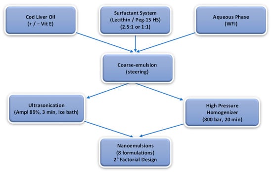
Nanoemulsions were prepared using two high-energy emulsification techniques for comparative evaluation: high-pressure homogenization (HPH) and ultrasonication, in accordance with the factorial design described in Section 2.2. For all formulations, the oil phase, surfactant mixture, and aqueous phase were combined at fixed weight ratios (Table 1), ensuring that only the investigated variables (surfactant ratio, processing method, and antioxidant supplementation) were varied (Figure 1).
Figure 1.
Schematic representation of the experimental design and preparation process for cod liver oil nanoemulsions. The system was developed using a 23 factorial design, evaluating the effects of surfactant ratio, antioxidant supplementation, and emulsification method on nanoemulsion formation, highlighting the controlled formulation composition and the two high-energy emulsification techniques applied.
2.3.1. Coarse Emulsion Preparation
The lipid phase, consisting of cod liver oil with or without vitamin E acetate, was mixed with the surfactant blend (lecithin/PEG-15 hydroxystearate) under magnetic stirring at ambient temperature until a visually homogeneous mixture was obtained. The aqueous phase (purified water) was then gradually added under continuous stirring to form a coarse emulsion.
2.3.2. Ultrasonication
Ultrasonication was performed using a probe sonicator (Vibra-CellTM VCX 130PB, Sonics & Materials, Inc., Newtown, CT, USA) operating at an amplitude of 89%. Each formulation was subjected to three consecutive ultrasonication cycles of 1 min duration (total effective sonication time: 3 min), with 30 s intervals between cycles. During these intervals, samples were vortex-mixed to ensure homogeneity [17].
All samples were maintained in an ice bath during processing, and the temperature was monitored to ensure that it remained below 25 °C, minimizing thermal or oxidative degradation.
2.3.3. High-Pressure Homogenization (HPH)
The coarse emulsions were processed using an EmulsiFlexTM-C5 high-pressure homogenizer (Avestin Inc., Ottawa, ON, Canada). Homogenization was performed at a constant pressure of 800 bar (11,600 psi) in continuous recirculation mode for 20 min. Temperature was controlled using the integrated heat exchanger to prevent excessive thermal buildup and minimize oxidative degradation of the PUFA-rich lipid phase [21,22].
2.3.4. General Handling Conditions
All formulations were prepared under subdued light conditions to limit photooxidation. Freshly prepared nanoemulsions were stored in sealed containers at 4 °C prior to further characterization. All experiments were performed in triplicate.
2.4. Physicochemical Characterization
2.4.1. Droplet Size and Polydispersity Index
Mean droplet size (Z-average) and polydispersity index (PdI) were determined by dynamic light scattering (DLS) using a Zetasizer Nano ZS (Malvern Instruments, Malvern, UK). An aliquot of 100 μL of each nanoemulsion sample was diluted to a final volume of 600 μL using WFI corresponding to the external aqueous phase of the formulations. Measurements were conducted at 25 °C following appropriate dilution with purified water to avoid multiple scattering effects. All measurements were performed in triplicate [11,17,28].
2.4.2. ζ-Potential Measurement
Surface charge (ζ-potential) was measured for each sample immediately after assessment of droplet size distribution, using electrophoretic light scattering (ELS) with the same instrument, and employing non-invasive backscattering at a detection angle of 173°. The results are reported as the mean ± standard deviation of triplicate measurements [17,28].
2.5. Oxidative Integrity/Lipid Preservation Analyses
2.5.1. Vitamin E Acetate Encapsulation Efficiency
Encapsulation efficiency (EE%) of vitamin E acetate was determined following separation of free and nanoemulsion-associated fractions using size exclusion chromatography (SEC).
Briefly, samples were applied to a Sephadex G-75 column (stationary phase) and eluted using water for injection (WFI) as the mobile phase. Nanoemulsion droplets eluted in the void volume, while free vitamin E acetate was retained within the column matrix.
The fraction corresponding to the nanoemulsion droplets was collected, diluted with methanol in order to disrupt the nanoemulsion structure and enable spectrophotometric determination of vitamin E acetate levels at 290 nm using a UV-1800 spectrophotometer (Shimadzu, Kyoto, Japan). Quantification was performed using a calibration curve constructed using standard solutions of vitamin E acetate in the same solvent system. Encapsulation efficiency was calculated using Equation (1), taking into account all the dilution steps [29].
where Associated Vitamin E acetate is the amount of vitamin E acetate associated with nanoemulsion droplets and Initial Vitamin E acetate is the total amount of vitamin E acetate that was initially added.
EE(%) = (Associated Vitamin E acetate/Initial Vitamin E acetate) × 100,
2.5.2. Peroxide Value Determination
Oxidative stability was evaluated by determining the peroxide value (PV) via iodometric titration using sodium thiosulfate (0.01 N) as the titrant and a 1% starch solution as the indicator [8,16].
Under the applied experimental conditions, the detection limit of the method was approximately 1.0 meq O2/kg oil. Therefore, the absence of a detectable color change was interpreted as peroxide values below this threshold rather than complete absence of oxidation.
2.5.3. Triglyceride Content
Triglyceride preservation was assessed using a commercial enzymatic colorimetric assay (Triglycerides FS). Samples were incubated for 20 min at 25 °C and absorbance was measured at 505 nm according to the manufacturer’s instructions. The results were expressed as absorbance units (AU) and used for relative comparison among formulations. The assay was therefore employed as an indicator of relative changes in triglyceride content associated with oxidative or processing-induced degradation. This approach is consistent with comparative stability assessment methodologies reported in the literature.
2.5.4. Cod Liver Oil Composition
The fatty acid composition of the nanoemulsion formulations was determined using a modified one-step in situ transesterification (IST) procedure adapted from previously reported methods [30].
Briefly, an accurately measured aliquot of each nanoemulsion sample (approximately 3 mL) was lyophilized using a LyoQuest (Telstar, Barcelona, Spain), after which an accurately measured amount of each lyophilized sample (corresponding to approximately 10–50 mg of total lipid content) was directly subjected to transesterification without prior lipid extraction. Samples were reacted with 5 mL of a CH3OH/CHCl3/H2SO4 mixture (10:1:0.3, v/v/v) at 90 °C for 2 h under vigorous stirring to ensure complete conversion of fatty acids into their corresponding fatty acid methyl esters (FAMEs). The reaction was terminated by the addition of 2 mL of water, and FAMEs were subsequently extracted using a 4:1 (v/v) organic phase (hexane/chloroform).
The obtained FAMEs were analyzed by gas chromatography (GC) using an Agilent Technologies 7890A GC system (Agilent, Santa Clara, CA, USA) with a Flame Ionization Detector (FID), based on calibration with a certified FAME reference standard (FAMQ-005, AccuStandard Inc., New Haven, CT, USA). An internal standard (heptadecanoic acid ≥98%; Sigma-Aldrich, St. Louis, MO, USA) was added prior to analysis for quantitative purposes. Separation was achieved using a DB-WAX capillary column (10 m length, 0.1 mm internal diameter, 0.1 μm film thickness; Agilent J&W GC Columns, Agilent, Santa Clara, CA, USA). The oven temperature program was set as follows: from 40 °C to 195 °C at 25 °C/min, from 195 °C to 205 °C at 3 °C/min, and from 205 °C to 230 °C at 8 °C/min, with a final hold of 1 min. Helium was used as the carrier gas at an average linear velocity of 30.34 cm/s, while injector and detector temperatures were maintained at 250 °C [31].
2.6. Stability Studies
The stability of the cod liver oil nanoemulsions was evaluated by monitoring their physicochemical properties during storage and after exposure to stress conditions. Stability assessment was based on changes in mean droplet size, polydispersity index (PdI), ζ-potential and oxidative integrity of the lipid phase.
Nanoemulsions were stored at 4 °C, 25 °C and 40 °C for a period of 60 days. At predetermined time intervals, samples were appropriately diluted with purified water and used to measure mean droplet size (Z-average) and PdI by dynamic light scattering (DLS), as well as ζ-potential by electrophoretic light scattering (ELS), using a Zetasizer Nano ZS (Malvern Instruments, Malvern, UK). Stability was evaluated by comparing the physicochemical parameters obtained during storage with the initial values.
Resistance to thermal stress was further assessed using accelerated aging at 40 °C and using thermal cycling consisting of six alternating cycles between 4 °C and at 40 °C, each lasting 24 h. Mechanical stability was evaluated using centrifugation at 5000 rpm for 20 min. After each stress condition, nanoemulsions were visually inspected for signs of phase separation, creaming or sedimentation and subsequently reanalyzed for changes in droplet size distribution and PdI.
Oxidative stability of the PUFA-rich lipid core was assessed by peroxide value (PV) determination using iodometric titration. The absence of detectable peroxide formation was considered indicative of preserved oxidative integrity of the encapsulated cod liver oil.
Overall stability of the nanoemulsions was assessed based on the maintenance of nanometric droplet size, low PdI values (<0.30), stable ζ-potential values and the absence of visible physical instability throughout storage and stress testing [17,28,32].
2.6.1. Long-Term Storage Stability
Nanoemulsions were stored at 4 °C, 25 °C and 40 °C for a period of 60 days.
Droplet size, PdI and ζ-potential were monitored at predetermined intervals to evaluate changes in physicochemical stability over time [17,28].
2.6.2. Accelerated Thermal Aging and Thermal Cycling
Accelerated thermal aging was conducted by storing samples at 40 °C for 14 days. Thermal cycling involved six alternating cycles between 4 °C and 40 °C, with each cycle lasting 24 h [28].
2.6.3. Mechanical and Photostability Testing
Mechanical stability was assessed using centrifugation at 5000 rpm for 20 min. Following centrifugation, formulations were visually inspected for phase separation, creaming or sedimentation and reanalyzed for droplet size and polydispersity index [25,28].
Photostability was evaluated under environmentally relevant conditions by exposing samples to natural solar radiation for 7 consecutive days. Samples were placed indoors in proximity to a glass window, allowing for the transmission of primarily UV-A radiation (320–400 nm), while shorter wavelength UV-B radiation was largely attenuated by the glass.
Based on meteorological data for the study period (December), the daily UV Index ranged between 1 and 2, corresponding to low-intensity ultraviolet exposure. Based on meteorological UV Index data (UVI 1–2), the theoretical erythemally weighted UV dose was estimated to be approximately 270 J/m2 per day. However, due to indoor exposure conditions and UV filtering by glass, the effective UV dose reaching the samples was substantially lower.
Physicochemical properties were assessed before exposure (Day 1) and after the 7-day irradiation period. Following exposure, formulations were visually inspected for instability phenomena (phase separation, creaming, sedimentation) and reanalyzed in terms of droplet size and polydispersity index [25].
It should be noted that this approach provides a qualitative assessment of photostability under mild environmental conditions rather than a controlled photostability study with a defined irradiation intensity.
2.7. Antioxidant Activity
Antioxidant activity was evaluated using the DPPH radical scavenging assay.
Nanoemulsion samples were tested at concentrations ranging from 600 to 2200 μg/mL, and absorbance was measured at 540 nm. Ascorbic acid was used as a reference standard to validate assay performance, which yielded an IC50 value of 0.059 mg/mL under the same experimental conditions. The IC50 value was calculated as the concentration required to inhibit 50% of DPPH radicals [17,32,33]. The goodness-of-fit of the ascorbic acid reference calibration curve was high (R2 = 0.993), confirming the validity and reliability of the assay.
2.8. In Vitro Cytotoxicity Assessment
2.8.1. Reagents and Cell Culture
Primary normal human dermal fibroblasts (NHDFs) obtained from adult human skin (Lonza, Walkersville, MD, USA) were used in this study. Cells were maintained according to the manufacturer’s instructions in FGM-2 BulletKit culture medium supplemented with 2% serum and were passaged until they reached approximately 70–80% confluency.
2.8.2. Cytotoxicity Assay
The cytocompatibility of the selected nanoemulsion (F4) was evaluated in normal human dermal fibroblasts (NHDFs). Formulation F4 was selected based on its favorable overall physicochemical characteristics, colloidal stability, oxidative performance and vitamin E acetate encapsulation efficiency among the investigated formulations. Cell viability was evaluated using an MTT-based colorimetric assay (Vybrant® MTT Cell Proliferation Assay Kit, Thermo Fisher Scientific, Waltham, MA, USA) in accordance with the manufacturer’s instructions. Following cellular exposure to the tested samples, 100 μL of fresh basal medium (FBM) and 10 μL of MTT reagent (5 mg/mL) were added to each well. The plates were then incubated at 37 °C for 3 h. After incubation, 50 μL of DMSO was introduced to dissolve the formed formazan crystals, and the plates were further incubated for 10 min at 37 °C. Absorbance was subsequently recorded at 570 nm using a microplate reader (Infinite 200M Pro, Tecan, Männedorf, Switzerland). Cell viability was expressed as a percentage relative to untreated control cells, calculated according to Equation (2).
Cell viability (%) = (mean OD of treated cells/mean OD of control cells) × 100,
A range of concentrations (0.5–100 μg/mL) was tested for all samples to assess cytotoxic effects [32,33]. The tested concentrations were selected based on previous studies reflecting levels relevant to potential topical applications [34,35]. The cells were incubated with the different samples for 24 h prior to the cytotoxicity assessments. The cytotoxicity assessments have been performed in three independent experiments.
2.9. Film-Forming Capacity Assessment
The occlusive properties of nanoemulsions were evaluated using a modified in vitro occlusion test [25]. Briefly, glass beakers of 50 mL capacity and 51 mm internal diameter were filled with tap water (40 mL), covered with cellulose substrate (CA Membrane Filter, 47 mm, 0.45 um; Filter-Bio, Nantong, China), and sealed perimetrically with parafilm, leaving the upper surface exposed.
An aliquot of 500 μL of each nanoemulsion containing 2.5% w/v lipid (corresponding to 1.16 mg/cm2) was uniformly spread onto the membrane. Each nanoemulsion was tested in triplicate while three untreated beakers served as references. The beakers were maintained at 32 ± 0.5 °C and 50–55% relative humidity (RH) for 48 h. Their weight was recorded immediately after sample application (baseline) and at 6, 24, and 48 h to monitor water loss.
The occlusion factor (F) was calculated using Equation (3):
where R and S represent the water loss (i.e., weight reduction from baseline) of the reference and sample beakers, respectively. The occlusion factor ranges from 0 to 100, where 0 indicates no occlusive effect and 100 represents maximum occlusiveness. Based on the method of Liakopoulou et al., an F ≥ 10 is commonly considered indicative of measurable occlusive performance [25,36].
F = [(R − S)/R] × 100,
2.10. Statistical Analysis
All experiments were performed in three independent experiments (triplicate, n = 3). Results are presented as mean ± standard deviation. Statistical comparisons between groups were performed using Student’s t-test (two-tailed, unpaired), implemented in Microsoft Excel (Microsoft Office 365, version 2504, Redmond, WA, USA), and differences were considered statistically significant at p < 0.05. The statistical analysis was intended to support comparative evaluation rather than full factorial modeling.
3. Results
3.1. Physicochemical Properties
The physicochemical properties of the cod liver oil nanoemulsions were strongly influenced by the combined effects of surfactant ratio (Factor A), emulsification method (Factor B), and antioxidant supplementation (Factor C) (Table 2). Within the explored experimental space, the selected/best-performing formulation (F4) refers to the nanoemulsion exhibiting the most favorable overall physicochemical characteristics, colloidal stability and oxidative integrity among the tested conditions.
Table 2.
Physicochemical characteristics of nanoemulsions on Day 1 (mean value ± SD, n = 3).
A significant interaction between surfactant ratio (Factor A) and emulsification method (Factor B) was observed, with respect to droplet size. High-pressure homogenization (HPH) produced significantly smaller droplets than ultrasonication only in formulations prepared with the higher lecithin/PEG-15 hydroxystearate ratio (2.5:1; F3, F4 versus F1, F2; p < 0.05). In contrast, at the lower surfactant ratio (1:1), ultrasonication produced smaller droplets than HPH (F5 and F6 versus F7 and F8). These findings indicate that the effect of the processing method depended on the surfactant composition rather than representing a consistent main effect across all formulations.
Specifically, nanoemulsions F3 and F4, formulated with a lecithin/PEG-15 hydroxystearate ratio of 2.5:1 (w/w) and processed via HPH, exhibited the smallest (p < 0.05) mean droplet diameters (66.46 nm and 67.95 nm, respectively) with a low polydispersity index (PdI = 0.29 and 0.23, respectively), indicating a narrow and homogeneous size distribution. In contrast, ultrasonication of the same formulation resulted in significantly larger droplets (82–85 nm) despite identical compositions, highlighting the influence of processing conditions.
From a topical application perspective, this size range (≈70–85 nm) was considered appropriate because it provides excellent physicochemical stability together with homogeneous surface coverage and efficient interaction with the stratum corneum. Although nanosystems below 100 nm have been reported to facilitate deeper skin penetration under certain formulation and application conditions, droplet size alone is not the determining factor governing skin permeation. In the present study, the nanoemulsions were designed for topical delivery and interaction with the skin surface rather than for maximizing transdermal transport; therefore, a further reduction in droplet size was not considered necessary [26,27].
Surface charge measurements further supported these findings. All nanoemulsions exhibited negative ζ-potential values (−44.34 mV to −82.60 mV), indicating electrostatic stabilization. The most negative ζ-potential was recorded for the formulation F3 (−82.6 mV), followed by F4 (−63.12 mV), suggesting enhanced interfacial coverage and strong electrostatic repulsion between droplets in HPH-processed systems. A combined effect of surfactant ratio and processing method (A × B interaction) was evident, as ultrasonicated formulations (F1, F2, F5, F6) consistently exhibited less negative ζ-potential values, indicative of comparatively weaker interfacial stabilization.
The encapsulation efficiency (EE%) of vitamin E acetate was influenced by both formulation composition and processing conditions. High-pressure homogenization (HPH) markedly increased the encapsulation efficiency at the higher lecithin/PEG-15 hydroxystearate ratio (2.5:1), with formulation F4 exhibiting a significantly higher EE% (32.59%) than formulation F2 (8.98%, p < 0.05). In contrast, at the lower surfactant ratio (1:1), formulations F6 and F8 exhibited comparable encapsulation efficiencies (20.21% and 20.05%, respectively), indicating that the beneficial effect of HPH was not observed under these formulation conditions. These findings suggest that encapsulation efficiency depended on the combined influence of formulation composition and processing conditions, with the greatest improvement achieved when HPH was applied to the higher lecithin-containing formulations.
3.2. Antioxidant Activity of Nanoemulsions
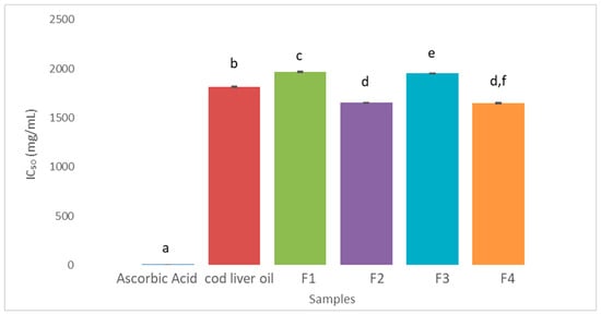
The antioxidant activity of the formulations, assessed by the DPPH assay, showed a modest improvement for vitamin E-containing nanoemulsions (F2 and F4) compared to pure cod liver oil (p < 0.05). Formulation F4 exhibited a lower IC50 value compared to pure cod liver oil (1649 μg/mL vs. 1816 μg/mL), and this difference was statistically significant (p < 0.05) (Figure 2). These findings indicate that antioxidant supplementation (Factor C) contributed to oxidative protection within the nanoemulsion system.
Figure 2.
IC50 values (mg/mL) of the tested samples for radical scavenging activity, as determined by the DPPH assay. Ascorbic acid (positive control); cod liver oil; F1: nanoemulsion without vitamin E, prepared by ultrasonication; F2: nanoemulsion with vitamin E, prepared by ultrasonication; F3: nanoemulsion without vitamin E, prepared by high-pressure homogenization; F4: nanoemulsion with vitamin E, prepared by high-pressure homogenization. Nanoemulsions (F1–F4) were formulated using a lecithin/PEG-15 hydroxystearate ratio of 2.5:1 (w/w). Lower IC50 values indicate higher antioxidant activity. Statistical significance between formulations was assessed using Student’s t-test. Different letters denote statistically significant differences (p < 0.05), whereas shared letters indicate no significant difference. Data are presented as mean ± standard deviation (n = 3).
3.3. Colloidal Stability and Stress Resistance
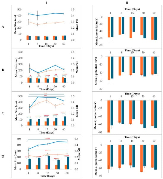
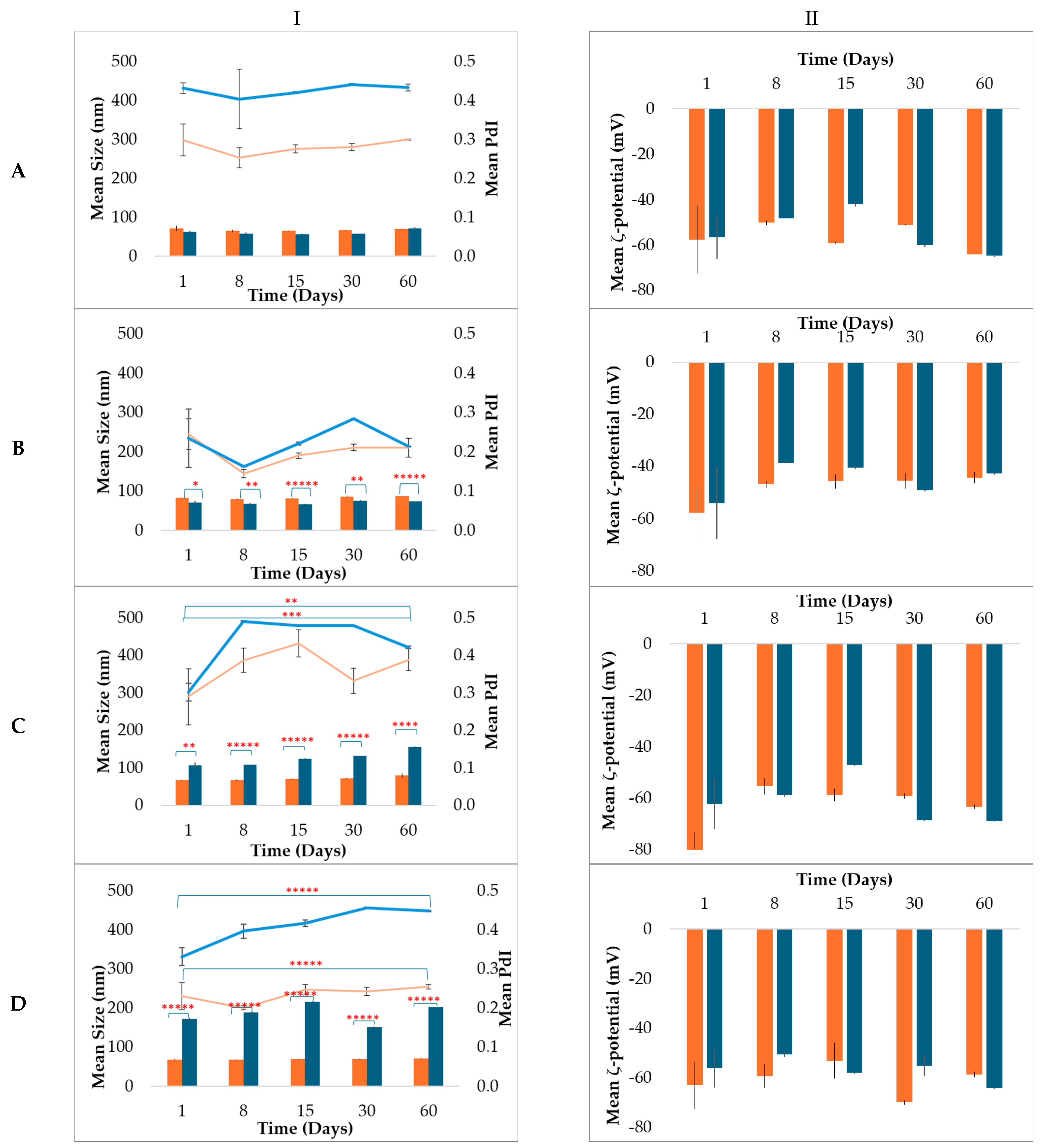
Colloidal stability of the nanoemulsions (F1–F8) was strongly influenced by surfactant ratio (Factor A) and its interaction with processing method (Factor B) (Figure 3 and Figure S1–S4). Formulations prepared at a 2.5:1 surfactant ratio and processed by high-pressure homogenization (F3 and F4) exhibited excellent stability over 60 days at 4 °C, 25 °C and 40 °C, with no significant changes in droplet size or PdI (p > 0.05), indicating resistance to aggregation and coalescence (Figure 3 and Figures S1 and S3). This stability was maintained irrespective of antioxidant presence, suggesting that Factors A and B were the dominant determinants of colloidal stability, with minimal direct contribution from Factor C.
Figure 3.
Time-dependent evolution of cod liver oil nanoemulsions stabilized with lecithin/PEG-15 hydroxystearate at a 2.5:1 (w/w) ratio (■) or 1:1 (w/w) ratio (■) during storage at 25 °C for 60 days. Mean droplet size (bars) and polydispersity index (lines) are presented in column I and ζ-potential are presented in column II. Nanoemulsions were prepared by ultrasonication (panel (A): F1, F5; panel (B): F2, F6) and high-pressure homogenization (panel (C): F3, F7; panel (D): F4, F8), either without (A,C) or with (B,D) vitamin E. Data are presented as mean ± SD (n = 3). * p < 0.05, ** p < 0.005, *** p < 0.0005, **** p < 0.00005, ***** p < 0.000005.
In contrast, formulations prepared using a 1:1 surfactant ratio (F5–F8) exhibited significant droplet growth under accelerated conditions (40 °C), exceeding 216 nm (p < 0.001) (Figure S4). This highlights a strong main effect of surfactant ratio (Factor A) and suggests insufficient interfacial coverage at lower lecithin contents.
Mechanical, thermal, and photostability assessments further supported these observations. Centrifugation, thermal cycling (six cycles), and UV exposure did not significantly affect the physicochemical properties of the formulations, as no phase separation or notable changes in droplet size, polydispersity index, or ζ-potential were observed. All formulations maintained consistent physicochemical characteristics under these stress conditions, confirming their robustness (Figures S5–S7).
These findings indicate that colloidal stability is primarily governed by interfacial composition (A), with processing method (B) enhancing structural robustness, while antioxidant presence (C) plays a secondary role in physical stability.
3.4. Oxidative Integrity and Lipid Preservation
Oxidative integrity of the nanoemulsions was evaluated through peroxide detection and triglyceride preservation analysis. No detectable peroxide formation was observed in formulations exhibiting the most favorable oxidative stability profiles (F3 and F4), indicating that primary lipid oxidation remained below the detection limit of the applied method during processing and storage evaluation.
Lipid preservation was further assessed through triglyceride-associated absorbance measurements (Table S1), where higher absorbance values were considered indicative of greater triglyceride retention. Among the evaluated formulations, the nanoemulsion prepared using a lecithin/PEG-15 hydroxystearate ratio of 2.5:1, high-pressure homogenization, and vitamin E acetate incorporation (F4) exhibited the highest mean absorbance value (0.944 ± 0.02), followed by F3 (0.895 ± 0.04), F2 (0.838 ± 0.03) and F1 (0.829 ± 0.05). In contrast, lower absorbance values were observed for F5–F8, ranging from 0.498 ± 0.06 to 0.575 ± 0.03 (Table S1).
These results suggest that oxidative integrity and lipid preservation were influenced by the combined effects of surfactant composition (Factor A), processing method (Factor B), and antioxidant supplementation (Factor C). In particular, formulations prepared with the higher lecithin/PEG-15 hydroxystearate ratio and processed by high-pressure homogenization generally exhibited higher triglyceride-associated absorbance values, while vitamin E acetate incorporation was associated with a tendency toward enhanced oxidative protection. Because not all observed differences reached statistical significance, these findings should be interpreted as trends within the investigated formulation space rather than definitive quantitative relationships.
3.5. Fatty Acid Composition and Preservation of Unsaturation
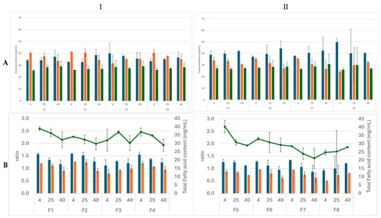
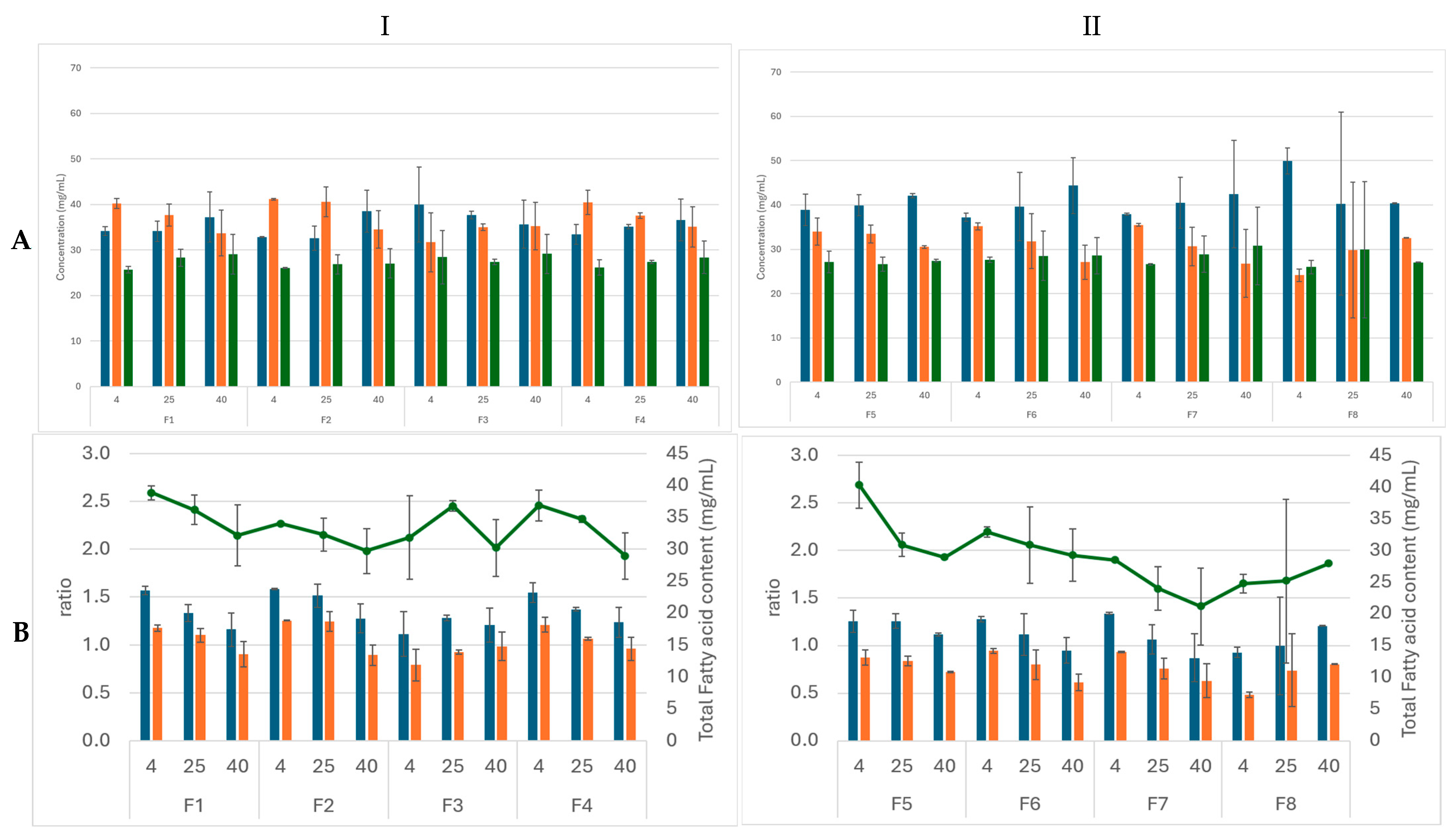
To obtain a more comprehensive assessment of lipid oxidation beyond primary oxidation products, the fatty acid composition of the formulations (F1–F8) was analyzed by gas chromatography following storage under different temperature conditions and compared with the initial composition of the cod liver oil (Figure 4, Table S2). The preservation of the fatty acid composition reflects the combined influence of formulation structure (Factors A and B) and antioxidant supplementation (Factor C).
Figure 4.
Fatty acid content of nanoemulsion formulations prepared with (I) 2.5:1 w/w lecithin/PEG-15 hydroxystearate and (II) 1:1 w/w lecithin/PEG-15 hydroxystearate. (A) Relative contents of monounsaturated fatty acids (MUFAs, ■), polyunsaturated fatty acids (PUFAs, ■) and saturated fatty acids (SFAs, ■), and (B) PUFA/SEA ratio (■), PUFA/MUFA ratio (■) and total fatty acid content (−) after storage at 4 °C, 25 °C and 40 °C for a period of 60 days. Data are presented as mean ± SD (n = 3).
The initial fatty acid profile of cod liver oil was characterized by a high content of polyunsaturated fatty acids (PUFAs), predominantly eicosapentaenoic acid (EPA) and docosahexaenoic acid (DHA). Following storage, all formulations (F1–F8) exhibited a decrease in PUFA content, accompanied by a relative increase in monounsaturated (MUFAs) and saturated fatty acids (SFAs), indicating progressive oxidative degradation. Although no statistically significant differences in PUFA content were observed among the formulations under the tested storage conditions (p > 0.05), a numerical trend toward greater PUFA loss was evident at 40 °C and in formulations prepared with the lower emulsifier ratio (1:1) (F5–F8).
The reduction in PUFA content was further reflected in the decrease in PUFA/SFA and PUFA/MUFA ratios, indicating a shift toward less unsaturated lipid profiles compared to the initial oil composition. Nevertheless, formulations containing vitamin E acetate (F2, F4, F6, and F8) generally showed comparatively improved retention of PUFAs during storage, particularly under thermal stress conditions, suggesting a partial protective effect against oxidative degradation.
These findings are in agreement with the peroxide value (PV) results and provide a more direct indication of lipid integrity at the molecular level. Overall, the data demonstrate that nanoencapsulation partially preserves the unsaturated lipid fraction relative to the initial composition, although degradation is progressively enhanced with increasing temperature.
3.6. Cytotoxicity Assessment
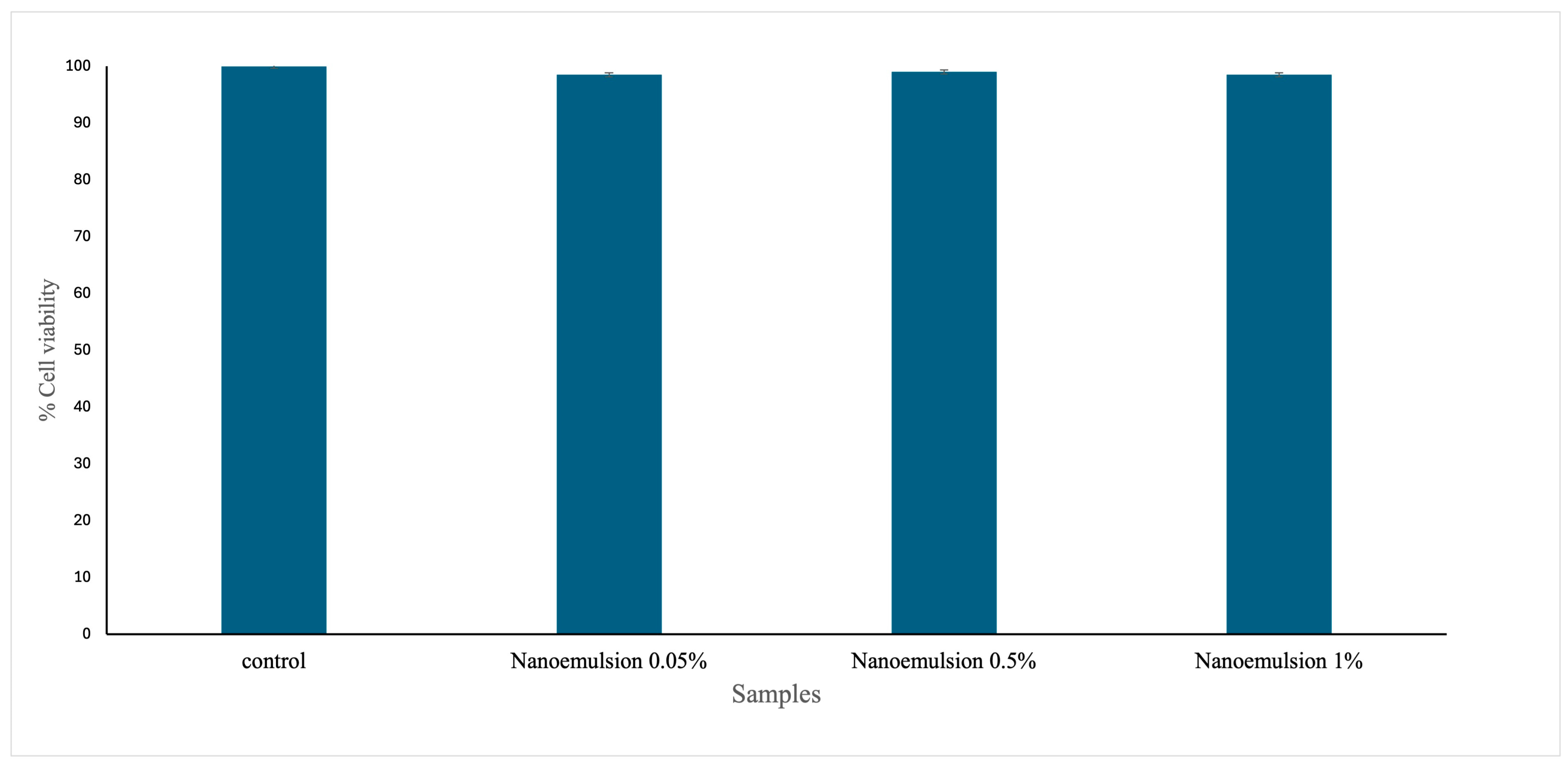
Cell viability of NHDF cells following exposure to formulation F4 (2.5:1 HPH nanoemulsion, with vitamin E acetate) at various concentrations (0.05–1.00 μg/mL) was evaluated using an MTT assay. The principal results are illustrated in Figure 5. Treatment of the cells with the sample across the examined concentration range (0.05–1.00 μg/mL) did not induce cytotoxic effects in NHDF cells. Notably, the cytotoxicity profile of the cod liver oil nanoemulsions without vitamin E was similar to that of the formulations containing vitamin E.
Figure 5.
Cell viability assessments for the control (untreated cells) and different concentrations (0.05–1%) of the selected nanoemulsion (F4) supplemented with vitamin E acetate (nanoemulsion 0.05%, nanoemulsion 0.5%, nanoemulsion 1%) in cells. The results are presented as the percentage ± SEM with respect to the control and represent the mean ± SEM of three independent experiments.
3.7. Film-Forming Capacity
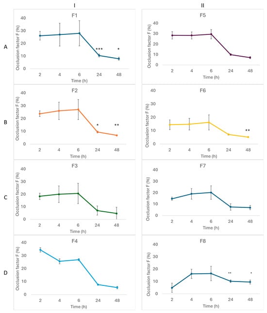
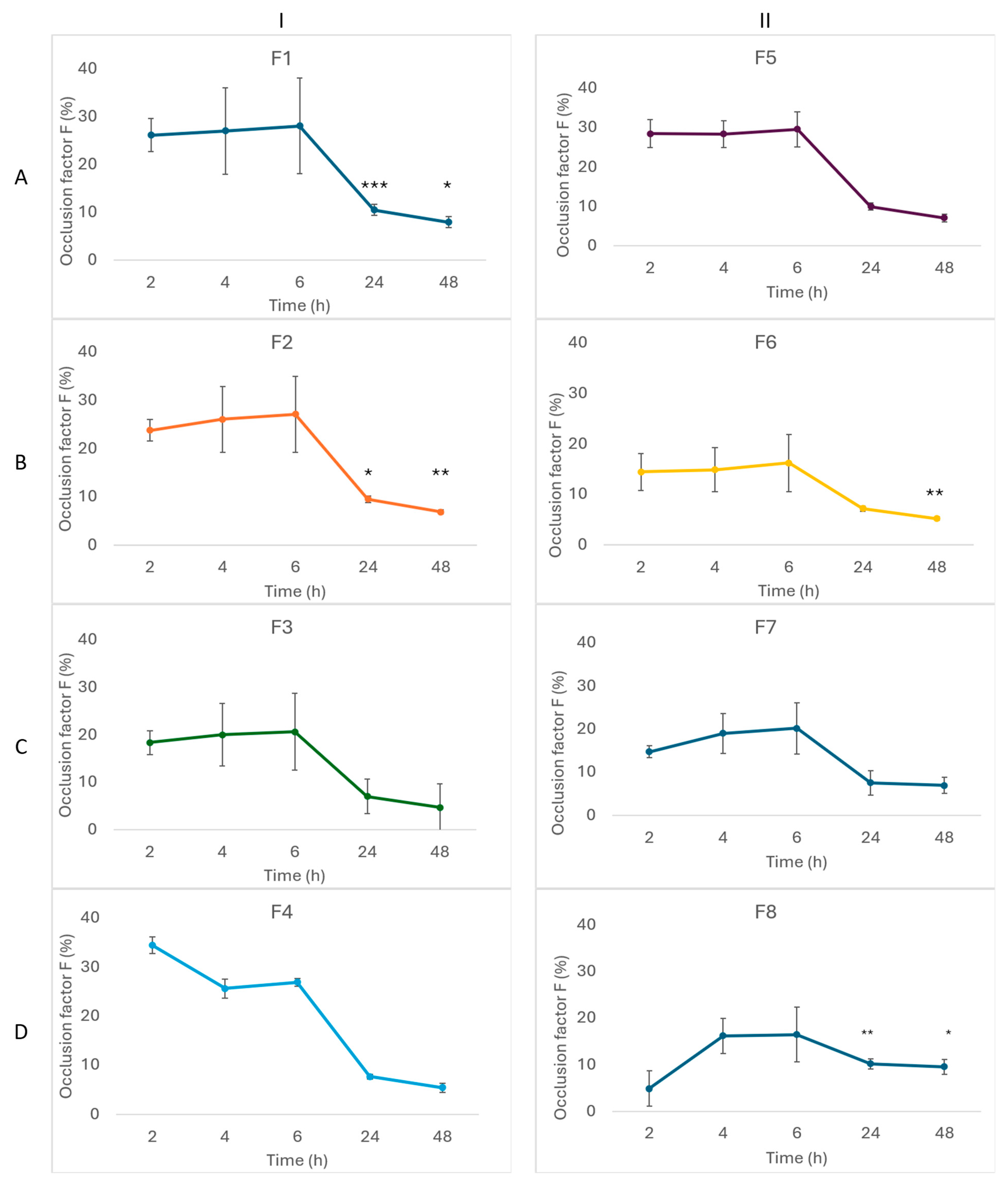
The film-forming capacity of the developed nanoemulsions was evaluated as an additional indicator of their suitability for topical application. Film formation was observed for all formulations following solvent evaporation. The occlusion factor exhibited a time-dependent decrease over 24–48 h, indicating that the barrier effect of the formed films was progressively reduced over time. At the early evaluation time points (2–6 h), most formulations exhibited occlusion factors exceeding the commonly reported reference value of F = 10, whereas after 24–48 h, the occlusion factors decreased to values close to or below this level, depending on the formulation [25]. For the selected formulation (F4), the occlusion factor decreased from 26.93 ± 0.79 at 6 h to 7.71 ± 0.51 at 24 h and 5.44 ± 0.93 at 48 h, indicating that its occlusive effect was initially above the reference threshold but became progressively lower during the evaluation period.
Formulations F2, F4, F5, and F6 showed statistically significant decreases (p < 0.05) in occlusion factor compared to earlier time points (Figure 6). These findings suggest that, while the nanoemulsions display initial film-forming behavior, their measurable occlusive properties are dynamic and may diminish over time.
Figure 6.
Occlusion factor (%) as a function of time for nanoemulsion formulations prepared with (I) 2.5:1 w/w lecithin/PEG-15 hydroxystearate and (II) 1:1 w/w lecithin/PEG-15 hydroxystearate. Formulations were produced by ultrasonication ((A)—F1, F5, (B)—F2, F6) and high-pressure homogenization ((C)—F3, F7, (D)—F4, F8), in the absence ((A)—F1, F5, (C)—F3, F7) or presence ((B)—F2, F6, (D)—F4, F8) of vitamin E. Data are expressed as mean ± standard deviation (n = 3). Statistically significant differences relative to earlier time points are denoted as follows: * p < 0.05, ** p < 0.01, *** p < 0.001.
4. Discussion
The present study demonstrates that the physicochemical stability and oxidative integrity of cod liver oil nanoemulsions intended for topical application are governed by combined and interdependent effects of formulation composition (surfactant ratio, Factor A), processing methodology (Factor B), and antioxidant supplementation (Factor C) [19,25,28]. By evaluating these parameters within a structured experimental framework, this work provides insight into how selected formulation parameters collectively influence the physicochemical stability and oxidative integrity of PUFA-rich marine oil nanoemulsions [22,25,28].
A dominant main effect of surfactant ratio (Factor A) was observed, particularly with respect to interfacial stabilization and resistance to destabilization phenomena. The superior performance of the 2.5:1 lecithin/PEG-15 hydroxystearate systems can be attributed to the formation of a more robust and highly organized interfacial film. Lecithin, due to its amphiphilic and zwitterionic characteristics, contributed to the development of a strongly negative surface charge, with ζ-potential values reaching −82.60 mV. These values exceed commonly accepted thresholds for electrostatic stabilization, indicating strong repulsive forces that effectively limit droplet aggregation [9,37]. The incorporation of PEG-15 hydroxystearate provided complementary steric stabilization through hydrated polyethylene glycol chains forming an additional barrier at the oil–water interface [3,4,5]. The combination of electrostatic and steric mechanisms resulted in enhanced resistance to droplet coalescence and Ostwald ripening, particularly under accelerated thermal conditions [18].
The influence of the processing method on droplet size was strongly dependent on the surfactant ratio, indicating a significant interaction between Factors A and B rather than a uniform main effect of processing. High-pressure homogenization (HPH) at 800 bar produced significantly smaller droplets than ultrasonication in formulations prepared with the higher lecithin/PEG-15 hydroxystearate ratio (2.5:1), suggesting that this interfacial composition was better able to stabilize the newly generated interface during high-energy homogenization. In contrast, at the lower surfactant ratio (1:1), ultrasonication yielded smaller droplets than HPH, indicating that reduced surfactant availability limited the effectiveness of high-pressure homogenization under these formulation conditions. These findings demonstrate that the efficiency of the emulsification process depended on the interplay between formulation composition and processing conditions rather than on the processing method alone [21,22]. This difference can be attributed to the uniform and reproducible application of high shear and elongational forces during HPH, in contrast to the localized and potentially less controlled energy input associated with ultrasonication. A notable interaction between surfactant ratio and processing method (A × B) was observed. The beneficial effect of HPH was most pronounced at the higher lecithin ratio (2.5:1), suggesting that favorable interfacial composition enhances the efficiency of droplet disruption and stabilization during processing. In contrast, systems with the lower lecithin content (1:1) exhibited reduced stability, particularly under thermal stress, indicating that processing alone cannot compensate for less favorable interfacial composition.
In the context of topical formulations, droplet size plays a multifaceted role, influencing physicochemical stability, surface area, film formation, and potentially skin penetration. However, the extent of penetration depends not only on droplet size but also on formulation composition, interfacial characteristics, the encapsulated compound, and the physiological state of the skin. Although droplets below 100 nm may enhance penetration in certain systems, the objective of the present nanoemulsions was not to maximize transdermal delivery but to achieve stable formulations with efficient interaction at the stratum corneum. Consequently, droplet size was optimized to balance stability and topical performance rather than minimized as an end in itself [13,26,27].
Preservation of oxidative integrity was influenced by both formulation structure (Factors A and B) and antioxidant supplementation (Factor C). The absence of detectable peroxide formation suggests that primary oxidation remained below the detection limit under the applied conditions, indicating that the combination of interfacial protection and controlled processing effectively mitigates oxidative stress during nanoemulsion formation [8,16,38]. Triglyceride preservation and fatty acid composition analysis further indicated that the nanoemulsion systems contributed to partial preservation of the PUFA-rich lipid phase during processing and storage, although progressive degradation of unsaturated fatty acids was still observed under storage conditions, particularly at elevated temperatures [39].
The incorporation of vitamin E acetate provided an additional layer of oxidative protection within the nanoemulsion systems. While its direct effect on physicochemical stability was limited, formulations containing vitamin E acetate exhibited its potential protective contribution against oxidative degradation of the unsaturated lipid fraction, as reflected by the observed factorial interactions (A × B × C) although its effect was limited under the studied conditions. Since the antioxidant activity of vitamin E acetate was not evaluated independently, the present results should be interpreted as reflecting the overall antioxidant performance of the nanoemulsion systems rather than the isolated contribution of the antioxidant compound itself. The improved antioxidant performance observed in vitamin E-containing formulations may be associated with the presence of vitamin E acetate within the lipid phase and its reported role in limiting lipid peroxidation reactions [29,34]. In this context, the preservation of antioxidant activity following processing indicates that incorporation within the nanoemulsion system did not adversely affect the oxidative resilience of the formulations. While the DPPH assay represents a simplified chemical model, it provides valuable comparative insight into the oxidative resilience of the formulations [8,30].
The cytotoxicity evaluation indicated that the vitamin E-enriched nanoemulsion caused only minimal cytotoxic effects in human dermal fibroblasts, with cell viability exceeding 90% for all tested samples. These preliminary results suggest that the formulations are well-tolerated at the tested concentrations in vitro. Consistent with previous studies, the observed high cell viability may reflect the maintenance of mitochondrial activity and basic cellular functions under the tested conditions [25,30]. While these findings are encouraging, they should be interpreted as preliminary, reflecting short-term in vitro exposure effects rather than a comprehensive safety evaluation [39].
From a formulation perspective, the results highlight that colloidal stability is primarily governed by interfacial composition (Factor A) and enhanced by processing conditions (Factor B), while antioxidant supplementation (Factor C) predominantly contributes to oxidative protection rather than physical stabilization. Collectively, the selected formulation conditions resulted in nanoemulsions with favorable physicochemical characteristics, resistance to environmental stress, and preserved lipid functionality [11,21].
The film-forming evaluation provides additional information regarding the potential performance of the nanoemulsions following topical application. All formulations formed a film upon solvent evaporation and exhibited an initial occlusive effect that progressively decreased over the 24–48 h evaluation period. At the early time points (2–6 h), most formulations exhibited occlusion factors above the commonly used reference value of F = 10, indicating measurable occlusive behavior according to the adopted in vitro method. In contrast, after 24–48 h, the occlusion factor decreased to values close to or below this threshold, suggesting that the barrier effect was transient rather than persistent. Although the underlying mechanism was not investigated in the present study, the observed reduction in occlusion may reflect changes in the properties of the formed film during drying or increased water vapor permeability. Compared with highly occlusive materials such as petrolatum, which are expected to produce substantially greater and more sustained occlusion, the developed nanoemulsions exhibited a moderate and temporary occlusive effect. Such a transient occlusive profile may be advantageous for topical formulations, where a temporary reduction in water loss is often desirable, while avoiding the sustained occlusion associated with highly impermeable films. Further studies evaluating film morphology, mechanical properties and skin adhesion and retention would be required to establish the relationship between film characteristics, occlusive performance and topical performance.
Despite these promising findings, several limitations should be acknowledged. First, the explored compositional space was restricted to two surfactant ratios and a fixed oil and surfactant concentration. While this targeted approach enabled systematic evaluation of key formulation and processing variables, it does not represent a comprehensive mapping of the nanoemulsion design space. In particular, ternary phase diagrams where not constructed, limiting insight into phase behavior and compositional regions associated with improved stability and performance.
Second, although the formulations were generated according to a 23 factorial experimental design, the statistical analysis was limited to pairwise comparisons and was not intended to provide a full factorial ANOVA or predictive modeling of main effects and interactions. Therefore, the identified factor effects should be interpreted as comparative trends within the tested experimental space, rather than as predictive relationships. Future studies employing factorial ANOVA and response-surface methodology would enable a more comprehensive characterization of the formulation design space.
Third, the photostability assessment was conducted under environmentally relevant conditions using natural sunlight exposure behind glass, resulting in primarily UV-A irradiation of low intensity. While this approach reflects realistic storage conditions, it does not provide a controlled or quantitatively defined UV dose, and therefore the results should be interpreted accordingly.
Additionally, the oxidative stability assessments relied on iodometric titration and enzymatic assays, which provide comparative rather than absolute quantification, particularly at low levels of oxidation.
Finally, although the formulations were designed for topical application, no direct skin permeation or retention studies were performed. While physicochemical characterization, film-forming properties and cytocompatibility support their suitability for topical application, further studies using ex vivo or in vivo skin models that include transepidermal water loss (TEWL) measurements, skin imaging and skin retention/permeation studies are required to confirm the relationship between these properties and their performance at the level of the skin barrier [3,40].
Overall, this study demonstrates that the performance of cod liver oil nanoemulsions is governed by the interplay of formulation composition, processing conditions, and antioxidant incorporation. These findings contribute to a more structured understanding of how targeted formulation parameters influence the stability and functionality of PUFA-rich nanoemulsion systems intended for topical applications.
5. Conclusions
This study demonstrates that the performance of cod liver oil nanoemulsions intended for topical application is governed by the combined effects of surfactant composition (Factor A), processing methodology (Factor B), and antioxidant supplementation (Factor C). Within the explored experimental space, a targeted formulation approach enabled the identification of a robust nanoemulsion system with favorable physicochemical and stability characteristics.
Surfactant composition emerged as the principal formulation parameter influencing the physicochemical characteristics of the nanoemulsions. Formulations prepared with the higher lecithin/PEG-15 hydroxystearate ratio of 2.5:1 exhibited smaller droplet sizes, more negative ζ-potential values, improved vitamin E acetate encapsulation efficiency and enhanced resistance to destabilization phenomena, particularly when processed by high-pressure homogenization.
The influence of the processing method depended on formulation composition. High-pressure homogenization was only associated with smaller droplets and enhanced vitamin E acetate encapsulation in formulations prepared with the higher lecithin ratio, whereas these advantages were not observed at the lower surfactant ratio. These findings demonstrate that the effectiveness of the processing method was formulation-dependent rather than universally applicable across all nanoemulsions.
Vitamin E acetate supplementation primarily contributed to oxidative protection of the PUFA-rich lipid phase under the tested experimental conditions, while its effect on the physicochemical properties of the nanoemulsions was comparatively limited.
Among the investigated formulations, F4 (2.5:1 lecithin/PEG-15 hydroxystearate, high-pressure homogenization, with vitamin E acetate) emerged as the optimal formulation, providing a physicochemically stable, oxidatively protected and cytocompatible nanoemulsion suitable as a topical carrier for cod liver oil intended for interaction with the stratum corneum.
Despite these promising findings, the study is limited by the restricted compositional space explored and the absence of comprehensive design-space mapping, such as ternary phase diagrams. Furthermore, the statistical analysis was not intended to provide predictive modeling of factor interactions, and oxidative and photostability assessments were conducted under conditions that provide comparative rather than fully quantitative evaluation. Finally, although the formulations are designed for topical use, further studies are required to assess skin permeation and retention.
Overall, this work highlights how targeted manipulation of formulation and processing parameters can be used to develop stable nanoemulsion systems for the incorporation and protection of marine-derived lipids. These findings provide a structured basis for future formulation development and further investigation of such systems in biologically relevant skin models. Future work should include skin permeation and retention studies, and in vivo evaluations of skin hydration and transepidermal water loss. Expansion of the formulation design space using response-surface methodology should be performed to further optimize nanoemulsion performance.
Supplementary Materials
The following supporting information can be downloaded at: https://www.mdpi.com/article/10.3390/cosmetics13040173/s1, Figure S1. Time-dependent evolution of (I) mean droplet size (bars) and polydispersity index (lines) and (II) ζ-potential of cod liver oil nanoemulsions with 2.5:1 w/w lecithin/PEG-15 hydroxystearate prepared by (A,B) high-pressure homogenization and (C,D) ultrasonication, (B,D) with or (A,B) without vitamin E, and stored at 4 °C for 60 days (n = 3); Figure S2. Time-dependent evolution of (I) mean droplet size (bars) and polydispersity index (lines) and (II) ζ-potential of cod liver oil nanoemulsions with 1:1 w/w lecithin/PEG-15 hydroxystearate prepared by (A,B) high-pressure homogenization and (C,D) ultrasonication, (B,D) with or (A,B) without vitamin E, and stored at 4 °C, over 60 days (n = 3); Figure S3. Time-dependent evolution of (I) mean droplet size (bars) and polydispersity index(lines) and (II) ζ-potential of cod liver oil nanoemulsions with 2.5:1 w/w lecithin/PEG-15 hydroxystearate prepared by (A,B) high-pressure homogenization and (C,D) ultrasonication, (B,D) with or (A,B) without vitamin E, and stored at 40 °C, over 60 days (n = 3); Figure S4. Time-dependent evolution of (I) mean droplet size (bars) and polydispersity index (lines) and (II) ζ-potential of cod liver oil nanoemulsions with 1:1 w/w lecithin/PEG-15 hydroxystearate prepared by (A,B) high-pressure homogenization and (C,D) ultrasonication, (B,D) with or (A,C) without vitamin E, and stored at 40 °C over 60 days (n = 3). Asterisks indicate statistically significant differences in mean droplet size compared to Day 1 for samples F7 and F8 (* p < 0.05, ** p < 0.01); Figure S5. Effect of centrifugation on the physicochemical properties of cod liver oil nanoemulsions: (I) mean droplet size (bars) and polydispersity index (lines) and (II) ζ-potential, measured after 1 day of storage and after centrifugation. Panel (A) shows formulations without vitamin E, whereas panel (B) shows formulations containing vitamin E. Data are presented as mean ± SD (n = 3); Figure S6. Effect of thermal cycling (6 cycles) on the physicochemical properties of cod liver oil nanoemulsions: (I) mean droplet size (bars) and polydispersity index (lines) and (II) ζ-potential, measured after 1 day of storage and after thermal cycling. Panel (A) shows formulations without vitamin E, whereas panel (B) shows formulations containing vitamin E. Data are presented as mean ± SD (n = 3); Figure S7. Effect of UV exposure on the physicochemical properties of cod liver oil nanoemulsions: (I) mean droplet size (bars) and polydispersity index (lines) and (II) ζ-potential, measured after 1 day of storage and after UV aging. Panel (A) shows formulations without vitamin E, whereas panel (B) shows formulations containing vitamin E. Data are presented as mean ± SD (n = 3); Table S1. Absorbance values obtained from triglyceride determination, expressed as mean ± SD (n = 3); Table S2. Relative fatty acid composition (% of total fatty acids) of nanoemulsions prepared with 2.5:1 w/w lecithin/PEG-15 hydroxystearate after storage at 4 °C, 25 °C and 40 °C for 60 days; Table S3. Relative fatty acid composition (% of total fatty acids) of nanoemulsions prepared with 1:1 w/w lecithin/PEG-15 hydroxystearate after storage at 4 °C, 25 °C and 40 °C for 60 days; Table S4. Relative fatty acid composition (% of total fatty acids) of cod liver oil and nanoemulsion formulations at initial time point (t0).
Author Contributions
Conceptualization, S.H. methodology, S.H., M.K., F.N.L. and S.L.; validation, K.A.; formal analysis, A.I., C.C., Ε.P. and S.L. investigation, A.I.; data curation, A.I., C.C. and Ε.P.; writing—original draft preparation, A.I. and S.H.; writing—review and editing, S.H., F.N.L., S.L., M.K. and K.A.; supervision, S.H. All authors have read and agreed to the published version of the manuscript.
Funding
This research received no external funding.
Institutional Review Board Statement
Not applicable.
Informed Consent Statement
Not applicable.
Data Availability Statement
Data available on request from the authors.
Acknowledgments
During the preparation and revision of this manuscript, the authors used ChatGPT (OpenAI, GPT-5.5) to assist with language editing, and improving the clarity and readability of the manuscript.
Conflicts of Interest
The authors declare no conflicts of interest.
Abbreviations
The following abbreviations are used in this manuscript:
| DHA | Docosahexaenoic acid |
| DLS | Dynamic light scattering |
| DPPH | 2,2-Diphenyl-1-picrylhydrazyl |
| EE | Encapsulation efficiency |
| ELS | Electrophoretic light scattering |
| EPA | Eicosapentaenoic acid |
| HPH | High-pressure homogenization |
| IC50 | Half maximal inhibitory concentration |
| MTT | 3-(4,5-dimethylthiazol-2-yl)-2,5-diphenyltetrazolium bromide |
| NE | Nanoemulsion |
| NHDF | Normal human dermal fibroblast |
| PdI | Polydispersity index |
| PEG | Polyethylene glycol |
| PUFA | Polyunsaturated fatty acid |
| PV | Peroxide value |
| SEM | Standard error of the mean |
| US | Ultrasonication |
| UV | Ultraviolet |
| ζ-potential | Zeta potential |
References
- Falch, E. Physico-Chemical Properties and Nutrition of Marine Lipids. Foods 2023, 12, 4078. [Google Scholar] [CrossRef] [Scilit] [PubMed] [PubMed Central]
- Chakraborty, N.; Narayanan, V.; Gautam, H.K. Nano-Therapeutics to Treat Acne Vulgaris. Indian J. Microbiol. 2022, 62, 167–174. [Google Scholar] [CrossRef] [Scilit] [PubMed]
- Hatziantoniou, S.; Rallis, M.; Demetzos, C.; Papaioannou, G.T. Pharmacological activity of natural lipids on a skin barrier disruption model. Pharmacol. Res. 2000, 42, 55–59. [Google Scholar] [CrossRef] [Scilit] [PubMed]
- Elias, P.M. Epidermal Lipids, Barrier Function, and Desquamation. J. Investig. Dermatol. 1983, 80, S44–S49. [Google Scholar] [CrossRef] [Scilit] [PubMed]
- Bouwstra, J.A.; Ponec, M. The Skin Barrier in Healthy and Diseased State. Biochim. Biophys. Acta Biomembr. 2006, 1758, 2080–2095. [Google Scholar] [CrossRef] [Scilit]
- Tanuku, S.; Velisila, D.; Thatraju, D.; kumar Vadaga, A. Nanoemulsion Formulation Strategies for Enhanced Drug Delivery. J. Pharma Insights Res. 2024, 2, 125–138. [Google Scholar] [CrossRef] [Scilit]
- Elsewedy, H.S. Insights of Nanoemulsion as a Drug Delivery System: An Overview of Current Trends and Applications. Indian J. Pharm. Educ. Res. 2025, 59, 472–492. [Google Scholar] [CrossRef] [Scilit]
- Bush, L.; Stevenson, L.; Lane, K.E. The Oxidative Stability of Omega-3 Oil-in-Water Nanoemulsion Systems Suitable for Functional Food Enrichment: A Systematic Review of the Literature. Crit. Rev. Food Sci. Nutr. 2019, 59, 1154–1168. [Google Scholar] [PubMed]
- Ling, J.K.U.; Chan, Y.S.; Nandong, J. Insights into the Release Mechanisms of Antioxidants from Nanoemulsion Droplets. J. Food Sci. Technol. 2022, 59, 1677–1691. [Google Scholar] [PubMed]
- Musakhanian, J.; Osborne, D.W. Understanding Microemulsions and Nanoemulsions in (Trans)Dermal Delivery. AAPS PharmSciTech 2025, 26, 31. [Google Scholar] [CrossRef] [Scilit] [PubMed]
- Tadros, T.; Izquierdo, P.; Esquena, J.; Solans, C. Formation and Stability of Nano-Emulsions. Adv. Colloid Interface Sci. 2004, 108–109, 303–318. [Google Scholar] [CrossRef] [PubMed]
- Wilson, R.J.; Li, Y.; Yang, G.; Zhao, C.-X. Nanoemulsions for Drug Delivery. Particuology 2022, 64, 85–97. [Google Scholar] [CrossRef] [Scilit]
- Shaker, D.S.; Ishak, R.A.H.; Ghoneim, A.; Elhuoni, M.A. Nanoemulsion: A Review on Mechanisms for the Transdermal Delivery of Hydrophobic and Hydrophilic Drugs. Sci. Pharm. 2019, 87, 17. [Google Scholar] [CrossRef] [Scilit]
- Gupta, A.; Eral, H.B.; Hatton, T.A.; Doyle, P.S. Nanoemulsions: Formation, Properties and Applications. Soft Matter 2016, 12, 2826–2841. [Google Scholar] [CrossRef] [Scilit] [PubMed]
- Patel, D.; Patel, B.; Thakkar, H. Lipid Based Nanocarriers: Promising Drug Delivery System for Topical Application. Eur. J. Lipid Sci. Technol. 2021, 123, 2000264. [Google Scholar] [CrossRef] [Scilit]
- Keramat, M.; Ehsandoost, E.; Golmakani, M.-T. Recent Trends in Improving the Oxidative Stability of Oil-Based Food Products by Inhibiting Oxidation at the Interfacial Region. Foods 2023, 12, 1191. [Google Scholar] [CrossRef] [Scilit] [PubMed]
- Chatzopoulou, N.; Chaikali, C.; Mourkogianni, E.; Mikelis, C.M.; Andriopoulos, V.; Kornaros, M.; Avgoustakis, K.; Lamari, F.N.; Hatziantoniou, S. Encapsulation of Microalgae Tisochrysis Lutea Extract in Nanostructured Lipid Carriers (NLCs) and Evaluation of Their Sunscreen, Wound Healing, and Skin Hydration Properties. Mar. Drugs 2024, 22, 487. [Google Scholar] [CrossRef] [Scilit] [PubMed]
- Koroleva, M.Y.; Yurtov, E.V. Ostwald Ripening in Macro- and Nanoemulsions. Russ. Chem. Rev. 2021, 90, 293–323. [Google Scholar] [CrossRef] [Scilit]
- Lucia, M. Lipid-Based Nanoparticles as Carriers for Dermal Delivery of Antioxidants. Curr. Drug Metab. 2017, 18, 469–480. [Google Scholar] [CrossRef] [Scilit] [PubMed]
- Kumar, M.; Bishnoi, R.S.; Shukla, A.K.; Jain, C.P. Techniques for Formulation of Nanoemulsion Drug Delivery System: A Review. Prev. Nutr. Food Sci. 2019, 24, 225–234. [Google Scholar] [CrossRef] [Scilit] [PubMed] [PubMed Central]
- Gawin-Mikołajewicz, A.; Nawrot, U.; Malec, K.H.; Krajewska, K.; Nartowski, K.P.; Karolewicz, B.L. The Effect of High-Pressure Homogenization Conditions on the Physicochemical Properties and Stability of Designed Fluconazole-Loaded Ocular Nanoemulsions. Pharmaceutics 2024, 16, 11. [Google Scholar] [CrossRef] [Scilit] [PubMed]
- Kotta, S.; Khan, A.W.; Ansari, S.H.; Sharma, R.K.; Ali, J. Formulation of Nanoemulsion: A Comparison between Phase Inversion Composition Method and High-Pressure Homogenization Method. Drug Deliv. 2015, 22, 455–466. [Google Scholar] [CrossRef] [Scilit] [PubMed]
- Koppel, D.E. Analysis of Macromolecular Polydispersity in Intensity Correlation Spectroscopy: The Method of Cumulants. J. Chem. Phys. 1972, 57, 4814–4820. [Google Scholar] [CrossRef] [Scilit]
- da Silva, T.L.M.; de Oliveira Capote, A.C.M.; Beltrame, F.L.; Ferrari, P.C. Nanoemulsions for Skin Delivery of Essential Oils: A Systematic Review. Phytother. Res. 2026, 40, 847–872. [Google Scholar] [CrossRef] [Scilit] [PubMed] [PubMed Central]
- Liakopoulou, A.; Mourelatou, E.; Hatziantoniou, S. Exploitation of Traditional Healing Properties, Using the Nanotechnology’s Advantages: The Case of Curcumin. Toxicol. Rep. 2021, 8, 1143–1155. [Google Scholar] [CrossRef] [Scilit] [PubMed]
- Jacob, S.; Nair, A.B. Nanoemulgels as Advanced Topical Drug Delivery Systems: Mechanistic Insights and Therapeutic Applications in Skin Disorders, Infections, Wound Healing, and Cancer. Pharmaceuticals 2026, 19, 247. [Google Scholar] [CrossRef] [Scilit] [PubMed]
- Souto, E.B.; Müller, R.H. Cosmetic features and applications of lipid nanoparticles (SLN, NLC). Int. J. Cosmet. Sci. 2008, 30, 157–165. [Google Scholar] [CrossRef] [Scilit] [PubMed]
- Flekka, K.; Dimaki, V.D.; Mourelatou, E.; Avgoustakis, K.; Lamari, F.N.; Hatziantoniou, S. Stability and Retention of Nanoemulsion Formulations Incorporating Lavender Essential Oil. Cosmetics 2024, 11, 65. [Google Scholar] [CrossRef] [Scilit]
- Coward, K.H.; Dyer, F.J.; Morton, R.A. The determination of vitamin A in cod-liver oils, (a) biologically, (b) chemically, (c) physically, with a statistical examination of the results: Further evidence that the intensity of absorption at 328mmu gives the best agreement with the biological measure of vitamin A in cod-liver oils. Biochem. J. 1932, 26, 1593–1600. [Google Scholar] [CrossRef] [Scilit] [PubMed]
- Mateu-Arrom, L.; Mora, I.; Garrote, L. Therapeutic Benefits of Topical Omega-3 Polyunsaturated Fatty Acids in Skin Diseases and Cosmetics: An Updated Systematic Review. J. Cosmet. Dermatol. 2025, 24, e70341. [Google Scholar] [CrossRef] [Scilit] [PubMed]
- Koutra, E.; Grammatikopoulos, G.; Kornaros, M. Microalgal post-treatment of anaerobically digested agro-industrial wastes for nutrient removal and lipids production. Bioresour. Technol. 2017, 224, 473–480. [Google Scholar] [CrossRef] [Scilit] [PubMed]
- Papaioannou, A.; Liakopoulou, A.; Papoulis, D.; Gianni, E.; Gkolfi, P.; Zygouri, E.; Letsiou, S.; Hatziantoniou, S. Effect of Peptides on the Synthesis, Properties and Wound Healing Capacity of Silver Nanoparticles. Pharmaceutics 2023, 15, 2471. [Google Scholar] [CrossRef] [Scilit] [PubMed]
- Liakopoulou, A.; Letsiou, S.; Avgoustakis, K.; Patrinos, G.P.; Lamari, F.N.; Hatziantoniou, S. Curcumin-Loaded Lipid Nanocarriers: A Targeted Approach for Combating Oxidative Stress in Skin Applications. Pharmaceutics 2025, 17, 144. [Google Scholar] [CrossRef] [Scilit]
- Blois, M.S. Antioxidant Determinations by the Use of a Stable Free Radical. Nature 1958, 181, 1199–1200. [Google Scholar] [CrossRef] [Scilit]
- Vater, C.; Adamovic, A.; Ruttensteiner, L.; Steiner, K.; Tajpara, P.; Klang, V.; Elbe-Bürger, A.; Wirth, M.; Valenta, C. Cytotoxicity of Lecithin-Based Nanoemulsions on Human Skin Cells and Ex Vivo Skin Permeation: Comparison to Conventional Surfactant Types. Int. J. Pharm. 2019, 566, 383–390. [Google Scholar] [CrossRef] [Scilit] [PubMed]
- Shrotriya, S.; Ranpise, N.; Satpute, P.; Vidhate, B. Skin Targeting of Curcumin Solid Lipid Nanoparticles-Engrossed Topical Gel for the Treatment of Pigmentation and Irritant Contact Dermatitis. Artif. Cells Nanomed. Biotechnol. 2018, 46, 1471–1482. [Google Scholar] [CrossRef] [Scilit] [PubMed]
- Rodriguez-Loya, J.; Lerma, M.; Gardea-Torresdey, J.L. Dynamic Light Scattering and Its Application to Control Nanoparticle Aggregation in Colloidal Systems: A Review. Micromachines 2023, 15, 24. [Google Scholar] [CrossRef] [Scilit] [PubMed]
- Magrode, N.; Poomanee, W.; Kiattisin, K.; Ampasavate, C. Microemulsions and Nanoemulsions for Topical Delivery of Tripeptide-3: From Design of Experiment to Anti-Sebum Efficacy on Facial Skin. Pharmaceutics 2024, 16, 554. [Google Scholar] [CrossRef] [Scilit] [PubMed]
- Mosmann, T. Rapid colorimetric assay for cellular growth and survival: Application to proliferation and cytotoxicity assays. J. Immunol. Methods 1983, 65, 55–63. [Google Scholar] [CrossRef] [Scilit] [PubMed]
- Jacob, S.; Kather, F.S.; Boddu, S.H.S.; Shah, J.; Nair, A.B. Innovations in Nanoemulsion Technology: Enhancing Drug Delivery for Oral, Parenteral, and Ophthalmic Applications. Pharmaceutics 2024, 16, 1333. [Google Scholar] [CrossRef] [Scilit] [PubMed]

Disclaimer/Publisher’s Note: The statements, opinions and data contained in all publications are solely those of the individual author(s) and contributor(s) and not of MDPI and/or the editor(s). MDPI and/or the editor(s) disclaim responsibility for any injury to people or property resulting from any ideas, methods, instructions or products referred to in the content. |
© 2026 by the authors. Licensee MDPI, Basel, Switzerland. This article is an open access article distributed under the terms and conditions of the Creative Commons Attribution (CC BY) license.