


Abstract
Background: Dermatology and aesthetic medicine make extensive use of microneedling, a minimally invasive and safe treatment. Across the research, it has been shown that microneedling combined with chemical peels is also more effective than chemical peels alone. However, data on procedures in dark-skinned individuals is rather scarce. Aim/Objective: This study aimed to determine the efficacy of using a 4% retinol solution product containing novel TGF-β activators and antioxidants combined with a microneedling technique in the treatment of hyperpigmentation, atrophic acne scars, and enlarged pores in patients with skin of color, generally corresponding to Fitzpatrick skin phototypes IV–VI. Methods: Each of the 10 patients underwent three peel treatment series, with a 30-day interval between each session. Moreover, skin hydration, elasticity, and pigmentation were examined using the Multi Skin Test MC 1000 Courage + Khazaka, and the Observ 520x device. Results: All patients reported an overall improvement and an enhancement in skin tone after the procedure. The majority of them stated subjective improvement in the reduction of facial skin issues: redness, hyperpigmentation, uneven structure, wrinkles, dehydration, dryness, and sebaceous gland activity. The least improvement was noted in scar reduction or liquidation. An objective evaluation revealed a statistically significant improvement in hyperpigmentation and elasticity in the study group. An improvement, however, not statistically significant, in hydration parameters was demonstrated during the study. Conclusions: This study suggests that a combined peel therapy of 4% retinol serum product containing novel TGF-β activators and antioxidants, together with a microneedling technique, may improve facial hyperpigmentation of the skin, as well as regulate sebaceous gland activity, their size, and reduce sebum production. The recommended method is relatively simple to use, low-cost, has minimal adverse effects, and is well tolerated by patients with skin of color.
1. Introduction
Microneedling is a safe and minimally invasive procedure widely utilized in dermatology and aesthetic medicine [1]. Used instruments comprise rows of fine needles, which are rolled over the skin to create promptly healing perforations. This leads to a wound healing response and collagen and elastin formation. Moreover, through microchannels made in the stratum corneum, this method is also utilized to improve transdermal drug delivery [1]. Compared to traditional resurfacing methods, microneedling treatment carries a lower chance of several cutaneous side effects and has been found to be effective in treating acne scars, enlarged pores, hyperpigmentation, and wrinkles and fine lines [2,3,4,5].
Patients with skin of color (SOC), most commonly encompassing Fitzpatrick skin phototypes IV to VI, include individuals of African, Asian, Latin American, Middle Eastern, and Indigenous descent, as well as those of mixed ancestry [6]. This classification is based on pigmentation levels and the skin’s physiological response to ultraviolet (UV) radiation [7]. Compared to lighter skin types, SOC patients demonstrate significant biological and clinical differences, which influence their dermatologic presentation and treatment responses. People with SOC frequently suffer from dry skin, which is further supported by lower ceramide levels, poorer cellular cohesiveness, and greater transepidermal water loss (TEWL) [8]. It is interesting to note that fibroblasts are large, numerous, and multinucleated in SOC, which explains why keloids, which are frequently observed in people of African origin, emerge. Moreover, the increased melanin content in darker skin, predominantly in the form of eumelanin, provides enhanced photoprotection; however, heightened melanin density predisposes individuals to pigmentary disorders, such as post-inflammatory hyperpigmentation (PIH) and melasma [9].
Acne scarring and hyperpigmentation are two of the most prevalent and distressing side effects of inflammatory skin diseases, significantly affecting patients’ quality of life [10]. Acne scars are caused by aberrant wound healing mechanisms that accompany inflammation, resulting in atrophic tissue alterations [11]. Simultaneously, pigmentation issues are common, particularly in people with darker skin tones. These include post-inflammatory hyperpigmentation (PIH), which occurs following cutaneous injury or inflammation [12], and melasma, a chronic, relapsing condition characterized by symmetric hyperpigmented macules that are frequently influenced by hormonal and environmental factors [13]. These disorders provide a significant treatment challenge due to their complex origin and tendency for persistence and recurrence.
There is a significant dearth of knowledge on safe and efficient treatment approaches for patients with Fitzpatrick skin types IV–VI. Recognizing these differences is essential for tailoring dermatologic treatments, including microneedling, lasers, and chemical peels, to the unique needs of this population. Moreover, understanding the distinct biological characteristics of SOC is critical for addressing disparities in dermatologic research and ensuring equitable care. Although microneedling and chemical peeling (a minimally invasive procedure that uses chemical agents to induce controlled skin exfoliation and subsequent regeneration) are well-established modalities, limited evidence exists regarding their combined use with retinol-based formulations containing TGF-β activators in patients with skin of color. The aim of this study was to determine the efficacy of a 4% retinol-based chemical peeling solution product containing novel TGF-β activators and antioxidants combined with a microneedling technique in the treatment of inflammatory hyperpigmentation, atrophic acne scars, and enlarged pores in patients with SOC. The primary outcome measure was the improvement in hyperpigmentation, reflecting the principal clinical indication for the intervention. Secondary outcome measures included skin elasticity, hydration, sebum levels, and patient-reported satisfaction.
2. Materials and Methods
2.1. Study Design
The study was conducted between September 2023 and October 2024. The Independent Bioethics Committee for Scientific Research at the Medical University of Gdansk, Poland, gave its approval (NKBBN/618/2022). Each subject provided written informed consent before the investigation. According to the inclusion and exclusion criteria, 10 healthy women with skin phototypes IV–VI were enrolled in total.
Chemical peel sessions were administered to patients in accordance with the same protocol as suggested by the manufacturer. Each patient had three peel treatment series combined with a microneedling procedure, with a 30-day interval between each session. Patients were instructed to utilize the same daily at-home skin care and photoprotection products that the producer supplied during the study. The MultiSkin Test MC 1000 (Courage + Khazaka electronic GmbH, Cologne, Germany) was used to assess the patients’ skin pigmentation, hydration, and elasticity prior to the study and before each peel procedure on the day of the treatment. Skin visualization was performed with the Observ® 520x (Sylton BV, Nijmegen, The Netherlands) device. All instrumental measurements were performed under standardized conditions, including controlled room temperature and humidity. Participants were acclimatized prior to measurement, and assessments were conducted at the same anatomical sites. Repeated measurements were performed and averaged to improve reliability. The last examination evaluating the above-mentioned parameters was completed 30 days after the last peel procedure. A total of five patient evaluations were conducted.
Prior to the study, participants were asked questions about the presence of various subjective facial skin symptoms, including itching, burning, roughness, redness, exfoliation, and tightness. After completion of the study, patients subjectively evaluated the attained improvement based on the following symptoms: redness, skin tone, dehydration, dryness, skin pigmentation, wrinkles, structure, sebaceous gland activity, scar appearance, and overall improvement. Patients rated the reduction of the aforementioned symptoms on a scale from 1 to 5, from mild to severe.
2.2. Inclusion Criteria
- (i)
- Signed informed consent;
- (ii)
- Women with IV–VI Fitzpatrick skin type, including features such as melasma, post-inflammatory hyperpigmentation (PIH), atrophic acne scars, enlarged pores;
- (iii)
- Age: 24–50 years;
- (iv)
- Sex: women;
- (v)
- Good general health condition.
2.3. Exclusion Criteria
- (i)
- Pregnancy;
- (ii)
- Breastfeeding period;
- (iii)
- Open wounds, violation of the continuity of the epidermis;
- (iv)
- Hypersensitivity to any of the treatment ingredients;
- (v)
- Acute and chronic infectious processes, e.g., tuberculosis, active herpes simplex virus infection;
- (vi)
- Treatment with isotretinoin within the last 6 months;
- (vii)
- Epilepsy;
- (viii)
- Emotional instability;
- (ix)
- Surgical procedures in the area of treatment procedures during the last 6 months;
- (x)
- Tendency to form keloids;
- (xi)
- Occurrence of active skin disease, e.g., rosacea, acne tarda, atopic dermatitis, contact dermatitis, and seborrheic dermatitis.
2.4. Procedure
Following the manufacturer’s protocol, the chemical procedure was carried out in the Department of Dermatology, Venereology, and Allergology. In order to prevent irritation, patients’ eyes were covered during the peeling process, and the procedure environment was well ventilated. The skin was first cleansed, then treated with Retix C® PRE-PEEL, a degreasing agent. One ampoule of Retix C® BOOSTER Serum was then applied to the whole area of the face and allowed to absorb completely. Thereafter, the microneedling procedure was completed with a Retix C® R Pen. The needles were inserted perpendicular to the pre-stretched skin. The treatment was carried out over the entire facial area. A 36-needle cartridge was applied. The needle depth was progressively increased across subsequent sessions, reaching 0.25 mm in the first treatment, 0.5 mm in the second, and 0.75 mm in the third session. The clinical endpoint was defined as the appearance of pinpoint bleeding. Excess blood was gently removed with sterile gauze. The procedure was performed using multiple passes over each treatment area in different directions to ensure even coverage. The number of passes was adjusted individually depending on the skin’s response. At last, the contents of the vial of Retix C Retinol TGF Activator were spread with a brush over the entire surface of the face, omitting the eye area. The solution was kept on the skin for 6 h before being removed with lukewarm water. Following the procedure, the patients were advised to use daily at-home skin care: Ultra Repair Moisturizer and SPF50+ cream in the morning, and Retimodeling serum for mixed and oily skin or Anti-Aging Face Cream Retix C for dry skin in the evening.
2.5. Products Utilized
Two products were used to perform the chemical peeling procedure. An antioxidant product, Retix C® BOOSTER Serum, was applied to the facial skin as the first step. Thereafter, the patients were treated with Retix C® R. Pen. Retix C® Retinol TGF Activator, containing 4% retinol and TGF activators, was applied afterwards. The recommended home care included products containing moisturizing and regenerating ingredients, as well as active anti-aging compounds such as retinol, lactic acid, antioxidants, and soothing agents, along with broad-spectrum UV filters for photoprotection.
2.6. Statistical Analysis
Completed surveys were downloaded for statistical analysis. The collected data were analyzed in Statistica 12.0 software. Quantitative variables were characterized by the arithmetic mean, standard deviation, median, minimum and maximum value (range), and a 95% CI (confidence interval). Qualitative variables were presented by counts and percentages. The Shapiro–Wilk W test was used to check whether the quantitative variable came from a population with a normal distribution. Levene’s (Brown–Forsythe) test was used to test the hypothesis of equal variances. The significance of differences between more than two groups was checked by the F test (ANOVA) or Kruskal–Wallis. In the case of statistically significant differences between groups, post hoc tests were used (Tukey’s test for F, Dunn’s test for Kruskal–Wallis). The significance of differences between more than two related variables in the model was tested by analysis of variance with repeated measures or Friedman’s test. Chi-square tests of independence were used for qualitative variables (with the use of Yates’ correction for cell numbers below 10, checking Cochran’s conditions and Fisher’s exact test, as appropriate). In all calculations, the level of significance was assumed to be p = 0.05.
3. Results
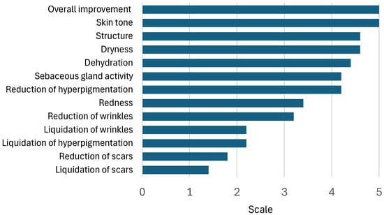
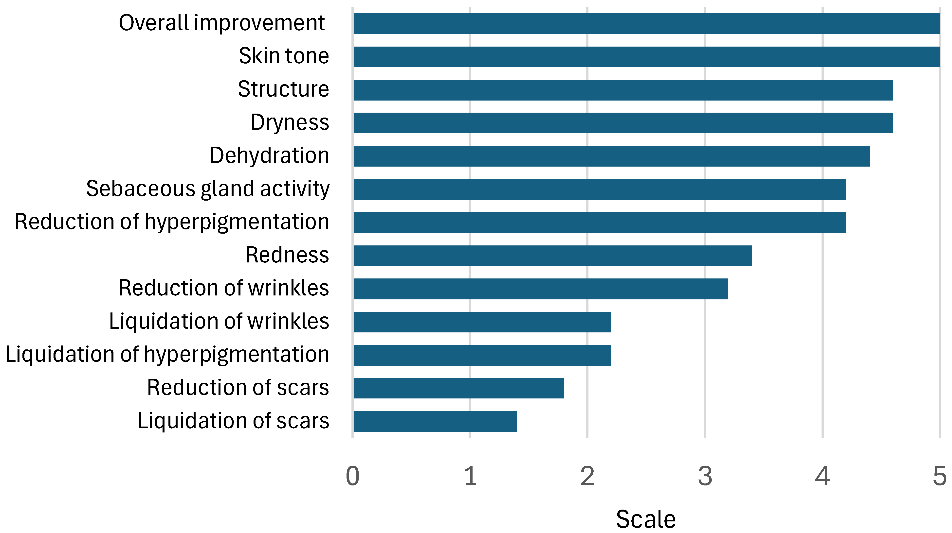
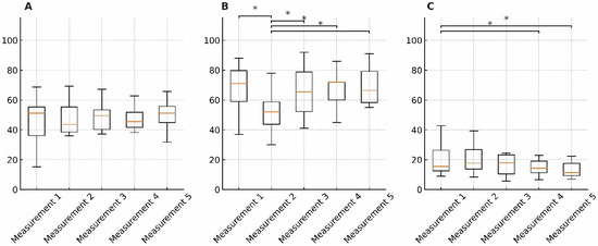
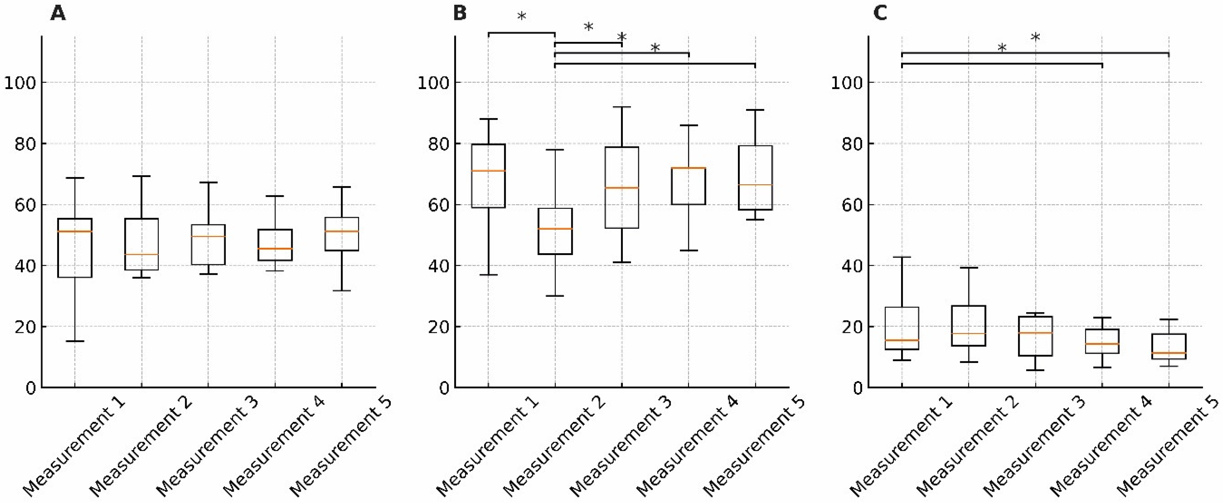
A total of 10 patients, all females, with an age range of 24–47 (mean age: 31.9), completed the study. Objective and subjective outcomes were analyzed separately. According to the survey data collected from 10 patients during the last peel session, subjective evaluation showed a satisfactory efficacy of the therapy. Improvement in the reduction in facial skin redness, hyperpigmentation, uneven structure, wrinkles, dehydration, dryness, and sebaceous gland activity, was reported by the majority of the patients. After the peeling procedure, all patients reported an enhancement in skin tone and overall improvement of their skin condition. The least improvement was noted for scar reduction or liquidation (Figure 1). Objective outcomes are presented in Figure 2.
Figure 1.
Patients’ subjective assessment of facial skin symptom changes.
Figure 2.
Changes over time in skin hydration (A), skin elasticity (B), and skin pigmentation (C) across five time points (Measurement 1–5). Each plot displays the distribution of values, including medians, interquartile ranges, and whiskers without outliers. Mean values and standard deviations are reported below. For skin hydration, the mean values ranged from 44.6 ± 8.7 to 51.2 ± 9.3 across the five time points, with no statistically significant differences observed between measurements. In contrast, skin elasticity showed a statistically significant change over time (Kruskal–Wallis test, p = 0.0050). Post hoc analysis revealed that elasticity significantly increased in Measurement 3 (p = 0.0227), Measurement 4 (p = 0.0488), and Measurement 5 (p = 0.0065) compared to Measurement 2. Additionally, elasticity in Measurement 2 was significantly lower than in Measurement 1 (p = 0.0148). Mean values for elasticity ranged from 56.2 ± 13.1 to 66.8 ± 12.6. Skin pigmentation also showed significant differences between time points. Specifically, a statistically significant decrease in pigmentation was observed in both Measurement 4 and Measurement 5 compared to Measurement 1 (p < 0.05). Mean pigmentation values ranged from 17.4 ± 5.2 to 25.8 ± 7.9. Statistically significant pairwise differences are indicated on the plots with asterisks (*).
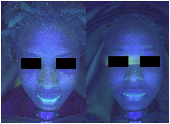
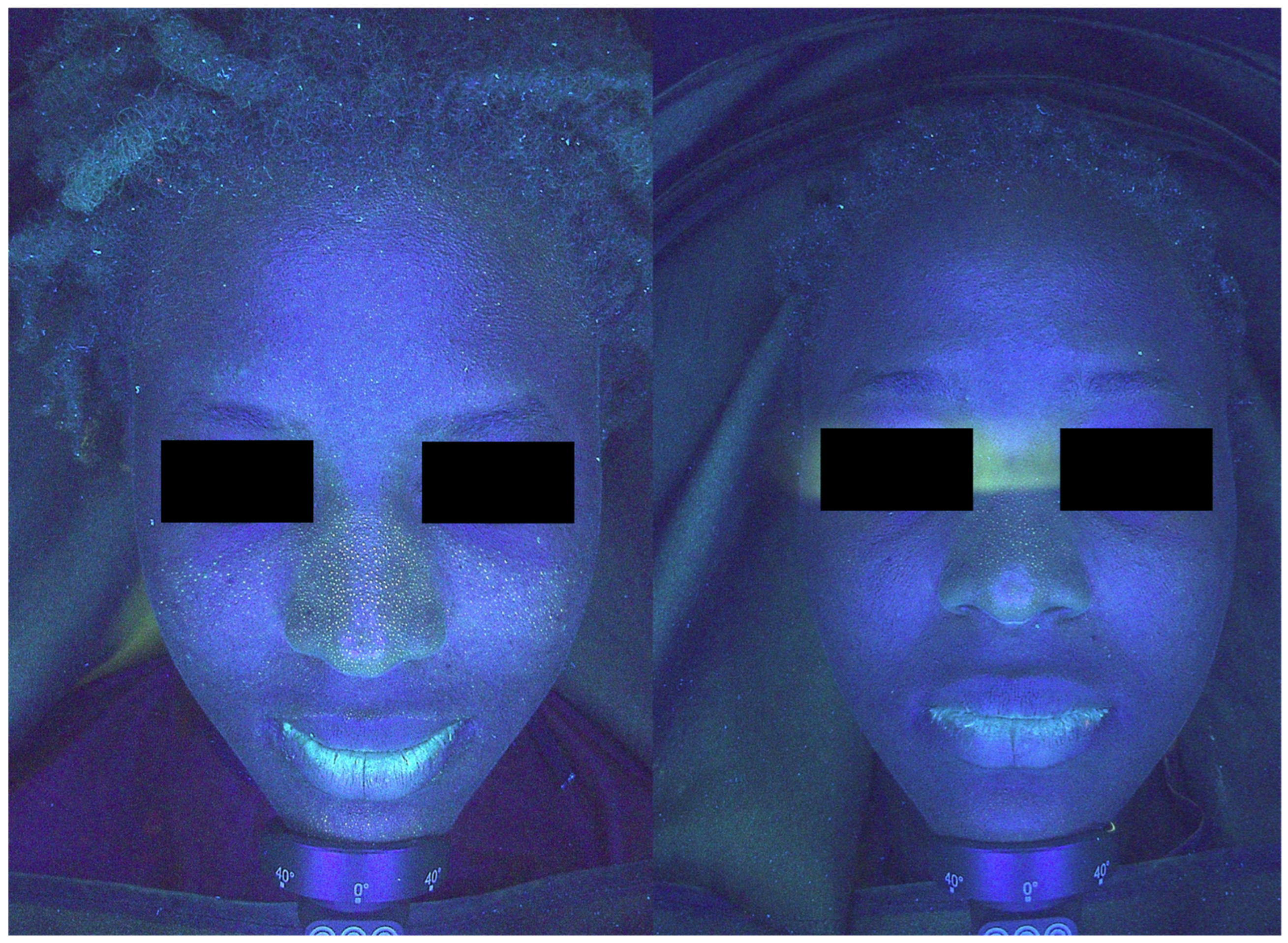
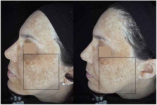
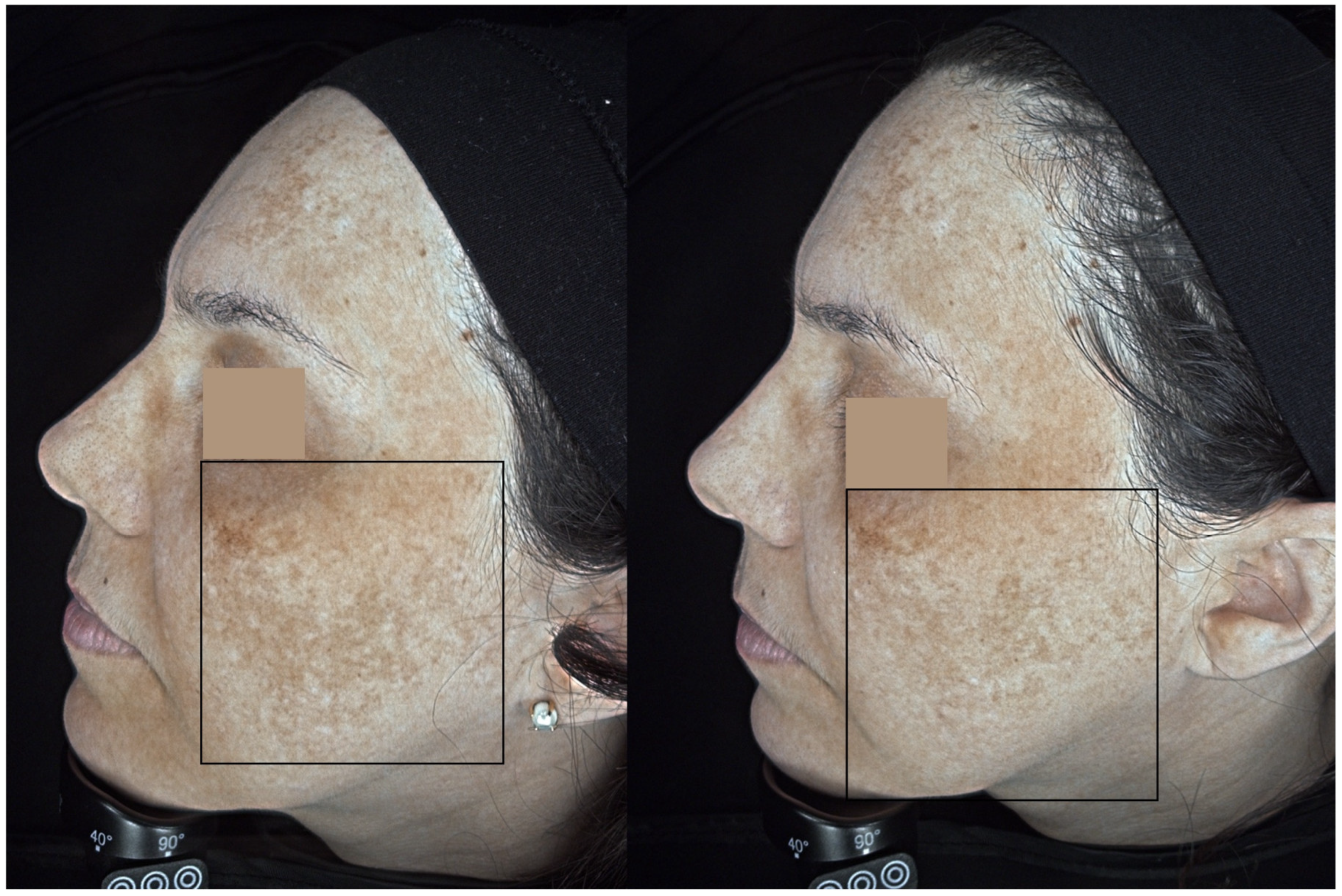
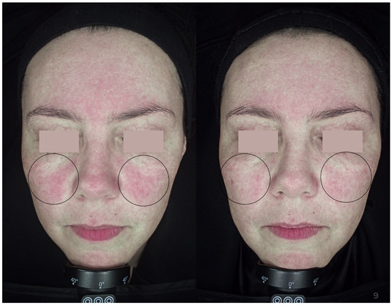
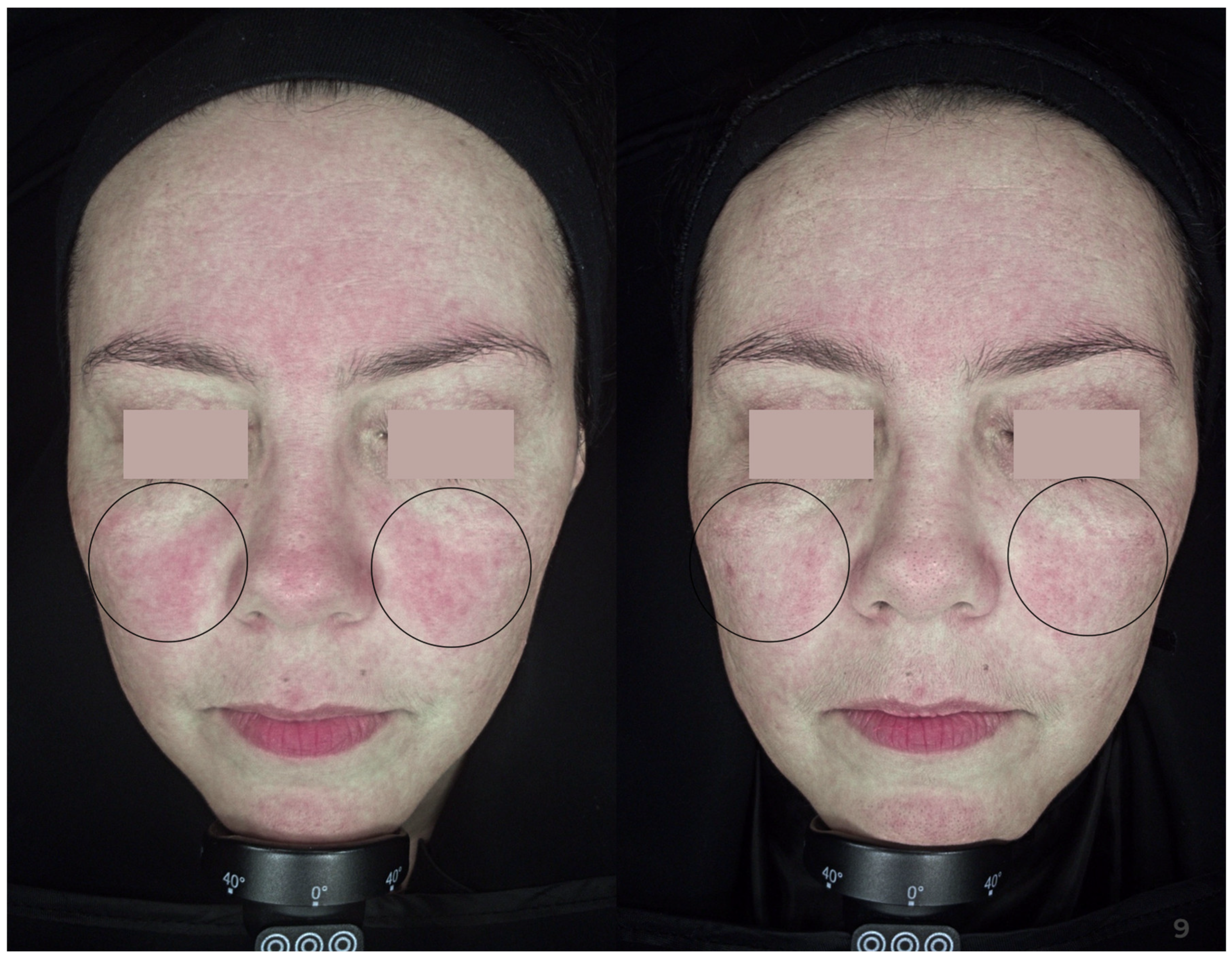
Improvement in skin condition, as documented in photographs, is shown in Figure 3, Figure 4 and Figure 5, respectively.
Figure 3.
Patient before (left side) and after (right side) the peel treatment. A significant reduction of enlarged, clogged sebaceous glands and a reduction of sebum production are visible.
Figure 4.
Patient before (left side) and after (right side) the peel treatment. A reduction in superficial hyperpigmentation is noted.
Figure 5.
Patient before (left side) and after (right side) the peel treatment. A reduction in skin redness is notable.
Adverse Events
No serious adverse events were observed during the study. Five patients reported increased skin exfoliation, as well as mild itching, redness, and skin irritation after the procedure.
4. Discussion
Data on the safety and efficacy of cosmetic procedures among patients with SOC remains limited. To the best of our knowledge, this was the first study to assess such a novel therapy in dark-skinned individuals. As aesthetic medicine develops, practitioners must also improve their expertise for treating patients with darker Fitzpatrick skin types who seek laser treatments, microneedling, or chemical peels. Especially considering the fact that individuals with darker skin types are more likely to experience adverse skin events after cosmetic treatments [14]. According to a review by Amechi et al. [14], choosing the right peeling agent is essential for successful chemical peels on people with darker skin tones. Dark-skinned people can safely undergo superficial chemical peels; however, extreme caution is required when using a medium-depth peel. Moreover, deep peels may increase the likelihood of side effects for patients with skin types IV–VI; thus, they should be avoided.
To get optimal results, patients undergoing chemical peels should be carefully chosen, with consideration given to the right peeling agent, its concentration, and its application technique. Each chemical peel agent has a unique mode of action, and various skin tones will provide varied results. The fact that individuals with colored skin may react differently to chemical peeling is therefore crucial for clinicians to comprehend. According to a 5-year, single-center retrospective study assessing the incidence of adverse events associated with superficial chemical peels in patients with Fitzpatrick skin types III–VI, the most common complications were crusting (2.3%), post-inflammatory hyperpigmentation (1.9%), and erythema (1.9%) [15]. Risk of complications varied among patients in the comparative analysis, with skin type VI having higher odds of experiencing a side effect.
In a single-center prospective study by Sadick et al. [16], 24 patients (photodamage group, n = 14 [with an acne subgroup, n = 5]; Fitzpatrick skin types IV–VI [Skin of Color group, n = 5; with mild to moderate melasma or hyperpigmentation group, n = 5]) were treated with a 3% retinol peel. A series of 2–4 peels in 6-week intervals with a supportive at-home skin care regimen was performed [16]. For each group, a dermatologist’s clinical grading of fine lines, wrinkles, pore size, laxity, mottled pigmentation, lack of clarity/radiance, and overall photodamage was conducted. The results confirmed that a 3% retinol superficial peel is well tolerated and effective in all skin types. However, in comparison to our study, the subjective appraisal of research findings is a major limitation. Such a novel procedure with a 4% retinol solution product containing TGF-β activators and antioxidants has only been evaluated in patients with Fitzpatrick skin types I–III, highlighting the need for further research [17]. Transforming growth factor-beta (TGF-β) is a key regulatory protein that influences cell proliferation, differentiation, and tissue remodeling [18]. It helps maintain structural integrity by facilitating communication between epidermal and dermal cells through specific surface receptors [19]. TGF-β also contributes to skin rejuvenation by stimulating the production of extracellular matrix components such as collagen, thereby counteracting signs of aging [20]. TGF-β plays a crucial role in maintaining normal skin homeostasis and mending or regenerating old skin due to its multiple actions.
Furthermore, it has been shown that combining different treatment methods yields greater outcomes than relying solely on one. Budamakuntla et al. [21] examined 60 SOC patients with dermal and epidermal melasma. Patients were treated with microneedling and topical tranexamic acid (TA) (4 mg/mL) or with localized microinjections of TA alone. There was a 35.72% improvement in the Melasma Area Severity Index (MASI) score in the microinjection group compared to 44.41% in the microneedling group after three treatments. The authors concluded that the improved therapeutic response to therapy in the microneedling group might be related to the deeper and uniform administration of the agents through microchannels generated by microneedling. Furthermore, in 2011, Fabbrocini et al. [22] performed a split-face study on 20 melasma patients (Fitzpatrick III–V) to compare the effectiveness of a depigmentation serum containing rucinol and sophora-alpha with and without microneedling. After two months of therapy with depigmenting serum alone, the baseline MASI score improved by 7.1 points, while the combination treatment resulted in a 10.1-point decrease. Although we did not compare microneedling with chemical peels to chemical peels alone in our study, the presented treatment protocol was also found to be helpful in treating skin hyperpigmentation.
Additionally, according to some research, microneedling combined with chemical peels is also more effective than chemical peels alone in treating acne scars in patients with SOC, even though our trial did not show any improvement in this area. In their trial, Sharad et al. administered five sessions of either microneedling alone (group 1, n = 30) or microneedling in conjunction with 35% glycolic acid peels (group 2, n = 30) to 60 patients (Fitzpatrick III–V) with acne scars [23]. Group 1′s mean improvement was 31%, while group 2′s was 62% at the 3-month mark. Transitory bruising, edema, milia, and PIH were among the side effects of the treatment. In another study, the efficacy of combination therapy using subcision, microneedling, and a 15% trichloroacetic acid (TCA) peel was evaluated on 50 patients (Fitzpatrick III–V) with atrophic scars [24]. Each patient had one subcision session with a 24-gauge needle at the initial phase of the research. Subsequently, for a total of six treatments each, microneedling and a 15% TCA peel were done alternately at 2-week intervals. The Goodman and Baron qualitative scale, which assigns scar categories of macular (grade 1), mild (grade 2), moderate (grade 3), and severe (grade 4), was used to evaluate the results. While 63% and 38% of patients with grade 4 scars at baseline progressed to grade 2 and 3, respectively, all patients with grade 2 scars had complete scar clearance. The procedure was well tolerated with only mild and temporary side effects experienced.
This study has several limitations. Primary drawbacks were its small sample size and its single-center design, which resulted from the preliminary nature of the research. An additional constraint was the absence of a comparison group with either microneedling or chemical peel alone. Third, the use of adjunctive home care products containing active ingredients may have influenced the observed outcomes. Finally, the inclusion of subjective patient-reported outcomes introduces the possibility of expectation bias.
5. Conclusions
To conclude, a combined peel therapy of a 4% retinol serum product containing novel TGF-β activators and antioxidants combined with a microneedling technique proved good efficacy in reducing superficial and deep facial hyperpigmentation of the skin, as well as regulation of sebaceous gland activity, their size, and reduction of sebum production. Moreover, enhancement of the skin barrier, improvement of skin hydration, and its general condition were also noted. In accordance with the study, the suggested approach is relatively simple to use, low-cost, has minimal adverse reactions, and is well tolerated by patients with SOC. However, due to the small sample size and the lack of a control group, these findings should be interpreted with caution and require confirmation in larger, controlled studies.
Author Contributions
Conceptualization, D.S. and W.B.-R.; methodology, D.S. and W.B.-R.; software, D.S.; validation, D.S. and W.B.-R.; formal analysis, D.S. and W.B.-R.; investigation, D.S.; resources, D.S. and W.B.-R.; data curation, D.S. and W.B.-R.; writing—original draft preparation, D.S. and W.B.-R.; writing—review and editing, D.S. and W.B.-R.; visualization, D.S. and W.B.-R.; supervision, W.B.-R. All authors have read and agreed to the published version of the manuscript.
Funding
This research received no external funding.
Institutional Review Board Statement
The study was conducted in accordance with the Declaration of Helsinki and approved by the Ethics Committee of the Medical University of Gdańsk (NKBBN/618/2022). Approval Date: 16 November 2022.
Informed Consent Statement
Informed consent was obtained from all subjects involved in the study. Written informed consent has been obtained from the patient(s) to publish this paper.
Data Availability Statement
The original contributions presented in this study are included in the article. Further inquiries can be directed to the corresponding author.
Acknowledgments
We would like to give special thanks to the Retix C® brand for providing us with materials that allowed us to perform this study.
Conflicts of Interest
The authors declare no conflicts of interest.
References
- Cohen, B.E.; Elbuluk, N. Microneedling in skin of color: A review of uses and efficacy. J. Am. Acad. Dermatol. 2016, 74, 348–355. [Google Scholar] [CrossRef] [Scilit]
- Nautiyal, A.; Wairkar, S. Management of hyperpigmentation: Current treatments and emerging therapies. Pigment Cell Melanoma Res. 2021, 34, 1000–1014. [Google Scholar] [CrossRef] [Scilit] [PubMed]
- Shen, Y.C.; Chiu, W.K.; Kang, Y.N.; Chen, C. Microneedling Monotherapy for Acne Scar: Systematic Review and Meta-Analysis of Randomized Controlled Trials. Aesthetic Plast. Surg. 2022, 46, 1913–1922. [Google Scholar] [CrossRef] [Scilit] [PubMed]
- Pająk, J.; Szepietowski, J.C.; Nowicka, D. Prevention of Ageing-The Role of Micro-Needling in Neck and Cleavage Rejuvenation: A Narrative Review. Int. J. Environ. Res. Public Health 2022, 19, 9055. [Google Scholar] [CrossRef] [Scilit]
- Litchman, G.; Nair, P.A.; Badri, T.; Kelly, S.E. Microneedling. In StatPearls; StatPearls Publishing: Tampa, FL, USA; St. Petersburg, FL, USA, 2022. [Google Scholar]
- Taylor, S.C. Skin of color: Biology, structure, function, and implications for dermatologic disease. J. Am. Acad. Dermatol. 2002, 46, S41–S62. [Google Scholar] [CrossRef] [Scilit]
- Gupta, V.; Sharma, V.K. Skin typing: Fitzpatrick grading and others. Clin. Dermatol. 2019, 37, 430–436. [Google Scholar] [CrossRef] [Scilit]
- Iwuala, C.; Taylor, S.C. Structural and functional differences in skin of colour. Clin. Exp. Dermatol. 2022, 47, 247–250. [Google Scholar] [CrossRef] [Scilit]
- Callender, V.D.; Baldwin, H.; Cook-Bolden, F.E.; Alexis, A.F.; Stein Gold, L.; Guenin, E. Effects of Topical Retinoids on Acne and Post-inflammatory Hyperpigmentation in Patients with Skin of Color: A Clinical Review and Implications for Practice. Am. J. Clin. Dermatol. 2022, 23, 69–81. [Google Scholar] [CrossRef] [Scilit]
- Chilicka, K.; Rusztowicz, M.; Szyguła, R.; Nowicka, D. Methods for the Improvement of Acne Scars Used in Dermatology and Cosmetology: A Review. J. Clin. Med. 2022, 11, 2744. [Google Scholar] [CrossRef] [Scilit] [PubMed]
- Goodman, G.J.; Baron, J.A. Postacne scarring: A qualitative global scarring grading system. Dermatol. Surg. Off. Publ. Am. Soc. Dermatol. Surg. 2006, 32, 1458–1466. [Google Scholar] [CrossRef] [Scilit]
- Kashetsky, N.; Feschuk, A.; Pratt, M.E. Post-inflammatory hyperpigmentation: A systematic review of treatment outcomes. J. Eur. Acad. Dermatol. Venereol. JEADV 2024, 38, 470–479. [Google Scholar] [CrossRef] [Scilit]
- Ali, L.; Al Niaimi, F. Pathogenesis of Melasma Explained. Int. J. Dermatol. 2025, 64, 1201–1212. [Google Scholar] [CrossRef] [Scilit]
- Amechi, M.; Halpin, J. Considerations for Laser Therapy, Microneedling, and Chemical Peels When Treating Patients with Skin of Color. Plast. Aesthetic Nurs. 2023, 43, 14–21. [Google Scholar] [CrossRef] [Scilit]
- Vemula, S.; Maymone, M.B.C.; Secemsky, E.A.; Widjajahakim, R.; Patzelt, N.M.; Saade, D.; Vashi, N.A. Assessing the safety of superficial chemical peels in darker skin: A retrospective study. J. Am. Acad. Dermatol. 2018, 79, 508–513.e2. [Google Scholar] [CrossRef] [Scilit]
- Sadick, N.; Edison, B.L.; John, G.; Bohnert, K.L.; Green, B. An Advanced, Physician-Strength Retinol Peel Improves Signs of Aging and Acne Across a Range of Skin Types Including Melasma and Skin of Color. J. Drugs Dermatol. JDD 2019, 18, 918–923. [Google Scholar]
- Sołdacka, D.; Podgórska, M.; Barańska-Rybak, W. Unique retinol therapy with antioxidant and anti-inflammaging complex for naturally reborn skin: The clinical case series study. Dermatol. Ther. 2023, 2023, 5588525. [Google Scholar] [CrossRef] [Scilit]
- Deng, Z.; Fan, T.; Xiao, C.; Tian, H.; Zheng, Y.; Li, C.; He, J. TGF-β signaling in health, disease, and therapeutics. Signal Transduct. Target. Ther. 2024, 9, 61. [Google Scholar] [CrossRef] [Scilit] [PubMed]
- Li, X.; Xie, R.; Luo, Y.; Shi, R.; Ling, Y.; Zhao, X.; Xu, X.; Chu, W.; Wang, X. Cooperation of TGF-β and FGF signalling pathways in skin development. Cell Prolif. 2023, 56, e13489. [Google Scholar] [CrossRef] [Scilit] [PubMed] [PubMed Central]
- Xu, X.; Zheng, L.; Yuan, Q.; Zhen, G.; Crane, J.L.; Zhou, X.; Cao, X. Transforming growth factor-β in stem cells and tissue homeostasis. Bone Res. 2018, 6, 2. [Google Scholar] [CrossRef] [Scilit] [PubMed] [PubMed Central]
- Budamakuntla, L.; Loganathan, E.; Suresh, D.H.; Shanmugam, S.; Suryanarayan, S.; Dongare, A.; Venkataramiah, L.D.; Prabhu, N. A Randomised, Open-label, Comparative Study of Tranexamic Acid Microinjections and Tranexamic Acid with Microneedling in Patients with Melasma. J. Cutan. Aesthetic Surg. 2013, 6, 139–143. [Google Scholar] [CrossRef] [Scilit] [PubMed] [PubMed Central]
- Fabbrocini, G.; De Vita, V.; Fardella, N.; Pastore, F.; Annunziata, M.C.; Mauriello, M.C.; Monfrecola, A.; Cameli, N. Skin needling to enhance depigmenting serum penetration in the treatment of melasma. Plast. Surg. Int. 2011, 2011, 158241. [Google Scholar] [CrossRef] [Scilit] [PubMed] [PubMed Central]
- Sharad, J. Combination of microneedling and glycolic acid peels for the treatment of acne scars in dark skin. J. Cosmet. Dermatol. 2011, 10, 317–323. [Google Scholar] [CrossRef] [Scilit] [PubMed]
- Garg, S.; Baveja, S. Combination therapy in the management of atrophic acne scars. J. Cutan. Aesthetic Surg. 2014, 7, 18–23. [Google Scholar] [CrossRef] [Scilit] [PubMed] [PubMed Central]
Disclaimer/Publisher’s Note: The statements, opinions and data contained in all publications are solely those of the individual author(s) and contributor(s) and not of MDPI and/or the editor(s). MDPI and/or the editor(s) disclaim responsibility for any injury to people or property resulting from any ideas, methods, instructions or products referred to in the content. |
© 2026 by the authors. Licensee MDPI, Basel, Switzerland. This article is an open access article distributed under the terms and conditions of the Creative Commons Attribution (CC BY) license.