


Abstract
Sunscreens are essential for photoprotection, but conventional inorganic UV filters raise concerns regarding marine toxicity. This study investigated hydroxyapatite (HAp) derived from skipjack, tongol, and salmon bone waste as a potential synergistic booster for Tinosorb® S (TS). HAp powders were prepared via alkaline treatment and calcination at 900 °C. XRD and XRF results confirmed highly crystalline HAp as the dominant phase. While 10% HAp alone provided negligible UV protection, a pronounced synergistic effect was observed in 1:1 hybrid formulations (5% HAp:5% TS), significantly enhancing Sun Protection Factor (SPF) and Ultraviolet A Protection Factor (UVAPF). Notably, particle-size refinement of salmon-derived HAp (SM–HAp) yielded an SPF of approximately 35, comparable to a commercial HAp counterpart. This improvement was suggested to be associated with enhanced dispersion, film uniformity, and particle–matrix interactions, which might contribute to achieving PA++++ protection. All formulations complied with microbiological and heavy metal safety standards. These results indicated that fish bone-derived HAp could potentially serve as a viable and sustainable functional additive derived from marine biowaste for the development of high-performance hybrid sunscreens, promoting biomaterial valorization in the cosmetic industry.
1. Introduction
Protecting the skin against the harmful effects of ultraviolet (UV) radiation is crucial, given its link to numerous dermatological issues, including sunburn, photoaging, immunosuppression, and cancer [1,2]. Excessive UV exposure initiates inflammation and the breakdown of collagen; simultaneously, it drives the formation of reactive oxygen species (ROS), resulting in cellular damage and oxidative stress [3]. Clinical manifestations such as erythema, pigmentation, wrinkles, and actinic keratosis are strongly associated with UV overexposure, with the latter potentially progressing to malignancies if untreated [4,5,6]. These negative consequences emphasize the critical importance of safe and effective photoprotection strategies.
Sunscreens are widely used to mitigate UV-induced skin damage and are generally categorized into inorganic (physical) and organic (chemical) filters. Inorganic filters, such as titanium dioxide (TiO2) and zinc oxide (ZnO), provide photoprotection by reflecting and scattering UV radiation. However, their use presents esthetic issues such as skin whitening and serious environmental risks, including coral reef bleaching and marine toxicity [7,8]. Chemical filters, such as avobenzone (BMDBM), octocrylene (OC), and bis-ethylhexyloxyphenol methoxyphenyl triazine (Tinosorb® S), absorb UV radiation efficiently and provide broad-spectrum protection, but some exhibit limitations such as narrow absorption ranges, potential skin penetration leading to endocrine-disrupting effects, and allergic reactions [9,10,11,12,13]. Current efforts are largely focused toward minimizing these drawbacks by developing sustainable or natural photoprotection alternatives that deliver high efficacy alongside the enhanced safety and environmental compatibility [14,15,16,17,18,19,20,21,22].
Hydroxyapatite (Ca10(PO4)6(OH)2), a calcium phosphate mineral chemically similar to vertebrate bone, is gaining increasing attention as a cosmetic ingredient due to its high biocompatibility, biosafety, and favorable optical properties [23,24,25]. Unlike common inorganic filters such as TiO2 and ZnO, HAp does not exhibit photocatalytic activity under UV exposure, which prevents the generation of harmful free radicals [26,27,28,29]. While both synthetic and natural HAp can attenuate UV radiation through absorption and scattering, they still offer limited SPF performance [24,25]. A major benefit is that HAp can be extracted from biogenic waste, such as animal and fish bones, providing an eco-friendly strategy to reduce seafood industry waste while creating valuable cosmetic ingredients [24,25]. Among biogenic sources, fish bone waste is preferred over terrestrial animal sources (bovine or porcine) as it precludes the risks of zoonotic disease transmission, such as bovine spongiform encephalopathy (BSE), and avoids religious or cultural restrictions [30,31]. Furthermore, the utilization of marine-derived byproducts aligns with circular economy valorization strategies and supports the development of marine-safe formulations [30,31,32,33]. Although fish bone-derived HAp has been demonstrated to reduce UV transmittance, its SPF enhancement is modest and requires further optimization. For instance, the incorporation of Fe2O3 can significantly boost photoprotective performance. However, it introduces an undesirable reddish or brownish tint to the final product [25,26,34].
Combining organic UV filters with inorganic or hybrid additives has emerged as a promising strategy to enhance SPF performance through synergistic effects, including improved UV attenuation, filter utilization, and formulation stability. Such approaches have been demonstrated using mesoporous silica, zeolites, and mineral-based systems, where inorganic components act as optical enhancers or carriers for organic UV filters [19,20,21,25,26,34]. Although TiO2 and ZnO are conventionally used for this purpose, the major concerns regarding their environmental impact necessitate the potential alternatives. Nonetheless, the synergy between bio-derived HAp and organic filters has not been comprehensively investigated. In particular, the interaction of fish bone-derived HAp with Tinosorb® S, a highly effective, oil-soluble, and photostable UV filter widely employed in sunscreens, facial moisturizers, anti-aging formulations, and color cosmetics, remains unexplored. Tinosorb® S provides broad-spectrum UVA and UVB protection with excellent photostability, retains efficacy under prolonged sun exposure, and is favored for its minimal irritation potential, non-comedogenic properties, and formulation flexibility [10,35].
Despite the growing use of bio-derived materials in cosmetic formulations, the synergistic potential of hydroxyapatite with the high-performance UV filter Tinosorb® S has not been elucidated. While previous studies have explored the UV attenuation capability of HAp powders or their modification with metal oxides [25], a systematic investigation of the synergy between undoped, fish bone-derived HAp and Tinosorb® S within complete sunscreen systems is still needed. Our study addresses this research gap by demonstrating a quantifiable synergistic enhancement of SPF when fish bone-derived HAp powders are combined with Tinosorb® S in oil-in-water sunscreen systems. Specifically, HAp powders derived from skipjack tuna, tongol tuna, and salmon bones, without additional elemental dopants were evaluated relative to a commercial HAp reference. These findings might provide new insight into mineral–organic UV filter synergy involving Tinosorb® S in cosmetic formulations and propose biogenic HAp as an effective, non-whitening, and sustainably sourced optical enhancer. Furthermore, this work represents a circular-economy-driven approach toward the development of marine-friendly sunscreens, offering a biocompatible alternative to conventional UV filter strategies while minimizing environmental impact on aquatic ecosystems.
2. Materials and Methods
2.1. Treatments of Fish Bones for Hydroxyapatite (HAp) Preparation
Skipjack tuna (Katsuwonus pelamis; SKJ), tongol tuna (Thunnus tonggol; TG), and salmon (Oncorhynchus spp.; SB) bones were obtained from local seafood processing industries in Thailand. The bones were cut longitudinally using an electric saw, thoroughly cleaned with a high-pressure water jet (Andaman, Zinsano, Bangkok, Thailand) at 120 bar to remove residual tissues, and sealed in polyethylene bags before storage at −20 °C until further use. Protein removal was carried out by immersing the bones in 2 M NaOH solution (1:10, w/v) at 50 °C for 1 h under continuous stirring, followed by filtration, rinsing with running water for 3–4 h until neutral pH was achieved, and oven drying at 50 °C for 2–6 h. Dried bones were coarsely ground to <4 cm fragments and further subjected to defatting by immersing in hexane (1:10, w/v) for 1 h at 25 °C with continuous stirring. The defatted bones were air dried at room temperature, milled into fine powders using a planetary ball mill (PM 100, Retsch GmbH, Haan, Germany), and sieved to a particle size <75 µm [30,31]. These processing steps were adopted to ensure the effective removal of organic residues (proteins and lipids) from different fish bone sources and to produce fine powders with consistent physicochemical properties, enabling reliable comparison of HAp derived from skipjack, tongol, and salmon bones and their suitability for sunscreen application [30,31,32].
2.2. Calcination and Particle Size Reduction
Calcination at 900 °C for 5 h was adopted based on previous studies showing that fish bone powders calcined at 900 °C contained no protein or fat, undetectable hydroxyproline, and >99.99% ash, confirming complete removal of organic matter [34,35]. By eliminating oxidizable residues, lipid and protein oxidation pathways in the O/W cream base could be suppressed, thus preventing color and odor development, emulsion destabilization, and ultimately lengthening shelf-life [33,36,37,38]. After calcination, the powders were divided into two processing routes to investigate the effect of particle size on UV performance and formulation rheology. One portion was used directly as micron-scale calcined HAp, whereas the second portion was further milled via planetary ball milling to obtain HAp with reduced particle size. Both materials were characterized and used in the subsequent sunscreen formulations to evaluate particle size dependent synergistic effects with Tinosorb® S (TS).
2.3. Characterization of Fish Bone and HAp Powders
2.3.1. X-Ray Diffraction (XRD)
X-ray diffraction (XRD) (Rigaku, Tokyo, Japan) was employed for phase identification. The analysis was conducted by using Cu Kα radiation at 40 kV, 40 mA. Diffraction patterns were collected over a 2θ range of 20–70° at a scan rate of 3°/min with a step size of 0.05° [30]. Phase identification was carried out using the ICDD PDF-2 database. Crystallite size was estimated using the Halder-Wagner (H-W) method implemented in PDXL-2. The crystallite size was taken from the intercept of the linear regression, considering only reflections belonging to the hydroxyapatite phase [39].
2.3.2. X-Ray Fluorescence (XRF)
The elemental profile and Ca/P molar ratios were determined using a wavelength dispersive X-ray fluorescence spectrometer (WDXRF, Bruker S8 Tiger, Bruker AXS, Karlsruhe, Germany) equipped with a Rh anode (50 kV, 20 mA). Spectral acquisition and evaluation were performed using the Quant-Express standardless analysis software (version 1.0.0.13, Bruker AXS, Karlsruhe, Germany). This non-destructive technique was selected for stoichiometric analysis to avoid cumulative dilution errors typically associated with the high-ratio serial dilutions required for ICP-OES analysis of high-concentration Ca and P minerals. To isolate the mineral phase stoichiometry from the residual organic matrix, all detected oxide compositions were normalized to 100% prior to the calculation of the Ca/P molar ratios.
2.3.3. Scanning Electron Microscopy (SEM)
Samples were oven-dried at 105 °C for 24 h, mounted on carbon tape, and gold-coated using ion sputtering (SPI-Module Sputter Coater, SPI Supplies, West Chester, PA, USA). Morphological observations were performed using a SEM (Quanta 400, Thermo Scientific, Hillsboro, OR, USA) operated at an accelerating voltage of 5.0 kV and beam current 6.3 pA. Images were collected with a secondary electron detector at 25,000 magnifications [30]. Particle size measurements were performed using ImageJ software (version 1.54g, National Institutes of Health, Bethesda, MD, USA).
2.4. Application of Fish Bone-Derived HAp in Sunscreen Formulations
2.4.1. Preparation of Base Cream and Sunscreen Formulation
An oil-in-water (O/W) emulsion was prepared, consisting of 19% oil phase (A), 71% water phase (B), and 10% UV filters (Table 1). UV filters included the chemical filter Bis-Ethylhexyloxyphenol Methoxyphenyl Triazine (Tinosorb® S; cosmetic grade, BASF SE, Ludwigshafen, Germany) and the physical filters TiO2 (Sigma-Aldrich, St. Louis, MO, USA, product 637254, 99.7% trace metals basis; <25 nm, as specified by the manufacturer), commercial HAp (C-HAp; Sigma-Aldrich, St. Louis, MO, USA, product 04238, puriss), or fish bone-derived HAp. While the TiO2 particle size was provided by the manufacturer, the C-HAp was independently characterized via SEM. Subsequently, ImageJ (version 1.54g) analysis revealed an average particle size of 88.40 ± 2.42 nm (Figure S1).
Table 1.
Composition of model sunscreen formulations (100 g batch).
Oil and water phases were prepared separately and preheated to 70–80 °C with continuous agitation. The heated phases were then combined and subjected to high-shear homogenizer T 25 digital ULTRA-TURRAX (IKA, Staufen, Germany) at 6000 rpm for 1 min, followed by 1 min rest. This process was repeated 15 times. Finally, the emulsions were cooled to room temperature and stored in amber glass bottles wrapped with aluminum foil.
2.4.2. Evaluation of Sunscreen Formulations
The physicochemical properties of the formulations were analyzed as follows:
Viscosity was measured at 25 ± 0.5 °C using a Brookfield viscometer (DV2TLVTJ0, Brookfield Engineering, Middleboro, MA, USA) with a spindle LV-T 94 at 20 rpm. Samples were gently loaded to avoid air entrapment, equilibrated for 10 min, and recorded in triplicate.
Color of sunscreen formulations was measured using a HunterLab colorimeter (Colour Flex, Hunter Lab Inc., Reston, VA, USA) in accordance with CIE L*a*b* color model [40]. The total color difference (ΔE*) among samples was subsequently calculated using the equation present below:
where , , and correspond to control values, and , , and to the tested samples. The instrument was calibrated with the manufacturer’s white standard tile (L* = 93.63, a* = −0.94, b* = 0.40). The whiteness index (WI) was determined [41]:
These combined analyses provided a quantitative assessment of brightness, hue, and overall whiteness of each sunscreen formulation.
pH measurements were carried out at 25 ± 2 °C using a pH meter calibrated with standard buffer solutions of pH 4.0, 7.0, and 10.0.
SPF and UVAPF (UVA protection factors) were determined using an SPF Analyzer (UV-2000s, Labsphere, North Sutton, NH, USA). All measurements were performed in vitro; however, the erythema action spectrum E(λ) and the reference solar spectrum used in the SPF calculation were sourced from ISO 24444:2010 [42]. UVAPF and critical wavelength (λc) were evaluated according to the calculation framework of ISO 24443:2012 [43], and PA grades were assigned in accordance with Japan Cosmetic Industry Association (JCIA) guidelines [44]; formulations exhibiting UVAPF < 2 were not assigned a PA grade. Sunscreen films were evenly applied at 1.3 mg/cm2 on polymethylmethacrylate (PMMA) plates (HD6; Ra ≈ 6 µm), incubated in the dark (25 ± 2 °C) and measured at nine positions on three plates (n = 3). The in vitro SPF was calculated as follows:
where E(λ) is the erythema Action Spectrum, and I(λ) is the reference solar irradiance spectrum, and T(λ) is the spectral transmittance of the sunscreen film normalized to an uncoated PMMA.
2.4.3. Microbial and Heavy Metal Contamination
Microbial contamination, including Staphylococcus aureus, Pseudomonas aeruginosa, yeast, and molds, were evaluated in accordance with the Notification of Ministry of Public Health 2016 [45] standards using AOAC Performance Tested Methods (PTM), including AOAC PT No. 120901 for S. aureus and AOAC PT No. 041001 for yeasts and molds [46]. P. aeruginosa was identified by MALDI-TOF MS (MALDI Biotyper, Microflex, Bruker Daltonics, Bremen, Germany). Heavy metals (As, Cd, and Pb) were quantified using ICP-OES (Optima 8000, PerkinElmer, Waltham, MA, USA), while mercury (Hg) was determined using a mercury analyzer (RA-915M, Lumex, Instruments, Mississauga, ON, Canada). All results were compared with cosmetic safety limits specified in national regulations [45].
2.4.4. Accelerated Stability Testing
Accelerated stability testing was performed by storing the optimized formulation (5% HAp + 5% Tinosorb® S) at 50 °C for 30 days. pH was measured using a calibrated pH meter at 25 ± 2 °C. Viscosity was determined using a Brookfield viscometer (DV2T, spindle LV-T 96, 5 rpm, 60 s). Physical appearance, including phase separation and sedimentation, was visually assessed before and after storage. Measurements were conducted in triplicate.
2.4.5. Statistical Analysis
All experiments were performed in triplicate unless otherwise specified. Results were reported as mean ± standard deviation (SD). Prior to statistical testing, the data were checked for normality and homogeneity of variance to ensure the assumptions of parametric analysis were met. Statistical differences among groups were evaluated using one-way analysis of variance (ANOVA) followed by appropriate post hoc tests at a significance level of p < 0.05. Tukey’s honestly significant difference (HSD) was employed for pair-wise comparisons among HAp variants. Dunnett’s test was utilized to compare optimized hybrid formulations against the designated control groups in synergistic and stability studies. Statistical analyses were conducted using IBM SPSS Statistics Version 31.0 (IBM Corp., Armonk, NY, USA).
3. Results and Discussion
3.1. Characterization of Fish Bone-Derived Hydroxyapatite
3.1.1. Elemental Profile and Semi-Quantitative XRF Analysis
The semi-quantitative chemical compositions of hydroxyapatite powders derived from skipjack (SKJ), tongol (TG), and salmon (SM) fish bones before and after calcination at 900 °C are presented in Table 2.
Table 2.
Semi-quantitative chemical analysis and Ca/P ratios of fish bone derived and commercial HAp powders.
Calcium (Ca) and phosphorus (P) were the major elements in all samples, while sodium (Na) and sulfur (S) appeared as minor components. Trace levels of strontium (Sr), magnesium (Mg), zinc (Zn), potassium (K), chlorine (Cl), iron (Fe), copper (Cu), and zirconium (Zr) were also detected. The presence of these elements is consistent with natural ionic substitutions commonly observed in biological apatites [47,48]. The analytical precision of the instrument was stable, with standard deviations (SDs) < 10% for all measurable elements. Comparative analysis and statistical significance (ANOVA) were specifically evaluated for the Ca/P ratios, as this stoichiometry directly influences the physicochemical properties of the fish bone-derived samples.
The Ca/P molar ratios of the raw powders were 2.16 for SKJ, 2.18 for TG, and 1.99 for SM, which exceeded the stoichiometric value of 1.67 for pure synthetic HAp. Since the apatite lattice is inherently non-stoichiometric due to B-type carbonate and trace element substitutions such as Na+, Mg2+, Sr2+, and Zn2+. These substitutions typically lower the Ca/P molar ratio below the theoretical value of 1.67, as the replacement of PO43− by CO32− induces calcium deficiency to maintain charge neutrality [47,48]. Consequently, the observed bulk ratios above 1.67 strongly indicate the presence of non-apatite, calcium-rich impurity phases, primarily calcium carbonate, within the raw matrix. Ca/P values have been documented in biogenic apatites ranging from near stoichiometric up to >2.0, depending on species, age, and thermal treatment [48,49,50], confirming that such variations are intrinsic to natural bone mineral. These compositional variations and elemental substitutions are known to critically influence the solubility, crystallinity, and overall physicochemical behavior of biological HAp in functional applications [48,50].
After calcination at 900 °C, the Ca/P ratios decreased to 1.84 (SKJ), 1.80 (TG), and 1.78 (SM). Despite this decrease, the samples remained significantly Ca-rich (>1.67), which was associated with the thermal decomposition of initial CaCO3 impurities into calcium oxide (CaO). The absence of crystalline CaO reflections in the XRD patterns (Section 3.1.2) is consistent with Raynaud et al., who noted that excess calcium in apatite is difficult to characterize as CaO rapidly transforms to Ca(OH)2, its stable form under ambient conditions, upon exposure to ambient humidity [51]. Simultaneously, the observed reduction in Ca/P ratios was consistent with a partial transformation toward β-TCP (Ca/P = 1.50), a phenomenon widely reported for biogenic hydroxyapatite during high temperature processing [48,52]. The minimal formation of β-TCP was supported by both the small magnitude of the Ca/P decrease and the absence of characteristic β-TCP reflections in the corresponding XRD analysis. In general, thermally induced secondary phases in fish bone HAp often occur at concentrations below the typical XRD detection limit of 3–5% [48,52,53]. For comparison, commercial HAp (C-HAp) showed a Ca/P ratio of 1.62 with negligible impurities, serving as a reference material for evaluating the suitability of fish bone derived HAp for sunscreen formulations.
3.1.2. Phase Composition (XRD)
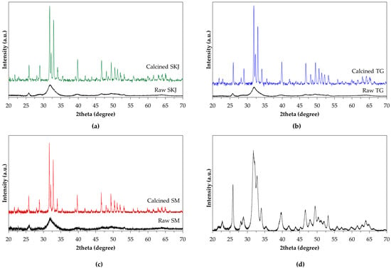
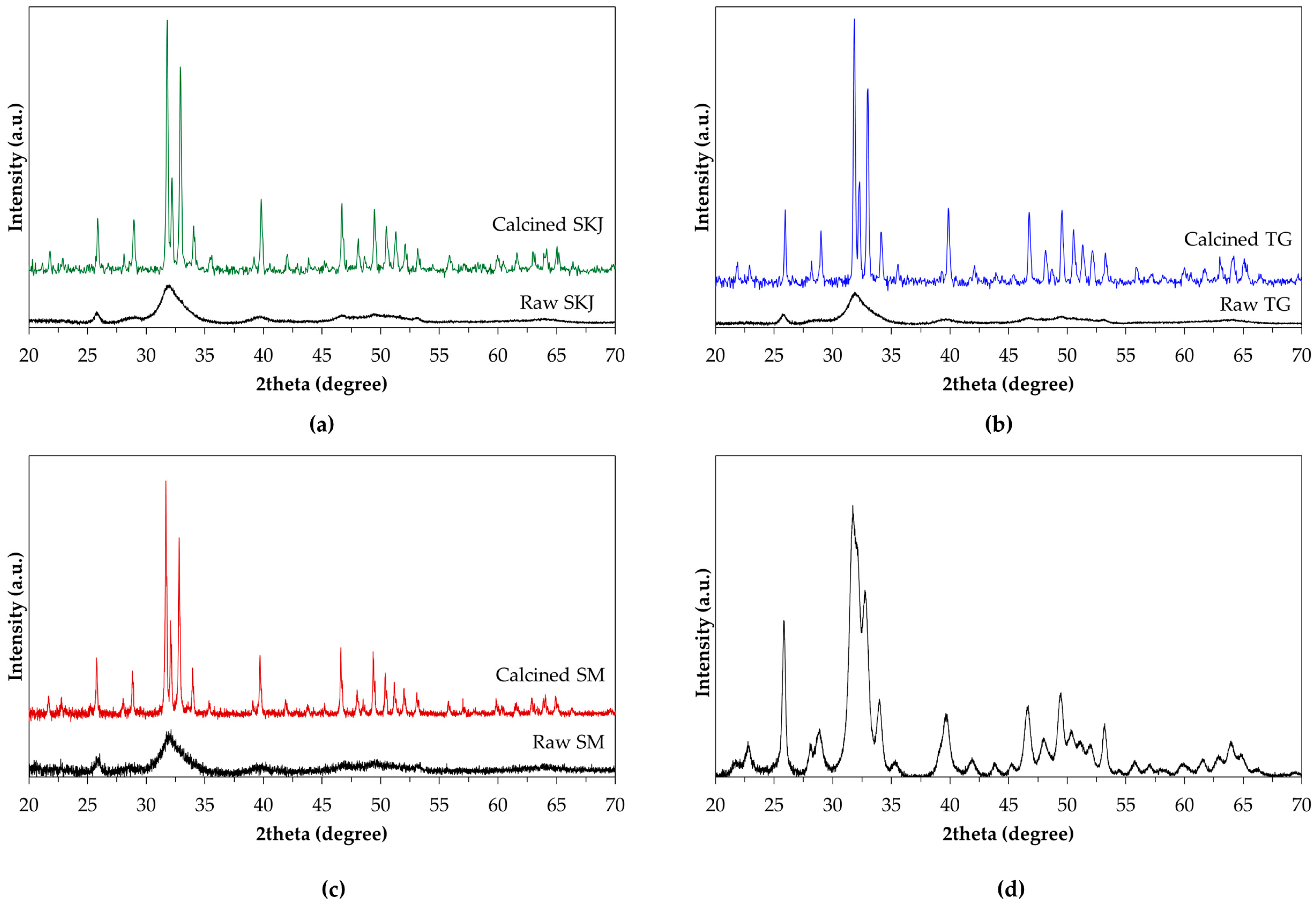
The crystalline phases of hydroxyapatite derived from skipjack (SKJ), tongol (TG), and salmon (SM) fish bones were examined by XRD (Figure 1), together with commercial HAp (C-HAp) as a reference. The raw bone powders exhibited broad, low intensity peaks at 2θ = 25.9°, 31.7–34.1°, 39.8°, 46.7°, and 49.5°, the characteristic of a poorly crystalline or nanocrystalline calcium phosphate phase in bone matrix. After calcination at 900 °C for 5 h, the diffraction patterns of all samples displayed sharp, well-defined reflections, corresponding to hexagonal HAp (JCPDS No. 09-0432), confirming the predominant crystalline phase. Although no secondary reflections were detected, the slight decrease in Ca/P ratio observed by XRF after calcination (Section 3.1.1) suggested the minor formation of β-TCP, as discussed previously. This β-TCP fraction was expected to be below the detection limit of laboratory XRD, accounting for the absence of distinct β-TCP peaks in the diffractograms.
Figure 1.
XRD patterns of hydroxyapatite powders derived from (a) skipjack tuna (SKJ), (b) tongol tuna (TG), (c) salmon bone (SM) powders before and after calcination at 900 °C and (d) commercial hydroxyapatite.
The commercial HAp exhibited a similar diffraction profile but with notably broader peaks, consistent with its significantly smaller crystallite size compared to those from fish bones. Calcination at 900 °C produced nearly identical diffraction patterns for all three fish bone powders (SKJ, TG, and SM), with enhanced peak sharpness indicating consistent crystal growth across all species. Minor variations in relative intensity and peak positions were observed among the samples, likely associated with inherent compositional factors of the source material, such as the initial Ca/P ratio and trace element substitutions within each specific bone type. Crystallite sizes, calculated using the Halder-Wagner (H-W) method [39], were 61.9 nm for SKJ, 67.6 nm for TG, and 55.7 nm for SM, compared with 12.7 nm for the commercial reference (Table 3). These results confirmed the formation of highly crystalline HAp in all calcined fish bone samples, with species-specific differences in crystallite size, more likely arising from the unique mineral compositions and trace element profiles inherent to each biogenic source. To further visualize the morphological evolution associated with phase development, SEM analysis was conducted to examine the surface microstructure and particle characteristics of the calcined HAp powders.
Table 3.
Crystallographic parameters of calcined fish bone-derived hydroxyapatite (SKJ-HAp, TG-HAp, SM-HAp) and commercial HAp.
3.1.3. Microstructure (SEM)
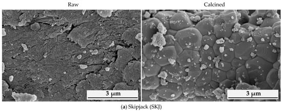
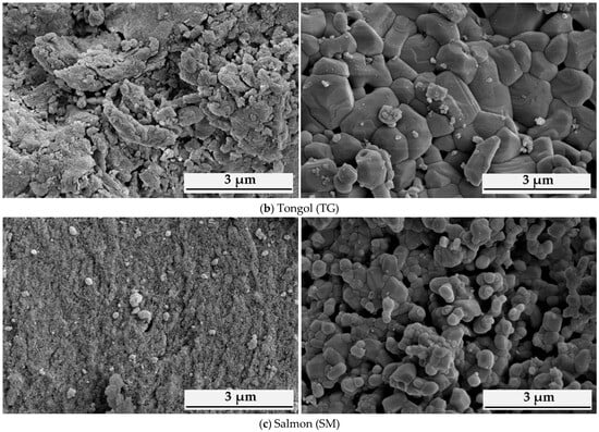
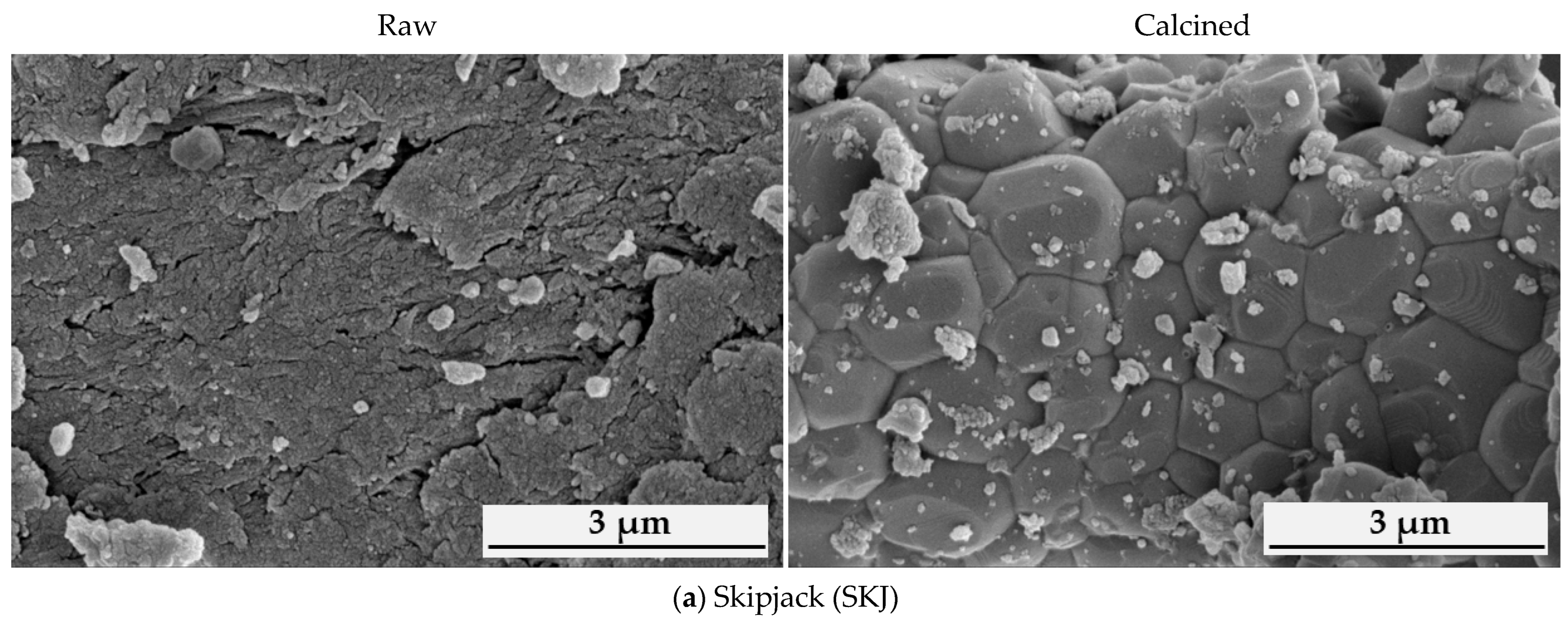
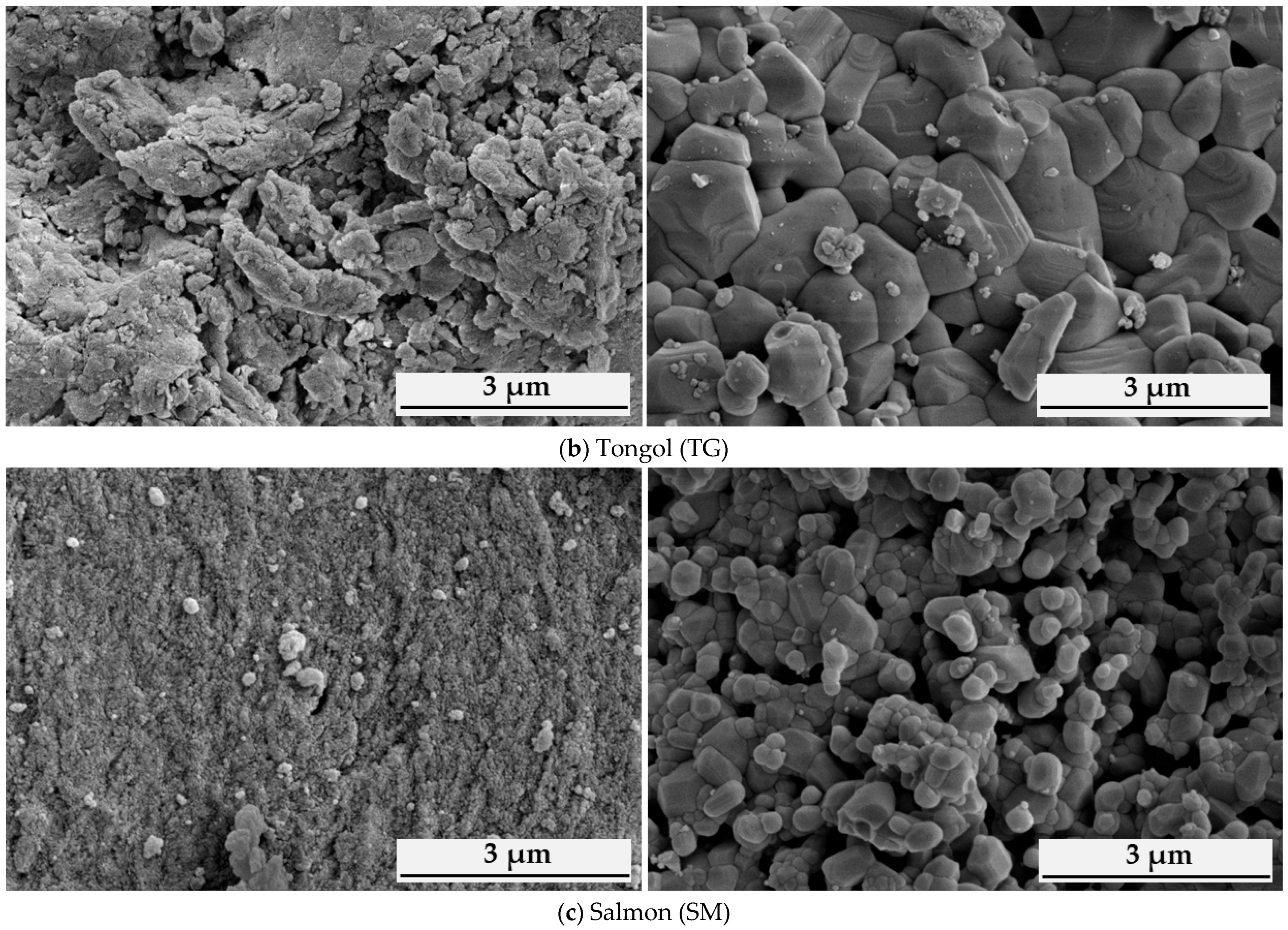
SEM images of hydroxyapatite powders derived from skipjack (SKJ), tongol (TG), and salmon (SM) fish bones are shown in Figure 2 (see also Figure S2 in Supplementary Materials). The raw powders (left panels), which had already undergone alkaline treatment, consisted of fine particles that were highly agglomerated, forming irregular clusters with rough and porous surfaces. While primary particles were visible, their boundaries remained indistinct due to strong aggregation, likely driven by high surface energy and residual organic components. Consequently, quantitative particle size analysis was deemed inappropriate for the raw samples owing to this significant degree of clustering.
Figure 2.
SEM micrographs of fish bone derived-hydroxyapatite powders (SKJ, TG, SM) before (Left) and after (Right) calcination at 900 °C (25,000× magnification, 5 kV, scale bar = 3 μm). Original images are provided in Figure S2.
After calcination at 900 °C (Figure 2, right panels), the powders exhibited markedly clearer particle boundaries and smoother surfaces, consistent with the complete removal of organic matter. The calcined samples contained compact mineral particles with species-dependent sizes and aggregation levels. Salmon derived HAp (SM-HAp) produced the smallest particles (0.43 ± 0.14 µm), whereas skipjack (SKJ-HAp) and tongol (TG-HAp) samples yielded significantly larger particles (1.10 ± 0.34 µm and 0.93 ± 0.26 µm, respectively). In all cases, clusters of fine particles attached to larger grains were observed, suggesting thermal coalescence during the heating process.
The resulting morphological changes correlated with the increased crystallite size observed in XRD data (Section 3.1.2) and the consolidation of HAp as the dominant crystalline phase. Species-dependent variations in particle size might be attributed to differences in initial mineral composition and trace element profiles [54,55], which influenced grain growth kinetics during calcination. Such size-dependent characteristics appeared to be relevant for cosmetic formulations, as finer particles potentially disperse more uniformly, which might contribute to enhanced film homogeneity and dispersion stability within the system. Conversely, larger particles could possibly impart different tactile properties or influence the resulting UV-shielding performance and rheology of the final sunscreen formulation.
3.2. Synergistic UV-Shielding Effects and Booster Performance of Fish Bone-Derived HAp
3.2.1. Physicochemical Properties of Sunscreen Formulations Containing 10% HAp
The physicochemical properties of oil-in-water (O/W) sunscreen formulations containing 10% hydroxyapatite derived from skipjack (SKJ), tongol (TG), and salmon (SM) bones were evaluated and compared with formulations incorporating commercial HAp, TiO2, and Tinosorb® S (Table 4). In the initial state, all samples appeared visually uniform and homogeneous, without visible phase separation.
Table 4.
Physicochemical properties of sunscreen formulations containing 10% HAp and reference UV filters.
The pH values of all formulations remained within the acceptable range for topical applications. Fish bone-derived HAp formulations exhibited mildly alkaline values (8.1–8.4), whereas commercial HAp, TiO2, and TS formulations remained near neutral (7.0–7.2), similar to the base cream (6.4 ± 0.1). This alkalinity was likely linked to the presence of residual calcium-rich phases within the fish bone HAp. Upon contact with the aqueous phase of the emulsion, these phases potentially reacted to form Ca(OH)2, a phenomenon consistent with the distinct Ca/P ratios (>1.67) discussed in Section 3.1.
Colorimetric analysis revealed that the base cream possessed high brightness (L* = 95.66) and a Whiteness Index (WI) of 95.48. Formulations containing SKJ-HAp and commercial HAp showed the excellent visual compatibility with the base cream, exhibiting minimal color deviations (ΔE* = 2.87 and 2.13, respectively). In contrast, TG-HAp introduced a slight yellowish tint (b* = 1.91), while SM-HAp shifted toward a subtle green–blue hue (a* = −4.51, b* = −1.90), resulting in a slightly higher ΔE* (5.19). The most pronounced deviation was observed in the TS formulation (ΔE* = 18.39), which exhibited a distinct yellow–green coloration. Notably, the TiO2 formulation achieved the highest WI (97.05); however, such extreme whiteness is often associated with an undesirable white cast on the skin. Conversely, the HAp-based formulations maintained more moderate WI values (≈89–95), offering a superior optical profile and ensuring a more natural-looking formulation finish.
The rheological behavior of the emulsions was significantly influenced by the particle properties of each HAp source. The base cream exhibited a baseline viscosity of 988 ± 45 cPs, which unexpectedly decreased in the SKJ- and TG-HAp formulations (≈620 cPs). This reduction suggested that the coarser particles of these fish bone-derived HAps might have disrupted or interfered with the formation of the internal emulsion network [56,57]. Conversely, SM-HAp and commercial HAp formulations showed a dramatic increase in viscosity, reaching 7094 and 12,900 cPs, respectively. This substantial thickening was therefore attributed to the finer particle sizes and higher specific surface area [56,58], which promoted the extensive particle–matrix interactions and the development of a dense, rigid 3D structural network [57,59]. Despite the homogeneity and phase stability during the initial state, our preliminary observations revealed that formulations with a high HAp loading (10%) exhibited visible phase separation during accelerated stability testing (50 °C for 30 days, assessed by visual inspection). Although both the 10% HAp and the 5% HAp + 5% TS formulations exhibited similar rheological profiles with high viscosity, the latter demonstrated superior structural integrity and physical stability [57]. Therefore, the HAp concentration was optimized at 5% in combination with 5% TS to ensure long-term formulation stability with potential synergistic UV-protection effects without compromising cosmetic esthetics [16].
3.2.2. Physicochemical Properties of Sunscreen Formulations Containing 5% HAp and 5% Tinosorb® S
Following the optimization results from the preliminary stability testing, O/W emulsions combining 5% HAp (from either fish bone or commercial sources) with 5% TS were prepared to evaluate their synergistic performance (Table 5). In their initial state, all hybrid formulations appeared smooth and homogeneous. However, they exhibited a more pronounced yellowish hue compared to the 10% HAp series, primarily due to the inherent yellow tint of the TS organic filter.
Table 5.
Physicochemical properties of sunscreen formulations containing 5% Tinosorb® S combined with physical UV filters in (TS).
Colorimetric analysis indicated that HAp-TS hybrid formulations exhibited green–yellow shades, with L* values ranging from 91.65 to 94.22, a* values from −5.02 to −3.34, and b* values from 8.33 to 10.91 (Table 5). Among the fish bone samples, the SM-HAp formulation showed the highest whiteness index (WI = 88.07). In contrast, the TiO2-TS formulation exhibited the highest L* value (95.98) and whiteness index (WI = 88.97), consistent with the earlier observation that TiO2 tended to produce a more visible white cast. Importantly, the total color difference (ΔE*) of HAp containing formulations (8.93–11.01) was substantially lower than that of the 5% TS control (ΔE* = 14.31). This suggested that the inclusion of HAp helped modulate the intense coloration of the organic filter, thereby improving the overall visual compatibility with the base formulation.
The pH values of HAp-TS creams ranged from 7.0 to 8.1, which was more neutralized compared to the 10% HAp formulations and remained within the acceptable range for topical applications. The fish bone-derived HAp formulations exhibited values of 7.5 ± 0.1 for SM-HAp, 7.8 ± 0.2 for SKJ-HAp, and 8.1 ± 0.1 for TG-HAp. The reduction in pH relative to the 10% loading series was likely due to the lower concentration of residual calcium-rich phases, bringing the formulations closer to the pH of the commercial HAp and TS controls (pH ≈ 7.0–7.1).
Viscosity remained strongly influenced by the HAp source, though the values were generally lower than the 10% loading series, contributing to better spreadability. The SKJ- and TG-HAp formulations exhibited lower viscosities (499–815 cPs), whereas the SM-HAp formulation maintained a more structured profile at 3377 ± 135 cPs. This remained consistent with the finer particle size and higher surface area of SM-HAp, which promoted stronger particle–emulsion interactions. Commercial HAp continued to produce the highest viscosity (16,894 cPs), whereas the TiO2-TS system showed a moderate profile (2370 ± 95 cPs). Overall, these optimized formulations containing 5% HAp and 5% TS achieved a superior balance of physical stability, pH compatibility, and cosmetic esthetics, providing a robust basis for subsequent UV-protection evaluations.
3.2.3. UV Protection Efficacy: SPF, UVAPF, and PA Values
Commercial and fish bone-derived HAp powders were evaluated for their UV protection performance. As summarized in Table 6, the formulations containing 10% HAp alone, regardless of source, exhibited negligible UV protection (SPF = 1.03–1.13), failing to meet the Thai Industrial Standard requirement (TIS 2804–2017: SPF > 15). Similarly, the 5% TS alone yielded an SPF of 10.54 ± 0.48, confirming that neither component at these concentrations provided sufficient efficacy.
Table 6.
SPF and UVAPF performance of sunscreen formulations containing HAp and Tinosorb® S.
Nevertheless, the combination of 5% HAp and 5% TS resulted in a pronounced synergistic enhancement. The SPF values for SKJ- and TG-HAp hybrid systems reached approximately 29–30 (~180% increase relative to 5% TS). Notably, the SM-HAp system exhibited a higher SPF of 32.19 ± 1.46 (205% enhancement), slightly less than the commercial HAp-TS system (SPF 35.21 ± 1.35). UVA protection followed a similar trend, with all HAp-TS hybrids achieving PA+++ or higher.
These results suggested that fish bone-derived HAp acts primarily as a synergistic booster rather than an intrinsic UV absorber. This synergistic effect was proposed to involve two plausible contributions, including optical and potential photochemical boosting as illustrated in the conceptual model provided in Figure S3. As particulate scattering centers within the emulsion matrix, the SM-HAp particles could possibly provide optical boosting by inducing light scattering and increasing the optical path length of incident UV photons, thereby improving the absorption efficiency of Tinosorb® S [60,61]. Concurrently, it was hypothesized that a contribution might arise from surface-mediated interactions. The high density of surface hydroxyl groups and exposed calcium sites on biogenic HAp could potentially interact with the electronic energy states of Tinosorb® S molecules. This synergistic enhancement was experimentally supported by the UV absorbance spectra (see Section 3.2.4), which demonstrated a significant increase in absorption intensity for the SM-HAp/TS system compared to the organic filter alone. These hypothesized interfacial interactions could possibly facilitate improved molecular distribution, reduce filter aggregation, and promote more homogeneous dispersion, contributing to enhanced performance beyond simple additive behavior. The proposed dual-action model was consistent with previous reports on fish-derived calcium phosphates in hybrid systems [25]. Consequently, the influence of particle size refinement was further evaluated to maximize these potential scattering and interfacial contributions [60,62,63].
3.2.4. Influence of HAp Particle Size and on Sunscreen Performance
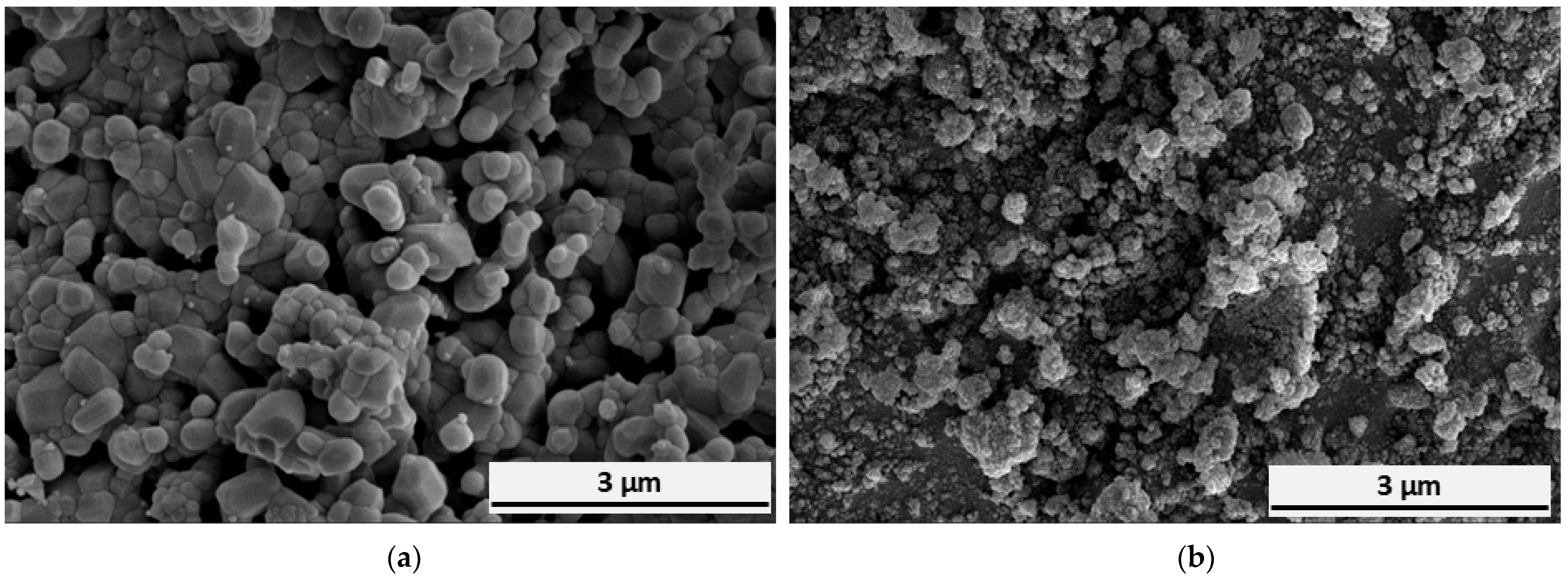
To examine the influence of particle size reduction in enhancing photoprotective performance, salmon-derived HAp was subjected to planetary ball milling. This mechanical process successfully reduced the mean particle size from irregular submicron aggregates (430 ± 140 nm) to a nanoscale distribution of 117.39 ± 61 nm (Figure 3a,b; original micrographs in Figure S4). The reduction in size significantly increased the specific surface area, thereby enabling superior dispersion and more effective interfacial contact within the sunscreen matrix. To provide a comprehensive comparison, commercial HAp and TiO2 were included as reference physical filters (Table 7).
Figure 3.
SEM images of calcined (900 °C) salmon-derived hydroxyapatite (SM-HAp) (a) before and (b) after planetary ball milling (25,000× magnification, 5 kV, scale bar = 3 μm). Original micrographs are provided in Figure S4.
Table 7.
Physicochemical properties and UV protection performance of sunscreen formulations containing 5% Tinosorb® S and physical UV filters.
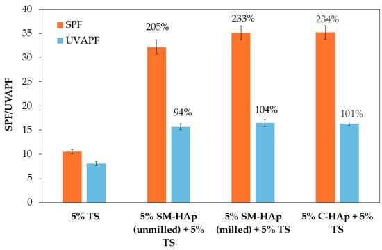
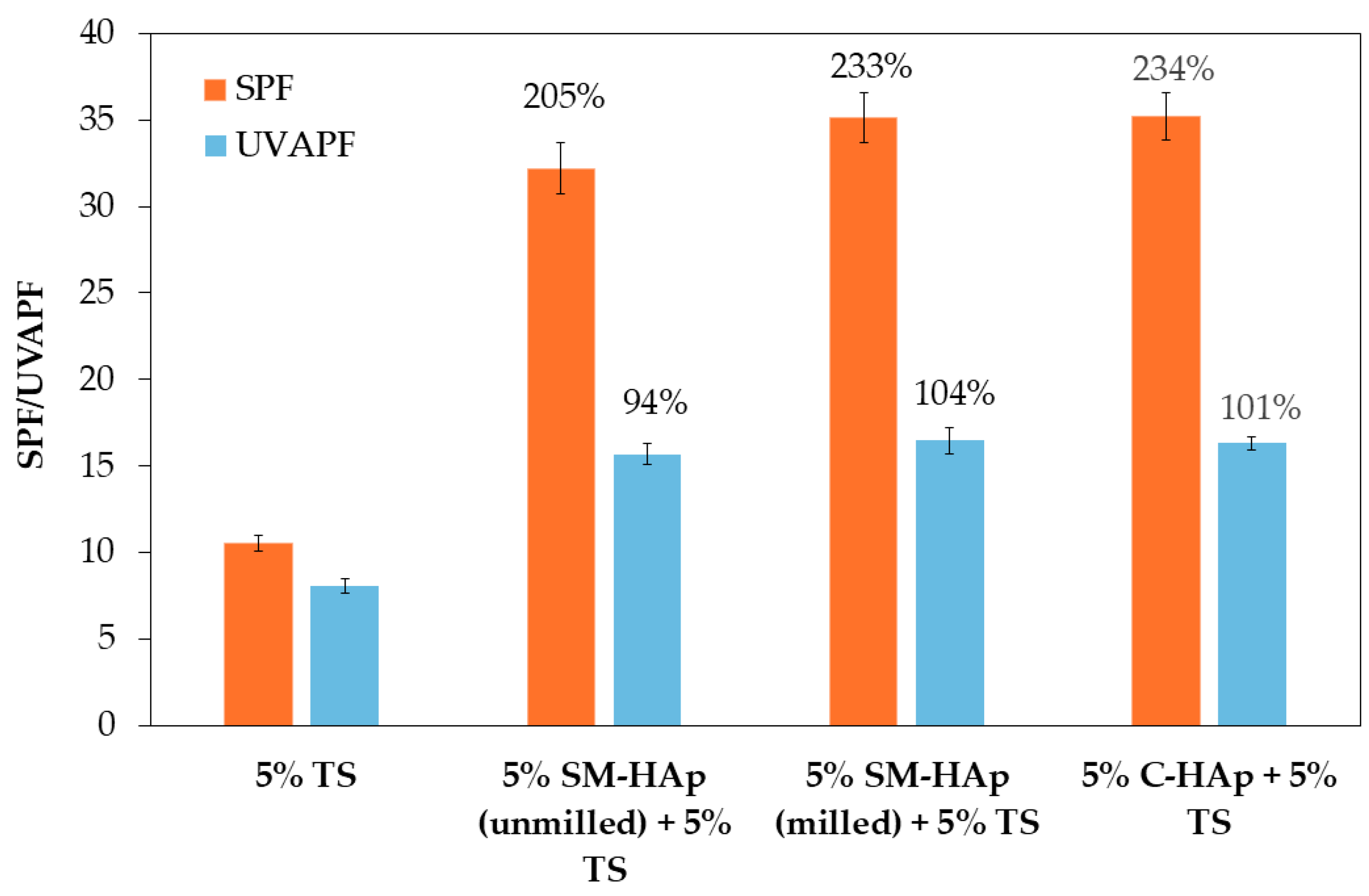
The reduction in SM-HAp particle size resulted in a statistically discernible enhancement in UV protection performance (p < 0.05). As shown in Figure 4 and Table 7, the SPF of the SM-HAp hybrid system increased from 32.19 ± 1.46 to 35.15 ± 1.44 following particle refinement. While this numerical increase was modest, it represented a meaningful functional transition. The corresponding increase in UVAPF from 15.68 ± 0.60 to 16.49 ± 0.75 was sufficient to elevate the protection grade from PA+++ to PA++++. In all formulations, the critical wavelength (λc) exceeded 371 nm, confirming robust broad-spectrum UVA coverage. Interestingly, the milled SM-HAp system achieved SPF and UVAPF values comparable to the commercial HAp-TS system, despite the commercial HAp possessing a smaller primary particle size. This suggested that the 117 nm SM-HAp particles might reach an optimal size range for scattering UV radiation within this specific emulsion system [56,60,61].
Figure 4.
SPF and UVAPF performance 5% SM-HAp (unmilled and milled) and commercial HAp combined with 5% Tinosorb® S. Percentage enhancement relative to 5% TS formulation is indicated above each bar.
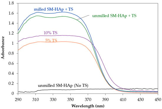
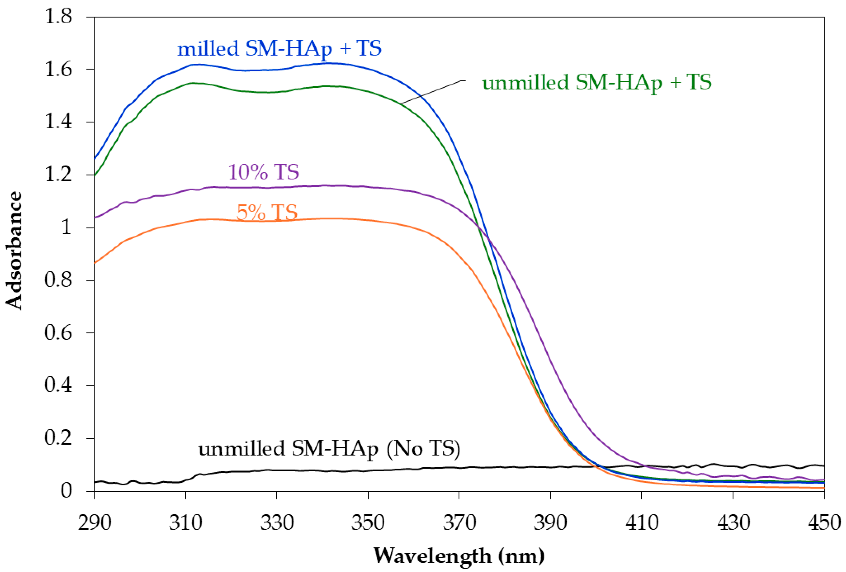
To further examine the effect of particle size refinement on UV attenuation behavior, UV absorbance spectra were analyzed (Figure 5). The milled SM-HAp/TS formulation exhibited higher absorbance intensity across the 290–400 nm range compared with the unmilled SM-HAp/TS system, covering both the UVB and UVA regions. Since the unmilled SM-HAp without Tinosorb® S showed negligible absorbance, it was further confirmed that HAp did not function as an intrinsic UV absorber. The enhanced absorbance observed following particle size reduction was consistent with the higher SPF and UVAPF values reported in Table 7 and with previous reports demonstrating particle size dependent enhancement of UV protection in inorganic UV filter systems [53].
Figure 5.
UV absorbance spectra of salmon-derived HAp formulations, Tinosorb® S, and their combination.
Furthermore, particle size reduction significantly increased the formulation’s apparent viscosity (Table 7), fostering stronger particle–matrix interactions and enhancing the emulsion’s structural integrity. This rheological shift promoted the formation of a more coherent and homogeneous film on the skin, ensuring a uniform distribution of synergistic boosters [57]. While TiO2 achieved the highest absolute SPF (64.29), its use is increasingly limited by environmental concerns regarding marine ecosystem toxicity and coral bleaching [16,62]. In contrast, milled SM-HAp offers a sustainable, bio-renewable alternative that achieves significant PA++++ protection and high SPF with superior cosmetic esthetics.
Overall, these results suggested that particle-size refinement optimized the potential roles of SM-HAp as a structural stabilizer and an optical booster, providing a bio-renewable mineral alternative with efficacy comparable to a traditional mineral counterpart. While these in vitro results indicated a significant synergistic enhancement, it is important to acknowledge the limitations of this method compared to in vivo testing. In vitro measurements, while excellent for screening and comparative studies, may not fully account for complex interactions with human skin surface, such as absorption, perspiration, or the topography of PMMA substrates. Furthermore, for regulatory approval and commercial SPF labeling, in vivo testing remains the global standard. The current in vitro data serve as a robust preliminary validation of the mineral–organic synergy, providing a foundation for future clinical assessments. While standard evaluation of sunscreens is performed in vivo, preliminary studies for new cosmetic ingredients are typically conducted using accredited in vitro testing to meet practical and ethical requirements. Following the performance evaluation, the optimized formulation of milled SM-HAp and Tinosorb® S was subsequently evaluated for its microbiological and chemical safety, as well as its physicochemical suitability for topical application (Section 3.3).
3.3. Safety Assessment and Physicochemical Suitability
This section evaluates the safety, stability, and physicochemical suitability of sunscreen formulations incorporating fish bone-derived HAp. Microbiological quality, heavy metal content, and accelerated stability were assessed to verify the compliance with cosmetic regulations and to support the suitability of fish bone HAp for topical sunscreen applications.
3.3.1. Microbial and Heavy Metal Safety Evaluation
Microbiological testing was performed in accordance with the Ministry of Public Health Notification (2016) [45], which specifies indicator microorganisms and permissible limits for cosmetic products in Thailand. The selected species, Staphylococcus aureus, Pseudomonas aeruginosa, and total yeasts and molds, represent common contaminants of topical formulations and are required for regulatory compliance. The sunscreen formulation containing 5% SM-HAp and 5% TS demonstrated satisfactory microbiological quality. S. aureus was <10 CFU/g, P. aeruginosa was not detected, and yeast and mold counts were below 10 CFU/g. Those counts were well within the permitted limits for cosmetic products (Table 8).
Table 8.
Microbial contamination analysis of 5% SM-HAp and 5% Tinosorb® S formulation.
Heavy metal analysis further confirmed chemical safety of the formulations. Arsenic (As), cadmium (Cd), and lead (Pb) were quantified by ICP-OES, while mercury (Hg) was measured using mercury analyzer. All measured concentrations were below corresponding detection limits of the analytical procedure (reported as <0.91 mg/kg), and Hg was below 0.00050 mg/kg (Table 9). These results verified that incorporation of salmon derived HAp did not introduce hazardous metal contamination into the sunscreen system.
Table 9.
Heavy metal content of 5% SM-HAp and 5% Tinosorb® S formulation compared with regulatory limits.
Accelerated stability testing (50 °C for 30 days) further demonstrated that the formulation maintained stable physicochemical properties throughout the storage period. The pH remained at 7.2 ± 0.1, and viscosity was 13,200 ± 400 cPs after storage, with no statistically significant deviation from the initial values (p > 0.05). No phase separation or sedimentation was observed, supporting its suitability for topical use. While particle size reduction produced HAp in the nanoscale range (mean size ≈ 117 nm), its role as a synergistic booster, rather than a primary filter, mitigated certain safety concerns. According to current safety frameworks, nanoscale materials embedded in complex emulsion systems typically exhibit limited dermal penetration due to their tendency to form larger aggregates within the matrix [65]. Together with its inherent biocompatibility as a calcium phosphate mineral, these findings support the safety of biogenic HAp for human application.
Nevertheless, considering the nanoscale nature of the refined particles, further studies focusing on potential ecotoxicity and long-term impacts on marine ecosystems are warranted. Such investigations would provide a more comprehensive understanding of its environmental safety profile, ensuring that biogenic HAp remains a truly sustainable and marine-friendly alternative to traditional mineral filters.
3.3.2. Nanoscale Safety and Formulation Integrity
Although mechanical processing resulted in HAp particles within the nanoscale range (mean size ~117 nm), their application as a synergistic booster, rather than a primary UV filter, was associated with a favorable safety profile. According to established safety frameworks, nanomaterials incorporated into complex emulsion systems typically exhibit negligible dermal penetration. This is primarily attributed to the tendency of nanoparticles to form larger aggregates within the emulsion matrix, thereby limiting their transdermal mobility [66]. When combined with the inherent biocompatibility of biogenic calcium phosphate, these factors substantiate the safety of salmon-derived HAp for topical applications. The enhanced performance of the SM-HAp system was fundamentally consistent with its balanced rheological profile and reduced whitening tendency (WI) discussed in previous sections. Such findings align with the existing literature indicating that inorganic particle size, morphology, and dispersion states strongly influence the rheological characteristics through complex vehicle–particle interactions [66,67]. Consequently, the optimized interaction between the biogenic HAp and the emulsion vehicle suggested that this salmon-derived material could potentially be integrated into hybrid sunscreens while maintaining the structural integrity required for modern cosmetic systems.
4. Conclusions
This study suggested that hydroxyapatite powders derived from skipjack, tongol, and salmon fish bones could be successfully converted into high-purity HAp suitable for cosmetic sunscreen formulation. Calcination at 900 °C produced HAp as the dominant phase, while species-dependent variations in Ca/P ratio, trace element compositions, and particle morphologies appeared to influence dispersion behavior and subsequent UV-protective performance. When incorporated into hybrid sunscreen formulations, 5% fish bone-derived HAp acted as a synergistic booster, markedly enhancing the efficacy of Tinosorb® S. This synergy was evidenced by a substantial increase in SPF, UVAPF, and broad-spectrum coverage, likely driven by a combination of optical scattering and potential surface-mediated molecular stabilization. The particle size refinement of salmon derived HAp further amplified this effect, yielding UV protection performance comparable to commercial HAp benchmarks. These improvements highlight the potential role of particle engineering in promoting film uniformity, and optimizing the electronic environment for the organic filter, rather than intrinsic UV absorption by the HAp additive.
All optimized formulations complied with microbiological and heavy metal regulatory requirements. Notably, the SM-HAp/TS system demonstrated high physicochemical suitability, offering effective UV enhancement with the potential to reduce white cast compared to TiO2-based systems. While TiO2 provided higher absolute SPF, its use is increasingly limited by ecological concerns. Collectively, these findings establish fish bone-derived HAp as a viable, sustainable functional booster for the development of modern hybrid sunscreens. While the present study demonstrates physicochemical safety and in vitro photoprotective enhancement, it is noted that clinical in vivo testing and direct marine ecotoxicity assessments should be addressed in future investigations to meet full regulatory and environmental certification standards.
Future work should extend these findings to more complex commercial-type formulations containing antioxidants, fragrances, and specialized stabilizers to assess their influence on long-term dispersion stability and film-forming dynamics. Furthermore, wavelength-resolved optical modeling, long-term photostability assessments, and in-depth evaluations of impacts on marine ecosystems are essential. Such investigations will be critical to fully validate the real-world performance and ecological safety of biogenic HAp as a sustainable alternative in the global sunscreen market.
5. Patents
As a result of the work described in this manuscript, Thai patent application No. 2501006021, entitled “Sunscreen Product with Synergistic Enhancement of Ultraviolet Protection Efficiency Using Hydroxyapatite Microparticles”, was filed on 10 September 2025.
Supplementary Materials
The following supporting information can be downloaded at: https://www.mdpi.com/article/10.3390/cosmetics13020071/s1, Figure S1: SEM micrograph of commercial hydroxyapatite (C-HAp); Figure S2: Original high-resolution SEM micrographs of fish bone-derived HAp powders before and after calcination; Figure S3: Conceptual schematic of the synergistic SPF-boosting mechanism; Figure S4: Original high-resolution SEM micrograph of milled salmon-derived HAp.
Author Contributions
Conceptualization, P.S. and S.B.; methodology, P.S. and T.A.; software, P.S.; validation, P.S. and S.L.-u.; formal analysis, P.S. and S.L.-u.; investigation, P.S. and S.L.-u.; resources, S.B. and T.P.; data curation, P.S. and S.L.-u.; writing—original draft preparation, P.S. and S.L.-u.; writing—review and editing, P.S., S.B., T.A. and T.P.; visualization, P.S.; supervision, S.B. and T.P.; project administration, P.S.; funding acquisition, P.S., T.P. and S.B. All authors have read and agreed to the published version of the manuscript.
Funding
This research was sponsored by Prince of Songkla University, grant number AGR6302074S and by the Coordinating Center for Thai Government Science and Technology Scholarship Students (CSTS), National Science and Technology Development Agency (NSTDA), grant number AGR6205084S.
Institutional Review Board Statement
Not applicable.
Informed Consent Statement
Not applicable.
Data Availability Statement
The original contributions presented in this study are included in the article; further inquiries can be directed to the corresponding author.
Acknowledgments
The authors gratefully acknowledge the research facility supported by the Center of Excellence in Bio-Based Materials and Packaging Innovation and the International Center of Excellence in Seafood Science and Innovation, Faculty of Agro-Industry, Prince of Songkla University, Hat Yai, Thailand. During the preparation of this manuscript, the authors used AI tools (ChatGPT 5.2 and Gemini 1.5 Flash) to improve language clarity and readability. The authors have reviewed and edited the generated content and take full responsibility for the integrity, accuracy, and originality of the work.
Conflicts of Interest
The authors declare no conflicts of interest.
References
- Hart, P.H.; Norval, M. More than effects in skin: Ultraviolet radiation-induced changes in immune cells in human blood. Front. Immunol. 2021, 12, 694086. [Google Scholar] [CrossRef] [Scilit] [PubMed]
- Gromkowska-Kępka, K.J.; Puścion-Jakubik, A.; Markiewicz-Żukowska, R.; Socha, K. The impact of ultraviolet radiation on skin photoaging—Review of In Vitro Studies. J. Cosmet. Dermatol. 2021, 20, 3427–3431. [Google Scholar] [CrossRef] [Scilit]
- Tang, X.; Yang, T.; Yu, D.; Xiong, H.; Zhang, S. Current insights and future perspectives of ultraviolet radiation (UV) exposure: Friends and foes to the skin and beyond the skin. Environ. Int. 2024, 185, 108535. [Google Scholar] [CrossRef] [Scilit]
- Mohania, D.; Chandel, S.; Kumar, P.; Verma, V.; Digvijay, K.; Tripathi, D.; Choudhury, K.; Mitten, S.K.; Shah, D. Ultraviolet radiations: Skin defense–damage mechanism. In Ultraviolet Light in Human Health, Diseases and Environment; Ahmad, S., Ed.; Advances in Experimental Medicine and Biology; Springer: Cham, Switzerland, 2017; Volume 996, pp. 71–87. [Google Scholar]
- U.S. Environmental Protection Agency. Health Effects of UV Radiation. EPA Sun Safety. 2025. Available online: https://www.epa.gov/sunsafety/health-effects-uv-radiation (accessed on 7 September 2025).
- Wang, Z.; Wang, X.; Shi, Y.; Wu, S.; Ding, Y.; Yao, G.; Chen, J. Advancements in elucidating the pathogenesis of actinic keratosis: Present state and future prospects. Front. Med. 2024, 11, 1330491. [Google Scholar] [CrossRef] [Scilit]
- Schneider, S.L.; Lim, H.W. A review of inorganic UV filters zinc oxide and titanium dioxide. Photodermatol. Photoimmunol. Photomed. 2019, 35, 442–446. [Google Scholar] [CrossRef] [Scilit]
- Egambaram, O.P.; Kesavan Pillai, S.; Sinha Ray, S. Materials science challenges in skin UV protection: A review. Photochem. Photobiol. 2020, 96, 779–797. [Google Scholar] [CrossRef] [Scilit] [PubMed]
- Santander Ballestín, S.; Luesma Bartolomé, M.J. Toxicity of different chemical components in sun cream filters and their impact on human health: A Review. Appl. Sci. 2023, 13, 712. [Google Scholar] [CrossRef] [Scilit]
- Pniewska, A.; Kalinowska-Lis, U. A survey of UV filters used in sunscreen cosmetics. Appl. Sci. 2024, 14, 3302. [Google Scholar] [CrossRef] [Scilit]
- Chavda, V.P.; Acharya, D.; Hala, V.; Daware, S.; Vora, L.K. Sunscreens: A comprehensive review with the application of nanotechnology. J. Drug Deliv. Sci. Technol. 2023, 86, 104720. [Google Scholar] [CrossRef] [Scilit]
- Nitulescu, G.; Lupuliasa, D.; Adam-Dima, I.; Nitulescu, G.M. Ultraviolet filters for cosmetic applications. Cosmetics 2023, 10, 101. [Google Scholar] [CrossRef] [Scilit]
- Hodge, A.A.; Hopkins, F.E.; Saha, M.; Jha, A.N. Ecotoxicological effects of sunscreen derived organic and inorganic UV filters on marine organisms: A critical review. Mar. Pollut. Bull. 2025, 213, 117627. [Google Scholar] [CrossRef] [Scilit] [PubMed]
- Sadeghifar, H.; Ragauskas, A.J. Lignin as a UV light blocker—A review. Polymers 2020, 12, 1134. [Google Scholar] [CrossRef] [Scilit]
- Piccinino, D.; Capecchi, E.; Tomaino, E.; Gabellone, S.; Gigli, V.; Avitabile, D.; Saladino, R. Nano-structured lignin as green antioxidant and uv shielding ingredient for sunscreen applications. Antioxidants 2021, 10, 274. [Google Scholar] [CrossRef] [Scilit]
- Hayden, D.R.; Kibbelaar, H.V.M.; Imhof, A.; Velikov, K.P. Fully-biobased UV-absorbing nanoparticles from ethyl cellulose and zein for environmentally friendly photoprotection. RSC Adv. 2018, 8, 25104–25111. [Google Scholar] [CrossRef] [Scilit]
- Shabrina, A.M.; Azzahra, R.S.S.; Permata, I.N.; Dewi, H.P.; Safitri, R.A.; Maya, I.; Aulia, R.N.; Sriwidodo, S.; Mita, S.R.; Amalia, E.; et al. Potential of natural-based sun protection factor (SPF): A systematic review of curcumin as sunscreen. Cosmetics 2025, 12, 10. [Google Scholar] [CrossRef] [Scilit]
- Milutinov, J.; Pavlović, N.; Ćirin, D.; Atanacković Krstonošić, M.; Krstonošić, V. The potential of natural compounds in UV protection products. Molecules 2024, 29, 5409. [Google Scholar] [CrossRef] [Scilit]
- Wang, W.-H.; Liang, H.-T.; Yang-Wang, Y.-T.; Shih, C.-J. Synthesis of hierarchically mesoporous silica with encapsulated avobenzone as a UV protection filter. RSC Adv. 2020, 10, 15846–15852. [Google Scholar] [CrossRef] [Scilit] [PubMed]
- Fantini, R.; Argenziano, M.; Cavalli, R.; Arletti, R. Zeofilters for potentially innovative sunscreen products: Formulation, stability and spectroscopic studies. J. Photochem. Photobiol. A Chem. 2024, 452, 115585. [Google Scholar] [CrossRef] [Scilit]
- Chrétien, M.-N.; Heafey, E.; Scaiano, J.C. Reducing adverse effects from UV sunscreens by zeolite encapsulation: Comparison of oxybenzone in solution and in zeolites. Photochem. Photobiol. 2010, 86, 153–161. [Google Scholar] [CrossRef] [Scilit]
- Gupta, V.; Mohapatra, S.; Mishra, H.; Farooq, U.; Kumar, K.; Ansari, M.J.; Aldawsari, M.F.; Alalaiwe, A.S.; Mirza, M.A.; Iqbal, Z. Nanotechnology in cosmetics and cosmeceuticals—A review of latest advancements. Gels 2022, 8, 173. [Google Scholar] [CrossRef] [Scilit] [PubMed]
- Righi, S.; Prato, E.; Magnani, G.; Lama, V.; Biandolino, F.; Parlapiano, I.; Carella, F.; Iafisco, M.; Adamiano, A. Calcium phosphates from fish bones in sunscreen: An LCA and toxicity study of an emerging material for circular economy. Sci. Total Environ. 2023, 862, 160751. [Google Scholar] [CrossRef] [Scilit]
- Carella, F.; Degli Esposti, L.; Adamiano, A.; Iafisco, M. The use of calcium phosphates in cosmetics: State of the art and future perspectives. Materials 2021, 14, 6398. [Google Scholar] [CrossRef] [Scilit]
- Adamiano, A.; Carella, F.; Degli Esposti, L.; Piccirillo, C.; Iafisco, M. Calcium phosphates from fishery byproducts as a booster of the sun protection factor in sunscreens. ACS Biomater. Sci. Eng. 2022, 8, 4987–4995. [Google Scholar] [CrossRef] [Scilit]
- Piccirillo, C.; Rocha, C.; Tobaldi, D.M.; Pullar, R.C.; Labrincha, J.A.; Ferreira, M.O.; Castro, P.M.L.; Pintado, M.M.E. A hydroxyapatite–Fe2O3 based material of natural origin as an active sunscreen filter. J. Mater. Chem. B 2014, 2, 5999–6009. [Google Scholar] [CrossRef] [Scilit] [PubMed]
- Rigano, L.; Gazzaniga, G.; Rastrelli, G. Sunscreen Product Comprising Hydroxyapatite as Physical Filter. European Patent WO 2010/109400 A2, 24 September 2013. [Google Scholar]
- Engsuwan, J.; Kaowphong, S.; Girdthep, S.; Chumha, N. One-pot synthesis of silver–hydroxyapatite nanocomposites for sunscreens against high-energy visible light radiation. Mater. Lett. 2025, 398, 138973. [Google Scholar] [CrossRef] [Scilit]
- Iafisco, M.; Adamiano, A.; Piccirillo, C. Physical Sunscreen Comprising Hydroxyapatite or Modified Hydroxyapatite Obtained from Fisheries and Aquaculture Waste, Process for Its Production and Photoprotective Compositions Comprising It. European Patent WO 2020/193750 A1, 16 June 2022. [Google Scholar]
- Idowu, A.T.; Benjakul, S.; Sinthusamran, S.; Sae-Leaw, T.; Suzuki, N.; Kitani, Y.; Sookchoo, P. Effect of alkaline treatment on characteristics of bio-calcium and hydroxyapatite powders derived from salmon bone. Appl. Sci. 2020, 10, 4141. [Google Scholar] [CrossRef] [Scilit]
- Benjakul, S.; Mad-Ali, S.; Senphan, T.; Sookchoo, P. Biocalcium powder from precooked skipjack tuna bone: Production and its characteristics. J. Food Biochem. 2017, 41, e12412. [Google Scholar] [CrossRef] [Scilit]
- Benjakul, S.; Mad-Ali, S.; Senphan, T.; Sookchoo, P. Characteristics of biocalcium from pre-cooked skipjack tuna bone as affected by different treatments. Waste Biomass Valor. 2018, 9, 1369–1377. [Google Scholar] [CrossRef] [Scilit]
- Toppe, J.; Albrektsen, S.; Hope, B.; Aksnes, A. chemical composition, mineral content and amino acid and lipid profiles in bones from various fish species. Comp. Biochem. Physiol. B 2007, 146, 395–401. [Google Scholar] [CrossRef] [Scilit]
- Teixeira, M.A.C.; Piccirillo, C.; Tobaldi, D.M.; Pullar, R.C.; Labrincha, J.A.; Ferreira, M.O.; Castro, P.M.L.; Pintado, M.M.E. Effect of preparation and processing conditions on UV absorbing properties of hydroxyapatite–Fe2O3 sunscreen. Mater. Sci. Eng. C 2017, 71, 141–149. [Google Scholar] [CrossRef] [Scilit]
- Jesus, A.; Augusto, I.; Duarte, J.; Sousa, E.; Cidade, H.; Cruz, M.T.; Lobo, J.M.S.; Almeida, I.F. Recent trends on UV filters. Appl. Sci. 2022, 12, 12003. [Google Scholar] [CrossRef] [Scilit]
- Hennebelle, M.; Villeneuve, P.; Durand, E.; Lecomte, J.; Van Duynhoven, J.; Meynier, A.; Yesiltas, B.; Jacobsen, C.; Berton-Carabin, C. lipid oxidation in emulsions: New insights from the past two decades. Prog. Lipid Res. 2024, 94, 101275. [Google Scholar] [CrossRef] [Scilit]
- Springer, A.; Ziegler, H.; Bach, K. The Influence of antioxidant plant extracts on the oxidation of O/W emulsions. Cosmetics 2023, 10, 40. [Google Scholar] [CrossRef] [Scilit]
- Sullivan, M.; Gonzalez Obezo, C.; Lipsky, Z.; Panchal, A.; Jensen, J. Frontiers in topical photoprotection. Cosmetics 2025, 12, 96. [Google Scholar] [CrossRef] [Scilit]
- Halder, N.C.; Wagner, C.N.J. Separation of particle size and lattice strain in integral breadth measurements. Acta Crystallogr. 1966, 20, 312–313. [Google Scholar] [CrossRef] [Scilit]
- León, K.; Mery, D.; Pedreschi, F.; León, J. Color Measurement in Lab* Units from RGB Digital Images. Food Res. Int. 2006, 39, 1084–1091. [Google Scholar] [CrossRef] [Scilit]
- Muley, A.B.; Chaudhari, S.A.; Mulchandani, K.H.; Singhal, R.S. Extraction and characterization of chitosan from prawn shell waste and its conjugation with cutinase for enhanced thermo-stability. Int. J. Biol. Macromol. 2018, 111, 1047–1058. [Google Scholar] [CrossRef] [Scilit]
- ISO 24444:2010; Cosmetics—Sun Protection Test Methods—In Vivo Determination of the Sun Protection Factor (SPF). ISO: Geneva, Switzerland, 2010.
- ISO 24443:2012; Determination of Sunscreen UVA Photoprotection In Vitro. ISO: Geneva, Switzerland, 2012.
- Japan Cosmetic Industry Association (JCIA). Measurement Standard for UVA Protection Efficacy of Sunscreen Products; JCIA: Tokyo, Japan, 2013. [Google Scholar]
- Thai Food and Drug Administration. Notification of the Ministry of Public Health (No. 283) B.E. 2559 (2016) Re: Characteristics of Cosmetics Prohibited for Manufacture, Import or Sale. Royal Thai Government Gazette; Thai Food and Drug Administration: Bangkok, Thailand, 2016. [Google Scholar]
- AOAC International. Official Methods of Analysis of AOAC International; AOAC: Gaithersburg, MD, USA, 2019. [Google Scholar]
- Cestari, F.; Agostinacchio, F.; Galotta, A.; Chemello, G.; Motta, A.; Sglavo, V. Nano-hydroxyapatite derived from biogenic and bioinspired calcium carbonates: Synthesis and in vitro bioactivity. Nanomaterials 2021, 11, 264. [Google Scholar] [CrossRef] [Scilit]
- Forero-Sossa, P.A.; Salazar-Martínez, J.D.; Giraldo-Betancur, A.L.; Segura-Giraldo, B.; Restrepo-Parra, E. Temperature effect in physicochemical and bioactive behavior of biogenic hydroxyapatite obtained from porcine bones. Sci. Rep. 2021, 11, 11069. [Google Scholar] [CrossRef] [Scilit]
- Kubiak-Mihkelsoo, Z.; Kostrzębska, A.; Błaszczyszyn, A.; Pitułaj, A.; Dominiak, M.; Gedrange, T.; Nawrot-Hadzik, I.; Matys, J.; Hadzik, J. Ionic doping of hydroxyapatite for bone regeneration: Advances in structure and properties over two decades—A narrative review. Appl. Sci. 2025, 15, 1108. [Google Scholar] [CrossRef] [Scilit]
- Seredin, P.V.; Goloshchapov, D.L.; Buylov, N.S.; Kashkarov, V.M.; Emelyanova, A.A.; Eremeev, K.A.; Ippolitov, Y.A. Bioengineering of hybrid nanomaterials based on non-stoichiometric nanocrystalline hydroxyapatite and nanometrology of small carbonate content in their structure. Results Eng. 2023, 17, 100900. [Google Scholar] [CrossRef] [Scilit]
- Raynaud, S.; Champion, E.; Bernache-Assollant, D.; Thomas, P. Calcium phosphate apatites with variable Ca/P atomic ratio I. Synthesis, characterisation and thermal stability of powders. Biomaterials 2002, 23, 1065–1072. [Google Scholar] [CrossRef] [Scilit]
- Patil, S.; Thanikodi, S.; Palaniyappan, S.; Giri, J.; Hourani, A.O.; Azizi, M. Sustainable synthesis of marine-derived hydroxyapatite for biomedical applications: A systematic review on extraction methods and bioactivity. Adv. Manuf. Polym. Compos. Sci. 2025, 11, 2508547. [Google Scholar] [CrossRef] [Scilit]
- Liu, Q.; Matinlinna, J.P.; Chen, Z.; Ning, C.; Nid, G.; Pan, H.; Darvell, B.W. Effect of thermal treatment on carbonated hydroxyapatite: Morphology, composition, crystal characteristics and solubility. Ceram. Int. 2015, 41, 6149–6157. [Google Scholar] [CrossRef] [Scilit]
- Jaber, H.; Mousa, M.H.; Qadir, A.; Dusza, J.; Tóth, L.; Pinke, P.; Kovács, T. Comprehensive investigation into thermal stability of ab-type bio-carbonate hydroxyapatite synthesized via heat-treated bovine bone. Open Ceram. 2024, 20, 100701. [Google Scholar] [CrossRef] [Scilit]
- Fara, A.N.K.A.; Pragash, G.; Abdullah, H.Z. Effect of calcination on the properties of hydroxyapatite from tilapia fish bones. Adv. Mater. Res. 2015, 1125, 474–478. [Google Scholar] [CrossRef] [Scilit]
- Quiñones, R.; Moreno, S.; Shoup, D.; Klein, M.; Westfall, T.D.; Damai, A. Examining particle size of inorganic active ingredients within sunscreens using dynamic light scattering. J. Chem. Educ. 2021, 98, 1371–1380. [Google Scholar] [CrossRef] [Scilit]
- Infante, V.H.P.; Maia Campos, P.M.B.G.; Calixto, L.S.; Darvin, M.E.; Kröger, M.; Schanzer, S.; Lohan, S.B.; Lademann, J.; Meinke, M.C. Influence of physical–mechanical properties on SPF in sunscreen formulations on ex vivo and in vivo skin. Int. J. Pharm. 2021, 598, 120262. [Google Scholar] [CrossRef] [Scilit]
- Ghamarpoor, R.; Fallah, A.; Jamshidi, M. Investigating the use of titanium dioxide (TiO2) nanoparticles on the amount of protection against UV irradiation. Sci. Rep. 2023, 13, 9793. [Google Scholar] [CrossRef] [Scilit]
- Tortini, G.; Ziosi, P.; Cesa, E.; Molesini, S.; Baldini, E.; De Lucia, D.; Rossi, C.; Durini, E.; Vertuani, S.; Manfredini, S. Criticisms in the development of high-protection and broad-spectrum “Natural/Organic” certifiable sunscreen. Cosmetics 2022, 9, 56. [Google Scholar] [CrossRef] [Scilit]
- Chen, L.; Wang, J.; Wu, X.; Coulthard, C.T.; Qian, Y.; Chen, C.; O’Hare, D. Boosting the effectiveness of UV filters and sunscreen formulations using photostable, non-toxic inorganic platelets. Chem. Commun. 2024, 60, 1039–1042. [Google Scholar] [CrossRef] [Scilit] [PubMed]
- Dai, K.; Cao, S.; Yuan, J.; Wang, Z.; Li, H.; Yuan, C.; Yan, X.; Xing, R. Recent advances of sustainable UV shielding materials: Mechanisms and applications. ACS Appl. Mater. Interfaces 2025, 17, 30402–30422. [Google Scholar] [CrossRef] [Scilit]
- Araki, S.M.; Baby, A.R. New Perspectives on titanium dioxide and zinc oxide as inorganic UV filters: Advances, safety, challenges, and environmental considerations. Cosmetics 2025, 12, 77. [Google Scholar] [CrossRef] [Scilit]
- Amin, R.M.; Elfeky, S.A.; Verwanger, T.; Krammer, B. A new biocompatible nanocomposite as a promising constituent of sunscreens. Mater. Sci. Eng. C 2016, 63, 46–51. [Google Scholar] [CrossRef] [Scilit]
- Ministry of Public Health (Thailand). Notification of the Ministry of Public Health: List of Prohibited Substances for Use in Cosmetic Products, B.E. 2565 (2022); Royal Thai Government Gazette: Bangkok, Thailand, 2022. [Google Scholar]
- Coimbra, S.C.; Sousa-Oliveira, I.; Ferreira-Faria, I.; Peixoto, D.; Pereira-Silva, M.; Mathur, A.; Pawar, K.D.; Raza, F.; Mazzola, P.G.; Mascarenhas-Melo, F.; et al. Safety assessment of nanomaterials in cosmetics: Focus on dermal and hair dyes products. Cosmetics 2022, 9, 83. [Google Scholar] [CrossRef] [Scilit]
- Arranz, C.; Anna, M.M.; Sinico, C.; Valenti, D. Stability studies of new cosmetic formulations with vegetable extracts as functional agents. II Farm. 2001, 56, 427–431. [Google Scholar]
- Ali, A.; Skedung, L.; Burleigh, S.; Lavant, E.; Lodén, M.; Yagmur, A.; Hall, A. Relationship between sensorial and physical characteristics of topical creams: A comparative study on effects of excipients. Int. J. Pharm. 2022, 613, 121370. [Google Scholar] [CrossRef] [Scilit] [PubMed]

Disclaimer/Publisher’s Note: The statements, opinions and data contained in all publications are solely those of the individual author(s) and contributor(s) and not of MDPI and/or the editor(s). MDPI and/or the editor(s) disclaim responsibility for any injury to people or property resulting from any ideas, methods, instructions or products referred to in the content. |
© 2026 by the authors. Licensee MDPI, Basel, Switzerland. This article is an open access article distributed under the terms and conditions of the Creative Commons Attribution (CC BY) license.