Review

Genomic and Epigenetic Landscapes of Keloid Scarring: Ancestry–Dependent Insights and Therapeutic Implications—A Narrative Review

by
José Fernando Llanos-Rodríguez
1,
Alan David De La Fuente Malvaez
2,
Angélica Saraí Jiménez-Osorio
3,
Luz Berenice López-Hernández
4,
Jacqueline Solares-Tlapechco
5,
Gerardo Marín
6,
Carlos Castillo-Rangel
6,
Cristofer Zarate-Calderon
7 and
Martha Eunice Rodríguez-Arellano
8,*
1
Department of General Surgery, “1° de Octubre” Regional Hospital, Institute of Social Security and Services for State Workers (ISSSTE), Mexico City 07300, Mexico
2
Department of Plastic and Reconstructive Surgery, “Lic. Adolfo López Mateos” Regional Hospital, Institute of Social Security and Services for State Workers (ISSSTE), Mexico City 01030, Mexico
3
Nursing Academic Area, Instituto de Ciencias de la Salud, Universidad Autónoma del Estado de Hidalgo, San Agustín Tlaxiaca 42160, Mexico
4
Academic Unit of Medicine, Autonomous University of Guadalajara, Zapopan 45129, Mexico
5
Blood Bank Department, “Lic. Adolfo López Mateos” Regional Hospital, Institute of Social Security and Services for State Workers (ISSSTE), Mexico City 01030, Mexico
6
Department of Neurosurgery, “1° de Octubre” Regional Hospital, Institute of Social Security and Services for State Workers (ISSSTE), Mexico City 07300, Mexico
7
Institute of Brain Research, Universidad Veracruzana, Xalapa 91190, Mexico
8
Department of Genomic Medicine, “Lic. Adolfo López Mateos” Regional Hospital, Institute of Social Security and Services for State Workers (ISSSTE), Mexico City 01030, Mexico
*
Author to whom correspondence should be addressed.
Cosmetics 2026, 13(2), 70; https://doi.org/10.3390/cosmetics13020070
Submission received: 25 December 2025 / Revised: 5 February 2026 / Accepted: 13 March 2026 / Published: 16 March 2026
(This article belongs to the Section Cosmetic Dermatology)

Abstract

Background: Keloid scarring is a fibroproliferative disorder driven by a complex interplay of genetic, epigenetic, and environmental factors, resulting in significant cosmetic and functional impairment. Despite its high prevalence in African, Asian, and Hispanic populations, the molecular mechanisms underlying ancestry-dependent susceptibility remain incompletely understood. Methods: This narrative review synthesizes current genomic, epigenetic, and multi-omic evidence related to keloid scarring. Relevant literature was identified through a targeted, structured, non-systematic search of PubMed, Scopus, Web of Science, SciELO, and Google Scholar up to August 2025, focusing on genetic susceptibility loci, epigenetic regulation, and ancestry-related differences. PRISMA-ScR guidelines were used as a reporting framework to enhance transparency, without implying a formal systematic review methodology. Results: This synthesis identifies recurrent susceptibility loci at 1q41, 3q22.3, and 15q21.3 across multiple populations. Variants in NEDD4 and regulatory regions near BMP2 emerge as key modulators of profibrotic signaling pathways, including TGF-β/SMAD and NF-κB. Additionally, epigenetic reprogramming and long non-coding RNA networks, such as CACNA1G-AS1, appear to sustain fibroblast hyperactivation. A persistent limitation is the marked underrepresentation of Latin American populations in current genomic studies. Conclusions: Integrating ancestry-specific genomic variation with epigenetic markers is essential for advancing precision diagnostic and therapeutic strategies in keloid scarring. Future research should prioritize diverse, multicenter cohorts and integrative multi-omics approaches to improve risk stratification and enable targeted interventions for this disfiguring condition.

1. Introduction

Keloid scarring acts as a persistent fibroproliferative disorder in which the body’s wound-healing mechanisms fail to resolve. Unlike hypertrophic scars, keloids aggressively invade healthy tissue beyond the initial wound margins, frequently recur post-surgery, and often inflict debilitating pain and pruritus [1,2,3]. But the impact is not merely physical. The condition carries a heavy psychosocial burden, significantly impairing quality of life, a reality that elevates it to a clinical priority far exceeding cosmetic concerns [4,5].
At a cellular level, the pathology stems from dermal fibroblasts that remain locked in a hyperactive state, releasing dysregulated cytokine signals and churning out excessive type I and III collagen [6,7]. Deciphering genetic architecture driving these molecular errors is vital, not least because our current treatment arsenal remains largely empirical.
Central to this dysfunction is the hyperactivation of core profibrotic pathways, specifically TGF-β/SMAD and BMP signaling, alongside inflammatory cascades such as NF-κB and IL-17 [6,8,9]. This molecular convergence drives cell hyperproliferation and the relentless extracellular matrix (ECM) accumulation that characterizes invasive keloid growth [9,10]. Integrative molecular studies increasingly support the concept that keloid formation results from a complex interaction between genetic susceptibility and dysregulated wound healing pathways [11]. Intercellular communication mediated by extracellular vesicles and exosomes has also been implicated in the propagation of profibrotic signaling between fibroblasts and surrounding cells within the keloid microenvironment [12].
Recent transcriptomic studies have further highlighted complex regulatory networks linking TGF-β signaling with downstream transcriptional programs that sustain fibroblast activation in keloid tissue [13].
Although factors such as trauma, anatomical site, age, and hormonal status often trigger the onset, genetic predisposition serves as the decisive driver [6,9,14]. The epidemiological data make this starkly clear: prevalence reaches 16% in populations of African ancestry and Hispanics, and ranges between 4% and 16% in Asian groups; by comparison, rates in European populations remain vanishingly low, staying below 0.1% [4,7,8,15]. Such a profound disparity cannot be explained away by environmental factors or mechanical skin tension alone; instead, it points directly to ancestry-dependent genetic determinants.
Pathological scarring results from dysregulated wound healing processes characterized by persistent inflammatory signaling, sustained fibroblast activation, and abnormal extracellular matrix deposition. Contemporary reviews of scar biology emphasize that these processes interact to sustain a profibrotic microenvironment that ultimately drives keloid formation [16].
Familial clustering reinforces this view, typically following an autosomal dominant pattern with incomplete penetrance [17,18]. Early linkage studies identified susceptibility regions on chromosomes 2q23 and 7p11, establishing genetic heterogeneity well before the modern genomic era [19,20]. More recently, genome-wide association studies (GWAS) have pinpointed canonical loci at 1q41, 3q22.3, and 15q21.3, predominantly within Asian and African-descendant cohorts [21,22]. In addition, multi-ancestry GWAS analyses have identified further susceptibility loci associated with pathological scarring in European populations, including variants at 1q32.1 and 15q21.3, highlighting the ancestry-dependent genetic architecture underlying keloid susceptibility [23].
In parallel, epigenetic mechanisms, including DNA methylation and non-coding RNAs, appear to lock in these profibrotic programs, effectively trapping fibroblasts in a state of hyperactivity [24,25,26,27].
Recent experimental studies further support the role of epigenetic regulation in keloid fibroblast behavior, as pharmacological inhibition of chromatin-modifying complexes has been shown to significantly reduce fibroblast proliferation, migration, and invasion, underscoring the importance of epigenomic regulation in keloid pathogenesis [28].
Yet, a critical gap persists: the current molecular landscape is heavily skewed toward non-Latin American populations, leaving a significant blind spot regarding Mexico and other admixed groups [6,29,30]. Given the region’s unique genetic admixture, this omission represents both a clinical challenge and a major scientific opportunity to define more precise, population-specific risk profiles.
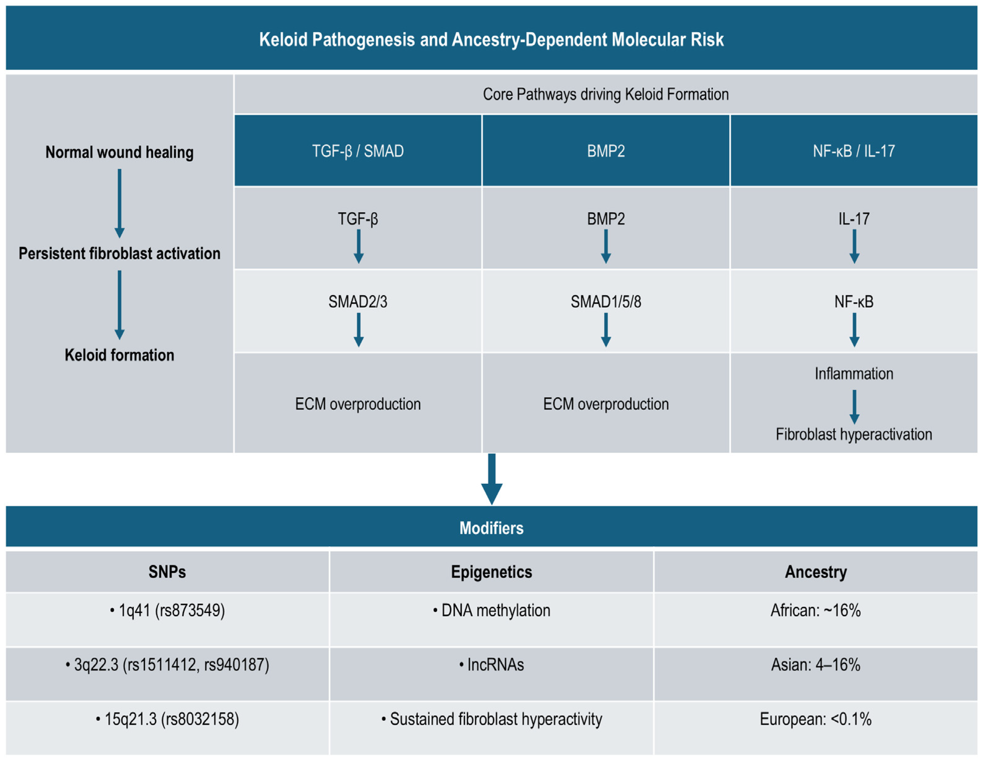
An integrated overview of the molecular pathways, genetic susceptibility, and epigenetic regulation involved in keloid pathogenesis is summarized in Figure 1.
In this context, this narrative review synthesizes existing genomic and epigenetic evidence regarding keloid scarring, placing specific emphasis on ancestry-dependent susceptibility. We summarize key genetic variants across diverse populations, examine the regulatory networks governing fibroblast activity, and discuss how translating these findings could refine risk stratification and pave the way for precision therapeutic strategies.

2. Materials and Methods

2.1. Study Design and Search Strategy

We designed this study as a narrative review to provide a critical, integrative synthesis of the genomic and epigenetic literature surrounding keloid scarring. To ensure transparency in the literature selection process, we adopted reporting standards from the PRISMA-ScR guidelines (Preferred Reporting Items for Systematic Reviews and Meta-Analyses extension for Scoping Reviews). Notably, this work functions as a narrative synthesis rather than a formal systematic review or meta-analysis; as such, formal registration in databases like PROSPERO was not applicable, and we did not perform quantitative risk-of-bias assessments (e.g., ROBINS-I or Newcastle-Ottawa scales).
Search operations covered major electronic databases, including PubMed, Scopus, Web of Science, SciELO, and Google Scholar, for articles published up to December 2025. To capture the full scope of the topic, we used combinations of English and Spanish keywords such as “keloid”, “keloid scarring”, “genetics”, “SNP”, “genome-wide association study”, “fibroblasts”, “BMP2”, “epigenetics”, and “Mexican population”.

2.2. Eligibility Criteria

Our inclusion criteria focused on original research, specifically GWAS, candidate gene association studies, gene expression analyses, and epigenetic or functional studies, involving human subjects of any age. We also incorporated relevant systematic reviews and book chapters that offered essential context regarding epidemiology or molecular mechanisms. By contrast, we excluded isolated case reports lacking molecular analysis, animal models not validated in humans, duplicate records, and studies focused exclusively on physiological wound healing without specific relevance to keloid pathology.

3. Synthesis of Genomic and Epigenetic Evidence

3.1. Study Selection

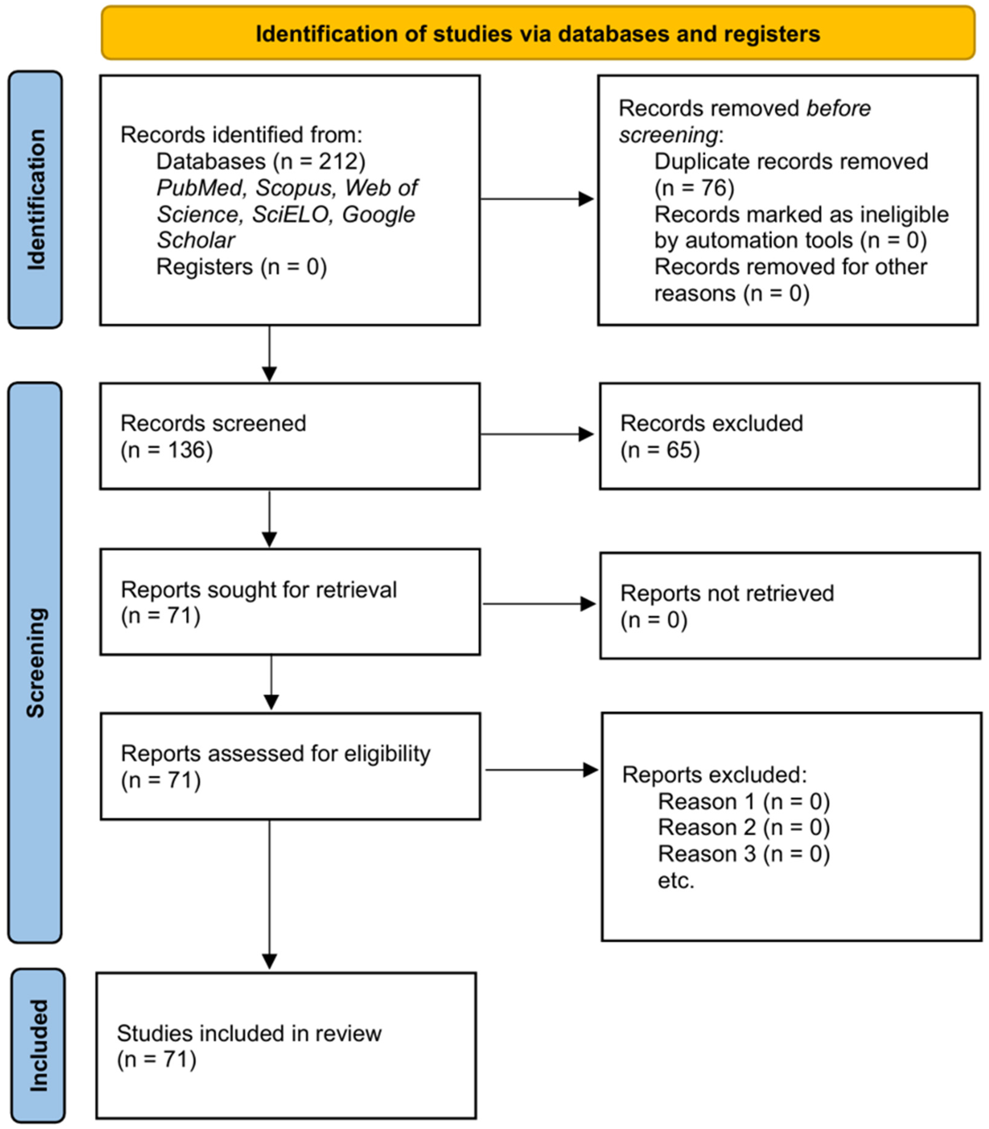
Our initial search identified 212 records. After removing 76 duplicates, we screened 136 unique articles by title and abstract. From this pool, we excluded 65 studies that did not meet specific eligibility criteria regarding genomic or molecular relevance. This left 71 full-text publications to form the analytical corpus of this review (Figure 2). These articles, comprising original GWAS, candidate gene association studies, and functional analyses, provide the evidentiary basis for the ancestry-dependent synthesis presented below.

3.2. Genomic Basis and Epidemiological Patterns

Substantial epidemiological evidence points to a strong genetic component in keloid scarring. Prevalence rates vary drastically across populations: they reach 16% in Zaire, 8.5% in Kenya, and 9% in Zambia, yet estimates in England remain as low as 0.09% [5,14,15,19]. In East Asia, prevalence sits at approximately 0.1% in Japan but climbs notably higher in Taiwan, confirming distinct regional susceptibility [22,31]. By comparison, cohorts of European ancestry, such as those in the UK Biobank, show a considerably lower disease burden [5]. Crucially, studies in Latin America are currently limited to clinical reports with minimal genomic characterization, leaving a significant gap in regional knowledge [4,14,29].
Familial clustering further supports a hereditary basis. Although autosomal dominant inheritance with incomplete penetrance is the most widely accepted model [17,18,32,33], genetic heterogeneity was apparent even before the genomic era. Additionally, rare syndromic associations have historically reinforced this genetic predisposition [30,34]. Pioneering linkage studies by Marneros et al. [20] identified loci on 2q23 (TNFAIP6) and 7p11 (EGFR) in Japanese and African American families. These foundational findings paved the way for GWAS, which later confirmed three canonical susceptibility loci: 1q41, 3q22.3, and 15q21.3 (Figure 3).

3.3. Key Susceptibility Loci and Variants

Identifying specific Single-Nucleotide Polymorphisms (SNPs) has clarified the mechanisms driving keloid pathogenesis. A comprehensive summary of these variants appears in Table 1.

3.3.1. Locus 1q41 (The DEIK-BMP2 Axis)

Nakashima et al. (2010) first pinpointed rs873549 in a Japanese GWAS [21]. This finding was soon replicated in Han Chinese cohorts, where it showed a strong association with clinical severity [22]. This polymorphism resides in strong linkage disequilibrium (LD) with rs1348270, a variant that functionally disrupts an enhancer element. As a result, DEIK expression drops while BMP2, a key driver of fibroblast proliferation and ECM synthesis, becomes overexpressed [35,36]. Complementing this, rs1442440 (located near BMP2/POSTN) has been linked to epigenetic modifications that further promote collagen synthesis [22,25]. Expanding the landscape, a recent multi-ancestry meta-analysis identified novel signals in LINC01705 and PHLDA3 within this locus [37].

3.3.2. Locus 3q22.3 (FOXL2)

The variants rs1511412 and rs940187 map to the FOXL2 region. While the association appears robust in Japanese populations [18], signals in Han Chinese cohorts have proven weaker after stringent statistical correction, suggesting ancestry-dependent effect sizes [22]. A meta-analysis confirmed that rs1511412 correlates with both susceptibility and clinical severity in Asian populations [38,39]. FOXL2 encodes a transcriptional regulator involved in cell differentiation and apoptosis, and it may interact indirectly with the TGF-β/SMAD pathway [38,39].

3.3.3. Locus 15q21.3 (NEDD4)

The SNP rs8032158 in NEDD4 shows consistent replication across Japanese, Chinese, and Egyptian populations [21,22,40,41]. Functionally, NEDD4 encodes an E3 ubiquitin ligase that regulates SMAD4 stability; the risk allele appears to impede SMAD4 degradation, amplifying TGF-β signaling [51]. Interestingly, in European cohorts, this association reached significance only under a recessive model [52], reflecting clear genetic heterogeneity. Within the same region, rs2271289 in FUT8 has been associated with susceptibility in Han Chinese groups [22,36]. Adding to this, multi-ancestry data have validated additional variants in ITGA11 and CORO2B, consolidating 15q21.3 as a global risk region [37].

3.3.4. Emerging Variants

Recent integrative genomic studies have broadened the field. Transcriptomic analyses identified candidates such as SIRT3 (rs181924090) in oxidative stress regulation and MYH8 (rs151091483) in fibroblast contractility [43,44]. In metabolic pathways, variants in the leptin receptor (LEPR) have been linked to increased risk in Han Chinese populations [8,42]. Ancestry-specific associations include ASAH1 in Yoruba families (Nigeria) [46], MYO1E/MYO7A in African Americans [50], and ADAM33 in Han Chinese [47]. Finally, HLA-DRB1*15 shows a positive association in Caucasians but no signal in Afro-Caribbean populations [48,49].

3.3.5. Integrated Perspective

Taken together, genomic, epigenetic, and immunogenetic studies delineate a complex model of susceptibility to keloid scarring. Common variants such as rs873549, rs8032158, and rs1511412, alongside less frequent polymorphisms and rare mutations, converge with epigenetic mechanisms and microenvironmental factors to explain clinical heterogeneity and interpopulation variability. The strongest evidence corresponds to 1q41 and 15q21.3 (supported by multi-population replication and functional validation), whereas 3q22.3 and emerging genes contribute ancestry-dependent nuances. The multi-ancestry meta-analysis [37] identified more than twenty additional loci and reinforced the convergence of pathways such as TGF-β/SMAD and NF-κB/STAT3, linking them to inflammation, cell migration, and dermal remodelling. To date, none of these findings have been replicated in Mexican or Latin American populations, underscoring the urgent need for regional genomic studies that provide representative molecular profiles (See Table 1).

3.4. Functional Convergence and Epigenetic Regulation

Genomic variants rarely act in isolation; instead, they converge on core signaling pathways that define the fibroinflammatory phenotype of keloids (Table 2).

3.4.1. Pathway Convergence

The primary loci (1q41, 15q21.3) and emerging candidates (EGFR, TNFAIP6) consistently dysregulate the TGF-β/SMAD and NF-κB/inflammatory axes. For instance, BMP2 overexpression (driven by 1q41) and SMAD4 stabilization (driven by NEDD4) work synergistically to promote excessive collagen deposition. Emerging biomarkers like TNFAIP6 and EGFR further integrate fibrosis with chronic inflammation, engaging TNF-α and IL-17 pathways [44,45,53]. Secondary modulators such as Syndecan-1 (SDC1) and ATF3 amplify these routes by potentiating MAPK cascades [10,54].

3.4.2. Epigenetic Modulation

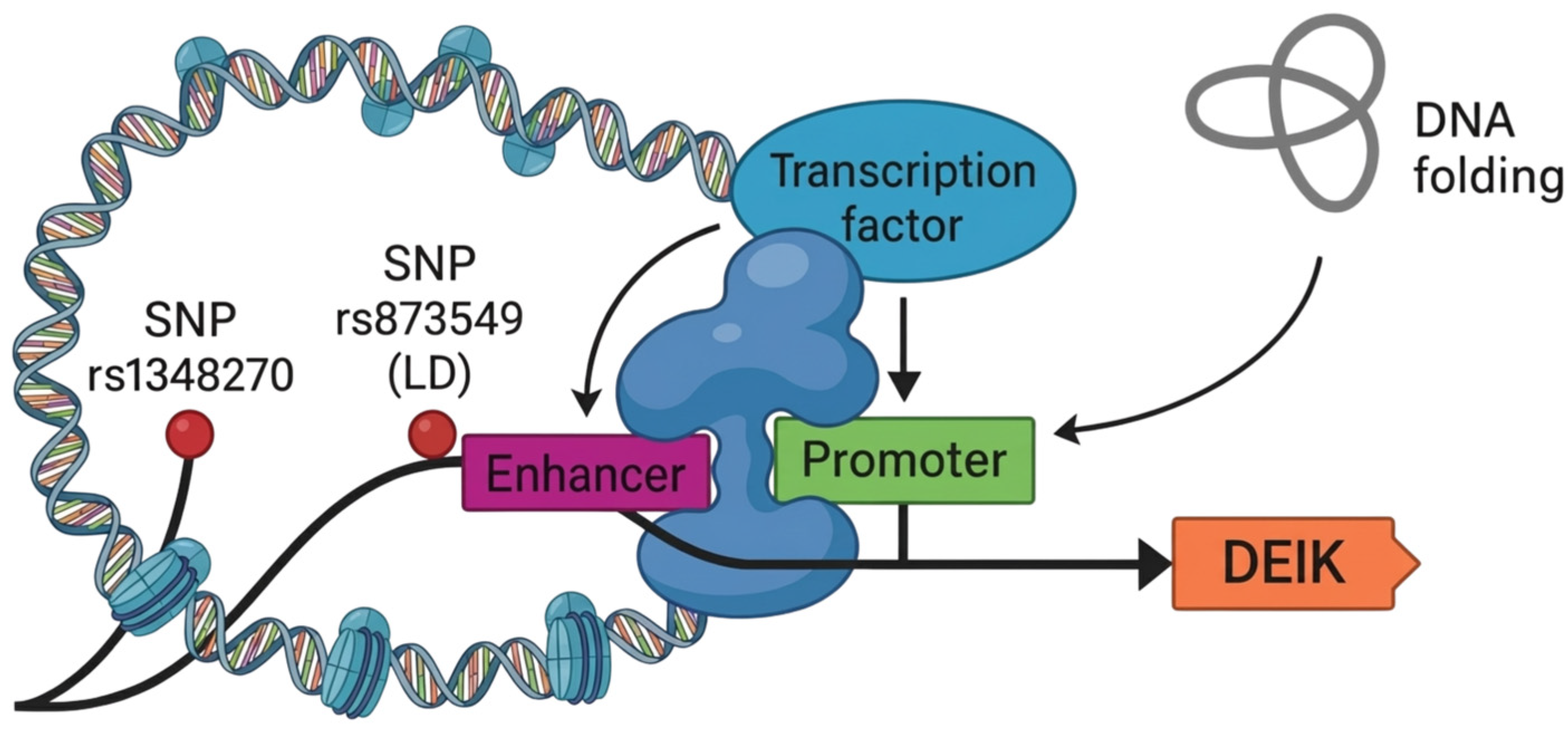
Beyond DNA sequence variations, epigenetic mechanisms sustain the “keloid memory.” Global hypomethylation has been observed in profibrotic gene promoters. The rs1348270 variant (1q41) exerts its effect via long-range chromatin looping (Figure 4). This aligns with epigenetic patterns in other inflammatory contexts, where regulators like HDAC6 and miR-9 modulate dermal immune responses [55]. In addition, over 2500 lncRNAs are differentially expressed in keloids; notably, CACNA1G-AS1 plays a prominent role in regulating intracellular calcium and fibroblast hyperactivation [56].

3.5. Susceptibility and Inheritance Models

Population-based evidence supports complex hereditary architecture. Autosomal dominant patterns with incomplete penetrance are commonly described in African and Asian families [17,18]. Rare high-penetrance mutations, such as ASAH1 p.Leu401Pro, can drive the phenotype in specific pedigrees [46]. By contrast, common variants like rs8032158 (NEDD4) may follow dominant models in Asia but recessive patterns in Europe [52]. Similarly, IL6 variants show associations in Egyptian populations that are absent in Polish cohorts [40,57].
Taken together, these findings indicate that keloid scarring is a polygenic disorder shaped by ancestry-specific architecture, where common low-effect variants and rare mutations interact within a broader multifactorial background (Table 3).

4. Discussion

The extensive genetic and epigenetic heterogeneity observed across diverse populations reinforces the concept of keloid scarring as a complex polygenic condition, shaped by the dynamic interplay between ancestral background and environmental triggers. Foundational epidemiological research established a clear ethnic–geographical gradient early on, with prevalence rates ranging from a negligible 0.09% in England to 16% in Zaire [14,58]. These disparities locate the highest susceptibility within African populations, followed by intermediate rates in Asian groups, while European cohorts show the lowest incidence [3,26,59].
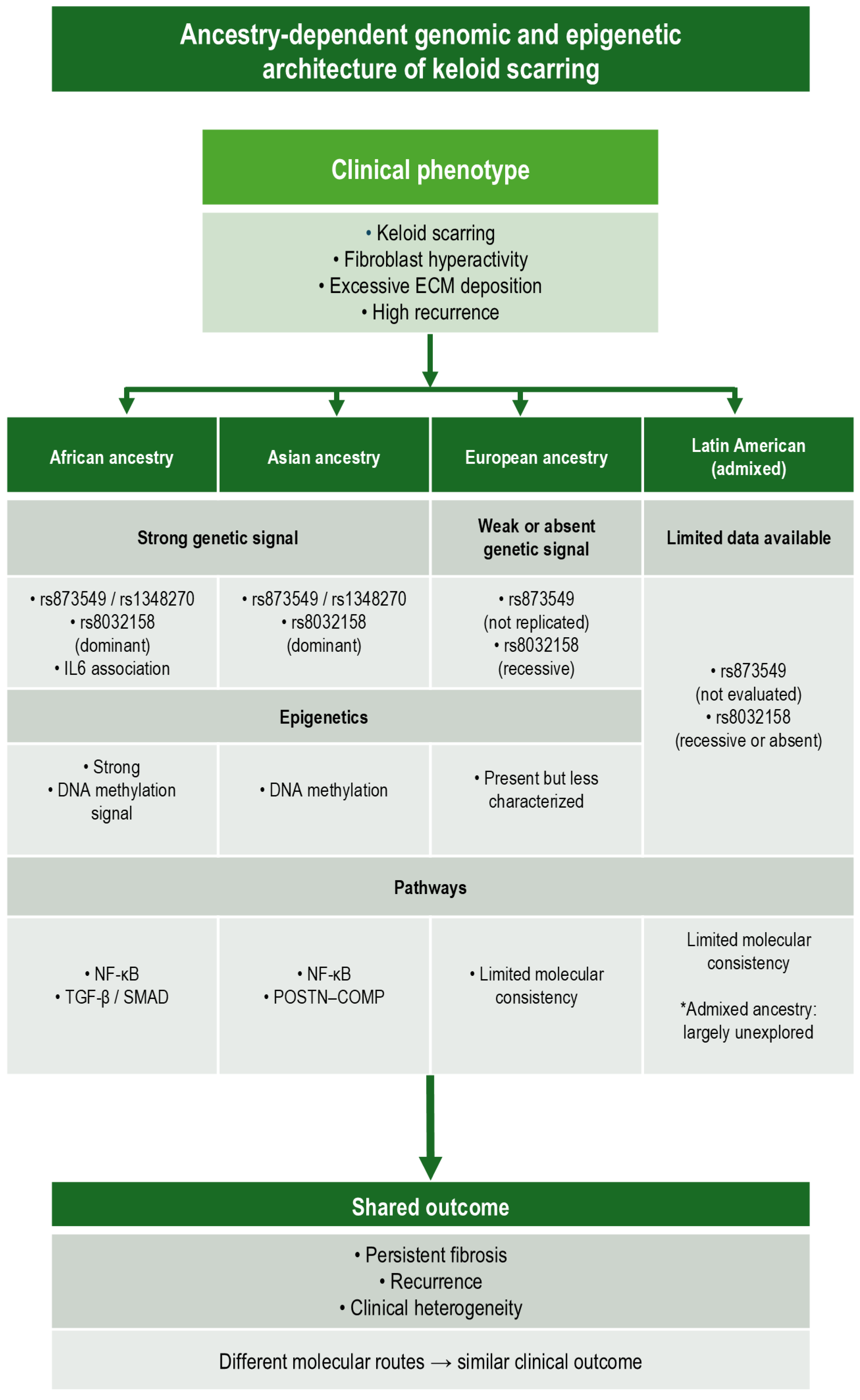
While these epidemiological patterns remain consistent across studies, the underlying molecular drivers appear to diverge by ancestry, suggesting that similar clinical phenotypes can arise from distinct genetic and epigenetic architectures [21,22,37] (Table 4). Recent evidence from Taiwan has consolidated the susceptibility profile of East Asian populations [25]; by contrast, a profound gap persists in Latin America, where research focuses largely on clinical assessments rather than genomic characterization [4,14,29].
From a hereditary perspective, familial studies have predominantly identified autosomal dominant inheritance patterns with variable expressivity, particularly among African American and Asian pedigrees [17,18]. Yet early linkage scans identifying loci at 2q23 and 7p11 highlighted the condition’s inherent heterogeneity even before the era of GWAS [19,20]. These early findings anticipated the population-specific effects later confirmed by GWAS, emphasizing that no single genetic model fully explains keloid susceptibility across all ancestries.
The shift to GWAS marked a turning point, identifying canonical susceptibility loci at 1q41, 3q22.3, and 15q21.3 [21,22]. The functional relevance of these regions is becoming clearer. At 1q41, variants such as rs873549 and rs1348270 appear to regulate enhancer–promoter interactions involving DEIK, BMP2, and POSTN, driving the characteristic overexpression of ECM proteins [35,36]. This locus shows consistent replication in Asian populations, supporting a shared pathogenic mechanism centered on fibroblast-driven ECM dysregulation.
Similarly, at 15q21.3, the rs8032158 variant in NEDD4 has been validated across Japanese, Chinese, and Egyptian populations [21,22,40], with evidence suggesting the NEDD4-TV3 isoform promotes fibroblast activation via NF-κB and STAT3 signaling [51]. European cohorts, however, often require alternative genetic models or yield weaker associations, such as the recessive effect observed for rs8032158 or the lack of signal for IL6, highlighting the critical role of ancestry in modulating genetic risk [52,57]. Rare high-penetrance variants like ASAH1 p.Leu401Pro further exemplify this heterogeneity, confirming that monogenic drivers can coexist within a broader polygenic architecture [46].
Genetic predisposition offers a plausible explanation for familial clustering [60], while recent studies highlight that pathways such as JAK/STAT and PI3K/AKT contribute to fibroblast hyperactivity beyond the TGF-β axis [61]. Epigenetic regulation adds another layer to keloid pathophysiology. The documentation of over 100,000 differentially methylated sites and the role of lncRNAs like CACNA1G-AS1 illustrate sustained reprogramming that governs fibroblast hyperactivity [24,26,36]. These mechanisms may partially account for the persistence and recurrence of keloids, as well as for tissue- and ancestry-specific phenotypic variability, even in the absence of high-penetrance mutations.
Recent multi-ancestry meta-analyses identifying 26 associated loci represent a milestone toward including underrepresented groups, such as admixed Latin American populations via the All of Us cohort [37]. Complementing these advances, recent reviews have systematized the landscape of molecular biomarkers and epigenetic alterations [27,62,63], reinforcing the need for an integrative perspective. Collectively, these findings support a model where genetic susceptibility establishes a permissive background subsequently reinforced by epigenetic dysregulation.
Over the past decade, rapid advances in both molecular research and clinical management have significantly expanded our understanding of keloid pathogenesis and therapeutic strategies, highlighting the need for more integrated and individualized treatment approaches [64]. Clinically, the high recurrence rates associated with conventional therapies, often exceeding 50%, emphasize the need for precision management [63,64,65,66]. The convergence of ancestry-dependent genetic risk, epigenetic persistence, and immune modulation suggests that uniform therapeutic approaches may be insufficient across populations. Current evidence points to the integration of genomic, epigenetic, and immunological markers into clinical algorithms as essential for effective risk stratification [67]. For instance, therapies such as non-thermal plasma show promise in differentially modulating keloid fibroblasts [53], suggesting that genotype- or pathway-informed strategies could improve outcomes.
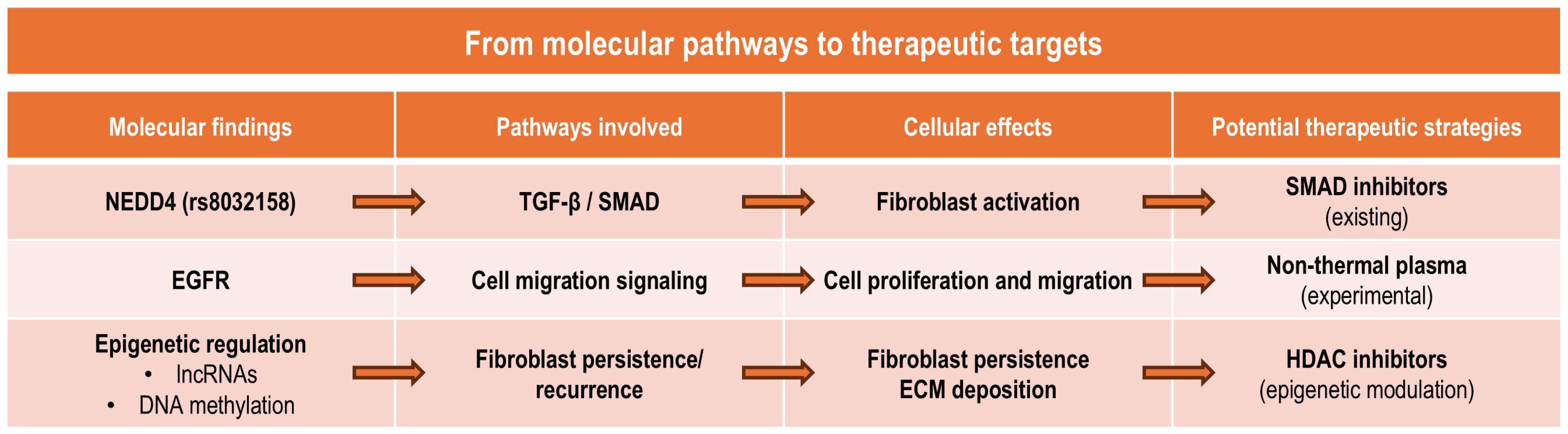
The literature supports a heterogeneous model where common variants, rare mutations, and epigenetic mechanisms converge. Crucially, ancestry emerges as a central modifier influencing genetic risk, epigenetic regulation, and disease expression. Addressing the underrepresentation of Latin American populations remains a priority; future multicenter studies must incorporate regional diversity to enable ancestry-informed strategies. Figure 5 illustrates the translational implications of these findings, linking genomic and epigenetic alterations to potential therapeutic targets.
From a translational perspective, emerging therapeutic strategies are increasingly targeting molecular pathways involved in fibrosis, including TGF-β signaling, mechanotransduction pathways, and immune-mediated fibroblast activation, reflecting a shift toward mechanism-based interventions for pathological scarring [68]. Looking beyond current insights, emerging technologies promise to refine our understanding of keloid pathogenesis.
Approaches such as single-cell RNA sequencing and spatial transcriptomics may help disentangle fibroblast heterogeneity and define ancestry-specific cellular states. [69]. Recent multi-omics single-cell analyses have further revealed substantial cellular heterogeneity within keloid tissue, identifying fibroblast subpopulations with distinct profibrotic transcriptional programs [70].
Integrative multi-omics strategies could enable more precise mapping of regulatory networks underlying fibroblast hyperactivation. In parallel, functional genomics tools, including CRISPR-based perturbation models, may further facilitate the validation of candidate susceptibility loci. Applying these technologies to underrepresented and admixed populations will be essential to translate molecular findings into clinically relevant strategies. Ultimately, novel molecular targets derived from genomic and transcriptomic studies are currently being explored as potential therapeutic approaches for preventing pathological scar formation [71].

Limitations

Although this narrative review provides an integrative overview of genomic and epigenetic mechanisms underlying keloid scarring, we must acknowledge several limitations. First, genomic evidence remains uneven across populations, with a marked underrepresentation of Latin American and other admixed cohorts, which limits the generalizability of current findings. Second, while we focused on genomic, epigenetic, and regulatory mechanisms, detailed clinical trial data and non-genetic risk factors fell outside our primary scope. Finally, emerging areas such as microbiome, fibrosis interactions, mechanical signaling, and advanced spatial multi-omics remain underexplored in this context due to limited evidence. Recognizing these gaps underscores the need for future integrative research and defines the boundaries of this narrative synthesis.

5. Conclusions

The evidence synthesized here establishes keloid scarring as a complex, multifactorial disorder defined by a polygenic architecture and modulated by ancestry-dependent epigenetic mechanisms. Instead of being driven by single causative factors, pathogenesis stems from the cumulative burden of common low-effect variants, rare high-penetrance mutations, and sustained transcriptional reprogramming. These factors converge on key profibrotic axes, most notably TGF-β/SMAD and NF-κB. Together, these genomic and epigenetic layers provide a coherent framework linking molecular dysregulation to the disease’s characteristic persistence, recurrence, and clinical heterogeneity.
Validating canonical loci alongside emerging epigenetic regulators confirms that genetic susceptibility is inherently dynamic and population-specific. Crucially, genetic associations do not replicate uniformly across populations, highlighting ancestry as a major modifier of disease susceptibility. Variants conferring high risk in African and Asian populations frequently exhibit weaker or absent effects in European cohorts. By contrast, epigenetic alterations, including DNA methylation changes and non-coding RNA regulation, offer a mechanistic explanation for phenotypic persistence and interindividual variability that inherited sequence variation alone cannot explain. Taken together, these findings show that keloid scarring cannot be adequately understood through single-population genetic models.
From a clinical perspective, these molecular insights hold direct translational relevance. The persistently high recurrence rates associated with conventional therapies expose the limitations of uniform management strategies and reinforce the need for precision-oriented approaches. Incorporating genomic, epigenetic, and immunological markers into clinical frameworks may enable improved risk stratification, prognostic assessment, and the identification of patient subgroups who are more likely to benefit from targeted or pathway-informed interventions.
Yet, despite substantial advances, a critical imbalance remains. Current genomic and epigenomic evidence leans heavily toward Asian, African, and European populations, leaving Latin American and other admixed groups markedly underrepresented. This skew limits the generalizability of existing findings and constrains the equitable translation of genomic discoveries into clinical practice. At the same time, the unique genetic admixture characterizing Mexico and Latin America presents a valuable scientific opportunity to identify ancestry-specific modifiers that might remain undetectable in more homogeneous cohorts.
Future research must therefore prioritize the inclusion of multi-ethnic and admixed populations through multicenter study designs, alongside integrative multi-omics approaches that combine genomic, epigenomic, transcriptomic, and functional data. Such strategies are essential to refine the molecular classification of keloid scarring, clarify ancestry-specific disease mechanisms, and ultimately advance personalized preventive and therapeutic strategies applicable across diverse populations.

Author Contributions

Conceptualization, J.F.L.-R., A.D.D.L.F.M., A.S.J.-O. and M.E.R.-A.; methodology, J.F.L.-R., A.D.D.L.F.M. and A.S.J.-O.; validation, L.B.L.-H., J.S.-T., G.M., C.C.-R. and C.Z.-C.; formal analysis, J.F.L.-R., A.D.D.L.F.M. and A.S.J.-O.; investigation, J.F.L.-R., A.D.D.L.F.M. and M.E.R.-A.; resources, M.E.R.-A.; data curation, L.B.L.-H., J.S.-T. and G.M.; writing—original draft preparation, J.F.L.-R., A.D.D.L.F.M., A.S.J.-O., L.B.L.-H. and C.C.-R.; writing—review and editing, J.S.-T., G.M., C.Z.-C. and M.E.R.-A.; visualization, J.F.L.-R., C.C.-R. and C.Z.-C.; supervision, M.E.R.-A.; project administration, J.F.L.-R. and M.E.R.-A. All authors have read and agreed to the published version of the manuscript.

Funding

This research received no external funding.

Institutional Review Board Statement

Not applicable.

Informed Consent Statement

Not applicable.

Data Availability Statement

No new data were created or analyzed in this study. Data sharing is not applicable to this article.

Acknowledgments

During the preparation of this manuscript, the authors used Gemini 3 (Google) for the purposes of improving language readability, refining academic tone, and assisting with translation from Spanish to English. The authors have reviewed and edited the output and take full responsibility for the content of this publication.

Conflicts of Interest

The authors declare no conflicts of interest.

Abbreviations

The following abbreviations are used in this manuscript:
GWASGenome-Wide Association Studies
SNPSingle-Nucleotide Polymorphism
ECMExtracellular Matrix
TGF-βTransforming Growth Factor Beta
SMADMothers Against Decapentaplegic Homolog
NF-κBNuclear Factor Kappa B
BMP2Bone Morphogenetic Protein 2
EGFREpidermal Growth Factor Receptor
lncRNALong Non-Coding RNA
HLAHuman Leukocyte Antigen
NEDD4Neuronal Precursor Cell Expressed Developmentally Down-Regulated 4
TNFAIPTumor Necrosis Factor Alpha-Induced Protein 6
MAPKMitogen-Activated Protein Kinase
JAK/STJanus Kinase/Signal Transducer and Activator of Transcription
LEPRLeptin Receptor

References

  1. Brown, J.J.; Ollier, W.; Arscott, G.; Ke, X.; Lamb, J.; Day, P.; Bayat, A. Genetic susceptibility to Keloid scarring: SMAD gene SNP frequencies in Afro-Caribbeans. Exp. Dermatol. 2008, 17, 610–613. [Google Scholar] [CrossRef]
  2. Kelly, A.P. Medical and surgical therapies for keloids. Dermatol. Ther. 2004, 17, 212–218. [Google Scholar] [CrossRef]
  3. Crockett, D.J. Regional keloid susceptibility. Br. J. Plast. Surg. 1964, 17, 245–253. [Google Scholar] [CrossRef]
  4. Morales-Sánchez, M.A.; Flores-Ruvalcaba, C.N.; Peralta-Pedrero, M.L.; De Villafranca-Dugelby, A.; Cruz, F.J.-S. Quality of life in adults with keloid scars. Cir. Cir. 2019, 86, 281–286. [Google Scholar] [CrossRef]
  5. Ung, C.Y.; Warwick, A.; Onoufriadis, A.; Barker, J.N.; Parsons, M.; McGrath, J.A.; Shaw, T.J.; Dand, N. Comorbidities of Keloid and Hypertrophic Scars Among Participants in UK Biobank. JAMA Dermatol. 2023, 159, 172. [Google Scholar] [CrossRef] [PubMed]
  6. Tsai, C.H.; Ogawa, R. Keloid research: Current status and future directions. Scars Burns Heal. 2019, 5, 2059513119868659. [Google Scholar] [CrossRef] [PubMed]
  7. Limandjaja, G.C.; Niessen, F.B.; Scheper, R.J.; Gibbs, S. The Keloid Disorder: Heterogeneity, Histopathology, Mechanisms and Models. Front. Cell Dev. Biol. 2020, 8, 360. [Google Scholar] [CrossRef] [PubMed]
  8. Liu, S.; Yang, H.; Song, J.; Zhang, Y.; Abualhssain, A.T.H.; Yang, B. Keloid: Genetic susceptibility and contributions of genetics and epigenetics to its pathogenesis. Exp. Dermatol. 2022, 31, 1665–1675. [Google Scholar] [CrossRef]
  9. Sadiq, A.; Khumalo, N.P.; Bayat, A. Genetics of Keloid Scarring. In Textbook on Scar Management; Springer: Cham, Switzerland, 2020; pp. 61–76. [Google Scholar]
  10. Cui, J.; Jin, S.; Jin, C.; Jin, Z. Syndecan-1 regulates extracellular matrix expression in keloid fibroblasts via TGF-β1/Smad and MAPK signaling pathways. Life Sci. 2020, 254, 117326. [Google Scholar] [CrossRef]
  11. Jing, S.L.; Suh, E.J.; Huang, K.X.; Griffin, M.F.; Wan, D.C.; Longaker, M.T. Understanding and Advancing Wound Healing in the Era of Multi-Omic Technology. Bioengineering 2025, 13, 51. [Google Scholar] [CrossRef] [PubMed] [PubMed Central]
  12. Li, J.; Li, Z.; Wang, S.; Bi, J.; Huo, R. Exosomes from human adipose-derived mesenchymal stem cells inhibit production of extracellular matrix in keloid fibroblasts via downregulating transforming growth factor-β2 and Notch-1 expression. Bioengineered 2022, 13, 8515–8525. [Google Scholar] [CrossRef] [PubMed] [PubMed Central]
  13. Cheng, X.; Gao, Z.; Shan, S.; Shen, H.; Zheng, H.; Jin, L.; Li, Q.; Zhou, J. Single cell transcriptomics reveals the cellular heterogeneity of keloids and the mechanism of their aggressiveness. Commun. Biol. 2024, 7, 1647. [Google Scholar] [CrossRef] [PubMed]
  14. Huang, C.; Wu, Z.; Du, Y.; Ogawa, R. The Epidemiology of Keloids. In Textbook on Scar Management; Springer: Cham, Switzerland, 2020; pp. 29–35. [Google Scholar]
  15. Kouotou, E.A.; Nansseu, J.R.; Guissana, E.O.; Menye, C.R.M.; Akpadjan, F.; Tounkara, T.M.; Bissek, A.Z.; Ndam, E.C.N. Epidemiology and clinical features of keloids in Black Africans: A nested case–control study from Yaoundé, Cameroon. Int. J. Dermatol. 2019, 58, 1135–1140. [Google Scholar] [CrossRef] [PubMed]
  16. Kohlhauser, M.; Mayrhofer, M.; Kamolz, L.P.; Smolle, C. An Update on Molecular Mechanisms of Scarring-A Narrative Review. Int. J. Mol. Sci. 2024, 25, 11579. [Google Scholar] [CrossRef] [PubMed] [PubMed Central]
  17. Marneros, A.G.; Norris, J.E.C.; Olsen, B.R.; Reichenberger, E. Clinical Genetics of Familial Keloids. Arch. Dermatol. 2001, 137, 1429–1434. [Google Scholar] [PubMed]
  18. Clark, J.A.; Turner, M.L.; Howard, L.; Stanescu, H.; Kleta, R.; Kopp, J.B. Description of familial keloids in five pedigrees: Evidence for autosomal dominant inheritance and phenotypic heterogeneity. BMC Dermatol. 2009, 9, 8. [Google Scholar] [CrossRef]
  19. Marneros, A.G.; Norris, J.E.C.; Watanabe, S.; Reichenberger, E.; Olsen, B.R. Genome Scans Provide Evidence for Keloid Susceptibility Loci on Chromosomes 2q23 and 7p11. J. Investig. Dermatol. 2004, 122, 1126–1132. [Google Scholar] [CrossRef]
  20. Marneros, A.G.; Krieg, T. Keloids—Clinical diagnosis, pathogenesis, and treatment options. J. Dtsch. Dermatol. Ges. 2004, 2, 905–913. [Google Scholar] [CrossRef]
  21. Nakashima, M.; Chung, S.; Takahashi, A.; Kamatani, N.; Kawaguchi, T.; Tsunoda, T.; Hosono, N.; Kubo, M.; Nakamura, Y.; Zembutsu, H. A genome-wide association study identifies four susceptibility loci for keloid in the Japanese population. Nat. Genet. 2010, 42, 768–771. [Google Scholar] [CrossRef]
  22. Zhu, F.; Wu, B.; Li, P.; Wang, J.; Tang, H.; Liu, Y.; Zuo, X.; Cheng, H.; Ding, Y.; Wang, W.; et al. Association Study Confirmed Susceptibility Loci with Keloid in the Chinese Han Population. PLoS ONE 2013, 8, e62377. [Google Scholar] [CrossRef]
  23. Dand, N.; Ung, C.Y.; Saklatvala, J.R.; Simpson, M.A.; Barker, J.N.; Shaw, T.J.; McGrath, J.A.; Onoufriadis, A. GWAS Meta-Analysis Identifies Susceptibility Loci for Keloids and Hypertrophic Scarring in Europeans. J. Investig. Dermatol. 2025, 145, 1538–1540.e8. [Google Scholar] [CrossRef] [PubMed]
  24. Jones, L.R.; Young, W.; Divine, G.; Datta, I.; Chen, K.M.; Ozog, D.; Worsham, M.J. Genome-Wide Scan for Methylation Profiles in Keloids. Dis. Markers 2015, 2015, 943176. [Google Scholar] [CrossRef] [PubMed]
  25. Stevenson, A.W.; Deng, Z.; Allahham, A.; Prêle, C.M.; Wood, F.M.; Fear, M.W. The epigenetics of keloids. Exp. Dermatol. 2021, 30, 1099–1114. [Google Scholar] [PubMed]
  26. Liang, X.; Ma, L.; Long, X.; Wang, X. LncRNA expression profiles and validation in keloid and normal skin tissue. Int. J. Oncol. 2015, 47, 1829–1838. [Google Scholar] [CrossRef]
  27. He, Y.; Deng, Z.; Alghamdi, M.; Lu, L.; Fear, M.W.; He, L. From genetics to epigenetics: New insights into keloid scarring. Cell Prolif. 2017, 50, e12326. [Google Scholar] [CrossRef] [PubMed]
  28. Almier, N.; Leibowitz, K.; Gower, A.C.; To, S.; Keller, M.R.; Connizzo, B.K.; Roh, D.S.; Alani, R.M.; Collard, M. Targeting the Epigenome Reduces Keloid Fibroblast Cell Proliferation, Migration, and Invasion. J. Investig. Dermatol. 2025, 145, 411–422.e7. [Google Scholar] [CrossRef] [PubMed]
  29. Borda, V.; Loesch, D.P.; Guo, B.; Laboulaye, R.; Veliz-Otani, D.; French, J.N.; Leal, T.P.; Gogarten, S.M.; Ikpe, S.; Gouveia, M.H.; et al. Genetics of Latin American Diversity Project: Insights into population genetics and association studies in admixed groups in the Americas. Cell Genom. 2024, 4, 100692. [Google Scholar] [CrossRef]
  30. Rubinstein, J.H. Broad Thumbs and Toes and Facial Abnormalities. Am. J. Dis. Child. 1963, 105, 588. [Google Scholar] [CrossRef]
  31. Sun, L.M.; Wang, K.H.; Lee, Y.C. Keloid incidence in Asian people and its comorbidity with other fibrosis-related diseases: A nationwide population-based study. Arch. Dermatol. Res. 2014, 306, 803–808. [Google Scholar]
  32. Omo-Dare, P. Genetic studies on keloid. J. Natl. Med. Assoc. 1975, 67, 428–432. [Google Scholar]
  33. Halim, A.S.; Emami, A.; Salahshourifar, I.; Kannan, T.P. Keloid Scarring: Understanding the Genetic Basis, Advances, and Prospects. Arch. Plast. Surg. 2012, 39, 184–189. [Google Scholar] [CrossRef]
  34. Goeminne, L. A New Probably X-Linked Inherited Syndrome: Congenital Muscular Torticollis, Multiple Keloids Cryptorchidism and Renal Dysplasia. Acta Genet. Med. Gemellol. 1968, 17, 439–467. [Google Scholar] [PubMed]
  35. Deng, C.-C.; Zhang, L.-X.; Xu, X.-Y.; Zhu, D.-H.; Cheng, Q.; Ma, S.; Rong, Z.; Yang, B. Risk single-nucleotide polymorphism-mediated enhancer–promoter interaction drives keloids through long noncoding RNA down expressed in keloids. Br. J. Dermatol. 2023, 188, 84–93. [Google Scholar] [CrossRef]
  36. Lv, W.; Ren, Y.; Hou, K.; Hu, W.; Yi, Y.; Xiong, M.; Wu, M.; Wu, Y.; Zhang, Q. Epigenetic modification mechanisms involved in keloid: Current status and prospect. Clin. Epigenet. 2020, 12, 183. [Google Scholar] [CrossRef]
  37. Greene, C.A.; Hampton, G.; Jaworski, J.; Shuey, M.M.; Khan, A.; Luo, Y.; Jarvik, G.P.; Namjou-Khales, B.; Edwards, T.L.; Edwards, D.R.V.; et al. Multi-ancestry meta-analysis of keloids uncovers novel susceptibility loci in diverse populations. Nat. Commun. 2025, 16, 7770. [Google Scholar] [CrossRef] [PubMed]
  38. Lu, W.; Zheng, X.; Liu, S.; Ding, M.; Xie, J.; Yao, X.; Zhang, L.; Hu, B. SNP rs1511412 in FOXL2 gene as a risk factor for keloid by meta-analysis. Int. J. Clin. Exp. Med. 2015, 8, 2766–2771. [Google Scholar] [PubMed]
  39. Lu, M.-Z.; Ang, Q.-Q.; Zhang, X.; Zhang, L.-F.; Yao, X.-H.; Lv, H.; Zheng, X.-D.; Lu, W.-S. Genomic risk variants at 3q22.3 are associated with keloids in a Chinese Han population. Am. J. Transl. Res. 2018, 10, 554–562. [Google Scholar]
  40. Farag, A.G.; Khaled, H.N.; Hammam, M.A.; Elshaib, M.E.; Tayel, N.R.; Hommos, S.E.I.; El Gayed, E.M.A. Neuronal Precursor Cell Expressed Developmentally Down Regulated 4 (NEDD4) Gene Polymorphism Contributes to Keloid Development in Egyptian Population. Clin. Cosmet. Investig. Dermatol. 2020, 13, 649–656. [Google Scholar] [CrossRef]
  41. Yang, Y.; Liang, Y.; Ma, X.; Su, Y.; Zhang, X. Genetic susceptibility to keloid scarring in Chinese Han population: NEDD4 gene single nucleotide polymorphism. Int. J. Clin. Exp. Med. 2017, 10, 4042–4048. [Google Scholar]
  42. Liu, J.; Cai, L.; Zhang, Z.; Ma, Y.; Wang, Y. Association of Leptin Receptor Gene Polymorphisms with Keloids in the Chinese Han Population. Med. Sci. Monit. 2021, 27, e928503-1. [Google Scholar]
  43. Teng, G.; Liu, C.; Chen, M.; Ma, K.; Liang, L.; Yan, T. Differential Susceptible Loci Expression in Keloid and Hypertrophic Scars in the Chinese Han Population. Ann. Plast. Surg. 2015, 74, 26–29. [Google Scholar] [CrossRef]
  44. Tang, Y.; Ren, K.; Yin, X.; Yang, Y.; Fang, F.; Zhou, B.; Bu, W. Tissue RNA Sequencing Reveals Novel Biomarkers Associated with Postoperative Keloid Recurrence. J. Clin. Med. 2023, 12, 5511. [Google Scholar] [CrossRef]
  45. Zhong, C.; Shi, K.; Li, P.; Qiu, X.; Wu, X.; Chen, S.; Liu, Y.; Li, F.; Zhao, Z.; Zhou, J.; et al. Single-cell sequencing analysis and bulk-seq identify IGFBP6 and TNFAIP6 as novel differential diagnosis markers for postburn pathological scarring. Burns 2024, 50, 107255. [Google Scholar] [PubMed]
  46. Santos-Cortez, R.L.P.; Hu, Y.; Sun, F.; Benahmed-Miniuk, F.; Tao, J.; Kanaujiya, J.K.; Ademola, S.; Fadiora, S.; Odesina, V.; A Nickerson, D.; et al. Identification of ASAH1 as a susceptibility gene for familial keloids. Eur. J. Hum. Genet. 2017, 25, 1155–1161. [Google Scholar] [CrossRef]
  47. Han, J.; Han, J.; Yu, D.; Xiao, J.; Shang, Y.; Hao, L. Association of ADAM33 Gene Polymorphisms with Keloid Scars in a Northeastern Chinese Population. Cell. Physiol. Biochem. 2014, 34, 981–987. [Google Scholar] [CrossRef]
  48. Brown, J.J.; Ollier, W.E.R.; Thomson, W.; Bayat, A. Positive association of HLA-DRB1*15 with keloid disease in Caucasians. Int. J. Immunogenet. 2008, 35, 303–307. [Google Scholar]
  49. Ashcroft, K.J.; Syed, F.; Arscott, G.; Bayat, A. Assessment of the influence of HLA class I and class II loci on the prevalence of keloid disease in Jamaican Afro-Caribbeans. Tissue Antigens 2011, 78, 390–396. [Google Scholar]
  50. Velez Edwards, D.R.; Tsosie, K.S.; Williams, S.M.; Edwards, T.L.; Russell, S.B. Admixture mapping identifies a locus at 15q21.2–22.3 associated with keloid formation in African Americans. Hum. Genet. 2014, 133, 1513–1523. [Google Scholar] [CrossRef] [PubMed]
  51. Fujita, M.; Yamamoto, Y.; Jiang, J.-J.; Atsumi, T.; Tanaka, Y.; Ohki, T.; Murao, N.; Funayama, E.; Hayashi, T.; Osawa, M.; et al. NEDD4 Is Involved in Inflammation Development during Keloid Formation. J. Investig. Dermatol. 2019, 139, 333–341. [Google Scholar] [CrossRef] [PubMed]
  52. Dmytrzak, A.; Boroń, A.; Łoniewska, B.; Clark, J.S.C.; Kaczmarczyk, M.; Ciechanowicz, A. Replication study of four keloid-associated polymorphisms in patients of European descent—A single centre study. Intractable Rare Dis. Res. 2020, 9, 40–42. [Google Scholar]
  53. Kang, S.U.; Kim, Y.S.; Kim, Y.E.; Park, J.K.; Lee, Y.S.; Kang, H.Y.; Jang, J.W.; Ryeo, J.B.; Lee, Y.; Shin, Y.S.; et al. Opposite effects of non-thermal plasma on cell migration and collagen production in keloid and normal fibroblasts. PLoS ONE 2017, 12, e0187978. [Google Scholar] [CrossRef]
  54. Wang, X.M.; Liu, X.M.; Wang, Y.; Chen, Z.Y. Activating transcription factor 3 (ATF3) regulates cell growth, apoptosis, invasion and collagen synthesis in keloid fibroblast through transforming growth factor beta (TGF-beta)/SMAD signaling pathway. Bioengineered 2021, 12, 117–126. [Google Scholar] [CrossRef]
  55. Kwon, Y.; Choi, Y.; Kim, M.; Jeong, M.S.; Jung, H.S.; Jeoung, D. HDAC6 and CXCL13 Mediate Atopic Dermatitis by Regulating Cellular Interactions and Expression Levels of miR-9 and SIRT1. Front. Pharmacol. 2021, 12, 691279. [Google Scholar] [CrossRef]
  56. Glasgow, E.; Mishra, L. Transforming growth factor-β signaling and ubiquitinators in cancer. Endocr. Relat. Cancer 2008, 15, 59–72. [Google Scholar] [CrossRef] [PubMed]
  57. Dmytrzak, A.; Lewandowska, K.; Boroń, A.; Łoniewska, B.; Grzesch, N.; Brodkiewicz, A.; Clark, J.S.C.; Ciechanowicz, A.; Kostrzewa-Nowak, D. No Association of Polymorphisms in the Genes Encoding Interleukin-6 and Interleukin-6 Receptor Subunit Alpha with the Risk of Keloids in Polish Patients. Int. J. Mol. Sci. 2024, 25, 5284. [Google Scholar] [CrossRef]
  58. Huang, C.; Ogawa, R. Keloidal pathophysiology: Current notions. Scars Burns Heal. 2021, 7, 2059513120980320. [Google Scholar] [CrossRef] [PubMed]
  59. Bayat, A.; Arscott, G.; Ollier, W.E.R.; Ferguson, M.W.J.; Mc Grouther, D.A. Description of site-specific morphology of keloid phenotypes in an Afrocaribbean population. Br. J. Plast. Surg. 2004, 57, 122–133. [Google Scholar] [CrossRef] [PubMed]
  60. Glass, D.A. Current Understanding of the Genetic Causes of Keloid Formation. J. Investig. Dermatol. Symp. Proc. 2017, 18, S50–S53. [Google Scholar] [CrossRef]
  61. Kim, H.J.; Kim, Y.H. Comprehensive Insights into Keloid Pathogenesis and Advanced Therapeutic Strategies. Int. J. Mol. Sci. 2024, 25, 8776. [Google Scholar] [CrossRef]
  62. Bakhtyar, N.; Amini-Nik, S.; Jeschke, M.G. OMICS Approaches Evaluating Keloid and Hypertrophic Scars. Int. J. Inflamm. 2022, 2022, 1490492. [Google Scholar] [CrossRef]
  63. Budiyanto, A.B.; Wirohadidjojo, Y.W. Epigenetic Alterations in Keloid a Possible Method to Find Novel Agents for Keloid Treatment. Berk. Ilmu Kesehat. Kulit Dan. Kelamin 2024, 36, 60–67. [Google Scholar]
  64. Qi, W.; Xiao, X.; Tong, J.; Guo, N. Progress in the clinical treatment of keloids. Front. Med. 2023, 10, 1284109. [Google Scholar] [CrossRef] [PubMed] [PubMed Central]
  65. Cosman, B.; Crikelair, G.F.; Ju, D.; Gaulin, J.C.; Lattes, R. The surgical treatment of keloids. Plast. Reconstr. Surg. 1961, 27, 335–358. [Google Scholar] [CrossRef]
  66. Berman, B.; Bieley, H.C. Adjunct therapies to surgical management of keloids. Dermatol. Surg. 1996, 22, 126–130. [Google Scholar] [CrossRef] [PubMed]
  67. Latoni, D.I.; McDaniel, D.C.; Tsao, H.; Tsao, S.S. Update on the Pathogenesis of Keloid Formation. JID Innov. 2024, 4, 100299. [Google Scholar] [CrossRef]
  68. Merlino, L.; Dominoni, M.; Pano, M.R.; Pasquali, M.F.; Senatori, R.; Zino, G.; Gardella, B. Recent Progress in Keloid Mechanism and Treatment: A Comprehensive Review. Biomedicines 2025, 13, 2276. [Google Scholar] [CrossRef] [PubMed] [PubMed Central]
  69. Li, Y.; Li, C.; Liu, W.; Gao, T.; Liu, Q.; Yang, L.; Li, S.; Tang, R.; Yang, L. Single-cell RNA sequencing reveals fibroblast heterogeneity and identifies CLOCK as a key regulator in fibrotic skin diseases. Sci. Rep. 2025, 16, 786. [Google Scholar] [CrossRef] [PubMed] [PubMed Central]
  70. Zhao, S.; Xie, J.; Zhang, Q.; Ni, T.; Lin, J.; Gao, W.; Zhao, L.; Yi, M.; Tu, L.; Zhang, P.; et al. New Anti-Fibrotic Strategies for Keloids: Insights from Single-Cell Multi-Omics. Cell Prolif. 2025, 58, e13818. [Google Scholar] [CrossRef] [PubMed] [PubMed Central]
  71. Luo, X.; Zhu, S.; Li, J.; Zeng, N.; Wang, H.; Wu, Y.; Wang, L.; Liu, Z. Potential genetic therapies based on m6A methylation for skin regeneration: Wound healing and scars/keloids. Front. Bioeng. Biotechnol. 2023, 11, 1143866. [Google Scholar] [CrossRef]
Figure 1. Integrated schematic overview of keloid pathogenesis. This diagram illustrates the interplay between dysregulated wound healing, profibrotic signaling pathways, and ancestry-dependent susceptibility. Persistent fibroblast activation, driven by altered inflammatory signaling and canonical profibrotic axes (TGF-β/SMAD, BMP2), converges to promote excessive ECM deposition and invasive scar growth. Genetic risk variants and epigenetic mechanisms modulate these pathways, shaping disease expression across different ancestral backgrounds.
Cosmetics 13 00070 g001
Figure 2. PRISMA 2020 flow diagram detailing the study selection process. The flowchart outlines the identification, screening, and eligibility assessment phases conducted to select the final articles included in this review.
Cosmetics 13 00070 g002
Figure 3. Ancestry-dependent genomic and epigenetic architecture of keloid scarring. This comparative schematic illustrates how similar clinical phenotypes arise from distinct molecular mechanisms across populations. While African and Asian ancestries exhibit strong, replicated genetic signals at canonical loci, European cohorts show weaker genetic associations, often requiring alternative inheritance models. Epigenetic dysregulation appears consistently involved across all groups but may be differentially regulated.
Cosmetics 13 00070 g003
Figure 4. Mechanism of DEIK modulation by distal variants. The SNPs rs1348270 and rs873549 (located in linkage disequilibrium at 1q41) constitute a distal enhancer element. Through 3D chromatin folding, this enhancer interacts physically with the DEIK promoter to regulate its transcription. This illustrates precisely how non-coding genetic variants can modulate gene expression in dermal fibroblasts by altering long-range chromatin architecture.
Cosmetics 13 00070 g004
Figure 5. Translating molecular findings into therapeutic targets. Overview of how specific genomic and epigenetic alterations converge on actionable signaling pathways. Genetic variants and epigenetic regulators (DNA methylation, lncRNAs) drive fibroblast activation and persistence. These molecular nodes serve as potential targets for precision interventions, ranging from existing SMAD inhibitors to experimental approaches like non-thermal plasma and epigenetic modulators.
Cosmetics 13 00070 g005
Table 1. Genetic variants: SNPs associated with keloid scarring. Organized by chromosomal locus, gene/region involved, possible affected molecular pathway, studied population and reference. Includes findings from linkage studies, GWAS, meta-analyses, and differential transcriptomic studies.
StudySNPLocusGene/Region InvolvedPossible Affected Molecular PathwayPopulation Studied
Nakashima, 2010 [21];
Zhu, 2013 [22]
rs8735491q41Intergenic (DEIK–BMP2–POSTN/COMP) (a)Enhancer in dermal fibroblasts; proliferation and ECM synthesisJapanese, Han Chinese
Deng, 2023 [35]rs13482701q41Enhancer in LD with rs873549↓DEIK → ↑BMP2 → ↑POSTN/COMPHan Chinese
Zhu, 2013 [22];
Lv, 2020 [36]
rs14424401q41Near BMP2/POSTN (LD with rs873549)Epigenetic regulation of ECM and fibroblast proliferationHan Chinese
Greene, 2025 [37]rs108636831qLINC01705Intergenic regulator; multi-ancestry replicationEuropean, African, East Asian, Latin American
Greene, 2025 [37]rs353839421qPHLDA3Apoptosis; fibroblast expressionEuropean, East Asian
Lu, 2015 [38];
Lu, 2018 [39]
rs15114123q22.3FOXL2 (b)Differentiation/apoptosis; TGF-β/SMAD interaction; clinical severityJapanese, Han Chinese
Nakashima, 2010 [21];
Lu, 2018 [39]
rs9401873q22.3Non-coding region (TF/lncRNA)Gene regulation, ECM; clinical severityJapanese, Han Chinese
Greene, 2025 [37]rs69063846q25.1TAB2Modulates TLR/IL-1 → NF-κBGlobal
Nakashima, 2010 [21];
Zhu, 2013 [22];
Farag, 2020 [40]
rs803215815q21.3NEDD4 (intron) (c)SMAD4 ubiquitination; TGF-β/SMAD; NF-κB/STAT3Japanese, Han Chinese, Egyptian
Yang, 2017 [41]rs2303579/
rs2303580/
rs10518830
15q21.3NEDD4 (haplotype)Missense variants and haplotypes of risk/protectionHan Chinese
Zhu, 2013 [22];
Lv, 2020 [36]
rs227128915q21.3FUT8 (intron) (c)ECM protein glycosylationHan Chinese
Greene, 2025 [37]rs3464766715qITGA11Integrins/fibrosisAfrican ≫ European
Liu, 2021 [42];
Liu, 2022 [8]
rs1137101/
rs1938496/
rs7555955
1p31.3LEPRLeptin signalling and dermal inflammationHan Chinese
Teng, 2015 [43];
Tang, 2023 [44]
rs1831786446p25.3HUS1B (d)DNA repair, abnormal cell proliferationHan Chinese
Greene, 2025 [37]rs22420267p14.1EPDR1Dermal ECM; skin expressionGlobal
Greene, 2025 [37]rs29193868p12NRG1Epithelial–mesenchymal signallingGlobal
Greene, 2025 [37]rs64768389p24.2GLIS3Transcription factor; inflammationGlobal
Teng, 2015 [43];
Tang, 2023 [44]
rs18192409011p15.5SIRT3 (d)Epigenetic regulation, oxidative stress, mitochondrial metabolism; fibroblast senescenceHan Chinese
Greene, 2025 [37]rs7602454011p15SLC22A18Imprinting; wound healingAfrican
Greene, 2025 [37]rs68672211p15.5LSP1Cell migration, cytoskeletonGlobal
Teng, 2015 [43];
Tang, 2023 [44]
rs15109148317p13.1MYH8 (d)Fibroblast migration and contractilityHan Chinese
Zhong, 2024 [45]TNFAIP62q23 (functional)Hyaluronan-binding protein↓ in keloids; AUC ~1.0; ECM and inflammation rolePost-burn cohorts (China)
Santos-Cortez, 2017 [46]ASAH18q23.3–p21.3Acid ceramidaseSphingolipid metabolismYoruba (Nigeria)
Han, 2014 [47]ADAM3320q13MetalloproteinaseECM remodellingHan Chinese
Marneros, 2004 [20]EGFR7p11EGFR (candidate, linkage) (e)Fibroblast proliferationAfrican American family
Brown, 2008 [48];
Ashcroft, 2011 [49]
HLA-DRB1*15, DQA1/DQB16p21.3HLA class IIAdaptive immune response; ancestry-dependent effectCaucasian (+), Afro-Caribbean (–)
Velez-Edwards, 2014 [50]MYO1E/MYO7AActin motor proteinsFibroblast adhesion/migrationAfrican American
Zhong, 2024 [45]IGFBP6Diagnostic biomarker↓ in keloids vs hypertrophic scars; AUC ~0.75China
Liang, 2015 [26]lncRNAs (e.g., CACNA1G-AS1)Differential lncRNAsECM–receptor interaction, Ca2+ signalling, focal adhesionHan Chinese
Footnotes: (a) Genes located within the 1q41 locus include DEIK (downregulated and associated with profibrotic pathways), BMP2 (implicated in dermal remodeling and extracellular matrix production), and POSTN/COMP, which encode extracellular matrix proteins consistently overexpressed in keloid fibroblasts. (b) Although FOXL2 has not yet undergone direct functional validation in dermal fibroblasts, extrapolated evidence from other tissues suggests potential interaction with the TGF-β/SMAD signalling axis. (c) At the 15q21.3 locus, both NEDD4 and FUT8 harbour susceptibility SNPs, indicating convergence of multiple molecular mechanisms, particularly ubiquitination and glycosylation, within the same genomic region. (d) Gene expression profiles reported by Teng et al. (2015) [43] corroborate the involvement of SIRT3, HUS1B, and MYH8 in oxidative stress, cellular proliferation, and DNA repair, providing indirect functional support for these transcriptomic findings of Tang et al. (2023) [44]. (e) Susceptibility loci at 2q23 and 7p11, originally identified through linkage studies [19], lack defined rsID variants but highlighted EGFR and nearby regulatory regions as candidate loci prior to the GWAS era, reflecting the population-specific genetic architecture underlying keloid formation.
Table 2. Molecular pathways involved in keloid scarring: main function and effect. This table summarizes the core signaling axes and molecular mechanisms dysregulated in keloid tissue, detailing the specific function of key proteins and their downstream effects on fibroblast activation and extracellular matrix deposition. Abbreviations: ECM, extracellular matrix; TGF-β, transforming growth factor beta; SMAD, mothers against decapentaplegic homolog; BMP2, bone morphogenetic protein 2; lncRNA, long non-coding RNA.
Gene/PathwayInvolved AxisMain FunctionEffect in Keloids
TGFBR2TGF-β/SMADSMAD-activating receptor promoting collagen synthesisExcessive fibrotic activation
NEDD4TGF-β/SMADUbiquitination of SMAD4Amplifies profibrotic signalling
DEIK (1q41)BMP2 pathwayRepressor of BMP2 and fibrosis regulator↓DEIK → ↑BMP2 → fibrosis
BMP2BMP2 (TGF-β superfamily)Growth factor promoting fibrosis and ECM depositionOverexpressed in keloid fibroblasts
POSTNECMCollagen adhesion and remodellingOverexpression linked to stiffness and keloid volume
COMPECMECM organisation↑ ECM accumulation
SDC1ECM/signallingTransmembrane proteoglycan↑ Fibroblast proliferation and ECM synthesis
ATF3TGF-β/SMADStress-induced transcription factor↑ Collagen production, proliferation, and apoptosis
lncRNAsEpigenetic regulationLong-range transcriptional regulationImbalance in lncRNAs (CAS1, DEIK-lncRNA) → fibroblast proliferation and profibrotic activation
Table 3. Main SNPs associated with keloid scarring according to population and inheritance models. This table synthesizes the hereditary architecture of keloid susceptibility, contrasting the clinical relevance of key variants across diverse ancestries and detailing the specific inheritance patterns, ranging from autosomal dominant to recessive or additive models, observed in different ethnic cohorts.
Variant/GeneInheritance ModelPopulation(s)Key Implication
rs873549/rs1348270 (1q41)Autosomal dominant/additiveJapanese, Han ChineseStrongly replicated susceptibility locus regulating the DEIK–BMP2 signaling axis
rs8032158 (NEDD4)Autosomal dominant/additive (Asian); autosomal recessive (European)Japanese, Han Chinese, Egyptian, EuropeanAncestry-dependent genetic effect with differential modulation of the TGF-β/SMAD pathway
IL6–572G>C (rs1800796)Population-specific associationEgyptian (associated), Polish (not associated)Illustrates ethnic heterogeneity in inflammatory genetic risk
ASAH1 (p.Leu401Pro)Autosomal dominant, high penetranceYoruba (Nigeria)Rare monogenic driver acting within a broader polygenic susceptibility background
HLA-DRB1*15Risk allele (non-Mendelian)Caucasian (associated), Afro-Caribbean (not associated)Ancestry-dependent immunogenetic contribution to keloid susceptibility
Global polygenic modelMultifactorial, additiveAfrican, Asian, EuropeanCombined effect of multiple low-effect common variants with epigenetic modulation
Table 4. Ancestry-dependent genomic and epigenetic architecture of keloid scarring. This comparative synthesis illustrates how similar clinical phenotypes arise from divergent molecular mechanisms across populations. It contrasts the robust involvement of BMP and TGF-β signaling pathways in African and Asian ancestries with the weaker genetic signals observed in European cohorts. ↑ indicates increased pathway activity or overexpression; ± indicates limited, variable, or inconsistent evidence across studies; * Latin American populations are genetically admixed and remain underrepresented in genomic and epigenomic studies of keloid scarring.
Pathway/Molecular MechanismAfrican AncestryAsian AncestryEuropean AncestryLatin American (Admixed) *
BMP Signaling & ECM (Locus 1q41)Replicated associationStrong replicated associationNo consistent association± Limited data
Functional Impact (1q41 Axis)↑ ECM-related pathways↑ ECM overproductionNot demonstratedUnknown
TGF-β/SMAD Signaling (Locus 15q21.3)Dominant/additive modelsDominant modelsRecessive or weak effect± Not evaluated
Inflammatory Signaling (IL-6 Variants)Positive association (Egyptian cohorts)± Variable evidenceNo associationNot studied
Epigenetic RegulationStrong (DNA methylation)StrongPresent± Largely unexplored
Dominant Pathogenic MechanismInflammatory–fibrotic balanceFibroblast hyperactivationLimited molecular signalAdmixture-dependent
Clinical PhenotypeHigh susceptibility, recurrenceHigh susceptibility, recurrenceLower prevalenceUnknown risk stratification
Disclaimer/Publisher’s Note: The statements, opinions and data contained in all publications are solely those of the individual author(s) and contributor(s) and not of MDPI and/or the editor(s). MDPI and/or the editor(s) disclaim responsibility for any injury to people or property resulting from any ideas, methods, instructions or products referred to in the content.