


Abstract
Achieving optimal skin penetration with bioactive cosmetic ingredients, such as epidermal growth factor (EGF), presents ongoing challenges. This study introduces a novel elastosome-based EGF delivery system co-loading dexpanthenol, which achieves superior skin penetration and multifunctional cosmetic efficacy compared with a conventional liposome formulation. The EGF FLEXIR-SOME formulation was characterized to determine its physicochemical properties measured for comparison against a conventional liposome control. Efficacy and safety were confirmed through in vitro and in vivo evaluations, including clinical trials of the formulation and primary skin irritation tests. The formulated EGF FLEXIR-SOME particles exhibited an average diameter of 124.8 nm and a zeta potential of −57.53 mV, demonstrating enhanced stability and skin penetration relative to the control. The results of clinical trials confirmed significant efficacy in anti-aging, moisture, skin barrier improvement, and hyperpigmentation reduction. Additionally, primary skin irritation tests classified the product as a non-irritant. In conclusion, an elastosome-based EGF formulation significantly enhances skin penetration and bioavailability. The formulation effectively improves skin elasticity, hydration, and barrier function while simultaneously reducing visible signs of aging and pigmentation. This study successfully developed an innovative formulation utilizing elastosome technology, maximizing the transdermal efficiency and stability of EGF, thereby offering a novel strategy for functional cosmeceutical development.
1. Introduction
With the increasing interest in skin health and beauty, the effective delivery of active ingredients to the skin has become a central focus in field of cosmetic research. The stratum corneum—the outermost layer of the skin—serves as a tightly packed lipid barrier, preventing the deep penetration of active molecules [1]. Consequently, there is growing demand for innovative delivery systems to overcome this physiological obstacle.
Among the most promising candidates for skin regeneration and anti-aging is epidermal growth factor (EGF). EGF, a polypeptide known for stimulating cell growth and proliferation, plays a critical role in skin regeneration and anti-aging effects [2,3]. Nevertheless, the topical efficacy of EGF is limited due to poor transdermal absorption, mainly stemming from its molecular size and protein structure, necessitating advanced delivery technologies.
To address this limitation, various delivery systems—including liposomes—have been developed to improve the stability and permeation of active ingredients in cosmetic formulations. Liposomes, composed of a lipid bilayer, are widely employed to encapsulate both hydrophilic and lipophilic molecules, thereby protecting the active compounds and improving their stability [4]. However, conventional liposomes exhibit relatively rigid membrane structures, diminishing their ability to promote deep skin penetration [5]. In response, elastosome technology has emerged as an innovative alternative. Unlike traditional liposomes, the elastosome membrane is composed of a lipid bilayer surrounding a hydrophilic core but is made flexible by incorporating one or more single-chain components, allowing for extreme deformability [5]. By mixing an appropriate ratio of surfactants, this highly flexible vesicle can easily pass through the microscopic gaps within the stratum corneum, significantly improving the transdermal delivery of encapsulated ingredients [6]. This enhanced penetration has been shown to be superior to that of traditional liposomes, highlighting elastosomes’ potential to revolutionize cosmetic and pharmaceutical applications [5].
In this study, we aimed to develop a novel cosmetic ingredient by encapsulating epidermal growth factor (EGF, 50 ppm) with dexpanthenol (5%) and Nicotinamide Mononucleotide (NMN) using elastosome technology. Dexpanthenol is widely recognized for its moisturizing, skin barrier repair, and anti-inflammatory properties [7,8,9]. Dexpanthenol also served as a stable surrogate marker to indirectly assess the enhanced penetration capability of the elastosome system. The efficacy of penetration enhancers (such as the surfactants used in elastosomes) is frequently assessed using dexpanthenol as a measurable index compound [10]. The improved delivery of dexpanthenol, a molecule with limited skin permeability, validates the effectiveness of this elastic delivery platform. Furthermore, Nicotinamide Mononucleotide (NMN) has demonstrated notable anti-aging benefits by supporting cellular energy production [11].
The physicochemical properties—including particle size, zeta potential, and stability—of the newly developed formulation were evaluated in vitro, with comparative analysis against a conventional liposome control. Its anti-aging, moisturizing, skin barrier improvement and pigmentation reduction effects were confirmed through clinical trials. Collectively, these results provide robust scientific evidence supporting the superiority of the EGF FLEXIR-SOME formulation as a multifunctional delivery platform in advanced cosmetic applications.
2. Materials and Methods
2.1. Formulation Development and Characterization
The EGF FLEXIR-SOME formulation was produced using a high-pressure microfluidizer(operating at 1000 bar), with three sequential passes to ensure uniform particle formation [12,13]. The composition included hydrogenated lecithin and lysolecithin as solid lipids, glycerin as a liquid lipid, and polyglyceryl-10 laurate as an emulsifying agent (Table 1).
Table 1.
Composition of the EGF FLEXIR-SOME formulation.
The particle structure was analyzed using cryo-transmission electron microscopy (Cryo-TEM, Tecnai F20 G2 Cryo, FEI, Eindhoven, The Netherland) [13]. Particle size and zeta potential were measured using a laser light scattering system (Photal, ELS-Z, Hirakata, Osaka, Japan), and stability was assessed using Turbiscan (TurbiscanLAB, Formulation, L’Union, France) at 25 °C and 40 °C over 24 h. The dexpanthenol content was quantified by means of HPLC.
- Dexpanthenol content analysis
The standard solution was prepared by dissolving 528 mg of dexpanthenol in 100 mL of solvent. A stock solution of 528 ppm was prepared, and then diluted to create standard solutions of 264 ppm and 132 ppm.
The composition of the solvent was as follows:
- (A) Mobile Phase: 0.01 M Na2HPO4–Methanol (92:8);
- (B) Solvent: Water–Methanol (70:30).
To prepare the test solution, 0.2 g of the sample was collected into a 50 mL volumetric flask. The sample was extracted by adding 50 mL of solvent, followed by sonication for approximately 20 min. The extracted solution was filtered through a 0.45 µm filter and used as the test solution.
2.2. Skin Penetration Methodology and Apparatus
The skin penetration rate of dexpanthenol encapsulated in the EGF FLEXIR-SOME formulation was compared to that of a control liposome using a Franz-type diffusion cell with an artificial skin membrane (Strat-M® Membrane, Merck Millipore, Darmstadt, Germany) [14]. The experiment was conducted at 33 °C for 6, 16, and 24 h.
2.2.1. Experimental Apparatus and Conditions
The evaluation of in vitro skin permeation was executed using a standardized Franz-type diffusion cell (Transdermal Semi-Auto Diffusion Cell, LOGAN Instruments, Somerset, NJ, USA) to evaluate the delivery capabilities of the developed formulation. For the skin barrier model, a commercially available Strat-M® Membrane [15,16] was utilized, securely mounted between the donor and acceptor compartments of the diffusion cell. The acceptor compartment (5 mL) was filled with a receptor fluid consisting of a 50 mM phosphate buffer (pH 7.4, 0.1 M NaCl). Throughout the experiment, the temperature was strictly maintained at 33 °C under continuous stirring at 600 rpm to ensure adequate sink conditions. The test materials, the EGF FLEXIR-SOME sample and the control liposome sample, were separately applied to the donor compartment for comparative analysis of their permeation kinetics.
2.2.2. Penetration Rate Measurement Method
The absorption of dexpanthenol was monitored at predetermined measurement time points (6, 16, and 24 h). At each interval, the entire receptor fluid was carefully collected from the acceptor compartment using a fixed syringe. Following completion of the diffusion process, residual emulsion remaining on the artificial skin membrane was gently removed by wiping or rinsing with 10 mL of ethanol. To account for components absorbed into the membrane itself, the artificial skin was recovered and homogenized using a tip-type homogenizer, ensuring the complete recovery of absorbed dexpanthenol [17]. Lastly, the skin penetration rate of dexpanthenol in the recovered medium solutions (both receptor fluid and the homogenized membrane extract) was quantitatively evaluated using UV-vis analysis [17].
2.3. Methodology for In Vitro Efficacy Assays
The formulation’s efficacy was evaluated through various in vitro tests: antioxidant activity (DPPH [18] and ABTS [19] radical scavenging), anti-aging effects (collagenase inhibition [20]), whitening effects (tyrosinase inhibition [21]), and anti-inflammatory effects (Nitric Oxide (NO) inhibition [22]).
2.4. Clinical Trials
2.4.1. Subject for Anti-Aging
This study was approved by the Institutional Review Board (IRB) at Humantest Co.(Seoul, Korea) (IRB No. HE-IRB-P23-0225, HE-IRB-P23-0226) and was conducted from 4 to 18 October 2023. A total of 22 healthy Korean adult women aged 30 and older were enrolled in the anti-aging trial.
- Inclusion criteria:
- –
- Healthy Korean women aged 30 years or older.
- –
- Individuals’ informed consent provided after full explanation of this study’s purpose and procedures.
- –
- No acute or chronic systemic diseases, including skin conditions.
- –
- Available for follow-up throughout the study period.
- Exclusion criteria:
- –
- Pregnant, breastfeeding, or planning pregnancy.
- –
- Psychiatric illness or infectious skin disease.
- –
- Use of topical steroid for more than one month.
- –
- Participation in a similar study within the last six months.
- –
- Sensitive or allergic skin.
- –
- Abnormal skin condition at the test site
- –
- Use of cosmetics or drugs with similar effects within the last three months.
- –
- Dermatological procedure at the test site within the last six months.
- –
- Employees of the company conducting this study.
- –
- Deemed unsuitable by the principal investigator.
During this study, subjects abstained from the intake or use of functional cosmetics and medicines and were advised not to alter usual skincare habits or undergo cosmetic procedures. Excessive sunlight exposure was strictly avoided.
2.4.2. Anti-Aging Evaluation
Anti-aging efficacy was evaluated by analyzing changes in skin parameters before and after 2 weeks of product use. The clinical trial was conducted using a randomized split-face method to minimize inter-individual variability [23]. Each subject applied EGF FLEXIR-SOME 2% (2% of the proprietary EGF elastosome raw material (equivalent to 1 ppm EGF)) and EGF FLEXIR-SOME 8% (8% of the proprietary EGF elastosome raw material (equivalent to 4 ppm EGF)) to the right and left sides of their face, respectively, divided along the center of the nose. The assignment of the test products to either the left or right side was randomized for each participant. The baseline measurements for each side served as the internal control, and clinical improvements were evaluated by comparing the changes between the two sides after 2 weeks of application.
Subjects were instructed to apply the test products (EGF FLEXIR-SOME 2% and 8%) once daily, either in the morning or evening, as the final step of their regular skincare routine for a total of 14 days. The dosage was determined based on each individual’s habitual amount for facial application.
To ensure the accuracy and reproducibility of the measurements, all clinical evaluations were performed in a strictly controlled environment maintained at a temperature of 22 ± 2 °C and relative humidity of 50 ± 5%, with no direct sunlight or significant air movement. Upon visiting the center, subjects washed the test area with lukewarm water and were required to rest for at least 30 min under these constant temperature and humidity conditions to allow for skin stabilization before any measurements were taken.
Parameters included the following: R7 (eye elasticity, mm) and arithmetic mean roughness (Ra, wrinkles, μm), skin sagging angle (cheek, °), and skin age index. Skin elasticity (R7) was measured with a Cutometer MPA580 (Courage+Khazaka GmbH, Cologne, Germany) [24], wrinkles (Ra) with an Antera 3D (Miravex Ltd., Dublin, Ireland) [25], and lifting with an F-ray (BEYOUNG Co., Ltd., Gyeonggi-do, Republic of Korea). The skin aging index was calculated from these three parameters. The Skin Age Index was calculated using the following formula.
R7: A measure of skin elasticity in the eye area.
Ra: Arithmetic mean roughness, an indicator of wrinkles in the eye area.
Sagging Angle: A measure of skin lifting in the cheek area.
2.4.3. Subjects for Skin Irritation Evaluation
This study was approved by the IRB at Humantest Co. (IRB No. HE-231011-IR-0018-21, HE-231011-IR-0018-22) and was conducted from October 11 to 13, 2023.
A total of 32 healthy adult subjects (male and female) aged 19 and above were recruited for this study. The number of subjects was determined to be at least 30, in accordance with Item 7 (1) ‘Human Patch Test Methods’ of Appendix 1 (Toxicological Test Methods) under the ‘Regulations on the Evaluation of Functional Cosmetics’ (Ministry of Food and Drug Safety, Republic of Korea).
- Inclusion criteria:
- –
- Healthy male and female adults aged 19 years or older.
- –
- Voluntarily informed consent after understanding this study’s procedures.
- –
- No acute or chronic systemic diseases, including skin diseases.
- –
- Availability for follow-up observations throughout the study period.
- Exclusion criteria:
- –
- Pregnant or breastfeeding.
- –
- Psychiatric illnesses or skin diseases.
- –
- Chronic debilitating diseases such as asthma, diabetes, or hypertension.
- –
- Abnormal skin conditions at the test site.
- –
- Atopic dermatitis.
- –
- Use of topical steroids for more than one month at the test site.
- –
- Sensitive or hyper-sensitive skin.
- –
- Severe irritation or allergies to adhesive tapes.
- –
- Participation in an identical study within the last three months.
- –
- Taking contraceptives, antihistamines, or anti-inflammatory drugs.
- –
- Severe irritation or allergy to cosmetics, drugs, or sun exposure.
- –
- Employees of the institution.
- –
- Deemed unsuitable by the investigator.
2.4.4. Skin Irritation Evaluation
To evaluate the primary skin irritation potential, a flat area of the back without pigmentation or skin damage (excluding the spine) was selected as the test site. For the liquid or cream-type test products, a volume of was applied, while solid or sheet-type products were cut into approximately 1 cm × 1 cm squares for application. These materials were applied via an occlusive patch for 24 h. Skin reactions were visually observed and evaluated at two distinct time points: 30 min and 24 h after patch removal. The presence and degree of skin irritation were assessed by trained investigators through visual inspection according to standardized evaluation criteria. The results were used to calculate the Skin Irritation Index based on the Frosch & Kligman (1979) and CTFA guidelines [26], and graded using the modified Draize method [27].
2.4.5. Subjects for Skin Hydration, Barrier Repair, and Pigmentation Reduction Evaluation
This study was approved by the IRB at Humantest Co. (IRB No. HM-IRB-P23-0266) and was conducted from 27 September to 10 October 2023.
- Inclusion criteria:
- –
- Healthy Korean adult females, 19 years of age or older.
- –
- Voluntary informed consent provided after full explanation of this study’s procedures.
- –
- No acute or chronic physical diseases, including skin diseases.
- –
- Availability for follow-up throughout the study period.
- Exclusion criteria:
- –
- Pregnant, breastfeeding, or planning pregnancy.
- –
- Psychiatric disorders or infectious skin diseases.
- –
- Use of topical steroids for more than one month.
- –
- Participation in a similar study within the last six months.
- –
- Sensitive or hypersensitive skin.
- –
- Abnormal skin conditions at the test site.
- –
- Use of cosmetics or drugs with the similar efficacy within the last three months.
- –
- Dermatological or cosmetic procedures at the test site within the last six months.
- –
- Employees of the institution.
- –
- Deemed unsuitable by the principal investigator.
2.4.6. Skin Hydration, Repairing Damaged Skin Barrier Repair, and Hyperpigmentation Reduction Evaluation
This study was divided into three evaluation segments: 1-time use for skin hydration and damaged skin barrier repair, and 2-week use for the improvement in hyperpigmentation and melasma. Upon visiting the center, subjects washed the test areas with lukewarm water and rested for at least 30 min in a stabilized environment (22 ± 2 °C, 50 ± 5% humidity) to achieve skin equilibrium before participating in the measurements.
To evaluate skin hydration and the repair of the damaged skin barrier, a specialized cross-allocation design was implemented to minimize systematic bias arising from anatomical differences between measurement sites. The potential for systematic bias was strictly controlled by balancing the measurement sites; subjects 1–11 had the test product applied to the wrist and the control to the popliteal area, while subjects 12–22 followed the opposite arrangement. This method accounts for anatomical differences in skin properties, providing a robust internal control for each participant [28]. For the forearm and wrist areas, a standardized dose of 2 was applied to quantify immediate barrier recovery after physical irritation.
For the improvement in hyperpigmentation and melasma, the melanin content (%) on both cheeks was analyzed before and after 2 weeks of use. During this period, subjects applied the product twice daily (morning and evening) as the final step of their skincare routine. Facial application followed the subject’s habitual appropriate amount based on their personal skincare habits. All clinical measurements were conducted in a space devoid of air movement and direct sunlight to ensure the precision and reproducibility of the data.
- Measurement devices
Skin hydration was quantified by measuring the average permittivity (ε) of the skin surface using the Epsilon E100 (Biox Systems Ltd., London, UK). The repair and integrity of the damaged skin barrier were evaluated by measuring Transepidermal Water Loss (TEWL) in g/m2h on the forearm using a Vapometer (Delfin Technologies Ltd., Kuopio, Finland). Lastly, the improvement in hyperpigmentation was determined by analyzing the melanin content in high-resolution images of the face captured with the DermaVision system (Opto Biomed Co., Ltd., Wonju, Republic of Korea).
2.4.7. Statistical Analysis
All statistical analyses were performed using SPSS Statistics 26 (IBM Corp., Armonk, NY, USA) and the normality of the data was verified using the Shapiro–Wilk test.
For in vitro evaluations, all experiments were performed at least in triplicate, and the results are expressed as the mean ± standard deviation (SD) for continuous variables, while categorical variables (survey data) are presented as frequencies and percentages. Statistical significance for in vitro assays was determined by comparing each treatment group to the untreated control group using the independent-samples two-tailed Student’s t-test.
For clinical efficacy evaluations—including anti-aging, hydration, barrier repair, and pigmentation—, in cases where the data followed a normal distribution, the paired t-test was employed to evaluate pre- versus post-application changes; otherwise, the Wilcoxon signed-rank test was utilized. Inter-group comparisons were performed using repeated measures ANOVA based on raw data to account for multiple measurement time points.
A p-value of less than 0.05 was considered statistically significant at a 95% confidence interval.
3. Results
3.1. Formulation Characteristics
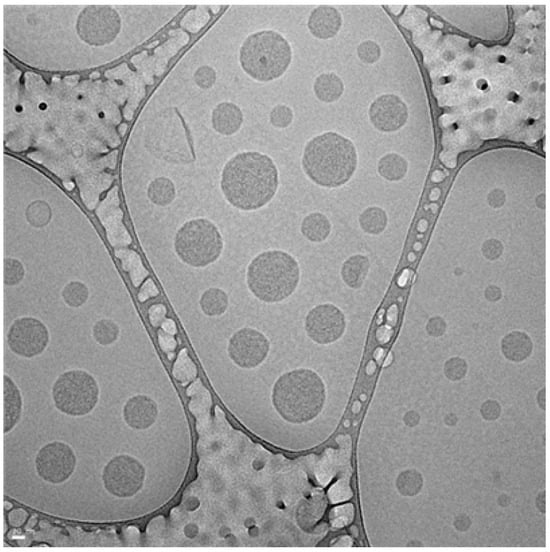
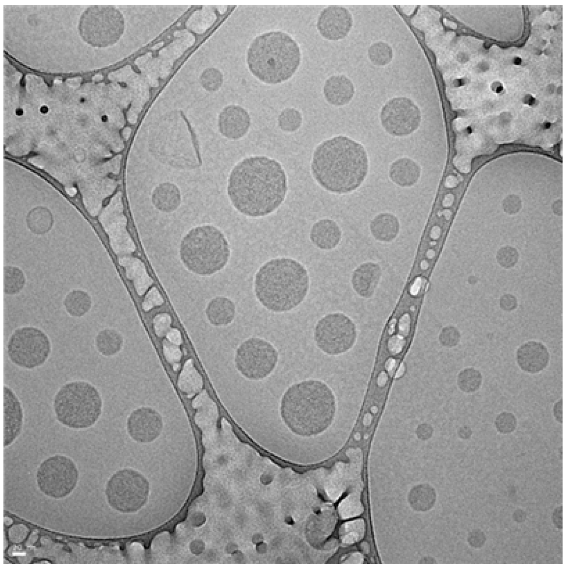
The EGF FLEXIR-SOME formulation was prepared using a high-pressure micro-emulsifier. The structure of the liposome particles was analyzed by transmission electron microscopy, as shown in Figure 1.
Figure 1.
Structure of EGF FLEXIR-SOME particles. Magnification: 100K.
3.1.1. Particle Size
The average particle size of the EGF FLEXIR-SOME formulation, measured in triplicate to ensure reproducibility, was 124.8 ± 1.5 nm, with individual measurements ranging from 123.6 nm to 126.5 nm. This size falls within the target range of 100 to 200 nm and was found to be slightly smaller than the average size of the conventional liposome control group (127.9 ± 2.2 nm) (Table 2).
Table 2.
Particle size analysis of the EGF FLEXIR-SOME.
3.1.2. Surface Charge
The surface charge of the particles was measured using a laser light scattering system (Photal, ELS-Z, Hirakata, Osaka, Japan), with the results shown in Table 3. The EGF FLEXIR-SOME formulation exhibited a greater magnitude of zeta potential (−57.53 mV) compared to the conventional liposome control (−45.08 mV).
Table 3.
Zeta potential of the EGF FLEXIR-SOME.
3.1.3. Stability
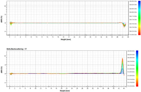
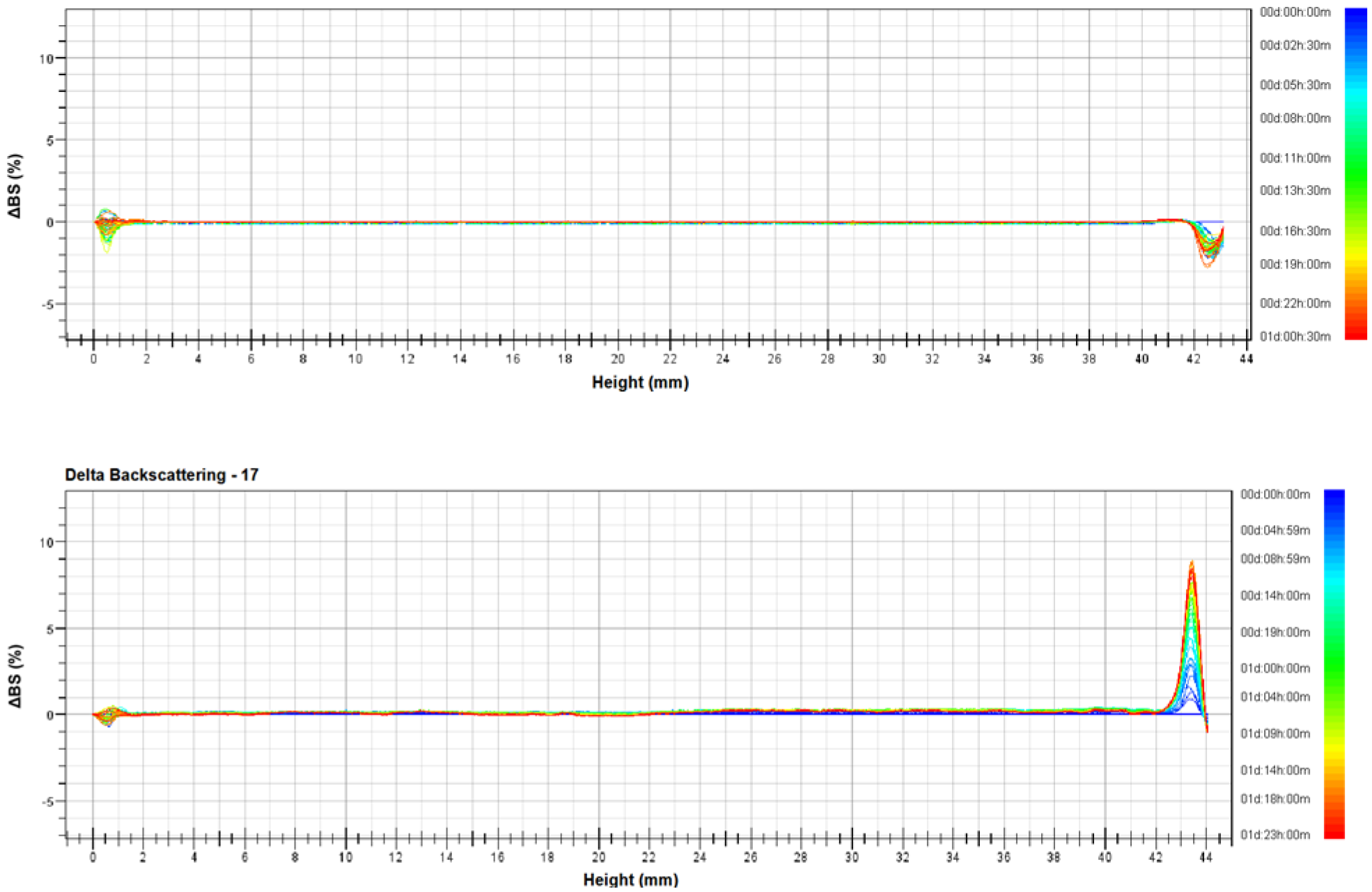
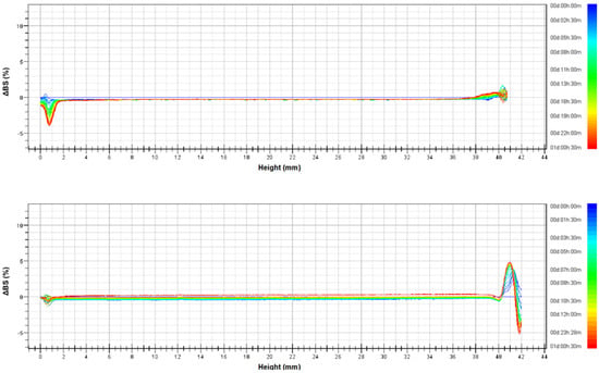
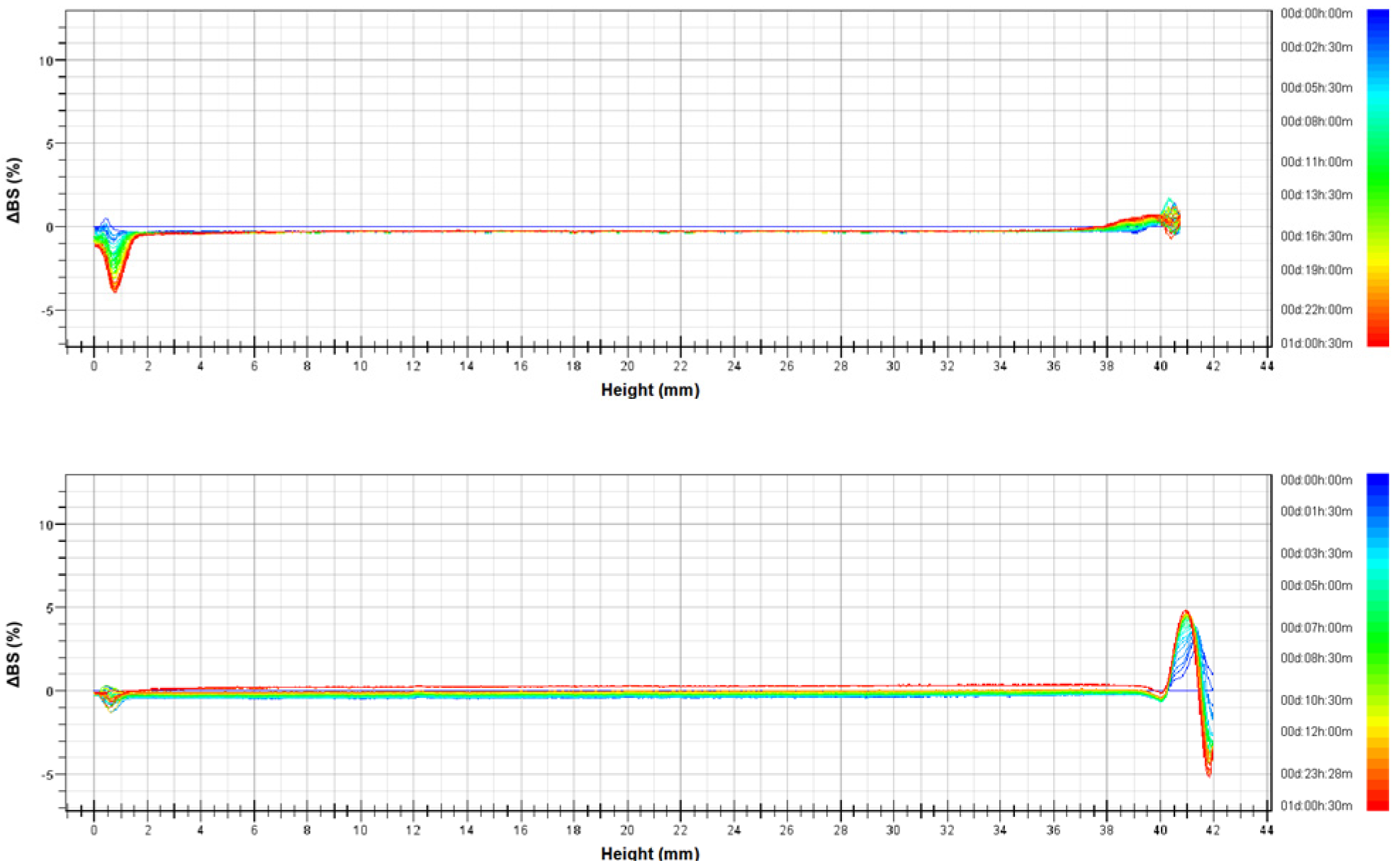
Particle stability was evaluated under both ambient (25 °C) and high-temperature (40 °C) conditions. After 50 scans over 24 h, no noticeable time-dependent changes were observed in the corresponding graphs (Figure 2 and Figure 3), compared to the control liposome group, confirming stable physical properties.
Figure 2.
Turbiscan of the EGF FLEXIR−SOME ((top): 25 °C, (bottom): 40 °C).
Figure 3.
Turbiscan of liposome (control) ((top): 25 °C, (bottom): 40 °C).
3.1.4. Dexpanthenol Content Analysis
The dexpanthenol content of the manufactured EGF FLEXIR-SOME was quantified using high-performance liquid chromatography(HPLC), and the results are shown in Table 4. The analysis confirmed that the active ingredient was accurately encapsulated at the target concentration, with a measured content of 5.05 ± 0.01%.
Table 4.
HPLC analysis of dexpanthenol content.
3.2. Analysis of Transdermal Absorption Results
The rate of skin penetration by dexpanthenol encapsulated within the EGF FLEXIR-SOME formulation was compared to that of a conventional liposome control.
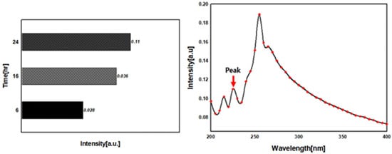
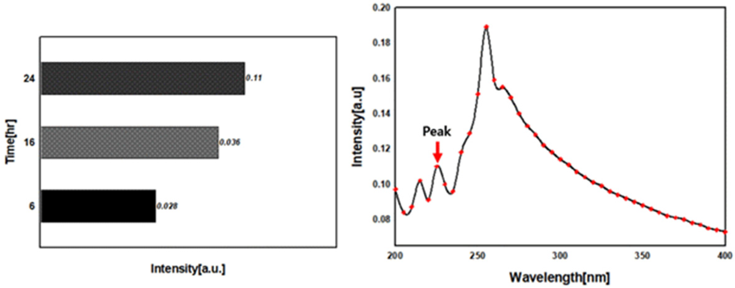
UV-Vis analysis of the EGF FLEXIR-SOME formulation containing dexpanthenol revealed the appearance of an absorption peak at a wavelength of 210–230 nm. Transdermal absorption of dexpanthenol from the EGF FLEXIR-SOME formulation was detected after 6 h, with peak intensity increasing over time (Figure 4). In contrast, the control liposome group showed no detectable absorption peak, even with extended time.
Figure 4.
Absorption intensity (UV-Vis analysis).
These results demonstrate that the elastosome-based formulation significantly enhances skin penetration compared to conventional liposomes.
3.3. Quantitative Analysis of In Vitro Efficacy
An experiment was conducted to evaluate the antioxidant, whitening, anti-aging, and anti-inflammatory effects of the aforementioned cosmetic formulation containing a proprietary EGF raw material. The results of the evaluations, including antioxidant, anti-aging, whitening, and anti-inflammatory assays, are summarized in Table 5, Table 6, Table 7, Table 8, Table 9 and Table 10, and the corresponding raw data of Tables S1–S5 are available in the Supplementary Materials.
3.3.1. Antioxidant Effect Evaluation (DPPH and ABTS Assay)
The antioxidant potential of the EGF raw material was evaluated using DPPH and ABTS radical scavenging assays1. In the DPPH assay, the EGF raw material exhibited a concentration-dependent increase in scavenging activity, reaching a maximum inhibition of 44.35 ± 2.35% at a 10% concentration (Table 5). Although the for DPPH was >10%, the EGF group showed significantly higher efficacy than the control group 23.81 ± 1.09% at 10%).
In the ABTS assay, the EGF raw material demonstrated superior potency with an value of 0.17% (Table 6). This was lower (more potent) than the control group’s of 0.21%, confirming that the EGF formulation is particularly effective at neutralizing ABTS radicals.
Table 5.
Antioxidant effect (DPPH assay).
Table 6.
Antioxidant effect (ABTS assay).
3.3.2. Anti-Aging Effect Evaluation (Collagenase Inhibition Assay)
The anti-aging efficacy was assessed through a collagenase inhibition assay. At a 5% concentration, the EGF raw material inhibited collagenase activity by 15.91 ± 1.21%, whereas the control group showed 10.30 ± 0.28% inhibition (Table 7). While the for collagenase inhibition was >5%, the superior performance of the EGF group indicates its potential in preventing dermal matrix degradation.
Table 7.
Anti-aging effect (collagenase inhibition assay).
3.3.3. Whitening Effect Evaluation (Tyrosinase Inhibition Assay)
The whitening effect was notably strong in the tyrosinase inhibition assay. The EGF raw material achieved an value of 0.96%, as it already showed 50.71 ± 0.41% inhibition at the lowest tested concentration (1%) (Table 8). In contrast, the control group exhibited minimal activity, with an exceeding 20%.
Table 8.
Whitening effect (tyrosinase inhibition assay).
3.3.4. Anti-Inflammatory Effect Evaluation (NO Inhibition Assay)
The anti-inflammatory capacity was determined by measuring Nitric Oxide (NO) production in LPS-stimulated cells. The EGF raw material inhibited NO production by 21.73 ± 2.51% at a 2% concentration (Table 9). Although the for NO inhibition was >2%, the EGF raw material showed significantly improved anti-inflammatory activity compared to the control group (1.98 ± 2.65% at 2%).
Table 9.
Anti-inflammatory effect (NO inhibition assay).
Overall, as shown in Table 10, the proprietary EGF raw material demonstrated superior performance across all assays, compared with the control group, including DPPH and ABTS radical scavenging, tyrosinase inhibition, collagenase inhibition, and NO inhibition. Statistical analysis revealed that all observed differences between the proprietary EGF raw material and control group were highly significant (p < 0.001). The 95% confidence intervals for the differences in efficacy parameters further confirm the robustness and reliability of these results (Table 10).
Table 10.
In vitro efficacy evaluation: EGF raw material and control group.
3.4. Clinical Efficacy and Safety
Using the proprietary EGF Elastosome method, two cosmetic formulations were developed with different concentrations of the EGF elastosome raw material. The first product, EGF FLEXIR-SOME Cream, was formulated with 2% elastosome raw material (equivalent to 1 ppm EGF). The second product, Dermafactory Pro-EGF FLEXIR-SOME 8% Repair Cream, contained 8% elastosome raw material (equivalent to 4 ppm EGF).
The anti-aging effects of these two products were evaluated after a two-week period of use. This clinical evaluation aimed to assess the real-world performance of the EGF FLEXIR-SOME Cream and Dermafactory Pro-EGF FLEXIR-SOME 8% Repair Cream on the skin of the study participants. Objective measurements included wrinkle depth, skin elasticity, and moisture content. A split-face test was conducted wherein each subject applied the 2% formulation to one side and the 8% formulation to the other side of their face. The pre-application state served as the control for comparison of post-application changes.
3.4.1. Subject Characteristics
A total of 22 healthy female subjects were enrolled in this study. Subject characteristics were collected through questionnaires and are summarized as follows (Table 11).
Table 11.
Subject characteristics for the anti-aging trial.
3.4.2. EGF FLEXIR-SOME 2% (Equivalent to 1 ppm EGF)
Anti-Aging Efficacy Evaluation Results of EGF FLEXIR-SOME 2%
- Skin Elasticity Improvement in EGF FLEXIR-SOME 2%
After 2 weeks of product use, the R7 value in the eye area showed a significant increase (from 0.3132 ± 0.0261 mm to 0.3441 ± 0.0285 mm, p < 0.001), with an improvement rate of 9.87% (Table 12).
Table 12.
Cutometer MPA580 R7 Measurement Results (significant difference (p < 0.05)).
- Wrinkle Reduction (Antera 3D) of EGF FLEXIR-SOME 2%
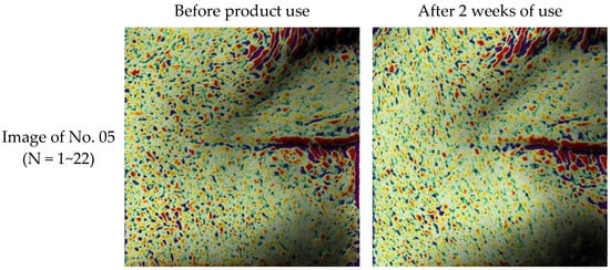
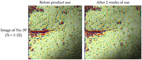
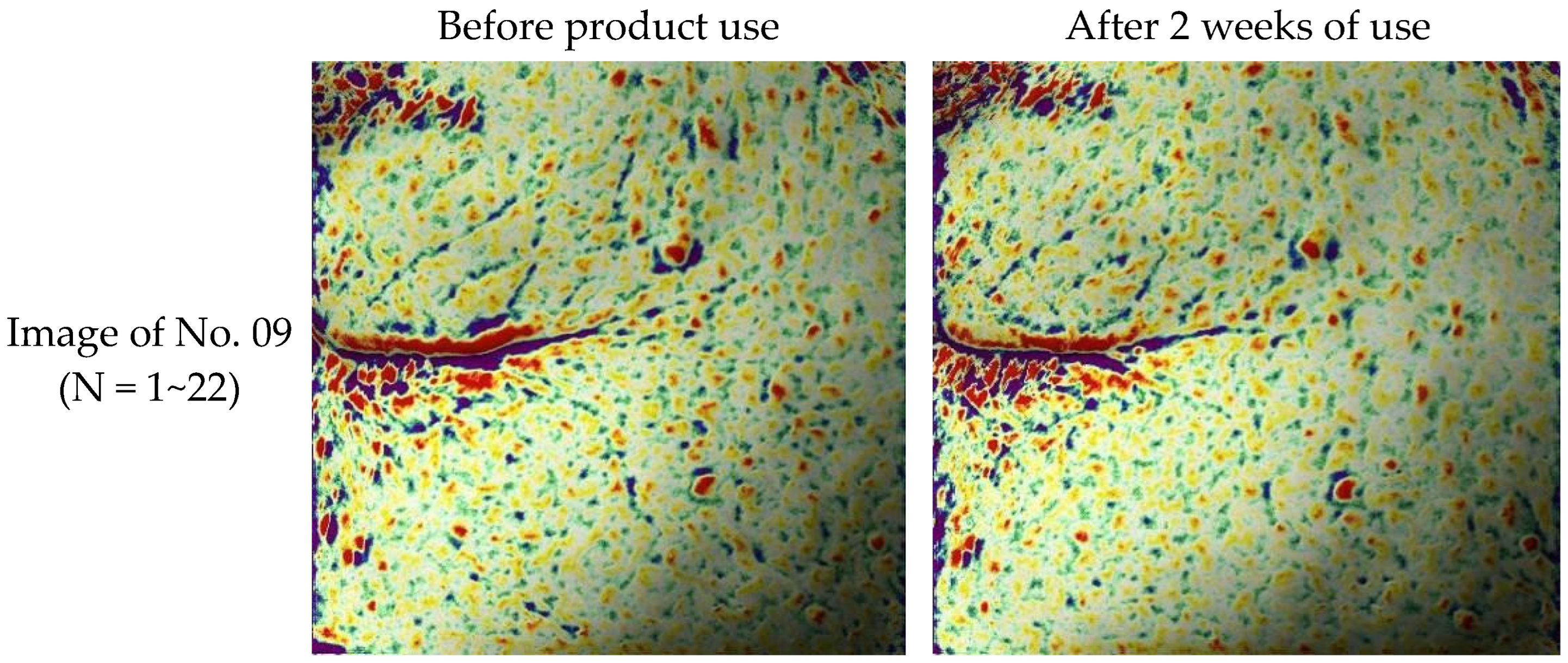
The arithmetic mean roughness (Ra, µm) measured using an Antera 3D significantly decreased from 8.36 ± 1.23 μm to 7.61 ± 1.14 μm (p < 0.001), an improvement rate of 8.97% (Table 13, Figure 5).
Table 13.
Antera 3D arithmetic mean roughness analysis results (EGF FLEXIR-SOME 2%).
Figure 5.
Wrinkle image obtained using an Antera 3D (EGF FLEXIR-SOME 2%).
- Lifting Effect (F-ray) of EGF FLEXIR-SOME 2%
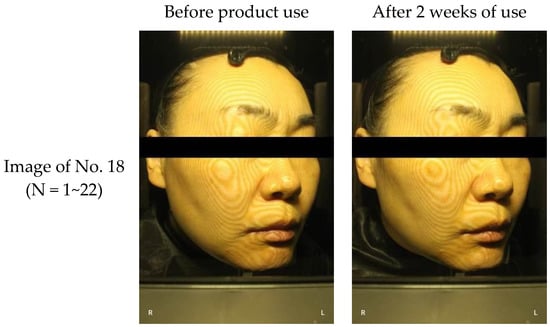
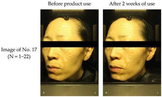
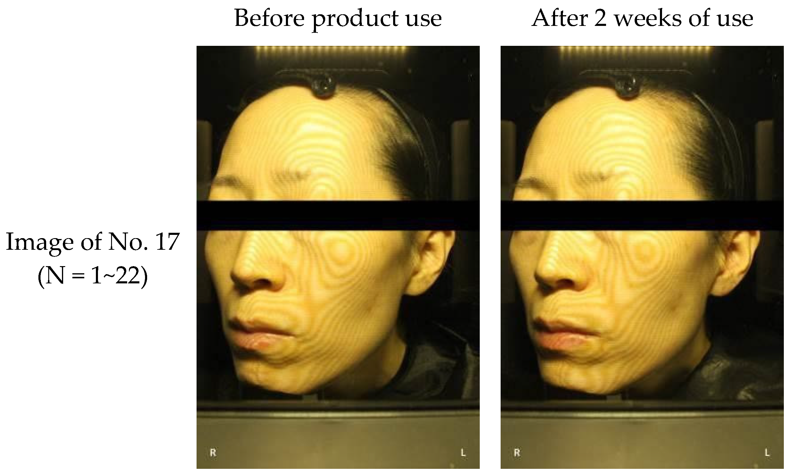
The skin sagging angle (°) on the cheek area decreased significantly from 37.86° ± 5.82 to 35.53° ± 5.74 (p < 0.001), an improvement of 6.15% (Table 14, Figure 6).
Table 14.
F-ray skin sagging angle analysis results (EGF FLEXIR-SOME 2%).
Figure 6.
Lifting image using F-ray (EGF FLEXIR-SOME 2%).
- Skin Aging Index Improvement in EGF FLEXIR-SOME 2%
The skin aging index significantly decreased from 39.47 ± 4.04 to 33.86 ± 4.06 (p < 0.001), representing a 14.21% improvement (Table 15).
Table 15.
Skin aging index analysis results (EGF FLEXIR-SOME 2%).
Patch Test Evaluation (EGF FLEXIR-SOME 2%)
Dermatological evaluation of skin irritation following 30 min and 24 h patch tests showed the product to be non-irritating (average skin reaction score 0.00) (Table 16). No adverse reactions or concomitant medication were reported.
Table 16.
Skin irritation assessment results (EGF FLEXIR-SOME 2%).
3.4.3. EGF FLEXIR-SOME 8% (EGF 4 ppm)
Anti-Aging Efficacy Evaluation Results
- Skin Elasticity Improvement (Cutometer MPA580) of EGF FLEXIR-SOME 8%
The R7 (mm) value improved significantly from 0.3158 ± 0.0305 mm to 0.3392 ± 0.0318 mm (p < 0.001), with a 7.41% improvement (Table 17).
Table 17.
Cutometer MPA580 R7 measurement results (EGF FLEXIR-SOME 8%).
- Wrinkle Reduction (Antera 3D) of EGF FLEXIR-SOME 8%
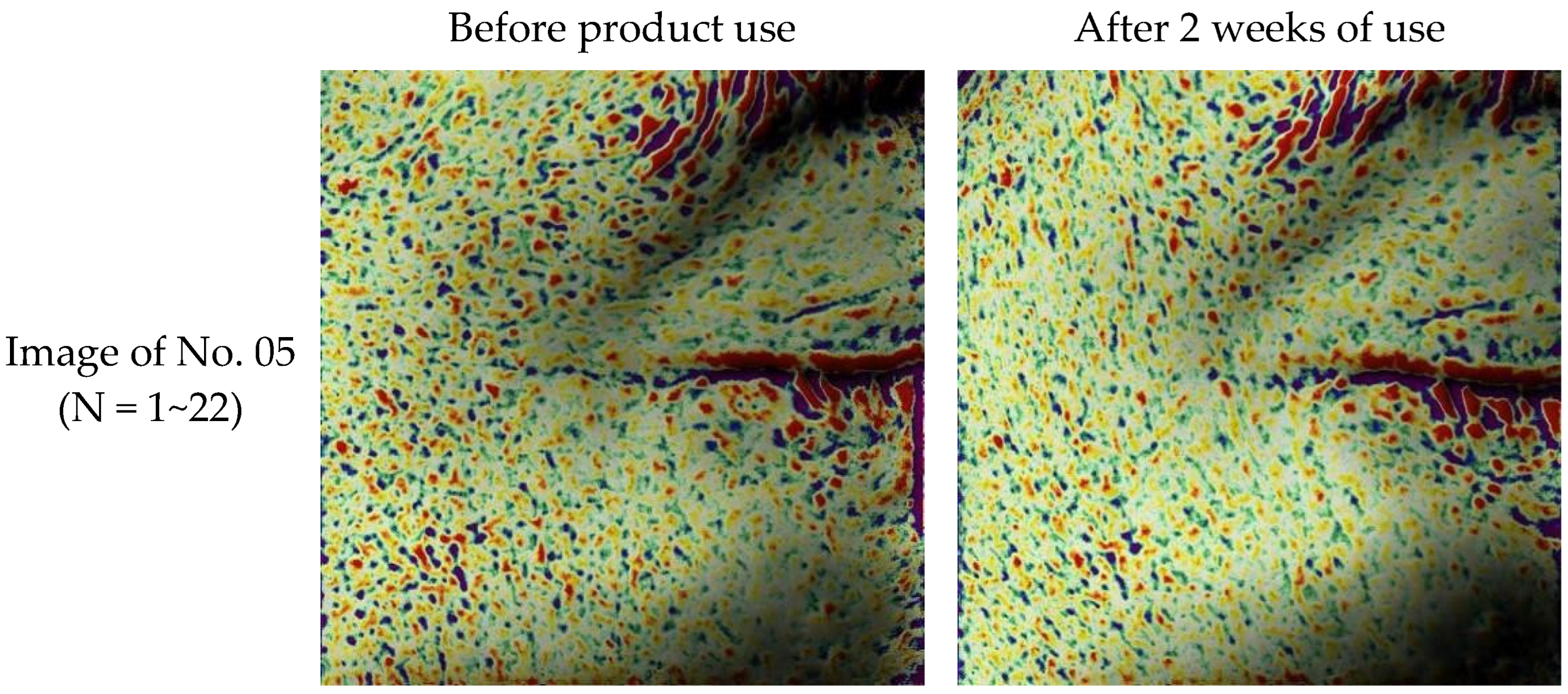
The arithmetic mean roughness (Ra, µm) decreased significantly from 8.08 ± 1.22 μm to 7.61 ± 1.12 μm (p < 0.001), with a 5.82% improvement (Table 18, Figure 7).
Table 18.
Antera 3D arithmetic mean roughness analysis results (EGF FLEXIR-SOME 8%).
Figure 7.
Wrinkle image obtained using an Antera 3D (EGF FLEXIR-SOME 8%).
- Lifting Improvement (F-ray) of EGF FLEXIR-SOME 8%
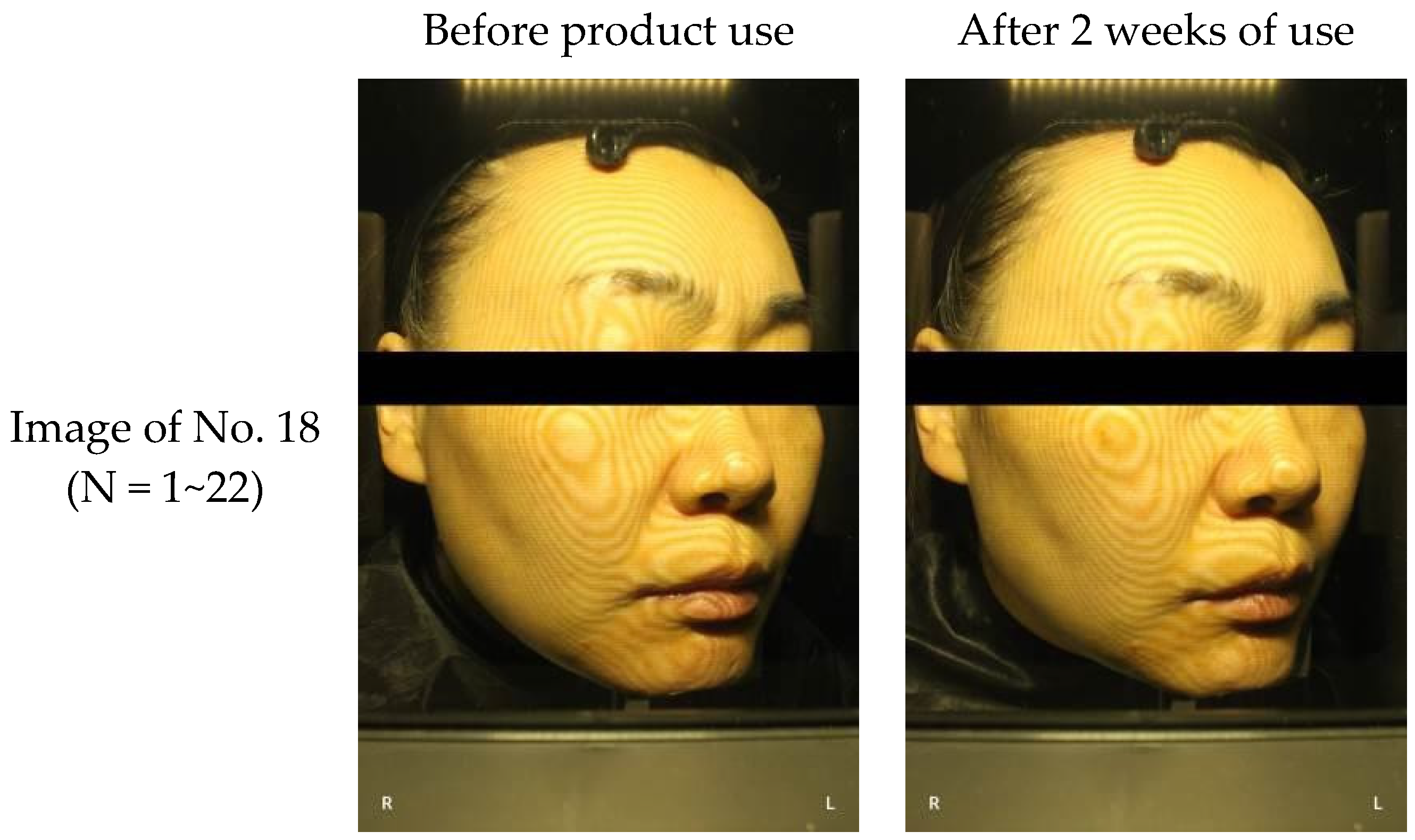
The skin sagging angle (°) of the cheek area was significantly reduced from 38.51° ± 5.19 to 36.79° ± 5.69 (p = 0.025), with a 4.23% improvement (Table 19, Figure 8).
Table 19.
F-ray skin sagging angle analysis results (EGF FLEXIR-SOME 8%).
Figure 8.
Lifting image using F-ray (EGF FLEXIR-SOME 8%).
- Skin Aging Index Improvement in EGF FLEXIR-SOME 8%
The skin aging index significantly decreased from 38.89 ± 4.54 to 34.81 ± 4.57 (p < 0.001), with a remarkable improvement rate of 10.49% (Table 20).
Table 20.
Skin aging index analysis results.
Patch Test Evaluation of EGF FLEXIR-SOME 8%
The Dermafactory Pro-EGF FLEXIR-SOME 8% Repair Cream was judged to be non-irritating through dermatological evaluation (average skin reaction score 0.00) (Table 21). No adverse reactions or concomitant medications were reported.
Table 21.
Skin irritation assessment results.
Skin Hydration, Repairing Damaged Skin Barrier, and Reducing Hyperpigmentation
- Subject characteristics
Twenty-one healthy Korean adult females (mean age 49.2 years, range 33–61) were included (one subject excluded for protocol violation). Detailed skin-type and lifestyle characteristics are summarized in Table 22.
Table 22.
Subject characteristics for the skin hydration, barrier repair and hyperpigmentation trial.
- Skin Hydration
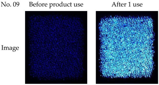
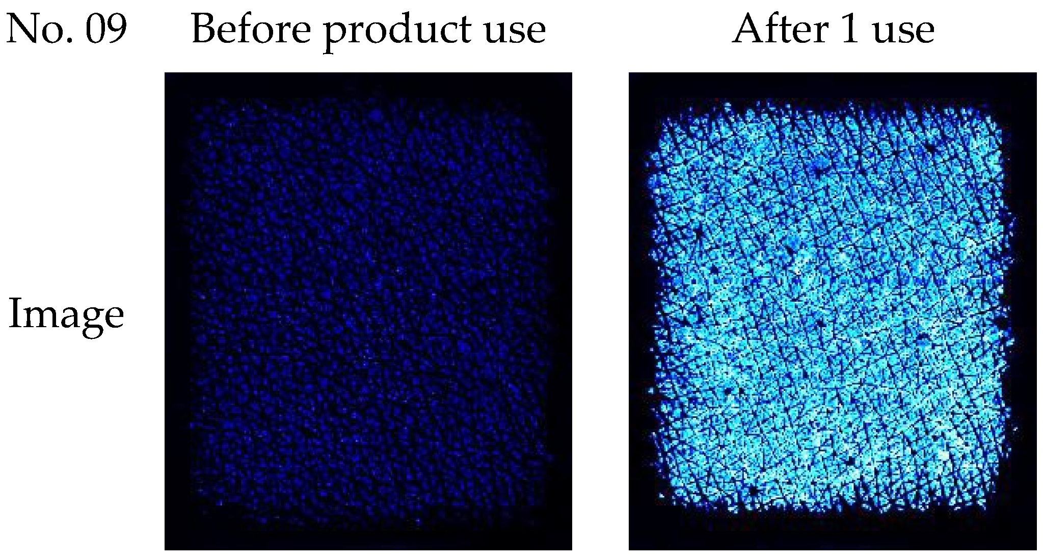
The average skin permittivity (ε) on the forearm increased significantly after one application of the test from 6.10 ± 1.97 to 33.46 ± 4.05 (p < 0.001), with an improvement of 448.52% (Table 23, Figure 9).
Table 23.
Epsilon E100 average permittivity measurement results.
Figure 9.
Image of skin hydration obtained using the Epsilon E100.
- Damaged Skin Barrier Repair
After physical irritation, transepidermal water loss (TEWL) significantly decreased in both the test and control groups (p < 0.001). The test group showed a significantly greater reduction than the control (31.57% vs. 10.59% improvement, p < 0.001), with a greater improvement of 197.96% (Table 24).
Table 24.
Transepidermal water loss (TEWL) measurement results.
- Hyperpigmentation and Melasma Improvement Efficacy Evaluation
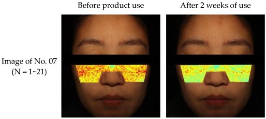
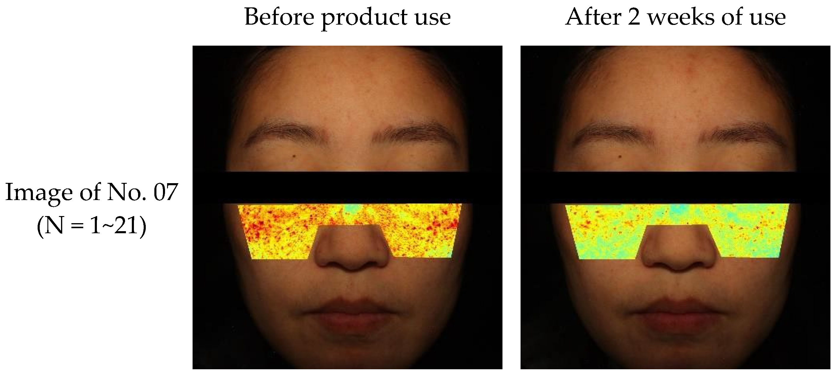
The cheek melanin content decreased significantly after two weeks of use from 73.55 ± 31.97 to 67.75 ± 33.85 (p = 0.009), with an improvement of 7.89% (Table 25, Figure 10).
Table 25.
DermaVision melanin content analysis results.
Figure 10.
Image of melanin content obtained using DermaVision.
- Adverse reactions or contact dermatitis
No adverse reactions, including allergic or irritant contact dermatitis, were reported or observed during this study.
4. Discussion
In this study, we successfully developed and comprehensively evaluated an EGF FLEXIR-SOME formulation, highlighting its enhanced delivery capabilities and multifunctional cosmetic efficacy. The primary innovation is the implementation of elastosome technology, which addresses a limitation of conventional liposomes: their rigid structure and suboptimal skin penetration [4,5,6]. By virtue of their flexible and deformable nature, elastosomes can transverse the narrow intercellular spaces of the stratum corneum more effectively, resulting in improved transdermal delivery of encapsulated bioactives [29]. The structural integrity and spherical morphology of these vesicles were visually confirmed via Cryo-TEM (Figure 3), providing a definitive anatomical basis for their barrier-navigating capability.
The EGF FLEXIR-SOME formulation demonstrated a smaller average particle size ( nm) and a significantly higher magnitude of zeta potential (−57.53 mV) than the control liposome ( nm, −45.08 mV). These values, derived from triplicate measurements (n = 3), confirm the high reproducibility of our microfluidization process. A high absolute zeta potential is indicative of colloidal stability, thereby supporting uniform dispersion and consistent skin delivery [30]. Physicochemical stability must be maintained over time to preserve the efficacy and integrity of bioactive components; furthermore, this stability directly promotes improved skin permeability. The above was further validated via the Franz-type diffusion cell skin penetration assay, wherein transdermal absorption of dexpanthenol was observed exclusively in the elastosome formulation, highlighting the advanced barrier-navigating capability of elastosomes.
The biological efficacy of the EGF FLEXIR-SOME formulation was substantiated by both in vitro and clinical evaluations. To ensure statistical transparency, all in vitro data are presented as Mean ± SD, and values were calculated to provide a standardized metric of potency. The EGF raw material exhibited superior performance, notably achieving an of 0.17% in the ABTS assay and an exceptional of 0.96% in the tyrosinase inhibition assay. While values for DPPH, collagenase, and NO inhibition exceeded the maximum tested concentrations, the experimental group consistently outperformed the control at all concentration levels (p < 0.05), validating its enhanced antioxidant and anti-aging potential. These findings were corroborated in clinical studies, which revealed statistically significant improvements in key anti-aging parameters. Following two weeks of product application, significant enhancements were observed in skin elasticity (R7 value) with a 9.87% increase, wrinkle reduction (Ra) with an 8.97% decrease, and skin lifting, contributing to a substantial improvement in the overall skin age index. Pigmentation reduction and skin barrier improvement were also confirmed, reinforcing the product’s multifunctional cosmetic utility.
The observed efficacy likely results from the synergistic effects of EGF’s regenerative function [2,3], facilitated by the superior penetration rate of the EGF FLEXIR-SOME, and the barrier-strengthening role of dexpanthenol [8]. This combined effect facilitates the deep and effective delivery of essential anti-aging components, including EGF and NMN, amplifying overall skin benefits. A notable finding of the clinical trials was the use of a split-face method, which allows for direct, side-by-side comparisons of two formulations within the same subject. This approach effectively controls for inter-individual variability and enhances data robustness [25]. Both 2% and 8% concentrations of the EGF Elastosome formulation demonstrated statistically significant improvements in anti-aging indices, indicating that even the lower concentration is clinically effective.
Safety assessments indicated excellent tolerability, with no adverse reactions, including allergic or irritant contact dermatitis, reported during the clinical trial. The product was classified as a non-irritant in primary skin irritation testing, an essential attribute for any cosmetic intended for daily use.
While this study successfully established the delivery system’s superiority over conventional liposomes through comprehensive in vitro comparative assays (Table 5, Table 6, Table 7, Table 8 and Table 9), a direct clinical comparison with a liposome-control cream was omitted to prioritize a focused dose–response analysis within the split-face protocol. In the clinical trial, both 2% and 8% concentrations demonstrated significant improvements; however, a clear dose-dependent differentiation was not observed. This lack of a distinct dose–response relationship is a primary limitation, likely due to the 2% concentration already reaching a biological saturation point within the 14-day study period, masking incremental benefits of the higher dose, or the potent efficacy of the elastosome platform masking incremental dose effects.
Nevertheless, limitations warrant further investigation. We primarily focused on the development of EGF FLEXIR-SOME and its clinical safety rather than conducting a comprehensive quantitative comparison of skin efficacy based on different delivery platforms in a clinical setting. Future studies should include additional clinical trials that directly compare EGF FLEXIR-SOME with a liposome-control cream to clinically validate the transdermal advantages observed in vitro. Furthermore, research involving larger cohorts and broader concentration ranges is needed to identify the precise biological saturation threshold for EGF delivery. Clinical trials comparing EGF FLEXIR-SOME against other advanced platforms, such as ethosomes or solid lipid nanoparticles, would also further define its competitive advantages. Since the current participants were restricted to healthy Korean adult females, expanded research involving diverse ethnic populations and across different age groups is essential to validate and refine the formulation’s generalizability and safety profile. Additionally, long-term studies are recommended to investigate the molecular synergies between EGF, NMN, and dexpanthenol at the cellular level.
5. Conclusions
The results of this study demonstrate that encapsulating epidermal growth factor (EGF) within elastosomes significantly enhances its skin penetration and bioavailability. Improved transdermal delivery was confirmed through permeability testing using dexpanthenol, which functions both as an active moisturizing agent and as a reference compound within the system. The EGF FLEXIR-SOME formulation significantly improved essential skin parameters, including elasticity, hydration, anti-aging effects, barrier repair, and pigmentation reduction. Collectively, these findings highlight the potential of this advanced elastosome delivery system as a multifunctional cosmetic platform for enhancing the efficacy of potent bioactive ingredients such as EGF while simultaneously supporting overall skin health.
Supplementary Materials
The following supporting information can be downloaded at: https://www.mdpi.com/article/10.3390/cosmetics13010010/s1, Table S1: Antioxidant effect (DPPH assay); Table S2: Antioxidant effect (ABTS assay); Table S3: Anti-aging effect (collagenase inhibition assay); Table S4: Whitening effect (tyrosinase inhibition assay); Table S5: Anti-inflammatory effect (NO inhibition assay).
Author Contributions
Conceptualization, S.G.H., G.S.C. and W.K.H.; Formulation Development and Characterization, E.M.K., M.S.K., S.Y.S., S.M.C., J.H.P. and J.H.K.; Clinical Trial Methodology and Data Collection, In Vitro Assays and Statistical Analysis, S.G.H., G.S.C. and W.K.H.; Writing—Original Draft Preparation, S.G.H.; Writing—Review and Editing, G.S.C.; Supervision, G.S.C. All authors have read and agreed to the published version of the manuscript.
Funding
This research received no external funding.
Institutional Review Board Statement
This study was approved by the Institutional Review Board (IRB) at Humantest Co., (Seoul, Republic of Korea) (IRB No. HE-IRB-P23-0225, HE-IRB-P23-0226) Approval Date: 22 September 2023.
Informed Consent Statement
Informed consent was obtained from all individual participants included in the study. Informed consent for publication was obtained from all identifiable human participants.
Data Availability Statement
The original contributions presented in this study are included in this article; further inquiries can be directed to the corresponding author.
Conflicts of Interest
The authors declare the following financial interests/personal relationships which may be considered potential competing interests: Authors Eun Mi Kim, Min Soo Kim, Si Young Song, Su Mi Choi, Jun Hyeong Park, and Ji Hye Kim are employees of B&B Korea Co., Ltd., which developed the proprietary EGF FLEXIR-SOME formulation evaluated in this study. Author Won Kyu Hong is an employee of Humantest Co., the institution that performed the clinical efficacy and safety evaluations. However, this study was conducted in strict accordance with the approved Institutional Review Board (IRB) protocols to ensure the objectivity and integrity of the clinical data. There are no other relevant financial or non-financial interests to disclose.
References
- Gorzelanny, C.; Mess, C.; Schneider, S.W.; Huck, V.; Brandner, J.M. Skin Barriers in Dermal Drug Delivery: Which Barriers Have to Be Overcome and How Can We Measure Them? Pharmaceutics 2020, 12, 684. [Google Scholar] [CrossRef] [Scilit]
- Shin, S.H.; Koh, Y.G.; Lee, W.G.; Seok, J.; Park, K.Y. The use of epidermal growth factor in dermatological practice. Int. Wound J. 2023, 20, 2414–2423. [Google Scholar] [CrossRef] [Scilit]
- Draelos, Z.D. The effect of a combination of recombinant EGF cosmetic serum and a crosslinked hyaluronic acid serum as compared to a fibroblast-conditioned media serum on the appearance of aging skin. J. Drugs Dermatol. 2016, 15, 738–741. [Google Scholar] [PubMed]
- Van Tran, V.; Moon, J.-Y.; Lee, Y.-C. Liposomes for delivery of antioxidants in cosmeceuticals: Challenges and development strategies. J. Control. Release 2019, 300, 114–140. [Google Scholar] [CrossRef] [Scilit] [PubMed]
- Aziz, D.E.; Abdelbary, A.A.; Elassasy, A.I. Fabrication of novel elastosomes for boosting the transdermal delivery of diacerein: Statistical optimization, ex-vivo permeation, in-vivo skin deposition and pharmacokinetic assessment compared to oral formulation. Drug Deliv. 2018, 25, 815–826. [Google Scholar] [CrossRef] [Scilit] [PubMed]
- Nayak, D.; Tippavajhala, V.K. A Comprehensive Review on Preparation, Evaluation and Applications of Deformable Liposomes. Iran. J. Pharm. Res. 2021, 20, 186–205. [Google Scholar] [CrossRef] [Scilit]
- Ebner, F.; Heller, A.; Rippke, F.; Tausch, I. Topical Use of Dexpanthenol in Skin Disorders. Am. J. Clin. Dermatol. 2002, 3, 427–433. [Google Scholar] [CrossRef] [Scilit]
- Proksch, E.; de Bony, R.; Trapp, S.; Boudon, S. Topical use of dexpanthenol: A 70th anniversary article. J. Dermatol. Treat. 2017, 28, 766–773. [Google Scholar] [CrossRef] [Scilit]
- Choi, S.J.; Lee, H.S. Dexpanthenol-containing cream enhances skin barrier repair and reduces inflammation after sodium lauryl sulphate-induced irritation. J. Korean Dermatol. Assoc. 2020, 58, 205–212. [Google Scholar]
- Laffleur, F.; Pschick, S.; Barthelmes, J.; Hauptstein, S.; Bernkop-Schnurch, A. Impact of Surfactants on Skin Penetration of Dexpanthenol. Curr. Drug Deliv. 2018, 15, 351–356. [Google Scholar] [CrossRef] [Scilit]
- Sun, Z.; Li, J.; Zheng, Y.; Zhang, J.; Bai, W.; Deng, X.; Wu, Z.; Xu, X.; Ding, W.; Qian, H.; et al. Small extracellular vesicles derived from mesenchymal stromal cells loaded with β-nicotinamide mononucleotide activate NAD+/SIRT3 signaling pathway-mediated mitochondrial autophagy to delay skin aging. Stem Cell Res. Ther. 2025, 1, 339. [Google Scholar] [CrossRef] [Scilit]
- Jafari, S.M.; Yinghe, H.; Bhesh, B. Production of sub-micron emulsions by ultrasound and microfluidization techniques. J. Food Eng. 2006, 82, 478–488. [Google Scholar] [CrossRef] [Scilit]
- Thomas, A.M.; Kapanen, A.I.; Hare, J.I.; Ramsay, E.; Edwards, K.; Karlsson, G.; Bally, M.B. Development of a liposomal nanoparticle formulation of 5-Fluorouracil for parenteral administration: Formulation design, pharmacokinetics and efficacy. J. Control. Release 2011, 150, 212–219. [Google Scholar] [CrossRef] [Scilit] [PubMed]
- Franz, T.J. Percutaneous absorption on the relevance of in vitro data. J. Investig. Dermatol. 1975, 64, 190–195. [Google Scholar] [CrossRef] [Scilit]
- Uchida, T.; Kadhum, W.R.; Kanai, S.; Todo, H.; Oshizaka, T.; Sugibayashi, K. Prediction of skin permeation by chemical compounds using the artificial membrane, Strat-MTM. Eur. J. Pharm. Sci. 2015, 25, 113–118. [Google Scholar] [CrossRef] [Scilit] [PubMed]
- Haq, A.; Goodyear, B.; Ameen, D.; Joshi, V.; Michniak-Kohn, B. Strat-M® synthetic membrane: Permeability comparison to human cadaver skin. Int. J. Pharm. 2018, 547, 432–437. [Google Scholar] [CrossRef] [Scilit] [PubMed]
- OECD. Test No. 428: Skin Absorption: In Vitro Method. In OECD Guidelines for the Testing of Chemicals, Section 4; OECD Publishing: Paris, France, 2004. [Google Scholar] [CrossRef] [Scilit]
- Brand-Williams, W.; Cuvelier, M.E.; Berset, C. Use of a free radical method to evaluate antioxidant activity. LWT-Food Sci. Technol. 1995, 28, 25–30. [Google Scholar] [CrossRef] [Scilit]
- Re, R.; Pellegrini, N.; Proteggente, A.; Pannala, A.; Yang, M.; Rice-Evans, C. Antioxidant activity applying an improved ABTS radical cation decolorization assay. Free Radic. Biol. Med. 1999, 26, 1231–1237. [Google Scholar] [CrossRef] [Scilit]
- Ghimeray, A.K.; Jung, U.S.; Lee, H.Y.; Kim, Y.H.; Ryu, E.K.; Chang, M.S. In vitro antioxidant, collagenase inhibition, and in vivo anti-wrinkle effects of combined formulation containing Punica granatum, Ginkgo biloba, Ficus carica, and Morus alba fruits extract. Clin. Cosmet. Investig. Dermatol. 2015, 8, 389–396. [Google Scholar] [CrossRef] [Scilit]
- Zolghadri, S.; Bahrami, A.; Hassan Khan, M.T.; Munoz-Munoz, J.; Garcia-Molina, F.; Garcia-Canovas, F.; Saboury, A.A. A comprehensive review on tyrosinase inhibitors. J. Enzym. Inhib. Med. Chem. 2019, 34, 279–309. [Google Scholar] [CrossRef] [Scilit]
- Korhonen, R.; Lahti, A.; Kankaanranta, H.; Moilanen, E. Nitric oxide production and signaling in inflammation. Curr. Drug Targets Inflamm. Allergy 2005, 4, 471–479. [Google Scholar] [CrossRef] [Scilit]
- Shin, D.M.; Lee, J.; Noh, H.; Jang, D.; Oh, S.J.; Park, J.H.; Lee, J.H. A Double-Blind, Split-Face, Randomized Study on the Effects and Safety of Intradermal Injection of Botulinum Toxin A (Incobotulinum Toxin A) in the Cheek. Ann. Dermatol. 2022, 34, 442–450. [Google Scholar] [CrossRef] [Scilit]
- Ryu, H.S.; Joo, Y.H.; Kim, S.O.; Park, K.C.; Youn, S.W. Influence of age and regional differences on skin elasticity as measured by the Cutometer. Skin Res. Technol. 2008, 14, 354–358. [Google Scholar] [CrossRef] [Scilit] [PubMed]
- Messaraa, C.; Metois, A.; Walsh, M.; Hurley, S.; Doyle, L.; Mansfield, A.; O’COnnor, C.; Mavon, A. Wrinkle and roughness measurement by the Antera 3D and its application for evaluation of cosmetic products. Skin Res. Technol. 2018, 24, 359–366. [Google Scholar] [CrossRef] [Scilit]
- Frosch, P.J.; Kligman, A.M. The soap chamber test. A new method for assessing the irritancy of soaps. J. Am. Acad. Dermatol. 1979, 1, 35–41. [Google Scholar] [CrossRef] [Scilit]
- Draize, J.H.; Woodard, G.; Calvery, H.O. Methods for the study of irritation and toxicity of substances applied topically to the skin and mucous membranes. J. Pharmacol. Exp. Ther. 1944, 82, 377–390. [Google Scholar] [CrossRef] [Scilit]
- Draelos, Z.D. Cosmetic Dermatology: Products and Procedures, 2nd ed.; Wiley-Blackwell: Hoboken, NJ, USA, 2012. [Google Scholar]
- Benson, H.A.E. Elastic Liposomes for Topical and Transdermal Drug Delivery. In Liposomes: Methods and Protocols; Springer: New York, NY, USA, 2017; Volume 1552, pp. 107–117. [Google Scholar] [CrossRef] [Scilit]
- Stachurski, J.; MichaŁek, M. The effect of the zeta potential on the stability of a non-polar oil-in-water emulsion. J. Colloid Interface Sci. 1996, 184, 433–436. [Google Scholar] [CrossRef] [Scilit] [PubMed]

Disclaimer/Publisher’s Note: The statements, opinions and data contained in all publications are solely those of the individual author(s) and contributor(s) and not of MDPI and/or the editor(s). MDPI and/or the editor(s) disclaim responsibility for any injury to people or property resulting from any ideas, methods, instructions or products referred to in the content. |
© 2026 by the authors. Licensee MDPI, Basel, Switzerland. This article is an open access article distributed under the terms and conditions of the Creative Commons Attribution (CC BY) license.