


Simple Summary
Rising global temperatures accelerate plant flowering, threatening agricultural productivity. While the transcriptional regulation of flowering is well understood, protein-level mechanisms such as protein degradation, ubiquitination, and liquid–liquid phase separation are now recognized as equally important mechanisms enabling rapid and reversible temperature responses. This review focuses on recently identified protein-level thermosensory modules that are important for flowering responses to elevated temperatures. Insights from these mechanisms open new avenues for engineering climate-resilient crops by targeting protein stability and phase separation behavior. However, most of the work in this field is from the model plant Arabidopsis, and the conservation of these posttranslational mechanisms has yet to be tested in crops.
Abstract
Global warming poses serious threats to plant reproduction and agricultural productivity by affecting the timing of flowering, a critical developmental transition. Although transcriptional regulation of flowering pathways has been extensively studied, posttranslational and protein-level regulatory mechanisms are gaining increasing attention as important thermosensory switches enabling rapid and reversible responses to temperature fluctuations. These mechanisms include temperature-dependent protein degradation, ubiquitination, liquid–liquid phase separation of intrinsically disordered proteins, protein sequestration, and dynamic protein–protein interactions. This review summarizes current understanding of posttranslational flowering time regulation under high-temperature conditions, focusing on the major interconnected thermosensory modules, such as the temperature-dependent proteostasis of floral repressors and the emergence of temperature-responsive liquid–liquid phase separation (LLPS) of intrinsically disordered proteins (IDPs). Recent discoveries indicate that temperature-responsive flowering relies not only on transcriptional networks but also on dynamic protein-level regulatory mechanisms, including ubiquitination, proteasomal degradation, and liquid–liquid phase separation. However, the fact that these mechanisms have not been validated in crop species leaves their translational potential an open question.
1. Introduction
Flowering is a critical developmental transition in the plant life cycle that determines reproductive success, seed production, and yield. Given its importance, the timing of this transition must be tightly regulated and synchronized with environmental conditions to maximize reproductive fitness. Several environmental factors can influence flowering; among them, temperature has emerged as a critical regulator of flowering time [1,2], especially under the current global warming conditions. Average global surface temperature has already increased by approximately 1.1 °C above pre-industrial levels, with an increasing frequency and intensity of heat waves [3]. Elevated temperatures impair reproductive development, shorten the grain-filling period, and reduce yield across major crops. In wheat, heat stress during anthesis and grain filling accelerates phenological development, shortens the grain-filling period, and reduces yield by 3–8% per °C above optimal temperatures [4]. In rice, high night-time temperatures during grain filling produce chalky grains with lower quality and yield losses of up to 7.1–8.1% per °C [5,6]. Temperatures above 35 °C reduce pollen germination and tube growth in maize, leading to kernel abortion and yield losses [7,8]. In soybean, heat stress during the reproductive phase causes yield losses of up to 25% [9]. In tomato, temperatures above 32 °C during anthesis negatively affect pollen tube growth and sugar accumulation, reducing both yield and quality [10].
The pronounced effects of elevated temperatures on reproductive development and yield underscore the need to understand the molecular mechanisms governing flowering, under changing environmental conditions. Plants have evolved complex regulatory networks that modulate flowering in response to both exogenous and endogenous cues. To date, over 400 genes have been functionally characterized as flowering regulators and categorized into eight genetically distinct pathways, including autonomous, vernalization, photoperiod, and ambient-temperature pathways [11]. Most of these pathways converge on a set of common genes, known as the floral integrators, such as the floral promoters FLOWERING LOCUS T (FT), TWIN SISTER OF FT (TSF), and SUPPRESSOR OF OVEREXPRESSION OF CONSTANS 1 (SOC1), as well as their upstream repressors FLOWERING LOCUS C (FLC), FLOWERING LOCUS M (FLM), and SHORT VEGETATIVE PHASE (SVP) [11,12]. However, transcriptional regulation alone does not fully explain the rapid, reversible, and quantitatively precise nature of temperature-responsive flowering.
FLC, FLM, and SVP are major floral repressors that interact to form floral repressor complexes, which fine-tune flowering across a broad range of temperatures [2,13,14]. FLC encodes an MADS-box transcription factor that acts as a potent floral repressor in many plant species, including Arabidopsis [15,16]. FLC is epigenetically regulated by Polycomb Repressive Complex 2 (PRC2) in response to prolonged cold exposure (vernalization) and transcriptionally regulated by the autonomous pathway [17,18,19]. FLC represses flowering by directly repressing the floral promoter genes FT and SOC1 [20]. FLM acts as a temperature-responsive floral repressor that is primarily regulated at the posttranscriptional level via temperature-sensitive alternative pre-mRNA splicing [13,21]. FLM predominantly produces the functionally repressive FLM-β splice variant at lower temperatures, while increasing temperatures shift splicing toward the generation of FLM-δ isoform, thereby relieving floral repression [13,22,23]. SVP is another highly potent floral repressor in the ambient-temperature pathway and plays a critical role in regulating temperature-responsive flowering, as svp mutants show temperature-insensitive early flowering across a broad range of temperatures (5–27 °C) [13]. Unlike FLC and FLM, SVP is primarily regulated at the posttranslational level. Its transcript levels are largely unperturbed across temperatures, whereas SVP protein abundance rapidly declines at higher temperatures through 26S proteasome-dependent degradation [13,24]. These three major floral repressors interact with each other to form floral complexes that directly repress the downstream floral promoter genes FT and SOC1 [14,21,25].
Alongside these MADS-box TFs, Phytochrome B (phyB) and EARLY FLOWERING 3 (ELF3) are also important floral repressors that modulate flowering in response to light and temperature [2]. PHYB is a prominent red light photoreceptor that indirectly represses flowering under different photoperiodic conditions, primarily by destabilizing the floral activator CO during the morning [26,27]. In contrast, ELF3 is a nuclear scaffold protein and a core component of the evening complex (EC), alongside ELF4 and LUX ARRHYTHMO (LUX), which repress downstream floral promoters such as GIGANTEA (GI) and FT [28,29]. PHYB and ELF3 directly interact, and this interaction enhances ELF3 protein stability by preventing its COP1-mediated proteasomal degradation [28,30]. phyB acts as a thermostat in Arabidopsis [31,32]. PhyB is converted from the inactive Pr state to the active Pfr state by both light and temperature; red light drives Pr-to-Pfr photoactivation, whereas the active Pfr form can spontaneously revert to the inactive Pr state through a high-temperature-accelerated process known as thermal reversion [33].
Liquid–liquid phase separation (LLPS) has recently emerged as a functionally important regulatory hub for fine-tuning protein activity. For instance, LLPS provides a specialized microenvironment that enables EMBRYO DEFECTIVE 1579 (EMB1579) to carry out diverse biological functions, including the regulation of flowering time through FLC [34] and the control of cell division required for root meristem maintenance through interactions with root developmental regulators [35]. PhyB has been shown to undergo phase separation to assemble liquid-like droplets, and the phase behavior of these droplets is directly regulated by temperature through its N-terminal extension (NTE) region [36]. High temperatures abolish phyB droplet formation and hence renders phyB nonfunctional at elevated temperatures [31], thereby promoting early flowering.
Although transcriptional mechanisms have long dominated studies of temperature-responsive flowering, recent discoveries reveal that protein-level regulation provides equally important thermosensory control. These mechanisms enable rapid, reversible, and highly dynamic responses to fluctuating temperatures. Given that core floral transition networks are highly conserved across angiosperms—particularly central thermal and photoperiodic regulators such as SVP, FLM, phyB, CO, and the evening complex component ELF3—the underlying posttranslational regulatory mechanisms are likely to be broadly conserved as well [37,38]. Therefore, mechanistic insights gained from model species may provide a valuable framework for identifying and engineering analogous thermosensory pathways in crop plants to enhance resilience under future climate-warming scenarios. The following sections discuss the major protein-level thermosensory modules that coordinate flowering under elevated temperatures.
2. The SVP–FLM Thermoregulatory Repressor Module
2.1. SVP as a Central Floral Repressor of Temperature-Responsive Flowering
SVP is one of the most important repressors of temperature-responsive flowering [39], especially under cooler ambient temperatures. SVP binds directly to the promoter regions of floral integrator genes FT and SOC1, causing their repression and thereby suppressing the reproductive transition [13,24]. A key breakthrough in ambient-temperature flowering research was the discovery that SVP protein levels strongly depend on temperature [13]. SVP accumulates at low temperatures but is rapidly degraded at higher temperatures, while its transcript levels remain largely unchanged [13], suggesting that warm-temperature-mediated regulation occurs primarily at the posttranslational level.
High temperatures accelerate SVP protein turnover via the 26S proteasome pathway. The half-life of the SVP protein significantly decreases as temperature increases, establishing a direct connection between temperature perception and flowering repression. The biological significance of this mechanism is demonstrated by svp mutants, which exhibit temperature-insensitive flowering across a broad range of temperatures [13]. This allows SVP to function as a core temperature-responsive protein switch, enabling flexible developmental responses.
2.2. Temperature-Dependent FLM Isoforms and Protein Interaction Dynamics
FLM, a major floral repressor, acts as a primary interaction partner of SVP. FLM undergoes temperature-dependent alternative splicing to generate two major isoforms, FLM-β and FLM-δ [23]. At cooler temperatures, plants accumulate higher levels of FLM-β, which forms a stable nuclear repressor complex with SVP [21]. This SVP–FLM-β complex strongly represses FT and SOC1 transcription. At higher temperatures, plants accumulate increased levels of the FLM-δ splice variant. FLM-δ lacks floral repressor activity and alters SVP stability and localization. FLM-δ promotes the cytoplasmic retention and ubiquitination of SVP, accelerating SVP degradation [21]. In short, warm temperatures regulate SVP-mediated flowering through two posttranslational mechanisms: reduced SVP protein stability and temperature-dependent alterations in SVP interaction partners [21]. Together, these posttranslational regulatory mechanisms confer high thermosensitivity to flowering regulation in Arabidopsis.
2.3. SVP Degradation via the CRL3-LFH1-UBC15 Complex
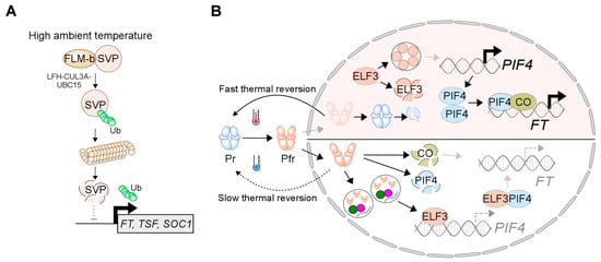
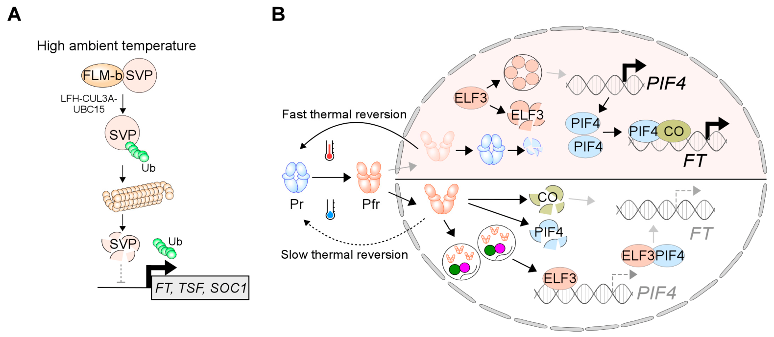
SVP has been shown to be regulated by the 26S proteasomal system in a temperature-dependent manner. However, the E3 ubiquitin ligase responsible for this process was identified only recently by Jin and colleagues [40]. Through protein pull-down screens of SVP interactors at different temperatures, they identified a BTB/POZ-domain protein, named LATE FLOWERING AT HIGH TEMPERATURE 1 (LFH1) as a high-temperature-specific interactor of SVP. LFH1 functions as a substrate adaptor within a CULLIN3–RING E3 ligase (CRL3) complex, bridging SVP to CUL3A and the cognate E2 ubiquitin-conjugating enzyme UBC15 [40] (Figure 1A).
Figure 1.
Temperature-dependent posttranslational regulation of flowering. (A) High temperature destabilizes the floral repressor SVP through LFH1–CUL3A–UBC15-mediated ubiquitination and 26S proteasomal degradation, relieving repression of FT, TSF, and SOC1. (B) Elevated temperature accelerates phyB reversion from the active Pfr to the inactive Pr state, affecting downstream regulators ELF3, PIF4, and CO. At low-temperature conditions (lower right), Pfr-phyB promotes degradation of CO and PIF4 and stabilizes ELF3 in nuclear photobodies, suppressing flowering. At warm temperature (upper right), Pfr-to-Pr reversion prevents photobody formation, and phyB is degraded, stabilizing CO and PIF4 to induce FT expression and promote flowering. Black arrows indicate induction, whereas gray arrows indicate failure to induce.
The lfh1 and cul3a mutants phenocopied SVP overexpression lines, as they displayed late flowering only at high ambient temperatures, highlighting that the regulatory complex is specifically required for high temperature-accelerated flowering. Furthermore, the SVP protein was highly stable in lfh1 mutants, without any difference in transcript levels. Interestingly, LFH1 protein accumulates to higher levels at higher temperatures. Because LFH1 promotes SVP degradation, this creates a feed-forward loop: rising temperatures increase LFH1 levels, which clear more SVP, further promoting flowering in a self-amplifying manner. This self-reinforcing mechanism distinguishes the CRL3–LFH1 module from simpler degradation pathways and likely contributes to the robustness and irreversibility of high-temperature-induced flowering. However, the role of FLM-β in modulating CRL3-LFH1-UBC15-mediated SVP degradation remains unclear and requires further research. While SVP degradation represents a major thermoregulatory mechanism, temperature-responsive flowering is also controlled through the stabilization and activity of transcriptional activators such as PIF4.
3. PIFs and Temperature-Regulated Protein Stability
PIF4 is a well-studied central integrator of temperature signaling, thermomorphogenesis, and flowering regulation [41]. At higher temperatures, PIF4 protein accumulates to higher levels and activates genes associated with hypocotyl elongation and flowering induction [42,43]. Among the flowering genes, PIF4 directly activates FT expression at high temperatures, especially under short-day photoperiods [42]. PIF4 activity is largely regulated at the protein level through dynamic interactions with photoreceptors such as phyB [44] and ubiquitin ligases such as BOPs [45].
The phyB-PIF4 module serves as a key hub for integrating light and temperature signals, with protein-level regulation of PIF4 protein being critical for thermomorphogenesis and flowering modulation. PIF4 acts as a key growth-promoting transcription factor that is stabilized at higher ambient temperatures. At elevated temperatures, phyB is less active and therefore impaired in its ability to interact with PIF4 and sequester it in the cytoplasm. This temperature-dependent reduction in phyB functionality allows PIF4 to accumulate in the nucleus and induce its target genes, thereby promoting flowering and hypocotyl elongation [31,32]. In addition to its nuclear translocation, PIF4 protein stability is modulated by the ubiquitin–proteasome system, with several E3 ubiquitin ligases, including BLADE-ON-PETIOLE (BOP) 1 and 2, triggering PIF4 degradation in a temperature-dependent manner. BOP proteins function as substrate adaptors in CUL3-based E3 ubiquitin ligase complexes that ubiquitinate PIF4 and hence promote its degradation via the 26S proteasome pathway. The efficiency of this degradation is fine-tuned by temperature and light conditions [45]. Under cool-to-moderate ambient temperatures (<23 °C), the BOP-mediated 26S proteasomal system rapidly degrades PIF4 to keep cell elongation in check. In contrast, elevated temperatures dampen this BOP-mediated degradation [45]. However, the fact that PIF4 ubiquitination and degradation are not completely abolished in bop mutants suggests the involvement of additional E3 ligases or proteolytic pathways that modulate PIF4 stability [45]. Contrary to BOPs, a recent report identified LIGHT-RESPONSE BRIC-A-BRACK/TRAMTRACK/BROAD (LRB) E3 ubiquitin ligases as positive regulators of PIF4 accumulation [46]. LRB1 and LRB2 physically interact with PIF4 and stabilize it, likely by inhibiting its degradation by phyB and possibly by counteracting the action of unknown E3 ubiquitin ligases. In lrb123 mutants, PIF4 protein levels were significantly reduced under both optimal and warm temperatures, confirming that LRBs are essential for normal PIF4 stability.
Additionally, LRBs also negatively regulate ELONGATED HYPOCOTYL 5 (HY5) by promoting its degradation via the 26S proteasomal pathway. HY5 functionally antagonizes PIF4 by competing for binding to the promoters of shared target genes [47]. LRB-mediated degradation of HY5 relieves this inhibitory effect on PIF4 resulting in enhanced activation of PIF4 target genes. Thus, LRBs constitute a dual regulatory model that simultaneously stabilize PIF4 and promotes the degradation of its functional antagonist, HY5, resulting in the fine-tuning of PIF4 activity [46]. However, additional regulators of PIF4 stability are likely to exist. Furthermore, as PIFs have functional redundancies, they are likely to undergo similar protein-level regulation, although this has yet to be experimentally validated. Collectively, these findings establish ubiquitin-mediated protein turnover as a key regulatory mechanism underlying temperature-responsive flowering, a theme that is conserved across multiple E3 ligase complexes in plants. Among these, the CONSTITUTIVE PHOTOMORPHOGENIC 1 (COP1) pathway represents one of the best-characterized mechanisms linking protein degradation to temperature-responsive flowering.
4. COP1-Mediated Protein Degradation at High Temperature
The COP1–SUPPRESSOR OF PHYA (SPA) E3 ubiquitin ligase complex integrates ambient-temperature signals from the photoperiodic, circadian, and hormonal flowering pathways into the plant flowering modules. Elevated temperatures promote the nuclear accumulation and importin-α/β-mediated transport of COP1, leading to the formation of tetrameric COP1–SPA holocomplexes [48]. This regulatory module is activated in the nucleus during the daytime and modulates proteasomal degradation cascades that target distinct master regulators of the photoperiodic, circadian, and hormonal flowering pathways.
In photoperiodic and circadian pathways, COP1 recognizes its target proteins through its WD40-repeat domains, and this interaction results in the polyubiquitination of HY5 and GI, marking them for rapid degradation via the 26S proteasome system [49,50]. The accelerated turnover of GI prevents it from forming a complex with FLAVIN-BINDING KELCH REPEAT F-BOX 1 (FKF1), which normally promotes the degradation of the flowering repressor CYCLING DOF FACTOR 1 (CDF1) [51]. Consequently, the accumulation of CDF1 transcriptionally represses FT, thereby delaying flowering under extreme heat [52]. Concurrently, the COP1–SPA1 module directly interacts with DELLA repressors, such as REPRESSOR OF GA1 (RGA) and GA-INSENSITIVE (GAI), leading to their degradation in Arabidopsis thaliana, as well as that of MdRGL1a in Malus domestica, under high-temperature stress independently of the canonical GA pathway [53,54]. Interestingly, this posttranslational regulation has divergent, species-specific developmental effects, accelerating flowering in some while causing severe floral repression in other species [53,54]. In addition to protein degradation, plants exploit the biophysical properties of proteins to sense temperature through phase separation.
5. Phase Separation: A New Paradigm for Temperature Sensing
Cells contain several membrane-less compartments and biomolecular condensates that offer unique microenvironments for macromolecules to concentrate and play their specific biological roles [55]. LLPS is a process in which a solution separates into two or more distinct liquid phases that coexist, thereby forming biomolecular condensates [56]. Phase separation primarily occurs for proteins with multivalency conferred by intrinsically disordered regions (IDRs) and/or multiple folded domains [57]. IDPs lack stable tertiary structures under physiological conditions and have emerged as a versatile platform for sensing and responding to environmental and developmental cues. Unlike properly structured proteins with fixed conformational states, IDPs are structurally flexible and undergo reversible, stimulus-responsive phase transitions, subsequently dissolving when favorable conditions are restored.
In recent years, LLPS has gained considerable attention as an increasing number of studies have demonstrated its biological significance in modulating plant development and responses to environmental cues [58]. For instance, the major floral repressor FLC has been shown to be regulated by LLPS. The transcriptional repressor REDUCED VERNALIZATION RESPONSE 1 (VRN1) undergoes LLPS driven by its two B3 domains and an IDR, resulting in dynamic nuclear condensates that results in the epigenetic silencing of FLC [59]. Similarly, the RNA-binding protein FLOWERING CONTROL LOCUS A (FCA) contains a prion-like domain (PrLD) that assembles into nuclear condensates with the help of coiled-coil protein FLL2, which acts as a scaffold. These condensates selectively recruit RNA 3′-end-processing factors that polyadenylate COOLAIR antisense transcripts, epigenetically repressing FLC and inducing flowering [60]. In contrast, the IDR-containing transcriptional activator SUF4 acts as a posttranslational temperature sensor and undergoes temperature-dependent phase separation, forming nuclear condensates that stabilize FLC transcription at warm temperatures to repress flowering. At cooler temperatures, these condensates dissolve, lowering the barrier to vernalization [61]. However, this review focuses on the role of LLPS in regulating temperature-responsive flowering at higher temperatures.
5.1. GI LLPS Modulate SVP-Mediated Temperature-Responsive Flowering
Recent studies have shown how plants utilize LLPS to sense and integrate temperature cues into endogenous photoperiodic pathway [62]. The FLAVIN-BINDING, KELCH REPEAT, F-BOX 1 (FKF1) protein and its interacting partner GI, core components of daytime photoperiodic signaling, play important roles in overriding photoperiodic constraints to drastically accelerate flowering. Under long-day (LD) conditions, the FKF1 and GI protein levels both peak in the late afternoon, resulting in the formation of FKF1–GI complexes that promote the degradation of CDF proteins and hence prevent CDF-mediated repression of CO and FT, thereby modulating day length-dependent flowering [51]. FKF1–GI complexes also physically interacts with CO protein, stabilizing it and enhancing its ability to induce FT under LD conditions [51,62].
This FKF1–GI complex also plays an important role in accelerating flowering time at elevated temperatures under non-inductive photoperiodic conditions by inducing FT transcript levels [63]. This high-temperature-induced FT expression is governed by the LLPS of GI protein. Lower ambient temperatures promote GI phase separation, resulting in its sequestration and functional inactivation within nuclear condensates. At warm ambient temperatures, FKF1 accumulates to high levels and interacts with GI, reversing GI phase separation and bringing GI back to its active state. The FKF1-GI complex then physically interacts with SVP, recruiting it to an E3 ubiquitin ligase complex for ubiquitination and subsequent 26S proteasomal degradation [63]. The rapid degradation of SVP relieves its direct transcriptional repression of FT and thereby facilitates flowering under warm conditions. Although this work clearly demonstrates that FKF1-mediated reversal of GI phase separation controls the turnover of a major floral repressor, several questions remain, particularly regarding the relative contributions of CO and PIF proteins to this high-temperature-mediated FT induction and floral acceleration.
5.2. The ELF3 Prion-like Domain as a Thermosensor: Phase Separation Links Temperature to Flowering Time Control
ELF3, a core scaffold component of the evening complex, contains a polyglutamine (polyQ) repeat embedded within its prion domain (PrD) [64,65]. The length of this polyQ repeat-containing PrD region positively correlates with high temperature-induced hypocotyl elongation and early flowering. Variations in the length of PrD result in differential temperature responsiveness and hence adaptation to climate conditions with different ambient temperatures. ELF3 orthologs from species naturally adapted to warmer climates, such as Brachypodium distachyon, lack a PrD and are therefore unable to confer temperature-responsive flowering when expressed in Arabidopsis. Swapping the Arabidopsis PrD with the B. distachyon PrD abolished the thermosensitivity of chimeric Arabidopsis ELF3 (AtELF3-BdPrD), suggesting that the PrD itself is the functional thermosensory unit. This PrD was shown to be important for regulation of flowering at elevated temperatures. Overexpression of Arabidopsis ELF3 did not alter flowering, implying that increased ELF3 protein levels alone are not sufficient to repress flowering [66]. The ELF4, another EV complex protein and binding partner of ELF3, stabilizes ELF3 activity. Under low-temperature conditions, ELF4 becomes dispensable, whereas overexpression of ELF4 maintains ELF3 in an active state and enables its chromatin-binding ability even at a warmer temperature of 27 °C [66].
5.3. PhyB Photobodies Integrate Light and Temperature Signals Through Phase Separation
Phytochrome B (phyB) acts as a dual receptor for both light and temperature cues in plants. Chen and colleagues [36] showed that light-activated phyB undergoes LLPS to form nuclear photobodies (PBs). The capacity for LLPS is modulated by two structurally distinct domains with complementary roles: the C-terminal domain, which mediates light-independent self-oligomerization and hence establishes the multivalency required for condensate assembly, and the intrinsically disordered N-terminal extension (NTE), which functions in phase separation and enables light-dependent regulation of condensate formation. Light-induced conformational changes expose the buried NTE and thereby enable NTE-mediated multivalent interactions that promote oligomerization and subsequent PB assembly. Deletion of the NTE abolishes both droplet formation and phyB activity, while replacing it with heterologous IDRs restores its condensate formation but eliminates light-dependent regulation, suggesting that NTE possesses both a general LLPS capacity and a sequence-specific function in connecting condensation to light status.
High-temperature-mediated regulation of phyB activity occurs through a mechanistically distinct pathway that converges on the same LLPS platform. phyB condensates are intrinsically thermosensitive; they disassemble as temperature rises and reassemble reversibly upon cooling, indicating that temperature directly modulates condensate dynamics independent of Pfr-to-Pr dark reversion. This thermosensitivity is entirely dependent on the NTE region, as loss of the NTE makes the condensates temperature-insensitive, which cannot be rescued by heterologous IDRs, suggesting that the NTE contains sequence-specific thermosensory capacity. Once formed, phyB photobodies selectively recruit interacting transcription factors, including PIF3 and PIF4, into concentrated microreactors, and transgenic plants expressing NTE-deleted phyB lose elevated temperature-induced hypocotyl elongation despite retaining partial light responses (Figure 1B). Collectively, phyB photobodies act as flexible sensory condensates that decode two distinct environmental cues through separate mechanisms. Light is processed through allosteric conformational changes that gate condensate assembly, whereas temperature is sensed through NTE-dependent regulation of condensate stability, enabling a single receptor to precisely integrate both signals [36]. While phyB photobodies sense temperature through condensate dynamics, plants have evolved parallel chromatin-based mechanisms that translate thermal signals into changes in gene expression. One of the most prominent chromatin-based thermosensory mechanisms involves the histone variant H2A.Z.
6. H2A.Z-Mediated Chromatin Remodeling in Temperature-Responsive Flowering
The histone variant H2A.Z is a key mediator of this chromatin-based thermosensory response, marking temperature-responsive genes across the genome [67]. In Arabidopsis thaliana, H2A.Z is encoded by HISTONE H2A (HTA) 8, HTA9, and HTA11 [68]. Its deposition into target chromatin is mediated by the ATP-dependent SWR1 chromatin-remodeling complex (SWR1-C), which catalyzes the exchange of H2A–H2B dimers for H2A.Z–H2B dimers [69,70]. H2A.Z incorporation occurs through an ATP-dependent process involving nucleosome priming, DNA unwrapping, and the subsequent replacement of H2A-containing dimers [71]. In Arabidopsis, SWR1-C consists of several conserved subunits, including PHOTOPERIOD INDEPENDENT EARLY FLOWERING 1 (PIE1), ACTIN-RELATED PROTEIN 6 (ARP6), SWR COMPLEX 6 (SWC6), ARP4, YEAST AF9 (YAF9), and other conserved subunits [69,72,73,74,75]. Recruitment of SWR1-C to target loci occurs through several mechanisms, including the recognition of AT-rich DNA elements by SWC4 and recruitment by ALFIN-LIKE proteins through H3K4me3 recognition [76,77]. Emerging evidence further suggests that LLPS-mediated transcriptional condensates may facilitate local concentration of chromatin remodelers at warm-temperature-responsive loci [78]. However, this hypothesis has yet to be experimentally validated.
Genetic and genomic studies have established H2A.Z as a central regulator of temperature-responsive transcription. A forward genetic screen identified arp6 mutants that exhibited aberrant HSP70 expression at low temperatures, demonstrating that H2A.Z normally represses heat-responsive genes under non-inductive conditions [68]. Consistently, genome-wide chromatin immunoprecipitation (ChIP) assays revealed the rapid depletion of H2A.Z from the gene body and transcription start site regions of warm-temperature-responsive genes in response to elevated temperatures [79]. This process occurs within 15 min of transfer to 27 °C and requires HEAT SHOCK FACTOR 1 (HSF1), which promotes H2A.Z-to-H2A exchange at heat-responsive loci [79]. The antagonistic relationship between H2A.Z and HSF1 provides an effective mechanism to prevent inappropriate transcription at low temperatures while enabling rapid activation upon warming [79].
Beyond warm-temperature-responsive gene expression, H2A.Z plays important roles in flowering time regulation. Mutants lacking H2A.Z or SWR1-C components, including pie1, arp6, swc6, arp4, and yaf9, exhibit early flowering and reduced sensitivity to ambient-temperature changes [69,72,75,80,81,82], phenocopying plants grown under warm conditions. These findings suggest that H2A.Z eviction is an early thermosensory event that promotes the floral transition.
Mechanistically, H2A.Z regulates the expression of several flowering repressors, including FLC, MAF4, and MAF5 [80,81,82,83]. However, the early flowering phenotypes of H2A.Z-deficient plants cannot be fully explained by these targets alone. Warm-temperature-induced eviction of H2A.Z from the promoter region of FT likely facilitates access by transcriptional activators such as CO and PIF4 [42,84,85], thereby enhancing FT expression and promoting flowering.
Despite significant progress, important questions remain unanswered. The molecular mechanisms governing the fate of evicted H2A.Z–H2B dimers, their redistribution between chaperone-bound pools and chromatin, and the spatiotemporal dynamics of H2A.Z turnover remain poorly understood. Furthermore, whether LLPS-mediated condensates regulate H2A.Z deposition or eviction at thermoresponsive loci has not been experimentally tested [78].
7. Future Directions: Engineering Resilience Through Posttranslational Precision
7.1. Manipulation of SVP-FLM Interaction
Biasing FLM splicing toward FLM-β by modifying splicing elements could reduce SVP cytoplasmic retention and subsequently its proteolytic turnover, preventing global warming-induced acceleration of flowering. The K53 and K165 residues are critical ubiquitination sites on SVP. They can be precisely altered through base editing to produce conservative lysine-to-arginine substitutions, which could stabilize SVP at elevated temperatures and hence mitigate heat-induced flowering acceleration in crops where extending the vegetative growth period under warming is agronomically valuable, such as leafy vegetables, forage grasses, or biomass crops. In addition, producing dominant-negative LFH1 variants or interrupting the LFH1–SVP interaction by manipulating the Kh2C domain of SVP or/and the C-terminal region of LFH1 could stabilize SVP in crops where premature flowering under warming reduces yield. However, because SVP is a major developmental regulator, its stabilization may impose trade-offs between prolonged vegetative growth and reproductive fitness. Excessive SVP activity could delay floral transition and reduce reproductive performance; therefore, engineering SVP stability would require careful optimization to balance heat resilience with agronomic productivity.
7.2. Manipulating the LLPS of ELF3 and PhyB
LLPS is a highly promising and rapidly evolving aspect underlying plants responses to diverse endogenous and environmental factors, including high temperatures. A promising avenue for climate-resilient agriculture is the manipulation of phase separation properties to engineer crops with tailored temperature sensitivities.
Variations in the length of the PrD region result in differential temperature responsiveness and, subsequently, adaptation to climates with different ambient temperatures. Because ELF3 functions as a direct thermosensor within the EV complex through a polyglutamine (polyQ) repeat embedded in its PrD, precision genome-editing strategies could be used to modulate the temperature threshold of its phase separation. Using tools like CRISPR-Cas9 to precisely shorten the polyQ or introduce stabilizing amino acid substitutions within the PrD could prevent the heat-induced LLPS and its subsequent transcriptional deactivation, which triggers premature flowering. By structurally hardening ELF3 against thermal inactivation, EV complex-mediated repression can be maintained at warmer temperatures, delaying premature reproductive transition. This provides a potential framework for engineering temperature-responsive traits in crops, although its agronomic benefits remain to be experimentally validated.
PhyB functions as a thermosensor and undergoes light-activated LLPS to form subnuclear photobodies. To suppress the selective recruitment of downstream transcription factors like PIF4, future bioengineering strategies should focus on precision editing of this regulatory module. CRISPR-Cas9 tools could be used to alter specific residues within the regulatory NTE, thereby structurally stabilizing phyB photobodies against heat-induced dissolution, maintaining active transcriptional regulation and preventing the premature, heat-accelerated flowering that typically penalizes crop yields. Furthermore, mapping the natural variation in phyB LLPS dynamics across diverse plant species will supply a valuable genetic toolkit for marker-assisted selection, establishing a tunable framework to safeguard global food security in a warming climate. A recent study has reported phyB variants, phyBG564E and phyBG515E, that stabilize the active Pfr form and were able to form high-temperature-insensitive nuclear photobodies, hence preventing high-temperature-induced thermal reversion and subsequent heat-induced hypocotyl elongation in Arabidopsis and stem elongation in rice [86]. Although the effects of these variants on flowering outcome have yet to be tested, they are likely to resist warm-temperature-induced flowering acceleration as well, offering a potential strategy for developing climate-resilient crops.
Despite recent progress in understanding temperature-responsive flowering at the posttranslational level, the picture remains incomplete. Several critical questions remain unresolved, and targeted work on posttranslational regulation under elevated temperatures is still needed. Notably, most posttranslational mechanisms identified to date in response to elevated temperatures promote flowering. To our knowledge, feedback loops that reset thermosensory posttranslational switches have not yet been identified. Future studies should investigate whether plants possess posttranslational mechanisms that actively prevent precocious flowering under warming conditions. Furthermore, most posttranslational research has been carried out in model plants, primarily Arabidopsis thaliana, and whether these regulatory mechanisms are conserved in agriculturally important crop species remains largely untested.
7.3. Beyond Arabidopsis: Posttranslational Regulation of Flowering in Crops Remains Largely Uncharted
Most of our understanding of posttranslational regulation of flowering has been derived from work in Arabidopsis, leaving a significant gap in knowledge for crop species. While orthologs of key regulators such as PIFs, FLC, SVP, FT, and CO exist in other plant species, whether the same ubiquitin-mediated degradation, phase separation, or temperature-sensitive protein interaction mechanisms operate in these species remains largely untested. Closing this gap will require systematic validation across diverse species and is critical for translating these mechanisms into crop-improvement strategies.
Recent evidence suggests that LLPS may represent a broadly conserved regulatory principle across plant species [87]. Large-scale proteomic screens have identified hundreds of potential phase separation proteins across diverse plant species, including rice, maize, wheat, and tomato. Interestingly, while the primary sequences of IDRs show limited conservation, their presence is highly conserved, indicating that the capacity for LLPS may be under positive selection throughout plant evolution [87]. However, these experiments were performed using a cell-free system and hence require validation under in vivo conditions.
Functional evidence linking LLPS to flowering regulation in crops has begun to emerge. In rice, the prion-like protein (PrLP) EHD6 (EARLY HEADING DATE 6) recruits YTH07, an m6A reader protein, to form ribonucleoprotein (RNP) condensates. These granules facilitate the sequestration of the mRNA of the flowering repressor OsCOL4 (CONSTANS-like 4), which is critical for reducing OsCOL4 abundance and accelerating flowering [88]. Nevertheless, mechanistic insights into how elevated temperatures influence condensate dynamics and flowering-related posttranslational networks in crops remain scarce.
8. Conclusions
Collectively, recent advances demonstrate that temperature-responsive flowering is governed not only by transcriptional regulation but also by an interconnected network of posttranslational mechanisms. Protein degradation, ubiquitination, sequestration, and phase separation collectively enable plants to rapidly adjust flowering time in response to fluctuating temperatures. Understanding and harnessing these processes may facilitate the development of climate-resilient crops adapted to a warming climate. However, despite substantial progress, our understanding of these mechanisms remains incomplete. Most protein-level thermosensory pathways discussed in this review have been characterized primarily in Arabidopsis thaliana, and their conservation, biological significance, and agronomic utility in crop species remain largely unexplored. Furthermore, the mechanistic integration of ubiquitin-mediated proteolysis, phase separation, chromatin remodeling, and temperature-sensitive protein interactions within a unified thermosensory network is not yet fully understood. Addressing these knowledge gaps will be essential for translating fundamental discoveries into practical crop-improvement strategies under future climate scenarios.
Author Contributions
Conceptualization, formal analysis, investigation, data curation, writing—original draft preparation, Z.N.; writing—review and editing, N.K.; visualization, Z.N. and N.K.; supervision, Z.N. All authors have read and agreed to the published version of the manuscript.
Funding
This work was supported by the National Research Foundation of Korea (NRF) Brain Pool Program (BP) grant (RS-2023–00263673) to Z.N.
Institutional Review Board Statement
Not applicable.
Informed Consent Statement
Not applicable.
Data Availability Statement
No new data were created or analyzed in this study. Data sharing is not applicable to this article.
Acknowledgments
We apologize to those authors whose work we were unable to cite because of space limitations.
Conflicts of Interest
The authors declare no conflicts of interest.
References
- Srikanth, A.; Schmid, M. Regulation of flowering time: All roads lead to Rome. Cell. Mol. Life Sci. 2011, 68, 2013–2037. [Google Scholar] [CrossRef] [Scilit] [PubMed]
- Susila, H.; Nasim, Z.; Ahn, J.H. Ambient temperature-responsive mechanisms coordinate regulation of flowering time. Int. J. Mol. Sci. 2018, 19, 3196. [Google Scholar] [CrossRef] [Scilit] [PubMed]
- Masson-Delmotte, V.; Zhai, P.; Pirani, A.; Connors, S.L.; Péan, C.; Berger, S.; Caud, N.; Chen, Y.; Goldfarb, L.; Gomis, M.I. Climate Change 2021: The Physical Science Basis. Contribution of Working Group I to the Sixth Assessment Report of the Intergovernmental Panel on Climate Change 2021; Cambridge University Press: Cambridge, UK; New York, NY, USA, In press; Volume 2, p. 2391.
- Asseng, S.; Ewert, F.; Martre, P.; Rötter, R.P.; Lobell, D.B.; Cammarano, D.; Kimball, B.A.; Ottman, M.J.; Wall, G.W.; White, J.W.; et al. Rising temperatures reduce global wheat production. Nat. Clim. Change 2015, 5, 143–147. [Google Scholar]
- Sreenivasulu, N.; Butardo, V.M., Jr.; Misra, G.; Cuevas, R.P.; Anacleto, R.; Kavi Kishor, P.B. Designing climate-resilient rice with ideal grain quality suited for high-temperature stress. J. Exp. Bot. 2015, 66, 1737–1748. [Google Scholar] [CrossRef] [Scilit] [PubMed]
- Song, Y.; Wang, C.; Linderholm, H.W.; Fu, Y.; Cai, W.; Xu, J.; Zhuang, L.; Wu, M.; Shi, Y.; Wang, G.; et al. The negative impact of increasing temperatures on rice yields in southern China. Sci. Total Environ. 2022, 820, 153262. [Google Scholar] [CrossRef] [Scilit] [PubMed]
- Schlenker, W.; Roberts, M.J. Nonlinear temperature effects indicate severe damages to US crop yields under climate change. Proc. Natl. Acad. Sci. USA 2009, 106, 15594–15598. [Google Scholar] [CrossRef] [Scilit] [PubMed]
- Lobell, D.B.; Bänziger, M.; Magorokosho, C.; Vivek, B. Nonlinear heat effects on African maize as evidenced by historical yield trials. Nat. Clim. Change 2011, 1, 42–45. [Google Scholar] [CrossRef] [Scilit]
- Puteh, A.B.; ThuZar, M.; Mondal, M.M.A.; Abdullah, A.P.; Halim, M.R.A. Soybean [Glycine max (L.) Merrill] seed yield response to high temperature stress during reproductive growth stages. Aust. J. Crop Sci. 2013, 7, 1472–1479. [Google Scholar]
- Wahid, A.; Gelani, S.; Ashraf, M.; Foolad, M.R. Heat tolerance in plants: An overview. Environ. Exp. Bot. 2007, 61, 199–223. [Google Scholar] [CrossRef] [Scilit]
- Bouché, F.; Lobet, G.; Tocquin, P.; Périlleux, C. FLOR-ID: An interactive database of flowering-time gene networks in Arabidopsis thaliana. Nucleic Acids Res. 2016, 44, D1167–D1171. [Google Scholar] [PubMed]
- Karim, N.; Nasim, Z.; Ahn, J.H.; Lee, H.-J. Natural variations in FLOWERING LOCUS C and MADS AFFECTING FLOWERINGs modulate both thermosensory and photoperiodic flowering in Arabidopsis. J. Exp. Bot. 2026, erag056. [Google Scholar] [CrossRef] [Scilit] [PubMed]
- Lee, J.H.; Ryu, H.-S.; Chung, K.S.; Posé, D.; Kim, S.; Schmid, M.; Ahn, J.H. Regulation of temperature-responsive flowering by MADS-box transcription factor repressors. Science 2013, 342, 628–632. [Google Scholar] [PubMed]
- Nasim, Z.; Susila, H.; Jin, S.; Youn, G.; Ahn, J.H. Polymerase II–Associated factor 1 complex-regulated FLOWERING LOCUS C-clade genes repress flowering in response to chilling. Front. Plant Sci. 2022, 13, 817356. [Google Scholar] [CrossRef] [Scilit] [PubMed]
- Michaels, S.D.; Amasino, R.M. FLOWERING LOCUS C encodes a novel MADS domain protein that acts as a repressor of flowering. Plant Cell 1999, 11, 949–956. [Google Scholar] [CrossRef] [Scilit] [PubMed]
- Ruelens, P.; De Maagd, R.A.; Proost, S.; Theißen, G.; Geuten, K.; Kaufmann, K. FLOWERING LOCUS C in monocots and the tandem origin of angiosperm-specific MADS-box genes. Nat. Commun. 2013, 4, 2280. [Google Scholar] [PubMed]
- Johanson, U.; West, J.; Lister, C.; Michaels, S.; Amasino, R.; Dean, C. Molecular analysis of FRIGIDA, a major determinant of natural variation in Arabidopsis flowering time. Science 2000, 290, 344–347. [Google Scholar] [CrossRef] [Scilit] [PubMed]
- Choi, K.; Kim, J.; Hwang, H.-J.; Kim, S.; Park, C.; Kim, S.Y.; Lee, I. The FRIGIDA complex activates transcription of FLC, a strong flowering repressor in Arabidopsis, by recruiting chromatin modification factors. Plant Cell 2011, 23, 289–303. [Google Scholar] [CrossRef] [Scilit] [PubMed]
- Amasino, R. Seasonal and developmental timing of flowering. Plant J. 2010, 61, 1001–1013. [Google Scholar] [CrossRef] [Scilit] [PubMed]
- Helliwell, C.A.; Wood, C.C.; Robertson, M.; James Peacock, W.; Dennis, E.S. The Arabidopsis FLC protein interacts directly in vivo with SOC1 and FT chromatin and is part of a high-molecular-weight protein complex. Plant J. 2006, 46, 183–192. [Google Scholar] [PubMed]
- Jin, S.; Kim, S.Y.; Susila, H.; Nasim, Z.; Youn, G.; Ahn, J.H. FLOWERING LOCUS M isoforms differentially affect the subcellular localization and stability of SHORT VEGETATIVE PHASE to regulate temperature-responsive flowering in Arabidopsis. Mol. Plant 2022, 15, 1696–1709. [Google Scholar] [CrossRef] [Scilit] [PubMed]
- Balasubramanian, S.; Weigel, D. Temperature induced flowering in Arabidopsis thaliana. Plant Signal. Behav. 2006, 1, 227–228. [Google Scholar] [CrossRef] [Scilit] [PubMed]
- Posé, D.; Verhage, L.; Ott, F.; Yant, L.; Mathieu, J.; Angenent, G.C.; Immink, R.G.; Schmid, M. Temperature-dependent regulation of flowering by antagonistic FLM variants. Nature 2013, 503, 414–417. [Google Scholar] [CrossRef] [Scilit] [PubMed]
- Lee, J.H.; Yoo, S.J.; Park, S.H.; Hwang, I.; Lee, J.S.; Ahn, J.H. Role of SVP in the control of flowering time by ambient temperature in Arabidopsis. Genes Dev. 2007, 21, 397–402. [Google Scholar] [CrossRef] [Scilit] [PubMed]
- Gu, X.; Le, C.; Wang, Y.; Li, Z.; Jiang, D.; Wang, Y.; He, Y. Arabidopsis FLC clade members form flowering-repressor complexes coordinating responses to endogenous and environmental cues. Nat. Commun. 2013, 4, 1947. [Google Scholar] [CrossRef] [Scilit] [PubMed]
- Reed, J.W.; Nagpal, P.; Poole, D.S.; Furuya, M.; Chory, J. Mutations in the gene for the red/far-red light receptor phytochrome B alter cell elongation and physiological responses throughout Arabidopsis development. Plant Cell 1993, 5, 147–157. [Google Scholar] [CrossRef] [Scilit] [PubMed]
- Valverde, F.; Mouradov, A.; Soppe, W.; Ravenscroft, D.; Samach, A.; Coupland, G. Photoreceptor regulation of CONSTANS protein in photoperiodic flowering. Science 2004, 303, 1003–1006. [Google Scholar] [CrossRef] [Scilit] [PubMed]
- Liu, X.L.; Covington, M.F.; Fankhauser, C.; Chory, J.; Wagner, D.R. ELF3 encodes a circadian clock–regulated nuclear protein that functions in an Arabidopsis PHYB signal transduction pathway. Plant Cell 2001, 13, 1293–1304. [Google Scholar] [CrossRef] [Scilit]
- Ezer, D.; Jung, J.-H.; Lan, H.; Biswas, S.; Gregoire, L.; Box, M.S.; Charoensawan, V.; Cortijo, S.; Lai, X.; Stöckle, D.; et al. The evening complex coordinates environmental and endogenous signals in Arabidopsis. Nat. Plants 2017, 3, 17087. [Google Scholar] [CrossRef] [Scilit] [PubMed]
- Nieto, C.; López-Salmerón, V.; Davière, J.-M.; Prat, S. ELF3-PIF4 interaction regulates plant growth independently of the evening complex. Curr. Biol. 2015, 25, 187–193. [Google Scholar] [CrossRef] [Scilit] [PubMed]
- Legris, M.; Klose, C.; Burgie, E.S.; Rojas, C.C.R.; Neme, M.; Hiltbrunner, A.; Wigge, P.A.; Schäfer, E.; Vierstra, R.D.; Casal, J.J. Phytochrome B integrates light and temperature signals in Arabidopsis. Science 2016, 354, 897–900. [Google Scholar] [CrossRef] [Scilit] [PubMed]
- Jung, J.-H.; Domijan, M.; Klose, C.; Biswas, S.; Ezer, D.; Gao, M.; Khattak, A.K.; Box, M.S.; Charoensawan, V.; Cortijo, S.; et al. Phytochromes function as thermosensors in Arabidopsis. Science 2016, 354, 886–889. [Google Scholar] [CrossRef] [Scilit] [PubMed]
- Delker, C.; van Zanten, M.; Quint, M. Thermosensing enlightened. Trends Plant Sci. 2017, 22, 185–187. [Google Scholar] [CrossRef] [Scilit] [PubMed]
- Zhang, Y.; Li, Z.; Chen, N.; Huang, Y.; Huang, S. Phase separation of Arabidopsis EMB1579 controls transcription, mRNA splicing, and development. PLoS Biol. 2020, 18, e3000782. [Google Scholar] [CrossRef] [Scilit] [PubMed]
- Aljedaani, F.; Luo, Y.; Deng, Y.; Smet, W.; Nasim, Z.; Xu, X.; Hameed, U.F.S.; Xiao, T.T.; Gonzalez-Kise, J.K.; Arold, S.; et al. The dual function of EMB1579 in transcription and splicing governs tissue patterning in the Arabidopsis root meristem. Cell Rep. 2025, 44, 115660. [Google Scholar] [CrossRef] [Scilit] [PubMed]
- Chen, D.; Lyu, M.; Kou, X.; Li, J.; Yang, Z.; Gao, L.; Li, Y.; Fan, L.-M.; Shi, H.; Zhong, S. Integration of light and temperature sensing by liquid-liquid phase separation of phytochrome B. Mol. Cell 2022, 82, 3015–3029.e6. [Google Scholar] [CrossRef] [Scilit] [PubMed]
- Takagi, H.; Hempton, A.K.; Imaizumi, T. Photoperiodic flowering in Arabidopsis: Multilayered regulatory mechanisms of CONSTANS and the florigen FLOWERING LOCUS T. Plant Commun. 2023, 4, 100552. [Google Scholar] [CrossRef] [Scilit] [PubMed]
- Naik, Y.D.; Bahuguna, R.N.; Garcia-Caparros, P.; Zwart, R.S.; Reddy, M.S.; Mir, R.R.; Jha, U.C.; Fakrudin, B.; Pandey, M.K.; Challabathula, D.; et al. Exploring the multifaceted dynamics of flowering time regulation in field crops: Insight and intervention approaches. Plant Genome 2025, 18, e70017. [Google Scholar] [CrossRef] [Scilit] [PubMed]
- Hartmann, U.; Höhmann, S.; Nettesheim, K.; Wisman, E.; Saedler, H.; Huijser, P. Molecular cloning of SVP: A negative regulator of the floral transition in Arabidopsis. Plant J. 2000, 21, 351–360. [Google Scholar] [CrossRef] [Scilit] [PubMed]
- Jin, S.; Youn, G.; Kim, S.Y.; Kang, T.; Shin, H.-y.; Jung, J.-Y.; Seo, P.J.; Ahn, J.H. The CUL3A–LFH1–UBC15 ubiquitin ligase complex mediates SHORT VEGETATIVE PHASE degradation to accelerate flowering at high ambient temperature. Plant Commun. 2024, 5, 100814. [Google Scholar] [PubMed]
- Quint, M.; Delker, C.; Franklin, K.A.; Wigge, P.A.; Halliday, K.J.; Van Zanten, M. Molecular and genetic control of plant thermomorphogenesis. Nat. Plants 2016, 2, 15190. [Google Scholar] [CrossRef] [Scilit] [PubMed]
- Kumar, S.V.; Lucyshyn, D.; Jaeger, K.E.; Alós, E.; Alvey, E.; Harberd, N.P.; Wigge, P.A. Transcription factor PIF4 controls the thermosensory activation of flowering. Nature 2012, 484, 242–245. [Google Scholar] [CrossRef] [Scilit] [PubMed]
- Sun, J.; Qi, L.; Li, Y.; Chu, J.; Li, C. PIF4–Mediated activation of YUCCA8 expression integrates temperature into the auxin pathway in regulating Arabidopsis hypocotyl growth. PLoS Genet. 2012, 8, e1002594. [Google Scholar] [CrossRef] [Scilit] [PubMed]
- Lorrain, S.; Allen, T.; Duek, P.D.; Whitelam, G.C.; Fankhauser, C. Phytochrome-mediated inhibition of shade avoidance involves degradation of growth-promoting bHLH transcription factors. Plant J. 2008, 53, 312–323. [Google Scholar] [PubMed]
- Zhang, B.; Holmlund, M.; Lorrain, S.; Norberg, M.; Bako, L.; Fankhauser, C.; Nilsson, O. BLADE-ON-PETIOLE proteins act in an E3 ubiquitin ligase complex to regulate PHYTOCHROME INTERACTING FACTOR 4 abundance. eLife 2017, 6, e26759. [Google Scholar] [PubMed]
- Singhal, C.; Upadhyaya, G.; Rajkumar, M.S.; Modak, A.; Sethi, V.; Singh, S.; Das, D.; Jain, M.; Gangappa, S.N. CUL3LRB E3 ubiquitin ligases control thermosensory growth in Arabidopsis by differentially regulating HY5 and PIF4 protein stability. Sci. Adv. 2026, 12, e7817. [Google Scholar] [CrossRef] [Scilit] [PubMed]
- Toledo-Ortiz, G.; Johansson, H.; Lee, K.P.; Bou-Torrent, J.; Stewart, K.; Steel, G.; Rodríguez-Concepción, M.; Halliday, K.J. The HY5-PIF regulatory module coordinates light and temperature control of photosynthetic gene transcription. PLoS Genet. 2014, 10, e1004416. [Google Scholar] [CrossRef] [Scilit] [PubMed]
- Park, Y.J.; Lee, H.J.; Ha, J.H.; Kim, J.Y.; Park, C.M. COP1 conveys warm temperature information to hypocotyl thermomorphogenesis. New Phytol. 2017, 215, 269–280. [Google Scholar] [CrossRef] [Scilit] [PubMed]
- Holm, M.; Hardtke, C.S.; Gaudet, R.; Deng, X.W. Identification of a structural motif that confers specific interaction with the WD40 repeat domain of Arabidopsis COP1. EMBO J. 2001, 20, 118–127. [Google Scholar] [CrossRef] [Scilit] [PubMed]
- Yu, J.-W.; Rubio, V.; Lee, N.-Y.; Bai, S.; Lee, S.-Y.; Kim, S.-S.; Liu, L.; Zhang, Y.; Irigoyen, M.L.; Sullivan, J.A.; et al. COP1 and ELF3 control circadian function and photoperiodic flowering by regulating GI stability. Mol. Cell 2008, 32, 617–630. [Google Scholar] [CrossRef] [Scilit] [PubMed]
- Sawa, M.; Nusinow, D.A.; Kay, S.A.; Imaizumi, T. FKF1 and GIGANTEA complex formation is required for day-length measurement in Arabidopsis. Science 2007, 318, 261–265. [Google Scholar] [CrossRef] [Scilit] [PubMed]
- Song, Y.H.; Ito, S.; Imaizumi, T. Flowering time regulation: Photoperiod-and temperature-sensing in leaves. Trends Plant Sci. 2013, 18, 575–583. [Google Scholar] [PubMed]
- Al Bolbol, M.; Costigliolo-Rojas, C.; Costes, E.; Alabadί, D.; Andrés, F. Apple DELLA Is Degraded Under Warm Temperature Conditions in Nicotiana benthamiana Leaves Through a COP1-Dependent Mechanism. Plant Direct 2025, 9, e70108. [Google Scholar] [PubMed]
- Blanco-Touriñán, N.; Legris, M.; Minguet, E.G.; Costigliolo-Rojas, C.; Nohales, M.A.; Iniesto, E.; García-León, M.; Pacín, M.; Heucken, N.; Blomeier, T.; et al. COP1 destabilizes DELLA proteins in Arabidopsis. Proc. Natl. Acad. Sci. USA 2020, 117, 13792–13799. [Google Scholar] [CrossRef] [Scilit] [PubMed]
- Banani, S.F.; Lee, H.O.; Hyman, A.A.; Rosen, M.K. Biomolecular condensates: Organizers of cellular biochemistry. Nat. Rev. Mol. Cell Biol. 2017, 18, 285–298. [Google Scholar] [CrossRef] [Scilit] [PubMed]
- Bergeron-Sandoval, L.-P.; Safaee, N.; Michnick, S.W. Mechanisms and consequences of macromolecular phase separation. Cell 2016, 165, 1067–1079. [Google Scholar] [CrossRef] [Scilit] [PubMed]
- Choi, J.-M.; Holehouse, A.S.; Pappu, R.V. Physical principles underlying the complex biology of intracellular phase transitions. Annu. Rev. Biophys. 2020, 49, 107–133. [Google Scholar] [CrossRef] [Scilit] [PubMed]
- Liu, Q.; Liu, W.; Niu, Y.; Wang, T.; Dong, J. Liquid–Liquid phase separation in plants: Advances and perspectives from model species to crops. Plant Commun. 2024, 5, 100663. [Google Scholar] [PubMed]
- Zhou, H.; Song, Z.; Zhong, S.; Zuo, L.; Qi, Z.; Qu, L.J.; Lai, L. Mechanism of DNA-induced phase separation for transcriptional repressor VRN1. Angew. Chem. 2019, 131, 4912–4916. [Google Scholar]
- Fang, X.; Wang, L.; Ishikawa, R.; Li, Y.; Fiedler, M.; Liu, F.; Calder, G.; Rowan, B.; Weigel, D.; Li, P.; et al. Arabidopsis FLL2 promotes liquid–liquid phase separation of polyadenylation complexes. Nature 2019, 569, 265–269. [Google Scholar] [CrossRef] [Scilit] [PubMed]
- Meyer, H.M.; Hotta, T.; Malkovskiy, A.V.; Zheng, Y.; Ehrhardt, D.W. Manipulating condensation of thermo-sensitive SUF4 protein tunes flowering time in Arabidopsis thaliana. Cell Rep. 2025, 44, 115912. [Google Scholar] [CrossRef] [Scilit] [PubMed]
- Song, Y.H.; Estrada, D.A.; Johnson, R.S.; Kim, S.K.; Lee, S.Y.; MacCoss, M.J.; Imaizumi, T. Distinct roles of FKF1, GIGANTEA, and ZEITLUPE proteins in the regulation of CONSTANS stability in Arabidopsis photoperiodic flowering. Proc. Natl. Acad. Sci. USA 2014, 111, 17672–17677. [Google Scholar] [CrossRef] [Scilit] [PubMed]
- Lee, H.G.; Kim, J.; Park, K.-H.; Lee, H.; Kim, S.-B.; Jung, J.-Y.; Gwak, E.; Ahn, J.H.; Jung, J.-H.; Lee, J.-C.; et al. High-temperature-induced FKF1 accumulation promotes flowering through the dispersion of GI and degradation of SVP. Nat. Plants 2025, 11, 1282–1297. [Google Scholar] [PubMed]
- Tajima, T.; Oda, A.; Nakagawa, M.; Kamada, H.; Mizoguchi, T. Natural variation of polyglutamine repeats of a circadian clock gene ELF3 in Arabidopsis. Plant Biotechnol. 2007, 24, 237–240. [Google Scholar] [CrossRef] [Scilit]
- Undurraga, S.F.; Press, M.O.; Legendre, M.; Bujdoso, N.; Bale, J.; Wang, H.; Davis, S.J.; Verstrepen, K.J.; Queitsch, C. Background-dependent effects of polyglutamine variation in the Arabidopsis thaliana gene ELF3. Proc. Natl. Acad. Sci. USA 2012, 109, 19363–19367. [Google Scholar] [CrossRef] [Scilit] [PubMed]
- Jung, J.-H.; Barbosa, A.D.; Hutin, S.; Kumita, J.R.; Gao, M.; Derwort, D.; Silva, C.S.; Lai, X.; Pierre, E.; Geng, F.; et al. A prion-like domain in ELF3 functions as a thermosensor in Arabidopsis. Nature 2020, 585, 256–260. [Google Scholar] [CrossRef] [Scilit] [PubMed]
- Long, J.; Carter, B.; Johnson, E.T.; Ogas, J. Contribution of the histone variant H2A.Z to expression of responsive genes in plants. Semin. Cell Dev. Biol. 2023, 135, 85–92. [Google Scholar] [PubMed]
- Kumar, S.V.; Wigge, P.A. H2A. Z-containing nucleosomes mediate the thermosensory response in Arabidopsis. Cell 2010, 140, 136–147. [Google Scholar] [PubMed]
- March-Díaz, R.; Reyes, J.C. The beauty of being a variant: H2A.Z and the SWR1 complex in plants. Mol. Plant 2009, 2, 565–577. [Google Scholar] [PubMed]
- Mizuguchi, G.; Shen, X.; Landry, J.; Wu, W.-H.; Sen, S.; Wu, C. ATP-driven exchange of histone H2A.Z variant catalyzed by SWR1 chromatin remodeling complex. Science 2004, 303, 343–348. [Google Scholar] [PubMed]
- Fan, J.; Moreno, A.T.; Baier, A.S.; Loparo, J.J.; Peterson, C.L. H2A. Z deposition by SWR1C involves multiple ATP-dependent steps. Nat. Commun. 2022, 13, 7052. [Google Scholar] [PubMed]
- Noh, Y.-S.; Amasino, R.M. PIE1, an ISWI family gene, is required for FLC activation and floral repression in Arabidopsis. Plant Cell 2003, 15, 1671–1682. [Google Scholar] [CrossRef] [Scilit] [PubMed]
- Deal, R.B.; Kandasamy, M.K.; McKinney, E.C.; Meagher, R.B. The nuclear actin-related protein ARP6 is a pleiotropic developmental regulator required for the maintenance of FLOWERING LOCUS C expression and repression of flowering in Arabidopsis. Plant Cell 2005, 17, 2633–2646. [Google Scholar] [PubMed]
- Kandasamy, M.K.; Deal, R.B.; McKinney, E.C.; Meagher, R.B. Silencing the nuclear actin-related protein AtARP4 in Arabidopsis has multiple effects on plant development, including early flowering and delayed floral senescence. Plant J. 2005, 41, 845–858. [Google Scholar] [PubMed]
- Zacharaki, V.; Benhamed, M.; Poulios, S.; Latrasse, D.; Papoutsoglou, P.; Delarue, M.; Vlachonasios, K.E. The Arabidopsis ortholog of the YEATS domain containing protein YAF9a regulates flowering by controlling H4 acetylation levels at the FLC locus. Plant Sci. 2012, 196, 44–52. [Google Scholar] [CrossRef] [Scilit] [PubMed]
- Gómez-Zambrano, Á.; Crevillén, P.; Franco-Zorrilla, J.M.; López, J.A.; Moreno-Romero, J.; Roszak, P.; Santos-González, J.; Jurado, S.; Vazquez, J.; Köhler, C.; et al. Arabidopsis SWC4 binds DNA and recruits the SWR1 complex to modulate histone H2A.Z deposition at key regulatory genes. Mol. Plant 2018, 11, 815–832. [Google Scholar] [PubMed]
- Xu, L.; Wang, Y.; Li, X.; Hu, Q.; Adamkova, V.; Xu, J.; Harris, C.J.; Ausin, I. H3K4me3 binding ALFIN-LIKE proteins recruit SWR1 for gene-body deposition of H2A.Z. Genome Biol. 2025, 26, 137. [Google Scholar] [PubMed]
- Huang, Y.; Liang, Z.; Xia, P. Liquid–Liquid phase separation of transcription factors facilitates environmental adaptation in plants. Plant Physiol. 2025, 199, kiaf377. [Google Scholar] [PubMed]
- Cortijo, S.; Charoensawan, V.; Brestovitsky, A.; Buning, R.; Ravarani, C.; Rhodes, D.; van Noort, J.; Jaeger, K.E.; Wigge, P.A. Transcriptional regulation of the ambient temperature response by H2A.Z nucleosomes and HSF1 transcription factors in Arabidopsis. Mol. Plant 2017, 10, 1258–1273. [Google Scholar] [CrossRef] [Scilit] [PubMed]
- Coleman-Derr, D.; Zilberman, D. Deposition of histone variant H2A.Z within gene bodies regulates responsive genes. PLoS Genet. 2012, 8, e1002988. [Google Scholar] [PubMed]
- Deal, R.B.; Topp, C.N.; McKinney, E.C.; Meagher, R.B. Repression of flowering in Arabidopsis requires activation of FLOWERING LOCUS C expression by the histone variant H2A.Z. Plant Cell 2007, 19, 74–83. [Google Scholar] [PubMed]
- Choi, K.; Kim, S.; Kim, S.Y.; Kim, M.; Hyun, Y.; Lee, H.; Choe, S.; Kim, S.-G.; Michaels, S.; Lee, I. SUPPRESSOR OF FRIGIDA3 encodes a nuclear ACTIN-RELATED PROTEIN6 required for floral repression in Arabidopsis. Plant Cell 2005, 17, 2647–2660. [Google Scholar] [PubMed]
- Jarillo, J.A.; Piñeiro, M. H2A. Z mediates different aspects of chromatin function and modulates flowering responses in Arabidopsis. Plant J. 2015, 83, 96–109. [Google Scholar] [PubMed]
- Capovilla, G.; Schmid, M.; Posé, D. Control of flowering by ambient temperature. J. Exp. Bot. 2015, 66, 59–69. [Google Scholar] [PubMed]
- Fernández, V.; Takahashi, Y.; Le Gourrierec, J.; Coupland, G. Photoperiodic and thermosensory pathways interact through CONSTANS to promote flowering at high temperature under short days. Plant J. 2016, 86, 426–440. [Google Scholar] [CrossRef] [Scilit] [PubMed]
- Jeon, J.; Rahman, M.M.; Yang, H.W.; Kim, J.; Gam, H.-J.; Song, J.Y.; Jeong, S.W.; Kim, J.-I.; Choi, M.-G.; Shin, D.-H.; et al. Modulation of warm temperature-sensitive growth using a phytochrome B dark reversion variant, phyB [G515E], in Arabidopsis and rice. J. Adv. Res. 2024, 63, 57–72. [Google Scholar] [PubMed]
- Zhang, H.; Peng, F.; He, C.; Liu, Y.; Deng, H.; Fang, X. Large-scale identification of potential phase-separation proteins from plants using a cell-free system. Mol. Plant 2023, 16, 310–313. [Google Scholar] [PubMed]
- Cui, S.; Song, P.; Wang, C.; Chen, S.; Hao, B.; Xu, Z.; Cai, L.; Chen, X.; Zhu, S.; Gan, X.; et al. The RNA binding protein EHD6 recruits the m6A reader YTH07 and sequesters OsCOL4 mRNA into phase-separated ribonucleoprotein condensates to promote rice flowering. Mol. Plant 2024, 17, 935–954. [Google Scholar] [PubMed]
Disclaimer/Publisher’s Note: The statements, opinions and data contained in all publications are solely those of the individual author(s) and contributor(s) and not of MDPI and/or the editor(s). MDPI and/or the editor(s) disclaim responsibility for any injury to people or property resulting from any ideas, methods, instructions or products referred to in the content. |
© 2026 by the authors. Licensee MDPI, Basel, Switzerland. This article is an open access article distributed under the terms and conditions of the Creative Commons Attribution (CC BY) license.