


Highlights
- A PCL/nanocapsule coating was developed on Mg alloy by spin-coating.
- Pluronic F127 modified nanocapsules enabled ultrasound-triggered drug release.
- The coating enhanced corrosion resistance and enabled self-healing at 50 °C.
- The coating showed antibacterial effect and promoted osteogenic differentiation.
Abstract
Magnesium alloys exhibit promising application prospects in medical orthopedic implants. However, their practical applications are limited by rapid corrosion, suboptimal osseointegration, and implant-related infections. Although conventional drug-eluting polymer coatings can provide various biological functions, the uncontrolled drug release often compromises long-term therapeutic efficacy. In this study, a self-healing Mg-poly(ε-caprolactone) (PCL)@OHF coating is designed and prepared on WE43 Mg by spin coating to achieve ultrasound-triggered release of osthole. OHF consists of osthole-loaded hollow mesoporous silica nanoparticles (HMSs) modified with Pluronic F127. Drug release studies show that the nanocapsules respond to ultrasound stimulation, with the cumulative release increasing from 39.94% to 75.93% after 7 days. Furthermore, the coating demonstrates intrinsic self-healing capacity upon thermal treatment at 50 °C. Electrochemical and immersion tests reveal that the composite coating provides good barrier protection for the WE43 Mg alloy, evidenced by a decrease in corrosion current density from 2.04 × 10⁻⁶ to 5.94 × 10⁻⁷ A/cm2. In vitro biological assays confirm the antibacterial efficacy against Staphylococcus aureus and Escherichia coli, as well as the ability to promote osteogenic differentiation. The results reveal a surface modification strategy that combines self-healing, anticorrosion, and on-demand drug release, offering a promising approach for advanced orthopedic implants.
1. Introduction
Magnesium (Mg) alloys possess proper biocompatibility [1], favorable mechanical properties (low density and an elastic modulus matching that of bone), and biodegradable characteristics [1]. However, their rapid corrosion in physiological environments [2] limits the clinical applications of Mg-based implants [1,3]. To address this issue, various surface modification strategies have been explored, including conversion coatings [4,5], deposition coatings [6,7,8], and polymeric coatings [9,10], which have been widely utilized to prolong the service lifetime of Mg alloys in physiological conditions. Nevertheless, these protective layers are prone to mechanical damage during implantation and in-service loading, which may accelerate localized degradation and coating failure [11]. Accordingly, self-healing polymeric coatings have attracted research interest due to their superior long-term protective performance and structural reliability. Han et al. [10] fabricated a self-healing composite coating composed of poly(ε-caprolactone) (PCL) and Prussian blue nanoparticles, which achieved a high damage repair efficiency of 98.8% while reducing the corrosion current density.
Despite these advantages, implant-related infection and insufficient osseointegration remain critical challenges for orthopedic implants. Surface coatings on Mg alloys not only regulate the interfacial biocompatibility upon contact with body fluids, but can also serve as carriers for therapeutic agents. Among various bioactive molecules, osthole, a natural coumarin derivative, has attracted attention due to its ability to enhance osteoblast-related bone formation and reduce osteoclast-related bone resorption [12,13]. Osthole is a natural coumarin derivative, which can enter osteoblasts and bone marrow mesenchymal stem cells (BMSCs), stabilize intracellular β-catenin and promote its nuclear translocation through the Wnt/β-catenin signaling pathway, thereby upregulating the expression of key osteogenic transcription factors and phenotypic markers such as Runx2, Osterix, ALP, Col I and OCN to accelerate osteogenic differentiation [14], and this activated Wnt/β-catenin pathway can also upregulate the expression of angiogenesis-related markers to realize the osteogenic–angiogenic coupling of bone marrow mesenchymal stem cells (BMSCs); meanwhile, osthole can activate the BMP-2/Smad1/5/8 signaling cascade to further enhance the expression of osteogenic genes and strengthen the proliferation and differentiation of osteoblasts, and it can also inhibit RANKL-induced osteoclast differentiation [14] and bone resorption activity, adjust the bone remodeling balance to the direction of bone formation, and ultimately achieve the dual effects of promoting osteogenesis and upregulating angiogenesis-related markers. As reported by Zheng et al. [14], osthole promotes osteogenesis and upregulates the expression of angiogenesis-related markers. Their study further demonstrated that osthole can induce the osteogenic–angiogenic coupling of BMSCs [15].
However, the clinical application of osthole is limited by its poor chemical stability, as it is prone to oxidation, epoxidation, and hydroxylation [15]. Previous studies have revealed that its conjugated double bonds and isopentenyl groups are susceptible to free radical oxidation, leading to the loss of bioactivity [16,17]. To improve the stability and therapeutic efficacy of such bioactive molecules, nanoparticle-based drug delivery systems have been widely explored. Nanoparticles enable the stable and sustained release of drugs, owing to their large specific surface area and high adsorption capacity [18]. When incorporated into protective coatings, these nanoparticles can achieve gradual drug release during the degradation of coatings [19,20].
Bone regeneration is a long-term process requiring sustained therapeutic stimulation [21]. However, conventional drug delivery systems often exhibit a rapid burst release during the initial stage, resulting in a short duration of therapeutic efficacy. Controlled drug delivery has profoundly influenced medicine by enabling precise administration of therapeutic agents at target sites and reducing adverse reactions [22,23,24]. Ultrasound features non-invasiveness, tunability, and deep tissue penetration, making it an effective and promising stimulus for achieving spatiotemporally controlled drug release at specific locations [25]. Ultrasound irradiation can generate cavitation bubbles, and the collapse of these bubbles triggers localized mechanical and thermal effects that facilitate drug release [26]. In addition, ultrasound can enhance tissue permeability, further accelerating the drug release and promoting cellular internalization of the loaded therapeutic molecules [26,27,28,29]. Xin et al. [30] developed ultrasound-responsive PLGA nanoparticles. The results showed that the elimination rate of liposome L3 after ultrasound stimulation was higher than that of unstimulated L3, indicating that this novel ultrasound-responsive liposome-encapsulated nanoparticle system has the potential to serve as an ultrasound-responsive drug delivery system for antitumor drugs. Wei et al. [31] synthesized two block copolymers denoted as mPEGx-b-P(DEAγ-co-MEMA_z), consisting of hydrophobic segments (i.e., pH-responsive 2-(diethylamino)ethyl acrylate (DEA) and ultrasound-responsive 2-methoxyethyl acrylate (MEMA)) and hydrophilic methoxy polyethylene glycol (mPEG). The results demonstrated that ultrasound could efficiently trigger drug release from the polymeric micelles, verifying the potential of ultrasound-responsive systems for controlled therapeutic delivery.
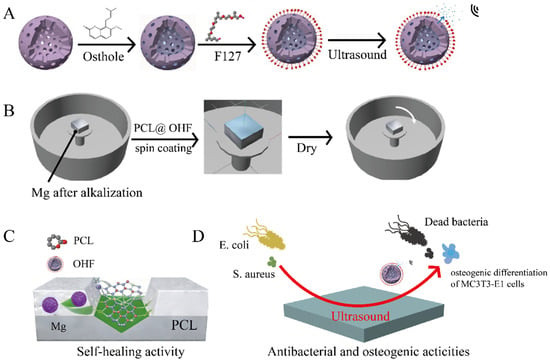
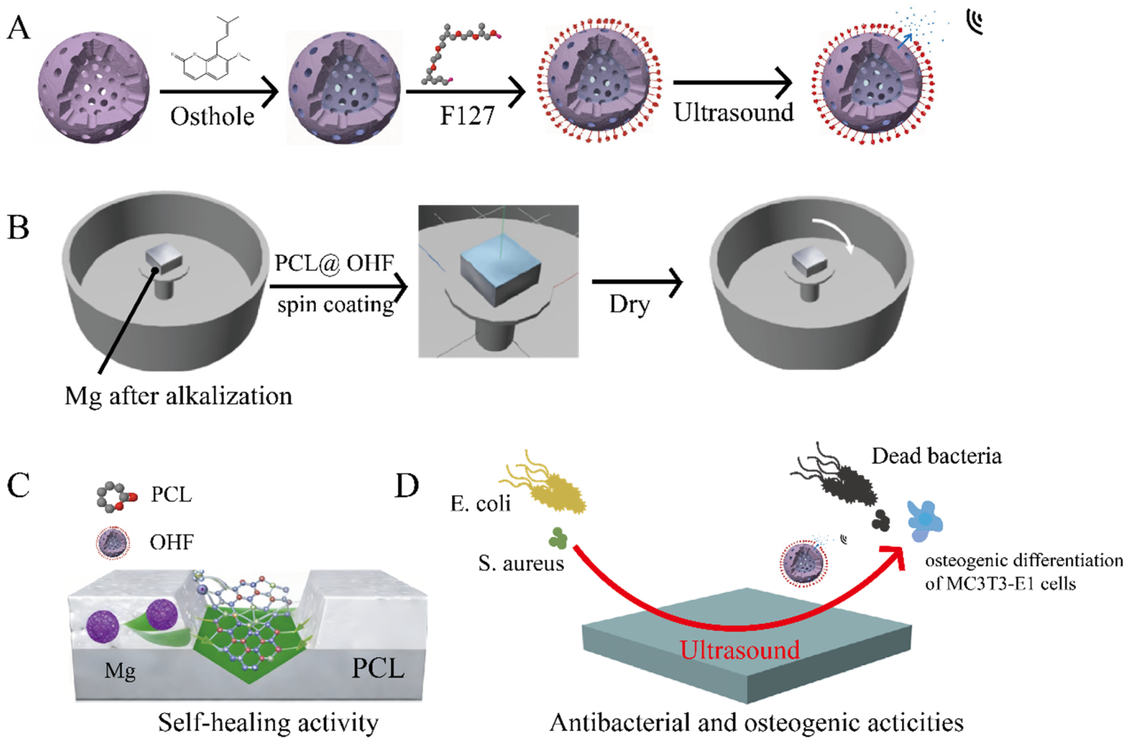
However, the development of ultrasound-responsive drug delivery coatings for Mg-based orthopedic implants remains unexplored. The goal of the present research is to develop a multifunctional PCL-based composite coating incorporated with ultrasound-responsive osthole-loaded nanocapsules. This coating is intended to endow magnesium alloy implants with self-healing ability, enhanced corrosion resistance, controllable on-demand drug release, effective antibacterial performance, and promoted osteogenic differentiation, ultimately overcoming the critical challenges restricting the clinical application of magnesium orthopedic implants. In this study, the thermosensitive block copolymer Pluronic F127 [32] was employed as a “switch” for drug release to modify the outer shell of hollow mesoporous silica (HMSs) nanoparticles. The hydrophilic–hydrophobic phase transition can be triggered by ultrasound, thereby facilitating controlled drug release [33,34]. The drug-loaded nanoparticles were incorporated into PCL and applied to Mg substrate to prepare a self-healing [35], functionally graded composite coating. This multifunctional coating is expected to simultaneously enhance corrosion protection while providing antibacterial activity and osteogenesis-promoting capability through ultrasound-triggered drug delivery (Figure 1).
Figure 1.
Overview of experimental process and coating performances: (A) Fabrication of HMSs, OH, and OHF. (B) Fabrication of Mg-PCL@OHF. (C) Evaluation of self-healing activity. (D) Antibacterial and osteogenic activities.
2. Experimental Details
2.1. Materials
The extruded WE43 Mg alloy was purchased from Suzhou Rongqian Rare Metal Products Co., Ltd. (Suzhou, China), and its chemical composition is listed in Table 1. The main chemicals included polycaprolactone (PCL, Mw = 50,000–60,000 g/mol, Aladdin, Shanghai, China), osthole (C15H16O3, Mw = 244.29, ≥99%, Aladdin), octadecyl trimethoxysilane (C18TMS, MW = 374.67 g/mol, 90%, Aladdin), tetraethyl orthosilicate (C8H20O4Si, Shanghai Chemical Reagent Co., Ltd., Shanghai, China, 99.7%), ammonia water (NH3·H2O, Mw = 35.05 g/mol, 25–28%), anhydrous sodium carbonate (Na2CO3, Mw = 105.99 g/mol, ≥99.8%), potassium carbonate (K2CO3, Mw = 138.206 g/mol, ≥98.0%), anhydrous magnesium sulfate (MgSO4, Mw = 120.37 g/mol, ≥97.0%). All the reagents were used directly without further purification. Ultrapure water was prepared using a Milli-Q water system.
Table 1.
Chemical composition of WE43 Mg alloy (wt.%).
2.2. Synthesis of Nanoparticles
Hollow mesoporous silica nanoparticles (HMSs) were synthesized using a modified Stöber method combined with the sol–gel technique [13,36]. The specific procedure was as follows: 140 mL of ethanol and 20 mL of ultrapure water were mixed to form an alcohol–water medium, followed by the addition of 3.14 mL of ammonia to create an alkaline reaction environment. Then, tetraethyl orthosilicate (TEOS) (7 mL) was rapidly injected under continuous stirring for 2 h, until the solution turned milky white. Subsequently, a mixture containing 5 mL of TEOS and an appropriate amount of C18TMS was added dropwise, and the mixture was stirred for 1 h to form a milky colloidal dispersion. After the reaction, the resulting emulsion was centrifuged at 10,000 rpm for 30 min to collect the precipitate, which was repeatedly washed with ethanol to remove residual organic impurities. The washed product was dispersed in a 0.6 M Na2CO3 solution and etched at 85 °C for 30 min to form a hollow structure through selective dissolution of the silica core by Na2CO3. The initial HMSs product was then collected by centrifugation at 8000 rpm for 30 min and washed three times with ultrapure water. The cleaned sample was placed in a vacuum drying oven and dried overnight at 100 °C. After grinding, the sample was transferred to a muffle furnace for calcination using the following program: heating rate of 1 K/min, final temperature of 813.15 K, and a 6 h hold to completely remove the template, yielding a pure HMSs product.
Osthole was dissolved in ethanol to prepare a stock solution at 5 mg/mL. This stock solution was mixed with 5 mg/mL of HMSs and continuously stirred for 5 h to allow sufficient drug adsorption and entry into the mesoporous channels of HMSs. Subsequently, the mixture was centrifuged at 6000 rpm to separate and collect the precipitate. The collected precipitate was dried at 37 °C, ground, and then the drug-loaded nanoparticles (denoted as OH) were obtained. Meanwhile, the supernatant was retained for the calculation of drug loading capacity.
To modify OH with an outer coating, 20 mg of Pluronic F127 copolymer was dissolved in 10 mL of deionized water. Then, 100 mg of the obtained OH particles were dispersed in this F127 solution and stirred for 20 min to ensure complete coating with F127 molecules. After the reaction, the mixture was centrifuged at 8000 rpm for 20 min to separate the solid from the liquid. The precipitate was collected, dried at 37 °C, and ultimately yielded F127-modified drug-loaded nanoparticles (denoted as OHF).
2.3. Characterization of Nanoparticles
Transmission electron microscopy (TEM, Talos F200X S/TEM, Thermo Fisher, Waltham, MA, USA) was employed to characterize the micromorphology and structure of HMSs, OH, and OHF nanoparticles. Fourier transform infrared spectroscopy (FTIR, Thermo Scientific Nicolet iS10, Thermo Fisher, Madison, WI, USA) was used to analyze the chemical composition and functional groups of the materials. The hydrodynamic diameter and size distribution of the nanoparticles were determined using a Malvern Zetasizer Nano ZS90 dynamic light scattering (DLS) system (Malvern Instruments, Malvern, UK). X-ray diffraction (XRD, Rigaku Smart Lab SE, Tokyo, Japan) using Cu-Kα radiation and 2θ of 10–90° was conducted to examine the crystal structure of the nanoparticles.
2.4. Preparation and Characterization of Coatings
Poly(ε-caprolactone) (PCL) was dissolved in dichloromethane and stirred hermetically at 500 rpm for 1 h to prepare a homogeneous PCL solution with a fixed concentration of 100 mg/mL. Then, OHF nanoparticles were added into the PCL solution to reach a concentration of 1 mg/mL, followed by sufficient vortexing to obtain a well-dispersed PCL@OHF composite solution.
WE43 magnesium alloy specimens with dimensions of 20 mm × 20 mm × 5 mm were successively ground with SiC sandpaper up to 3000# roughness, rinsed with deionized water, and dried with warm air. All samples were alkali-treated with 5 M NaOH for 20 min and then dried at 110 °C for 2 h to stabilize the surface state. The alkali-treated Mg alloy was fixed on the spin coater stage, and 1.5 mL of PCL or PCL@OHF solution was evenly spread onto the alloy surface. The spin-coating program was carried out at a rotation speed of 1000 rpm for 10–15 s to allow rapid evaporation of dichloromethane. To achieve a uniform, compact, and defect-free protective layer, the spin-coating procedure was repeated for 3 layers in total.
The surface morphology of the composite coating samples was observed using a scanning electron microscope (SEM, Nova Nano SEM450, Thermo Fisher, Hillsboro, OR, USA). The uniformity of the surface morphology was examined by SEM to determine the optimal PCL concentration.
2.5. Corrosion and Hydrogen Evolution Assessment
The electrochemical properties of the samples were evaluated on a PARSTAT 3000 electrochemical workstation (Princeton Applied Research, Princeton Applied Research, Oak Ridge, TN, USA) using the standard three-electrode system [37]. Potentiodynamic polarization tests were conducted with a saturated calomel electrode (SCE) as the reference, scanning from −2.5 V to −1.0 V at a rate of 1 mV·s−1. The corrosion current density (icorr) and corrosion potential (Ecorr) were determined by Tafel extrapolation. Electrochemical impedance spectroscopy (EIS) was performed at the open-circuit potential over the frequency range of 100 kHz to 0.1 Hz, with equivalent circuit modeling and impedance spectrum analysis performed using ZSimpWin software (version 3.60).
The corrosion rates were determined by the mass-loss method, with samples completely immersed in HBSS at a solution-to-surface area ratio of 100 mL/cm2. The specimens were retrieved after 1, 3, 7, 14, and 21 days of immersion. Surface corrosion products were chemically removed using a chromic acid solution (2 mol CrO3 saturated with 0.059 mol AgNO3), followed by ultrasonic cleaning and drying before mass-loss measurement. The annual corrosion rate of the samples was calculated according to the Formula [38,39]:
where Δm represents the mass loss (g), A denotes the exposed surface area of the sample (cm2), t is the cumulative immersion time (h), and ρ is the density of the alloy (g/cm3).
To characterize the composition and structure of the corrosion products, three parallel samples (Mg, Mg-PCL and Mg-PCL@OHF) were immersed in HBSS at 37.5 °C for 1, 3, and 10 days, respectively. A scanning electron microscopy–energy-dispersive spectroscopy (SEM-EDS) system was employed to analyze the morphology and elemental distribution of the corroded areas, enabling systematic characterization of the phase composition and chemical state of the corrosion products.
2.6. Self-Healing Test
To systematically evaluate the self-healing performance of the material, a through-scratch method was employed to simulate damage under long-term service conditions. The scratched samples were maintained at 50 °C for 20 min, after which their self-repairing morphology was observed using SEM. The chemical composition of the repaired area was characterized by EDS and X-ray photoelectron spectroscopy (XPS).
2.7. Drug Release
To investigate the controlled drug release behavior of the nanoparticles, 10 mg of drug-loaded nanoparticles were accurately weighed and placed in a dialysis bag with a molecular weight cutoff of 3.5 kDa. After adding 1 mL of phosphate-buffered saline (PBS), the bag was sealed and immersed in a centrifuge tube containing 9 mL of PBS. The release experiment was conducted at 37 °C. At predetermined time points (0, 24, 48, 72, 96, 120, 144, 168 h), the release medium (5 mL) was sampled and immediately replaced with an equal volume of fresh pre-warmed PBS to maintain the PBS volume.
To examine the effects of ultrasound irradiation on release at specific time points (1, 25, 49, 73, 97, 121, 145, 169 h), the samples of HMSs@ost@OHF(OHF) were subjected to ultrasound treatment at 0, 1, and 2 W/cm2 for 5 min. After treatment, the medium (5 mL) was sampled and replaced with an equal volume of PBS. The absorbance of osthole in all collected release solutions was measured using a dual-beam ultraviolet–visible spectrophotometer, and the drug concentration was calculated based on a pre-established standard curve.
The drug loading efficiency was calculated according to the following formula [13]:
where represents the drug concentration at the i-th sampling point, is the volume of the release medium (5 mL), and mtotal is the theoretical total drug loading amount in the nanoparticles.
2.8. Biological Evaluation
2.8.1. Live/Dead Staining Assay
Embryonic mouse osteoblast precursor cells (MC3T3-E1) were seeded into 24-well plates and cultured until reaching approximately 80% confluency. After observing cell adhesion, the culture medium was replaced with αMEM complete medium containing nanomaterial (Mg, Mg-PCL, and Mg-PCL@OHF) extracts, and the culture was continued. After culturing for 24 h, the cells were stained according to the instructions of the Calcein-AM/PI double staining kit. After staining, the cells were washed 3 times with PBS and observed using an inverted fluorescence microscope.
2.8.2. CCK-8 Assay for Cell Viability
The nanomaterials were sterilized by UV irradiation, then immersed in complete medium and sonicated for 20 min. After removing the nanomaterials, the extract was filtered through a 0.22 μm filter membrane to obtain the nanomaterial extract. MC3T3-E1 cells in good growth condition were seeded on 96-well plates at a density of 2 × 10³ cells/well. After the cells adhered to the wall, the extracts were added to the above culture wells for co-culture with the cells. After co-culture for 1, 2, and 3 days, respectively, the basic medium containing 10% CCK-8 reagent (100 μL) was added to each well. The plate was placed in a cell incubator in the dark for 2 h. It was then placed in a microplate reader in the dark, and the OD value of each well was recorded at 450 nm. The cell viability of each group was calculated by the following formula:
2.8.3. Hemolysis Test
The nanomaterials were sterilized by UV irradiation, then immersed in normal saline and sonicated for 20 min. After removing the nanomaterials, the extract was filtered through a 0.22 μm filter membrane to obtain the nanomaterial extract. A total of 3 mL of the SD rat blood sample was centrifuged at 1000 rpm for 15 min to obtain red blood cell (RBCs) precipitation. The RBC precipitate was washed 3 times with normal saline, and 1 mL of RBC precipitation was added to 3.67 mL of normal saline to prepare the RBC suspension. The RBC suspension (100 μL) was added to 1.1 mL of normal saline and deionized water, respectively, as the negative control group and positive control group. Meanwhile, the RBC suspension (100 μL) was added to 1 mL of normal saline and 100 μL of different nanomaterial extracts as the experimental group, which were then incubated in a 37 °C incubator for 3 h. Subsequently, each group was centrifuged at 1000 rpm for 15 min, and the supernatant was collected. The absorbance (Optical density, OD) value at 540 nm was measured using a multi-function microplate reader. The hemolysis rate was calculated by the following formula:
where ODx is the OD of the different nanomaterial groups, ODs is that of the negative control group, and ODw is that of the positive control group.
2.8.4. Colony Forming Unit (CFU) Assay
The nanomaterials were sterilized by UV irradiation, then immersed in LB medium and sonicated for 20 min. After removing the nanomaterials, the extract was filtered through a 0.22 μm filter membrane to obtain the nanomaterial extract. The bacterial strains used in this study included S. aureus (ATCC 49295) and E. coli (ATCC 25922). Viable bacterial cells were quantified via a CFU assay. Serial dilutions (10−4–10−5) of the treated bacterial suspensions were prepared and plated (100 µL) in triplicate on LB agar. After 24 h incubation at 37 °C, the colonies were enumerated and photographed. All tests were conducted in triplicate.
2.8.5. Detection Anti-Biofilm Assay
S. aureus and E. coli were cultured to the logarithmic growth phase, and the concentration was adjusted to 1 × 10⁷ CFU/mL. The bacterial solution (250 μL) was seeded into 48-well plates, and high-glucose LB medium containing different material extracts was added, followed by static culture at 37 °C for 48 h. After 48 h, the biofilm was washed once with PBS to gently remove planktonic bacteria. Total biofilm biomass was quantified via crystal violet staining, involving formalin fixation, dye binding, and dissolution for absorbance measurement at 590 nm.
2.8.6. Osteogenic Properties
The osteogenic differentiation ability of MC3T3-E1 cells were detected by alkaline phosphatase (ALP) staining kit (7 days) and Alizarin red S (ARS) staining kit (21 days). The experimental groups included: (1) Blank; (2) Mg-PCL; (3) Mg-PCL@OHF at 0 W/cm2 ultrasound; (4) Mg-PCL@OHF at 1 W/cm2 ultrasound; and (5) Mg-PCL@OHF at 2 W/cm2 ultrasound. Following 7 days of osteogenic induction, MC3T3-E1 cells were assessed for ALP expression and mineralization. ALP staining was performed by fixing cells with 4% paraformaldehyde fixative (Biosharp, Hefei, China) and developing with a BCIP/NBT kit (Beyotime, Shanghai, China). After 21 days of osteogenic induction, the cells were fixed with 4% paraformaldehyde fixative, washed, and incubated with 1% ARS solution (Fuheng, Shanghai, China). For quantification, 10% (w/v) cetylpyridinium chloride (Fuheng, China) was added to dissolve the ARS precipitate, and the absorbance was read at 560 nm.
3. Results and Discussion
3.1. Characterization of the Nanoparticles
Hollow mesoporous silica nanoparticles (HMSs) are synthesized by a combined Stöber and sol–gel method, in which the weak alkaline environment provided by ammonia catalyzes the hydrolysis and condensation of the silicon source, forming a solid silica matrix with a mesoporous structure. Differential dissolution of the sodium carbonate (Na2CO3) solution is utilized to etch the silica nanoparticles. Carbonate ions (CO32−) preferentially dissolve the less cross-linked and relatively loose core of the silica nanoparticles, while the more densely cross-linked outer shell is largely preserved.
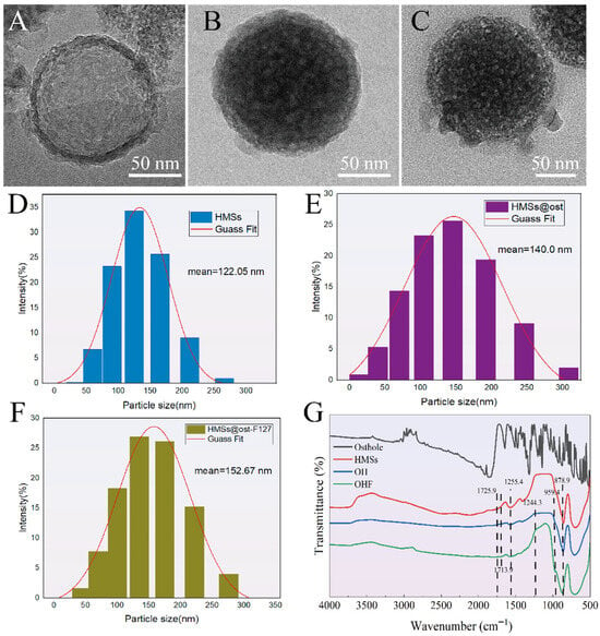
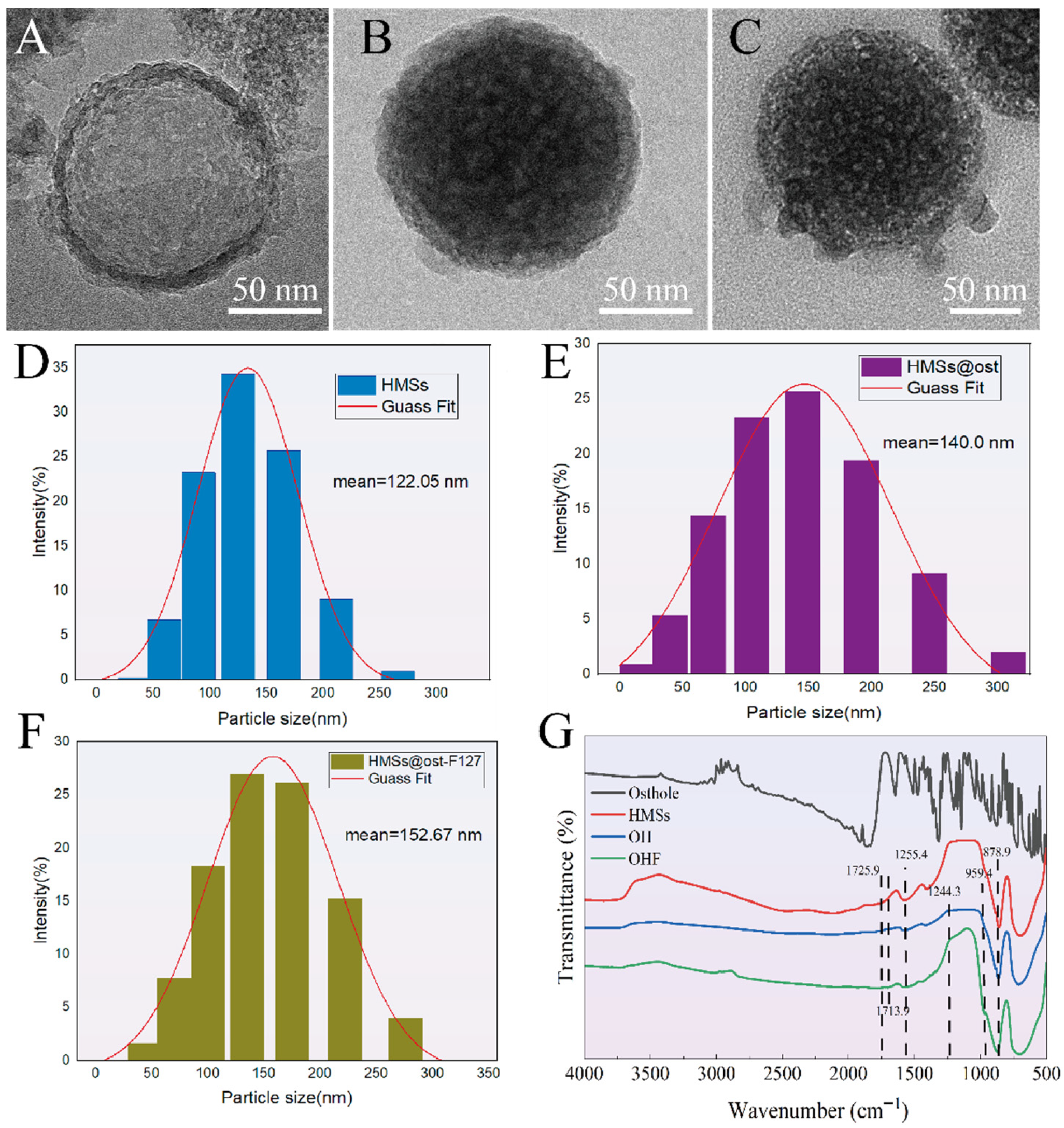
This selective etching produces a hollow structure (Figure 2A) with an average cavity size of 122.5 nm (Figure 2D). TEM reveals that the pristine HMSs exhibit a well-defined spherical core–shell structure, featuring a thin shell (~6.7 nm) and a large internal cavity. This architecture provides ample space for osthole encapsulation, while the thin shell facilitates rapid drug diffusion post-stimulation. After loading osthole to form OH (HMSs@osthole) nanoparticles (Figure 2B), the internal cavity is filled. A slight increase in diameter from 122.05 nm (HMSs) to 140.0 nm (OH), as shown in Figure 2E,F, confirms that osthole has entered and occupied the mesoporous cavities. Subsequent modification with the ultrasound-responsive polymer F127 produces OHF nanoparticles (Figure 2C), resulting in a further increase in diameter to 152.67 nm (Figure 2F) and a thicker outer shell. The HMSs, OH, and OHF (HMSs@osthole@F127) are all monodispersed spheres with a uniform morphology.
Figure 2.
Characterization of the nanoparticles: TEM images of (A) HMSs, (B) HMSs@ost(OH), (C) HMSs@ost@F127(OHF). DLS analysis of (D) HMSs, (E) HMSs@ost(OH), (F) HMSs@ost@F127(OHF). (G) FT-IR spectra of the material: Osthole, HMSs, HMSs@ost(OH) and HMSs@ost@F127(OHF).
As shown in Figure 2G, FTIR provides molecular-level information about the chemical composition and bonding interactions of the nanoparticles. The peaks at 878.9 cm−1 (OH) and 959.4 cm−1 (OHF) correspond to the asymmetric stretching of Si-O-Si bonds and surface silanol groups, indicating that the mesoporous silica framework remains intact during functionalization. The ester C=O stretching peaks at 1713.9 cm−1 (OH) and 1725.9 cm−1 (OHF), along with C-O-C stretching peaks of lactones and esters at 1255.4 cm−1 (OH) and 1244.3 cm−1 (OHF), confirm the presence of osthole molecules containing ester and lactone moieties. FTIR demonstrates that the loading of osthole and grafting of F127 do not compromise the structural integrity of HMSs.
3.2. Drug Loading and Release of the Nanoparticles
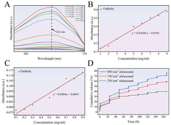
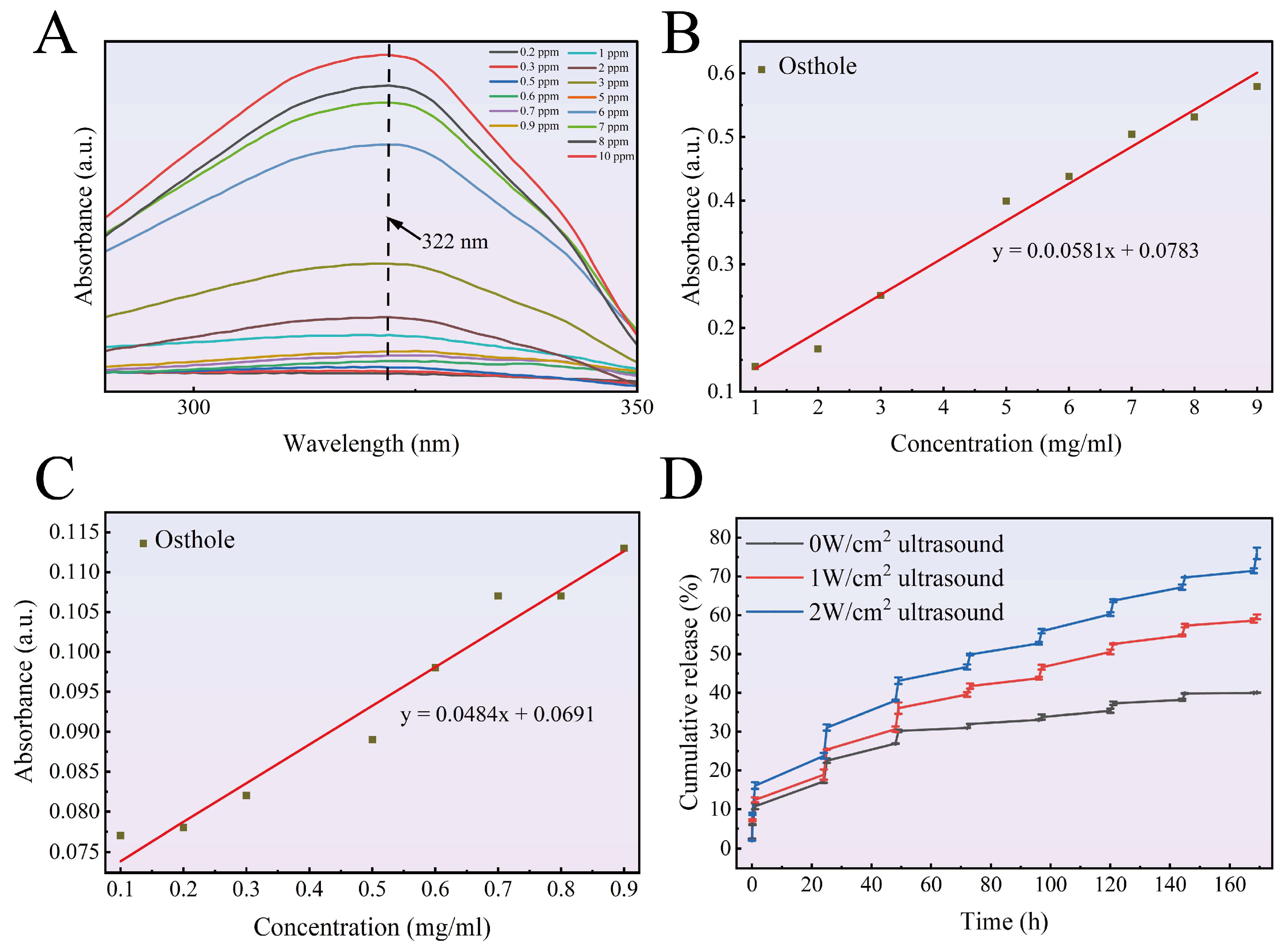
UV-vis spectrophotometry is employed to assess the release of osthole in HMSs. The UV-Vis spectrum in Figure 3A shows an absorption peak for osthole at 322 nm. The UV absorption spectra of the osthole solutions with different concentrations are shown in Figure 3B,C to investigate the linear relationship between drug concentration and UV absorption intensity. Figure 3D illustrates the cumulative release profiles of OHF under 0, 1, and 2 W/cm2 ultrasound irradiation. Under non-stimulated conditions, HMSs exhibit a slow and sustained diffusion-based release, with a cumulative release of only 39.94% after 169 h, which is characteristic of a diffusion-controlled mechanism. In this case, drug molecules diffuse through the mesoporous silica shell and the hydrated PEO layer of the F127 coating, which acts as a physical barrier, resulting in slow release.
Figure 3.
Drug loading, release profiles of OHF: (A) UV-vis spectra. (B) Standard curve of osthole (1–9 ppm). (C) Standard curve of osthole (0.1–0.9 ppm). (D) Drug release of osthole upon ultrasound irradiation.
As shown in Figure 3D, under 2 W/cm2 ultrasonic irradiation, the cumulative drug release increased to 75.93%, which was higher than the release of 59.57% observed under 1 W ultrasonic irradiation. The power-dependent acceleration of the release rate confirms that a higher ultrasound intensity (2 W/cm2) leads to more intense cavitation effects, giving rise to more bubble collapse and stronger mechanical effects, which in turn promote faster drug diffusion [40]. Cyclic release experiments confirm that F127 undergoes reversible dissociation and reorganization under ultrasound irradiation, enabling controlled drug release. Furthermore, HMSs exhibit a highly reproducible release behavior, boding well for practical applications [41,42].
3.3. Morphology of Coatings
Spin-coating experiments are conducted using PCL solutions with different mass fractions. Mg is pre-treated with alkalization to generate hydroxyl-binding sites, enhancing the adhesion of the polymer coating. These surface hydroxyl groups (-OH) act as binding sites, forming strong interactions with the carbonyl groups (-C=O) in PCL chains via hydrogen bonding and dipole–dipole interactions, thereby improving coating stability [43]. The shear force generated by the high-speed rotation of the spin coater rapidly removes the solvent, enabling effective bonding between the PCL coating and the Mg alloy.
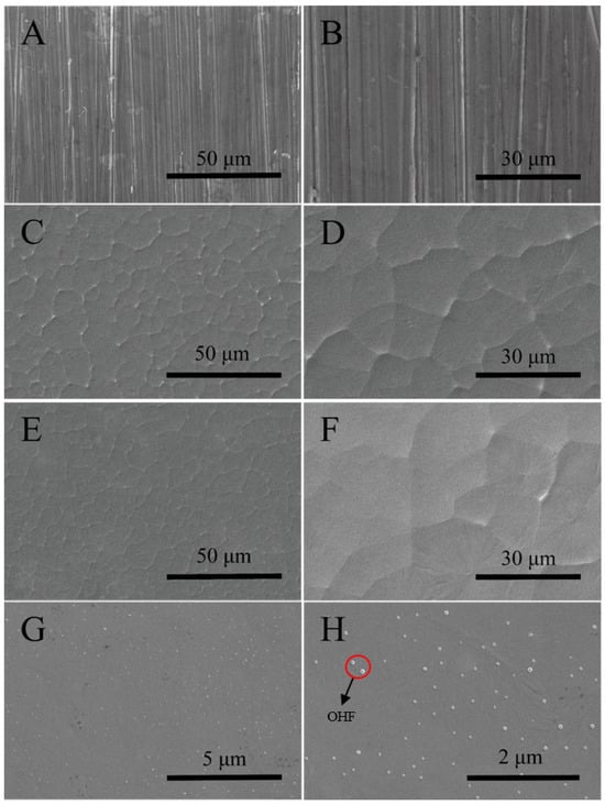
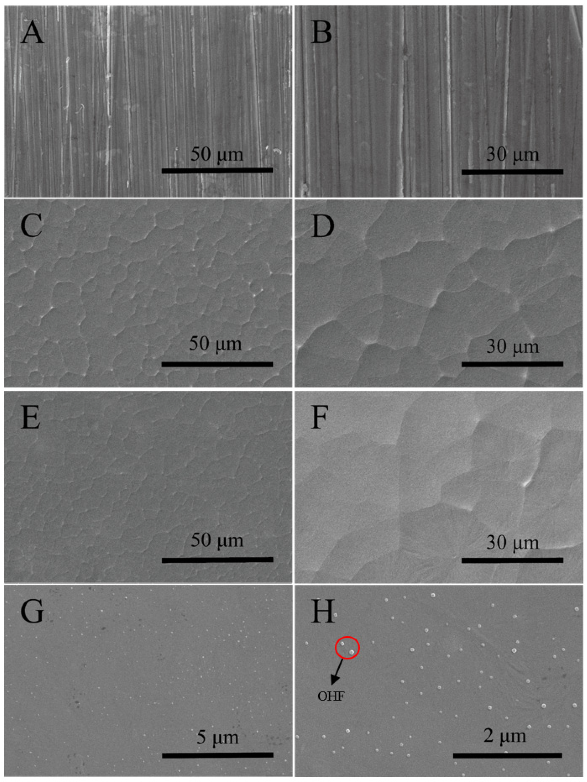
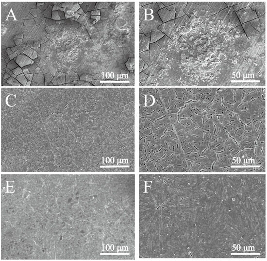
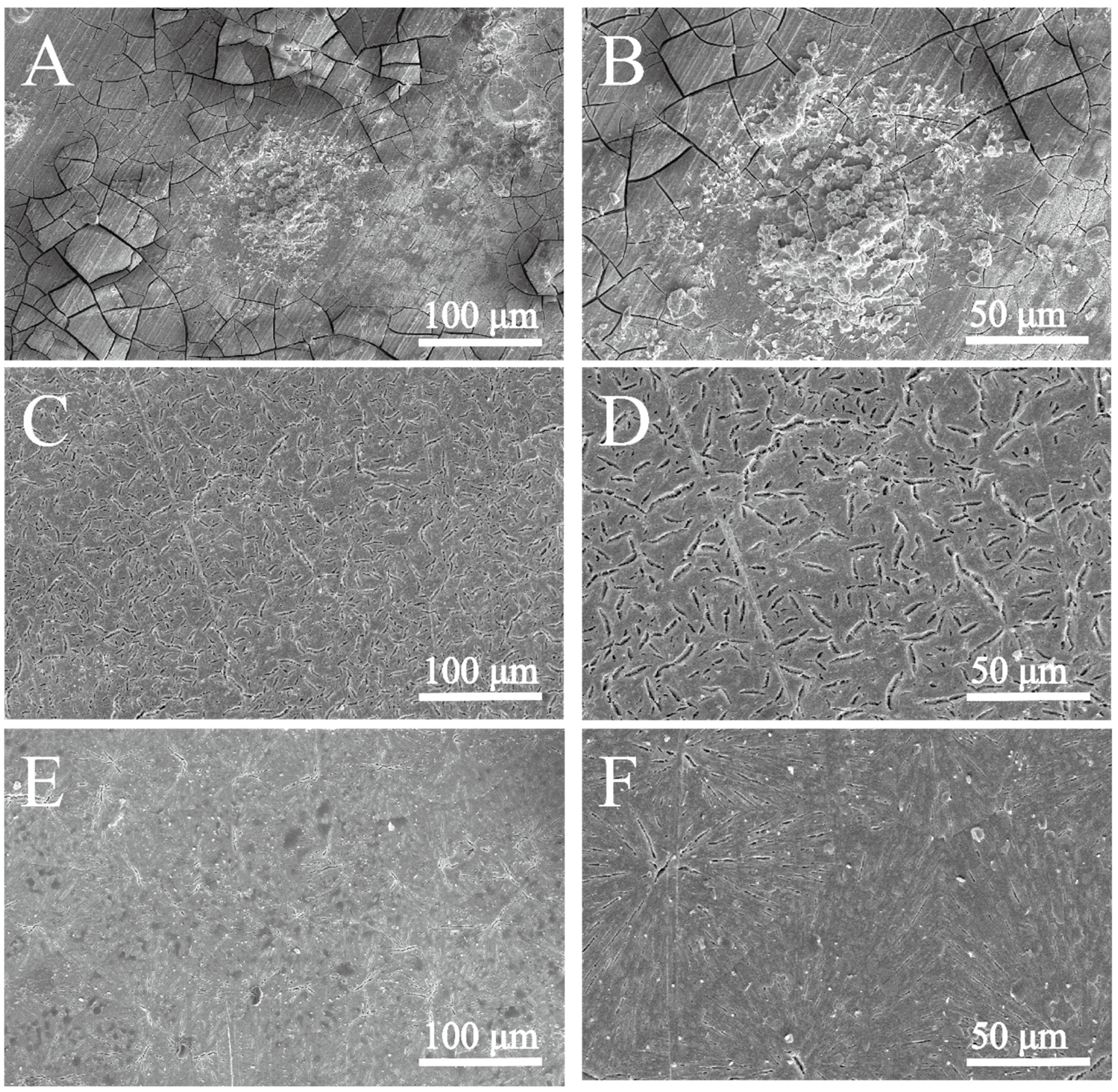
As shown in Figure 4A,B, the bare WE43 surface displays distinct parallel polishing scratches, constituting the surface morphology of the Mg alloy after polishing with 2000# sandpaper. This surface can generate mechanical interlocking sites upon alkalization to enhance the interfacial adhesion between the substrate and the subsequent polymer coating. For the Mg-PCL coating (Figure 4C,D), a continuous and uniform film completely covers the polishing textures of the substrate, verifying the good spreading and film-forming capacity of the PCL polymer solution. As illustrated, PCL with a concentration of 100 mg/mL forms a homogeneous and compact coating on the Mg alloy. The continuous film itself can also serve as a physical barrier to impede the corrosive medium, consistent with the electrochemical results in Figure 5. The Mg-PCL@OHF coating Figure 4E–H) exhibits superior surface uniformity at low magnification. High-magnification observation (Figure 4G,H) reveals that OHF nanoparticles with favorable dispersibility are distributed on the coating surface. These nanoparticles possibly restrict the mobility of PCL polymer chains during curing. This morphological feature further corroborates the conclusions in Figure 5 and Figure 6 that the Mg-PCL@OHF coating has the optimal corrosion resistance among the various specimens.
Figure 4.
SEM images of: (A,B) Bare WE43 Mg alloy after polishing and alkali treatment. (C,D) Mg alloy coated with PCL. (E–H) Mg alloy coated with PCL@OHF composite coating.
Figure 5.
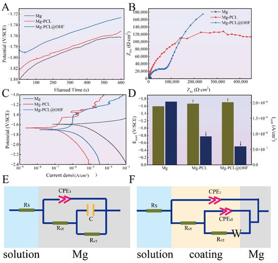
Electrochemical properties of Mg, Mg-PCL and Mg-PCL@OHF: (A) OCP curves. (B) Nyquist plots. (C) Polarization curves. (D) Icorr and Ecorr. (E) ECs of Mg. (F) ECs of Mg-PCL and Mg-PCL@OHF.
Figure 6.
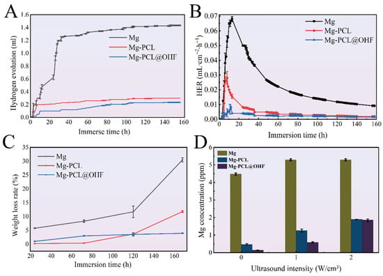
Immersion measurements during 160 h in HBSS: (A) Hydrogen evolution volume of Mg, Mg-PCL, and Mg-PCL@OHF. (B) Hydrogen evolution rate of Mg, Mg-PCL, and Mg-PCL@OHF. (C) Weight loss after hydrogen evolution of Mg, Mg-PCL, and Mg-PCL@OHF. (D) ICP of Mg2+ release concentration.
3.4. Electrochemical Tests and Degradation Behaviors
Figure 5 presents the electrochemical characteristics of the bare WE43, Mg-PCL, and Mg-PCL@OHF specimens. Figure 5A shows the open circuit potential (OCP) curves, with all three specimens exhibiting a gradual increase in OCP values, indicative of stable surface performance. The bare WE43 features a highly negative initial potential with an OCP of −1.84 V, demonstrating a pronounced corrosion kinetic process. In contrast, the OCP of the PCL-coated Mg-PCL specimen positively shifts to −1.82 V, and the Mg-PCL@OHF exhibits a further positive OCP shift to −1.78 V. This confirms that the surface coating suppresses the thermodynamic driving force for the oxidation reaction of Mg alloy and simultaneously reduces the kinetic rate of anodic dissolution.
Figure 5B displays the electrochemical impedance spectroscopy (EIS) results, wherein the capacitive loops of Mg-PCL and Mg-PCL@OHF are larger than that of the bare WE43, reflecting their superior average electrochemical corrosion resistance. Figure 5C,D show the electrochemical Tafel curves and fitting results, respectively. The corrosion current density (Icorr) results reveal that the Mg-PCL specimen with a PCL coating has an Icorr value of 949.92 nA/cm2, which is lower than that of the bare Mg (2.04 μA/cm2), while the Mg-PCL@OHF specimen loaded with OHF nanoparticles exhibits a further reduction in Icorr to 594.36 nA/cm2. The corrosion potential (Ecorr) follows the order of Ecorr (WE43) (−1.59 V) > Ecorr (Mg-PCL) (−1.67 V) > Ecorr (Mg-PCL@OHF) (−1.70 V). The charge transfer resistance (Rct) of pure Mg, Mg-PCL, and Mg-PCL@OHF are 1.076 × 10⁴ Ω·cm2, 5.87 × 10⁵ Ω·cm2, and 6.225 × 10⁵ Ω·cm2, respectively. The Rct value of Mg-PCL@OHF is 58 times that of bare Mg and approximately 6% higher than that of Mg-PCL, demonstrating a significantly enhanced physical barrier against corrosion. The combined results of Ecorr and Icorr indicate that the PCL coating endows the Mg alloy with superior corrosion resistance, which is further enhanced for the Mg-PCL@OHF specimen with nanoparticles (Table 2).
Table 2.
Fitted electrochemical parameters obtained by EIS.
Based on EIS, the equivalent circuits fitted by ZSimpWin are presented in Figure 5E,F, and each circuit component is listed in Figure 5E. The fitted equivalent circuit for the bare WE43 is Rs(Q1(Rf(CRct))), whereas that of both Mg-PCL and Mg-PCL@OHF is Rs(Q1(Rct(Q2(R2W)))), as shown in Figure 5F. This is because the coating forms a barrier against the surface migration of electrons. Here, W denotes the swelling of PCL in the electrolyte solution, Rc1 and Q1 in the low-frequency loop reflect the corrosion resistance of the corrosion products formed on the coating surface, the series connection of Rc2 and W in the medium-frequency loop characterizes the degradation behavior of the PCL and PCL@OHF coatings, and Rct(Q2(R2W)) in the high-frequency loop corresponds to the electric double layer structure.
The hydrogen evolution curves and weight loss results in Figure 6A,C impart information about the corrosion behavior of the bare Mg, Mg-PCL, and Mg-PCL@OHF specimens after immersion for 7 days in the simulated physiological fluid, HBSS. Rapid hydrogen evolution kinetics and a large cumulative hydrogen volume (1.435 mL on the 7th day) are observed from the bare WE43, accompanied by a substantial weight loss of 30.504%. This phenomenon probably arises from the inherently high reactivity of Mg in the aqueous environment, which leads to the spontaneous anodic dissolution of Mg coupled with cathodic water reduction and formation of a porous and unstable Mg(OH)2 layer. In HBSS, chloride ions (Cl−) with a high concentration readily penetrate this native oxide layer to disrupt the passive film and accelerate the initiation and propagation of pitting corrosion, thereby leading to continuous hydrogen evolution and material degradation. As shown in Figure 6B, the hydrogen evolution rate increases first, then decreases over time, and eventually levels off. In contrast to the bare WE43, the PCL-coated specimen (Mg-PCL) produces a reduced hydrogen evolution volume of 0.3 mL, demonstrating the effective physical barrier function of the polymer coating. During the early immersion stage, the Mg-PCL specimen shows minimal weight loss that is less than that of the bare Mg after 72 h. This confirms that PCL acts as a hydrophobic diffusion barrier to hinder the transport of corrosive species (e.g., water and chloride ions) to the Mg substrate. However, the gradual increase in hydrogen evolution with time indicates the limited long-term stability of the pristine PCL coating. Water molecules can permeate microstructural defects (e.g., pinholes and residual porosity) in the coating, and interfacial debonding eventually promotes localized corrosion at the coating-substrate interface [44].
Mass loss results (Figure 6C) show values of 30.504% for bare Mg, 11.801% for Mg-PCL, and 3.922% for Mg-PCL@OHF. Mg-PCL@OHF reduces mass loss by about 67% compared with Mg-PCL and by about 87% compared with bare Mg, confirming good long-term protective stability. The PCL coating loaded with OHF nanoparticles (Mg-PCL@OHF) has the best corrosion resistance, manifested by the lowest hydrogen evolution volume (0.235 mL) and a weight loss of 3.922% at 168 h, which are significantly lower than those of the bare Mg (30.504%) and Mg-PCL (11.801%). This superior performance arises from the synergistic effects of enhanced physical barrier, chemical passivation, and strengthened interfacial bonding enabled by the incorporation of OHF nanoparticles, As illustrated in Figure 6 [45]. These results are in agreement with Mg2+ release shown in Figure 6D, which is acquired from the coated Mg alloy after 20 min of immersion. Owing to the protective effect of the PCL@OHF coating, the lowest Mg2+ release is attained.
The post-corrosion SEM morphologies in Figure 7 further elucidate the differences in corrosion behavior among the three groups to corroborate the hydrogen evolution and weight loss data. For the bare WE43 (Figure 7A,B), the surface exhibits extensive interconnected cracks, severe corrosion pits, and large-scale spallation of the corrosion product layer. In contrast, the PCL-coated specimen (Mg-PCL, Figure 7C,D) shows a relatively intact surface with only minor microcracks and shallow corrosion pits. This confirms the physical barrier effect of the PCL coating, which initially blocks the transport of H2O and Cl− to the Mg substrate. However, the presence of microcracks and localized surface roughening indicates the limited long-term stability of the pristine PCL coating. The Mg-PCL@OHF specimen (Figure 7E,F) displays a more stable and dense surface morphology, with no visible corrosion pits, cracks, or product spallation. The SEM results for hydrogen evolution are consistent with the electrochemical results.
Figure 7.
SEM morphology after hydrogen evolution: (A,B) Mg, (C,D) Mg-PCL, (E,F) Mg-PCL@OHF.
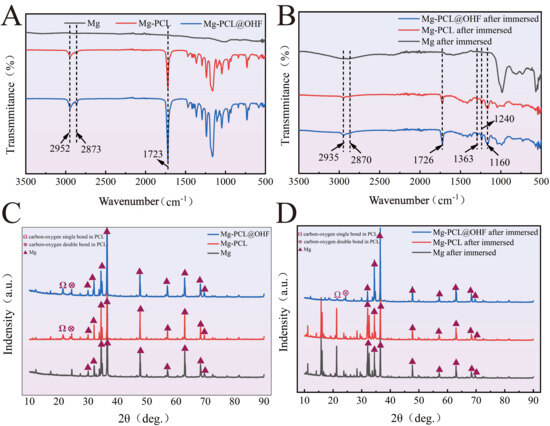
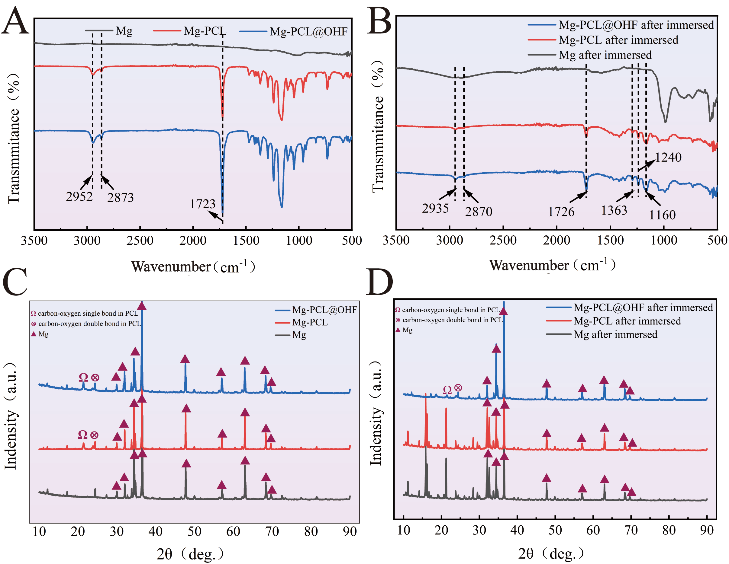
FTIR is employed to characterize the chemical evolution of the coatings and corrosion products before and after immersion in simulated body fluid (SBF). As shown in Figure 8A, the FTIR spectra of the pristine samples demonstrate the successful fabrication of the protective coatings. For Mg-PCL and Mg-PCL@OHF, the characteristic peaks at 2952/2873 cm−1 correspond to asymmetric/symmetric stretching of −CH2− in PCL, while the peak at 1723 cm−1 is associated with stretching of the C=O ester groups in PCL, verifying the presence of the PCL matrix. The Mg-PCL@OHF sample also exhibits characteristic peaks in the region, which are assigned to the Si-O-Si vibrations of OHF, confirming the successful incorporation of mesoporous silica nanoparticles into the PCL coating.
Figure 8.
Composition before and after hydrogen evolution test of Mg, Mg-PCL, and Mg-PCL@OHF: (A) FTIR spectra of the samples before immersion, (B) FTIR spectra of the samples after immersion, (C) XRD patterns of the samples before immersion, and (D) XRD patterns of the samples after immersion.
After immersion in SBF (Figure 8B), the FTIR spectra reveal significant differences in the coating stability and corrosion product formation. For the bare Mg, the attenuation of Mg-related signals and the emergence of new peaks at 1633 cm−1 (H-O-H bending of adsorbed water) and 1240 cm−1 (P-O stretching of phosphate groups) indicate rapid corrosion, with Mg(OH)2 being the main corrosion products. In contrast, Mg-PCL retains the residual peaks at 1726 cm−1 (C=O) and 2935/2870 cm−1 (−CH2−), demonstrating partial preservation of the PCL barrier. However, the appearance of peaks at 1633 cm−1 (water) and 1160 cm−1 (phosphate groups) indicates progressive coating degradation accompanied by the accumulation of corrosion products, reflecting partial failure of the PCL barrier over time. In contrast, the C=O peak (1726 cm−1) of Mg-PCL@OHF is preserved, and the P-O vibration peaks (1240 cm−1, 1160 cm−1) are clearly distinguishable, indicating significantly improved coating stability. This improvement may stem from OHF filling defects in the PCL matrix, thereby reducing the penetration of water and ions, delaying coating degradation, and ultimately mitigating the formation of corrosion products, consistent with the hydrogen evolution, SEM, and electrochemical results.
After immersion in Hank’s solution, the phase evolution characteristics and corresponding corrosion behaviors are determined, as shown in Figure 8C,D. For the bare Mg alloy, its characteristic diffraction peaks attenuate significantly, accompanied by the emergence of a new characteristic peak of magnesium hydroxide (Mg(OH)2) at 38.0°. This indicates the absence of a protective barrier on the bare Mg alloy. The generated Mg2+ rapidly combines with OH− in SBF to form Mg(OH)2, a typical corrosion product of Mg alloys. For the Mg-PCL specimen, partial Mg characteristic peaks and persistent crystalline signals of PCL are retained, and this phenomenon confirms the initial physical barrier effect of the PCL coating. Meanwhile, the Mg2+ produced by dissolution interacts with phosphate and calcium ions in Hanks’ SBF to generate a small amount of Mg(OH)2 and hydroxyapatite (HA), resulting in the appearance of weak characteristic peaks of corrosion products. The Mg-PCL@OHF specimen shows the strongest Mg characteristic peaks and the weakest signals from corrosion products (Mg(OH)2 and HA). This is because OHF nanoparticles can fill the micropores and defects in the PCL matrix to improve coating compactness, consequently forming a more effective physical barrier.
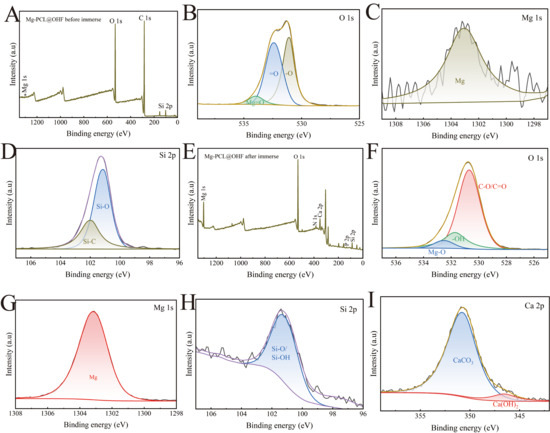
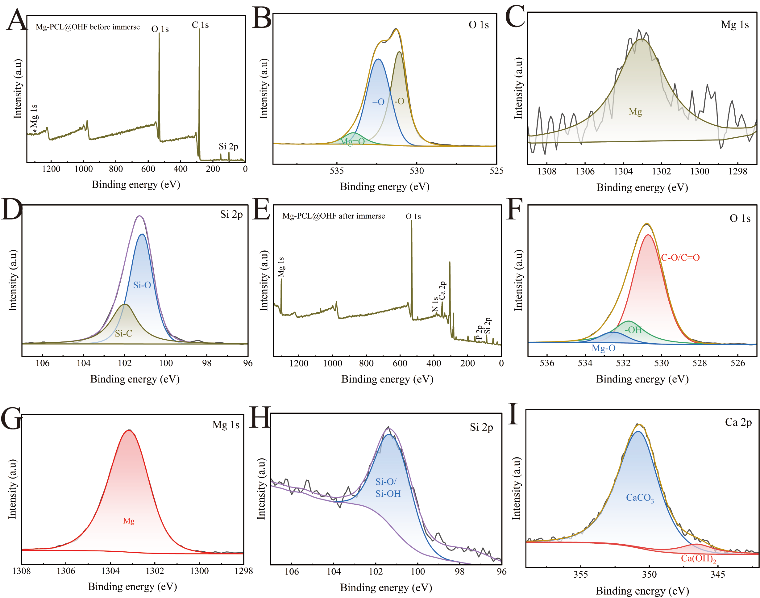
Figure 9 presents the XPS results of the WE43 sample coated with OHF nanoparticle-loaded PCL before corrosion in PBS. Distinct features are observed from the Mg 1s, C 1s, and Si 2p spectra, indicating that the PCL coating loaded with OHF nanoparticles provides better shielding for the bare Mg alloy surface compared to the pure PCL coating. The area ratio of the binding energy peaks for -O (531.8 eV) and =O (533.1 eV) in the O 1s spectrum is close to 1:1, demonstrating that the OHF-loaded PCL coating is densely and uniformly applied to the Mg alloy surface with good adhesion. The small peak corresponding to Mg-O (533.9 eV) suggests that the Mg alloy may have a passivated surface formed in air, while the Si-C (101.78 eV) and Si-O (99.88 eV) peaks indicate the presence of relatively uniform drug-loaded SiO2 nanoparticles on the coating surface.
Figure 9.
XPS spectra of Mg-PCL@OHF: (A–D) Before immersion. (E–I) After immersion.
XPS analysis of the OHF-loaded PCL coating reveals peaks of Mg 1s, O 1s, Ca 2p, and Si 2p, reflecting the elemental composition of the polymeric matrix, drug-loaded nanoparticles, and corrosion products. Deconvolution of the high-resolution O 1s spectrum (Figure 9B–F) identifies three distinct components. The peak at 530.98 eV is assigned to C=O/C–O, confirming the preservation of ester groups in the PCL matrix and oxygen-containing functional groups on OHF nanoparticles. The signal at 532.48 eV corresponds to Mg–O species, indicative of magnesium oxide/hydroxide formed during corrosion, and that at 531.88 eV is attributed to –OH groups originating from hydroxides in corrosion products and residual hydroxyl moieties on the surface of OHF nanoparticles. These results not only verify the retention of both the PCL matrix and OHF nanoparticle components after immersion but also the synergistic effect of residual coating constituents and in situ formed corrosion products, providing effective protection to the underlying WE43 substrate.
The OHF enhance the structural stability of the composite coating. The Si–O/Si–OH bonds corresponding to the Si 2p peak reveal a dense cross-linked network structure in the PCL matrix. This hinders the penetration of corrosive ions and reduces the formation of coating defects. In contrast, the pure PCL coating is prone to severe degradation and delamination under the same corrosion conditions, with a noticeable attenuation in the intensity of its C=O/C–O characteristic peaks. These results confirm that the PCL matrix and OHF nanoparticle components are preserved after immersion. The synergistic effects rendered by the residual coating components, SiO2-reinforced barrier structure, and in situ-generated corrosion products provide more efficient protection for the WE43 substrate.
3.5. Evaluation of Self-Healing Behavior
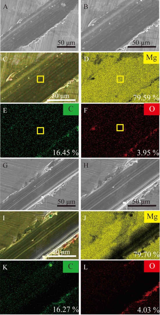
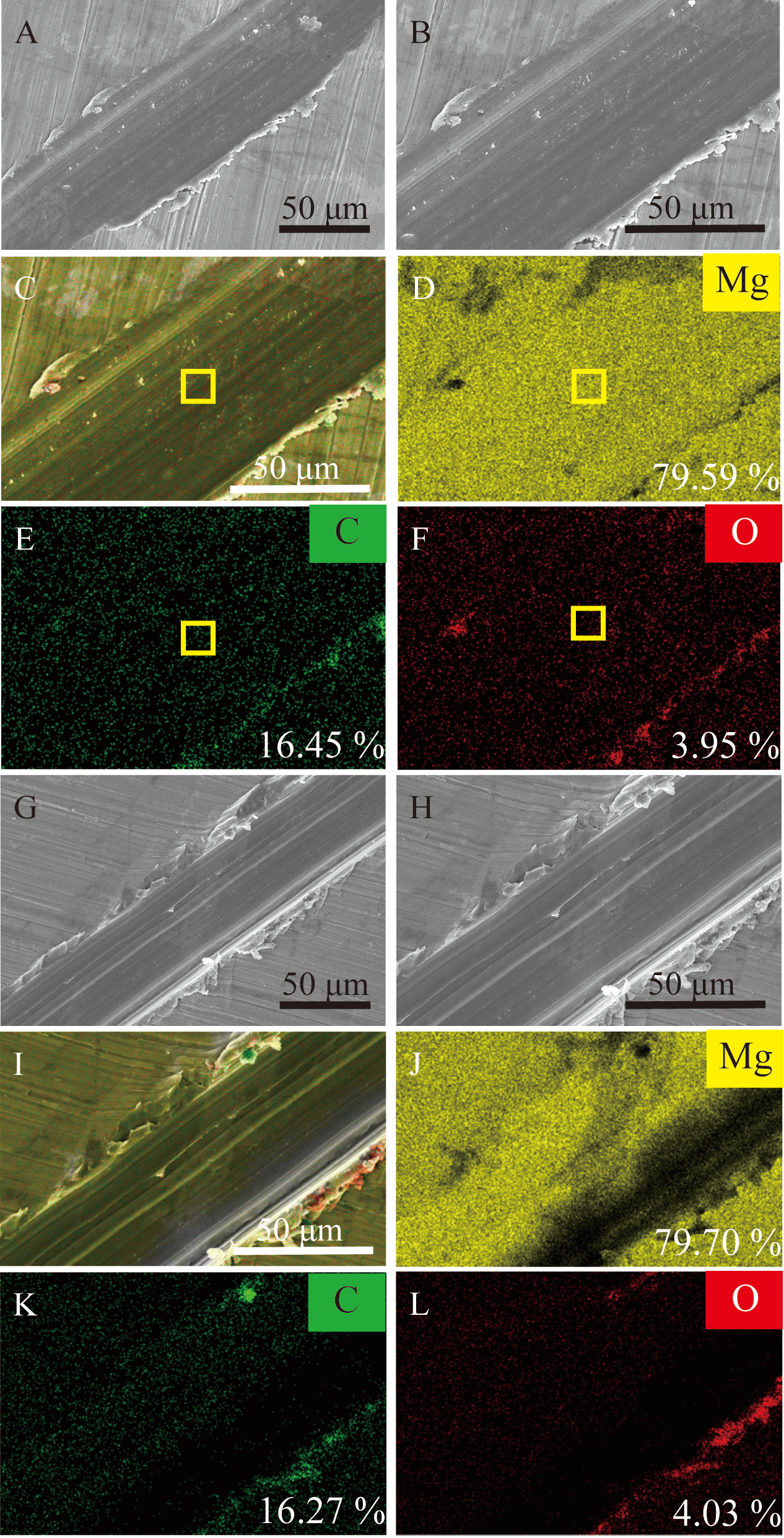
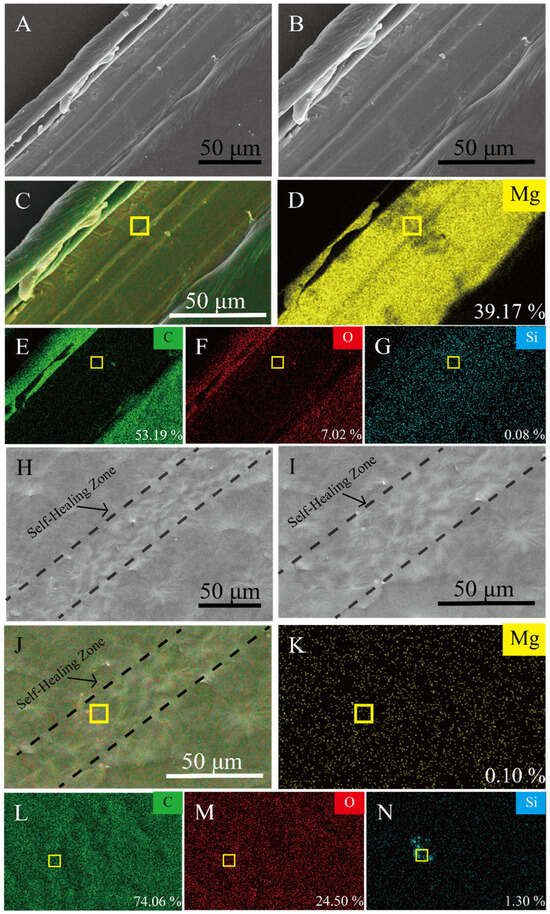
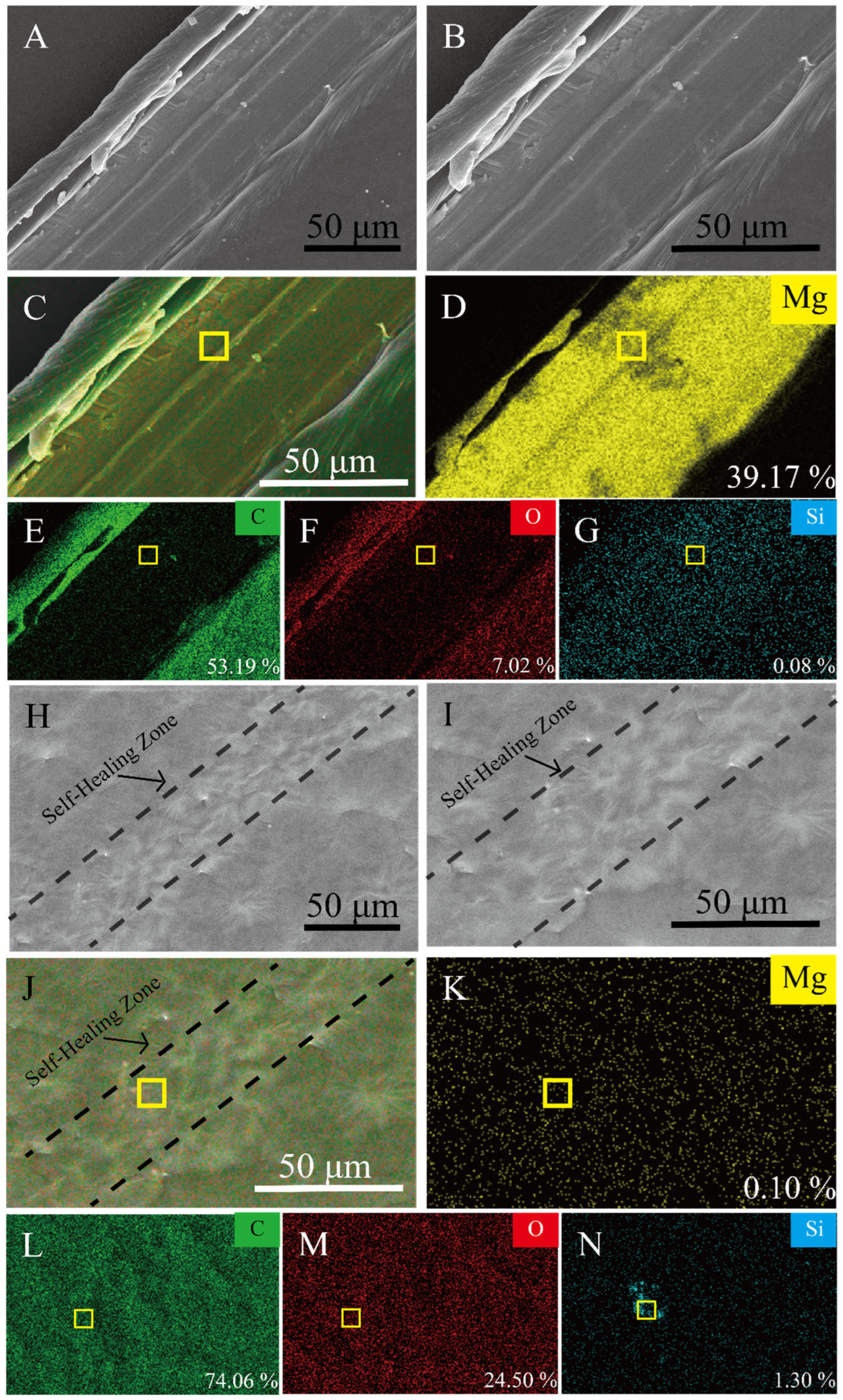
After making scratches approximately 100 μm in width on the coating surface, the samples are incubated at 50 °C for 30 min, and morphological changes are observed. The damaged areas on the Mg and Mg-PCL@OHF coating are shown in Figure 10 and Figure 11. Figure 10 displays the morphology of the Mg alloy following heat treatment at 50 °C for 30 min. Evidently, the alloy does not have the self-healing property, resulting in the exposure of the bare Mg matrix. Mg constitutes the primary elemental component, accompanied by minor contents of carbon and oxygen, which are derived from the surface oxide.
Figure 10.
(A,B) SEM morphologies of the bare Mg alloy before heat treatment, showing an artificial scratch with exposed substrate. (C–G) EDS elemental mapping of the scratched Mg alloy before heat treatment. (H,I) SEM morphologies of the bare Mg alloy after heat treatment. (J–L) EDS elemental mapping of the scratched Mg alloy after heat treatment.
Figure 11.
Self-healing activity ofMg-PCL@OHF: (A,B) SEM images of Mg-PCL@OHF before self-healing; (C–G) EDS maps of Mg-PCL@OHF before self-healing; (H,I) SEM images of Mg-PCL@OHF after self-healing; (J–N) EDS maps of Mg-PCL@OHF after self-healing.
The scratch on Mg-PCL@OHF narrows, accompanied by a decrease in the Mg content, indicating exposure of the Mg alloy substrate. Concurrent increases in carbon and oxygen content confirm the reformation of the polymer layer. The self-healing efficiency of the Mg-PCL@OHF coating is evaluated by SEM (Figure 11). Before self-healing (Figure 11A,B), distinct deep scratches are observed, with the Mg substrate exposed to the outside environment. EDS (Figure 11C–G) indicates that the scratch exposes the bare Mg alloy substrate, and point scan analysis of the scratch reveals a concentration of Mg of 39.17%. When the temperature reaches 50 °C, which is the melting point temperature (Tm) of PCL, driven by thermal motion, the diffused PCL macromolecular chains tend to entangle with each other at the scratch interface to form a continuous and dense physical cross-linking network.
As shown in Figure 11H,I, the scratch step disappears after self-healing, with its value decreasing to 0.10%. As revealed by the EDS spectra in Figure 11J–N, the proportions of C and O increase from 53.19% and 7.02% before healing to 74.06% and 24.50%, respectively. Meanwhile, Si originates from the SiO2 nanoparticles in OHF. This good self-healing behavior is the result of the synergistic effect between the viscoelastic flow of the PCL matrix and the interfacial regulation of the OHF nanoparticles. The seamless combination between the original region and the repaired region in the self-healing interface further proves the structural integrity of the self-healing coating.
3.6. Biocompatibility
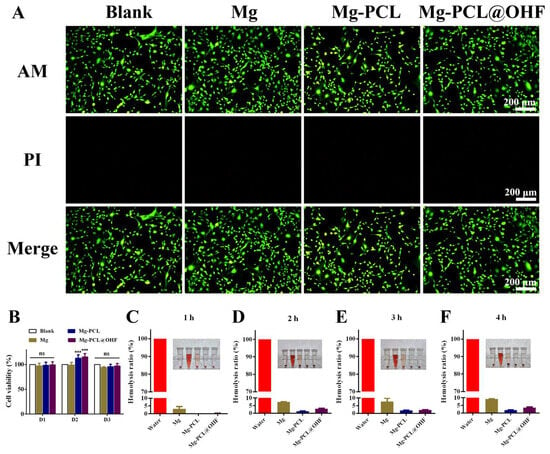
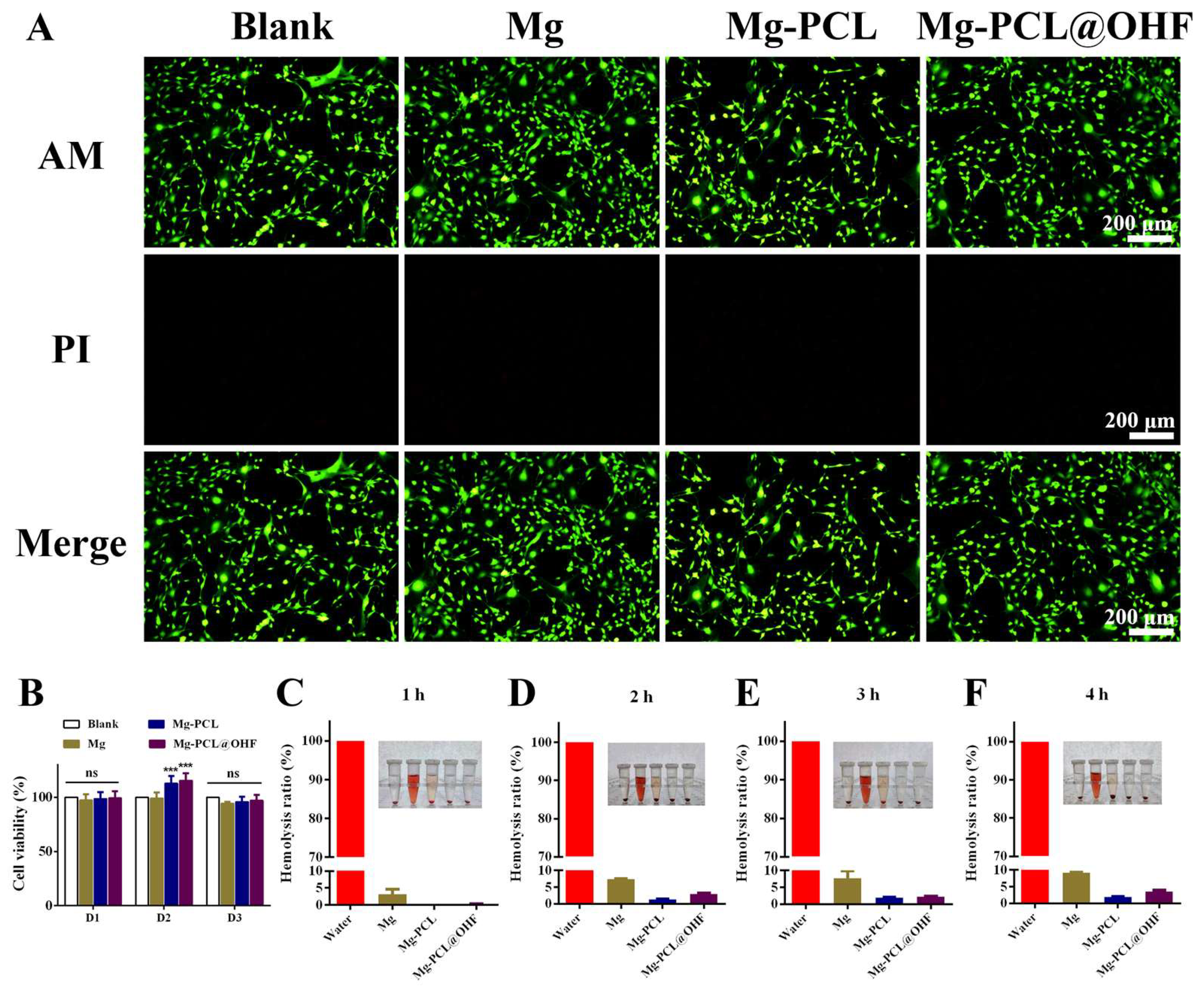
To evaluate the biocompatibility of the materials, MC3T3-E1 cells are co-cultured with material extracts, and the cell status is verified by live/dead cell staining and the CCK-8 assay. The live/dead staining profiles and morphology of MC3T3-E1 cells in each group after co-culturing are shown in the figure. Almost no red dead cells are observed from any group after co-culturing for 24 h (Figure 12A).
Figure 12.
(A) Live/dead staining of MC3T3-E1 cells co-cultured with each group for 24 h. Scale bar, 200 µm. (B) Cell viability of each group determined by CCK-8 assay via measuring absorbance at 450 nm. (C–F) Hemocompatibility and statistical analysis of hemolysis percentage for extracts of different nanomaterial groups. The data are the means ± SD. *** p < 0.001. N = 3.
Consistent with the live/dead staining results, the CCK-8 assay demonstrates that the cells in all groups maintain favorable proliferation, indicating that Mg, Mg-PCL, and Mg-PCL@OHF exhibit excellent biocompatibility (Figure 12B). Hemolysis tests are further performed to investigate the effects of the biomaterials on red blood cells and evaluate their hemocompatibility. The hemolytic response and degree of rat red blood cells are measured to assess the impact of the coatings on erythrocytes in blood. In vitro hemolysis results reveal that the nanomaterials cause no obvious hemolysis even at a concentration of 10 mg/mL, with a hemolysis rate of <5%. This meets the hemolysis rate threshold of <5% specified in the international standard ISO 10993-4:2002 [46] for the safe biomedical application of materials, confirming the biosafety of the nanomaterials for in vivo use (Figure 12C–F).
3.7. Antibacterial Evaluation
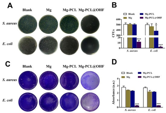
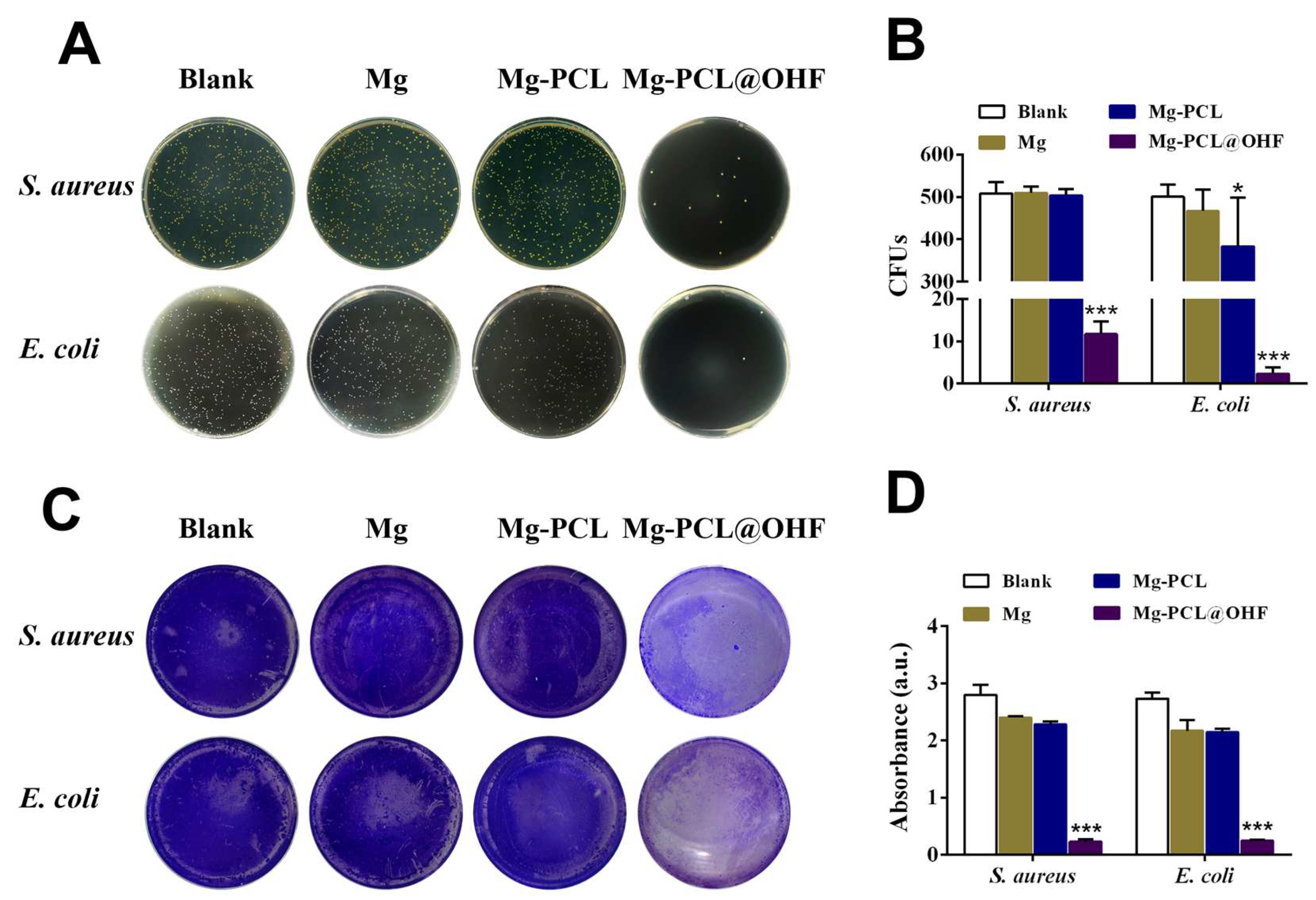
As shown in Figure 13, the antibacterial activity is evaluated by co-culturing with Staphylococcus aureus (S. aureus) and Escherichia coli (E. coli) for 24 h and analyzed by the plate counting method. Compared with the other groups, the Mg-PCL@OHF group exhibits a distinct antibacterial effect with the lowest number of colony-forming units (CFUs). As shown in Figure 13A, the samples co-incubated with extracts of Mg and Mg-PCL show a certain antibacterial capacity due to the release of Mg2+ and elevated pH caused by corrosion, resulting in a sharp reduction in bacterial colonies on bare Mg. In contrast, the coating of Mg-PCL@OHF after ultrasonic treatment shows excellent bactericidal effects due to the leached antibacterial components.
Figure 13.
Antibacterial assay: (A) Photographs of colony plating of S. aureus and E. coli co-incubated with extracts of different materials. (B) Colony counts of S. aureus and E. coli co-incubated with extracts of different materials. * p < 0.05, *** p < 0.001. (C) Bacterial biofilms of S. aureus and E. coli co-incubated with extracts of different materials stained with crystal violet. (D) Quantitative analysis of biofilm staining with crystal violet for S. aureus and E. coli. The data are the means ± SD. * p < 0.05, *** p < 0.001. N = 3.
The quantitative data in Figure 13B further confirm that Mg-PCL@OHF has the best antibacterial activity (p < 0.001). Bacterial biofilm formation is a vital mechanism for bacteria to adapt to the environment and improve their survival ability. Therefore, the capacity to inhibit biofilm formation is an indispensable step in evaluating the properties of antibacterial materials. To examine the inhibitory effect of the materials on bacterial biofilm formation, crystal violet staining of biofilms is carried out (Figure 13C). The biofilms co-cultured for 48 h are stained with 0.1% crystal violet. The control group (Blank) forms dense and uniform bacterial biofilms, whereas the Mg and Mg-PCL groups show relatively discontinuous biofilms with weaker staining. In the Mg-PCL@OHF group, only a small portion of the biofilm is stained, while most biofilm structures are destroyed, indicating that Mg-PCL@OHF releases the drug encapsulated inside the nanoparticles after ultrasonic vibration to inhibit the formation of bacterial biofilms.
3.8. Osteogenic Properties
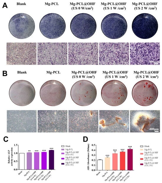
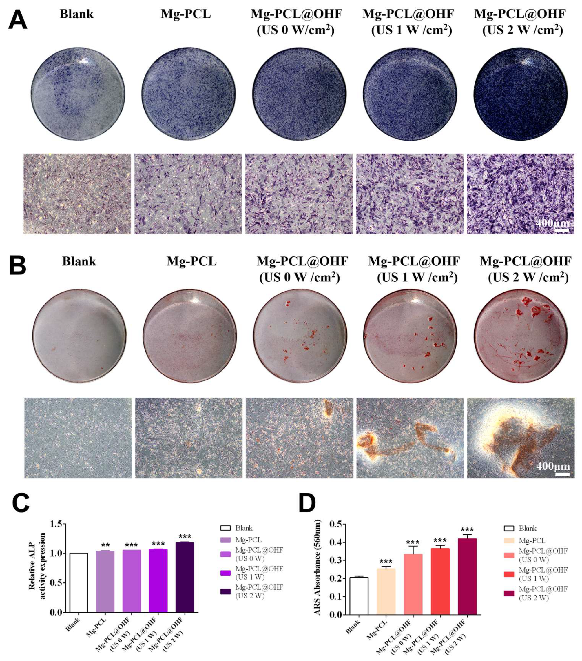
Osteogenesis is frequently employed to evaluate the potential therapeutic outcomes after surgery. Alkaline phosphatase (ALP), an early marker of osteogenic differentiation, reflects the formation of bioactive bone. As shown in Figure 14A, MC3T3-E1 cells are co-cultured for 7 days with osteogenic induction medium containing extracts of the above materials, and ALP staining is carried out to analyze the ability of the materials to promote osteogenic differentiation. The results show that the Mg-PCL@OHF ultrasonic groups exhibit stronger ALP staining intensity, and the osteogenic differentiation tendency of MC3T3-E1 cells were enhanced with increasing ultrasound intensity. As shown in Figure 14C, both the Mg-PCL and Mg-PCL@OHF ultrasonic groups show significantly higher ALP expression than the osteogenic induction medium group, and ALP expression in the Mg-PCL@OHF ultrasonic groups is further upregulated with increased ultrasound intensity.
Figure 14.
(A) ALP staining results of MC3T3-E1 cells after 7 days of co-culture with extracts of nanomaterials in each group. Scale bar, 400 µm. (B) ARS staining results of MC3T3-E1 cells after co-culturing for 21 days with extracts of nanomaterials in each group. Scale bar, 400 µm. (C) Quantitative results of ALP in MC3T3-E1 cells after 7 days of co-culturing with each group. (D) Quantitative results of ARS in MC3T3-E1 cells after co-culturing for 21 days with each group (Data analyzed by one-way ANOVA: ** p < 0.01, *** p < 0.001).
MC3T3-E1 cells are co-cultured for 21 days with the osteogenic induction medium containing the above material extracts, and Alizarin Red S staining is used to observe calcium nodule formation in each group. As shown in the figure, only small numbers of calcium nodules are formed in the Ctrl and Mg-PCL groups, whereas abundant calcified nodules are observed from all Mg-PCL@OHF ultrasonic groups. Among them, the Mg-PCL@OHF 2 W/cm2 ultrasonic group displays the highest calcium nodule content (Figure 14B), consistent with the quantitative results in Figure 14C. This can be explained by the fact that the ultrasound-responsive polymer F127 on the nanoparticle surface triggers the release of drugs from the nanoparticle interior under ultrasonic stimulation. The osteogenic agents derived from Humulus japonicus promote the osteogenic differentiation of MC3T3-E1 cells.
4. Conclusions
In this work, a novel self-healing PCL@OHF composite coating with ultrasound-triggered on-demand osthole release was successfully constructed on WE43 magnesium alloy via spin-coating for biodegradable orthopedic implant applications. The hollow mesoporous silica nanoparticles (HMSs) were loaded with osthole and further modified with Pluronic F127 to endow the coating with ultrasound-responsive controlled drug release behavior.
Electrochemical tests, hydrogen evolution measurements, and immersion experiments demonstrated that the PCL@OHF coating significantly enhanced the corrosion resistance of the magnesium alloy, reducing the corrosion current density from 2.04 × 10⁻⁶ A/cm2 to 5.94 × 10⁻⁷ A/cm2 and slowing down hydrogen evolution and Mg2+ release. Benefiting from the thermal-induced viscoelastic flow of PCL and the interfacial regulation of OHF nanoparticles, the composite coating exhibited excellent self-healing ability at 50 °C as shown by SEM, able to repair artificial scratches and restore the protective barrier of the substrate.
Drug release results verified that the composite coating realized on-demand osthole release under ultrasonic stimulation, with the cumulative release rate increasing from 39.94% (without ultrasound) to 75.93% (2 W/cm2 ultrasound) after 7 days. In vitro biological evaluations confirmed that the PCL@OHF coating possessed favorable biocompatibility and hemocompatibility, exhibited remarkable antibacterial activity against Staphylococcus aureus and Escherichia coli, inhibited bacterial biofilm formation, and significantly promoted the osteogenic differentiation of MC3T3-E1 cells. Moreover, the osteogenic effect was positively correlated with ultrasound intensity, indicating the flexible regulation of biological functions via external stimulation.
This study provides a versatile surface modification strategy that integrates self-healing, anti-corrosion, ultrasound-controlled drug release, antibacterial, and osteogenic properties in a single coating. The multifunctional coating effectively overcomes the critical limitations of rapid corrosion, implant-related infection, and insufficient osseointegration of magnesium-based implants. This intelligent responsive coating system shows high application potential in the field of biodegradable orthopedic implants and offers a reference for the design of next-generation smart biomedical implant materials.
Author Contributions
Methodology, Y.D., F.W., J.Y. (Junyan Yao), J.Y. (Juyi Yang), L.Z., S.W., C.C. and Y.Z.; Validation, J.Y. (Juyi Yang) and P.K.C.; Formal analysis, Y.F., S.J., Y.D. and F.W.; Investigation, Y.F., S.J., Y.D. and F.W.; Resources, F.X.; Data curation, Y.F., S.J., J.Y. (Junyan Yao) and S.W.; Writing—original draft, Y.F. and S.J.; Writing—review & editing, Y.D., F.W., J.Y. (Junyan Yao), C.W., J.B., C.C., J.M. and P.K.C.; Visualization, Y.Z.; Supervision, L.Z., C.W., J.B. and F.X.; Project administration, C.C.; Funding acquisition, C.C. and J.M. All authors have read and agreed to the published version of the manuscript.
Funding
This work was supported by the National Natural Science Foundation of China (Grant numbers 52571266, 52171236), State Key Program of National Natural Science Foundation of China (Grant number 52231005), Open Research Fund of Jiangsu Key Laboratory for Advanced Metallic Materials, Southeast University (Grant number AMM2024A01), Suzhou Science and Technology Project (Grant numbers SJC2023005, SZS2023023), City University of Hong Kong Donation Research Grants (Grant numbers DON-RMG 9229021 and 9220061), Guangdong-Hong Kong Technology Cooperation Funding Scheme (Grant numbers TCFS GHP/212/22GD and CityU 9440399), and Seeking Truth Talent Project, Hangzhou Medical College.
Institutional Review Board Statement
Not applicable.
Informed Consent Statement
Not applicable.
Data Availability Statement
Data will be made available on request.
Conflicts of Interest
The authors declare that they have no known competing financial interests or personal relationships that could have appeared to influence the work reported in this paper.
References
- Zhang, T.; Wang, W.; Liu, J.; Wang, L.; Tang, Y.; Wang, K. A Review on Magnesium Alloys for Biomedical Applications. Front. Bioeng. Biotechnol. 2022, 10, 953344. [Google Scholar] [CrossRef] [Scilit]
- Jafari, S.; Harandi, S.E.; Singh Raman, R.K. A Review of Stress-Corrosion Cracking and Corrosion Fatigue of Magnesium Alloys for Biodegradable Implant Applications. JOM 2015, 67, 1143–1153. [Google Scholar] [CrossRef] [Scilit]
- Zhou, P.; Xiao, X.; Zhu, X.; Chen, Y.; Lu, W.; Piao, M.; Cao, Z.; Lu, M.; Fang, F.; Li, Z.; et al. Machine Learning Enabled Customization of Performance-Oriented Hydrogen Storage Materials for Fuel Cell Systems. Energy Storage Mater. 2023, 63, 102964. [Google Scholar] [CrossRef] [Scilit]
- Yin, Z.-Z.; Qi, W.-C.; Zeng, R.-C.; Chen, X.-B.; Gu, C.-D.; Guan, S.-K.; Zheng, Y.-F. Advances in Coatings on Biodegradable Magnesium Alloys. J. Magnes. Alloys 2020, 8, 42–65. [Google Scholar] [CrossRef] [Scilit]
- Zhou, Q.; Zhou, P.; Bi, J.; Zhang, L.; Xiao, S.; Xiao, X.; Fan, X.; Chen, L. Micro-Alloying Design-Enabled Surface Activity Engineering for Enhanced Anti-Poisoning Properties of ZrCo-Based Hydrogen Isotope Storage Materials. J. Energy Chem. 2025, 108, 254–262. [Google Scholar] [CrossRef] [Scilit]
- Sun, R.X.; Liu, P.; Zhang, R.X.; Lv, Y.P.; Chen, K.Z. Hydrothermal Synthesis of Microstructured Fluoridated Hydroxyapatite Coating on Magnesium Alloy. Surf. Eng. 2016, 32, 879–884. [Google Scholar] [CrossRef] [Scilit]
- Singh, N.; Batra, U.; Kumar, K.; Ahuja, N.; Mahapatro, A. Progress in Bioactive Surface Coatings on Biodegradable Mg Alloys: A Critical Review towards Clinical Translation. Bioact. Mater. 2023, 19, 717–757. [Google Scholar] [CrossRef] [Scilit]
- Liu, X.; Wang, X.; Ren, L.; Dai, Y.; She, J.; Qi, F.; Wei, W.; Zhang, D.; Ouyang, X. Improved Deposition Quality of Calcium-Phosphate Coating on the Surface of WE43 Magnesium Alloy via FCVA Sputtering Pretreatment. J. Mater. Res. Technol. 2023, 26, 6672–6688. [Google Scholar] [CrossRef] [Scilit]
- Myagkikh, P.N.; Merson, E.D.; Poluyanov, V.A.; Begun, M.E.; Merson, D.L. Polymeric Coatings and Features of the Preliminary Surface Treatment of Biodegradable Magnesium Alloys: A Review. Russ. J. Non-Ferr. Met. 2024, 65, 364–371. [Google Scholar] [CrossRef] [Scilit]
- Han, Q.; Zhang, Z.; Liu, R.; Yu, L.; Liu, Y.; Zhao, Y.; Jin, F.; Chen, M. A Photocontrolled Self-Healing Composite Coating Based on Polycaprolactone for Enhancing the Corrosion Resistance and Antimicrobial Properties of Biodegradable Magnesium Alloys. Mater. Today Commun. 2026, 50, 114559. [Google Scholar] [CrossRef] [Scilit]
- Zhao, Y.; Bai, J.; Xue, F.; Zeng, R.; Wang, G.; Chu, P.K.; Chu, C. Smart Self-Healing Coatings on Biomedical Magnesium Alloys: A Review. Smart Mater. Manuf. 2023, 1, 100022. [Google Scholar] [CrossRef] [Scilit]
- Chen, J.; Liao, X.; Gan, J. Review on the Protective Activity of Osthole against the Pathogenesis of Osteoporosis. Front. Pharmacol. 2023, 14, 1236893. [Google Scholar] [CrossRef] [Scilit] [PubMed]
- Zhao, Y.; He, P.; Yao, J.; Li, M.; Wang, B.; Han, L.; Huang, Z.; Guo, C.; Bai, J.; Xue, F.; et al. pH/NIR-Responsive and Self-Healing Coatings with Bacteria Killing, Osteogenesis, and Angiogenesis Performances on Magnesium Alloy. Biomaterials 2023, 301, 122237. [Google Scholar] [CrossRef] [Scilit]
- Zheng, S.; Hu, G.; Zheng, J.; Li, Y.; Li, J. Osthole Accelerates Osteoporotic Fracture Healing by Inducing the Osteogenesis–Angiogenesis Coupling of BMSCs via the Wnt/β-Catenin Pathway. Phytother. Res. 2024, 38, 4022–4035. [Google Scholar] [CrossRef] [Scilit]
- Sun, M.; Sun, M.; Zhang, J. Osthole: An Overview of Its Sources, Biological Activities, and Modification Development. Med. Chem. Res. 2021, 30, 1767–1794. [Google Scholar] [CrossRef] [Scilit]
- Lv, M.; Ding, H.; Xu, H. Osthole: A Coumarin with Dual Roles in Biology and Chemistry. Biology 2025, 14, 588. [Google Scholar] [CrossRef] [Scilit]
- Mosebarger, A.; Reddi, R.N.; Menon, R.; Kammala, A.K. Computational Screening of the Natural Product Osthole and Its Derivates for Anti-Inflammatory Activity. Life 2022, 12, 505. [Google Scholar] [CrossRef] [Scilit]
- Mitchell, M.J.; Billingsley, M.M.; Haley, R.M.; Wechsler, M.E.; Peppas, N.A.; Langer, R. Engineering Precision Nanoparticles for Drug Delivery. Nat. Rev. Drug Discov. 2021, 20, 101–124. [Google Scholar] [CrossRef] [Scilit]
- Zhou, Y.; Quan, G.; Wu, Q.; Zhang, X.; Niu, B.; Wu, B.; Huang, Y.; Pan, X.; Wu, C. Mesoporous Silica Nanoparticles for Drug and Gene Delivery. Acta Pharm. Sin. B 2018, 8, 165–177. [Google Scholar] [CrossRef] [Scilit]
- Zhang, A.; Jung, K.; Li, A.; Liu, J.; Boyer, C. Recent Advances in Stimuli-Responsive Polymer Systems for Remotely Controlled Drug Release. Prog. Polym. Sci. 2019, 99, 101164. [Google Scholar] [CrossRef] [Scilit]
- Aydin, S.; Soares, A.P.; Fischer, H.; Knecht, R.S.; Kopp, A.; Schmidt-Bleek, K.; Heiland, M.; Rendenbach, C. In Vitro Study on the Osteoimmunological Potential of Magnesium Implants (WE43MEO). Biomed. Eng. OnLine 2025, 24, 90. [Google Scholar] [CrossRef] [Scilit]
- Thakur, N.S.; Saleh, N.; Khan, A.F.; Chakrabarty, B.; Agrahari, V. Progress and Promise of Photoresponsive Nanocarriers for Precision Drug Delivery in Cancer. J. Photochem. Photobiol. C Photochem. Rev. 2024, 59, 100665. [Google Scholar] [CrossRef] [Scilit]
- Zhang, H.; Ren, B.; Huang, Y.; Yao, X.; Zhai, Y.; Du, L. Functionalized Smart Platforms of ZIF-8: Progress in Orthopedic Therapeutic Applications and Challenges. Chem. Eng. J. 2025, 524, 169386. [Google Scholar] [CrossRef] [Scilit]
- Zhao, Y.; He, P.; Wang, B.; Bai, J.; Xue, F.; Chu, C. Incorporating pH/NIR Responsive Nanocontainers into a Smart Self-Healing Coating for a Magnesium Alloy with Controlled Drug Release, Bacteria Killing and Osteogenesis Properties. Acta Biomater. 2024, 174, 463–481. [Google Scholar] [CrossRef] [Scilit]
- Fan, J.; Xuan, M.; Zhao, P.; Loznik, M.; Chen, J.; Kiessling, F.; Zheng, L.; Herrmann, A. Ultrasound Responsive Microcapsules for Antibacterial Nanodrug Delivery. Nano Res. 2023, 16, 2738–2748. [Google Scholar] [CrossRef] [Scilit]
- Zhao, Y.; Li, Y.; Wang, B.; Yao, J.; Fan, Y.; He, P.; Bai, J.; Wang, C.; Xue, F.; Chu, C. An Injectable Magnesium-Based Cement Stimulated with NIR for Drug-Controlled Release and Osteogenic Potential. Adv. Healthc. Mater. 2024, 13, 2400207. [Google Scholar] [CrossRef] [Scilit]
- Cui, X.; Han, X.; Yu, L.; Zhang, B.; Chen, Y. Intrinsic Chemistry and Design Principle of Ultrasound-Responsive Nanomedicine. Nano Today 2019, 28, 100773. [Google Scholar] [CrossRef] [Scilit]
- Kooiman, K.; Roovers, S.; Langeveld, S.A.G.; Kleven, R.T.; Dewitte, H.; O’Reilly, M.A.; Escoffre, J.-M.; Bouakaz, A.; Verweij, M.D.; Hynynen, K.; et al. Ultrasound-Responsive Cavitation Nuclei for Therapy and Drug Delivery. Ultrasound Med. Biol. 2020, 46, 1296–1325. [Google Scholar] [CrossRef] [Scilit] [PubMed]
- Yang, B.; Du, J.-Z. Ultrasound-Responsive Homopolymer Nanoparticles. Chin. J. Polym. Sci. 2020, 38, 349–356. [Google Scholar] [CrossRef] [Scilit]
- Xin, Y.; Qi, Q.; Mao, Z.; Zhan, X. PLGA Nanoparticles Introduction into Mitoxantrone-Loaded Ultrasound-Responsive Liposomes: In Vitro and in Vivo Investigations. Int. J. Pharm. 2017, 528, 47–54. [Google Scholar] [CrossRef] [Scilit]
- Wei, H.-X.; Liu, M.-H.; Wang, T.-Y.; Shih, M.-H.; Yu, J.; Yeh, Y.-C. Fabrication of pH- and Ultrasound-Responsive Polymeric Micelles: The Effect of Amphiphilic Block Copolymers with Different Hydrophilic/Hydrophobic Block Ratios for Self-Assembly and Controlled Drug Release. Biomacromolecules 2025, 26, 2116–2130. [Google Scholar] [CrossRef] [Scilit] [PubMed]
- Hu, H.; Yu, J.; Li, Y.; Zhao, J.; Dong, H. Engineering of a Novel Pluronic F127/Graphene Nanohybrid for pH Responsive Drug Delivery. J. Biomed. Mater. Res. A 2012, 100A, 141–148. [Google Scholar] [CrossRef] [Scilit]
- Yao, J.; Wang, B.; Zhao, Y.; Xu, Y.; Wang, C.; Bai, J.; Xue, F.; Chu, P.K.; Chu, C. Modified Magnesium Phosphate Cement Composed of Hyaluronic Acid and Ultrasound-Responsive Nanocapsules with Improved Physicochemical and Osteogenic Properties. Chem. Eng. J. 2025, 525, 170175. [Google Scholar] [CrossRef] [Scilit]
- Li, S.; Yang, C.; Li, J.; Zhang, C.; Zhu, L.; Song, Y.; Guo, Y.; Wang, R.; Gan, D.; Shi, J.; et al. Progress in Pluronic F127 Derivatives for Application in Wound Healing and Repair. Int. J. Nanomed. 2023, 18, 4485–4505. [Google Scholar] [CrossRef] [Scilit]
- Zhao, Y.; He, P.; Yao, J.; Li, M.; Bai, J.; Xue, F.; Chu, C.; Cong, Y.; Chu, P.K. Self-Assembled Multilayered Coatings with Multiple Cyclic Self-Healing Capability, Bacteria Killing, Osteogenesis, and Angiogenesis Properties on Magnesium Alloys. Adv. Healthc. Mater. 2024, 13, 2302519. [Google Scholar] [CrossRef] [Scilit]
- Teng, Z.; Li, W.; Tang, Y.; Elzatahry, A.; Lu, G.; Zhao, D. Mesoporous Organosilica Hollow Nanoparticles: Synthesis and Applications. Adv. Mater. 2019, 31, 1707612. [Google Scholar] [CrossRef] [Scilit] [PubMed]
- Shen, C.; Zhou, P.; Guo, D. Potential Profile of Electrolytic Cell and Influence of Electrolyte Resistance in Three-Electrode Electrochemical Testing. Electrochim. Acta 2025, 530, 146428. [Google Scholar] [CrossRef] [Scilit]
- Cui, L.-Y.; Gao, S.-D.; Li, P.-P.; Zeng, R.-C.; Zhang, F.; Li, S.-Q.; Han, E.-H. Corrosion Resistance of a Self-Healing Micro-Arc Oxidation/Polymethyltrimethoxysilane Composite Coating on Magnesium Alloy AZ31. Corros. Sci. 2017, 118, 84–95. [Google Scholar] [CrossRef] [Scilit]
- Gupta, A.; Srivastava, C. Electrodeposition Current Density Induced Texture and Grain Boundary Engineering in Sn Coatings for Enhanced Corrosion Resistance. Corros. Sci. 2022, 194, 109945. [Google Scholar] [CrossRef] [Scilit]
- Manzano, M.; Vallet-Regí, M. Ultrasound Responsive Mesoporous Silica Nanoparticles for Biomedical Applications. Chem. Commun. 2019, 55, 2731–2740. [Google Scholar] [CrossRef] [Scilit]
- Shriky, B.; Kelly, A.; Isreb, M.; Babenko, M.; Mahmoudi, N.; Rogers, S.; Shebanova, O.; Snow, T.; Gough, T. Pluronic F127 Thermosensitive Injectable Smart Hydrogels for Controlled Drug Delivery System Development. J. Colloid Interface Sci. 2020, 565, 119–130. [Google Scholar] [CrossRef] [Scilit]
- Qian, X.; Wang, W.; Kong, W.; Chen, Y. Organic-Inorganic Hybrid Hollow Mesoporous Organosilica Nanoparticles for Efficient Ultrasound-Based Imaging and Controlled Drug Release. J. Nanomater. 2014, 2014, 972475. [Google Scholar] [CrossRef] [Scilit]
- Li, J.; He, Y.; Inoue, Y. Thermal and Infrared Spectroscopic Studies on Hydrogen-bonding Interactions between Poly-(Ε-caprolactone) and Some Dihydric Phenols. J. Polym. Sci. Part B Polym. Phys. 2001, 39, 2108–2117. [Google Scholar] [CrossRef] [Scilit]
- Park, M.; Lee, J.E.; Park, C.G.; Lee, S.H.; Seok, H.K.; Choy, Y.B. Polycaprolactone Coating with Varying Thicknesses for Controlled Corrosion of Magnesium. J. Coat. Technol. Res. 2013, 10, 695–706. [Google Scholar] [CrossRef] [Scilit]
- Zheng, Q.; Li, J.; Yuan, W.; Liu, X.; Tan, L.; Zheng, Y.; Yeung, K.W.K.; Wu, S. Metal–Organic Frameworks Incorporated Polycaprolactone Film for Enhanced Corrosion Resistance and Biocompatibility of Mg Alloy. ACS Sustain. Chem. Eng. 2019, 7, 18114–18124. [Google Scholar] [CrossRef] [Scilit]
- ISO 10993-4:2002; Biological Evaluation of Medical Devices—Part 4: Selection of Tests for Interactions with Blood. ISO: Geneva, Switzerland, 2002.

Disclaimer/Publisher’s Note: The statements, opinions and data contained in all publications are solely those of the individual author(s) and contributor(s) and not of MDPI and/or the editor(s). MDPI and/or the editor(s) disclaim responsibility for any injury to people or property resulting from any ideas, methods, instructions or products referred to in the content. |
© 2026 by the authors. Licensee MDPI, Basel, Switzerland. This article is an open access article distributed under the terms and conditions of the Creative Commons Attribution (CC BY) license.