


Abstract
Background: Growing research interest has focused on biosensing platforms as rapid, sensitive and economically advantageous tools for bacterial identification and antimicrobial resistance or susceptibility assessment in microbiological diagnostics. Conventional microbiological workflows remain essential reference methods but may require extended incubation and processing times. A wide range of biosensing strategies has therefore been investigated to accelerate bacterial detection and the assessment of antimicrobial response. Methods: This narrative review examines biosensing approaches relevant to bacterial detection and AST, including living-cell and whole-cell biosensors, electrochemical and impedimetric platforms, optical biosensors, bacteriophage-based systems, immunosensors, aptamer-based technologies, microfluidic approaches, and CRISPR-associated molecular detection. Particular attention is given to the distinction between platforms in which living cells function as sensing elements and systems in which bacterial cells or bacterial components represent the analytical target. Results: Reported analytical performance varied considerably according to bacterial species, recognition element, transduction strategy, sample matrix, sample-preparation requirements, and validation design. Selected experimental platforms achieved rapid detection or antimicrobial-response measurements; however, many studies were performed using buffer systems, artificially spiked samples, or relatively limited collections of bacterial isolates. Phenotypic AST platforms, molecular resistance-detection systems, and bacterial-identification technologies provide different types of information and should therefore not be considered interchangeable. Translation into routine microbiology remains limited by challenges related to sample preparation, reproducibility, sensor stability, standardization, validation against reference methods, and integration into clinical workflows. Conclusions: Cell-based and related biosensing approaches show potential to complement conventional microbiological methods, but further standardisation, validation and integration into routine workflows are required before widespread clinical implementation.
1. Introduction
Biosensors combine biological recognition elements and signal transduction technologies designed to detect and quantify specific substances in biological samples [1]. These analytical devices utilise living cells as sensing components, whether microbial, mammalian or genetically engineered, in conjunction with physicochemical transducers that convert biological activities into quantifiable signals [2].
With their rapid detection, enhanced sensitivity, and real-time monitoring capabilities for infectious agents and antimicrobial activities, biosensors have emerged as potential complements for traditional culture-based methods in microbiological diagnostics [3].
Timely bacterial identification and antimicrobial susceptibility testing (AST) support treatment decisions, infection control and antimicrobial stewardship. Conventional culture-based identification and antimicrobial susceptibility testing methods remain essential because they recover viable organisms and provide clinically interpretable results. Nevertheless, these methods may involve several sequential steps, delaying essential information for clinical decisions [4,5]. Biosensing technologies are being investigated as complementary approaches for obtaining diagnostic information more rapidly, provided that their results remain reliable in the clinical specimens and laboratory workflows in which they would be used [3,6].
In a cell-based or whole-cell biosensor, living microbial or mammalian cells act as sensing elements: exposure to a target produces a change in reporter expression, metabolism, viability or another cellular response [7,8]. In many other bacterial biosensors, the bacterium is instead the analytical target, captured or detected by an antibody, aptamer, bacteriophage or other recognition element. A bacterial cell measured by an impedance device is likewise the subject of analysis, not the device’s transducer. Nucleic-acid assays, including CRISPR-associated approaches, constitute a further category when they detect bacterial sequences rather than a response generated by a living sensing cell [9]. These distinctions are necessary to classify the technologies discussed in this review.
The diagnostic information provided by these platforms must also be distinguished. Bacterial detection or identification establishes whether a target is present or determines its identity. Detection of an antimicrobial-resistance gene provides genotypic information but does not, on its own, establish the organism’s phenotypic susceptibility under the conditions used for AST [10]. Phenotypic AST instead measures the response of viable bacteria following antimicrobial exposure, for example, through changes in growth, metabolism, morphology or electrical properties [11,12].
Immunological, phage-assisted, electrochemical, optical and microfluidic platforms provide additional options for bacterial detection or functional assessment. Their reported performance, however, depends on the target, specimen matrix, sample preparation, enrichment and the point at which assay time is measured. A short sensor readout does not necessarily translate into a short total time-to-result.
Advanced materials and nanotechnology have further expanded the capabilities of biosensor platforms [13]. Graphene, gold and carbon-based nanomaterials are examples of materials that improve signal transduction by maximising the surface area available for biomolecular interactions [1]. These developments have expanded the potential applications of biosensors in infectious disease diagnostics [14].
These differences make direct comparisons between published studies difficult. A detection limit measured in buffer or an artificially spiked sample cannot be assumed to apply to a naturally contaminated food product or a clinical specimen. Likewise, an analytical response to antibiotic exposure cannot be treated as validated AST without comparison with an appropriate reference method and reporting of agreement [3,6].
The distinctive contribution of this review is a cross-platform framework that distinguishes the biological recognition principle, transduction mechanism, microbiological information generated, and level of translational validation. We classify each platform by its biological sensing or recognition element, its transduction mechanism, and its intended output: bacterial detection or identification, genotypic resistance detection, or phenotypic AST. We then examine reported performance in the context of sample matrix, pretreatment or enrichment, total time to result, comparison with reference methods, and the level of analytical or clinical validation.
Accordingly, this review addresses three key questions: which biological component generates the signal, what microbiological conclusion that signal supports, and what evidence is available for use beyond proof-of-concept experiments. This approach is intended to identify where biosensing platforms could complement established microbiological workflows and where further validation is required.
2. Materials and Methods
This narrative review examines biosensing strategies for bacterial detection and antimicrobial susceptibility testing (AST), distinguishing platforms in which living cells function as sensing elements from those that detect bacterial cells or bacterial components. Relevant literature was identified through searches of PubMed, Web of Science, and Google Scholar. The literature was selected to cover early whole-cell biosensing systems and subsequent developments in microbiological detection and susceptibility assessment. Searches used combinations of keywords related to biological recognition, signal transduction, and microbiological applications, including: electrochemical and optical sensing platforms, fluorescence- and bioluminescence-based detection, bacteriophage-based biosensors, aptamer-based systems, microfluidic technologies, CRISPR-based detection methods, and artificial intelligence applications in microbiological diagnosis. The search terms included: “cell-based biosensors”, “whole-cell biosensors”, “bacterial detection”, “pathogen detection”, “antimicrobial susceptibility testing”, “rapid AST”, “biosensors in microbiology”, “electrochemical biosensors”, “optical biosensors”, “fluorescence biosensors”, “bioluminescent biosensors”, “bacteriophage biosensors”, “aptamer-based biosensors”, “lab-on-chip”, “CRISPR-based detection” and “artificial intelligence in microbiology”. The literature covered both the historical development of biosensing technologies and more recent advances in bacterial detection and antimicrobial susceptibility assessment.
2.1. Inclusion Criteria
Studies were selected based on the following criteria: (i) original research articles or reviews published in peer-reviewed journals; (ii) investigations describing biosensor-based approaches for bacterial detection, identification, characterisation, viability assessment, or antimicrobial susceptibility testing; (iii) studies involving whole-cell or cell-based sensing technologies, biological recognition elements, or biosensing platforms relevant to microbiological diagnosis; (iv) publications providing relevant methodological or analytical information, including, where available, limits of detection, assay duration, bacterial concentrations, sample matrices, sensitivity, specificity, or antimicrobial susceptibility results; (v) studies addressing emerging technologies with potential applications in microbiological diagnostics, including microfluidics, nanomaterials, bacteriophages, CRISPR-based systems and artificial-intelligence-assisted analysis; and (vi) articles published in English.
2.2. Exclusion Criteria
Publications were excluded when they did not provide adequate experimental evidence, contained limited methodological information, or were published in languages other than English.
Original research articles were considered for the comparative analysis of experimental biosensor performance, whereas review articles provided background information and contextualised technological developments. The comparative assessment focused on the intended microbiological application, biological recognition element, transduction mechanism, bacterial target, sample matrix, analytical performance, and validation procedures.
Where reported, detection limits, analytical ranges, assay duration, total time-to-result, sample pretreatment or enrichment, and agreement with reference methods were considered. Particular attention was given to distinguishing bacterial detection and identification from genotypic antimicrobial resistance detection and phenotypic AST.
Because the reviewed studies differed in experimental conditions, bacterial targets, sample matrices, and validation approaches, their findings were interpreted within the context of the individual study designs rather than used to establish direct rankings of platform performance.
3. Biosensing Strategies for Bacterial Detection and Antimicrobial Susceptibility Testing
3.1. Evolution of Biosensors
Cell-based biosensors have evolved from early-stage microbial sensors to complex diagnostic platforms that can detect individual pathogen cells [15].
The conceptual foundations of biosensing preceded the modern biosensor by several decades. The theoretical basis began in 1906 when M. Cremer observed that the concentration of an acid in a liquid is proportional to the electric potential difference across glass membranes. S.P.L. Sørensen introduced the pH concept in 1909, followed by W.S. Hughes’ pH measurement electrode in 1922 [16]. During this period (1909–1922), Griffin and Nelson demonstrated early enzyme immobilisation using invertase on aluminium hydroxide [16]. In the late 1950s, R.S. Yalow and S.A. Berson pioneered radioimmunoassays using radiolabeled antibodies as recognition and measurement elements [17] (Figure 1).
Figure 1.
Historical evolution of biosensors.
The following decade marked the diversification of biosensor architectures. In 1962, L. Clark and A. Lyons developed the first enzyme electrode (using glucose oxidase on an oxygen electrode), which measured the oxygen consumption rate, thereby enabling the measurement of glucose levels; establishing Clark as the “father of biosensors” [16,17,18]. S.J. Updike and G.P. Hicks introduced the first immobilised enzyme electrode in 1967 [17], and G.G. Guilbault and J.G. Montalvo created the first potentiometric biosensor for urea in 1969 [16,18,19]. In 1970, Bergveld developed the ion-sensitive field-effect transistor (ISFET), leading to Yellow Springs Instruments (YSI) launching the first commercial glucose biosensor in 1975 [16,19].
In the 1980s and early 1990s, biosensing expanded beyond classical enzyme electrodes through second-generation biosensors, which co-immobilise auxiliary enzymes and/or co-reactants with the analyte-converting enzyme, to improve analytical performance. The 1980s introduced synthetic electron mediators (e.g., ferrocene-mediated amperometric glucose detection in 1983–1984) to eliminate reliance on dissolved oxygen. Optical sensing advanced with fibre-optic pH (1980) and glucose sensors (1982), followed by Surface Plasmon Resonance (SPR) immunosensors in 1983. Commercial breakthroughs occurred with Pharmacia Biacore’s SPR system (1990) and i-STAT’s handheld blood analyser (1992) [16,17,18,19]. At the same time, advances in molecular biology enabled a second major transition: living microorganisms could themselves be engineered to function as biological sensing elements. The mer–lux system reported by O. Selifonova in 1993 demonstrated bioluminescent detection using genetically engineered bacteria, while subsequent systems incorporated reporter genes and electrochemical outputs for selective analyte detection [20].
During the 2000s and 2010s, bacterial biosensing evolved toward increasingly miniaturized and integrated platforms. Advances in MEMS/NEMS/BioNEMS, nanotechnology, and biotechnology supported the development of miniaturized and nanomaterial-enhanced sensing systems [18]. Carbon nanotubes, metallic nanoparticles, quantum dots, and other nanostructured materials were increasingly incorporated into biosensor architectures, while aptamers emerged as alternative molecular recognition elements compatible with electrochemical, optical, piezoelectric, and magnetic transducers [18]. In bacterial diagnostics, a notable cell-based example was the CANARY platform reported by Rider in 2003 [21], while Cheng et al. demonstrated the application of nanoporous electrochemical sensing to E. coli detection in 2011 [13]. During the same period, the repertoire of biological recognition elements expanded considerably. Researchers subsequently developed phage-based electrochemical and reporter systems for several bacterial targets. For example, bacteriophage-based amperometric sensing was applied to Shiga toxin-producing E. coli, whereas engineered reporter phages enabled bioluminescence-based detection of viable Listeria cells [22,23,24].
Since approximately 2020, bacterial biosensing has increasingly shifted from rapid detection alone toward functional assessment of viable bacteria and increasingly rapid phenotypic antimicrobial susceptibility testing. Microfluidic impedance-based approaches such as iFAST enabled rapid phenotypic AST by detecting antibiotic-induced changes in bacterial cells’ electrical properties [11]. Near-single-cell electrical impedance spectroscopy was subsequently used to monitor metabolic and electrical responses of individual bacteria to antibiotic exposure, with significant impedance changes reported within 30–60 min [25].
Molecular biosensing has developed in parallel with these phenotypic approaches. CRISPR/Cas-based platforms represent sequence-specific detection systems in which Cas proteins (Cas12a/Cas13a) recognise bacterial nucleic-acid targets and generate measurable signals through target-triggered reporter cleavage [26,27].
Recent research has expanded bacterial detection beyond culture-based confirmation toward faster molecular, biosensing, and point-of-care approaches, while culture remains important for recovering isolates and confirming results [6]. Modern detection incorporates advanced synthetic biology tools, such as programmable cell-free RNA toehold switch biosensors, engineered microbial whole-cell biosensors, and CRISPR-Cas (Cas12a/Cas13a) nucleases paired with isothermal amplification like RPA and LAMP for target-specific nucleic acid recognition [28,29]. Concurrently, functional nanomaterials, such as gold, magnetic, and upconversion nanoparticles, alongside two-dimensional materials such as MXenes, support multimodal optical and electrochemical signal transduction (SERS, LSPR, fluorescence), have reported low detection limits particularly in food-pathogen studies, although their performance varies by assay and sample matrix [6,30].
Overall, the historical development of biosensor technology reflects several parallel transitions in microbiology, from simple bacterial detection toward viability assessment, molecular resistance detection and rapid phenotypic AST. These technological advances have expanded analytical possibilities, but they have not eliminated the need for rigorous validation. Contemporary development therefore focuses not only on analytical sensitivity and rapid signal acquisition, but also on sample preparation, reproducibility, clinically relevant validation, scalability, regulatory requirements, and integration into routine diagnostic workflows [3,4,31].
3.2. Overview of Bacterial Detection and Clinical Importance
The burden of antimicrobial-resistant bacteria has increased the need for new methods of bacterial detection and antimicrobial susceptibility testing [5,32]. Timely identification is important for appropriate antimicrobial therapy, infection prevention, and antimicrobial stewardship, particularly because multidrug-resistant organisms such as vancomycin-resistant enterococci (VRE), extended-spectrum β-lactamase (ESBL)-producing Enterobacterales, and carbapenem-resistant Enterobacterales (CRE) present significant therapeutic and infection-control challenges [32].
Biosensor technologies have been developed for a broad range of clinically and epidemiologically significant bacterial targets, including common food-associated bacteria and other clinically important pathogens [4,33].
Gram-negative bacteria are important biosensor targets because several species cause foodborne disease, urinary tract infections, and healthcare-associated infections [4]. Escherichia coli (E. coli), particularly Shiga toxin-producing E. coli (STEC) O157 strains, is an important target for rapid identification because STEC infections can cause acute and frequently bloody diarrhoea and may progress to haemolytic uraemic syndrome (HUS), with approximately 8% of diagnosed O157 STEC infections developing HUS [22,34]. Salmonella species, particularly S. Enteritidis and S. Typhimurium, are major causes of infectious gastroenteritis and can also cause severe infections, including bloodstream infections [33,35]. Another clinically important Gram-negative bacterial target is Pseudomonas aeruginosa (P. aeruginosa), an opportunistic pathogen that commonly causes chronic pulmonary infections and contributes to progressive decline in lung function. P. aeruginosa can also cause acute and chronic infections in hospitalized and immunocompromised patients [36]. Cholera, an important diarrheal disease caused by Vibrio cholerae, is linked to inadequate access to sanitation and optimal hygiene, with approximately 1.3 to 4.0 million cases and 21,000 to 143,000 deaths each year [35].
Gram-positive bacteria are important targets for biosensor development, particularly because several clinically and foodborne-relevant species require rapid, sensitive detection [4,37]. Staphylococcus aureus (S. aureus) is a leading cause of infection, most notably bacteremia and device-related infections [38,39]. Rapid detection of MRSA can facilitate appropriate antimicrobial selection [4]. L. monocytogenes is a foodborne pathogen and a cause of severe infectious disease, particularly in immunocompromised individuals, making sensitive detection in food an important component of food-safety control [37,40].
Foodborne bacterial pathogens are important biosensor targets because they cause significant gastrointestinal infections and may have public-health consequences, creating a need for rapid, sensitive detection technologies [33].
Nosocomial pathogens such as P. aeruginosa and Acinetobacter baumannii (A. baumannii), as well as antimicrobial-resistant E. coli and S. aureus, are important targets for rapid biosensor-based diagnostic technologies in healthcare settings [41].
The diversity of bacterial targets, sample matrices and intended applications creates a need for adaptable biosensor platforms with tunable sensitivity, specificity, portability and analytical performance across clinical, food-safety, and environmental settings [42]. Multiplexed biosensors that can simultaneously identify numerous pathogens or antimicrobial-resistance indicators represent an important direction in biosensor development, particularly for applications involving mixed infections or samples containing multiple potential targets [43,44]. The translation of cell-based and whole-cell biosensors from research platforms into clinical, food-safety and environmental applications also requires attention to analytical validation, quality control, standardization and personnel competence [41,45].
Some biosensor platforms can detect and quantify microorganisms while simultaneously assessing their viability, providing information relevant to the evaluation of antimicrobial effects [46]. However, viability assessment alone does not establish phenotypic antimicrobial susceptibility without evaluating bacterial responses to antibiotic exposure and validating the resulting susceptibility assessment against an appropriate reference method.
3.3. Classification of Biosensing Platforms
3.3.1. Living-Cell and Whole-Cell Biosensors
Living-cell biosensors use viable microbial or mammalian cells as the biological sensing element and convert a cellular response to an analyte into a measurable output [8,47]. In engineered microbial systems, a regulatory protein or signalling pathway can control reporter genes such as lux, gfp, or lacZ, generating luminescent, fluorescent or colorimetric signals in response to a defined stimulus [8]. The signal may represent recognition of a molecule, activation of a stress pathway, or a change in cellular activity, depending on the sensor’s design [8,47].
Recent engineering strategies have broadened the inputs accessible to microbial whole-cell biosensors by modifying two-component systems and allosteric transcription factors [47]. Encapsulation can provide a controlled environment for viable sensing cells while allowing exchange with the surrounding sample, but researchers must assess how containment affects sensor function for each application [48]. Sample composition is equally important: A. Zúñiga et al. found that human faecal material inhibited several engineered bacterial metabolite sensors and that the magnitude of inhibition differed between sensor circuits and patient samples [49].
A direct example of antimicrobial-effect sensing was reported by Xu et al., who used the PghKR cell-wall stress response of Shewanella oneidensis MR-1 to activate a blaA promoter–luxCDABE luminescent reporter [29]. Deletion of both blaA and ampG improved the reported ampicillin detection limit to 0.06 µg/mL after one hour of exposure, compared with approximately 0.6–0.7 µg/mL for the corresponding single-deletion sensors [29]. When incorporated into a double-layer plate, the engineered cells signalled the presence of antibiotic-producing colonies from soil samples; 36 of 103 initially selected colonies retained sensor-activating activity on confirmation, and five showed activity against a multidrug-resistant indicator strain [29]. This assay identifies potential producers of compounds that induce cell-wall stress, but it does not determine the MIC or susceptibility category of a clinical bacterial isolate [29].
Mammalian-cell systems can instead report interactions between pathogens or toxins and host cells [33,50]. One such system detected Listeria monocytogenes (L. monocytogenes) at approximately 103–104 CFU/mL after 4–6 h of cell exposure and was also evaluated with artificially inoculated foods containing L. monocytogenes or Bacillus cereus [33]. In a more recent example, Eser et al. used HEK293 cells engineered to express human TLR5 and a secreted alkaline phosphatase reporter to detect responses to Salmonella flagellin [50]. Their pure-culture detection limit was approximately 105 CFU/mL, whereas approximately 50–100 CFU per 25 g of artificially inoculated food could be detected as the initial inoculum after enrichment, with a reported total sample-to-result time of 15 h [50]. The study assessed 20 Salmonella enterica serovars and 19 non-Salmonella cultures, but recognition of flagellin means that selectivity should be interpreted within the tested organism panel [50].
CANARY (Cellular Analysis and Notification of Antigen Risks and Yields) provides another mammalian-cell example: engineered B lymphocytes generate a rapid light signal after recognising a target pathogen [21]. The original study reported detection of 50 CFU of Yersinia pestis in less than three minutes under its experimental conditions [21]. That result illustrates rapid cellular recognition in a defined assay and does not establish the same performance across all clinical or environmental specimens [21].
These studies show distinct roles for living-cell biosensors in detecting pathogens and screening antimicrobial activity [21,29,33,50]. Evidence from artificially inoculated foods and environmental screening supports further development, while use for routine clinical identification or phenotypic AST requires validation with appropriate specimens, clinical isolates and reference methods [29,50].
3.3.2. Bacteriophage-Based Biosensing Platforms
Researchers have investigated bacteriophages as biological recognition elements for bacterial biosensing because of their natural specificity for susceptible bacterial hosts [23]. In these platforms, phages can be used either as selective bacterial capture elements or as infection-based recognition agents [22,23,24]. Researchers can then couple the recognition event to different signal-generation mechanisms, including electrochemical and bioluminescent readouts [22,23,24].
Electrochemical phage-based platforms have achieved high analytical sensitivity. Quintela and Wu developed a sandwich-type amperometric biosensor for Shiga toxin-producing E. coli O26, O157, and O179 [22]. Bacteriophages immobilised on streptavidin-functionalised screen-printed carbon electrodes captured viable STEC cells, while a secondary bacteriophage–gold nanoparticle detection system generated the electrochemical signal. The platform achieved detection limits of approximately 101–102 CFU/mL in pure cultures and achieved comparable detection limits of 101–102 CFU/g or mL in ground beef and apple juice, with an overall turnaround time below 1 h [22]. Farooq immobilised an S. aureus-specific bacteriophage on a conductive bacterial-cellulose interface and detected S. aureus at 3 CFU/mL in PBS and 5 CFU/mL in milk within 30 min. The system could also discriminate viable cells in mixtures containing live and dead bacteria [51].
Genetically engineered reporter phages provide another strategy in which bacterial recognition and signal generation are coupled through the phage infection process. Meile et al. engineered NanoLuc-expressing Listeria phages and demonstrated detection of 1 CFU of L. monocytogenes in 25 g of artificially contaminated milk, chicken cold cuts, and iceberg lettuce within approximately 24 h when combined with pre-enrichment. The assay was subsequently evaluated using 48 naturally purchased food samples and produced no false-positive or false-negative results compared with molecular detection and conventional selective culture in that sample set. In addition, narrow-host-range reporter phages A006::nluc and A500::nluc enabled differentiation of specific Listeria serovar groups, pertinent for bacterial differentiation [24].
Recent approaches have also focused on multiplexed phage recognition. Wu et al. developed dual-modified M13 phages for simultaneous detection of E. coli O157, S. Typhimurium, and P. aeruginosa, achieving a detection limit of approximately 102 cells/mL by flow cytometry [44].
More recently, phage recognition has been combined with magnetic bacterial separation. Zhang and Lin developed dual-bacteriophage superparamagnetic nanoprobes targeting S. aureus and P. aeruginosa. The optimised formulation, containing 107 PFU/mL of each phage, detected both bacteria at concentrations as low as 101 CFU/mL and enabled bacterial capture within 10 min. No cross-infection was observed between the phages and their respective non-target bacteria. However, capture efficiency depended on phage loading, with excessive phage concentrations reducing performance. The platform was validated only in controlled aqueous suspensions, so further evaluation in complex clinical, environmental, or food matrices is still required [52].
Bacteriophage-based detection offers high target specificity, low detection limits, and compatibility with electrochemical, optical, bioluminescent, and magnetic readouts. Infection-dependent reporter phages can also indicate viable bacteria because signal generation generally requires metabolically active host cells. However, narrow host ranges may limit strain coverage, while phage resistance, matrix interference, and low bacterial concentrations can reduce performance. In complex samples, enrichment may still be required, increasing the total time-to-result despite a rapid analytical readout. Moreover, several reported platforms have been evaluated mainly in buffer or artificially contaminated samples, highlighting the need for further validation in naturally contaminated and clinically relevant specimens.
3.3.3. Immunosensors and Antibody-Based Systems
Immunosensors use specific antibody–antigen interactions as the biological recognition mechanism for bacterial detection, while the resulting binding event is converted into a measurable signal by a physicochemical transducer [52]. Antibodies can recognise bacterial surface antigens and enable selective capture or identification of target organisms without functioning themselves as the signal-transduction component [52,53].
Accordingly, antibody-mediated recognition can be coupled to electrochemical, optical, or mass-sensitive transducers without changing the underlying biological recognition principle [52,53]. Electrochemical immunosensors detect changes in current, potential, or impedance following target binding, whereas optical systems can employ fluorescence or surface plasmon resonance, and mass-sensitive systems can detect binding through changes in resonant frequency [52,53]. This separation between recognition element and transducer is important because bacterial cells are the analytical targets in these platforms rather than the sensing or transducing elements [52].
Silva et al. developed a disposable paper-based potentiometric immunosensor for Salmonella Typhimurium [54]. The platform used immobilised antibodies directed against S. Typhimurium lipopolysaccharide and converted bacterial binding into a potentiometric response associated with changes in passive ion transport across the sensing membrane [54]. The reported detection limit was 5 cells/mL, and the platform was evaluated in apple juice with an analysis time of less than 60 min [54].
The influence of sample matrix becomes more relevant when immunosensors are applied to clinical specimens [55]. Hiraiwa developed an amperometric microtip immunosensor for Mycobacterium tuberculosis that detected cultured bacteria spiked into human sputum at a reported detection limit of 100 CFU/mL [55]. The platform concentrated target bacteria on a functionalised microtip before converting antibody-mediated capture into an electrical signal [55]. However, non-specific binding and interactions with non-target components of sputum affected the assay response, illustrating that performance obtained in simplified matrices cannot necessarily be transferred directly to complex clinical samples [55].
Antibody-based recognition has also been integrated into mass-sensitive platforms [56]. Masdor developed a nanoparticle-enhanced quartz crystal microbalance immunosensor for Campylobacter jejuni, with a reported detection limit of approximately 150 CFU/mL [56]. The sensor showed limited cross-reactivity with the tested non-target bacteria: 7% for Salmonella Typhimurium, 3% for L. monocytogenes and 0% for E. coli [56].
Recent developments increasingly combine antibody recognition with magnetic enrichment, nanomaterials, and microfluidic integration to improve target recovery and reduce interference before signal acquisition [53,57]. Immunomagnetic separation uses antibody-coated magnetic particles to capture and concentrate low-abundance bacteria and can be integrated upstream of ELISA, PCR, LFIA, LAMP, or other detection systems [6]. A 2026 review of nanoparticle-based Shigella biosensors similarly reported the use of antibody-coated magnetic particles for selective bacterial capture and concentration before signal transduction, particularly in complex matrices [30]. Microfluidic immunoassays further allow sample processing, immunoreactions, and detection to be incorporated within compact analytical devices, supporting reduced sample consumption [53].
These additions do not eliminate the effects of antibody stability, antigen accessibility, non-specific binding, or sample composition [52,57]. Enrichment and concentration steps must also be counted when reporting total time to result, while detection limits from buffer or artificially inoculated samples should be distinguished from performance in naturally contaminated food or clinical specimens [57]. Finally, antibody recognition indicates the presence of a target antigen but does not inherently establish bacterial viability or phenotypic antimicrobial susceptibility. Immunosensors should therefore be classified primarily as detection or identification platforms unless a separate functional measurement is incorporated [52,57].
3.3.4. Aptamer-Based Biosensors
Aptamers are DNA or RNA recognition elements selected to bind bacterial cells or specific bacterial components [58]. Whole-bacterium SELEX uses intact bacteria during aptamer selection; the selected aptamer subsequently serves as the sensor’s recognition element, while the bacterium is the analyte [58,59]. Aptamer binding can be measured through electrical, optical or mass-sensitive transducers, so the recognition element and signal-generation method should be described separately [58,59,60,61,62].
Studies already included in this review illustrate the range of aptasensor designs. Brosel-Oliu coupled a DNA aptamer recognising outer-membrane proteins E. coli O157 to a three-dimensional impedimetric electrode array, reporting a detection limit of approximately 102 CFU/mL and a 30-min detection time [60]. Their assessment of real samples involved bacterial preconcentration, which must be considered when interpreting the reported assay time [60]. Yu et al. selected a DNA aptamer against live E. coli O157 using whole-bacterium SELEX and incorporated it into a quartz crystal microbalance sensor; the published study reported a detection limit of 1.46 × 10³ CFU/mL within 50 min [59]. In an optical format, Khateb et al. detected S. aureus in artificially contaminated milk using an aptamer-functionalised localised surface plasmon resonance sensor, with a detection limit of 103 CFU/mL and a reported analysis time of 120 s without bacterial pre-enrichment [61]. Zhang et al. used an aptamer/DNA nanocomposite as part of an electrochemical signal probe for P. aeruginosa, reporting a detection limit of 2 CFU/mL and an assay time of up to 120 min; the platform was evaluated using urine samples spiked with the target bacterium [62].
These numerical results describe different assay designs and sample conditions and should not be interpreted as a direct ranking of clinical performance [58,59,61,63]. Léguillier et al. note that aptamer binding can change with temperature, pH, ionic strength and sample composition, making evaluation in the intended matrix essential [58]. Moreover, detecting an aptamer–bacterium binding event does not itself establish bacterial viability or provide a phenotypic antimicrobial susceptibility result [59,60,61,62]. The cited studies therefore support aptasensors as approaches to bacterial detection, while their relevance to routine microbiology depends on validation in representative samples and reporting of the complete sample-to-result workflow [58,59,60,61,62].
3.3.5. Molecular and CRISPR-Associated Detection
CRISPR-associated assays detect bacterial nucleic acid sequences through guide RNA-directed recognition. Target binding activates the collateral cleavage activity of Cas12 or Cas13, which generates a fluorescent or lateral-flow signal [26,27]. These systems can identify pathogens and selected resistance determinants, but they are molecular detection platforms rather than living-cell biosensors. Detection of a resistance gene provides genotypic information; phenotypic AST requires measurement of the bacterial response after antibiotic exposure [26].
Recent studies illustrate the difference between analytical sensitivity and clinical performance. In 588 stool specimens, a two-step RPA–Cas12a assay for Shigella flexneri 2a achieved 94.8% diagnostic sensitivity and 98.0% specificity; its reported limit of detection of 10 copies/µL was determined using purified DNA, rather than stool specimens [63]. A one-pot assay targeting two Mycobacterium tuberculosis complex sequences detected 1.1 genomes per reaction analytically, but detected 28 of 39 patients with positive sputum Xpert results when tested on tongue-swab extracts; only seven Xpert-negative patients were included [64]. For MRSA, a dual-target RPA–Cas12a assay identified all 29 phenotypically resistant and 10 susceptible S. aureus specimens in a small clinical series, while its 10 copies/µL limit of detection was measured using plasmid standards [65]. A Cas13a assay for Salmonella spp. detected seven of eight culture-positive cases in an initial stool cohort; subsequent cohorts showed high overall agreement with culture, although the number of positive cases was limited [66].
These results support rapid bacterial identification and targeted resistance-gene screening, while highlighting that analytical detection limits and short reaction times should not be equated with clinical sensitivity or total specimen-to-result time. Several workflows still require nucleic acid extraction, and performance may change with sample matrix and bacterial load. Broader clinical validation, including relevant non-target organisms and direct comparison with reference methods, is needed before results from these assays can be generalised to routine microbiology practice.
3.4. Signal-Transduction Strategies
3.4.1. Electrochemical and Impedimetric Detection
Electrochemical and impedimetric platforms convert bacterial capture or cellular activity into changes in current or impedance [13,60]. Electrochemical testing strategies have been widely used for bacterial biosensing because of their rapid signal acquisition, potential for miniaturisation, sensitivity, and portability [52]. Amperometric biosensors can use biorecognition components and signal-enhancing materials, such as antibodies and nanostructured components, to monitor changes in electrical current at a given applied potential [52].
In 2011, Cheng et al. captured E. coli on an antibody-functionalised nanoporous alumina membrane, where the cells restricted redox-probe transport and altered the electrochemical signal [13]. The reported detection limit was 22 CFU/mL across a linear range of 10–106 CFU/mL [13]. The authors also distinguished viable, viable but non-culturable, and dead cells through a subsequent enzyme-activity measurement, but did not assess antibiotic susceptibility [13].
In 2018, Brosel-Oliu et al. used an aptamer-functionalised interdigitated electrode array for label-free impedimetric detection of E. coli O157:H7, reporting a detection limit of approximately 102 CFU/mL and a 30-min detection step [60]. The reported analytical performance does not establish the complete time required to obtain a result from an original clinical specimen [13,60]. López-Téllez et al. (2022) [67] organised voltammetric approaches according to what the electrode measures. In direct detection, bacterial cells bind to a modified electrode and change its signal; in indirect detection, the measured signal comes from an electroactive product of bacterial activity, such as a secreted metabolite or a compound transformed by a bacterial enzyme. This distinction is useful for the present section because a voltammetric signal attributed to bacteria does not necessarily mean that intact bacterial cells were detected [67].
More recent impedimetric studies have examined environmental detection and biofilm monitoring [53,68,69]. Thanapaul et al. [68] detected the agricultural strain Bradyrhizobium sp. SUTN9-2 with an antibody-modified gold electrode over 103–107 CFU/mL and evaluated recovery from a spiked soil suspension. Zhang et al. [53] used a bacterial-imprinted polypyrrole/carbon-nanotube film to detect E. coli, reporting a calculated detection limit of 65 CFU/mL and a 30-min recognition period, with additional tests in spiked tap water and lemon juice [53]. Mijajlovic et al. [69] correlated impedance measurements with microscopy to monitor Bacillus cereus biofilm development on gold and indium tin oxide electrodes, observing faster development on indium tin oxide under the tested conditions.
Together, these studies illustrate development of electrical methods for detecting captured bacteria and monitoring biofilm formation [13,60,69]. Their reported sensitivities and times must be interpreted in relation to the starting material, sample matrix, preparation steps, and measured endpoint [53,60].
3.4.2. Optical, Fluorescence and Bioluminescence Detection
Early optical assays established that bacterial capture could be measured without labelling the cells. In 2003, Bokken et al. immobilised anti-Salmonella antibodies on a surface plasmon resonance (SPR) sensor and amplified the binding signal with a second, soluble antibody. The assay detected 53 Salmonella serovars at 107 CFU/mL, although the reported exceptions in groups C, G, L, and P limited its serogroup coverage [70]. Its cut-off of 1.7 × 10³ CFU per test portion should not be converted into a detection limit in CFU/mL. A later interferometric approach replaced the metal SPR surface with antibody-functionalised mesoporous silicon: Massad-Ivanir et al. (2013) [71] reported a 7 ± 1% signal decrease at 104 E. coli K-12 cells/mL, compared with <0.5% on an unmodified surface. That experiment required 30 min of exposure followed by 30 min of rinsing and remained a demonstration in a prepared bacterial suspension.
Subsequent plasmonic designs examined how the recognition layer and nanostructure affect detection in a more complex matrix. Khateb et al. (2020) [61] coupled S. aureus-binding aptamers to gold nanodisks for label-free localised SPR detection. In artificially contaminated milk, the reported limit of detection was 103 CFU/mL without pre-enrichment, with a 120 s analytical measurement. The 200 nm disks were more sensitive than the 100 nm disks, and the thinner aptamer layer produced a larger response than an antibody layer. These results extend testing beyond pure culture, but 120 s does not include every step from sample collection to result, and naturally contaminated samples were not assessed [61].
Optical sensing then expanded beyond measuring surface binding. Fluorescent platforms use materials such as quantum dots, carbon dots, upconversion nanoparticles, and metal–organic frameworks to detect intact bacteria, DNA, or metabolites through recognition elements that include antibodies, aptamers, and bacteriophages. In living-cell systems, fluorescent or bioluminescent reporters instead reflect a response of the sensing cell; CANARY, for example, uses engineered B cells to signal pathogen recognition. These outputs must be identified explicitly because a fluorescent signal, a cellular response, and bacterial viability are not interchangeable endpoints [10,21,72,73].
More recent optical work has combined bacterial identification with signal amplification and sample handling. SERS can analyse bacterial spectral fingerprints without labels or use targeted capture and Raman tags; reviewed microfluidic SERS systems also monitor responses after antibiotic exposure as a potential phenotypic AST readout. Multiplexed designs bring several pathogen targets into one assay through optical detection combined with microfluidics, amplification, or nanomaterials. Their practical value still depends on reproducible signals, recovery from the intended sample matrix, and a reported total sample-to-result time. For AST, the antibiotic-response result additionally requires comparison with a reference susceptibility method; detection of a pathogen alone cannot supply that result [44,74,75,76].
3.4.3. Microfluidic and Nanotechnology-Enhanced Platforms
Microfluidics and nanotechnology provide complementary strategies for developing platforms intended for bacterial detection or phenotypic antimicrobial susceptibility testing (AST), depending on the measured biological response and assay design [30]. Microfluidic devices control sample transport, compartmentalisation, and reaction conditions, whereas nanomaterials support target capture, recognition-element immobilisation, and signal amplification [30]. These features describe platform architecture and material function rather than a biological recognition category; bacterial cells may represent the analytical target or the organisms whose antibiotic response is measured [1,3,4,7]. Didarian and Azar, as well as Agarwalla et al., discuss miniaturisation, multiplexing, and optical/electrochemical integration, while identifying reproducibility and operational robustness as barriers to clinical translation [77,78].
For bacterial detection, nanoparticle-assisted systems combine antibodies, aptamers, or nucleic-acid probes with optical and electrochemical readouts [30]. Savas and Gharibzahedi emphasise that target selectivity originates from the recognition element, while nanoparticles enhance signal generation or magnetic separation [30]. Their review reports an AuNP-based colorimetric aptasensor for Shigella flexneri with an LOD of 80 CFU/mL and detection within 20 min without enrichment or DNA extraction; evaluation in smoked salmon yielded recoveries of 88.5–110.2% and agreement with plate counting [3]. Nevertheless, food-matrix recovery does not establish clinical diagnostic performance, and comparisons require consideration of enrichment, matrix interference, fabrication variability, and storage stability [30].
Microfluidic phenotypic AST platforms instead measure antibiotic-induced changes in bacterial growth or biophysical properties [11,12]. Baltekin et al. used physical trapping and automated phase-contrast imaging to detect differential E. coli growth within 3–11 min across nine antibiotics [12]. In a blinded comparison, ciprofloxacin classification agreed with EUCAST disk diffusion for all 49 evaluable clinical isolates within 10 min [12]. Although loading, measurement, and readout were reported to require less than 30 min, the experimental isolate workflow included overnight culture and a two-hour subculture [12]. The demonstrated evidence therefore supports rapid analysis of prepared isolates, with direct testing of uncultured patient urine requiring separate validation [12].
Electrical approaches provide label-free alternatives [11]. Spencer et al. developed iFAST, combining 30-min culture activation, 30-min antibiotic exposure, and 2–3-min impedance cytometry [11]. For ten Klebsiella pneumoniae (K. pneumoniae) isolates, meropenem electrical MICs derived using an assumed 50% response threshold were within one twofold dilution of broth microdilution [11]. Organism-dependent kinetics remained relevant, with longer exposure improving some P. aeruginosa results [11]. Pitruzzello et al. detected near-single-cell impedance responses after 30–60 min and proposed an approximately one-hour response limit under their experimental conditions [25]. This interpretation should remain assay-specific, given the shorter growth-response intervals reported using direct imaging [12,25].
Compartmentalised fluorescence assays extend these approaches to digital and heterogeneous responses [79,80]. Jakaratanopas et al. combined passive partitioning into 12,800 picoliter chambers with resazurin fluorescence, obtaining gentamicin susceptibility results within 4 h and 100% categorical agreement for ten clinical E. coli isolates [79]. Kim et al. combined droplet-based ROS fluorescence with bacterial enumeration, determining MICs within 90 min across eight antibiotics [80]. Twenty clinical isolates produced MICs within one twofold dilution of broth microdilution; reduced-ROS subpopulations also suggested persister-like heterogeneity, without independently establishing persistence [80].
These studies illustrate the importance of distinguishing between different validation endpoints, including categorical agreement, MIC agreement, and correlation, which should be reported separately [11,79,80]. Their limited isolate-based evaluations support the need for larger representative panels, direct-specimen testing, classification-error reporting, and complete specimen-to-result timing before routine applicability can be established [11,12,79,80].
3.5. Antimicrobial Effects and Susceptibility Testing
Biosensor-based antimicrobial susceptibility testing (AST) relies on measuring bacterial responses to antibiotic exposure rather than detecting bacterial presence alone. Unlike capture-based signals, changes in electrical properties, growth, or cellular activity following antimicrobial exposure may indicate a phenotypic response. However, such measurements require comparison with reference minimum inhibitory concentrations (MICs) or susceptibility categories to establish their relevance for AST [11,13].
The reviewed electrical platforms provide different levels of evidence for antimicrobial-response assessment. Brosel-Oliu et al. observed changes in electrical resistance in susceptible E. coli following exposure to 10 mg/L ampicillin, mainly within 1–2 h, whereas the resistant control did not show a comparable response [60]. Although these findings demonstrated antibiotic-response monitoring, the evaluation involved only two laboratory strains in a low-conductivity model solution and did not establish MIC determination or clinical AST performance [60].
In contrast, iFAST extended electrical-response measurements towards quantitative susceptibility assessment by comparing impedance-derived MICs with reference broth microdilution [11]. Nevertheless, its approximately one-hour assay relied on prepared bacterial cultures and did not include the time required for prior isolation from clinical specimens [11]. Similarly, Pitruzzello et al. demonstrated near-single-cell impedance changes within 30–60 min following antibiotic exposure, but these early physiological responses did not establish a complete clinical AST workflow or agreement with reference susceptibility categories [25].
Other electrical approaches have investigated bactericidal responses rather than conventional MIC-based endpoints. Swami et al. combined dielectrophoretic enrichment with impedance spectroscopy to measure ionic release associated with bacterial damage, generating minimum bactericidal concentration response profiles and preliminary differentiation between methicillin-susceptible and methicillin-resistant S. aureus within approximately 1 h [81]. However, these bactericidal measurements differ from MIC-based susceptibility classification, and the limited isolate evaluation did not establish categorical agreement across diverse organism–antibiotic combinations [81].
Overall, these studies demonstrate that antibiotic-induced electrical responses can be detected rapidly, but the resulting measurements differ in their microbiological interpretation and degree of validation [11,25,60,81]. Clinical applicability therefore depends not only on analytical response time but also on the ability to generate reproducible, clinically interpretable susceptibility results. Future evaluations should report the complete specimen-to-result interval, sample preparation and culture requirements, essential and categorical agreement, and classification errors against reference AST methods using representative collections of clinical isolates [11,80].
3.6. Analytical Performance and Validation
A comparison of the included studies reveals differences between analytical detection performance and evidence supporting clinical implementation. Electrochemical platforms reported low detection limits, including 2 CFU/mL for P. aeruginosa using the Cu–ZrMOF/Super P platform, 3 CFU/mL for S. aureus using peptide-based detection, and 22 CFU/mL for E. coli using a membrane immunosensor; however, their evaluation in spiked samples or prepared bacterial suspensions does not establish superior diagnostic sensitivity in patient specimens [13,62,82]. Similarly, the paper-based Salmonella immunosensor achieved 5 cells/mL in buffer, but mean recovery in processed apple juice was only 54%, demonstrating that favourable buffer performance may not translate directly to complex matrices [54]. The LSPR aptasensor detected S. aureus at 103 CFU/mL in spiked milk with a reported 120-s analysis time, whereas the QCM aptasensor detected E. coli O157 at 1.46 × 10³ CFU/mL within 50 min; these results illustrate different analytical trade-offs, but differences in targets and matrices, the absence of clinical accuracy estimates, and incomplete specimen-to-result reporting prevent a defensible ranking of identification performance [59,61].
Stronger evidence for pathogen detection was provided by molecular platforms evaluated in clinical specimens. Liao et al. assessed a CRISPR-Cas12a assay in 588 stool specimens, obtaining 94.8% sensitivity (365/385) and 98.0% specificity (199/203), with performance comparable to qPCR in the same specimen set [63]. This study provides a more substantial basis for assessing diagnostic accuracy than the predominantly analytical sensor studies, although its clinical evaluation was restricted to Shigella flexneri 2a-positive and Shigella-negative specimens and did not establish specificity against other serotypes in clinical samples [63]. Huang et al. provided broader prospective evidence for Salmonella detection across four cohorts comprising 2121 specimens, with agreement of 98.79–100% in three prospective cohorts; nevertheless, initial sensitivity was 87.5% based on only eight culture-positive specimens, illustrating why overall agreement must be interpreted alongside positive-case numbers and sensitivity [66]. Their approximately 90-min workflow included extraction, but testing of bloodstream infections began with positive blood-culture broth and therefore did not represent direct detection from whole blood [66].
Small clinical studies produced encouraging results but less precise estimates of performance. The dual-target MRSA assay showed complete concordance in 39 specimens containing MRSA or MSSA, supporting preliminary discrimination within this selected population rather than establishing accuracy across an unselected diagnostic population [65]. Conversely, the tongue-swab tuberculosis assay achieved an analytical LOD of approximately 1.1 genomes per reaction but a clinical sensitivity of 71.8% against sputum Xpert MTB/RIF Ultra, demonstrating that very low analytical detection limits do not ensure reliable detection in patient samples [64]. Its 100% specificity was based on only seven negative participants, limiting confidence in generalisability despite the practical appeal of non-sputum sampling [64].
For viable-cell detection in food, reporter phages provided stronger real-sample evidence than platforms assessed exclusively in artificially contaminated matrices. Meile et al. reported concordant results across reporter-phage, culture, and molecular testing in 48 retail food samples, including four positive samples, and detection of approximately 1 CFU/25 g in selected spiked foods [23]. However, this sensitivity required enrichment and an approximately 24-h workflow, with longer enrichment for smoked salmon, supporting food-testing feasibility rather than rapid clinical diagnosis [23].
Phenotypic AST requires a separate comparison because antibiotic-response measurements address susceptibility rather than bacterial identification. Baltekin et al. correctly classified all 49 evaluable clinical E. coli isolates for ciprofloxacin susceptibility in a blinded comparison with disk diffusion, with classification within 10 min and less than 30 min from chip loading to readout; this supports rapid isolate-based classification, although one isolate failed to grow and the clinical comparison covered only one organism–drug combination [12]. Jakaratanopas et al. reported 100% categorical agreement in ten clinical isolates within 4 h, whereas Kim et al. obtained MICs within one twofold dilution of broth microdilution results in 20 clinical isolates using a 90-min ROS-based droplet assay [79,80]. These findings support further evaluation of microfluidic AST, but the small panels and incomplete direct-specimen workflow evidence prevent conclusions about routine clinical readiness or overall superiority [79,80].
Table 1 provides a structured overview of representative experimental studies investigating biosensing strategies for bacterial detection, identification, and antimicrobial susceptibility testing. Its primary purpose is to summarise the methodological characteristics, reported analytical results, experimental conditions, and validation approaches of the included studies rather than to establish a direct performance ranking across different biosensing technologies. Because the studies differ in their bacterial targets, recognition mechanisms, sample matrices, assay protocols, and intended diagnostic outputs, the reported analytical parameters should be interpreted within their respective experimental contexts.
Table 1.
Overview of representative experimental studies on biosensing platforms for bacterial detection, identification, and antimicrobial susceptibility testing.
4. Challenges and Future Perspectives
Whole-cell biosensors have emerged as versatile analytical platforms in which living microbial cells convert interactions with target analytes into measurable biological signals, offering potential advantages in cost, adaptability and deployment compared with some conventional analytical technologies [8]. Despite their potential for rapid and low-cost biological detection, whole-cell biosensors still face important barriers to broader application, particularly regarding safe deployment, analytical performance, reproducibility and shelf-life [8].
For bacterial detection, a low detection limit obtained in buffer does not establish performance in clinical or food samples, where sample preparation and matrix interference can affect the result [3,68]. Clinical use depends on whether the assay remains accurate in the intended specimen, produces results consistent with established methods, and can be carried out reliably within a practical workflow. Validation should therefore extend from controlled experiments to relevant clinical samples and account for sample preparation, reproducibility, usability, and manufacturing requirements [2,3,57].
Detection and antimicrobial susceptibility testing (AST) require different endpoints: identifying a bacterium does not establish its susceptibility to an antibiotic [3,84]. Phenotypic AST platforms must connect the measured response to a clinically interpretable result and assess agreement and classification errors against a reference method across relevant isolates and antimicrobial agents [84]. A multicentre evaluation of one rapid phenotypic AST system reported 95.8% essential agreement and 97.6% categorical agreement with broth microdilution in clinical and contrived blood cultures, illustrating the type of comparison needed without establishing equivalent performance for other platforms [84]. Integrating sample preparation with detection or AST on the same device is therefore useful only if the complete workflow remains reproducible in the intended specimen [2,3].
Engineered genetic circuits and reporters may improve the specificity and signal output of living-cell biosensors, although response stability and safe deployment remain important challenges [8,85]. Nanomaterials can support signal transduction and device design, but their incorporation does not make a platform cell-based unless living cells serve as its sensing element [8]. Similarly, CRISPR/Cas assays detect molecular targets and should be discussed as complementary technologies rather than whole-cell biosensors [29].
Although molecular assays can rapidly identify resistance-associated genes or mutations, these findings provide genotypic information and do not directly establish phenotypic susceptibility without validation against reference AST methods [26,84,86].
Artificial intelligence (AI) and machine learning (ML) represent another direction for biosensor development, particularly in the interpretation of complex analytical signals and imaging data [6,31]. Machine-learning methods can model non-linear relationships between biological, clinical and microbiological variables and integrate different types of information, including laboratory results, images and surveillance data [87,88,89]. In biosensing applications, published examples include estimating E. coli concentrations from electrochemical signals and classifying bacterial growth or cellular responses during antibiotic exposure in microfluidic systems [89,90,91]. However, the reported agreement of AI-assisted AST platforms applies only to the organisms, antibiotics and samples evaluated in the respective studies and should not be generalised to other biosensors without further validation [90].
The performance of AI-assisted bacterial biosensors also depends on the quality and reproducibility of the underlying analytical data [6]. Matrix interference, inconsistent imaging conditions and variations between sensor batches may reduce accuracy when models are applied outside their original development settings [6]. Small or unrepresentative datasets increase the risk of overfitting, while data leakage during model development can produce misleading performance estimates [6,87]. Clinical implementation therefore requires representative training data, consistent signal acquisition and independent validation across relevant specimens, devices and laboratory settings, alongside assessment of the complete analytical workflow [3,6,87].
Future research should prioritise integrated sample preparation, direct testing of relevant specimens, and reliable interpretation of bacterial detection and phenotypic AST results [2,3,6,84]. Studies should clearly distinguish analytical response time from total specimen-to-result time and report agreement with established reference methods where applicable [2,3,5,6]. Whole-cell biosensors will additionally require improvements in cellular stability, reproducibility, and safe deployment, whereas AI-assisted systems must demonstrate the generalisability of both the underlying analytical platform and its interpretation model [6,8,85,87]. Addressing these limitations will provide a stronger basis for judging which approaches can become useful additions to routine microbiological practice [2,3].
5. Conclusions
The current literature highlights the significant potential of rapid antimicrobial susceptibility testing (AST) and biosensor-based technologies to reduce the time required for bacterial detection and antibiotic susceptibility assessment.
A comparison of available studies indicates considerable variability in reported analytical performance, as results depend on factors such as sample matrix, enrichment, total TTR, sample number, reference method, agreement, reproducibility and external or clinical validation. Differences in experimental protocols and validation approaches make direct comparison between platforms difficult. Although these technologies show considerable promise, further studies using clinically relevant samples, standardised methodologies, larger isolate collections and established reference AST methods are required to confirm their reproducibility and clinical applicability.
The strongest evidence for clinical translation was provided by platforms evaluated in patient specimens against reference methods: the Shigella CRISPR-Cas12a assay offered a substantial diagnostic-accuracy assessment, while the Salmonella CRISPR-Cas13a assay provided broader prospective validation [63,66]. Lower detection limits and faster readouts reported by electrochemical and optical sensors did not establish superior clinical identification because most evaluations used prepared suspensions or artificially contaminated samples [13,54,61].
For phenotypic AST, single-cell imaging and compartmentalized microfluidic platforms demonstrated agreement with reference testing, but their limited isolate panels and organism–antibiotic coverage restrict generalisability [12,79,80]. Consequently, the available evidence supports prioritising these clinically evaluated approaches for further validation rather than identifying a universally superior technology; implementation requires prospective assessment of complete specimen-to-result workflows, diagnostic or AST error rates, and reproducibility under routine laboratory conditions [12,63,66].
Overall, cell-based biosensors represent a promising component of next-generation microbiological diagnostics. Their clinical impact will ultimately depend less on analytical performance alone and more on successful translation into standardised, affordable, reproducible and clinically validated technologies that can complement or improve existing diagnostic and antimicrobial susceptibility testing workflows.
Author Contributions
Conceptualization, A.C., M.B. and A.R.; methodology, A.C. and M.B.; software, A.C. validation, all authors; formal analysis, A.C. and M.B.; investigation, A.C., M.B. and A.R.; resources, M.B. and S.M.P.; writing—original draft preparation, A.C. and M.B.; writing—review and editing, A.C. and M.B.; visualization, all authors; supervision, M.B.; project administration, A.C. and M.B.; funding acquisition, M.B. and S.M.P. All authors have read and agreed to the published version of the manuscript.
Funding
This research received no external funding.
Institutional Review Board Statement
Not applicable.
Informed Consent Statement
Not applicable.
Data Availability Statement
No new data were created or analyzed in this study. Data sharing is not applicable to this article.
Acknowledgments
During the preparation of this manuscript, the authors used Quillbot (QuillBot Inc., web-based application) to assist with language editing, text restructuring, and improving clarity. All AI-assisted content was critically reviewed, verified, and edited by the authors, who take full responsibility for the final manuscript.
Conflicts of Interest
The authors declare no conflicts of interest.
Abbreviations
The following abbreviations are used in this manuscript:
| ASTs | Antimicrobial susceptibility tests |
| DNA | Deoxyribonucleic acid |
| IDEAs | Interdigitated electrode arrays |
| CRISPR | Clustered regularly interspaced short palindromic repeats |
| Cas12 | CRISPR-associated protein 12 |
| Cas13 | CRISPR-associated protein 13 |
| ESBL | Extended-spectrum β-lactamase |
| CRE | Carbapenem-resistant Enterobacterales |
| VRE | Vancomycin-resistant Enterococci |
| STEC | Shiga toxin-producing Escherichia coli |
| CFU/mL | Colony-forming units per millilitre |
| HUS | Haemolytic uraemic syndrome |
| MRSA | Methicillin-resistant Staphylococcus aureus |
| BiMW | Bimodal waveguide |
| CANARY | Cellular Analysis and Notification of Antigen Risks and Yields system |
| SPR | Surface plasmon resonance |
| FFT | Fast Fourier transform |
| GFP | Green fluorescent protein |
| QCM | Quartz crystal microbalance |
| SERS | Surface-enhanced Raman spectroscopy |
| TF | Transcription factor |
| RSD | Relative standard deviation |
| ATP | Adenosine triphosphate |
| Pam2CSK4 | Synthetic diacylated lipopeptide, a TLR2/6 agonist |
| TLR | Toll-like receptor |
| TLR2/6 | Toll-like receptor 2/6 heterodimer |
| PEMC | Piezoelectric-excited millimeter-size cantilever |
| DEPIS | Dielectrophoresis-based impedance sensing/platform |
| ROS | Reactive oxygen species |
| MIC | Minimum inhibitory concentration |
| MIC values | Minimum inhibitory concentration values |
| ISFETs | Ion-selective field-effect transistors |
References
- Mohamed, A.G.M.; Vajravelu, A.; Othman, N.B.; Mahmud, W.M.W.; Ghazali, M.H. Biosensors: Preliminary concepts and their principles with applications in the engineering perspective. Int. J. Sci. Healthc. Res. 2021, 6, 77–81. [Google Scholar] [CrossRef] [Scilit]
- Arora, S.; Pastorella, G.; Byrne, B.; Enrico, M.; O’Kennedy, R. Microbial cells and biosensing: A dual approach—Exploiting antibodies and microbial cells as analytical/power systems. In Reviews in Pharmaceutical & Biomedical Analysis; Tzanavaras, P.D., Zacharis, C.K., Eds.; Bentham Books: Sharjah, United Arab Emirates, 2010; pp. 63–75. [Google Scholar] [CrossRef] [Scilit]
- D’Alton, L.; Souto, D.E.P.; Punyadeera, C.; Abbey, B.; Voelcker, N.H.; Hogan, C.; Silva, S.M. A holistic pathway to biosensor translation. Sens. Diagn. 2024, 3, 1234–1246. [Google Scholar] [CrossRef] [Scilit]
- Dicle, Y.; Karamese, M. Biosensors for the detection of pathogenic bacteria: Current status and future perspectives. Future Microbiol. 2024, 19, 281–291. [Google Scholar] [CrossRef] [Scilit] [PubMed]
- Nikkhoo, N.; Gulak, P.G.; Maxwell, K. A CMOS sensor for rapid testing of pathogen susceptibility to pore-forming antibiotics. In Proceedings of the 37th Annual International Conference of the IEEE Engineering in Medicine and Biology Society (EMBC); IEEE: New York, NY, USA, 2015; pp. 7534–7537. [Google Scholar] [CrossRef] [Scilit] [PubMed]
- Pan, X.; Ding, X. Advances in foodborne pathogen detection: From conventional confirmation to integrated and intelligent platforms. Foods 2026, 15, 1983. [Google Scholar] [CrossRef] [Scilit] [PubMed]
- Turner, K.; Raut, N.; Pasini, P.; Daunert, S.; Michelini, E.; Cevenini, L.; Mezzanotte, L.; Roda, A. Cell-based bioluminescent biosensors. In Chemiluminescence and Bioluminescence: Past, Present and Future; Roda, A., Ed.; Royal Society of Chemistry: Cambridge, UK, 2010; pp. 511–542. [Google Scholar] [CrossRef] [Scilit]
- Moraskie, M.; Roshid, M.H.O.; O’Connor, G.; Dikici, E.; Zingg, J.M.; Deo, S.; Daunert, S. Microbial whole-cell biosensors: Current applications, challenges, and future perspectives. Biosens. Bioelectron. 2021, 191, 113359. [Google Scholar] [CrossRef] [Scilit] [PubMed]
- Bueno, J. Biosensors in antimicrobial drug discovery: From biology until screening platforms. J. Microb. Biochem. Technol. 2015, S10, 002. [Google Scholar] [CrossRef]
- Zieliński, W.; Grabowska, I. Electrochemical biosensors as emerging alternatives to conventional detection of pathogens and antibiotic-resistance determinants. Talanta 2026, 301, 129323. [Google Scholar] [CrossRef] [Scilit] [PubMed]
- Spencer, D.C.; Paton, T.F.; Mulroney, K.T.; Inglis, T.J.J.; Sutton, J.M.; Morgan, H. A fast impedance-based antimicrobial susceptibility test. Nat. Commun. 2020, 11, 5328. [Google Scholar] [CrossRef] [Scilit] [PubMed]
- Baltekin, Ö.; Boucharin, A.; Tano, E.; Andersson, D.I.; Elf, J. Antibiotic susceptibility testing in less than 30 min using direct single-cell imaging. Proc. Natl. Acad. Sci. USA 2017, 114, 9170–9175. [Google Scholar] [CrossRef] [Scilit] [PubMed]
- Cheng, M.S.; Lau, S.H.; Chow, V.T.; Toh, C.S. Membrane-based electrochemical nanobiosensor for Escherichia coli detection and analysis of cells viability. Environ. Sci. Technol. 2011, 45, 6453–6459. [Google Scholar] [CrossRef] [Scilit] [PubMed]
- Chuang, H.S.; Fan, Y.J.; Ger, T.R.; Chiu, N.F.; Williams, S.J.; Bau, H.H. Editorial: Rapid detection of fungi, microbial, and viral pathogens based on emerging biosensing technology. Front. Bioeng. Biotechnol. 2022, 10, 1067322. [Google Scholar] [CrossRef] [Scilit] [PubMed]
- Patil, S.; Bhat, R.; Kurumbanshi, S.; Chaoudhary, H. Comprehensive assessment of usage of biosensors in biomedics. Int. J. Innov. Res. Electron. Commun. 2017, 4, 20–26. [Google Scholar] [CrossRef] [Scilit]
- Bhalla, N.; Jolly, P.; Formisano, N.; Estrela, P. Introduction to biosensors. Essays Biochem. 2016, 60, 1–8. [Google Scholar] [CrossRef] [Scilit] [PubMed]
- Bhatia, D.; Paul, S.; Acharjee, T.; Ramachairy, S.S. Biosensors and their widespread impact on human health. Sens. Int. 2024, 5, 100257. [Google Scholar] [CrossRef] [Scilit]
- Dede, S.; Altay, F. Biosensors from the first generation to nano-biosensors. Int. Adv. Res. Eng. J. 2018, 2, 200–207. [Google Scholar]
- Maji, S.; Kwak, M.; Kumar, R.; Lee, H. 3D printing assisted wearable and implantable biosensors. Biosensors 2025, 15, 619. [Google Scholar] [CrossRef] [Scilit] [PubMed]
- Selifonova, O.; Burlage, R.; Barkay, T. Bioluminescent sensors for detection of bioavailable Hg(II) in the environment. Appl. Environ. Microbiol. 1993, 59, 3083–3090. [Google Scholar] [CrossRef] [Scilit] [PubMed]
- Rider, T.H.; Petrovick, M.S.; Nargi, F.E.; Harper, J.D.; Schwoebel, E.D.; Mathews, R.H.; Blanchard, D.J.; Bortolin, L.T.; Young, A.M.; Chen, J.; et al. A B cell-based sensor for rapid identification of pathogens. Science 2003, 301, 213–215. [Google Scholar] [CrossRef] [Scilit] [PubMed]
- Quintela, I.A.; Wu, V.C.H. A sandwich-type bacteriophage-based amperometric biosensor for the detection of Shiga toxin-producing Escherichia coli serogroups in complex matrices. RSC Adv. 2020, 10, 35765–35775. [Google Scholar] [CrossRef] [Scilit] [PubMed]
- Meile, S.; Kilcher, S.; Loessner, M.J.; Dunne, M. Reporter phage-based detection of bacterial pathogens: Design guidelines and recent developments. Viruses 2020, 12, 944. [Google Scholar] [CrossRef] [Scilit] [PubMed]
- Meile, S.; Sarbach, A.; Du, J.; Schuppler, M.; Saez, C.; Loessner, M.J.; Kilcher, S. Engineered reporter phages for rapid bioluminescence-based detection and differentiation of viable Listeria cells. Appl. Environ. Microbiol. 2020, 86, e00442-20. [Google Scholar] [CrossRef] [Scilit] [PubMed]
- Pitruzzello, G.; Johnson, S.; Krauss, T.F. Exploring the fundamental limit of antimicrobial susceptibility by near-single-cell electrical impedance spectroscopy. Biosens. Bioelectron. 2023, 224, 115056. [Google Scholar] [CrossRef] [Scilit] [PubMed]
- Lan, H.; Shu, W.; Jiang, D.; Yu, L.; Xu, G. Cas-based bacterial detection: Recent advances and perspectives. Analyst 2024, 149, 1398–1415. [Google Scholar] [CrossRef] [Scilit] [PubMed]
- Li, T.; Cheng, N. Sensitive and portable signal readout strategies boost point-of-care CRISPR/Cas12a biosensors. ACS Sens. 2023, 8, 3988–4007. [Google Scholar] [CrossRef] [Scilit] [PubMed]
- Carballo-Uicab, V.M.; Casados-Vázquez, L.E.; Endy, D.; Barboza-Corona, J.E. Synthetic toehold biosensors for detecting Listeria monocytogenes in raw milk. Front. Microbiol. 2026, 17, 1891217. [Google Scholar] [CrossRef] [Scilit] [PubMed]
- Xu, J.; Gong, C.; Yu, J.; Hu, Y.; Zhang, J.; Cai, J.; Meng, Q.; Yu, Z.; Yin, J. A double-layer plate method for rapid screening of cell wall-targeting antibiotic producers using a microbial whole-cell biosensor. Appl. Environ. Microbiol. 2026, 92, e01207-26. [Google Scholar] [CrossRef] [Scilit] [PubMed]
- Savas, S.; Gharibzahedi, S.M.T. Recent progress in nanoparticle-based biosensors for monitoring Shigella spp. in food safety: A critical review. Biosensors 2026, 16, 435. [Google Scholar] [CrossRef] [Scilit] [PubMed]
- Frigoli, M.; Lowdon, J.W.; Caldara, M.; Cleij, T.J.; Diliёn, H.; Eersels, K.; Grinsven, B. Emerging biomimetic sensor technologies for the detection of pathogenic bacteria: A commercial viability study. ACS Omega 2024, 9, 23155–23171. [Google Scholar] [CrossRef] [Scilit] [PubMed]
- World Health Organization. WHO Bacterial Priority Pathogens List, 2024: Bacterial Pathogens of Public Health Importance to Guide Research, Development and Strategies to Prevent and Control Antimicrobial Resistance; World Health Organization: Geneva, Switzerland, 2024. [Google Scholar]
- Banerjee, P.; Bhunia, A.K. Cell-based biosensor for rapid screening of pathogens and toxins. Biosens. Bioelectron. 2010, 26, 99–106. [Google Scholar] [CrossRef] [Scilit] [PubMed]
- CDC’s Recommendations for Diagnosis of Shiga Toxin-Producing Escherichia coli Infections by Clinical Laboratories. Available online: https://www.cdc.gov/mmwr/preview/mmwrhtml/rr5812a1.htm (accessed on 15 August 2026).
- World Health Organization. Antimicrobial Resistance. Available online: https://www.who.int/news-room/fact-sheets/detail/antimicrobial-resistance (accessed on 15 August 2026).
- Malhotra, S.; Hayes, D., Jr.; Wozniak, D.J. Cystic fibrosis and Pseudomonas aeruginosa: The host-microbe interface. Clin. Microbiol. Rev. 2019, 32, 10–1128. [Google Scholar] [CrossRef] [Scilit] [PubMed]
- McLeod, J.; Park, C.; Cunningham, A.; O’Donnell, L.; Brown, R.S.; Kelly, F.; She, Z. Developing a toll-like receptor biosensor for Gram-positive bacterial detection and its storage strategies. Analyst 2020, 145, 6024–6032. [Google Scholar] [CrossRef] [Scilit] [PubMed]
- Tong, S.Y.C.; Davis, J.S.; Eichenberger, E.; Holland, T.L.; Fowler, V.G. Staphylococcus aureus infections: Epidemiology, pathophysiology, clinical manifestations, and management. Clin. Microbiol. Rev. 2015, 28, 603–661. [Google Scholar] [CrossRef] [Scilit] [PubMed]
- Turner, N.A.; Sharma-Kuinkel, B.K.; Maskarinec, S.A.; Eichenberger, E.M.; Shah, P.P.; Carugati, M.; Holland, T.L.; Fowler, V.G. Methicillin-resistant Staphylococcus aureus: An overview of basic and clinical research. Nat. Rev. Microbiol. 2019, 17, 203–218. [Google Scholar] [CrossRef] [Scilit] [PubMed]
- World Health Organization. Listeriosis. Available online: https://www.who.int/news-room/fact-sheets/detail/listeriosis (accessed on 15 August 2026).
- Mendoza, A.S.G.; Acosta, M.F.M.; Sánchez, J.A.M.; Vázquez, L.E.C. Principles and challenges of whole-cell microbial biosensors in the food industry. J. Food Sci. 2024, 89, 5255–5269. [Google Scholar] [CrossRef] [Scilit] [PubMed]
- Abdurehman, A.; Jafer, M. Biosensor technology as diagnostic tool for detection of pathogen. J. Microb. Biochem. Technol. 2020, 12, 439. [Google Scholar]
- Kumar, S.; Kumar, S.; Ali, M.A.; Anand, P.; Agrawal, V.V.; John, R.; Maji, S.; Malhotra, B.D. Microfluidic-integrated biosensors: Prospects for point-of-care diagnostics. Biotechnol. J. 2013, 8, 1267–1279. [Google Scholar] [CrossRef] [Scilit] [PubMed]
- Wu, Y.; Xu, X.; Zhu, Y.; Wan, J.; Wang, X.; Zhou, X.; Li, X.; Zhou, W. Research progress on multiplexed pathogen detection using optical biosensors. Biosensors 2025, 15, 378. [Google Scholar] [CrossRef] [Scilit] [PubMed]
- David, S.; Cârtoc, R.E.; Petcu, I.C.; Polonschii, C.; Petran, A.; Turcu, R.; Bratu, D.; Gheorghiu, M.; Gheorghiu, E. In situ detection and viability assessment of target microorganisms. Biosens. Bioelectron. 2024, 245, 115821. [Google Scholar] [CrossRef] [Scilit] [PubMed]
- Gupta, N.; Renugopalakrishnan, V.; Liepmann, D.; Paulmurugan, R.; Malhotra, B.D. Cell-based biosensors: Recent trends, challenges and future perspectives. Biosens. Bioelectron. 2019, 141, 111435. [Google Scholar] [CrossRef] [Scilit] [PubMed]
- Bergman, G.Ö.; Mecacci, S.; Martins dos Santos, V.A.P.; Asin-Garcia, E. Engineering chimeric signaling proteins for microbial whole-cell biosensors: From design to deployment. Trends Biotechnol. 2026, 44, 975–990. [Google Scholar] [CrossRef] [Scilit] [PubMed]
- Erdogan, Z.K.; Desai, K.; Baldwin, G.S.; Polizzi, K.M. Coexistence and collaboration: Engineering encapsulation for whole-cell biosensors. Trends Biotechnol. 2026, 44, 1576–1588. [Google Scholar] [CrossRef] [Scilit] [PubMed]
- Zúñiga, A.; Muñoz-Guamuro, G.; Boivineau, L.; Mayonove, P.; Conejero, I.; Pageaux, G.-P.; Altwegg, R.; Bonnet, J. A rapid and standardized workflow for functional assessment of bacterial biosensors in fecal samples. Front. Bioeng. Biotechnol. 2022, 10, 859600. [Google Scholar] [CrossRef] [Scilit] [PubMed]
- Eser, E.; Felton, V.A.; Drolia, R.; Bhunia, A.K. Salmonella Detection in Food Using a HEK-hTLR5 Reporter Cell-Based Sensor. Biosensors 2024, 14, 444. [Google Scholar] [CrossRef] [Scilit] [PubMed]
- Farooq, U.; Ullah, M.W.; Yang, Q.; Aziz, A.; Xu, J.; Zhou, L.; Wang, S. High-density phage particles immobilization in surface-modified bacterial cellulose for ultra-sensitive and selective electrochemical detection of Staphylococcus aureus. Biosens. Bioelectron. 2020, 157, 112163. [Google Scholar] [CrossRef] [Scilit] [PubMed]
- Janik-Karpinska, E.; Ceremuga, M.; Niemcewicz, M.; Podogrocki, M.; Stela, M.; Cichon, N.; Bijak, M. Immunosensors—The future of pathogen real-time detection. Sensors 2022, 22, 9757. [Google Scholar] [CrossRef] [Scilit] [PubMed]
- Zhang, J.-P.; Lin, H.-Y. Optimization of Dual-Bacteriophage Magnetic Nanoprobe for the Detection of Staphylococcus aureus and Pseudomonas aeruginosa. Macromol. Biosci. 2026, 26, e70237. [Google Scholar] [CrossRef] [Scilit] [PubMed]
- Silva, N.F.D.; Almeida, C.M.R.; Magalhães, J.M.C. Development of a disposable paper-based potentiometric immunosensor for real-time detection of a foodborne pathogen. Biosens. Bioelectron. 2019, 141, 111317. [Google Scholar] [CrossRef] [Scilit] [PubMed]
- Hiraiwa, M.; Kim, J.H.; Lee, H.B.; Inoue, S.; Becker, A.L.; Weigel, K.M.; Cangelosi, G.A.; Lee, K.H.; Chung, J.H. Amperometric immunosensor for rapid detection of Mycobacterium tuberculosis. J. Micromech. Microeng. 2015, 25, 055013. [Google Scholar] [CrossRef] [Scilit] [PubMed]
- Masdor, N.A.; Altintas, Z.; Tothill, I.E. Sensitive detection of Campylobacter jejuni using nanoparticles enhanced QCM sensor. Biosens. Bioelectron. 2016, 78, 328–336. [Google Scholar] [CrossRef] [Scilit] [PubMed]
- Halder, S.A. Advancements in the diagnosis of bacterial infections in modern era—A comprehensive review. Indian Sci. J. Res. Eng. Manag. 2025, 9, 6. [Google Scholar] [CrossRef] [Scilit]
- Léguillier, V.; Heddi, B.; Vidic, J. Recent advances in aptamer-based biosensors for bacterial detection. Biosensors 2024, 14, 210. [Google Scholar] [CrossRef] [Scilit] [PubMed]
- Yu, X.; Chen, F.; Wang, R.; Li, Y. Whole-bacterium SELEX of DNA aptamers for rapid detection of Escherichia coli O157 using a QCM sensor. J. Biotechnol. 2018, 266, 39–49. [Google Scholar] [CrossRef] [Scilit] [PubMed]
- Brosel-Oliu, S.; Ferreira, R.; Uria, N.; Abramova, N.; Gargallo, R.; Muñoz-Pascual, F.X.; Bratov, A. Novel impedimetric aptasensor for label-free detection of Escherichia coli O157. Sens. Actuators B Chem. 2018, 255, 2988–2995. [Google Scholar] [CrossRef] [Scilit]
- Khateb, H.; Klös, G.; Meyer, R.L.; Sutherland, D.S. Development of a label-free LSPR aptasensor for Staphylococcus aureus detection. ACS Appl. Bio Mater. 2020, 3, 3066–3077. [Google Scholar] [CrossRef] [Scilit] [PubMed]
- Zhang, X.; Xie, G.; Gou, D.; Luo, P.; Yao, Y.; Chen, H. A novel enzyme-free electrochemical biosensor for rapid detection of Pseudomonas aeruginosa based on high catalytic Cu-ZrMOF and conductive Super P. Biosens. Bioelectron. 2019, 142, 111486. [Google Scholar] [CrossRef] [Scilit] [PubMed]
- Liao, J.; Su, Y.; Jiang, F. CRISPR-Cas12a assay for rapid and specific detection of Shigella flexneri 2a in clinical samples. J. Clin. Microbiol. 2026, 64, e01615–e01625. [Google Scholar] [CrossRef] [Scilit] [PubMed]
- Cantera, J.L.; Banadaki, M.D.; Nithiananthan, G.; Khimani, K.; Pendergraft, J.L.; Madan, D.; Purcell, J.; Connelly, J.T.; Nalefski, E.A. A one-pot biplex RPA-Cas assay for sensitive detection of Mycobacterium tuberculosis from tongue swabs. Sci. Rep. 2026, 16, 26816. [Google Scholar] [CrossRef] [Scilit] [PubMed]
- Chen, L.; Luo, J.; Zhang, H.; Zhao, P. RPA combined with CRISPR/Cas12a for rapid and ultrasensitive detection dual-gene of methicillin-resistant Staphylococcus aureus (MRSA). J. Mol. Recognit. 2026, 39, e70035. [Google Scholar] [CrossRef] [Scilit] [PubMed]
- Huang, Y.; Liang, W.; Huang, M.; Deng, Y.; Huang, Z.; Ai, C.; Tan, W.; Jiang, L. Application of CRISPR/Cas13a system on the rapid detection of Salmonella spp. PLoS Neglected Trop. Dis. 2026, 20, e0014150. [Google Scholar] [CrossRef] [Scilit] [PubMed]
- López-Téllez, J.; Ramírez-Montes, S.; Ferreira, T.A.; Santos, E.M.; Rodríguez, J.A. Application of voltammetric sensors for pathogen bacteria detection: A review. Chemosensors 2022, 10, 424. [Google Scholar] [CrossRef] [Scilit]
- Thanapaul, N.B.; Bista, S.; Tittabutr, P.; Blay, V.; Pinyou, P.; Yamabhai, M. Sensitive detection of nitrogen-fixing soil bacteria by impedimetric immunosensing. ACS Omega 2025, 10, 44489–44498. [Google Scholar] [CrossRef] [Scilit] [PubMed]
- Mijajlovic, A.; Stankovic, D.; Sentic, M.; Costache, V.; Wang, S.; Jbara, H.; Deschamps, J.; Briandet, R.; Sojic, N.; Vidic, J. In situ electrochemical monitoring of Bacillus cereus biofilm formation. Chem. Biomed. Imaging 2026, 4, 1361–1369. [Google Scholar] [CrossRef] [Scilit] [PubMed]
- Bokken, G.C.A.M.; Corbee, R.J.; van Knapen, F.; Bergwerff, A.A. Immunochemical detection of Salmonella group B, D and E using an optical surface plasmon resonance biosensor. FEMS Microbiol. Lett. 2003, 222, 75–82. [Google Scholar] [CrossRef] [Scilit] [PubMed]
- Massad-Ivanir, N.; Shtenberg, G.; Segal, E. Optical detection of E. coli bacteria by mesoporous silicon biosensors. J. Vis. Exp. 2013, 81, e50805. [Google Scholar] [CrossRef] [Scilit] [PubMed]
- Daunert, S.; Barrett, G.; Feliciano, J.S.; Shetty, R.S.; Shrestha, S.; Smith-Spencer, W. Genetically engineered whole-cell sensing systems: Coupling biological recognition with reporter genes. Chem. Rev. 2000, 100, 2705–2738. [Google Scholar] [CrossRef] [Scilit] [PubMed]
- Zhang, J.; Zhou, M.; Li, X.; Fan, Y.; Li, J.; Lu, K.; Wen, H.; Ren, J. Recent advances of fluorescent sensors for bacteria detection—A review. Talanta 2023, 254, 124133. [Google Scholar] [CrossRef] [Scilit] [PubMed]
- Zhou, X.; Hu, Z.; Yang, D.; Xie, S.; Jiang, Z.; Niessner, R.; Haisch, C.; Zhou, H.; Sun, P. Bacteria detection: From powerful SERS to its advanced compatible techniques. Adv. Sci. 2020, 7, 2001739. [Google Scholar] [CrossRef] [Scilit] [PubMed]
- Liu, L.; Ma, W.; Wang, X.; Li, S. Recent progress of surface-enhanced Raman spectroscopy for bacteria detection. Biosensors 2023, 13, 350. [Google Scholar] [CrossRef] [Scilit] [PubMed]
- Péter, B.; Farkas, E.; Kurunczi, S.; Szittner, Z.; Bősze, S.; Ramsden, J.J.; Szekacs, I.; Horvath, R. Review of label-free monitoring of bacteria: From challenging practical applications to basic research perspectives. Biosensors 2022, 12, 188. [Google Scholar] [CrossRef] [Scilit] [PubMed]
- Didarian, R.; Azar, M.T. Microfluidic biosensors: Revolutionizing detection in DNA analysis, cellular analysis, and pathogen detection. Biomed. Microdevices 2025, 27, 10. [Google Scholar] [CrossRef] [Scilit] [PubMed]
- Agarwalla, S.; Singh, S.K.; Duraiswamy, S. Microbiosensors: Advancing diagnosis and prognosis through MEMS. Biomicrofluidics 2025, 19, 041504. [Google Scholar] [CrossRef] [Scilit]
- Jakaratanopas, S.; Lin, B.; Nasongkla, N.; Pongchaikul, P.; Liu, P.; Athamanolap, P. Standalone self-compartmentalizing microfluidic chip for digital single-cell antimicrobial susceptibility testing. J. Pharm. Biomed. Anal. 2026, 268, 117183. [Google Scholar] [CrossRef] [Scilit] [PubMed]
- Kim, J.S.; Hwang, B.-H.; Kim, J.; Heo, H.Y.; Song, H.; Hazra, A.; Kim, J.-S.; Kim, W.; Lee, C.-S. A ROS-based droplet microfluidic platform for rapid antimicrobial susceptibility testing and single-cell heterogeneity analysis. Sens. Actuators B Chem. 2026, 458, 139747. [Google Scholar] [CrossRef] [Scilit]
- Swami, P.; Sharma, A.; Anand, S.; Gupta, S. DEPIS: A combined dielectrophoresis and impedance spectroscopy platform for rapid cell viability and antimicrobial susceptibility analysis. Biosens. Bioelectron. 2021, 182, 113190. [Google Scholar] [CrossRef] [Scilit] [PubMed]
- Eissa, S.; Zourob, M. Ultrasensitive peptide-based multiplexed electrochemical biosensor for the simultaneous detection of Listeria monocytogenes and Staphylococcus aureus. Microchim. Acta 2020, 187, 486. [Google Scholar] [CrossRef] [Scilit] [PubMed]
- ISO 11290-1:2017; Microbiology of the Food Chain—Horizontal Method for the Detection and Enumeration of Listeria monocytogenes and of Listeria spp.—Part 1: Detection Method. International Organization for Standardization: Geneva, Switzerland, 2017.
- Göransson, J.; Sundqvist, M.; Ghaderi, E.; Lisby, J.G.; Molin, Y.; Eriksson, E.; Carlsson, S.; Cederlöf, A.; Ellis, L.; Melin, J. Performance of a system for rapid phenotypic antimicrobial susceptibility testing of Gram-negative bacteria directly from positive blood culture bottles. J. Clin. Microbiol. 2023, 61, e01525-22. [Google Scholar] [CrossRef] [Scilit] [PubMed]
- Tanniche, I.; Behkam, B. Engineered live bacteria as disease detection and diagnosis tools. J. Biol. Eng. 2023, 17, 65. [Google Scholar] [CrossRef] [Scilit] [PubMed]
- Ellington, M.J.; Ekelund, O.; Aarestrup, F.M.; Canton, R.; Doumith, M.; Giske, C.; Grundman, H.; Hasman, H.; Holden, M.T.; Hopkins, K.L.; et al. The role of whole genome sequencing in antimicrobial susceptibility testing of bacteria: Report from the EUCAST Subcommittee. Clin. Microbiol. Infect. 2017, 23, 2–22. [Google Scholar] [CrossRef] [Scilit] [PubMed]
- Lboukili, I.; McFadden, B.R.; Sethi, T.; Brüningk, S.C. An introduction to the machine learning lifecycle for clinical microbiology. Clin. Microbiol. Infect. 2026. [Google Scholar] [CrossRef] [Scilit] [PubMed]
- Jeon, K.; Song, W.; Shin, D.H.; Kim, H.S.; Kim, H.S.; Lee, J.; Jeong, S.H.; Jeong, S. Machine learning-based prediction of bloodstream infection in rectal carbapenem-resistant Enterobacterales carriers: A multicenter retrospective cohort study. Front. Cell. Infect. Microbiol. 2026, 16, 1791859. [Google Scholar] [CrossRef] [Scilit] [PubMed]
- Aliev, T.A.; Lavrentev, F.V.; Dyakonov, A.V.; Diveev, D.A.; Shilovskikh, V.V.; Skorb, E.V. Electrochemical platform for detecting Escherichia coli bacteria using machine learning methods. Biosens. Bioelectron. 2024, 259, 116377. [Google Scholar] [CrossRef] [Scilit] [PubMed]
- Riti, J.; Sutra, G.; Naas, T.; Volland, H.; Simon, S.; Perez-Toralla, K. Combining deep learning and droplet microfluidics for rapid and label-free antimicrobial susceptibility testing of colistin. Biosens. Bioelectron. 2024, 257, 116301. [Google Scholar] [CrossRef] [Scilit] [PubMed]
- Chatzimichail, S.; Turner, P.; Feehily, C.; Farrar, A.; Crook, D.; Andersson, M.; Oakley, S.; Barrett, L.; El Sayyed, H.; Kyropoulos, J.; et al. Rapid identification of bacterial isolates using microfluidic adaptive channels and multiplexed fluorescence microscopy. Lab Chip 2024, 24, 4843–4858. [Google Scholar] [CrossRef] [Scilit] [PubMed]
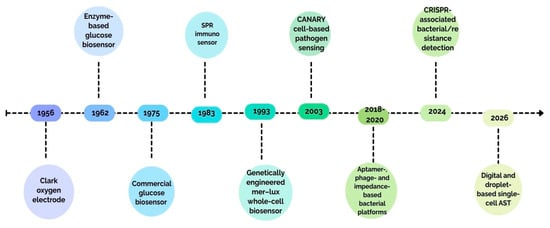
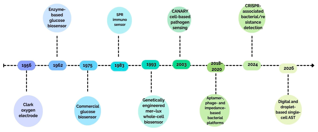

Disclaimer/Publisher’s Note: The statements, opinions and data contained in all publications are solely those of the individual author(s) and contributor(s) and not of MDPI and/or the editor(s). MDPI and/or the editor(s) disclaim responsibility for any injury to people or property resulting from any ideas, methods, instructions or products referred to in the content. |
© 2026 by the authors. Licensee MDPI, Basel, Switzerland. This article is an open access article distributed under the terms and conditions of the Creative Commons Attribution (CC BY) license.