


Abstract
Complex patient-specific bone defects remain difficult to reconstruct because the regenerative capacity of bone is limited and prefabricated implants cannot readily match defect geometry. In this work, methylcellulose-gelatin-hydroxyapatite (MC/GEL/HA) inks were formulated with varying methylcellulose content and hydroxyapatite incorporation, crosslinked with EDC/NHS, and 3D-printed into porous scaffolds with defined square-pore architectures. Inks were evaluated by oscillatory and steady-shear rheology, and printed scaffolds were characterized for morphology, chemical composition, mechanical performance, physicochemical stability, wettability, apatite-forming bioactivity, and osteogenic responses of human bone marrow mesenchymal stem cells. Methylcellulose content primarily governed ink rheology and printability, and increased compressive strength (up to ~0.38 MPa for 15MC/10GEL/30HA), whereas hydroxyapatite enhanced surface hydrophilicity, promoted apatite nucleation within 7 days in simulated body fluid, and markedly increased alkaline phosphatase activity (>10-fold over HA-free scaffolds), mineralization (~2-fold by Alizarin Red), and the highest osteocalcin expression among the HA-containing formulations (~2.45-fold at day 14). The 15MC/10GEL/30HA formulation showed the most favorable balance of printability, mechanical performance, and osteogenic performance. These complementary functions reconciled structural stability with osteogenic performance, supporting the MC/GEL/HA system as a tunable bioink platform for non-weight-bearing bone regeneration, while warranting further in vivo validation.
1. Introduction
Bone is a hierarchically organized connective tissue that fulfills critical structural and physiological roles in vertebrates. However, it remains susceptible to a range of injuries, including trauma, fractures, and tumor-related pathologies [1,2]. Autografts and allografts have long been the clinical gold standard, but each has inherent limitations. Autologous bone remains the preferred choice because it combines the three requirements of repair in a single graft: osteogenic cells, osteoinductive factors, and an osteoconductive matrix. However, its supply is limited, and harvesting may cause donor-site morbidity. Allografts avoid harvesting from the patient but bring their own drawbacks: the processing that reduces antigenicity also removes much of their osteoinductive potency, while some immunogenicity and a small risk of disease transmission remain [3]. These limitations have motivated the development of engineered substitutes that can be produced on demand and customized to each bone defect.
Additive manufacturing is particularly well suited to addressing the geometric aspect of this challenge. Skeletal defects are geometrically irregular, so prefabricated implants rarely conform to them and leave interfacial gaps that impair integration. Extrusion-based three-dimensional printing builds a construct layer by layer from a computer-aided design, giving precise, reproducible control over pore size, lattice geometry, and overall shape [1,2], which in turn improves cell infiltration and nutrient flow [4,5]. This enables patient-specific fabrication, particularly for geometrically complex non-weight-bearing defects such as those of the craniofacial skeleton.
The performance of a printed scaffold is largely determined by the choice of ink. Hydrogels emerge as an especially promising choice because of their hydrated, customizable network structure, mimicking properties of the extracellular matrix and capability of providing cells with a water-rich environment [6,7]. Gelatin (GEL), a denatured protein derived from collagen, maintains an Arg-Gly-Asp (RGD) tripeptide recognized by integrins, thus providing natural cell adhesive ability [8]. Nonetheless, gelatin suffers from considerable printability drawbacks as a result of its low viscosity causing sagging of filaments during extrusion, and thermoreversible gelation resulting in dissolution of printed constructs at physiological temperatures [9]. Methylcellulose (MC), a water-soluble cellulose derivative that carries hydrophobic methoxy groups, is extensively used for solving these problems through an increase in ink viscosity and introduction of thermoresponsive rheology, allowing for filament extrusion and self-support during the printing process [10]. Previous studies showed that the addition of MC improves printability and mechanical stability of gelatin-containing inks while maintaining biological compatibility [11,12,13,14]. For example, addition of 8% of MC to 5% GelMA raised the compressive modulus roughly 3-fold while osteoblasts encapsulated in this material remained viable with efficiency above 90% [11]. Hence, these results suggest that MC is the main ingredient responsible for printability, while gelatin provides a bioactive matrix containing RGD peptides for cell adhesion.
Despite these advances, the MC/GEL system still lacks the osteoconductive mineral component needed for active bone regeneration. Hydroxyapatite (HA), a calcium phosphate mineral comprising a major part of the inorganic fraction of bone, is osteoconductive and has direct bonding with surrounding bone, thus reinforcing the surrounding polymer [15]. While inherent brittleness and the slow resorption rate limit the utility of the HA degradation process as a bulk bone substitute, these problems can be overcome due to the dispersion of HA in a compliant hydrogel matrix, effectively restraining crack propagation [16]. Previous studies have shown that HA reinforces gelatin-based matrices while enhancing osteogenic activity [2,17,18]; for instance, incorporating hydroxyapatite (and strontium-doped HA) into a gelatin methacryloyl matrix improved the mechanical, swelling, and degradation behavior of the printed hydrogel while supporting osteoblast adhesion and proliferation [18].
Methylcellulose has previously been used to improve the printability of gelatin-based inks, while hydroxyapatite has been incorporated into gelatin- or GelMA-based scaffolds to promote osteogenesis, often using photocrosslinking [11,12,13,14]. However, the systematic integration of MC, gelatin, and HA within a single EDC/NHS-crosslinked, non-photocrosslinked ink, and the effects of MC content at a fixed HA loading on rheology, printing fidelity, mechanical performance, and osteogenic response, have not been systematically investigated.
Here, we developed an EDC/NHS-crosslinked MC/GEL/HA ink combining the cell-adhesive properties of gelatin, the rheological and printing functions of MC, and the osteoconductive properties of HA. MC content was systematically varied (10–15% w/v) across HA-containing formulations at a fixed HA loading, with corresponding HA-free formulations included as controls. We systematically assessed their rheological, mechanical, and biological performance using human bone marrow-derived mesenchymal stem cells (hBM-MSCs). We hypothesized that optimizing the formulation would improve printing fidelity while promoting osteogenic activity suitable for patient-specific repair of non-weight-bearing bone defects, including those of the craniofacial skeleton.
2. Materials and Methods
2.1. Hydrogel Preparation
MC/GEL/HA was blended at various concentrations (Table 1). MC powder was dissolved in distilled water at 50 °C under continuous stirring and hydrated overnight at 4 °C [19]. Gelatin was separately dissolved in distilled water at 50 °C [20], followed by gradual addition of HA powder and incorporation of the MC solution to form the composite hydrogel. The mixture was sonicated to remove air bubbles, loaded into a printing syringe, which was held at 50 °C in a water bath before printing. The fabrication protocol (Figure 1) used an extrusion-based 3D printer according to predefined parameters.
Table 1.
Percentages of MC/GEL/HA concentrations.
Figure 1.
Protocol for preparation of ink. Created in BioRender. Yuksel, C. (2026) https://BioRender.com/skhchow (Accessed on 26 July 2026).
2.2. 3D Printer Design and Scaffold Fabrication
Hydrogels were printed using the Printess: a custom-built, low-cost extrusion-based 3D printer developed at Stanford University, Stanford, CA, USA [21]. Scaffold geometry was defined by coordinate-based G-code to produce three-dimensional porous mesh structures. The hydrogel was maintained at 50 °C in a heated block and extruded through a 25 G needle [22]. Extrusion parameters were adjusted per formulation viscosity: E1.20 (E/mm = 0.08), E1.50 (E/mm = 0.10), and E1.80 (E/mm = 0.12) for 10%, 12%, and 15% MC, respectively, per 15 mm printing path. Following printing, scaffolds were crosslinked in 50 mM 1-Ethyl-3-(3-dimethylaminopropyl) carbodiimide/ N-Hydroxysuccinimide (EDC/NHS) at 4 °C for 20 h to form amide bonds between gelatin amino groups. After crosslinking, scaffolds were washed three times with phosphate-buffered saline (PBS) to remove residual reagents [22].
2.3. Rheological Analysis
Rheological measurements were acquired on an Anton Paar rotational rheometer (Anton Paar GmbH, Graz, Austria) using a 15 mm parallel-plate geometry and a 1 mm gap. The linear viscoelastic region (LVR) was determined by oscillatory amplitude sweeps at 37 °C and 10 rad s−1 over a strain range of 0.01–100%, from which the storage (G′) and loss (G″) moduli were obtained. Steady-shear measurements were then conducted over a shear rate range of 0.01–100 s−1 to evaluate the shear-thinning behavior. Thermal gelation behavior was characterized by temperature sweeps from 25 °C to 80 °C at a heating rate of 10 °C min−1, recording complex viscosity (η*) and G′/G″; the gelation temperature was taken as the G′-G″ crossover [23]. All rheological measurements were performed at Boğaziçi University (Istanbul, Türkiye).
2.4. Fourier-Transform Infrared Spectroscopy (FTIR)
FTIR spectroscopy was carried out to identify the functional groups and chemical interactions within the MC/GEL/HA hydrogels. Measurements were acquired over the 400–4000 cm−1 range on a Nicolet iS50 spectrometer (Thermo Fisher Scientific, Waltham, MA, USA).
2.5. Pore Size and Filament Diameter Analysis
Scaffold morphology, porous architecture, pore size, and filament diameter were examined by scanning electron microscopy (SEM) [24]. Prior to imaging, scaffolds were EDC/NHS crosslinked, frozen at −20 °C, and lyophilized, then mounted onto aluminum stubs with double-sided conductive carbon tape. Imaging was conducted on a FEI-Philips XL30 field-emission scanning electron microscope (Boğaziçi University, Istanbul, Türkiye) in low-vacuum mode at 20 kV, using a low-vacuum secondary-electron detector (LVD) [22]. Pore size and filament diameter were quantified from at least three randomly selected fields per scaffold using ImageJ 1.54 (National Institutes of Health, Bethesda, MD, USA) following scale-bar calibration, and results were expressed as mean ± standard deviation.
The printability index (Pr) was calculated from the perimeter (L) and area (A) of pores in the printed grid structures [25]:
An ideal square pore geometry yields Pr = 1, whereas Pr < 1 indicates a more circular pore shape from filament fusion or spreading, and Pr > 1 indicates an irregular, non-uniform pore geometry [25]. Pore perimeter and area were measured using the same ImageJ protocol described above, from at least four pores per scaffold across three independent samples.
To define the reference geometry for morphological analysis, the theoretical filament width and pore size specified by the printing design were calculated. The programmed filament width was 1.0 mm and was identical across formulations because the same G-code geometry was used. For the 15 × 15 mm lattice containing 11 filaments per direction and ten intervening pores, the theoretical pore width was calculated as (15 − 11 × 1.0)/10 = 0.4 mm (400 μm).
2.6. Swelling/Degradation Tests and In Vitro pH Stability
The swelling and degradation behavior of the scaffolds was studied using a separate set of scaffolds for each time point (n = 3 per group). Each scaffold was first dried in an incubator at 37 °C for 24 h, and its initial dry weight (W0) was recorded. Scaffolds were then immersed in PBS at 37 °C. On days 1, 3, 7, 14, and 18, the corresponding scaffolds were removed from PBS, gently blotted to remove surface water, and weighed to obtain the swollen mass (Ws). These scaffolds were then dried at 37 °C for 24 h and reweighed (Wd) to determine the mass loss for degradation analysis [26]. Swelling and degradation were expressed as percentages according to [27]:
The pH of the 3D-printed scaffolds was monitored over 18 days. Scaffolds from every experimental group were immersed in PBS at 37 °C (n = 3 per group). Fresh PBS was added daily to compensate for evaporation and maintain a constant medium volume during incubation [28]. The pH of the surrounding medium was measured daily with an Accumet pH meter (Fisher Scientific, Waltham, MA, USA). All experiments were conducted at Stanford University (Stanford, CA, USA).
In a complementary experiment, water uptake and mass loss of the MC/GEL and MC/GEL/HA scaffolds were monitored for 49 days in the complete cell culture medium used for the biological assays (DMEM Alpha supplemented with 10% fetal bovine serum and 1% penicillin-streptomycin), providing a biochemical environment that more closely resembles cell culture conditions, including exposure to serum proteins and growth factors [29]. After recording the initial dry weight (W0), scaffolds were incubated in the complete medium at 37 °C, with fresh medium replaced periodically to maintain nutrient availability. At predetermined time points (days 3, 7, 14, 21, 28, 35, 42, and 49), scaffolds were removed, gently blotted to remove excess medium, weighed (Ws), and then dried for 24 h to determine the final dry weight (Wd). Water uptake and mass loss were calculated using the same equations described above.
2.7. Mechanical Testing
The mechanical properties of the scaffolds, including compressive strength and elasticity, were evaluated [30]. Uniaxial compression testing was carried out on an Instron 5565 universal testing machine (Instron, Norwood, MA, USA) equipped with a 100 N load cell. Specimens measuring 15 × 15 × 2 mm were tested at room temperature (n = 5 per group). Samples were compressed between two parallel platens at a crosshead speed of 1 mm min−1 until failure, and load–displacement data were converted into stress–strain curves [31]. The compressive modulus was determined from the slope of the initial linear region of the stress–strain curve (between 5% and 10% strain). Compressive strength was taken as the maximum stress at the point of failure, and the corresponding strain at failure (%) was also recorded. All values are reported as mean ± standard deviation. All experiments were performed at Stanford University (Stanford, CA, USA).
2.8. Contact Angle Measurement
Static water contact angle measurements were performed using the sessile drop method on a KSV CAM 101 goniometer (KSV Instruments, Helsinki, Finland) at Boğaziçi University, Istanbul, Türkiye. A droplet of deionized water was carefully placed onto the surface of each sample, and images were captured after 1 s at room temperature to minimize the effect of droplet spreading. Measurements were conducted in three independent replicates for each sample group (n = 3), and the contact angle values were reported as the average. All analyses were performed on bulk scaffold samples.
2.9. Bioactivity Studies (Simulated Body Fluid Test)
The in vitro bioactivity of the scaffolds was evaluated based on their ability to induce apatite formation in simulated body fluid (SBF), prepared according to Kokubo and Takadama [32,33]. Rectangular specimens (15 × 15 × 2 mm) were immersed in 15 mL of SBF at 37 °C for up to 14 days (n = 3 per group per time point), with the SBF refreshed every 3 days to preserve ionic equilibrium. Samples were retrieved at day 7 and day 14 to monitor the onset and progression of apatite deposition. After immersion, samples were rinsed with distilled water and dried at 37 °C.
Apatite deposits on the scaffold surfaces were examined with a JEOL JSM-IT500HR environmental scanning electron microscope. Energy Dispersive X-ray Spectroscopy (EDS) elemental mapping was used to determine the spatial distribution of calcium (Ca) and phosphorus (P), and the Ca/P molar ratio was calculated to assess how closely it matched that of native hydroxyapatite [34]. Post-immersion samples at both time points were further characterized by FTIR to identify characteristic bands associated with bone-like apatite formation [35].
2.10. Human Bone Marrow Mesenchymal Stem Cell Culture
Human bone marrow mesenchymal stem cells (hBM-MSCs) were maintained in DMEM Alpha (Biowest) supplemented with 10% fetal bovine serum and 1% penicillin-streptomycin (Biowest) at 37 °C under a humidified 5% CO2 atmosphere. Prior to seeding, MC/GEL/HA scaffolds were sterilized by UV irradiation (45 min per side). To prevent non-specific cell attachment to the culture surface, the wells of 12-well plates were precoated with poly (2-hydroxyethyl methacrylate) (pHEMA), whereas TCP control wells were left uncoated [36]. hBM-MSCs were seeded directly onto the scaffolds at 1 × 10⁶ cells per well and allowed to attach for 2 h, after which 1 mL of complete medium was added and refreshed daily (n = 3 per group). Tissue culture plastic (TCP) seeded at the same density served as the positive control. All cell culture studies were performed at Stanford University (Stanford, CA, USA).
2.10.1. Cell Viability and Proliferation
Cell metabolic activity was evaluated on days 3 and 7 using the Alamar Blue assay [37]. Alamar Blue (10% v/v) was added at each time point, and scaffolds were incubated for 4 h at 37 °C. Aliquots (100 μL) of the supernatant were transferred to a 96-well plate, and absorbance was recorded at 570 and 600 nm on a SpectraMax iD3 microplate reader [38]. Values were normalized to the day-3 TCP control (set to 100%) [39].
2.10.2. DAPI/Phalloidin Staining
Cell attachment, morphology, and cytoskeletal organization were examined on days 3 and 7. Cell-seeded scaffolds were fixed with 4% paraformaldehyde for 15 min, permeabilized with 0.1% Triton X-100 for 10 min, and blocked with 1% bovine serum albumin for 30 min, with PBS washes between steps. F-actin was labeled with Phalloidin (30 min) [40] and nuclei counterstained with 4′,6-diamidino-2-phenylindole (DAPI) (20 min), with all steps from this point performed in the dark to limit photobleaching. Samples were imaged on a BZ-X810 fluorescence microscope and analyzed using BZ-X800 Viewer software (version 1.3.0.0, KEYENCE, Osaka, Japan).
2.10.3. Alkaline Phosphatase (ALP) Staining
Early osteogenic commitment was assessed by alkaline phosphatase staining after 7 days of culture in osteogenic medium (DMEM supplemented with 10% FBS, 1% penicillin-streptomycin, 100 μg mL−1 ascorbic acid, 10 mM β-glycerophosphate, and 100 nM dexamethasone). hBM-MSCs were seeded at 1 × 10⁶ cells per well (n = 3 per group), with acellular scaffolds processed identically serving as the background control [41]. Samples were rinsed with PBS, fixed with 4% paraformaldehyde for 15 min, and rinsed again with PBS. ALP activity was visualized using NBT/BCIP substrate (Thermo Fisher Scientific, Waltham, MA, USA), and samples were incubated at 37 °C until color development, after which the reaction was stopped by rinsing with distilled water [42]. Stained samples were imaged in bright-field mode on a BZ-X810 microscope (Keyence, Osaka, Japan). Stained area was measured from three independent samples per group using ImageJ (NIH, USA) [43].
2.10.4. Alizarin Red S Staining
Calcium deposition was evaluated by Alizarin Red S staining after 14 days of culture in osteogenic medium [44]. hBM-MSCs were seeded at 1 × 10⁶ cells per well in 12-well plates (n = 5 per group) and fixed with 4% paraformaldehyde for 15 min. To confirm the presence of cells within the mineralized regions, nuclei were first stained with DAPI (1 μg/mL, 10 min) and imaged under fluorescence microscopy. The same samples were then stained with 2% (w/v) Alizarin Red S (pH 4.2) for 30 min, after which excess dye was removed with distilled water. Mineralized nodules were imaged on a BZ-X810 fluorescence microscope (Keyence, Osaka, Japan) and analyzed with BZ-X800 Viewer software, with red-stained deposits taken as evidence of matrix mineralization. For quantification, bound stain was extracted in 20% (v/v) methanol/10% (v/v) acetic acid, and absorbance was measured at 450 nm [44]; acellular scaffolds processed identically were stained and quantified in parallel, and their absorbance values were subtracted as background.
2.10.5. Reverse Transcription Quantitative PCR (RT-qPCR)
Osteogenic gene expression was evaluated in the hydroxyapatite-containing groups. Osteocalcin (OCN), a late-stage marker of mature osteoblast activity and bone matrix formation, was quantified on days 7 and 14 of osteogenic culture [45]. Expression was normalized to GAPDH using the ΔCt method [46]. Primer sequences are listed in Table 2. All reactions were run in triplicate.
Table 2.
Primer sequences used for RT-qPCR.
RNA Extraction
Total RNA was isolated using TRIzol under RNase-free conditions. TRIzol reagent (Thermo Fisher Scientific, Waltham, MA, USA) was added directly to each well containing the cell-seeded scaffolds (n = 3 per group), and the lysate was collected by pipetting. Chloroform was added for phase separation, and samples were centrifuged at 12,000× g for 15 min at 4 °C. RNA in the aqueous phase was precipitated with isopropanol using glycogen as a carrier, washed with 75% ethanol, air-dried, and resuspended in RNase-free water [28,46]. RNA concentration and purity were assessed on a NanoDrop One spectrophotometer (Thermo Fisher Scientific, Waltham, MA, USA), with A260/A280 ratios of 1.8–2.0 considered acceptable.
Complementary DNA (cDNA) Synthesis
cDNA was synthesized from total RNA using iScript Reverse Transcription Supermix (Bio-Rad) in 20 μL reactions assembled on ice. Reverse transcription was performed in a MiniAmp Plus thermal cycler (Thermo Fisher Scientific, Waltham, MA, USA) using the following thermal profile: priming at 25 °C for 5 min, reverse transcription at 46 °C for 20 min, and reverse transcriptase inactivation at 95 °C for 1 min. Products were stored at −20 °C.
qPCR
cDNA was diluted 1:5 in RNase-free water and amplified using PowerUp SYBR Green Master Mix (Applied Biosystems, Waltham, MA, USA) with primers for OCN and GAPDH (Table 2), on a QuantStudio 6 Pro Real-Time PCR System (Applied Biosystems, Waltham, MA, USA); specificity was confirmed by melt-curve analysis. Relative OCN expression was expressed as 2−ΔCt. Data were processed in Design and Analysis Software v2.6.0 (Applied Biosystems).
2.11. Statistical Analyses
Statistical analysis was performed using GraphPad Prism (version 11.0.2; GraphPad Software, Boston, MA, USA). Group differences were assessed by one-way ANOVA with Tukey’s post hoc test, except for cell viability (7 × 2; group × time point), swelling and degradation (6 × 5; group × time point), pH (6 × 18; group × time point), and OCN expression (3 × 2; group × time point) data, which were analyzed by two-way ANOVA with Tukey’s post hoc test, with statistical significance set at p < 0.05 [47]. Effect size was calculated as eta-squared (η2) for one-way ANOVA and partial eta-squared (partial η2) for two-way ANOVA comparisons to determine the magnitude of observed differences [48,49].
3. Results and Discussion
3.1. Rheological Characterization of Hydrogel Formulations
The rheological properties of the MC/GEL/HA hydrogels were evaluated by amplitude sweep, shear-rate-dependent viscosity, temperature-dependent complex viscosity, and temperature-dependent modulus measurements (Figure 2a–d).
Figure 2.
Rheological properties of the MC/GEL/HA hydrogels evaluated through: (a) amplitude sweep, (b) shear rate- dependent viscosity, (c) complex viscosity variations with temperature, and (d) temperature sweep analysis.
The amplitude sweep measurements (Figure 2a) revealed composition-dependent differences in the viscoelastic behavior of the MC/GEL/HA hydrogels. Across all formulations, G′ exceeded G″ throughout the linear viscoelastic region before both moduli declined sharply at higher % strains, indicating predominantly elastic gel behavior followed by network disruption [50]. MC concentration was the primary determinant of gel stiffness, with G′ increasing progressively from 10MC/10GEL (the lowest modulus) to 15MC/10GEL [12]. The influence of HA depended on MC concentration, substantially increasing G′ in the 10MC/10GEL formulation but producing only minor changes in the 12MC/10GEL and 15MC/10GEL formulations [51,52].
Shear-rate-dependent viscosity (Figure 2b) characterized the flow behavior of the hydrogels across a shear-rate range of 0.01–100 s−1. All six formulations displayed pronounced shear-thinning, with viscosity decreasing by two to three orders of magnitude as the shear rate increased, a characteristic essential for extrusion-based printing [53,54]. The elevated viscosity at low shear rates (≤0.1 s−1), prior to the onset of shear-thinning, is indicative of yield-stress-like behavior. Viscosity also increased with MC concentration, confirming that higher MC content strengthened the network under flow. The HA-containing formulations, particularly 15MC/10GEL/30HA, showed the highest viscosities, suggesting that HA further contributed to flow resistance under shear.
Temperature-dependent complex viscosity (Figure 2c) revealed the temperature behavior of the hydrogels. All formulations displayed a reduction in complex viscosity upon initial heating, reaching a minimum between approximately 44 and 49 °C, followed by a rapid increase with further heating. This behavior reflects the coil-to-fibril transition characteristic of MC gels, in which polymer chains reorganize from disordered coils into fibrillar aggregates above a critical temperature, producing macroscopic stiffening [55,56,57,58]. The similar transition temperatures indicate that all formulations underwent the same thermal gelation mechanism, with only the magnitude of the response varying among compositions. Based on these results, 50 °C was chosen as the processing temperature for subsequent experiments, since it provided an intermediate viscosity following the onset of thermal gelation while avoiding the substantially higher viscosities observed at 70–80 °C.
Figure 2d presented the corresponding temperature sweep for G′ and G″. In contrast to hydrogel systems that display a sol–gel crossover upon heating [59], G′ remained above G″ across the entire 25–80 °C range for every formulation, confirming that gel-like character was preserved throughout the tested temperature range. Both moduli decreased until approximately 45–50 °C before increasing again, mirroring the trend observed in the complex viscosity data. Among the HA-free formulations, thermal stiffening became more pronounced with increasing MC content, from 10MC/10GEL to 12MC/10GEL and 15MC/10GEL. The contribution of HA to thermal stiffening was greatest at low MC content, consistent with the pattern observed in the amplitude sweep [60].
MC concentration emerged as the dominant factor governing the viscoelastic behavior of the MC/GEL/HA system, while HA acted as a composition-dependent reinforcing phase most effective in low-MC networks [51,52,60]. All formulations nonetheless showed gel-like behavior, pronounced shear-thinning, and thermally induced stiffening, supporting their suitability as inks for extrusion-based 3D printing.
3.2. FTIR Analysis and Surface Morphology of Scaffolds
FTIR spectra of all EDC/NHS-crosslinked formulations (Figure 3) retained the characteristic bands of MC and gelatin, including broad O-H/N-H stretching (3000–3600 cm−1), C-H stretching (~2900 cm−1), and the amide I and II bands (~1630–1650 and ~1540–1560 cm−1; corresponding to C=O stretching and combined N-H bending/C-N stretching, respectively), indicating preservation of the chemical spectral features of the MC/GEL matrix across all formulations [61].
Figure 3.
FTIR-ATR spectroscopy of EDC/NHS cross-linked MC/GEL and MC/GEL/HA scaffolds (vertically shifted for visibility). Shaded regions mark diagnostic bands: O-H/N-H stretching (3600–3000 cm−1), C-H stretching (~2900 cm−1), amide I (~1650 cm−1), amide II (~1550 cm−1), C-N stretching (~1250 cm−1), P-O stretching (1100–950 cm−1), and O-P-O bending (~600 cm−1).
The prominent amide I and II bands, together with the absence of a distinct free-carboxyl shoulder (~1710–1740 cm−1), suggest amide bond formation through EDC/NHS-mediated coupling, in agreement with previous reports describing carbodiimide crosslinking of gelatin-based systems [59,62].
HA-containing formulations displayed additional phosphate-associated absorptions, including P-O stretching (900–1100 cm−1) and O-P-O bending (~500–650 cm−1), characteristic of calcium phosphate phases [63]. Although the 900–1100 cm−1 region partially overlaps with C-O-C vibrations of the MC/GEL matrix, the increased intensity of these phosphate-related bands relative to HA-free controls is in line with the presence of HA.
These spectral features confirm that HA was successfully incorporated into the EDC/NHS-crosslinked MC/gelatin matrix.
3.3. 3D Printing of Scaffolds
SEM imaging demonstrated that all scaffold groups maintained a well-defined, interconnected square-pore structure following printing (Figure 4a). The 0°/90° strand deposition pattern remained intact, with no visible strand collapse or fusion observed in any group, indicating good shape fidelity across all compositions. Quantitative printability (Pr) analysis supported these SEM findings, with Pr values clustering near 1 across all groups (Figure 4b), indicating near-ideal pore geometry. Groups containing HA showed a slight increase in Pr, consistent with decreased strand spreading after extrusion.
Figure 4.
Morphological and structural characterization of MC/GEL and MC/GEL/HA scaffolds. (a) Representative SEM images of printed scaffolds showing the interconnected, square-pore architecture of 10MC/10GEL, 12MC/10GEL, and 15MC/10GEL formulations (top row) and their HA-containing counterparts, 10MC/10GEL/30HA, 12MC/10GEL/30HA, and 15MC/10GEL/30HA (bottom row). Scale bars, 50 μm. (b) Printability index (Pr), calculated from the perimeter and area of printed pores, with Pr = 1 indicating an ideal square pore geometry. (c) Filament diameter and (d) Pore size measured from SEM images. Data are presented as mean ± standard deviation (n = 3 per group); differences not indicated are non-significant.
Filament diameter (Figure 4c) ranged from roughly 850 to 1200 μm, with the highest value observed in the 15MC/10GEL group. This pattern is likely attributable to the greater viscosity of MC-rich inks (described in Figure 2b), which produces thicker extruded filaments. The effect of HA on filament diameter depended on MC content: HA increased filament diameter at the two lower MC concentrations (10MC/10GEL, 12MC/10GEL) but reduced it at the highest MC concentration (15MC/10GEL), suggesting reduced post-extrusion filament expansion at higher MC content.
The pore sizes (Figure 4d) ranged from approximately 350 to 610 μm, falling within the range generally considered favorable for bone regeneration, where interconnected pores support cell infiltration, nutrient transport, and vascularization [64]. Even the smallest pores exceeded the lower thresholds commonly associated with restricted cell migration. Accordingly, MC and HA provided complementary control over filament and pore dimensions without compromising print fidelity.
Relative to the theoretical design values (Section 2.5; 1.0 mm programmed filament width and 400 μm theoretical pore width), the SEM-measured filament diameters ranged from approximately 850 to 1200 μm, whereas pore sizes ranged from approximately 350 to 610 μm. Thus, the printed filaments generally exceeded the programmed width, with a corresponding reduction in pore size. The magnitude of this deviation varied with formulation and was most pronounced at higher MC content, consistent with the greater viscosity and flow resistance of these inks (Figure 4c,d).
This trend is consistent with the rheological behavior described in Section 3.1. HA incorporation increased viscosity and low-shear resistance to flow (Figure 2b), particularly in the lower-MC formulations, which can limit strand spreading and relaxation after deposition. Reduced spreading preserves more defined strand boundaries and alters pore geometry, contributing to the observed changes in pore size and printability index. Because pore size is constrained by the fixed strand spacing of the printed lattice and the deposited filament width, formulation-dependent changes in filament dimensions directly translate into corresponding changes in pore size.
3.4. Compressive Behavior of the Scaffolds
The mechanical properties of the MC/GEL-based hydrogels were evaluated under uniaxial compression (Figure 5). Stress–strain curves (Figure 5a) exhibited the characteristic J-shaped behavior, with an initially compliant response followed by progressive strain stiffening, typical of hydrated polymer networks [65,66].
Figure 5.
Compressive mechanical behavior of MC/GEL and MC/GEL/HA scaffolds. (a) Representative uniaxial compressive stress–strain curves, (b) compressive modulus, (c) compressive strength, and (d) % strain, derived from the curves in (a). Data are presented as mean ± s.d. (n = 5). Statistical significance was assessed by one-way ANOVA with Tukey’s post hoc test (** p < 0.01, *** p < 0.001, **** p < 0.0001; differences not indicated are non-significant).
The compressive modulus of the MC/GEL-based scaffolds is shown in Figure 5b. Neither increasing MC content nor incorporating HA produced a statistically significant change in compressive modulus, with values clustering between ~0.065 and ~0.093 MPa across all six groups. As the compressive modulus was determined within the low-strain compliant region governed by the soft, water-swollen polymer network, HA appears to contribute minimally to the initial mechanical response, with its reinforcing effect becoming more evident at larger compressive deformations [65,66].
Figure 5c shows the ultimate compressive strength of the MC/GEL-based scaffolds. Ultimate compressive strength increased progressively with increasing MC content in both the HA-free and HA-containing groups. Mechanistically, the MC-driven increase in compressive strength is consistent with the higher storage modulus and shear viscosity observed with increasing MC content (Section 3.1), suggesting a more physically interconnected polymer network with greater resistance to deformation.
Within the HA-containing groups, ultimate compressive strength increased from approximately 0.27 MPa (10MC/10GEL/30HA) to approximately 0.38 MPa (15MC/10GEL/30HA), with the greatest strength observed for 15MC/10GEL/30HA (p < 0.001 versus 10MC/10GEL/30HA). Compared with its HA-free counterpart, the 15MC/10GEL/30HA scaffold also exhibited higher ultimate compressive strength (p < 0.0001), suggesting additional reinforcement through interactions between HA particles and the surrounding polymer network, allowing the rigid ceramic phase to contribute more effectively to compressive load bearing in non-weight-bearing applications [67,68]. Additional non-covalent interactions, including hydrogen bonding between HA surface groups and the polymer network, may further contribute to interfacial load transfer. However, these interactions were not directly characterized here and would require dedicated interfacial studies to establish their contribution. Together, these findings indicate that MC and HA enhance compressive strength through complementary mechanisms.
In Figure 5d, the % strain at ultimate compressive strength showed little variation with increasing MC content, indicating that MC addition alone did not substantially alter network deformability. Although 15MC/10GEL/30HA showed a numerically higher % strain (~117%) compared with 15MC/10GEL (~101%), this difference did not reach statistical significance, and no significant differences in % strain were observed across any of the six formulations. Accordingly, neither MC content nor HA incorporation substantially altered scaffold deformability at the point of ultimate compressive strength.
3.5. Swelling, Degradation and pH Behavior of the Scaffolds
Swelling, weight loss (%) and pH were monitored in PBS at 37 °C over an 18 day degradation period (n = 3 per group), to evaluate scaffold stability under physiological conditions and the effect of HA incorporation.
Water uptake (%) (Figure 6a) declined progressively over the immersion period rather than exhibiting classical swelling behavior: following a moderate initial decrease by day 1, values partially plateaued (or slightly recovered in some HA-containing groups) through day 3, after which all groups underwent a pronounced decline between days 3 and 14, before stabilizing at low residual levels (<20%) through day 18. HA-containing scaffolds retained higher water uptake than their HA-free counterparts for longer, with the greatest difference observed at day 7, before both groups converged to comparably low values by day 14.
Figure 6.
Physicochemical properties of MC/GEL and MC/GEL/HA scaffolds after treatment with PBS at 37 °C. (a) Percent water uptake over 18 days. (b) Percent weight loss over 18 days. (c) pH measurements of the 3D-printed scaffolds over 18 days. Data are presented as mean ± standard deviation (n = 3).
Weight loss (%) (Figure 6b) followed the opposite trend. Scaffolds retained the majority of their initial mass during the first week, followed by a marked decline after day 7, with HA-containing groups degrading somewhat more rapidly than HA-free groups, consistent with their water uptake, which facilitated enhanced fluid penetration and promoted hydrolytic degradation of the polymer network [69,70]. By day 18, all groups had converged toward a low residual mass, indicating progressive degradation. Such gradual degradation is generally regarded as advantageous for bone tissue engineering, as it may provide transient structural support while allowing progressive tissue replacement [71].
Solution pH (Figure 6c) exhibited an initial decrease followed by recovery and stabilization. All groups revealed mild acidification of the environment in the first two days (minimum pH ≈ 6.9), which can be related to hydration of the matrix and release of gelatin’s residual species into the medium [72]. Starting from day 3, pH increased gradually before stabilizing by approximately day 7. HA-containing scaffolds equilibrated at distinctly alkaline pH values (approximately pH = 8.0–8.3), whereas HA-free scaffolds remained closer to neutral pH, consistent with the buffering and ion-exchange behavior of HA [73,74]. Among HA-containing materials, 15MC/10GEL/30HA maintained the highest pH throughout, suggesting the strongest buffering capacity.
HA incorporation accelerated degradation while sustaining a mildly alkaline microenvironment, a physicochemical profile that may support osteogenesis during tissue regeneration [75,76].
These trends indicate an inverse temporal relationship between water uptake and mass loss: formulations and time windows associated with greater water retention (Figure 6a) preceded and overlapped with periods of more rapid mass loss (Figure 6b), most clearly reflected by the decline in water uptake between days 3 and 14 and the onset of marked mass loss after day 7. This pattern is consistent with a water-mediated degradation process, in which greater fluid uptake may facilitate polymer-network chain scission, such that formulations retaining more water for longer, notably the HA-containing scaffolds, subsequently exhibit earlier and somewhat faster mass loss than their HA-free counterparts.
3.6. Surface Wettability
Scaffold wettability was assessed through static water contact angle measurements, recorded 1 s after droplet deposition (Figure 7b), with representative sessile drop profiles presented in Figure 7a. All six formulations exhibited contact angles well below 90°, indicating hydrophilic surfaces [77]. In the series without HA, increasing the MC fraction resulted in progressively higher contact angles, from 46.2 ± 1.3° for 10MC/10GEL to 49.7 ± 1.1° for 12MC/10GEL and 57.1 ± 3.4° for 15MC/10GEL, with all pairwise differences being statistically significant (Figure 7b). The progressive increase in contact angle with MC content suggests enhanced hydrophobic association among methoxy-substituted chains, thereby reducing surface wettability, consistent with previous observations [78]. Incorporation of 30 wt % HA consistently decreased the contact angle across all formulations, to 30.4 ± 1.5°, 37.8 ± 2.2°, and 43.6 ± 1.4° for the 10, 12, and 15MC variants, respectively, while preserving the MC-dependent trend. This indicates that HA enhanced surface hydrophilicity while maintaining the compositional influence of the polymer network, a behavior commonly ascribed to the abundance of surface hydroxyl groups on HA, which readily engage in hydrogen bonding with water molecules [79].
Figure 7.
Surface wettability of the hydrogel formulations assessed by water contact angle measurement. (a) Representative sessile-drop images for the six formulations (10MC/10GEL, 12MC/10GEL, 15MC/10GEL, and their %30wt HA-containing counterparts). (b) Corresponding mean contact angles. All formulations exhibited hydrophilic surfaces (contact angle < 90°). Data are presented as mean ± SD (n = 3); asterisks denote statistical significance (** p < 0.01, *** p < 0.001, **** p < 0.0001); differences not indicated are non-significant.
3.7. Surface Morphology and Chemical Analysis of Apatite Formation
Apatite-forming ability was evaluated after SBF immersion for up to 14 days using complementary SEM, EDS and FTIR analyses (Figure 8) to probe in vitro bioactivity, a widely used method for assessing in vivo bone-bonding potential [33].
Figure 8.
Apatite formation on the scaffolds after immersion in simulated body fluid. (a) SEM images and corresponding Ca/P elemental maps of all groups at day 7, showing Ca and P signals co-localized with surface deposits, with intensity increasing with MC content. Scale bars, 50 µm. (b) SEM images of the scaffold surface at day 14 in all groups (arrows mark apatite deposition). Scale bars, 50 µm. (c) FTIR spectra of the scaffolds at day 7, demonstrating phosphate and carbonate bands of the carbonated apatite phase. (d) FTIR spectra of the scaffolds at day 14, revealing restoration and enhancement of the P-O stretching band in the presence of HA.
SEM revealed particulate surface deposits (arrows, Figure 8a, bottom row), indicative of apatite nucleation. By day 7, isolated nodules were already present in the HA-containing formulations, while HA-free formulations showed no discernible particulate deposits at this time point (Figure 8a, top row). By day 14, the HA-containing groups displayed a denser, more continuous apatite layer (Figure 8b, bottom row), whereas the HA-free groups exhibited only a smooth, fibrous surface morphology without evidence of apatite deposition (Figure 8b, top row).
EDS was employed to quantify surface Ca/P atomic ratios, providing elemental confirmation that the SEM-observed deposits corresponded to an apatite-like calcium phosphate phase. At day 7, Ca/P ratios for 10MC/10GEL/30HA, 12MC/10GEL/30HA, and 15MC/10GEL/30HA were 1.83, 1.84, and 1.99, respectively, exceeding the 1.67 value expected for stoichiometric hydroxyapatite [80], consistent with the onset of apatite mineralization. By day 14, Ca/P ratios remained at similar values (1.85, 1.87, and 1.96), reflecting continued apatite formation. HA-free groups remained near the detection limit at both time points (0.33, 0.00, and 0.00 at day 7; 0.00–0.01 at day 14), indicating that the polymer matrix alone did not support heterogeneous apatite nucleation within this window.
Although phosphate-associated absorptions, together with weaker carbonate bands, were detectable from day 7 (Figure 8c), the intensity of the P-O stretching region (950–1150 cm−1) did not increase monotonically with immersion time. Instead, it was transiently attenuated alongside the N-H, C=O, and glycosidic bands during early immersion, before recovering by day 14 (Figure 8d), particularly in the HA-containing groups. This behavior likely reflects two concurrent effects. First, ATR-FTIR probes only the outermost ~0.5–2 µm of the surface [81], so a thin early apatite layer adds little signal against the strong intrinsic P-O background from the 30 wt % HA already present in the scaffold. Second, early hydrolytic degradation of the gelatin/MC matrix likely released soluble, non-crosslinked chains, explaining the concurrent decline in matrix bands. By day 14, as degradation progressed, recovery of the P-O signal was consistent with continued maturation of the apatite layer.
3.8. In Vitro Cytocompatibility
Water uptake (%) and mass loss (%) of the MC/GEL and MC/GEL/HA scaffolds were monitored over 49 days in the complete cell culture medium used for the biological assays (DMEM Alpha supplemented with 10% fetal bovine serum and 1% penicillin-streptomycin) (Figure 9a,b). This approach more closely reflects the biochemical environment experienced by cells during culture, including exposure to serum proteins and growth factors, thereby offering a more simulated physiologically relevant context for interpreting cell behavior on the scaffolds [30]. Water uptake (%) remained high (~90–95%) and relatively stable through day 21 for all formulations, then declined gradually between days 28 and 42, before dropping sharply for all groups by day 49. Weight loss (%) remained low and comparatively stable through day 21, then increased markedly after day 28, most notably for the higher-MC, HA-containing scaffold, before all groups converged toward near-complete mass loss by day 49.
Figure 9.
In vitro cytocompatibility and cell behavior of the hydrogel scaffolds. (a) Water uptake (%) of the six scaffold formulations over 49 days (days 3, 7, 14, 21, 28, 35, 42, 49) in cell culture medium. (b) Weight loss (%) of the same formulations over the same time course in cell culture medium. (c) Relative cell viability (%) of TCP (control) and the six formulations at days 3 and 7. (d) Representative fluorescence micrographs (DAPI/phalloidin; nuclei, blue; F-actin, green) of the six formulations at day 3. Scale bar, 100 μm. (e) Representative fluorescence micrographs of the same formulations at day 7. Scale bar, 100 μm. Data are mean ± s.d. (n = 3 per group); *** p < 0.001 (two-way ANOVA with Tukey’s post hoc test); differences not indicated are non-significant.
The cytocompatibility of hBM-MSCs cultured on the MC/GEL and MC/GEL/HA scaffolds was assessed against a tissue-culture plastic (TCP) reference at days 3 and 7 (Figure 9c). At the earlier time point, most composite formulations supported a lower relative viability than TCP, most markedly for the 10MC/10GEL and 10MC/10GEL/30HA groups (both p < 0.001 vs. TCP). Increasing the MC content progressively improved day-3 viability, with 15MC/10GEL/30HA producing the highest response, although this did not differ significantly from TCP. This graded, dose-dependent effect of MC was also evident among the composite formulations, independent of the TCP comparison (e.g., 10MC/10GEL vs. 15MC/10GEL/30HA). By day 7, every composite condition had not only recovered but exceeded the TCP reference, with the HA-containing scaffolds showing the highest viability, and no statistically significant differences detected among the groups at this time point.
The composition-dependent response at day 3 is unlikely to be governed by wettability alone, since contact angle increased with MC content while day-3 viability increased in parallel, despite all formulations remaining within the hydrophilic range generally considered favorable for cell adhesion (30–57°; approximately 20–80°) [82,83]. Matrix stability at higher MC contents is a more likely explanation, consistent with the concentration-dependent gelation behavior of MC [84]. This interpretation is further supported by the swelling and degradation data (Figure 9a,b), which showed a delayed onset of degradation in the higher-MC, HA-containing scaffolds, indicative of greater matrix stability.
By day 7, the superior viability of the HA-containing scaffolds suggests that HA-related surface properties become increasingly influential once the initial adaptation phase is complete. Together with the cell-adhesive RGD motifs provided by gelatin and the improved surface hydrophilicity of HA-containing scaffolds and likely favors adsorption of adhesion-mediating serum proteins (Figure 7), which likely promote sustained cell attachment and proliferation [85,86,87,88].
Taken together, the temporary decline observed at day 3 for the lower-MC formulations is most consistent with an early cell-adaptation phase rather than inherent cytotoxicity, since all groups rebounded and surpassed the TCP control within four days, consistent with the transient lag phase commonly reported in three-dimensional culture systems [89,90,91]. This pattern of an initial deficit relative to conventional monolayer culture followed by a later crossover, where three-dimensional culture systems ultimately support cell numbers exceeding those on planar controls, has similarly been reported for other 3D biomaterial platforms [92]. By day 7, HA incorporation appeared to support sustained proliferation, potentially through its wettability-enhancing surface chemistry and intrinsic bioactivity.
3.8.1. Cell Attachment and Migration (DAPI/Phalloidin Staining)
hBM-MSC behavior varied over time and with scaffold composition, as reflected in cytoskeletal organization. Consistent with the day-3 viability reduction described above (Figure 9c), phalloidin/DAPI staining at this time point showed predominantly rounded cells forming small clusters with little evidence of organized actin stress fibers (Figure 9d), indicative of an early attachment stage [93,94].
By day 7, the cellular phenotype had changed markedly. In parallel with the metabolic recovery described above, phalloidin staining revealed elongated, spindle-shaped cells with aligned actin filaments (Figure 9e). The emergence of organized stress fibers together with pronounced cell elongation is widely associated with stable cell adhesion and enhanced cell-material interactions [93,94]. These observations are consistent with the improved cytocompatibility observed by day 7 and with the reported role of HA in promoting cell adhesion, migration, and cytoskeletal organization through surface microtopography and calcium/phosphate ion release [86,87,95].
3.8.2. Early Osteogenic Differentiation Assessed by ALP Activity
ALP activity at day 7 was used to assess early osteogenic differentiation (Figure 10b). The six formulations segregated into two distinct groups. In the absence of HA, the HA-free constructs (10MC/10GEL, 12MC/10GEL and 15MC/10GEL) exhibited only near-background ALP activity, with relative values remaining below 10% of the reference level regardless of MC fraction. Incorporation of 30 wt % HA markedly increased ALP activity, with all HA-containing groups reaching 78–88% relative activity, representing more than a 10-fold increase compared with the corresponding HA-free scaffold (p <0.0001). No significant differences were observed among the HA-containing formulations, indicating that HA incorporation, rather than the MC/GEL ratio, was the primary determinant of the early osteogenic response.
Figure 10.
Osteogenic differentiation of hBM-MSCs on the MC/GEL and MC/GEL/HA scaffolds. (a) Representative brightfield images of BCIP/NBT histochemical staining showing ALP activity at day 7; red arrows indicate ALP-positive nodules. (b) Quantification of relative ALP activity (%) at day 7. (c) Representative fluorescence images of DAPI-stained nuclei (blue) and Alizarin Red-stained mineralized matrix (red) after 14 days of osteogenic induction. (d) Spectrophotometric quantification of extracted Alizarin Red at day 14. (e) OCN transcript expression (normalized to GAPDH) for the three HA-containing scaffolds at days 7 and 14. Data are mean ± SD; ** p < 0.01, *** p < 0.001, **** p < 0.0001. Scale bars = 500 µm; images acquired at 2× magnification; differences not indicated are non-significant.
This pattern was further supported histochemically by BCIP/NBT staining images (Figure 10a). HA-free scaffolds (top row) exhibited only sparse, weakly stained deposits, indicating limited early osteogenic activity. In contrast, HA-containing scaffolds (bottom row, red arrows) developed denser, more uniformly distributed dark purple/black ALP-positive nodules. The close agreement between the quantitative ALP measurements and the histochemical staining indicates that HA incorporation increased both the magnitude and the uniformity of ALP activity throughout the scaffolds. This is consistent with HA’s osteoconductive activity [96,97].
3.8.3. Osteogenic Mineralization by Alizarin Red Staining and Quantification
After 14 days of osteogenic induction, mineralized matrix deposition was observed on all scaffolds, as distinct Alizarin Red-positive nodules (Figure 10c). HA apatite was deposited even in the HA-free formulations, indicating that the polymer matrix alone supported calcium-phosphate mineralization under osteogenic culture conditions. This effect may be partly due to the presence of gelatin, whose RGD-like sequences and charged residues support integrin-mediated adhesion and may create a favorable microenvironment for calcium phosphate nucleation [98,99,100].
Spectrophotometric quantification of the extracted dye revealed a graded, composition-dependent increase in mineral deposition (Figure 10d), with HA as the principal compositional driver. Incorporating 30 wt % HA raised the Alizarin Red signal, and the majority of HA versus HA-free contrasts reached significance (p < 0.01 to p < 0.0001). Calcium-phosphate mineralization peaked in 15MC/10GEL/30HA, approximately 2-fold above the 15MC/10GEL control, consistent with HA acting as a bone-mimetic template that lowers the energetic barrier to heterogeneous calcium phosphate deposition [101,102,103].
MC exerted an additional modulatory influence on mineralization. Increasing MC from 10% to 15% raised the Alizarin Red signal within each HA group, an effect most evident where HA was present, suggesting that MC modulates rather than independently drives the osteogenic influence of HA. This effect may instead reflect a direct physicochemical interaction between MC and the mineral phase rather than a swelling-mediated mechanism. Previous studies have suggested that methylcellulose can influence calcium phosphate mineralization by interacting with calcium and phosphate ions and modifying crystal growth behavior in HA-containing composites [88]. Other factors, such as MC-mediated dispersion of HA particles or resulting changes in protein and ion adsorption at the composite surface [104,105,106], were not directly examined here and are noted as candidate directions for future work rather than established mechanisms.
Fluorescence imaging supported these quantitative findings. DAPI-stained nuclei were uniformly distributed under all conditions, whereas the Alizarin Red signal progressed from sparse, diffuse staining in 10MC/10GEL to dense, merged nodules in the high-MC, HA-containing scaffolds (Figure 10c,d). Because DAPI and Alizarin Red images were acquired from the same field of view, co-registration showed that mineral deposition was concentrated in nuclei-rich regions rather than acellular areas, supporting a cell-associated mineralization process.
Comparison with the day-7 ALP results further suggests distinct roles for HA and MC during osteogenic differentiation. Whereas HA alone markedly increased early ALP activity, with little influence of MC content (Figure 10a,b), the later mineralization stage showed an additional MC-dependent increase superimposed on the HA effect (Figure 10c,d). This divergence suggests that HA predominantly drives early osteogenic commitment, whereas the MC/GEL ratio becomes influential only later, in regulating the extent of mineralization.
3.8.4. Osteocalcin Gene Expression Analysis by RT-qPCR
Formulations with higher HA content were prioritized for OCN evaluation based on their favorable performance across earlier viability, ALP, and Alizarin Red assays. Osteocalcin, a non-collagenous matrix protein secreted by mature osteoblasts, is widely recognized as a molecular marker of late-stage osteogenic differentiation and matrix mineralization [107,108]. To assess this terminal stage of differentiation, OCN transcript levels (normalized to GAPDH) were measured in MC/GEL/HA scaffolds at days 7 and 14 (n = 3) (Figure 10e). At day 7, OCN expression remained low across all three formulations, with no significant differences observed between groups, consistent with cultures that had not yet entered the mineralization phase at this early stage. By day 14, the three formulations diverged markedly: the 15MC/10GEL/30HA scaffold showed the highest OCN expression (~2.45-fold).
The day-14 pattern is consistent with the expected late-stage increase in OCN expression during osteogenic differentiation [108]. It also aligns with the earlier viability, cytoskeletal findings, and mineralization results (Figure 9c–e and Figure 10c,d), where the 15MC/10GEL/30HA scaffold consistently showed the most favorable biological response among the HA-containing groups. These findings suggest that the more favorable early cell response may be associated with the enhanced late-stage osteogenic outcome, although the underlying mechanotransduction pathways (e.g., focal adhesion maturation or YAP/TAZ signaling) were not examined.
4. Conclusions
This work demonstrates that an EDC/NHS-crosslinked MC/GEL/HA ink can combine composition-dependent rheological control with reliable extrusion printability, mechanical performance, and osteoconductive bioactivity in a single bone-mimetic construct. MC content governed the viscoelastic and shear-thinning behavior of the ink, while HA acted as a composition-dependent reinforcing phase, particularly in formulations with lower MC content. The complementary roles of MC and HA were reflected in printed constructs with square-pore architectures, near-unity printability indices, and filament and pore dimensions within ranges considered favorable for cellular interaction and nutrient transport. Swelling, degradation, and pH measurements further indicated that HA incorporation modulated matrix stability and degradation behavior while sustaining a mildly alkaline microenvironment, a combination consistent with gradual structural evolution rather than premature structural failure. The progressive apatite layer detected by SEM, EDS, and FTIR after immersion in simulated body fluid confirmed that HA retained its intrinsic mineral-nucleating capacity even after being embedded within the crosslinked polymer network.
These compositional effects were mirrored in the biological response of hBM-MSCs. Although hBM-MSCs showed a transient dip in metabolic activity during initial attachment, all scaffold groups demonstrated recovery and increased metabolic activity over time, accompanied by a shift from rounded, clustered cells to elongated morphologies with organized actin architecture. Rather than contributing redundantly, HA and MC appeared to influence distinct stages of osteogenic differentiation, with HA promoting early commitment and MC-dependent matrix properties contributing to later-stage mineral deposition and maturation. Among the evaluated formulations, 15MC/10GEL/30HA exhibited the most favorable balance of mechanical properties, matrix mineralization, and late-stage osteogenic maturation. Together, these findings indicate that balancing MC content and HA incorporation (Figure 11) provides a means of reconciling the competing demands of printability, structural integrity, and osteogenic performance within the range of formulations investigated. Overall, these results provide a rational design strategy for extrusion-printed osteogenic biomaterials suited to patient-specific reconstruction of irregular, non-weight-bearing bone defects, while underscoring the need for in vivo validation prior to clinical translation.
Figure 11.
Schematic representation of the structural and functional properties of MC (left side) and HA (right side). Created in BioRender. Yuksel, C. (2026) https://BioRender.com/skhchow (Accessed on 26 July 2026).
Author Contributions
Conceptualization, D.E., C.Y., S.K.-H.C. and S.G.; methodology, D.E., C.Y., S.K.-H.C. and S.G.; software, C.Y. and S.K.-H.C.; validation, D.E. and C.Y.; investigation, D.E., R.H., S.K.-H.C., M.M., Q.G., T.I. and M.T.; resources, D.E., C.M., T.T., S.C. and M.S.S.; writing—original draft preparation, C.Y.; writing—review and editing, D.E., C.Y., S.K.-H.C., R.H., M.M., Q.G., T.I., M.T. and S.G.; visualization, C.Y.; supervision, D.E. and S.G.; project administration, D.E., C.Y. and S.K.-H.C.; funding acquisition, D.E. and S.G. All authors have read and agreed to the published version of the manuscript.
Funding
This research was supported in part by the Robert L. and Mary Ellenburg Chair in Surgery, Stanford University; the Fulbright Türkiye Doctoral Research Scholarship Program, which supported the author’s doctoral research period at Stanford University from 18 August 2025, to 18 May 2026; the Boğaziçi University Research Fund (Grant No. 20325 and 20413).
Institutional Review Board Statement
The study was approved by the Administrative Panel on Human Subjects in Medical Research at Stanford University (approval No. 35711, approved on 31 October 2025).
Informed Consent Statement
For this IRB protocol, the human bone marrow samples were collected as medical waste after the surgical procedure had been completed. Therefore, obtaining a patient informed consent form was not required for the use of these samples.
Data Availability Statement
The original contributions presented in this study are included in the article. Further inquiries can be directed to the corresponding authors.
Acknowledgments
The authors acknowledge BioRender.com for providing the platform used to create the original figures included in this manuscript. The authors have reviewed and edited the output and take full responsibility for the content of this publication.
Conflicts of Interest
The authors declare no conflicts of interest.
Abbreviations
The following abbreviations are used in this manuscript:
| MC | Methylcellulose |
| GEL | Gelatin |
| HA | Hydroxyapatite |
| EDC | 1-Ethyl-3-(3-dimethylaminopropyl) carbodiimide |
| NHS | N-Hydroxysuccinimide |
| hBM-MSC | human Bone Marrow-derived Mesenchymal Stem Cell |
| RGD | Arginine-Glycine-Aspartate |
| SBF | Simulated Body Fluid |
| FTIR | Fourier-Transform Infrared Spectroscopy |
| SEM | Scanning Electron Microscopy |
| EDS | Energy Dispersive X-ray Spectroscopy |
| LVR | Linear Viscoelastic Region |
| G′ | Storage modulus |
| G″ | Loss modulus |
| η* | Complex viscosity |
| Pr | Printability index |
| PBS | Phosphate-Buffered Saline |
| W0/Ws/Wd | Initial dry weight/Swollen (wet) weight/Dry weight |
| ALP | Alkaline Phosphatase |
| NBT/BCIP | Nitro Blue Tetrazolium/5-Bromo-4-Chloro-3-Indolyl Phosphate |
| DAPI | 4′,6-Diamidino-2-Phenylindole |
| OCN | Osteocalcin |
| RT-qPCR | Reverse Transcription Quantitative Polymerase Chain Reaction |
| cDNA | Complementary DNA |
| GAPDH | Glyceraldehyde-3-Phosphate Dehydrogenase |
| Ct/ΔCt | Cycle threshold/Delta Cycle threshold |
| TCP | Tissue Culture Plastic |
| pHEMA | poly (2-Hydroxyethyl Methacrylate) |
| Ca/P | Calcium/Phosphorus ratio |
| ANOVA | Analysis of Variance |
| η2/partial η2 | Eta-squared/Partial eta-squared |
| DMEM | Dulbecco’s Modified Eagle Medium |
| FBS | Fetal Bovine Serum |
References
- Oryan, A.; Alidadi, S.; Moshiri, A.; Maffulli, N. Bone regenerative medicine: Classic options, novel strategies, and future directions. J. Orthop. Surg. Res. 2014, 9, 18. [Google Scholar] [CrossRef] [Scilit] [PubMed]
- Kim, H.; Hwangbo, H.; Koo, Y.; Kim, G. Fabrication of mechanically reinforced gelatin/hydroxyapatite bio-composite scaffolds by core/shell nozzle printing for bone tissue engineering. Int. J. Mol. Sci. 2020, 21, 3401. [Google Scholar] [CrossRef] [Scilit] [PubMed]
- Kahraman, E.; Ribeiro, R.; Lamghari, M.; Neto, E. Cutting-edge technologies for inflamed joints on chip: How close are we? Front. Immunol. 2022, 13, 802440. [Google Scholar] [CrossRef] [Scilit] [PubMed]
- Zhang, Q.; Zhou, J.; Zhi, P.; Liu, L.; Liu, C.; Fang, A.; Zhang, Q. 3D printing method for bone tissue engineering scaffold. Med. Nov. Technol. Devices 2023, 17, 100205. [Google Scholar] [CrossRef] [Scilit] [PubMed] [PubMed Central]
- Budharaju, H.; Suresh, S.; Sekar, M.P.; De Vega, B.; Sethuraman, S.; Sundaramurthi, D.; Kalaskar, D.M. Ceramic materials for 3D printing of biomimetic bone scaffolds—Current state-of-the-art and future perspectives. Mater. Des. 2023, 237, 112064. [Google Scholar] [CrossRef] [Scilit]
- Tan, S.H.; Chua, D.A.C.; Tang, J.R.J.; Bonnard, C.; Leavesley, D.; Liang, K. Design of hydrogel-based scaffolds for in vitro three-dimensional human skin model reconstruction. Acta Biomater. 2022, 153, 13–37. [Google Scholar] [CrossRef] [Scilit] [PubMed]
- Short, A.R.; Koralla, D.; Deshmukh, A.; Wissel, B.; Stocker, B.; Calhoun, M.; Dean, D.; Winter, J.O. Hydrogels that allow and facilitate bone repair, remodeling, and regeneration. J. Mater. Chem. B 2015, 3, 7818–7830. [Google Scholar] [CrossRef] [Scilit] [PubMed]
- Meissner, S.; Raos, B.; Svirskis, D. Hydrogels can control the presentation of growth factors and thereby improve their efficacy in tissue engineering. Eur. J. Pharm. Biopharm. 2022, 181, 1–21. [Google Scholar] [CrossRef] [Scilit]
- Iranmanesh, P.; Gowdini, M.; Khademi, A.; Dehghani, M.; Latifi, M.; Alsaadi, N.; Hemati, M.; Mohammadi, R.; Saber-Samandari, S.; Toghraie, D.; et al. Bioprinting of three-dimensional scaffold based on alginate-gelatin as soft and hard tissue regeneration. J. Mater. Res. Technol. 2021, 14, 2853–2864. [Google Scholar] [CrossRef] [Scilit]
- Rajabi, N.; Rezaei, A.; Kharaziha, M.; Bakhsheshi-Rad, H.R.; Luo, H.; RamaKrishna, S.; Berto, F. Recent advances on bioprinted gelatin methacrylate-based hydrogels for tissue repair. Tissue Eng. Part A 2021, 27, 679–702. [Google Scholar] [CrossRef] [Scilit] [PubMed]
- Rastin, H.; Ormsby, R.T.; Atkins, G.J.; Losic, D. 3D bioprinting of methylcellulose/gelatin-methacryloyl (MC/GelMA) bioink with high shape integrity. ACS Appl. Bio Mater. 2020, 3, 1815–1826. [Google Scholar] [CrossRef] [Scilit] [PubMed]
- Reakasame, S.; Dranseikiene, D.; Schrüfer, S.; Zheng, K.; Schubert, D.W.; Boccaccini, A.R. Development of alginate dialdehyde gelatin based bioink with methylcellulose for improving printability. Mater. Sci. Eng. C 2021, 128, 112336. [Google Scholar] [CrossRef] [Scilit] [PubMed]
- Züger, F.; Berner, N.; Gullo, M.R. Towards a novel cost-effective and versatile bioink for 3D-bioprinting in tissue engineering. Biomimetics 2023, 8, 27. [Google Scholar] [CrossRef] [Scilit] [PubMed]
- Bernhardt, A.; Liu, S.; Kara Özenler, A.; Wirsig, K.; Gelinsky, M. Bioprinted constructs of differentiated primary osteocytes: A co-culture model with osteoclasts for biomedical research. Biofabrication 2026, 18, 035007. [Google Scholar] [CrossRef] [Scilit] [PubMed]
- Bal, Z.; Kaito, T.; Korkusuz, F.; Yoshikawa, H. Bone regeneration with hydroxyapatite-based biomaterials. Emergent Mater. 2020, 3, 521–544. [Google Scholar] [CrossRef] [Scilit]
- Almawash, S.; Osman, S.K.; Mustafa, G.; El Hamd, M.A. Current and future prospective of injectable hydrogels—Design challenges and limitations. Pharmaceuticals 2022, 15, 371. [Google Scholar] [CrossRef] [Scilit] [PubMed]
- Cakmak, A.M.; Unal, S.; Sahin, A.; Oktar, F.N.; Sengor, M.; Ekren, N.; Gunduz, O.; Kalaskar, D.M. 3D printed polycaprolactone/gelatin/bacterial cellulose/hydroxyapatite composite scaffold for bone tissue engineering. Polymers 2020, 12, 1962. [Google Scholar] [CrossRef] [Scilit] [PubMed]
- Codrea, C.I.; Baykara, D.; Mitran, R.-A.; Çalıkoğlu Koyuncu, A.C.; Gunduz, O.; Ficai, A. 3D-bioprinted gelatin methacryloyl strontium-doped hydroxyapatite composite hydrogels scaffolds for bone tissue regeneration. Polymers 2024, 16, 1932. [Google Scholar] [CrossRef] [Scilit] [PubMed]
- Tate, M.C.; Shear, D.A.; Hoffman, S.W.; Stein, D.G.; LaPlaca, M.C. Biocompatibility of methylcellulose-based constructs designed for intracerebral gelation following experimental traumatic brain injury. Biomaterials 2001, 22, 1113–1123. [Google Scholar] [CrossRef] [Scilit] [PubMed]
- Fang, J.; Yang, Z.; Tan, S.; Tayag, C.; Nimni, M.E.; Urata, M.; Han, B. Injectable gel graft for bone defect repair. Regen. Med. 2014, 9, 41–51. [Google Scholar] [CrossRef] [Scilit] [PubMed]
- Weiss, J.D.; Mermin-Bunnell, A.; Solberg, F.S.; Tam, T.; Rosalia, L.; Sharir, A.; Rütsche, D.; Sinha, S.; Choi, P.S.; Shibata, M.; et al. A low-cost, open-source 3D printer for multimaterial and high-throughput direct ink writing of soft and living materials. Adv. Mater. 2025, 37, 2414971. [Google Scholar] [CrossRef] [Scilit] [PubMed]
- Arıcı, Ş.; Ege, D. Effect of gelatin concentration on the mechanical properties and in vitro degradation behaviour of gelatin/carboxymethyl cellulose-based 3D printed scaffolds. MRS Adv. 2024, 9, 1598–1604. [Google Scholar] [CrossRef] [Scilit]
- Demir Oğuz, Ö.; Ege, D. Rheological and mechanical properties of thermoresponsive methylcellulose/calcium phosphate-based injectable bone substitutes. Materials 2018, 11, 604. [Google Scholar] [CrossRef] [Scilit] [PubMed]
- Krishnakumar, G.S.; Sampath, S.; Muthusamy, S.; John, M.A. Importance of crosslinking strategies in designing smart biomaterials for bone tissue engineering: A systematic review. Mater. Sci. Eng. C 2019, 96, 941–954. [Google Scholar] [CrossRef] [Scilit] [PubMed]
- Ouyang, L.; Yao, R.; Zhao, Y.; Sun, W. Effect of bioink properties on printability and cell viability for 3D bioplotting of embryonic stem cells. Biofabrication 2016, 8, 035020. [Google Scholar] [CrossRef] [Scilit] [PubMed]
- Kaya, H.; Bulut, O.; Kamali, A.R.; Ege, D. L-Arginine modified multi-walled carbon nanotube/sulfonated poly(ether ether ketone) nanocomposite films for biomedical applications. Appl. Surf. Sci. 2018, 444, 168–176. [Google Scholar] [CrossRef] [Scilit]
- Ma, T.; Lv, L.; Ouyang, C.; Hu, X.; Liao, X.; Song, Y.; Hu, X. Rheological behavior and particle alignment of cellulose nanocrystal and its composite hydrogels during 3D printing. Carbohydr. Polym. 2021, 253, 117217. [Google Scholar] [CrossRef] [Scilit] [PubMed]
- Chomczynski, P.; Sacchi, N. The single-step method of RNA isolation by acid guanidinium thiocyanate-phenol-chloroform extraction: Twenty-something years on. Nat. Protoc. 2006, 1, 581–585. [Google Scholar] [CrossRef] [Scilit] [PubMed]
- Fazel Anvari-Yazdi, A.; Tahermanesh, K.; Ejlali, M.; Blivet-Bailly, L.; Singh, V.; Acharya, B.; MacPhee, D.J.; Badea, I.; Chen, X. One-step decellularization of porcine uterine tissue for developing alginate-decellularized uterine ECM hydrogel for uterine tissue engineering. Int. J. Bioprint. 2026, 12, 026150132. [Google Scholar] [CrossRef] [Scilit]
- Zhao, K.; Wang, L.; Chang, Y.; Yan, J. Identification of post-necking stress-strain curve for sheet metals by inverse method. Mech. Mater. 2016, 92, 107–118. [Google Scholar] [CrossRef] [Scilit]
- Wu, D.; Isaksson, P.; Ferguson, S.J.; Persson, C. Young’s modulus of trabecular bone at the tissue level: A review. Acta Biomater. 2018, 78, 1–12. [Google Scholar] [CrossRef] [Scilit] [PubMed]
- Kokubo, T.; Takadama, H. How useful is SBF in predicting in vivo bone bioactivity? Biomaterials 2006, 27, 2907–2915. [Google Scholar] [CrossRef] [Scilit] [PubMed]
- Kokubo, T.; Kushitani, H.; Sakka, S.; Kitsugi, T.; Yamamuro, T. Solutions able to reproduce in vivo surface-structure changes in bioactive glass-ceramic A-W. J. Biomed. Mater. Res. 1990, 24, 721–734. [Google Scholar] [CrossRef] [Scilit] [PubMed]
- Morgan, E.F.; Unnikrisnan, G.U.; Hussein, A.I. Bone mechanical properties in healthy and diseased states. Annu. Rev. Biomed. Eng. 2018, 20, 119–143. [Google Scholar] [CrossRef] [Scilit] [PubMed]
- Rehman, I.; Bonfield, W. Characterization of hydroxyapatite and carbonated apatite by photo acoustic FTIR spectroscopy. J. Mater. Sci. Mater. Med. 1997, 8, 1–4. [Google Scholar] [CrossRef] [Scilit] [PubMed]
- Folkman, J.; Moscona, A. Role of cell shape in growth control. Nature 1978, 273, 345–349. [Google Scholar] [CrossRef] [Scilit] [PubMed]
- O’Brien, J.; Wilson, I.; Orton, T.; Pognan, F. Investigation of the Alamar Blue (resazurin) fluorescent dye for the assessment of mammalian cell cytotoxicity. Eur. J. Biochem. 2000, 267, 5421–5426. [Google Scholar] [CrossRef] [Scilit] [PubMed]
- Longhin, E.M.; El Yamani, N.; Rundén-Pran, E.; Dusinska, M. The Alamar Blue assay in the context of safety testing of nanomaterials. Front. Toxicol. 2022, 4, 981701. [Google Scholar] [CrossRef] [Scilit] [PubMed]
- Osaka, I.; Hefty, P.S. Simple resazurin-based microplate assay for measuring Chlamydia infections. Antimicrob. Agents Chemother. 2013, 57, 2838–2840. [Google Scholar] [CrossRef] [Scilit] [PubMed]
- Wulf, E.; Deboben, A.; Bautz, F.A.; Faulstich, H.; Wieland, T. Fluorescent phallotoxin, a tool for the visualization of cellular actin. Proc. Natl. Acad. Sci. USA 1979, 76, 4498–4502. [Google Scholar] [CrossRef] [Scilit] [PubMed]
- Rivoallan, N.; Mueller, M.; Baudequin, T.; Vigneron, P.; Hébraud, A.; Jellali, R.; Dermigny, Q.; Le Goff, A.; Schlatter, G.; Glasmacher, B.; et al. Comparison of hydroxyapatite and honeycomb micro-structure in bone tissue engineering using electrospun beads-on-string fibers. Int. J. Artif. Organs 2024, 47, 642–649. [Google Scholar] [CrossRef] [Scilit] [PubMed]
- Lee, J.-M.; Kim, M.-G.; Byun, J.-H.; Kim, G.-C.; Ro, J.-H.; Hwang, D.-S.; Choi, B.-B.; Park, G.-C.; Kim, U.-K. The effect of biomechanical stimulation on osteoblast differentiation of human jaw periosteum-derived stem cells. Maxillofac. Plast. Reconstr. Surg. 2017, 39, 7. [Google Scholar] [CrossRef] [Scilit] [PubMed]
- Schneider, C.A.; Rasband, W.S.; Eliceiri, K.W. NIH Image to ImageJ: 25 years of image analysis. Nat. Methods 2012, 9, 671–675. [Google Scholar] [CrossRef] [Scilit] [PubMed]
- Gregory, C.A.; Gunn, W.G.; Peister, A.; Prockop, D.J. An Alizarin red-based assay of mineralization by adherent cells in culture. Anal. Biochem. 2004, 329, 77–84. [Google Scholar] [CrossRef] [Scilit] [PubMed]
- Livak, K.J.; Schmittgen, T.D. Analysis of relative gene expression data using real-time quantitative PCR and the 2−ΔΔCT method. Methods 2001, 25, 402–408. [Google Scholar] [CrossRef] [Scilit] [PubMed]
- Chomczynski, P.; Sacchi, N. Single-step method of RNA isolation by acid guanidinium thiocyanate-phenol-chloroform extraction. Anal. Biochem. 1987, 162, 156–159. [Google Scholar] [CrossRef] [Scilit] [PubMed]
- Brown, A.M. A new software for carrying out one-way ANOVA post hoc tests. Comput. Methods Programs Biomed. 2005, 79, 89–95. [Google Scholar] [CrossRef] [Scilit] [PubMed]
- Tukey, J.W. Comparing individual means in the analysis of variance. Biometrics 1949, 5, 99–114. [Google Scholar] [CrossRef] [Scilit]
- Sokal, R.R.; Rohlf, F.J. Biometry: The Principles and Practice of Statistics in Biological Research, 4th ed.; W.H. Freeman: New York, NY, USA, 2012. [Google Scholar] [CrossRef] [Scilit]
- Robertson, C.G.; Vaikuntam, S.R.; Heinrich, G. A nonequilibrium model for particle networking/jamming and time-dependent dynamic rheology of filled polymers. Polymers 2020, 12, 190. [Google Scholar] [CrossRef] [Scilit] [PubMed]
- Liang, J.Z. Reinforcement and quantitative description of inorganic particulate-filled polymer composites. Compos. Part B Eng. 2013, 51, 224–232. [Google Scholar] [CrossRef] [Scilit]
- Adhikari, J.; Perwez, M.S.; Das, A.; Saha, P. Development of hydroxyapatite reinforced alginate-chitosan based printable biomaterial-ink. Nano-Struct. Nano-Objects 2021, 25, 100630. [Google Scholar] [CrossRef] [Scilit]
- Schwab, A.; Levato, R.; D’Este, M.; Piluso, S.; Eglin, D.; Malda, J. Printability and shape fidelity of bioinks in 3D bioprinting. Chem. Rev. 2020, 120, 11028–11055. [Google Scholar] [CrossRef] [Scilit] [PubMed]
- Paxton, N.; Smolan, W.; Böck, T.; Melchels, F.; Groll, J.; Jungst, T. Proposal to assess printability of bioinks for extrusion-based bioprinting and evaluation of rheological properties governing bioprintability. Biofabrication 2017, 9, 044107. [Google Scholar] [CrossRef] [Scilit] [PubMed]
- Hirrien, M.; Chevillard, C.; Desbrières, J.; Axelos, M.A.V.; Rinaudo, M. Thermogelation of methylcelluloses: New evidence for understanding the gelation mechanism. Polymer 1998, 39, 6251–6259. [Google Scholar] [CrossRef] [Scilit]
- Niemczyk-Soczynska, B.; Sajkiewicz, P.; Gradys, A. Toward a better understanding of the gelation mechanism of methylcellulose via systematic DSC studies. Polymers 2022, 14, 1810. [Google Scholar] [CrossRef] [Scilit] [PubMed]
- Lott, J.R.; McAllister, J.W.; Arvidson, S.A.; Bates, F.S.; Lodge, T.P. Fibrillar structure of methylcellulose hydrogels. Biomacromolecules 2013, 14, 2484–2488. [Google Scholar] [CrossRef] [Scilit] [PubMed][Green Version]
- Coughlin, M.L.; Liberman, L.; Ertem, S.P.; Edmund, J.; Bates, F.S.; Lodge, T.P. Methyl cellulose solutions and gels: Fibril formation and gelation properties. Prog. Polym. Sci. 2021, 112, 101324. [Google Scholar] [CrossRef] [Scilit]
- Bigi, A.; Cojazzi, G.; Panzavolta, S.; Roveri, N.; Rubini, K. Stabilization of gelatin films by crosslinking with genipin. Biomaterials 2002, 23, 4827–4832. [Google Scholar] [CrossRef] [Scilit] [PubMed]
- Korkeamäki, J.T.; Silen, R.; Syverud, K.; Mustafa, K.; Rashad, A.; Miettinen, S. Optimizing gelatin methacryloyl-based bioinks with nanohydroxyapatite for 3D bioprinting in bone tissue engineering. Bioprinting 2026, 56, e00479. [Google Scholar] [CrossRef] [Scilit]
- Karaca, M.A.; Yilmaz, I.; Ozbek, T.; Güven, A.; Kafes, G.; Gok, O.; Ege, D. Antibacterial and bioactive 3D-printed hexagonal boron nitride incorporated gelatin/methyl cellulose scaffolds for bone tissue engineering applications. Macromol. Mater. Eng. 2026, 311, e70198. [Google Scholar] [CrossRef] [Scilit]
- Olde Damink, L.H.H.; Dijkstra, P.J.; van Luyn, M.J.A.; van Wachem, P.B.; Nieuwenhuis, P.; Feijen, J. Glutaraldehyde as a cross-linking agent for collagen-based biomaterials. J. Mater. Sci. Mater. Med. 1995, 6, 460–472. [Google Scholar] [CrossRef] [Scilit] [PubMed]
- Hossain, M.S.; Ahmed, S. FTIR spectrum analysis to predict the crystalline and amorphous phases of hydroxyapatite: A comparison of vibrational motion to reflection. RSC Adv. 2023, 13, 14625–14630. [Google Scholar] [CrossRef] [Scilit] [PubMed]
- Mukasheva, F.; Moazzam, M.; Yernaimanova, B.; Shehzad, A.; Zhanbassynova, A.; Berillo, D.; Akilbekova, D. Design and characterization of 3D printed pore gradient hydrogel scaffold for bone tissue engineering. Bioprinting 2024, 39, e00341. [Google Scholar] [CrossRef] [Scilit]
- Ma, Y.; Feng, X.; Rogers, J.A.; Huang, Y.; Zhang, Y. Design and application of “J-shaped” stress-strain behavior in stretchable electronics: A review. Lab Chip 2017, 17, 1689–1704. [Google Scholar] [CrossRef] [Scilit] [PubMed]
- Shi, Y.; Wu, B.; Sun, S.; Wu, P. Aqueous spinning of robust, self-healable, and crack-resistant hydrogel microfibers enabled by hydrogen bond nanoconfinement. Nat. Commun. 2023, 14, 1370. [Google Scholar] [CrossRef] [Scilit] [PubMed]
- Eshraghi, S.; Das, S. Micromechanical finite-element modeling and experimental characterization of the compressive mechanical properties of polycaprolactone-hydroxyapatite composite scaffolds prepared by selective laser sintering for bone tissue engineering. Acta Biomater. 2012, 8, 3138–3143. [Google Scholar] [CrossRef] [Scilit] [PubMed]
- Raucci, M.G.; Demitri, C.; Soriente, A.; Fasolino, I.; Sannino, A.; Ambrosio, L. Gelatin/nano-hydroxyapatite hydrogel scaffold prepared by sol-gel technology as filler to repair bone defects. J. Biomed. Mater. Res. A 2018, 106, 2007–2019. [Google Scholar] [CrossRef] [Scilit] [PubMed]
- Dan, Y.; Liu, O.; Liu, Y.; Zhang, Y.-Y.; Li, S.; Feng, X.-B.; Shao, Z.-W.; Yang, C.; Yang, S.-H.; Hong, J.-B. Development of novel biocomposite scaffold of chitosan-gelatin/nanohydroxyapatite for potential bone tissue engineering applications. Nanoscale Res. Lett. 2016, 11, 487. [Google Scholar] [CrossRef] [Scilit] [PubMed]
- Mahdavi, R.; Belgheisi, G.; Haghbin-Nazarpak, M.; Omidi, M.; Khojasteh, A.; Solati-Hashjin, M. Bone tissue engineering gelatin-hydroxyapatite/graphene oxide scaffolds with the ability to release vitamin D: Fabrication, characterization, and in vitro study. J. Mater. Sci. Mater. Med. 2020, 31, 97. [Google Scholar] [CrossRef] [Scilit] [PubMed]
- Koons, G.L.; Diba, M.; Mikos, A.G. Materials design for bone-tissue engineering. Nat. Rev. Mater. 2020, 5, 584–603. [Google Scholar] [CrossRef] [Scilit]
- Priya, G.; Madhan, B.; Narendrakumar, U.; Suresh Kumar, R.V.; Manjubala, I. In vitro and in vivo evaluation of carboxymethyl cellulose scaffolds for bone tissue engineering applications. ACS Omega 2021, 6, 1246–1253. [Google Scholar] [CrossRef] [Scilit] [PubMed]
- Jeong, J.; Kim, J.H.; Shim, J.H.; Hwang, N.S.; Heo, C.Y. Bioactive calcium phosphate materials and applications in bone regeneration. Biomater. Res. 2019, 23, 4. [Google Scholar] [CrossRef] [Scilit] [PubMed]
- Szałaj, U.; Chodara, A.; Gierlotka, S.; Wojnarowicz, J.; Łojkowski, W. Enhanced release of calcium ions from hydroxyapatite nanoparticles with an increase in their specific surface area. Materials 2023, 16, 6397. [Google Scholar] [CrossRef] [Scilit] [PubMed]
- Ruan, C.; Hu, N.; Ma, Y.; Li, Y.; Liu, J.; Zhang, X.; Pan, H. The interfacial pH of acidic degradable polymeric biomaterials and its effects on osteoblast behavior. Sci. Rep. 2017, 7, 6794. [Google Scholar] [CrossRef] [Scilit] [PubMed]
- Liu, W.; Wang, T.; Yang, C.; Darvell, B.W.; Wu, J.; Lin, K.; Chang, J.; Pan, H.; Lu, W.W. Alkaline biodegradable implants for osteoporotic bone defects—Importance of microenvironment pH. Osteoporos. Int. 2016, 27, 93–104. [Google Scholar] [CrossRef] [Scilit] [PubMed]
- Paxton, N.C.; Woodruff, M.A. Measuring contact angles on hydrophilic porous scaffolds by implementing a novel raised platform approach: A technical note. Polym. Adv. Technol. 2022, 33, 3759–3765. [Google Scholar] [CrossRef] [Scilit]
- Niemczyk-Soczynska, B.; Gradys, A.; Kolbuk, D.; Krzton-Maziopa, A.; Sajkiewicz, P. Crosslinking kinetics of methylcellulose aqueous solution and its potential as a scaffold for tissue engineering. Polymers 2019, 11, 1772. [Google Scholar] [CrossRef] [Scilit] [PubMed]
- Zimina, A.; Senatov, F.; Choudhary, R.; Kolesnikov, E.; Anisimova, N.; Kiselevskiy, M.; Orlova, P.; Strukova, N.; Generalova, M.; Manskikh, V.; et al. Biocompatibility and physico-chemical properties of highly porous PLA/HA scaffolds for bone reconstruction. Polymers 2020, 12, 2938. [Google Scholar] [CrossRef] [Scilit] [PubMed]
- LeGeros, R.Z. Calcium phosphate-based osteoinductive materials. Chem. Rev. 2008, 108, 4742–4753. [Google Scholar] [CrossRef] [Scilit] [PubMed]
- Oelichmann, J. Surface and depth-profile analysis using FTIR spectroscopy. Fresenius Z. Anal. Chem. 1989, 333, 353–359. [Google Scholar] [CrossRef] [Scilit]
- Arima, Y.; Iwata, H. Effect of wettability and surface functional groups on protein adsorption and cell adhesion using well-defined mixed self-assembled monolayers. Biomaterials 2007, 28, 3074–3082. [Google Scholar] [CrossRef] [Scilit] [PubMed]
- Webb, K.; Hlady, V.; Tresco, P.A. Relative importance of surface wettability and charged functional groups on cell attachment, spreading, and cytoskeletal organization. J. Biomed. Mater. Res. 1998, 41, 422–430. [Google Scholar] [CrossRef] [Scilit] [PubMed]
- Thirumala, S.; Gimble, J.M.; Devireddy, R.V. Methylcellulose based thermally reversible hydrogel system for tissue engineering applications. Cells 2013, 2, 460–475. [Google Scholar] [CrossRef] [Scilit] [PubMed]
- Davidenko, N.; Schuster, C.F.; Bax, D.V.; Farndale, R.W.; Hamaia, S.; Best, S.M.; Cameron, R.E. Evaluation of cell binding to collagen and gelatin: A study of the effect of 2D and 3D architecture and surface chemistry. J. Mater. Sci. Mater. Med. 2016, 27, 148. [Google Scholar] [CrossRef] [Scilit] [PubMed]
- Shi, H.; Zhou, Z.; Li, W.; Fan, Y.; Li, Z.; Wei, J. Hydroxyapatite-based materials for bone tissue engineering: A brief and comprehensive introduction. Crystals 2021, 11, 149. [Google Scholar] [CrossRef] [Scilit]
- Wang, Y.; Wang, F.; Gong, M.; Chen, L.; Wang, Y.; Xu, P.; Zeng, Z.; Hu, Z.; Chen, J. Engineering stepped structures on hydroxyapatite surfaces: A potential strategy to modulate bone marrow mesenchymal stem adhesion, spreading, and proliferation. J. Funct. Biomater. 2025, 16, 165. [Google Scholar] [CrossRef] [Scilit] [PubMed]
- Woo, K.M.; Seo, J.; Zhang, R.; Ma, P.X. Suppression of apoptosis by enhanced protein adsorption on polymer/hydroxyapatite composite scaffolds. Biomaterials 2007, 28, 2622–2630. [Google Scholar] [CrossRef] [Scilit] [PubMed][Green Version]
- Divieto, C.; Sassi, M.P. A first approach to evaluate the cell dose in highly porous scaffolds by using a nondestructive metabolic method. Future Sci. OA 2015, 1, FSO58. [Google Scholar] [CrossRef] [Scilit] [PubMed]
- faCellitate. Introduction to Cell Culture Growth Stages. Available online: https://facellitate.com/introduction-to-cell-culture-growth-stages/ (accessed on 26 July 2026).
- Schorl, C.; Sedivy, J.M. Analysis of cell cycle phases and progression in cultured mammalian cells. Methods 2007, 41, 143–150. [Google Scholar] [CrossRef] [Scilit] [PubMed]
- Blanco-Elices, C.; Oruezabal, R.I.; Sánchez-Porras, D.; Chato-Astrain, J.; Campos, F.; Alaminos, M.; Garzón, I.; Campos, A. A novel 3D biofabrication strategy to improve cell proliferation and differentiation of human Wharton’s jelly mesenchymal stromal cells for cell therapy and tissue engineering. Front. Bioeng. Biotechnol. 2023, 11, 1235161. [Google Scholar] [CrossRef] [Scilit] [PubMed]
- Dede Eren, A.; Lucassen, A.W.A.; Tuvshindorj, U.; Truckenmüller, R.; Giselbrecht, S.; Eren, E.D.; Tas, M.O.; Sudarsanam, P.; de Boer, J. Cells dynamically adapt to surface geometry by remodeling their focal adhesions and actin cytoskeleton. Front. Cell Dev. Biol. 2022, 10, 863721. [Google Scholar] [CrossRef] [Scilit] [PubMed]
- Greiner, A.M.; Chen, H.; Spatz, J.P.; Kemkemer, R. Cyclic tensile strain controls cell shape and directs actin stress fiber formation and focal adhesion alignment in spreading cells. PLoS ONE 2013, 8, e77328. [Google Scholar] [CrossRef] [Scilit] [PubMed]
- Meesuk, L.; Suwanprateeb, J.; Thammarakcharoen, F.; Tantrawatpan, C.; Kheolamai, P.; Palang, I.; Tantikanlayaporn, D.; Manochantr, S. Osteogenic differentiation and proliferation potentials of human bone marrow and umbilical cord-derived mesenchymal stem cells on the 3D-printed hydroxyapatite scaffolds. Sci. Rep. 2022, 12, 19654. [Google Scholar] [CrossRef] [Scilit] [PubMed]
- Müller, P.; Bulnheim, U.; Diener, A.; Lüthen, F.; Teller, M.; Klinkenberg, E.-D.; Neumann, H.-G.; Nebe, B.; Liebold, A.; Steinhoff, G.; et al. Calcium phosphate surfaces promote osteogenic differentiation of mesenchymal stem cells. J. Cell. Mol. Med. 2008, 12, 281–291. [Google Scholar] [CrossRef] [Scilit] [PubMed]
- Hu, J.; Zhou, Y.; Huang, L.; Liu, J.; Lu, H. Effect of nano-hydroxyapatite coating on the osteoinductivity of porous biphasic calcium phosphate ceramics. BMC Musculoskelet. Disord. 2014, 15, 114. [Google Scholar] [CrossRef] [Scilit] [PubMed][Green Version]
- Ruoslahti, E. RGD and other recognition sequences for integrins. Annu. Rev. Cell Dev. Biol. 1996, 12, 697–715. [Google Scholar] [CrossRef] [Scilit] [PubMed]
- Van Vlierberghe, S.; Dubruel, P.; Schacht, E. Biopolymer-based hydrogels as scaffolds for tissue engineering applications: A review. Biomacromolecules 2011, 12, 1387–1408. [Google Scholar] [CrossRef] [Scilit] [PubMed]
- Echave, M.C.; Sáenz del Burgo, L.; Pedraz, J.L.; Orive, G. Gelatin as biomaterial for tissue engineering. Curr. Pharm. Des. 2017, 23, 3567–3584. [Google Scholar] [CrossRef] [Scilit] [PubMed]
- LeGeros, R.Z. Properties of osteoconductive biomaterials: Calcium phosphates. Clin. Orthop. Relat. Res. 2002, 395, 81–98. [Google Scholar] [CrossRef] [Scilit] [PubMed]
- Habibovic, P.; Barrère, F.; van Blitterswijk, C.A.; de Groot, K.; Layrolle, P. Biomimetic hydroxyapatite coating on metal implants. J. Am. Ceram. Soc. 2002, 85, 517–522. [Google Scholar] [CrossRef] [Scilit]
- Dorozhkin, S.V.; Epple, M. Biological and medical significance of calcium phosphates. Angew. Chem. Int. Ed. 2002, 41, 3130–3146. [Google Scholar] [CrossRef] [Scilit] [PubMed]
- Zakharov, N.A.; Sentsov, M.Y.; Kiselev, M.R.; Klyuev, V.A.; Kalinnikov, V.T. The influence of methylcellulose (MC) on solubility of calcium hydroxyapatite (HA) crystals in HA/MC nanocomposites. Prot. Met. Phys. Chem. Surf. 2016, 52, 89–99. [Google Scholar] [CrossRef] [Scilit]
- Wei, G.; Ma, P.X. Structure and properties of nano-hydroxyapatite/polymer composite scaffolds for bone tissue engineering. Biomaterials 2004, 25, 4749–4757. [Google Scholar] [CrossRef] [Scilit] [PubMed]
- Solechan, S.; Suprihanto, A.; Widyanto, S.A.; Triyono, J.; Fitriyana, D.F.; Siregar, J.P.; Cionita, T. Characterization of PLA/PCL/nano-hydroxyapatite (nHA) biocomposites prepared via cold isostatic pressing. Polymers 2023, 15, 559. [Google Scholar] [CrossRef] [Scilit] [PubMed]
- Zoch, M.L.; Clemens, T.L.; Riddle, R.C. New insights into the biology of osteocalcin. Bone 2016, 82, 42–49. [Google Scholar] [CrossRef] [Scilit] [PubMed]
- Rutkovskiy, A.; Stensløkken, K.-O.; Vaage, I.J. Osteoblast differentiation at a glance. Med. Sci. Monit. Basic Res. 2016, 22, 95–106. [Google Scholar] [CrossRef] [Scilit] [PubMed][Green Version]
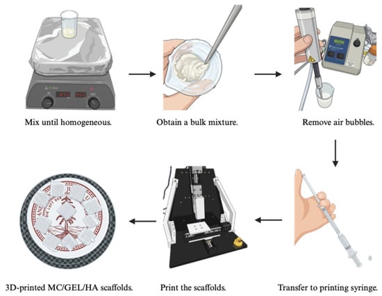
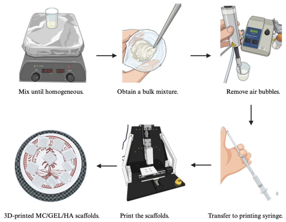
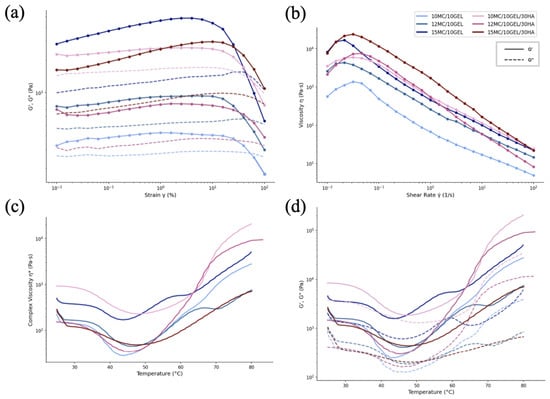
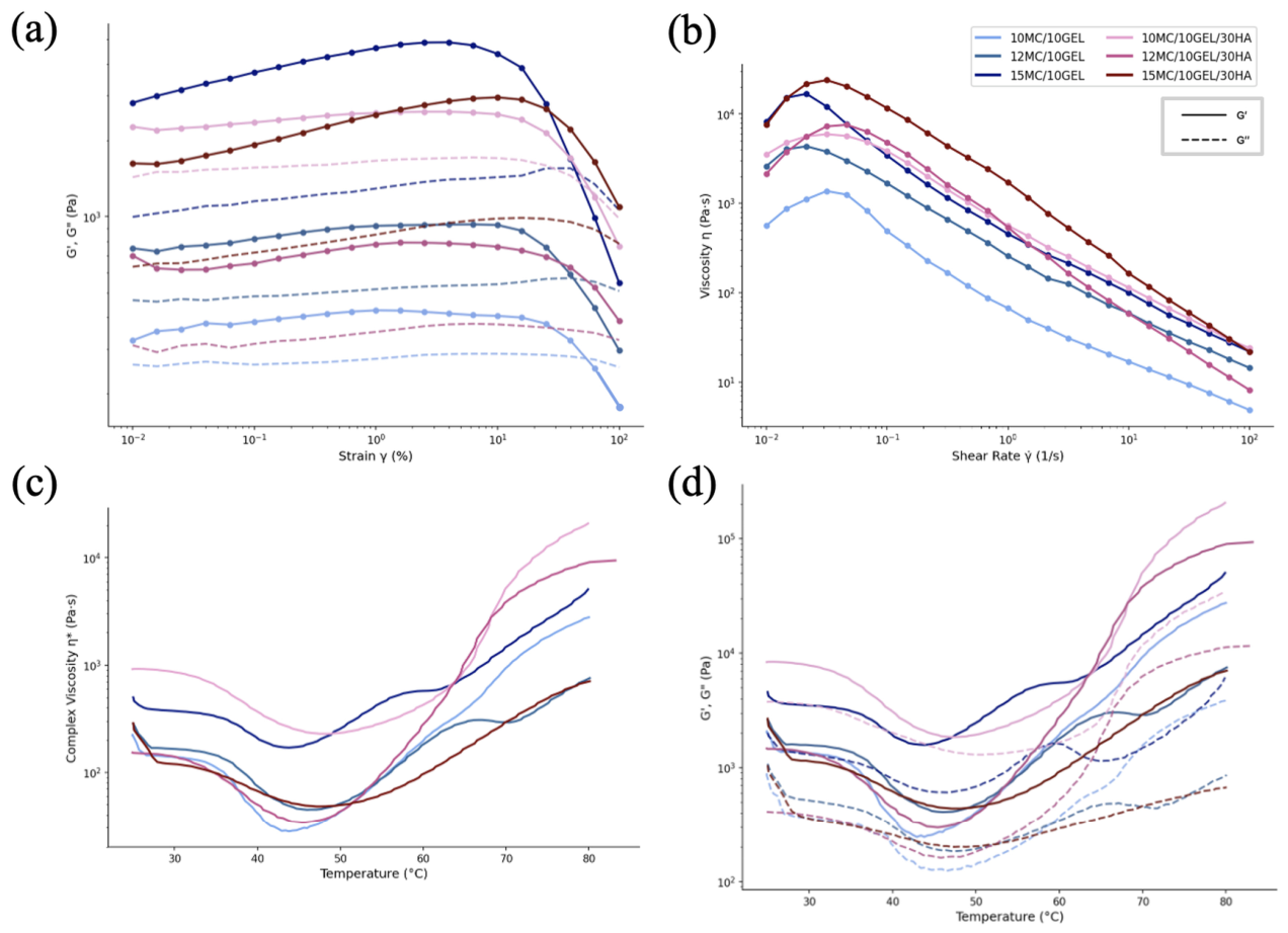
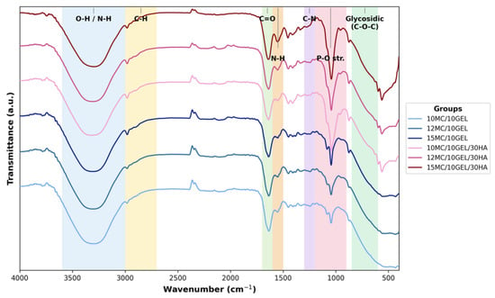
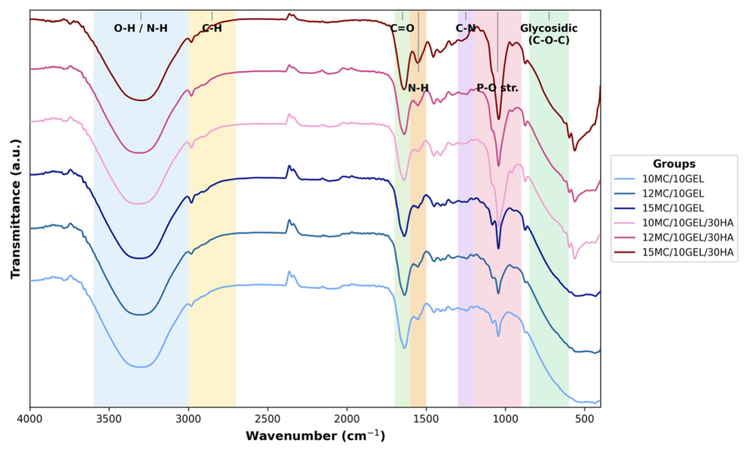
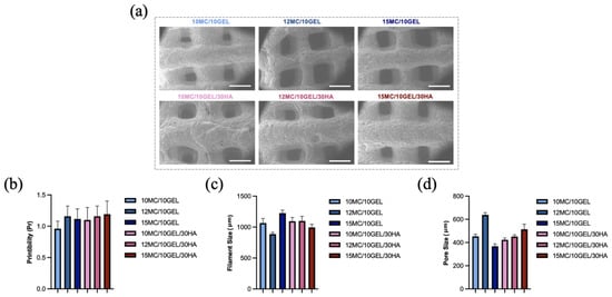
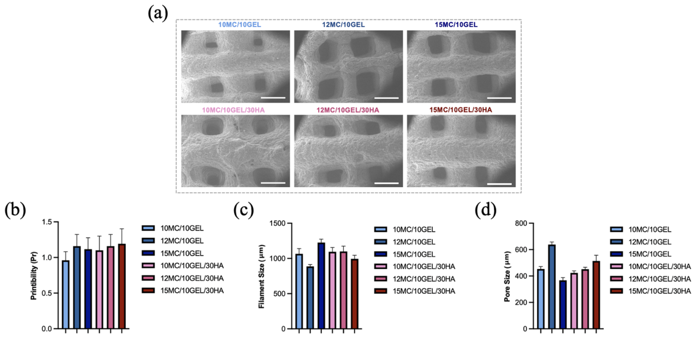
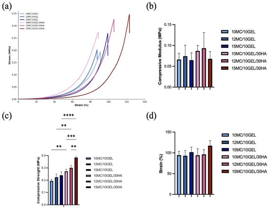
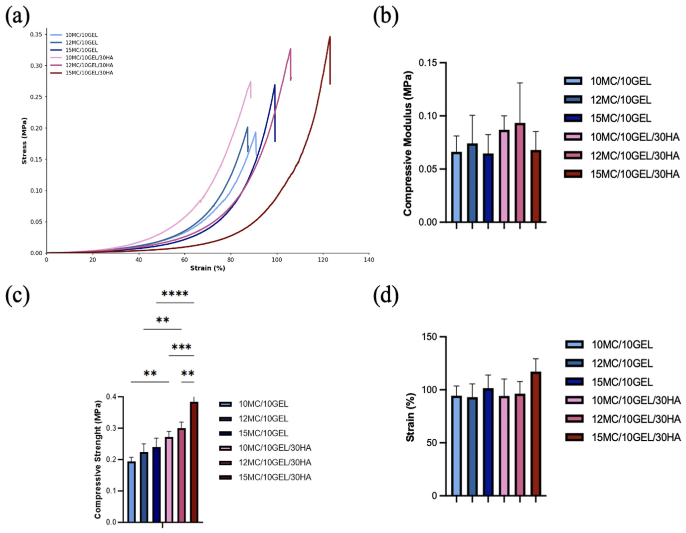
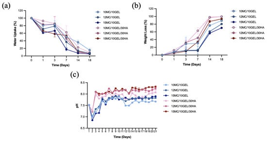
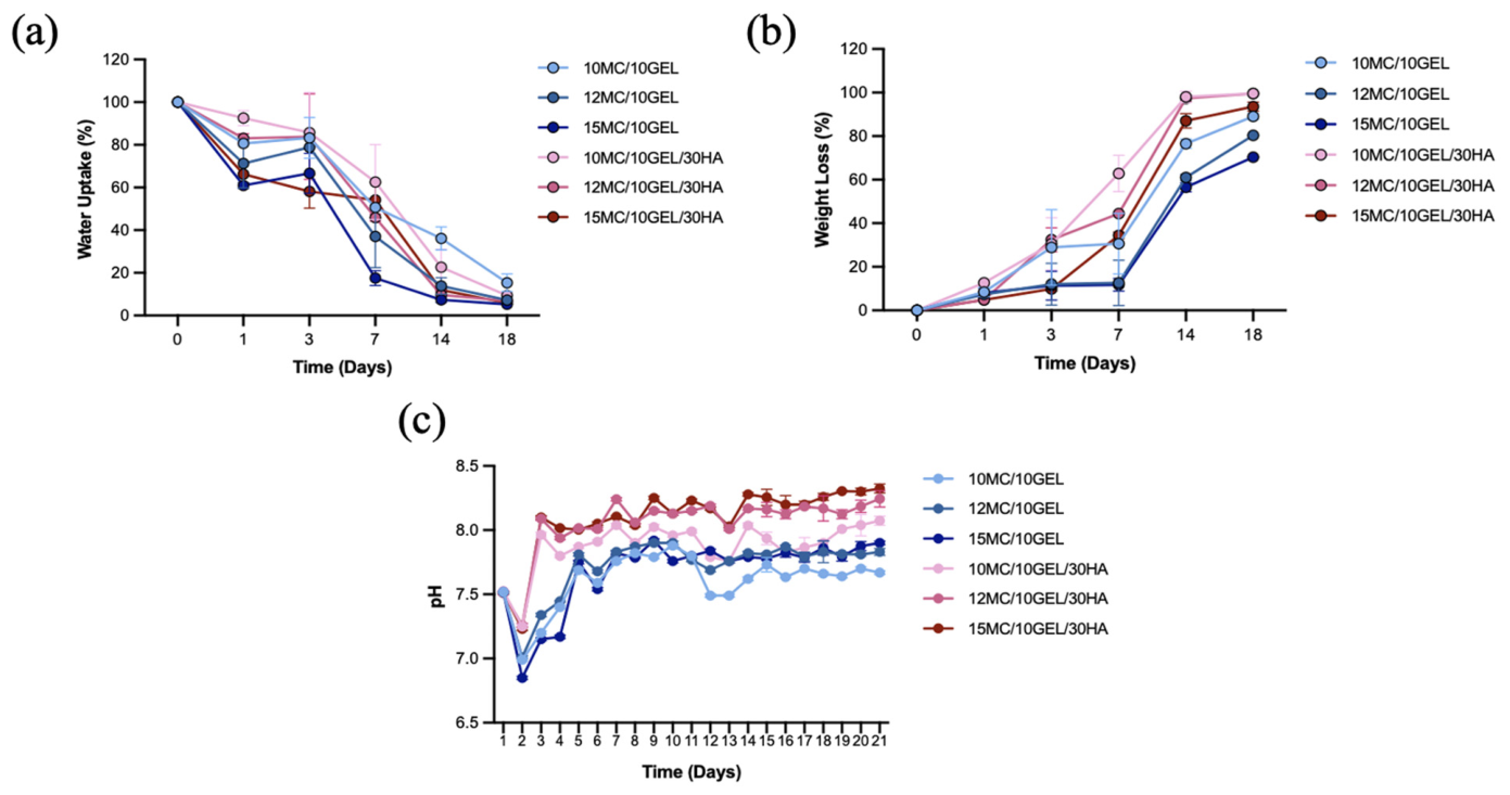
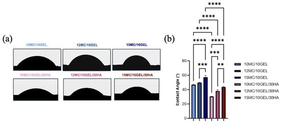
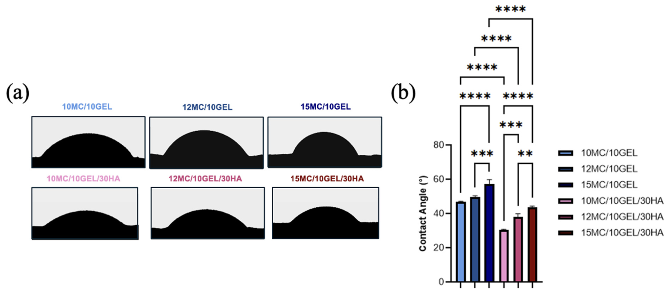
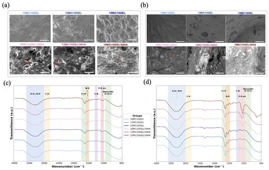
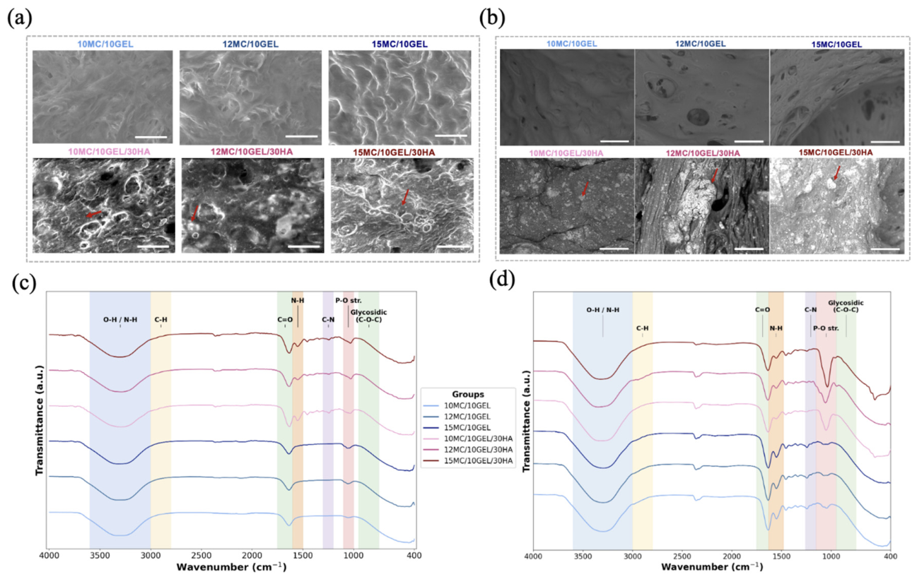
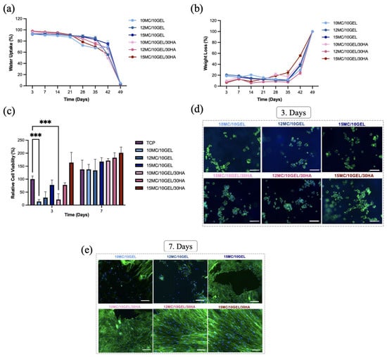
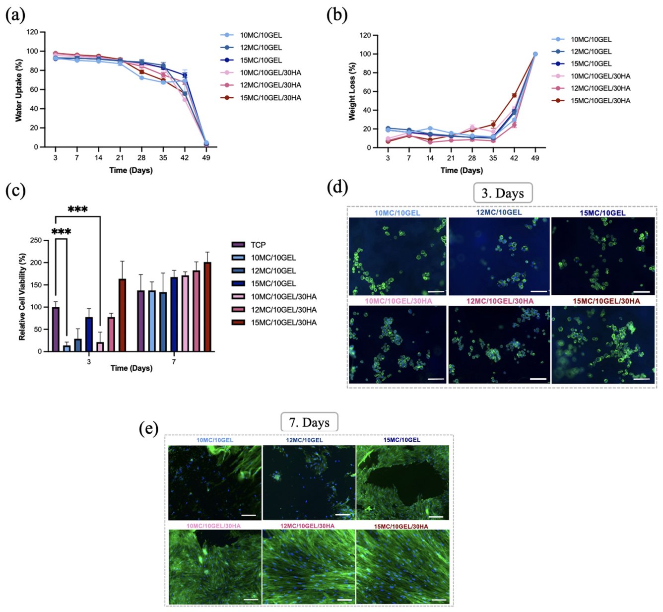
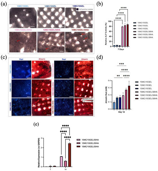
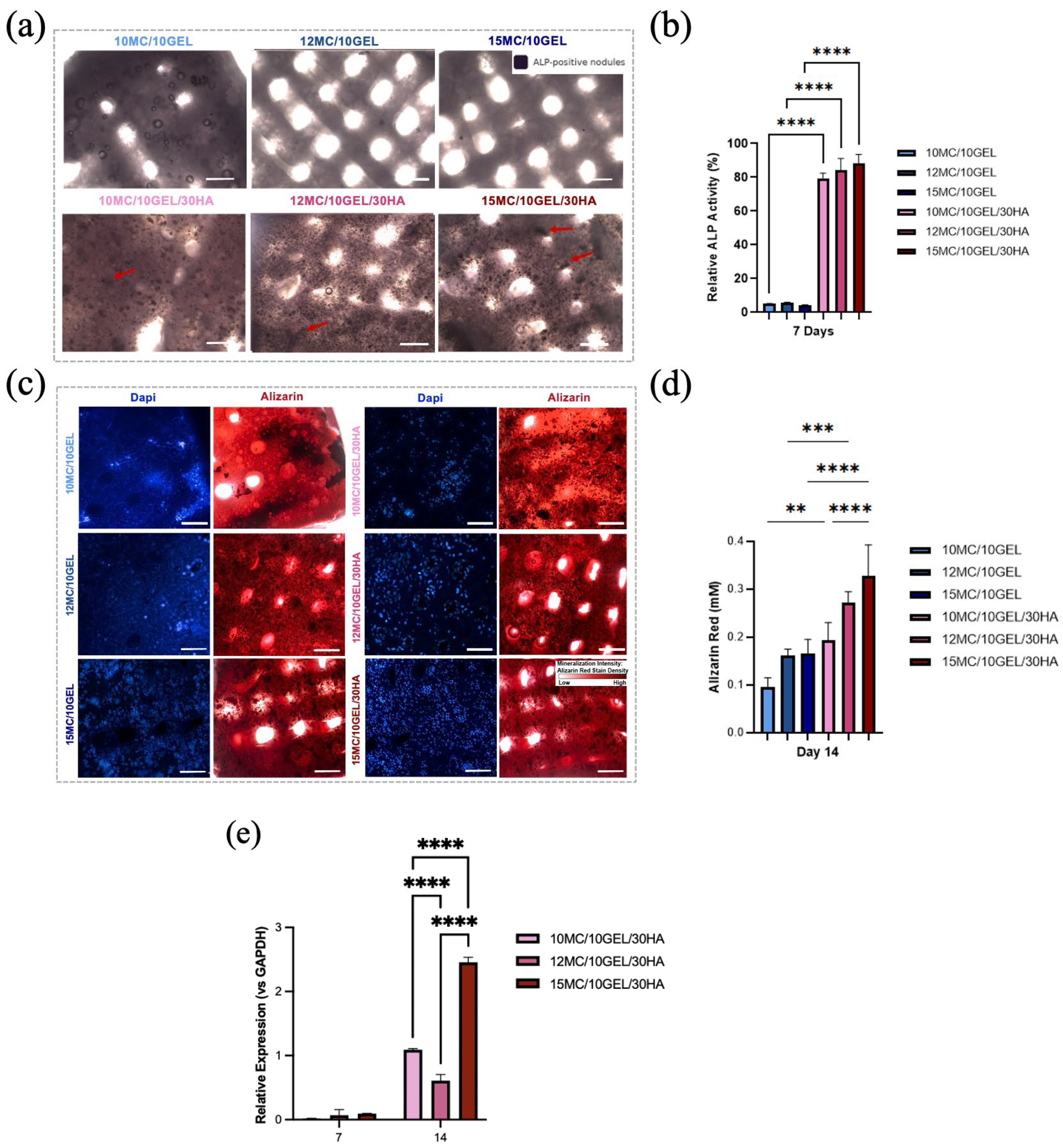
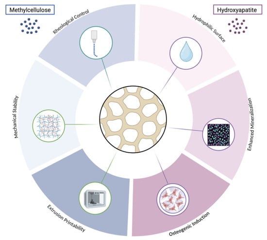
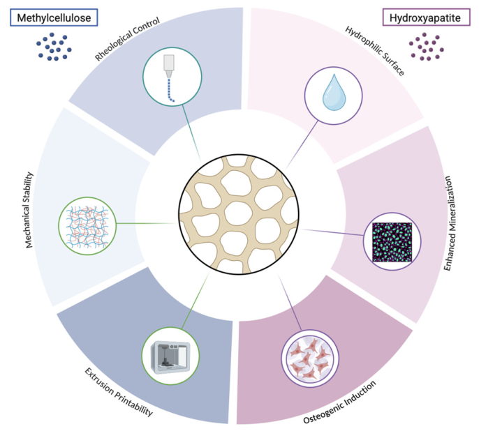

Disclaimer/Publisher’s Note: The statements, opinions and data contained in all publications are solely those of the individual author(s) and contributor(s) and not of MDPI and/or the editor(s). MDPI and/or the editor(s) disclaim responsibility for any injury to people or property resulting from any ideas, methods, instructions or products referred to in the content. |
© 2026 by the authors. Licensee MDPI, Basel, Switzerland. This article is an open access article distributed under the terms and conditions of the Creative Commons Attribution (CC BY) license.