


Abstract
Maxillary second molars with root fusion exhibit considerable anatomical variability that complicates dental treatment planning. Cone-beam computed tomography (CBCT) has become an essential imaging modality for evaluating such complex morphology. This study aimed to develop a deep learning (DL)-based computer-aided diagnosis (CAD) system for the automated classification of root fusion types in maxillary second molars, and to systematically evaluate the impact of image preprocessing strategies on model performance. CBCT images from 172 maxillary second molars (3188 two-dimensional slices) were collected and classified into seven root fusion types. A ResNet50 convolutional neural network was trained with tooth-level 5-fold cross-validation. The effects of grayscale bit depth (4096 vs. 256 levels), preprocessing mode (original, binarized, and segmented images), and contrast enhancement techniques (histogram equalization [HE] and contrast-limited adaptive histogram equalization [CLAHE] with varying parameters) on classification accuracy were systematically compared. The model trained on segmented, non-binarized images with 4096 grayscale levels achieved the highest overall accuracy of 91.6%. In descending order of performance: CLAHE-1 (90.5%), CLAHE-3 (90.5%), CLAHE-40 (88.2%), and HE (83.9%). The unprocessed original image group yielded the lowest accuracy (57.5%). A ResNet50-based DL system can effectively classify complex root fusion morphology in maxillary second molars. Preserving the natural radiopacity gradient of CBCT images, rather than applying artificial contrast enhancement, was critical for optimal model performance. These findings provide a reference for preprocessing strategy selection in dental CBCT-based AI systems.
1. Introduction
Maxillary second molars present a higher prevalence of root fusion and more complex root canal systems compared to maxillary first molars, contributing to increased unpredictability in various dental procedures [1]. Molars with fused roots are predisposed to deeper probing depths and greater clinical attachment loss, which can accelerate the progression of localized periodontal destruction [2]. From a prosthodontic perspective, the unfavorable crown-to-root ratio associated with root fusion may adversely affect the long-term prognosis of prosthetic restorations [3,4]. In endodontic treatment, the complex internal morphology of fused roots and the presence of danger zones significantly increase the difficulty of canal debridement and shaping, elevating the risk of instrument separation and root perforation [5].
In clinical practice, periapical radiographs have traditionally been used to assess root morphology. However, the superimposition of adjacent anatomical structures frequently obscures critical details. Consequently, cone-beam computed tomography (CBCT) has emerged as an indispensable imaging tool before complex endodontic interventions, providing three-dimensional visualization of root and canal anatomy with greater accuracy [6].
In recent years, computer-aided detection and diagnosis (CAD) based on deep learning (DL) algorithms has been increasingly applied to dental radiographic imaging, where it has demonstrated strong performance in tasks ranging from caries detection to the automated segmentation of dental CT images, offering image interpretation with reduced observer bias, improved precision, and shortened analysis time [7,8]. While a growing body of literature has explored DL applications in dental imaging—including detection of carious lesions, evaluation of the mandibular canal, and identification of periapical pathologies—automated analysis of complex root fusion morphology remains relatively underexplored, particularly in maxillary molars. Moreover, few studies have addressed the critical trade-off in image preprocessing between artificial contrast enhancement and the preservation of natural radiopacity gradients.
Deep learning has been increasingly applied to dental imaging tasks, including caries detection, automated mandibular canal segmentation, and periapical lesion detection [9,10,11]. By comparison, evidence concerning the automated analysis of root morphology remains limited. Representative studies have used deep learning to identify additional distal roots in mandibular first molars on panoramic radiographs, with CBCT serving as the reference standard [12], while a recent systematic review summarized the diagnostic performance of artificial intelligence for detecting second mesiobuccal canals in maxillary first molars on CBCT [13]. Root fusion in maxillary molars, however, has primarily been characterized through observer-based morphological assessment of CBCT datasets rather than automated classification [14]. Although Sherwood et al. [15] applied deep learning to CBCT-based segmentation and classification of C-shaped canal configurations in mandibular second molars, that study addressed internal canal anatomy rather than external root-fusion morphology in maxillary molars.
Image preprocessing represents an additional methodological consideration. A systematic review of panoramic radiography-based machine-learning studies identified substantial variation in the management of image-quality concerns and preprocessing procedures [16]. Furthermore, HE and CLAHE improved the perceived quality of periapical radiographs but altered fractal-dimension measurements, indicating that contrast enhancement can modify quantitative image characteristics [17]. Although the addition of CLAHE improved deep-learning performance for classifying C-shaped canal anatomy in mandibular second molars [15], the effects of contrast enhancement are likely to depend on the imaging modality and classification task. The comparative effects of grayscale reduction, background removal, binarization, HE, and CLAHE on deep-learning classification of external root-fusion morphology in maxillary second molars using CBCT-derived axial images therefore remain insufficiently established.
The present study aimed to establish an automated CAD system capable of accurately classifying seven morphological types of root fusion in maxillary second molars from CBCT images, and to identify the optimal image preprocessing strategy for this task.
In previous studies referring to image enhancement, Liang et al. (2017) described the issue of ring artifacts in CT images, which can be caused by various factors such as detector non-uniformity, beam hardening, and scattering [18]. They proposed an iterative estimation method to effectively eliminate these artifacts while preserving image structures, demonstrating its utility in CBCT-guided radiotherapy despite limitations like requiring prior artifact pattern knowledge and potential over-correction. Miki et al. (2018) also proposed a method for classifying tooth types on dental cone-beam computed tomography (CBCT) images using deep convolutional neural networks (DCNN) [19]. The authors suggested selecting rectangular regions of interest (ROIs) around the teeth on axial slices and using a DCNN-based method to classify teeth into seven types. In addition, the authors investigated the impact of data augmentation on classification performance and found that using samples with different contrast and rotation angles could improve the overall classification accuracy. They concluded that the proposed method may have important reference value for dentists in identifying and treating dental problems. Jiang et al. (2022) proposed a new approach to improving image quality in CBCT using deep learning techniques [20]. The suggested technique produces high-quality CBCT images in the projection domain and increases scatter estimate accuracy by utilizing several spectral CT labels along with the Pix2pix GAN algorithm. The difficulties with CBCT imaging, such as low contrast-to-noise ratio and scatter contamination, are demonstrated in this work.
2. Materials and Methods
2.1. Data Acquisition
The retrospective single-center study was approved by the Institutional Review Board of Ditmanson Medical Foundation Chia-Yi Christian Hospital (IRB-20211114). CBCT scan data were retrospectively collected from patients who attended the dental department of Chia-Yi Christian Hospital between January 2010 and September 2021. All images were acquired using a Promax 3D CBCT device (Planmeca, Helsinki, Finland) at 84 kV and 12 mA, with a voxel size of 200 μm and an exposure time of 12 s. The original grayscale resolution was 4096 levels (12-bit).
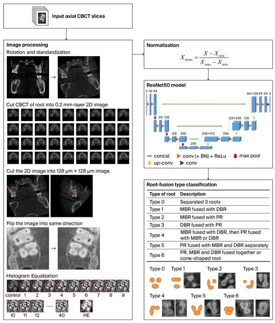
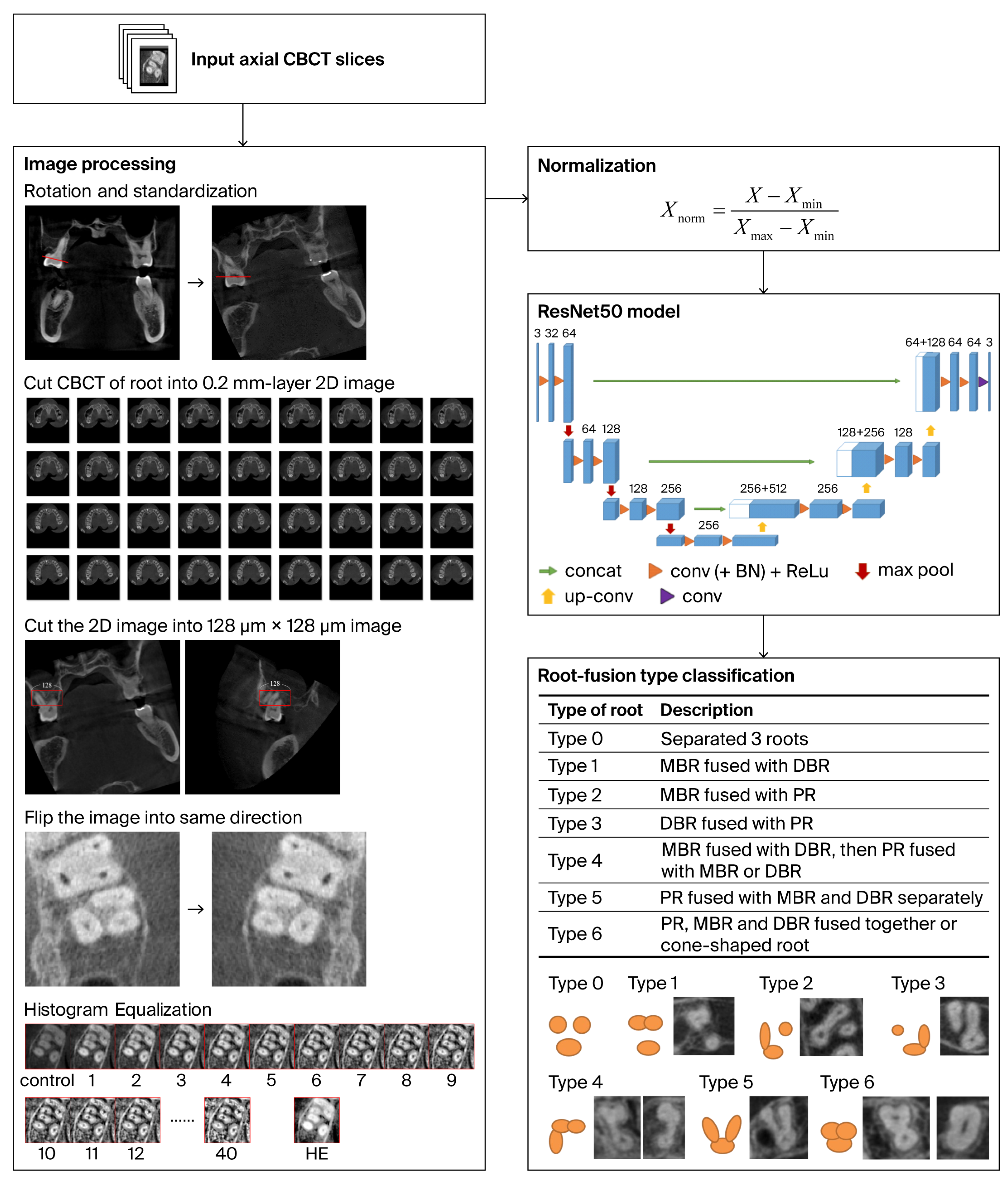
Patients were eligible for inclusion if they were aged 18 years or older. Additionally, the target maxillary second molars were required to have fully formed roots. Complete visualization of the target tooth from the cemento-enamel junction to the root apex within the CBCT field of view was also required. The maxillary arch also had to be intact with no history of molar loss. This was verified through a review of the complete clinical records. Conversely, teeth with prior root canal treatment were excluded from the study. Specimens showing any evidence of root resorption were also excluded. Furthermore, scans were omitted if they contained imaging artifacts or possessed a voxel size greater than 0.2 mm. These included severe motion, metal-related, beam-hardening, or other reconstruction artifacts that interfered with visualization of root boundaries. Based on these selection criteria, CBCT scans of 172 maxillary second molars were enrolled. These scans ultimately yielded a total of 3188 two-dimensional (2D) image slices for analysis. At the same time, two professional clinical dentists assigned the ground-truth labels to the individual slices. The primary objective of this study was to accurately identify and classify the fused root types in maxillary second molars. To achieve this, a novel evaluation method was proposed. The detailed workflow of this proposed method is illustrated in Figure 1.
Figure 1.
Flowchart of the proposed deep learning-based classification system for root fusion morphology in maxillary second molars.
2.2. Root Fusion Classification
All CBCT scans and 2D image slices were reviewed and classified into seven types of root fusion (Types 0–6), based on a modified and simplified version of the classification system proposed by Martins et al. [14]. The classification scheme is summarized in Table 1. Image annotation was performed independently by two endodontic specialists with 10 and 15 years of clinical experience, respectively. Inter-rater reliability was assessed using Cohen’s kappa coefficient, yielding a value of 0.88, indicating near-perfect agreement. Disagreements were resolved through joint discussion.
Table 1.
Type of root fusion.
2.3. Image Preprocessing
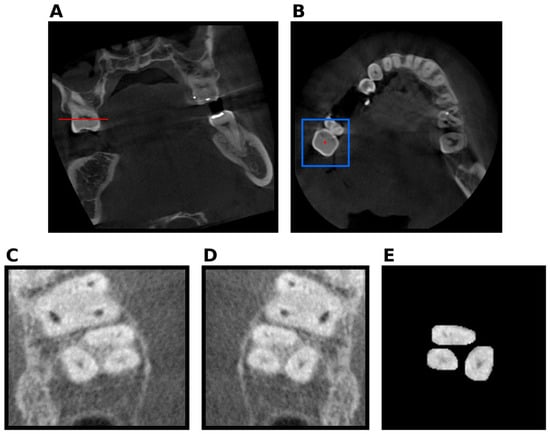
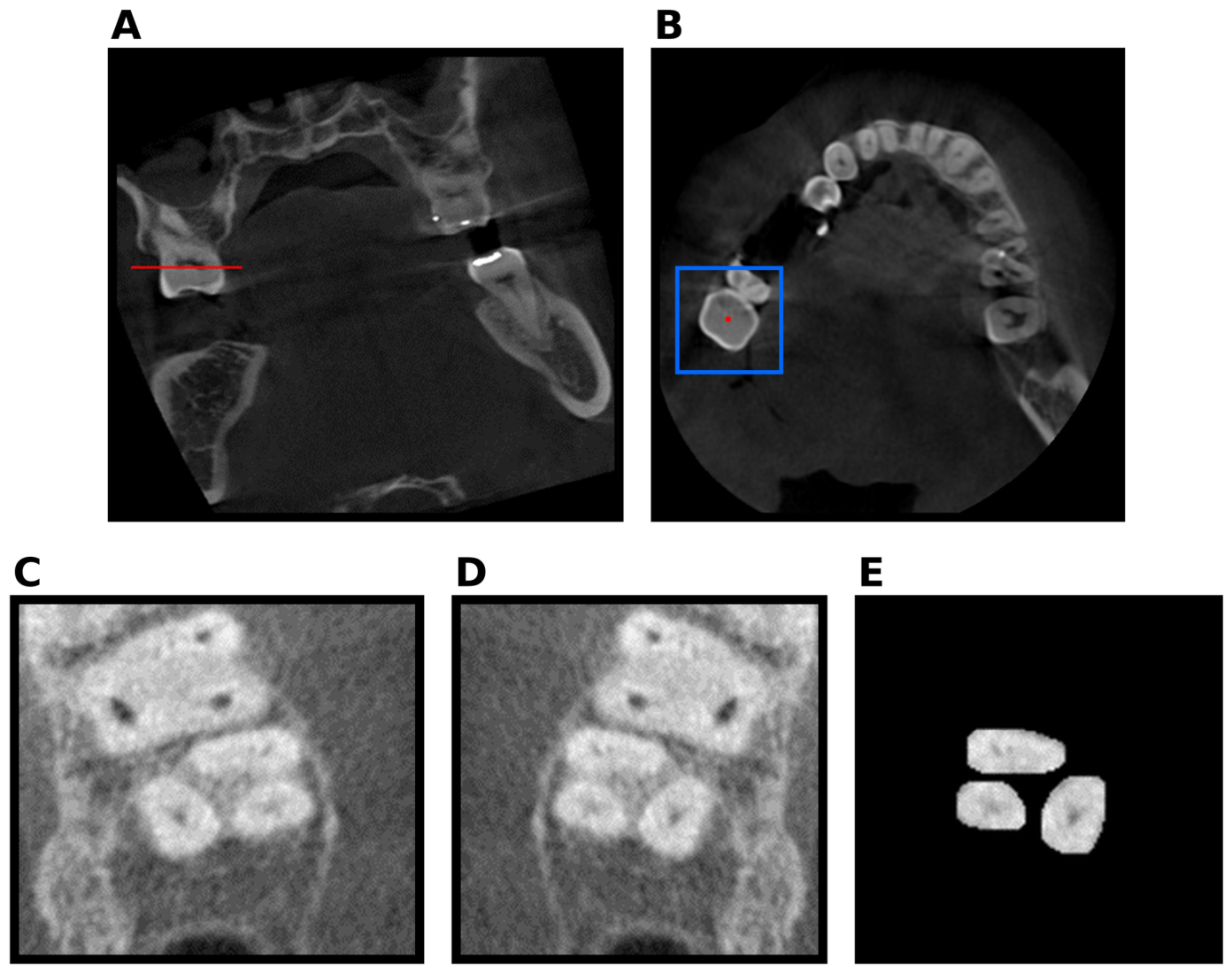
To mitigate variability arising from inconsistent patient positioning, Figure 2 shows a standardized preprocessing pipeline to reorient all scans to a uniform anatomical coordinate system. Each 3D CBCT volume was first rotated to align the cemento-enamel junction (CEJ) with the horizontal plane, ensuring uniform tooth angulation across cases. Each axial slice based on the horizontal plane was used to classify the type by the model. Continuous two-dimensional (2D) cross-sectional images were extracted from the cone-beam computed tomography (CBCT) volumetric dataset at 0.2 mm intervals along the transverse plane, spanning the region from the first root bifurcation to the root apex. This systematic slice-sampling strategy was designed to capture the spatial variations in root morphology at fine anatomical intervals and to generate a consistent set of image samples for subsequent deep learning-based classification. At each sampling level, a fixed-size region of interest (ROI) of 128 × 128 pixels was extracted, with the ROI center aligned to the centroid of the target tooth crown. This spatially standardized cropping procedure ensured consistent image dimensions and anatomical positioning across samples while retaining the complete root cross-section within the ROI. To further reduce orientation-related variability, images of left-sided teeth were horizontally flipped to establish a consistent anatomical orientation across teeth from both sides of the dental arch. These preprocessing operations standardized the spatial representation of the input images and reduced variations unrelated to the target root morphology, thereby improving the consistency of the image dataset for model training and evaluation.
Figure 2.
Procedure of CBCT image preprocessing. (A) Rotation of the 3D CBCT volume to align the cemento-enamel junction (CEJ) with the horizontal plane (red reference line), with sequential 0.2 mm transverse slicing from the first root bifurcation to the apex. (B) The blue box indicates the 128 × 128-pixel region of interest (ROI) centered on the crown centroid (red dot). (C) Example ROI for the left maxillary second molar. (D) Horizontally flipped image for standardization of left-sided teeth. (E) Manually segmented root image after removal of background anatomical structures.
Subsequently, manual image segmentation was performed to isolate the target root cross-section and suppress irrelevant background information. Specifically, surrounding anatomical structures, including alveolar bone, the periodontal ligament space, and adjacent teeth, were excluded from the images, while the root boundaries and morphological features relevant to classification were preserved. From a deep learning perspective, this segmentation step served as a targeted background-reduction procedure, minimizing potential interference from non-target anatomical structures and encouraging the model to learn discriminative features associated with root morphology rather than incidental contextual information. The resulting images underwent standardized spatial preprocessing, including fixed-size ROI extraction, orientation normalization, and background removal, to produce anatomically focused input samples with consistent dimensions and alignment. These processed cross-sectional images were subsequently organized into the image dataset used for deep learning model development, enabling the convolutional neural networks to extract hierarchical morphological features and learn patterns associated with different root morphology classes. By integrating anatomically guided image sampling with standardized preprocessing and segmentation, the proposed pipeline established a consistent input representation for robust model training and systematic performance evaluation.
Although the teeth on the left side are flipped horizontally, the principle of data confidentiality is still adhered during the model training and testing phases. This meant that the left and right teeth of a patient would not appear in the training and testing data simultaneously.
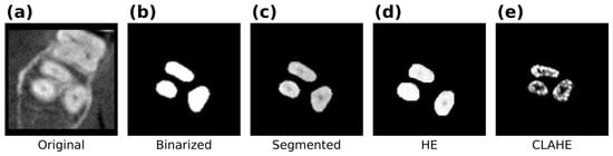
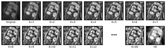
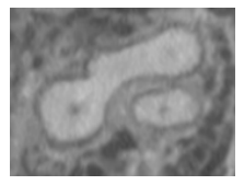
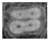
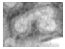
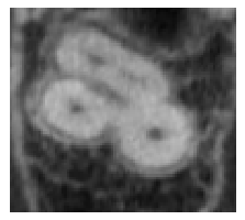
Three sets of comparative experiments were conducted to systematically identify the optimal preprocessing approach. The first set of experiments evaluated the effect of grayscale bit depth reduction by comparing 4096 levels against 256 levels. The second set examined different preprocessing modes. This comparison involved the original images shown in Figure 3a, the binarized images in Figure 3b, and the manually segmented non-binarized images in Figure 3c. The third set evaluated contrast enhancement methods. This evaluation compared histogram equalization (HE) [21] in Figure 3d with contrast-limited adaptive histogram equalization (CLAHE) [22] in Figure 3e. Finally, Figure 4 illustrates how varying the clip limit parameter (K = 1, 3, and 40) affects the performance of the enhancement algorithm.
Figure 3.
Representative root cross-sectional images illustrating different preprocessing results: (a) original, (b) binarized, (c) segmented (non-binarized), (d) HE-processed, (e) CLAHE-processed.
Figure 4.
Representative root cross-sectional images after CLAHE processing across a range of clip-limit parameters (K = 1–12 and K = 40), shown alongside the original image and the histogram-equalized (HE) result. Progressive increases in clip limit enhance local contrast but also amplify image noise, most evident at K = 40.
2.4. Data Normalization
Min-max normalization was applied to all image pixel values prior to model training, scaling each value to the range [0, 1] according to the following equation:
where X denotes the original pixel value, and Xmin and Xmax represent the minimum and maximum pixel values of the dataset, respectively. This normalization preserves the relative grayscale relationships between pixels while stabilizing gradient descent and accelerating model convergence.
2.5. Algorithm Architecture and Model Training
A ResNet50 convolutional neural network was employed for seven-class classification of root fusion types. ResNet50 was selected because its residual learning framework enables the training of substantially deeper networks without suffering from the vanishing-gradient and degradation problems that limit conventional architectures, while maintaining a favorable balance between representational capacity and computational cost. This depth is advantageous for capturing the subtle, hierarchical morphological features that distinguish closely related root fusion subtypes. Feature extraction was performed through five groups of residual convolutional blocks, followed by a Global Average Pooling layer and a 7-dimensional fully connected layer with Softmax activation. The use of residual connections effectively mitigates vanishing gradient and degradation issues in deep network training. The detailed architecture parameters are summarized in Supplementary Table S1.
All experiments were conducted using TensorFlow 2.9.1 on an NVIDIA GeForce RTX 3090 GPU (Intel Core i9-10900X CPU, 36 GB RAM). Training parameters were: 50 epochs, batch size of 32, initial learning rate of 0.001, Adam optimizer, Categorical Cross-Entropy loss function, and ReLU activation with Batch Normalization enabled.
This study evaluates the effectiveness of 5-fold cross-validation with tooth-level data partitioning to ensure that all 2D slices from the same tooth were confined to either the training or the test set within each fold. 5-fold cross-validation is a robust resampling technique used to evaluate machine learning models and prevent overfitting by partitioning a dataset into five equally sized subsets, or “folds.” The training and evaluation process is then repeated five times; in each iteration, a unique fold is held out as the validation set while the remaining four folds are combined to train the model. Finally, the performance metrics from all five iterations are averaged to yield a reliable, low-variance estimate of how well the model generalizes to unseen data. To avoid data leakage, Xmin and Xmax were calculated only from the training data for each fold.
Accuracy, the micro-averaged F1-score (micro-F1), and the macro-averaged F1-score (macro-F1) were calculated for each test fold and summarized as mean values across the five folds [23]. No inferential statistical hypothesis testing was performed.
The F1-score is the harmonic mean of precision and recall and therefore accounts for both false-positive and false-negative predictions. Micro-F1 is calculated by pooling the true-positive, false-positive, and false-negative counts across all classes. In a single-label multiclass classification task, micro-F1 equals overall accuracy. Macro-F1 is the unweighted mean of the class-specific F1-scores and therefore assigns equal weight to each class, regardless of its sample size, providing an overall summary of performance across classes.
2.6. Output Aggregation Strategy
To account for the intricate anatomical variability of maxillary second molars—where root fusion configurations frequently transition across different axial depths—this study implemented a cumulative union aggregation strategy to integrate prediction outputs from all 2D constitutive cross-sectional slices of each tooth. Unlike conventional majority-voting schemes that reduce multi-slice outputs to a single dominant label and risk suppressing transient morphological variations, this non-reductive approach establishes the final diagnostic classification as the mathematical union of all distinct fusion patterns identified across the entire axial sequence. For example, if a single specimen demonstrates Type 1, Type 2, and Type 3 features at different axial levels, the system systematically records the tooth as exhibiting a multi-level composite of all three classifications rather than forcing a single consensus.
By preserving every detected configuration along the coronal-to-apical trajectory, this methodological framework accurately captures the continuous, three-dimensional nature of root morphogenesis. It eliminates slice-selection bias and ensures that subtle, localized anatomical shifts are fully accounted for. Consequently, this aggregate profile provides clinicians with a high-fidelity, comprehensive anatomical map that is essential for optimizing access cavity design, locating extra or merged canals, and guiding precise endodontic and surgical treatment planning.
3. Results
A total of 3188 CBCT image slices from 172 maxillary second molars were analyzed. The distribution of root fusion types is presented in Table 2. Type 0 (completely separated roots) was the most prevalent. Among fused root types, Type 6 (cone-shaped single root) was the most common, followed by Type 1 (MB-DB fusion). The remaining types were relatively evenly distributed.
Table 2.
Distribution of root fusion types by 2D image slices and number of teeth.
3.1. Experiment 1: Effect of Grayscale Bit Depth
Reducing the grayscale range from 4096 to 256 levels had minimal impact on classification performance. The average accuracy based on patient-level for the 4096-level group was 0.916 (m-F1: 0.916, M-F1: 0.819), compared to 0.906 (m-F1: 0.906, M-F1: 0.807) for the 256-level group (Supplementary Table S2). Given that the 4096-level images retain more anatomical detail, all subsequent experiments used the full 12-bit grayscale range.
3.2. Experiment 2: Effect of Preprocessing Mode
The original (unprocessed) images yielded the poorest model performance, with an average accuracy of only 0.575 (m-F1: 0.575, M-F1: 0.249), reflecting substantial misclassification across multiple fusion types. Binarized images showed intermediate performance, while segmented non-binarized images achieved the highest overall accuracy (0.916, m-F1: 0.916, M-F1: 0.819). Performance was particularly robust for Type 3 and Type 4. Residual misclassifications occurred primarily between morphologically similar types (e.g., Type 2 and Type 3), reflecting the inherent anatomical ambiguity at fusion boundaries as shown in Supplementary Table S3.
3.3. Experiment 3: Effect of Contrast Enhancement
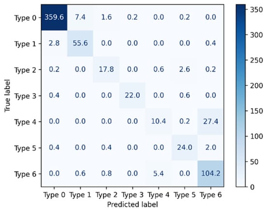
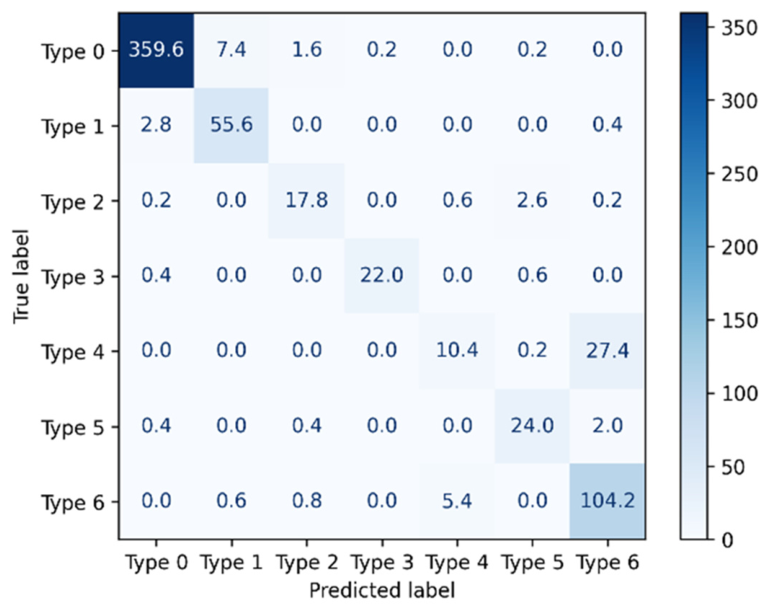
Visual inspection revealed that HE tended to over-amplify background noise, while CLAHE with a high clip limit (K = 40) introduced prominent imaging artifacts. Quantitatively, none of the contrast-enhanced groups outperformed the segmented non-binarized control (accuracy: 0.916). Model performance declined progressively with increasing contrast enhancement: CLAHE-1 (0.905), CLAHE-3 (0.905), CLAHE-40 (0.882), and HE (0.839) (Table 3). These results indicate that contrast enhancement is detrimental to model performance in this context. To identify which root-fusion types are most frequently misclassified in non-binarized slices, a confusion matrix that is the mean of five confusion matrices for five folds is shown in Figure 5. From the figure, we can understand that 27.4 slices of data from type 4 were misclassified as type 6. At the same time, Figure 5 also demonstrates the model performance for each root-fusion type under substantial class imbalance.
Table 3.
Accuracy (patient-level) across contrast enhancement methods.
Figure 5.
The confusion matrix of mean for five folds.
4. Discussion
The prevalence of root fusion in maxillary second molars varies considerably across studies, ranging from 10.71% to 52.9% [1,5,24,25,26]. Using micro-computed tomography (micro-CT), Zhang et al. reported a fusion prevalence of 42.25% in extracted maxillary second molars [24]. Martins et al., employing CBCT, found a prevalence of 25.2%, with 8.6% of cases showing simultaneous root and canal fusion [14]. These discrepancies likely reflect differences in imaging resolution, study populations, and classification criteria. The present study further observed that different fusion types could be identified at different cross-sectional levels within the same tooth, suggesting that current static classification systems may not fully capture the three-dimensional complexity of root fusion morphology.
Although micro-CT provides superior image clarity and smaller voxel size compared to CBCT, it is limited to in vitro specimens and cannot be used clinically. CBCT, while subject to various factors that may affect image quality, remains the standard method for three-dimensional preoperative assessment in clinical endodontics. The present study therefore focused on optimizing CBCT image preprocessing to maximize the performance of DL-based classification under clinically realistic imaging conditions.
Recent studies have demonstrated the feasibility of applying deep learning to CBCT-based analysis of complex endodontic anatomy. Sherwood et al. [15] developed U-Net-based models to segment and classify C-shaped canal configurations in mandibular second molars, reporting mean sensitivities ranging from 0.720 to 0.786 across the evaluated architectures. More recently, Mansour et al. [27] used CNN- and U-Net-based models to detect and segment the second mesiobuccal canal in maxillary first molars on CBCT images, achieving a testing accuracy of 0.87 and an F1-score of 0.93 for classification. These studies support the applicability of deep learning to complex dental anatomy on CBCT images. However, they primarily addressed internal canal anatomy and relatively limited classification targets, whereas the present study investigated seven categories of external root-fusion morphology in maxillary second molars.
A key finding of this study was the decisive influence of image preprocessing on model performance. Raw CBCT slices contain substantial anatomical background structures—including alveolar bone, periodontal ligament space, and adjacent teeth—that can interfere with feature extraction by the convolutional neural network. Manual segmentation effectively eliminated this background noise. Critically, preserving the original 12-bit grayscale gradient was equally important: binarization discards subtle radiopacity differences between dentine and surrounding tissues, which appear to be informative for boundary delineation. By retaining the natural gradient, the ResNet50 model could capture fine-grained root boundary features necessary for accurate subtype differentiation.
This observation aligns with a recent CBCT-based caries classification study by Chen et al. [28], in which models incorporating pre-segmentation consistently achieved performance metrics above 0.917, whereas the direct classification model using unsegmented images achieved a maximum accuracy of 0.700. Although the diagnostic target differed from that of the present study, both findings suggest that isolating the relevant anatomical region may reduce interference from surrounding structures during feature extraction.
The finding that HE and CLAHE did not improve—and in several cases significantly degraded—model performance diverges from their common application in general computer vision tasks. A plausible explanation is that the radiopacity of the root and surrounding hard tissues (cementum, alveolar bone) is inherently similar, and artificial contrast amplification may exaggerate minor density variations, introducing spurious boundaries that mislead the network. Since this interpretation was not validated using explainability tools such as Grad-CAM, it should be regarded as a preliminary hypothesis warranting further investigation. Notably, low-parameter CLAHE (K = 1 and K = 3) performed comparably to the non-enhanced segmented control, suggesting that moderate contrast adjustment may be tolerable, whereas excessive enhancement is clearly detrimental.
This finding differs from that of Sherwood et al. [15], who reported a mean improvement of 4.6% after applying CLAHE to U-Net-based models for C-shaped canal classification. This discrepancy may reflect differences in the classification targets: Sherwood et al. evaluated internal canal configurations, whereas the present study classified external root-fusion patterns based on root boundaries and grayscale transitions between dentine and adjacent tissues.
A further important observation concerns the cross-sectional heterogeneity of root fusion morphology. During the annotation process, it was noted that a single tooth frequently exhibited different fusion types at different cross-sectional levels along the root axis, indicating that root fusion represents a continuous three-dimensional anatomical phenomenon rather than a static, categorically defined feature. This observation is consistent with findings by Fan et al. regarding mandibular C-shaped canals, which were shown to display distinct morphological configurations at the canal orifice, mid-root, and apical levels [29]. In response, the present study adopted a union aggregation strategy rather than conventional majority voting, allowing the system to represent the full spectrum of morphological types present within a single tooth. From a clinical standpoint, this approach better captures the information needed for treatment planning—clinicians require knowledge of the complete root morphology along the entire length of the root, not merely the most common configuration at a representative cross-section. These observations also suggest that existing static classification frameworks may be insufficient for fully characterizing this anatomical complexity, and future work may benefit from developing continuous or multi-label classification systems that reflect the dynamic variation in root fusion along the root axis.
The main limitations of this study include: (1) data were collected from a single institution using a single CBCT device model, which may limit external generalizability; (2) image segmentation was performed manually, precluding full automation of the diagnostic pipeline; (3) unequal sample sizes across fusion type categories may affect the reliability of performance estimates for minority types. Future studies should incorporate multicenter data, integrate automated segmentation architectures such as U-Net, and expand sample sizes to enhance the clinical applicability and generalizability of the system; (4) a false-positive prediction in a single slice may lead to an overestimation of the number of root fusion types identified for a given tooth. These additional predicted types may not actually be present in the tooth’s root fusion morphology, potentially resulting in a certain degree of misclassification; and (5) the confusion matrix shows that type 4 samples have significant limitations, with most type 4 samples being misclassified as type 6. Based on observation and further analysis, the main reason is the high morphological similarity between type 4 and type 6 samples.
Automated tooth-root segmentation on CBCT images is technically feasible but remains challenging because of complex root morphology and the similar grayscale appearance of tooth roots and alveolar bone. Li et al. [30] combined an attention U-Net with a recurrent neural network and reported a Dice similarity coefficient of 0.955 for automated tooth-root segmentation. Chen et al. [31] subsequently developed the CTA-UNet architecture to address complex root morphology and indistinct boundaries between tooth roots and alveolar bone in dental CBCT images.
5. Conclusions
This study demonstrated that a ResNet50-based deep learning CAD system can effectively classify seven types of root fusion morphology in maxillary second molars from CBCT images, achieving an overall accuracy of 91.6% under optimal preprocessing conditions. Manually segmented, non-binarized images preserving the full 12-bit grayscale gradient yielded the best performance, whereas artificial contrast enhancement—particularly HE and high-parameter CLAHE—was detrimental to model accuracy. The adoption of a union aggregation strategy for multi-slice prediction integration more accurately reflects the three-dimensional, continuously varying nature of root fusion morphology and may better serve clinical decision-making. These findings offer practical guidance for preprocessing strategy selection in dental CBCT-based AI systems. Validation through large-scale multicenter studies remains an important direction for future research.
Supplementary Materials
The following supporting information can be downloaded at: https://www.mdpi.com/article/10.3390/jfb17110564/s1, Table S1: Network parameters for ResNet50; Table S2: Classification performance using 4096- and 256-level grayscale images; Table S3: Classification performance across preprocessing modes: original, segmented (non-binarized), and segmented (binarized) images; Table S4: m-F1 and M-F1 scores across contrast enhancement methods (format: m-F1/M-F1).
Author Contributions
Conceptualization, T.-N.W. and H.-Y.H.; methodology, T.-N.W., H.-W.W. and Y.-M.K.; software, C.-H.Y., H.-W.W. and Y.-M.K.; validation, T.-N.W. and H.-W.W.; formal analysis, C.-H.Y., H.-W.W. and Y.-M.K.; investigation, T.-N.W. and H.-W.W.; resources, T.-N.W., H.-Y.H. and Y.-M.K.; data curation, T.-N.W. and H.-W.W.; writing—original draft preparation, T.-N.W., C.-H.Y. and Y.-M.K.; writing—review and editing, T.-N.W., C.-H.Y., H.-Y.H., H.-W.W. and Y.-M.K.; visualization, T.-N.W., C.-H.Y., H.-W.W. and Y.-M.K.; supervision, H.-Y.H. and Y.-M.K.; project administration, T.-N.W.; funding acquisition, T.-N.W. All authors have read and agreed to the published version of the manuscript.
Funding
This research was funded by Ditmanson Medical Foundation Chia-Yi Christian Hospital, Taiwan (grant number R111-28).
Institutional Review Board Statement
The study was approved by the Institutional Review Board of Ditmanson Medical Foundation Chia-Yi Christian Hospital (approval No. IRB-20211114; approval date: 19 November 2021).
Informed Consent Statement
This study was approved by our Institutional Review Board (IRB) in 2021 as a retrospective study. According to the IRB-approved protocol, the requirement for individual patient informed consent was waived. Therefore, no patient-informed consent form was obtained or generated for this study.
Data Availability Statement
The CBCT image data analyzed in this study are not publicly available due to patient privacy and ethical restrictions. Requests for access may be directed to the corresponding author and are subject to institutional and ethical approval.
Conflicts of Interest
The authors declare no conflicts of interest.
References
- Ross, I.F.; Evanchik, P.A. Root fusion in molars: Incidence and sex linkage. J. Periodontol. 1981, 52, 663–667. [Google Scholar] [CrossRef] [Scilit] [PubMed]
- Hou, G.L.; Tsai, C.C.; Huang, J.S. Relationship between molar root fusion and localized periodontitis. J. Periodontol. 1997, 68, 313–319. [Google Scholar] [CrossRef] [Scilit] [PubMed]
- Huysmans, M.C.D.N.J.M.; Klein, M.H.J.; Kok, G.F.; Whitworth, J.M. Parallel post-space preparation in different tooth types ex vivo: Deviation from the canal centre and remaining dentine thickness. Int. Endod. J. 2007, 40, 778–785. [Google Scholar] [CrossRef] [Scilit] [PubMed]
- Gu, Y.C.; Zhang, Y.P.; Liao, Z.G.; Fei, X.D. A micro-computed tomographic analysis of wall thickness of C-shaped canals in mandibular first premolars. J. Endod. 2013, 39, 973–976. [Google Scholar] [CrossRef] [Scilit] [PubMed]
- al Shalabi, R.M.; Omer, O.E.; Glennon, J.; Jennings, M.; Claffey, N.M. Root canal anatomy of maxillary first and second permanent molars: Rewarming of cooled teeth. Int. Endod. J. 2000, 33, 405–414. [Google Scholar] [PubMed]
- Fayad, M.I.; Nair, M.; Levin, M.D.; Benavides, E.; Rubinstein, R.A.; Barghan, S.; Hirschberg, C.S.; Ruprecht, A. AAE and AAOMR joint position statement: Use of cone beam computed tomography in endodontics 2015 update. Oral Surg. Oral Med. Oral Pathol. Oral Radiol. 2015, 120, 508–512. [Google Scholar]
- Mehri Shervedani, A.; Khodadadi, H.; Mousavian, S.I. Development a computer-aided diagnosis system for dental caries detection applying radiographic images. Comput. Biol. Med. 2025, 196, 110966. [Google Scholar] [CrossRef] [Scilit] [PubMed]
- Wu, D.; Jiang, J.; Wang, J.; Bi, Z.; Yu, G. Segmentation algorithms of dental CT images: A comprehensive review from classical to deep learning trend. Expert Syst. Appl. 2025, 275, 126853. [Google Scholar] [CrossRef] [Scilit]
- Lee, J.-H.; Kim, D.-H.; Jeong, S.-N.; Choi, S.-H. Detection and diagnosis of dental caries using a deep learning-based convolutional neural network algorithm. J. Dent. 2018, 77, 106–111. [Google Scholar] [CrossRef] [Scilit] [PubMed]
- Ni, F.-D.; Xu, Z.-N.; Liu, M.-Q.; Zhang, M.-J.; Li, S.; Bai, H.-L.; Ding, P.; Fu, K.-Y. Towards clinically applicable automated mandibular canal segmentation on CBCT. J. Dent. 2024, 144, 104931. [Google Scholar] [CrossRef] [Scilit] [PubMed]
- Çelik, B.; Savaştaer, E.F.; Kaya, H.I.; Çelik, M.E. The role of deep learning for periapical lesion detection on panoramic radiographs. Dentomaxillofac. Radiol. 2023, 52, 20230118. [Google Scholar] [CrossRef] [Scilit] [PubMed]
- Hiraiwa, T.; Ariji, Y.; Fukuda, M.; Kise, Y.; Nakata, K.; Katsumata, A.; Fujita, H.; Ariji, E. A deep-learning artificial intelligence system for assessment of root morphology of the mandibular first molar on panoramic radiography. Dentomaxillofac. Radiol. 2019, 48, 20180218. [Google Scholar] [CrossRef] [Scilit] [PubMed]
- Dashti, M.; Khosraviani, F.; Ghadimi, N.; Baghaei, K.; Esmaeili, S.; Entezar-E-Ghaem, M.; Khurshid, Z.; Osathanon, T. Use of artificial intelligence for detection of MB2 canals in maxillary first molars on CBCT: A systematic review and meta-analysis. BMC Oral Health 2025, 25, 1860. [Google Scholar] [CrossRef] [Scilit] [PubMed]
- Martins, J.N.R.; Mata, A.; Marques, D.; Caramês, J. Prevalence of root fusions and main root canal merging in human upper and lower molars: A cone-beam computed tomography in vivo study. J. Endod. 2016, 42, 900–908. [Google Scholar] [CrossRef] [Scilit] [PubMed]
- Sherwood, A.A.; Sherwood, A.I.; Setzer, F.C.; K, S.D.; Shamili, J.V.; John, C.; Schwendicke, F. A deep learning approach to segment and classify C-shaped canal morphologies in mandibular second molars using cone-beam computed tomography. J. Endod. 2021, 47, 1907–1916. [Google Scholar] [CrossRef] [Scilit] [PubMed]
- Delamare, E.; Fu, X.; Huang, Z.; Kim, J. Panoramic imaging errors in machine learning model development: A systematic review. Dentomaxillofac. Radiol. 2024, 53, 165–172. [Google Scholar] [CrossRef] [Scilit] [PubMed]
- Mehdizadeh, M.; Tavakoli Tafti, K.; Soltani, P. Evaluation of histogram equalization and contrast limited adaptive histogram equalization effect on image quality and fractal dimensions of digital periapical radiographs. Oral Radiol. 2023, 39, 418–424. [Google Scholar] [CrossRef] [Scilit] [PubMed]
- Liang, X.; Zhang, Z.; Niu, T.; Yu, S.; Wu, S.; Li, Z.; Zhang, H.; Xie, Y. Iterative image-domain ring artifact removal in cone-beam CT. Phys. Med. Biol. 2017, 62, 5276–5292. [Google Scholar] [CrossRef] [Scilit] [PubMed]
- Miki, Y.; Muramatsu, C.; Hayashi, T.; Zhou, X.; Hara, T.; Katsumata, A.; Fujita, H. Classification of teeth in cone-beam CT using deep convolutional neural network. Comput. Biol. Med. 2017, 80, 24–29. [Google Scholar] [CrossRef] [Scilit] [PubMed]
- Jiang, Y.; Zhang, Y.; Luo, C.; Yang, P.; Wang, J.; Liang, X.; Zhao, W.; Li, R.; Niu, T. A generalized image quality improvement strategy of cone-beam CT using multiple spectral CT labels in Pix2pix GAN. Phys. Med. Biol. 2022, 67, 11. [Google Scholar] [CrossRef] [Scilit] [PubMed]
- Mohammed, I.M.; Isa, N.A.M. Contrast limited adaptive local histogram equalization method for poor contrast image enhancement. IEEE Access 2025, 13, 62600–62632. [Google Scholar] [CrossRef] [Scilit]
- Yuan, Z.; Zeng, J.; Wei, Z.; Jin, L.; Zhao, S.; Liu, X.; Zhang, Y.; Zhou, G. CLAHE-based low-light image enhancement for robust object detection in overhead power transmission system. IEEE Trans. Power Deliv. 2023, 38, 2240–2243. [Google Scholar] [CrossRef] [Scilit]
- Takahashi, K.; Yamamoto, K.; Kuchiba, A.; Koyama, T. Confidence interval for micro-averaged F1 and macro-averaged F1 scores. Appl. Intell. 2022, 52, 4961–4972. [Google Scholar] [CrossRef] [Scilit] [PubMed]
- Zhang, Q.; Chen, H.; Fan, B.; Fan, W.; Gutmann, J.L. Root and root canal morphology in maxillary second molar with fused root from a native Chinese population. J. Endod. 2014, 40, 871–875. [Google Scholar] [CrossRef] [Scilit] [PubMed]
- Ghoncheh, Z.; Zade, B.M.; Kharazifard, M.J. Root morphology of the maxillary first and second molars in an Iranian population using cone beam computed tomography. J. Dent. 2017, 14, 115–122. [Google Scholar]
- Wu, D.; Zhang, G.; Liang, R.; Zhou, G.; Wu, Y.; Sun, C.; Fan, W. Root and canal morphology of maxillary second molars by cone-beam computed tomography in a native Chinese population. J. Int. Med. Res. 2017, 45, 830–842. [Google Scholar] [CrossRef] [Scilit] [PubMed]
- Mansour, S.; Anter, E.; Mohamed, A.K.; Dahaba, M.M.; Mousa, A. Two step approach for detecting and segmenting the second mesiobuccal canal of maxillary first molars on cone beam computed tomography (CBCT) images via artificial intelligence. BMC Oral Health 2025, 25, 1404. [Google Scholar] [CrossRef] [Scilit] [PubMed]
- Chen, S.; Yang, Y.; Wu, W.; Wei, R.; Wang, Z.; Tay, F.R.; Hu, J.; Ma, J. Classification of Caries Based on CBCT: A Deep Learning Network Interpretability Study. J. Imaging Inform. Med. 2024, 37, 3160–3173. [Google Scholar] [CrossRef] [Scilit] [PubMed]
- Fan, B.; Cheung, G.S.P.; Fan, M.; Gutmann, J.L.; Bian, Z. C-shaped canal system in mandibular second molars: Part I—Anatomical features. J. Endod. 2004, 30, 899–903. [Google Scholar] [CrossRef] [Scilit] [PubMed]
- Li, Q.; Chen, K.; Han, L.; Zhuang, Y.; Li, J.; Lin, J. Automatic tooth roots segmentation of cone beam computed tomography image sequences using U-net and RNN. J. Xray Sci. Technol. 2020, 28, 905–922. [Google Scholar] [CrossRef] [Scilit] [PubMed]
- Chen, Z.; Chen, S.; Hu, F. CTA-UNet: CNN-transformer architecture UNet for dental CBCT images segmentation. Phys. Med. Biol. 2023, 68, 17. [Google Scholar] [CrossRef] [Scilit] [PubMed]

Disclaimer/Publisher’s Note: The statements, opinions and data contained in all publications are solely those of the individual author(s) and contributor(s) and not of MDPI and/or the editor(s). MDPI and/or the editor(s) disclaim responsibility for any injury to people or property resulting from any ideas, methods, instructions or products referred to in the content. |
© 2026 by the authors. Licensee MDPI, Basel, Switzerland. This article is an open access article distributed under the terms and conditions of the Creative Commons Attribution (CC BY) license.