


Abstract
Following myocardial infarction (MI), the injured myocardium undergoes substantial metabolic changes that contribute to cardiomyocyte death, adverse ventricular remodeling, and impaired cardiac function. Both ischemia and the restoration of blood flow through reperfusion contribute to these changes, which include disrupted oxidative phosphorylation, increased reactive oxygen species production, mitochondrial dysfunction, ferroptosis, and alterations in glucose and reactive carbonyl metabolism. Although therapies have been developed to target these pathways, their effectiveness is often limited by poor myocardial retention and an inability to respond to changes within the infarct environment. Injectable hydrogels provide a way to deliver these therapies locally, prolong their activity, and incorporate stimulus-responsive properties that allow for more controlled release. This review examines hydrogel-based approaches designed to reduce oxidative stress, alleviate hypoxia, preserve or replace damaged mitochondria, inhibit ferroptosis, regulate glycolysis, and limit methylglyoxal-associated damage following MI. The different biological rationales and material designs are discussed, along with their effects on cardiac repair. Finally, the challenges associated with translating these systems into clinical applications are considered, including the need for more representative preclinical models, practical delivery methods, and therapeutic strategies that account for the changing post-infarct environment.
1. Introduction
The ischemic injury caused by myocardial infarction (MI) leads to the irreversible loss of cardiomyocytes, and despite advances in reperfusion, pharmacological, and secondary prevention techniques, many MI patients will still experience adverse ventricular remodeling, ultimately leading to impaired cardiac function and chronic heart failure [1,2]. Early reperfusion strategies are prioritized to restore blood flow to the coronary arteries and salvage the ischemic myocardium after MI, particularly for ST-elevation myocardial infarction (STEMI) where a major coronary artery is completely blocked. However, reperfusion itself can initiate a cascade of secondary injuries including oxidative stress, inflammation, mitochondrial dysfunction, and cardiomyocyte death [3,4]. These adverse effects that occur after revascularization has been achieved can be linked to the treatment’s inability to adequately address the complex pathological remodeling that occurs during the days to weeks following infarction [5].
Increasing evidence suggests that metabolic dysfunction contributes substantially to post-infarct remodeling. Under normal physiological conditions, the adult heart relies predominantly on mitochondrial oxidative phosphorylation and fatty acid oxidation to satisfy its exceptionally high energetic demand [6,7]. However, ischemia rapidly disrupts oxygen availability, forcing cardiomyocytes to shift toward anaerobic glycolysis while simultaneously impairing mitochondrial ATP production [8]. Although this metabolic adaptation initially supports cell survival, persistent metabolic remodeling contributes to ATP depletion, lactate accumulation, mitochondrial dysfunction, excessive reactive oxygen species (ROS) generation, lipid peroxidation, ferroptosis, and reactive carbonyl stress [9,10,11]. These challenges have prompted growing interest in developing therapies that can restore normal metabolic function. One attractive approach is the use of hydrogels that can be applied directly within the post-infarct myocardium (Figure 1).
Figure 1.
Injectable hydrogels for metabolic modulation after myocardial infarction. Image created using BioRender.com.
A hydrogel is a 3D structured polymer or molecular crosslinked network, composed of hydrophilic groups, which swell in water without dissolving. Once fully crosslinked, a hydrogel can retain its shape [12]. Injectable hydrogels have gained attention for tissue engineering and drug delivery applications, and they are emerging as a platform for supporting cardiac repair post-MI [13,14]. Once injected, these liquid or partially crosslinked hydrogels undergo gelation via multiple mechanisms, including self-assembly, non-covalent interactions with ionic species, covalent crosslinking, and thermal transitions [14]. Injectable hydrogels for cardiac repair can be synthesized using natural-based or synthetic materials, or a combination of the two. For more details on different hydrogel compositions, chemical modifications, crosslinking strategies and physical properties, readers are referred to these comprehensive reviews [15,16]. These hydrogels can serve multiple purposes within the context of cardiac repair. Due to their structural and functional similarity to the natural extracellular matrix, they can provide mechanical support to infarcted hearts and attenuate pathological remodeling, consequently reducing wall stress and limiting adverse ventricular dilation [17]. They can also function as localized delivery vehicles, allowing bioactive molecules and/or cells to be delivered directly to the infarcted tissue to promote repair while avoiding the systemic side effects and rapid clearance associated with systemically administered therapies [18]. More advanced hydrogel systems can be engineered with environment-responsive properties, including responsiveness to pathological stimuli such as elevated ROS, acidic pH, hypoxia, or disease-associated enzymes, enabling controlled release of therapeutic agents specifically within the infarct tissue [19,20].
These properties make injectable hydrogels particularly well-suited for metabolic modulation of the post-MI environment. By acting on the interconnected processes that control cardiomyocyte survival and cardiac repair, hydrogels can be used to modulate ROS scavenging, mitochondrial bioenergetics, hypoxia alleviation, ferroptosis suppression, glycolytic remodeling, and reactive carbonyl stress [21]. Acting upstream of these pathways offers the potential to collectively reduce inflammation, fibrosis, and endothelial dysfunction while simultaneously promoting angiogenesis and functional recovery.
This rationale over the past decade has driven the development of various injectable hydrogel platforms such as those incorporating antioxidants, nanozymes, oxygen-generating materials, mitochondria-targeting, and stimuli-responsive chemistries. Existing reviews of injectable hydrogels for MI are predominantly organized according to biomaterial composition, therapeutic cargo, engineering properties, or broad biological topics such as angiogenesis and inflammation [22,23,24,25]. To our knowledge, there has been no review focused on hydrogel-based strategies targeting post-MI metabolism. Herein, we provide a summary of injectable hydrogels that target metabolic dysfunction following MI, including approaches to scavenge ROS, alleviate hypoxia, preserve mitochondrial function, suppress ferroptosis, and regulate glycolytic remodeling and reactive carbonyl stress. Furthermore, the biological rationale, engineering strategies, and/or translational challenges associated with these systems are discussed to inform the future development of injectable hydrogels for cardiac repair. The hydrogel literature presented in this review was identified through searches of Google Scholar and the University of Ottawa’s Omni library’s search platform for studies published between 2011 and 2026 using the following keywords: myocardial infarction, injectable hydrogels, and metabolic modulation. Studies were selected based on their relevance to injectable hydrogel-based modulation of metabolic dysfunction after MI. The review is not intended to be comprehensive, but rather an emphasis was put on providing examples for a range of different therapeutic mechanisms and effects on cardiac repair.
2. Metabolic Pathophysiology of the Post-Infarct Microenvironment
In this section, we briefly describe some of the main metabolic changes that occur post-MI and after reperfusion therapy that can be targeted by hydrogel treatments for improving cardiac repair, as summarized in Figure 2. For more information on this topic, we refer the reader to more comprehensive reviews [6,8,26].
Figure 2.
Metabolic changes during the overlapping phases of healing post-MI. During ischemia, coronary occlusion limits oxygen delivery, suppressing mitochondrial oxidative phosphorylation and fatty acid oxidation while increasing anaerobic glycolysis, ATP depletion, lactate accumulation, Ca2+ overload, and MG formation. Early reperfusion restores oxygen but also drives rapid succinate oxidation, mitochondrial ROS generation, permeability transition, lipid peroxidation, and ferroptosis. During the inflammatory phase, activated neutrophils and macrophages sustain ROS production, while damaged mitochondria release mtDNA and activate cGAS-STING and NLRP3 signaling. Oxidative stress further promotes ferroptosis and the accumulation of MG-derived AGEs. The reparative phase is characterized by mitochondrial quality control, angiogenesis, macrophage polarization, and fibroblast-mediated scar deposition, with glycolysis exerting cell-dependent effects. During maturation, inflammatory, angiogenic, and fibroblast activity declines as the scar stabilizes. However, persistent mitochondrial dysfunction, carbonyl stress, inflammation, and fibrosis in the surviving myocardium can promote adverse ventricular remodeling, contractile dysfunction, and heart failure. Image created using BioRender.com. Abbreviations: AGEs, advanced glycation end-products; ATP, adenosine triphosphate; cGAS-STING, cyclic GMP-AMP synthase–stimulator of interferon genes; ECM, extracellular matrix; ETC, electron transport chain; GAPDH, glyceraldehyde 3-phosphate dehydrogenase; MG, methylglyoxal; MI, myocardial infarction; mPTP, mitochondrial permeability transition pore; mtDNA, mitochondrial DNA; NLRP3, NOD-like receptor family pyrin domain-containing 3; ROS, reactive oxygen species.
2.1. Energetic Collapse and Metabolic Remodeling After MI
Coronary occlusion abruptly deprives the myocardium of oxygen, immediately disrupting mitochondrial oxidative phosphorylation, a primary pathway that the healthy adult heart relies on, as well as fatty acid oxidation, to meet its substantial ATP demand. In the absence of an adequate oxygen supply, cardiomyocytes are forced to shift towards anaerobic glycolysis as an alternative fuel source to maintain ATP production. However, because glycolysis yields much less ATP per glucose molecule compared to oxidative phosphorylation, glycolytic compensation is not enough to meet the cellular energy demands, resulting in progressive depletion of ATP reserves. As glycolytic flux increases, its anaerobic end-product (lactate) increases and accumulates within the ischemic tissue, resulting in a decreased intracellular pH and acidosis [7,21]. While this compensatory mechanism initially serves as a protective measure and supports cardiomyocyte survival during initial MI, prolonged ischemia causes these metabolic events to become maladaptive to the healing heart, leading to mitochondrial dysfunction and oxidative injury [8].
2.2. Reperfusion Injury and Oxidative Stress
Timely reperfusion, achieved through percutaneous coronary intervention or thrombolysis, remains the standard of care for STEMI and is essential for salvaging viable myocardium and preventing further ischemic injury [1]. However, despite restoring oxygen delivery to the heart, the reintroduction of electron flow through a mitochondrial electron transport chain that has been functionally and structurally compromised during the ischemic period results in a surge of ROS generation. Damaged mitochondria are not the only source of this oxidative burst, as enzymatic sources that were activated during ischemia—including xanthine oxidase and NADPH oxidases—further amplifies ROS production upon reperfusion [27,28]. The resulting excess of ROS promotes the opening of the mitochondrial permeability transition pore (mPTP), a large-conductance channel whose sustained opening collapses the mitochondrial membrane potential, uncouples oxidative phosphorylation, and accelerates ATP depletion [21]. This mitochondrial dysfunction is driven further by intracellular calcium overload, as impaired ATP-dependent calcium handling allows calcium to accumulate within cardiomyocytes and further sensitize the mPTP to opening [29].
2.3. Mitochondrial Dysfunction
Mitochondrial dysfunction persists well into the post-infarct period. The chronically impaired oxidative phosphorylation and electron transport chain activity leaves surviving cardiomyocytes unable to restore adequate ATP synthesis even after reperfusion has occurred, leading to an energy deficit within the peri-infarct region. This is largely due to defects in mitochondrial fusion and fission dynamics, which shifts heavily towards excessive fission and reduced fusion [30]. This imbalance impairs ATP production and triggers cardiomyocyte apoptosis, and also leads to defective mitophagy as excessive fragmentation overwhelms the clearance system [31]. The persisting damaged mitochondria contribute to ROS production and disturb calcium handling which impairs neighbouring mitochondria and activates downstream inflammatory pathways such as the NLRP3 inflammasome [32]. Thus, mitochondrial dysfunction becomes both a consequence and a driver of oxidative stress, establishing a self-perpetuating cycle that impacts metabolism long after the ischemic insult.
2.4. Ferroptosis and Lipid Peroxidation
Ferroptosis is an iron-dependent, non-apoptotic form of regulated cell death characterized by the extensive peroxidation of membrane lipids, and within the last decade has emerged as an important contributor to cardiomyocyte loss following MI [33,34]. This process is driven by the interaction of mitochondrial-derived ROS with intracellular iron (Fe2+). An excess of Fe2+ can react with hydrogen peroxide via the Fenton reaction to generate highly reactive hydroxyl radicals, which catalyzes the oxidation of polyunsaturated phospholipids within the cellular membrane to generate lipid hydroperoxides [33]. Under healthy metabolic conditions, the enzyme glutathione peroxidase 4 (GPX4) catalyzes the reduction of lipid hydroperoxides. However, in the post-infarct heart, GPX4 activity decreases leading to a loss in the ability of cells to detoxify lipid hydroperoxides, thus allowing them to accumulate, compromise membrane integrity and trigger ferroptotic cell death [35,36]. Ferroptosis has also been shown to play a role in post-MI inflammation, which contributes to the pathogenesis of adverse cardiac remodeling [37,38]. Ultimately, increased lipid peroxidation and impaired antioxidant management as a result of upstream mitochondrial ROS accumulation create a pro-ferroptotic environment following MI, which furthers cardiomyocyte loss and adverse cardiac remodeling.
2.5. Glycolysis and Reactive Carbonyl Stress
There is a metabolic cost associated with the glycolytic upregulation that is initiated to sustain ATP production during cardiac ischemia. As glycolytic flux increases, the intracellular pool of glyceraldehyde-3-phosphate and dihydroxyacetone phosphate expands, and a fraction of these intermediates spontaneously degrades to methylglyoxal (MG), a highly reactive dicarbonyl. This effect is compounded in the post-infarct heart, where oxidative inhibition of glyceraldehyde-3-phosphate dehydrogenase creates a bottleneck in lower glycolysis and further diverts accumulated triose phosphates toward MG formation [39]. Under physiological conditions, MG is efficiently detoxified by the glyoxalase system, in which glyoxalase-1 (GLO1) converts the MG-glutathione hemithioacetal to S-D-lactoylglutathione for subsequent hydrolysis by glyoxalase-2. However, this defense depends on an adequate supply of reduced glutathione, which is progressively consumed by the oxidative burden described above, and GLO1 expression and activity are themselves reduced in ischemic and failing myocardium [40]. The resulting imbalance between MG generation and clearance permits dicarbonyl accumulation and the irreversible modification of arginine and lysine residues. This process yields hydroimidazolone adducts and other advanced glycation end products (AGEs) on sarcomeric, calcium-handling, mitochondrial, and extracellular matrix proteins. These modifications impair contractile and mitochondrial function directly, while AGEs binding to their RAGE receptors leads to activation of NF-κB-dependent inflammatory signaling and NADPH oxidase-derived ROS production, promoting endothelial dysfunction, reduced nitric oxide bioavailability, and impaired microvascular perfusion [41,42]. Ultimately, the accumulation of MG and MG-derived AGEs in the post-infarct heart directly contribute to adverse structural remodeling and cardiac function loss, demonstrating the impact of reactive carbonyl stress on the post-MI metabolic environment [11].
The following sections will highlight some strategies that have been tested in the design of hydrogels for modulating dysregulated metabolic function after MI. Each section is not meant to exhaustively summarize all the hydrogel materials that have been reported, but rather to provide examples of some of the different approaches that have demonstrated success in limiting metabolic-mediated damage post-MI.
3. ROS-Scavenging and Antioxidant Hydrogels
3.1. Biological Rationale
Oxidative stress following MI is a sustained condition that persists through reperfusion and into the remodeling phase [27]. There are two features of this process that are particularly relevant in terms of guiding the design of hydrogel therapies. The first feature is the duration of the burden, as infiltrating neutrophils and pro-inflammatory macrophages sustain ROS production through the respiratory burst well into the inflammatory and proliferative phases of healing, and dysfunctional mitochondria continue to generate ROS for weeks thereafter [43,44]. Thus, any therapeutic strategy is likely to be most effective if applied over a timescale of days to weeks, making retention of the therapeutic agent an important factor. The second feature is chemical heterogeneity, as ROS comprise chemically distinct species with different reactivities and biological lifetimes. For example, superoxide (O2•−) is a short-lived radical with relatively restricted reactivity, whereas hydrogen peroxide (H2O2) is comparatively stable and can diffuse across biological membranes. In contrast, hydroxyl radicals (•OH), generated from H2O2 through iron-catalyzed Fenton chemistry, react rapidly and indiscriminately with nearby biomolecules [45]. These differences have potential important implications for the design of ROS-scavenging biomaterials, since antioxidant mechanisms effective against one species may not eliminate others. For example, superoxide dismutase (SOD)-like catalysts convert O2•− into H2O2, whereas catalase-like catalysts subsequently decompose H2O2, and H2O2-responsive boronate chemistries can consume H2O2 during oxidative cleavage [46]. Iron chelation provides a complementary approach by limiting the redox-active iron available for Fenton chemistry and thereby suppressing •OH generation upstream [47].
Endogenous defenses, including the nuclear factor erythroid 2-related factor 2 (Nrf2) axis that transcriptionally governs the expression of antioxidant genes such as superoxide dismutase, catalase, and glutathione peroxidase, are quickly overwhelmed in the infarct microenvironment [48]. Despite promising preclinical findings, systemically administered antioxidant therapies have shown limited clinical efficacy, partly because of short circulating half-lives, poor bioavailability, and non-specific biodistribution making it difficult to achieve sufficient antioxidant activity within the injured myocardium. Moreover, because ROS also participate in physiological and cardioprotective signaling, indiscriminate scavenging may disrupt beneficial redox-dependent processes [49,50]. Injectable hydrogels address these limitations directly by localizing the antioxidant activity to the infarct and border zone, sustaining it across the relevant therapeutic window, and, in responsive designs, coupling scavenging capacity to local ROS concentration, while simultaneously providing mechanical support to the thinning ventricular wall. The subsections below discuss the development of injectable hydrogel systems that regulate oxidative stress through distinct mechanisms, including direct ROS scavenging, catalytic ROS conversion, and ROS-responsive systems (Figure 3).
3.2. Antioxidant and Free Radical Scavenging Injectable Hydrogels
The most direct approach to achieving redox control is to build antioxidant function into the material itself, so that radicals are neutralized on contact with the network rather than through a released drug. In practice, this is most often achieved through phenolic chemistry, since the ortho-dihydroxyphenyl group can scavenge radicals by hydrogen atom donation, passing through a resonance-stabilized semiquinone state before oxidizing to the corresponding quinone. The same functionality can chelate transition metal ions and suppress Fenton chemistry independent of direct radical quenching [51,52]. Gong et al. exploited both these properties in an injectable hydrogel composed of dopamine-grafted hyaluronic acid and deferoxamine (DFO)-loaded gelatin microspheres [53]. The catechol groups provided direct radical-scavenging activity, while the DFO chelated Fe2+ to suppress Fenton-mediated hydroxyl radical generation, thereby simultaneously targeting oxidative stress and ferroptosis. The microspheres further enabled controlled DFO release in response to the acidic, matrix metalloproteinase-2 (MMP-2)-rich post-injury microenvironment. In a rat ischemia–reperfusion model, this hydrogel treatment reduced oxidative stress and iron accumulation, restored GPX4 expression, and improved cardiac function and revascularization by 28 days.
Figure 3.
Injectable hydrogel systems that regulate oxidative stress and oxygen responses after MI. Following delivery to the infarcted myocardium, hydrogels can reduce oxidative stress through direct ROS scavenging, iron chelation, catalytic ROS conversion, and ROS-triggered cargo release. Other formulations increase local oxygen availability through controlled release from peroxide-containing carriers or conversion of H2O2 to O2 by catalase or MnO2. Hydrogels can also release DPCA to inhibit PHD and stabilize HIF-1α, supporting adaptation to hypoxia without generating oxygen. Image created using BioRender.com. Abbreviations: DPCA, 1,4-dihydrophenonthrolin-4-one-3-carboxylic acid; HIF-1α, hypoxia-inducible factor-1α; MI, myocardial infarction; MMP, matrix metalloproteinase; PDA, polydopamine; PHD, prolyl hydroxylase domain-containing enzyme; PLGA, poly(lactic-co-glycolic acid); ROS, reactive oxygen species.
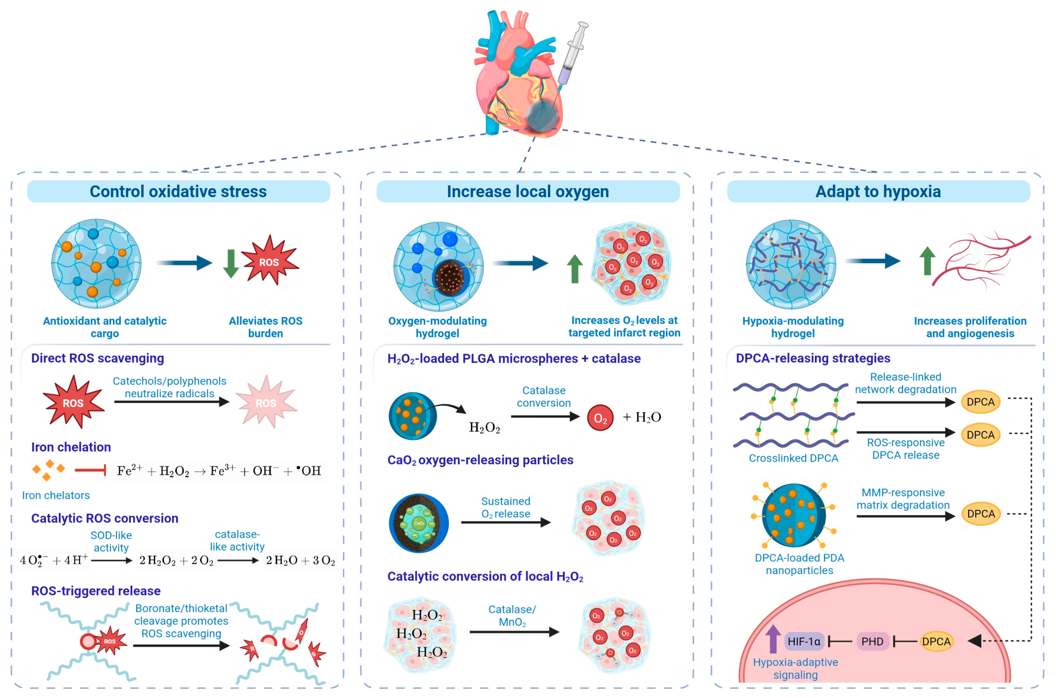
A second strategy is to use hydrogels to locally deliver polyphenolic antioxidants. Wang et al. incorporated epigallocatechin gallate (EGCG)-loaded mesoporous polydopamine (MPDA) nanoparticles into a thermosensitive chitosan hydrogel, providing sustained EGCG release while both EGCG and MPDA contributed to ROS scavenging [54]. Beyond direct antioxidant activity, the system activated the Nrf2/heme oxygenase-1 pathway, thereby reinforcing endogenous antioxidant defenses, and reduced oxidative stress and apoptosis while improving cardiac function after MI in rats. Similarly, Hu et al. encapsulated curcumin-loaded poly(lactic-co-glycolic acid) (PLGA) nanoparticles within a pH- and ROS-responsive hydrogel alongside recombinant humanized collagen type III, coupling antioxidant and anti-inflammatory activity with support for angiogenesis and tissue repair [55]. In rats with MI, this treatment resulted in reduced ROS, apoptosis, and inflammation while limiting infarct expansion and improving cardiac function.
3.3. Enzyme and Nanozyme-Loaded Hydrogels
The ROS scavengers described in Section 3.2 are consumed during redox reactions, thereby limiting the duration of their antioxidant capacity, whereas catalytic systems can continuously convert ROS without being depleted. Native antioxidant enzymes are limited by poor stability, high production costs, and restricted cellular permeability, motivating the development of nanozymes, or nanomaterials with intrinsic enzyme-like activity [56]. Many antioxidant nanozymes combine SOD- and catalase-like activities, allowing O2•− to be sequentially converted to H2O2 and subsequently to water and oxygen. Zhong et al. incorporated the Zn-based metal–organic framework ZIF-8 (zeolitic imidazolate framework-8) into an injectable alginate hydrogel, where it provided both SOD- and catalase-like activity while gradually releasing Zn2+ [57]. In addition to reducing ROS, the presence of Zn2+ promoted reparative macrophage polarization and angiogenesis, and treatment in a rat MI model reduced fibrosis and infarct size while improving ventricular function. This design therefore illustrates how a nanozyme can combine sustained catalytic ROS removal with secondary reparative effects.
Other systems have sought to target the catalytic activity towards mitochondria, a major source of oxidative stress after MI. Xu et al. incorporated tannic acid-coated Mn-substituted Co3O4 nanozymes into an injectable collagen/chondroitin sulfate hydrogel, enabling its mitochondrial localization while limiting potentially harmful metal-mediated pro-oxidant reactions [56]. The system reduced mitochondrial ROS and preserved mitochondrial function, ultimately improving cardiac recovery in the MI rat model. Similarly, Ling et al. incorporated selenium-doped Prussian blue nanozymes into a hyaluronic acid hydrogel, combining SOD- and catalase-like activity with activation of PINK1/Parkin-mediated mitophagy [58]. Since mitophagy clears the damaged mitochondria that generate ROS, while moderate ROS scavenging is itself required to keep the PINK1/Parkin axis functional, the two activities form a closed loop that effectively restores mitochondrial quality control. In a rat MI model, treatment reduced infarct size and fibrosis, while promoting angiogenesis.
3.4. ROS-Cleavable Injectable Hydrogels
The antioxidant systems discussed above scavenge ROS without requiring degradation of the hydrogel network. An alternative strategy incorporates oxidation-labile bonds directly into the material, allowing elevated ROS to trigger network degradation and therapeutic release while simultaneously consuming the oxidant. Arylboronate esters are commonly used for this purpose because they undergo oxidative cleavage in response to H2O2. Zheng et al. developed an injectable hydrogel containing ROS-sensitive boronate ester and pH-sensitive Schiff-base crosslinks, together with puerarin-loaded MnO2 nanozymes and black phosphorus nanosheets [59]. Exposure to the acidic, ROS-rich infarct microenvironment accelerated hydrogel network degradation and payload release, while the MnO2 nanozyme provided additional ROS scavenging and oxygen generation. In an MI mouse model, this multifunctional system reduced infarct expansion and fibrosis while improving vascularization and cardiac function.
Thioketal linkages provide another ROS-cleavable chemistry and have been incorporated into several injectable cardiac hydrogels. For example, Ding et al. synthesized a thioketal-containing hyperbranched polymer within a methacrylated hyaluronic acid network, allowing elevated ROS to drive polymer degradation and radical consumption [60]. Because thioketal cleavage itself does not generate oxygen, catalase was incorporated into the materials to simultaneously decompose H2O2 and alleviate hypoxia. In rats with MI, the combined system reduced oxidative stress, apoptosis, and inflammation while promoting angiogenesis and improving cardiac function. Zhen et al. similarly incorporated a thioketal linkage into a self-assembling peptide hydrogel carrying Apelin-13 (an adipokine involved in several processes, including oxidative stress and energy metabolism [61]) [62]. ROS-dependent cleavage disrupted the nanofibrous network and accelerated therapeutic release, allowing for greater Apelin-13 release within the ROS-rich infarct core while maintaining slower delivery into the border zone. This spatially responsive delivery reduced cardiomyocyte apoptosis and inflammation, promoted vascular repair, and improved cardiac function in a myocardial ischemia–reperfusion mouse model.
There are, however, limitations to ROS-cleavable hydrogels. Unlike catalytic nanozymes, ROS-cleavable groups are consumed during oxidation, making their scavenging capacity dependent on the amount of responsive material present. Because ROS exposure simultaneously promotes network degradation and therapeutic release, the extent of oxidative stress can directly influence hydrogel stability and residence time. For this reason, ROS-cleavable hydrogels have also been combined with complementary therapeutic components, such as catalase or nanozymes, to simultaneously address other metabolic features of the infarct microenvironment [60,63].
4. Oxygen-Generating and Hypoxia-Modulating Injectable Hydrogels
4.1. Biological Rationale
Due to its high energy demand, the myocardium relies almost entirely on aerobic metabolism in the presence of a continuous oxygen supply. Cardiomyocytes devote 30–40% of their cell volume to mitochondria and derive more than 90% of their ATP from oxidative phosphorylation, sustaining a turnover of several kilograms of ATP per day [64]. Because myocardial oxygen extraction is already near-maximal at rest (about 75%, the highest of any organ), the heart has essentially no reserve and can compensate for increased demand only through increased coronary flow [65]. Acute coronary occlusion therefore rapidly deprives the downstream myocardium of oxygen, arresting oxidative phosphorylation and forcing a shift toward anaerobic glycolysis. The consequence of this shift is lactate accumulation, intracellular acidosis, ATP depletion, impaired Na+/K+-ATPase and sarco/endoplasmic reticulum Ca2+-ATPase activity, and progressive cytosolic Ca2+ overload that ultimately contributes to cardiomyocyte death [26,66]. Irreversible injury begins within approximately 20 min of complete occlusion and propagates as a transmural wavefront from sub-endocardium to epicardium over the following hours [67]. The restoration of epicardial coronary blood flow does not necessarily normalize tissue oxygenation. Microvascular obstruction and no-reflow, interstitial edema, and subsequent reductions in functional microvascular density can further impair oxygen delivery by reducing tissue perfusion and increasing the diffusion distance between perfused vessels and metabolically active cells [68]. Regional hypoxia can persist throughout the inflammatory and proliferative phases of infarct healing, overlapping with the deployment window of many therapeutic interventions [69]. This hostile microenvironment has direct consequences for cell-based therapies, for which less than 10% of injected cells are typically retained within the targeted myocardium, while severe ischemia, apoptosis, and loss of cell–matrix attachment leading to anoikis further limit the survival of engrafted cells [70]. Restoring local oxygen availability is thus prioritized for the survival of both resident and transplanted cells, and some hydrogel-based approaches to achieve this are described below (Figure 3).
4.2. Oxygen-Releasing/Generating Hydrogels
Given the central role of oxygen deprivation in ischemic injury, a complementary strategy to ROS scavenging is to restore oxygen availability directly within the infarct. Injectable hydrogels can enable localized and sustained oxygen delivery, with H2O2 and solid peroxides such as CaO2 serving as widely used oxygen-generating sources [71]. For example, Fan et al. established an early in vivo approach by incorporating H2O2-loaded PLGA microspheres and catalase into a thermosensitive injectable hydrogel, allowing controlled H2O2 release and its subsequent conversion to oxygen and water [72]. Sustained oxygen generation improved cardiomyocyte survival and vascularization while reducing inflammation and fibrosis following MI in rats. Subsequent systems have coupled oxygen delivery with regenerative cues. Hassan et al. incorporated CaO2-based oxygen releasing microparticles and stromal cell-derived factor-1α into an adhesive silk-based hydrogel, which simultaneously alleviated hypoxia and promoted progenitor-cell recruitment and neovascularization in an MI rat model [73]. Similarly, Wang et al. combined catalase-mediated oxygen generation with mesenchymal stem cell-derived exosomes in a gelatin methacryloyl/hyaluronic acid hydrogel, improving cellular oxygenation and bioenergetics while reducing cardiomyocyte death and fibrosis and promoting angiogenesis in rats after MI [74].
More recent designs have integrated oxygen generation into multifunctional hydrogels that simultaneously address other features of the infarct microenvironment. In one such approach, Xu et al. incorporated CaO2 and adipose-derived stem cell exosomes into an injectable conductive hydrogel, providing sustained oxygen generation alongside ROS scavenging and electrical support and improving angiogenesis and ventricular recovery following ischemia–reperfusion injury in rats [75]. Jiang et al. similarly encapsulated CaO2 within ZIF-8-based nanoparticles embedded in a conductive, adhesive hydrogel, using the nanoparticle architecture to moderate CaO2 hydrolysis and prolong oxygen generation while incorporating additional antioxidant and electrical functions in an MI rat model [76]. Alternatively, oxygen can be generated from endogenous H2O2 through catalytic components, simultaneously removing a damaging oxidant and supplying oxygen. The catalase-containing hydrogel developed by Ding et al. and the MnO2 nanozyme system reported by Zheng et al. (discussed in Section 3) exploit this dual mechanism and are noted here for their oxygen-generating function [59,60]. Despite encouraging preclinical outcomes, precise control of oxygen delivery remains challenging, as release kinetics must be tailored to tissue oxygen requirements while avoiding the oxidative stress and cellular damage associated with excessive oxygen exposure [71].
4.3. Hypoxia-Responsive and Hypoxia-Signaling-Modulating Hydrogels
Rather than directly restoring oxygen availability in the infarcted myocardium, an alternative strategy is to modulate the cellular response to hypoxia. Hypoxia-inducible factor-1α (HIF-1α) is normally hydroxylated by prolyl hydroxylase domain (PHD) enzymes and rapidly degraded under normoxic conditions, whereas its stabilization during hypoxia activates adaptive pathways involved in angiogenesis, cell survival, and metabolic adaptation [77]. Consequently, several injectable hydrogels have been designed to prolong HIF-1α signaling through sustained delivery of the PHD inhibitor 1,4-dihydrophenonthrolin-4-one-3-carboxylic acid (DPCA). Deng et al. covalently incorporated DPCA into a thermosensitive polymer network in which drug release progressively altered hydrogel hydration and degradation, enabling sustained and tunable DPCA delivery [78]. The fast-releasing formulation produced greater HIF-1α stabilization and more effectively reduced cardiomyocyte apoptosis and fibrosis and preserved ventricular function after MI in rats, demonstrating that the kinetics of HIF-1α modulation can influence therapeutic efficacy.
Other systems are designed to make DPCA release responsive to features of the infarct microenvironment. Wang et al. incorporated a disulfide-linked DPCA prodrug into a methacrylated hyaluronic acid hydrogel, coupling drug release to the presence of ROS, while simultaneously providing antioxidant activity [79]. Sustained HIF-1α stabilization was associated with reduced apoptosis and infarct size in rats and a shift toward a reparative macrophage phenotype. Wei et al. instead incorporated DPCA-loaded polydopamine nanoparticles into an MMP-responsive, conductive alginate/hyaluronic acid hydrogel, allowing protease-mediated degradation to promote DPCA release [80]. This treatment enhanced HIF-1α signaling, reduced infarct expansion and fibrosis, and improved ventricular function in a rat MI model. Despite these studies demonstrating the potential of injectable hydrogels to prolong and regulate HIF-1α activation, their efficacy ultimately depends on matching drug-release kinetics to the evolving infarct microenvironment. This remains challenging, particularly for stimuli-responsive systems, as in vitro release profiles may not accurately predict drug release within the dynamic, protease- and ROS-rich myocardium [19].
5. Mitochondria-Targeting and Bioenergetic Injectable Hydrogels
5.1. Biological Rationale
During ischemia, oxygen deprivation inhibits the electron transport chain and drives Ca2+ overload, while reperfusion converts the succinate accumulated during ischemia into a superoxide burst at complex I within minutes of reflow. The combination of matrix ROS, Ca2+ loading, and pH normalization opens the mitochondrial permeability transition pore, dissipating membrane potential and releasing cytochrome c [81,82]. Damage then becomes self-sustaining as oxidative injury to cardiolipin and respiratory complexes increases electron leak, while mitochondrial DNA released from dysfunctional mitochondria activates the cGAS-STING pathway and inflammasome signaling. The resulting inflammation, along with impaired mitophagy and suppressed mitochondrial biogenesis, allows damaged organelles to persist into the remodeling phase [83,84]. Since adult cardiomyocytes have limited regenerative capacity, preserving, repairing, or replacing the mitochondrial population within the surviving cells would provide therapeutic benefit. Mitochondria, therefore, are not just another source of oxidative stress, but constitute a distinct therapeutic compartment that is not directly addressed by the untargeted approaches that were discussed in Section 4. This has led to the development of carriers such as triphenylphosphonium conjugates, which accumulate in response to membrane potential, and cardiolipin-binding Szeto–Schiller peptides [85,86]. These moieties provide subcellular specificity but not myocardial retention, as systemic delivery gives short residence times and off-target accumulation. Injectable hydrogels provide a complementary level of spatial and temporal control by retaining therapeutic cargo within the infarcted myocardium and permitting sustained or stimuli-responsive release [87,88] for improving mitochondrial function (see Figure 4).
5.2. Mitochondria-Targeted Protective Systems
The most direct approach to mitigating mitochondrial dysfunction after MI is the local delivery of mitochondria-targeted therapeutics. Several injectable hydrogel systems have employed the Szeto–Schiller peptide SS31 (elamipretide), which associates with cardiolipin in the inner mitochondrial membrane and can therefore protect mitochondrial function independently of the membrane-potential-dependent accumulation required by other mitochondria-targeting agents [86]. Zhang et al. developed a pH- and ROS-responsive hyaluronic acid hydrogel, dually crosslinked through imine and boronic ester bonds, containing SS31-functionalized PLGA-thioketal-polyethylene glycol (PEG) nanomicelles loaded with cyclosporine A (CsA) [89]. Their selection of a fluorinated phenylboronic acid allowed boronate ester crosslinking at physiological pH, and the crosslinks additionally consumed ROS in the extracellular microenvironment. Following microenvironment-responsive release, SS31 facilitated mitochondrial targeting while CsA inhibited mPTP opening, reducing mitochondrial ROS production and mitochondria-mediated apoptosis. In a rat ischemia–reperfusion model, this treatment restored mitochondrial energetics and structural integrity and improved cardiac function. Zheng et al. similarly incorporated SS31 into a ROS-responsive hydrogel, in this case a thioketal-crosslinked conductive gelatin network tuned to the stiffness and conductivity of native myocardium, but co-delivered sphingosine-1-phosphate (S1P) to couple mitochondrial protection with angiogenesis. ROS-mediated degradation of the hydrogel promoted the release of SS31/S1P-loaded liposomes, allowing SS31 to suppress mitochondrial oxidative damage while S1P stimulated angiogenic signaling [90]. This resulted in reduced cardiomyocyte injury and fibrosis and improved vascularization and cardiac function after MI in rats.
Figure 4.
Injectable hydrogel strategies that target mitochondrial dysfunction after MI. Ischemia–reperfusion causes calcium overload, mitochondrial ROS production, mPTP opening, and mitochondrial DNA release, contributing to apoptosis and inflammation. Mitochondria-targeted hydrogels have been used to deliver SS31 with CsA, sphingosine-1-phosphate, or the QK peptide to protect mitochondrial function and support angiogenesis. Other systems promote PINK1/Parkin-mediated mitophagy or limit mtDNA-cGAS-STING inflammatory signaling. For mitochondrial transplantation, PEG-gelatin and Q11-MOTS-c hydrogels protect donor mitochondria during delivery; the latter also promotes macrophage mitochondrial uptake and metabolic reprogramming. Image created using BioRender.com. Abbreviations: AMPK, AMP-activated protein kinase; cGAS-STING, cyclic GMP-AMP synthase–stimulator of interferon genes; CsA, cyclosporine A; MI, myocardial infarction; MOTS-c, mitochondrial ORF of the 12S rRNA-c; mPTP, mitochondrial permeability transition pore; mtDNA, mitochondrial DNA; PEG, polyethylene glycol; PINK1, PTEN-induced kinase 1; ROS, reactive oxygen species.
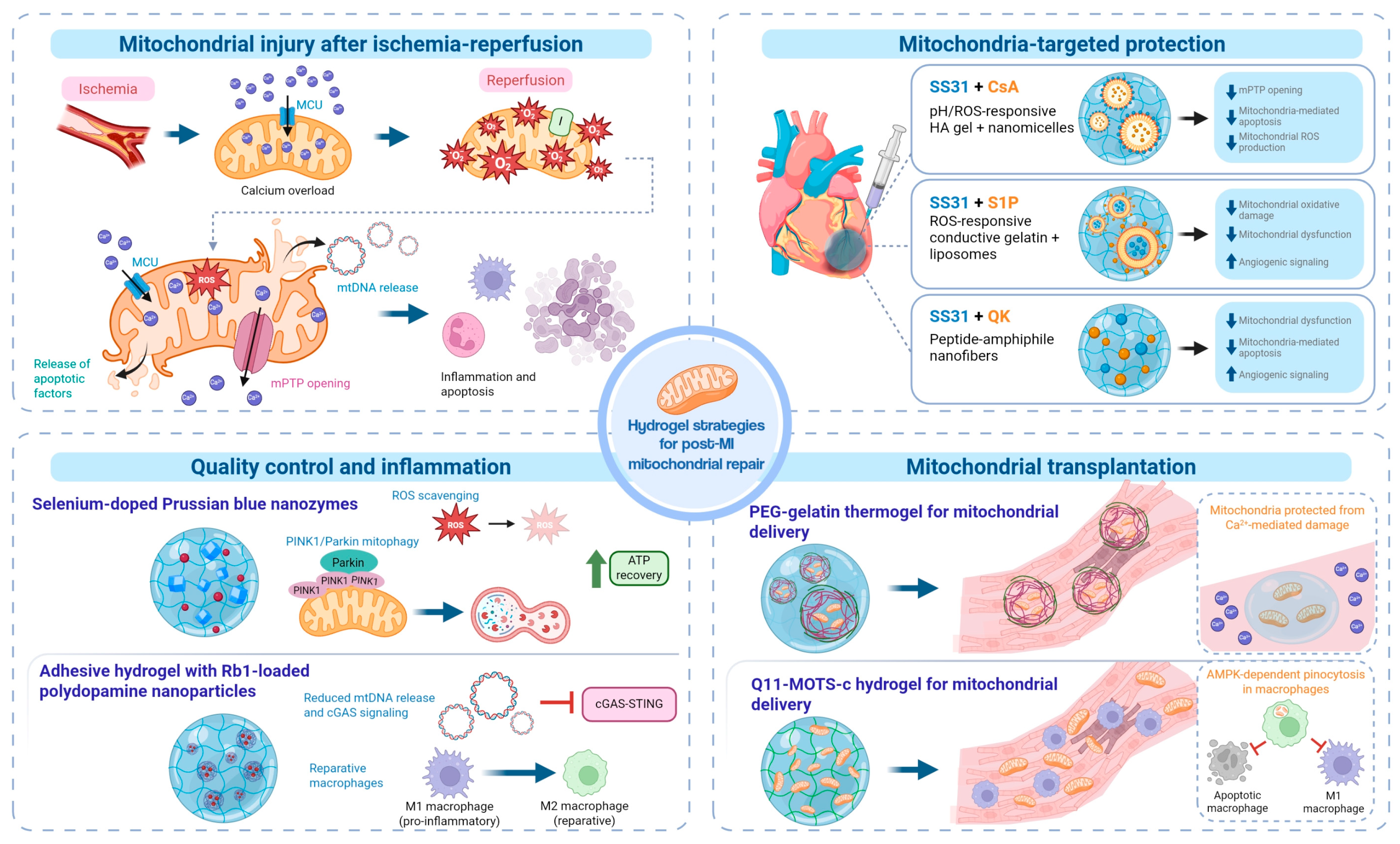
Xu et al. simplified this approach by incorporating SS31 and the vascular endothelial growth factor-mimetic peptide QK directly into self-assembling peptide-amphiphile nanofibers [91]. Rather than relying on stimulus-responsive release, the resulting injectable hydrogel degraded gradually under physiological conditions over approximately one month, providing sustained local SS31-mediated mitochondrial protection alongside QK-mediated angiogenic stimulation. This treatment reduced mitochondrial dysfunction and cardiomyocyte apoptosis, increased vascular regeneration, and improved cardiac structure and function in a mouse MI model.
5.3. Mitochondrial Quality Control and Bioenergetic Restoration
Protecting existing mitochondria alone does not resolve the persistence of dysfunctional organelles, which can continue producing ROS and releasing mitochondrial DNA [92]. To address this problem, Ling et al. developed an injectable hyaluronic acid hydrogel containing selenium-doped Prussian blue nanozymes that were crosslinked through dynamic Schiff base bonds [58]. The nanozymes combined superoxide dismutase- and catalase-like activity with activation of PINK1/Parkin-mediated mitophagy, while selenium doping supported electron transport and the clearance of damaged mitochondria. Because the imine crosslinks destabilized under acidic conditions, nanozyme release was stimulated within the infarct microenvironment. In a rat MI model, this system restored mitochondrial membrane potential and ATP production, reduced infarct size and fibrosis, promoted angiogenesis, and improved cardiac function.
Rather than directly promoting mitochondrial clearance, Zheng et al. targeted the inflammatory response triggered by mitochondrial damage [93]. They developed an injectable, adhesive hydrogel containing ginsenoside Rb1 (Rb1)-loaded polydopamine nanoparticles. Phenylborate ester crosslinks made the hydrogel pH-responsive, self-healing, and adhesive under wet conditions, allowing it to be delivered intramyocardially or applied as an epicardial patch. By scavenging ROS and preserving mitochondrial integrity, the system limited mitochondrial DNA release, while Rb1 suppressed the downstream cGAS-STING signaling and promoted reparative macrophage polarization. The combination of these two delivery systems reduced fibrosis, increased ventricular wall thickness, and improved cardiac function after MI in rats.
5.4. Hydrogel-Mediated Mitochondrial Transplantation
Mitochondrial transplantation aims to restore bioenergetic capacity by delivering functional mitochondria to the injured myocardium. However, a concern with such an approach is the likelihood that extracellular Ca2+ and ROS will damage the transplanted organelles before they are taken up by recipient cells [94]. Ai et al. addressed this challenge using a thermosensitive phase-separated PEG-gelatin hydrogel for mitochondrial transplantation [95]. This hydrogel environment protected mitochondrial integrity and bioenergetic function from Ca2+-mediated damage, while allowing their rapid release after injection. In a rat ischemia–reperfusion model, this treatment increased mitochondrial retention and tissue ATP levels, reduced infarct size and fibrosis, and improved ventricular structure and function compared to free mitochondria.
Li et al. developed a complementary strategy by combining mitochondrial transplantation with MOTS-c, a mitochondria-derived peptide involved in metabolic regulation [96]. Conjugating MOTS-c to the self-assembling peptide, Q11, produced a nonfibrous hydrogel that protected transplanted mitochondria from oxidative- and Ca2+-mediated damage. As the hydrogel degraded, MOTS-c activated AMPK-dependent micropinocytosis in macrophages, increasing mitochondrial uptake and shifting their metabolism from glycolysis toward oxidative phosphorylation. This metabolic shift promoted a more reparative macrophage phenotype. In a rat MI model, the system reduced inflammation and fibrosis while improving ventricular structure and function. However, fluorescence-based tracking could not distinguish intact mitochondria from residual mitochondria material, making the long-term survival and functional integration of transplanted mitochondria difficult to determine.
6. Anti-Ferroptosis Injectable Hydrogels
6.1. Biological Rationale
Targeting ferroptosis after MI requires therapeutics to reach and remain within the injured myocardium, which can be difficult to achieve as typical therapies such as iron chelators and lipophilic antioxidants are poorly suited to systemic administration, even though their molecular targets are well-defined [97]. Conventional iron chelation in post-ischemic tissue is limited by ineffective intracellular chelation in the myocardium, rapid clearance, and systemic toxicity, with hydrophilic chelators, such as deferoxamine, constrained by poor cell membrane permeability [98,99]. Injectable hydrogels provide a way to localize these therapeutics and extend their availability within the infarct and border zone through in situ gelation and controlled degradation at the site of injury. Elevated ROS and local acidification, for example, have been used to trigger material degradation or therapeutic release, allowing the treatment to respond to conditions associated with ongoing tissue injury [100]. These properties make it possible to intervene at different points involved in ferroptotic injury, including reducing the labile iron pool through iron chelation, limiting lipid peroxidation or strengthening endogenous antioxidant defenses, and regulating signaling pathways that influence ferroptotic susceptibility [101].
6.2. Iron-Chelating and Iron-Handling Hydrogels
Since ferroptosis is fundamentally iron-dependent, reducing the labile iron pool provides one of the most direct ways to limit its progression after MI. Deferoxamine (DFO) is a potent iron chelator, but its short plasma half-life and toxicity at higher concentrations can limit its use as a freely administered drug [102]. Gong et al. addressed this using a hyaluronic acid/dopamine hydrogel containing DFO-loaded gelatin microspheres [53]. The hydrogel was designed with acid-labile Schiff base bonds and MMP-2-degradable gelatin, allowing DFO release to respond to conditions within the post-ischemic environment. The material itself also contributed to iron removal, as the carboxyl groups in hyaluronic acid and the catechol groups from dopamine were able to bind and trap Fe2+ and Fe3+, while dopamine provided additional radical-scavenging activity. In a rat ischemia–reperfusion model, the hydrogel treatment reduced myocardial iron accumulation and ferroptosis, including restoration of GPX4 and reduced ACSL4 expression. These changes were accompanied by a smaller infarct size and improved cardiac function.
Lv et al. targeted the same iron pool but instead used a chitosan-DFO nanosponge designed to enter cells and remove intracellular iron [98]. Although the nanosponge is a nanoparticulate system rather than an in situ-forming hydrogel, this study provides relevant complementary context on the use of DFO to target intracellular iron handling. Once internalized, both DFO and functional groups within the chitosan matrix contributed to iron chelation, with greater DFO release occurring under acidic conditions. This nanosponge also increased ferroportin-1 expression without reducing the transmembrane divalent metal transporter 1, suggesting that its effects may extend beyond iron sequestration to promoting its export from the cell. Following intramyocardial injection in a mouse MI model, the nanosponge remained in the heart for at least 7 days, reduced ferroptosis and fibrosis, and improved cardiac function at 28 days. This study shows the potential importance of reaching the intracellular labile iron pool when designing iron-chelating biomaterials. Both studies also reported increased vascular endothelial growth factor expression and vascularization, which may reflect the known effects of DFO on HIF-1α stabilization. In this way, iron-chelating biomaterials may provide benefits beyond ferroptosis inhibition alone. Both materials were administered during the acute response to injury, which is consistent with targeting the early accumulation of labile iron and lipid peroxidation. Whether iron-chelating hydrogels would remain beneficial if delivered later, once the myocardium has entered the reparative and fibrotic phases, has not been established.
6.3. Antioxidant and Gene-Regulatory Anti-Ferroptosis Hydrogels
While the systems in Section 6.2 target ferroptosis by reducing the labile iron pool, other hydrogels have focused further downstream on lipid peroxidation and the pathways that protect against ferroptotic injury. Wang et al. developed an injectable chitosan/PEGDA hydrogel containing ZIF-8 nanoparticles loaded with dihydromyricetin, which improved its retention and allowed for sustained, pH-responsive release within the acidic infarct environment [100]. In cardiomyocytes exposed to ferroptotic stress, the hydrogel reduced intracellular Fe2+ and lipid ROS, preserved mitochondrial structure and increased GPX4 while reducing ACSL4, which was linked to sirtuin 1/Nrf2/GPX4 pathway signaling. Similar effects were seen in rats, with less myocardial iron accumulation and fibrosis alongside improved cardiac function. Therefore, unlike the chelating systems described above, dihydromyricetin acts primarily by strengthening the cell’s own antioxidant defenses instead of directly removing iron.
An alternative approach was taken by Ji et al. who targeted ferroptosis through gene regulation using a dynamic covalent injectable hydrogel containing photothermal nanoparticles and a miR-196c-3p mimic [103]. Near-infrared-II irradiation was used to trigger the local release of the miRNA, which suppressed NOX4, P53 and lysyl oxidase, while restoring GPX4 and ferritin heavy chain 1 and reducing intracellular Fe2+, ROS and lipid peroxidation. The timing of release was also built into the design. Since the decrease in GPX4 expression begins at 30 min after reperfusion and is lowest at 45 min, irradiation was started at 30 min to activate miR-196c-3p mimic release and target this early period of ferroptotic injury. Treatment ultimately reduced cardiomyocyte ferroptosis and improved cardiac function and survival in rats. Although Wang and Ji’s studies regulate different pathways, both show that ferroptosis can be targeted without directly chelating iron, either by restoring endogenous antioxidant defenses or interfering with signals that promote ferroptotic injury. The study by Ji et al. also suggests that the timing during which these therapies are delivered may matter, particularly following reperfusion when ferroptotic activity can change rapidly.
7. Glycolysis- and Carbonyl Stress-Modulating Injectable Hydrogels
7.1. Biological Rationale
Changes in glucose metabolism after MI are difficult to target because the effects of glycolysis vary between different cell populations. Increasing glycolytic activity in cardiomyocytes can support cell-cycle re-entry and cardiac recovery, whereas increased glycolysis in cardiac fibroblasts promotes their activation and contributes to fibrosis [104,105,106]. Glucose metabolism also changes in macrophages throughout the inflammatory response after MI, meaning that the timing and cellular target of metabolic intervention are both important [107]. Instead of systemically increasing or suppressing glycolysis, injectable hydrogels provide a way to deliver metabolic modulators locally within the injured myocardium and maintain their availability over time. This allows glycolytic pathways to be targeted more selectively, including enzymes such as PKM2 that have different roles depending on the cell population involved [108]. Hydrogels can also target the downstream consequences of altered glucose metabolism without directly changing glycolytic activity. In particular, scavenging MG or limiting MG-mediated glycation may reduce cardiac injury while leaving glycolytic flux intact, as reductions in myocardial MG-derived AGEs have previously been associated with improved cardiac function, vascularization, and cardiomyocyte survival after MI [11]. Thus, modifying glucose metabolism itself or limiting the damaging metabolites it produces provide two different approaches for hydrogel-based therapies.
7.2. Glycolysis-Modulating Hydrogels
Glycolytic modulation after MI appears to be highly cell-dependent, and different approaches have been taken in the development of hydrogel-based therapies to target the same metabolic pathway. Wei et al. delivered recombinant human checkpoint kinase 1 (rhCHK1) using an injectable thermosensitive PLGA-PEG-PLGA hydrogel in a porcine ischemia–reperfusion model [109]. Localized rhCHK1 delivery activated pyruvate kinase M2 and increased the glycolytic and pentose phosphate pathway activity in cardiomyocytes, promoting their proliferation while reducing apoptosis and fibrosis. These effects were largely lost following PKM2 knockdown, supporting that PKM2-dependent metabolic reprogramming was an important mediator of the regenerative response. Consistent with this mechanism, the rhCHK1 hydrogel treatment reduced scar formation and preserved cardiac function at 28 days.
Rather than stimulating glycolysis, Tang et al. suppressed it in inflammatory and fibrotic cell populations using a pH-responsive hydrogel containing hyaluronic acid modified ZIF-8 nanoparticles loaded with a shikonin/Cu2+ complex [110]. Shikonin inhibited PKM2-dependent glycolysis in activated macrophages and cardiac fibroblasts, while Cu2+ further disrupted cellular energy metabolism through cuproptosis. This reduced pro-inflammatory macrophage activity and fibroblast activation, weakening the macrophage–fibroblast crosstalk associated with adverse remodeling and improving early cardiac recovery following ischemia–reperfusion injury in mice. Thus, Wei and Tang’s studies reflect the different roles of glycolysis within the injured myocardium, and the consideration needed in developing hydrogel-based therapies that target this process.
7.3. Methylglyoxal-Targeting Hydrogels
The accumulation of MG following MI contributes to both extracellular matrix and cellular damage. Cimenci et al. addressed this using a collagen type I hydrogel containing the MG scavenger fisetin [111]. In a mouse MI model, local intramyocardial delivery of this therapy increased myocardial retention of fisetin and reduced methylglyoxal-derived hydroimidazolone-1 (MG-H1) accumulation, oxidative stress and scar formation while improving vascularization and cardiac function. The unloaded collagen hydrogel also reduced MG-H1 and improved myocardial healing (e.g., reduced oxidative stress and scar size, and improved cardiac function), suggesting that collagen itself may contribute to MG removal.
This was further explored by McLaughlin et al., who found that a hydrogel made from recombinant human collagen type I became rapidly glycated after its injection into the infarcted myocardium, which was accompanied by reductions in MG-H1 within cardiomyocytes and endogenous collagen [112]. These changes were associated with less oxidative damage and cardiomyocyte death and improved regional contractility in MI mice. Using the same mouse model, Guo et al. later demonstrated that pre-glycation of the collagen hydrogel to block its available MG-binding sites before injection largely abolished the functional and structural benefits observed with collagen hydrogel treatment [113]. This provided further evidence that glycation of the biomaterial contributes directly to its therapeutic activity, a strategy summarized in Figure 5. These studies thus show that collagen hydrogels can serve as both a delivery matrix and a local sink for reactive carbonyl species, introducing a material-based approach to reducing the MG burden after MI.
Figure 5.
Collagen hydrogel-mediated reduction of methylglyoxal stress after myocardial infarction. MI, ischemia and reperfusion trigger responses that lead to increased MG accumulation. MG reacts with proteins to form AGEs, contributing to extracellular matrix crosslinking, inflammatory signaling, impaired angiogenesis, and cardiomyocyte injury. An injectable collagen hydrogel contains MG-binding sites that can sequester MG, reducing its bioavailability and AGE formation in the myocardium. This MG trapping leads to several protective responses, reduces adverse ventricular remodeling and improves cardiac function. Image created using BioRender.com. Abbreviations: AGEs, advanced glycation end-products; ECM, extracellular matrix; MG, methylglyoxal; MI, myocardial infarction; RAGE, receptor for advanced glycation end-products; ROS, reactive oxygen species.
Table 1 compares the metabolic targets, the release/response mechanisms and the therapeutic outcomes for some of the hydrogels reviewed herein.
Table 1.
Select examples of injectable hydrogel systems targeting metabolic dysfunction following MI.
8. Translational Challenges and Future Perspectives
Despite promising preclinical results, many challenges still lie between metabolically targeted hydrogels and clinical use. One key limitation of the evidence reviewed is that the mechanistic links between hydrogel treatment, metabolic modulation, and improved cardiac repair were not consistently demonstrated. Changes in metabolic markers alongside improved cardiac outcomes do not directly establish whether the metabolic changes mediated the therapeutic benefit or resulted from a reduction in the degree of tissue injury. This distinction is also quite relevant for multifunctional hydrogels, as their mechanical support and other biological activities may contribute simultaneously to the observed functional outcomes. Future studies combining direct measurements of metabolic activity with targeted inhibition or genetic manipulation of proposed pathways would help establish their contribution to therapeutic efficacy. This understanding could help inform target selection, treatment timing, and biomarkers for evaluating therapeutic responses during clinical translation.
Other limitations include the experimental models used and the technical challenges associated with hydrogel manufacturing, delivery, and long-term safety. Most work to date comes from short-term rodent studies, which do not reflect the larger human heart, older patients, or the risk factors and/or comorbidities these patients usually carry. Future work should incorporate large-animal models, both sexes, and longer follow-up periods, as well as clinically relevant injury models, since permanent ligation and ischemia–reperfusion follow distinct metabolic timelines [114]. The manufacturing and delivery of the materials also pose challenges as polymer-engineered hydrogels are often hard to reproduce from batch to batch, and sterilization, storage, and scale-up can each introduce their own failure points. In terms of administration of the hydrogel, an open-chest injection is not an option for most patients, so catheter-based and intracoronary routes deserve far more attention, which in turn puts specific demands on gelation timing and the use of chemistry that presents non-toxic side effects. The material has to set quickly enough to resist washout, but not so quickly that it clogs the needle or catheter on the way in [115]. Long-term safety is still largely unmapped, particularly in relation to the immune response, the fate of degradation products, and whether some of these materials may be arrhythmogenic. Another challenge is determining when these therapies should be delivered. The post-infarct environment changes considerably during healing, with metabolic, inflammatory, and fibrotic processes becoming more or less active at different stages [116]. To address this, hydrogels can be designed to respond to changes within the infarct environment, such as ROS or pH, so that therapeutic release is better matched to the conditions present in the tissue [90,100]. However, consideration should be given to the possibility that the increased complexity of such hydrogels may make manufacturing and eventual clinical translation more difficult. Omics studies may help identify cell-specific metabolic targets and appropriate treatment windows, but these findings require functional validation and integration into hydrogel designs compatible with clinical manufacturing and delivery.
9. Conclusions
Injectable hydrogels are no longer limited to their traditional role in only providing structural support after MI, as newer scaffolds are designed to directly modify the metabolic environment of the injured myocardium and/or to deliver therapeutics targeted at restoring many different metabolic pathways that are dysregulated post-MI. These metabolic targets include oxidative stress, hypoxia, mitochondrial dysfunction, ferroptosis, dysregulated glycolysis, and reactive carbonyl stress. Hydrogels can improve the local retention of the therapeutics and, in some cases, respond to conditions within the infarct to control their release, providing an approach to target metabolic pathways that can otherwise be difficult to influence locally. Although these systems remain at the preclinical stage, the range of strategies explored so far shows how metabolic modulation can be incorporated into biomaterial-based cardiac repair and provides a foundation for the continued development of injectable hydrogels for treating the infarcted heart.
Author Contributions
S.A. and E.J.S.: conceptualization and writing—original draft; W.L.S. and E.J.S.: supervision and funding; V.G.P., E.I.A., and W.L.S.: writing—review and editing. All authors have read and agreed to the published version of the manuscript.
Funding
Funding was provided by an operating grant from the Canadian Institutes of Health Research (PJT-183748; to E.J.S). E.I.A. and E.J.S. also thank the Natural Sciences and Engineering Research Council of Canada’s support of the INTBIOTECH-CREATE program.
Institutional Review Board Statement
Not applicable.
Informed Consent Statement
Not applicable.
Data Availability Statement
No new data was generated in this review article.
Conflicts of Interest
The authors declare no conflicts of interest.
Abbreviations
The following abbreviations are used in this manuscript:
| ACSL4 | Acyl-CoA synthetase long-chain family member 4 |
| AGEs | Advanced glycation end-products |
| AMPK | Adenosine monophosphate (AMP)-activated protein kinase |
| ATP | Adenosine triphosphate |
| cGAS-STING | Cyclic GMP–AMP synthase–Stimulator of interferon genes |
| CsA | Cyclosporine A |
| DFO | Deferoxamine |
| DPCA | 1,4-dihydrophenonthrolin-4-one-3-carboxylic acid |
| EGCG | Epigallocatechin gallate |
| GLO1 | Glyoxalase-1 |
| GPX4 | Glutathione peroxidase 4 |
| MG | Methylglyoxal |
| MG-H1 | Methylglyoxal-derived hydroimidazolone-1 |
| MI | Myocardial infarction |
| MMP | Matrix metalloproteinase |
| MOTS-c | Mitochondrial ORF of the 12S rRNA Type-C |
| MPDA | Mesoporous polydopamine |
| mPTP | Mitochondrial permeability transition pore |
| NADPH | Nicotinamide adenine dinucleotide phosphate |
| NF-κB | Nuclear factor kappa-light-chain-enhancer of activated B cells |
| NLRP3 | NOD-like receptor family pyrin domain-containing 3 |
| NOX4 | NADPH oxidase 4 |
| Nrf2 | Nuclear factor erythroid 2-related factor 2 |
| PEG | Polyethylene glycol |
| PHD | Prolyl hydroxylase domain |
| PINK1 | Phosphatase and tensin homolog (PTEN)-induced kinase 1 |
| PLGA | Poly(lactic-co-glycolic acid) |
| Rb1 | Ginsenoside Rb1 |
| rhCHK1 | Recombinant human checkpoint kinase 1 |
| ROS | Reactive oxygen species |
| S1P | Sphingosine-1-phosphate |
| SOD | Superoxide dismutase |
| STEMI | ST-elevation myocardial infarction |
| ZIF-8 | Zeolitic imidazolate framework-8 |
References
- Saha, T.; Soliman-Aboumarie, H. Review of Current Management of Myocardial Infarction. J. Clin. Med. 2025, 14, 6241. [Google Scholar] [CrossRef] [Scilit] [PubMed]
- Konstam, M.A.; Kramer, D.G.; Patel, A.R.; Maron, M.S.; Udelson, J.E. Left Ventricular Remodeling in Heart Failure. JACC Cardiovasc. Imaging 2011, 4, 98–108. [Google Scholar] [CrossRef] [Scilit] [PubMed]
- Thomas, K.S.; Puthooran, D.M.; Edpuganti, S.; Reddem, A.L.; Jose, A.; Akula, S.S.M. Reperfusion Injury in STEMI: A Double-Edged Sword. Egypt. Heart J. 2025, 77, 83. [Google Scholar] [CrossRef] [Scilit] [PubMed]
- Frangogiannis, N.G. Pathophysiology of Myocardial Infarction. Compr. Physiol. 2015, 5, 1841–1875. [Google Scholar] [CrossRef] [Scilit]
- Zhang, J.; Guo, Y.; Bai, Y.; Wei, Y. Application of Biomedical Materials in the Diagnosis and Treatment of Myocardial Infarction. J. Nanobiotechnol. 2023, 21, 298. [Google Scholar] [CrossRef] [Scilit] [PubMed]
- Peng, X.; Du, J.; Wang, Y. Metabolic Signatures in Post-Myocardial Infarction Heart Failure, Including Insights into Prediction, Intervention, and Prognosis. Biomed. Pharmacother. 2024, 170, 116079. [Google Scholar] [CrossRef] [Scilit] [PubMed]
- Lopaschuk, G.D.; Karwi, Q.G.; Tian, R.; Wende, A.R.; Abel, E.D. Cardiac Energy Metabolism in Heart Failure. Circ. Res. 2021, 128, 1487–1513. [Google Scholar] [CrossRef] [Scilit] [PubMed]
- Zuurbier, C.J.; Bertrand, L.; Beauloye, C.R.; Andreadou, I.; Ruiz-Meana, M.; Jespersen, N.R.; Kula-Alwar, D.; Prag, H.A.; Botker, H.E.; Dambrova, M.; et al. Cardiac Metabolism as a Driver and Therapeutic Target of Myocardial Infarction. J. Cell Mol. Med. 2020, 24, 5937–5954. [Google Scholar] [CrossRef] [Scilit] [PubMed]
- Leancă, S.A.; Crișu, D.; Petriș, A.O.; Afrăsânie, I.; Genes, A.; Costache, A.D.; Tesloianu, D.N.; Costache, I.I. Left Ventricular Remodeling after Myocardial Infarction: From Physiopathology to Treatment. Life 2022, 12, 1111. [Google Scholar] [CrossRef] [Scilit] [PubMed]
- Mastoor, Y.; Murphy, E.; Roman, B. Mechanisms of Postischemic Cardiac Death and Protection Following Myocardial Injury. J. Clin. Investig. 2025, 135, e184134. [Google Scholar] [CrossRef] [Scilit] [PubMed]
- Blackburn, N.J.R.; Vulesevic, B.; McNeill, B.; Cimenci, C.E.; Ahmadi, A.; Gonzalez-Gomez, M.; Ostojic, A.; Zhong, Z.; Brownlee, M.; Beisswenger, P.J.; et al. Methylglyoxal-Derived Advanced Glycation End Products Contribute to Negative Cardiac Remodeling and Dysfunction Post-Myocardial Infarction. Basic. Res. Cardiol. 2017, 112, 57. [Google Scholar] [CrossRef] [Scilit] [PubMed]
- Temenoff, J.S.; Mikos, A.G. Materials for biomedical applications. In Biomaterials: The Intersection of Biology and Materials Science, 2nd ed.; Temenoff, J.S., Mikos, A.G., Eds.; Pearson: Hoboken, NJ, USA, 2023; Chapter 1; pp. 1–33. [Google Scholar]
- Hasan, A.; Khattab, A.; Islam, M.A.; Hweij, K.A.; Zeitouny, J.; Waters, R.; Sayegh, M.; Hossain, M.M.; Paul, A. Injectable Hydrogels for Cardiac Tissue Repair after Myocardial Infarction. Adv. Sci. 2015, 2, 1500122. [Google Scholar] [CrossRef] [Scilit] [PubMed]
- Tous, E.; Purcell, B.; Ifkovits, J.L.; Burdick, J.A. Injectable Acellular Hydrogels for Cardiac Repair. J. Cardiovasc. Transl. Res. 2011, 4, 528–542. [Google Scholar] [CrossRef] [Scilit] [PubMed]
- Gyles, D.A.; Castro, L.D.; Silva, J.O.C.; Ribeiro-Costa, R.M. A Review of the Designs and Prominent Biomedical Advances of Natural and Synthetic Hydrogel Formulations. Eur. Polym. J. 2017, 88, 373–392. [Google Scholar] [CrossRef] [Scilit]
- Zhang, Z.; Lang, X.; Zhang, Y.; Dong, X.; Han, X.; Xu, C.; Zhuang, X. The Application of Injectable Hydrogels in Myocardial Infarction Repair: Material Design, Biological Functions and Clinical Translation. Int. J. Mol. Sci. 2026, 27, 6464. [Google Scholar] [CrossRef] [Scilit] [PubMed]
- Lee, M.; Kim, M.C.; Lee, J.Y. Nanomaterial-Based Electrically Conductive Hydrogels for Cardiac Tissue Repair. Int. J. Nanomed. 2022, 17, 6181–6201. [Google Scholar] [CrossRef] [Scilit] [PubMed]
- Saeed, S.; Khan, S.U.; Khan, A.J.; Ullah, M.; Gouda, M.M. Recent Advances in Biomimetic Hydrogel Materials for Cardiac Medicine Applications. Discov. Mater. 2025, 5, 239. [Google Scholar] [CrossRef] [Scilit]
- Li, J.; Gao, J.; Zhu, K.; Gao, Y. Research Advances in Intelligent Microenvironment-Responsive Hydrogels for Myocardial Infarction Therapy. APL Bioeng. 2026, 10, 021501. [Google Scholar] [CrossRef] [Scilit] [PubMed]
- El-Husseiny, H.M.; Mady, E.A.; El-Dakroury, W.A.; Doghish, A.S.; Tanaka, R. Stimuli-Responsive Hydrogels: Smart State of-the-Art Platforms for Cardiac Tissue Engineering. Front. Bioeng. Biotechnol. 2023, 11, 1174075. [Google Scholar] [CrossRef] [Scilit] [PubMed]
- Ma, R.; Lu, D.; Yang, Z.; Ji, X.; Tian, R.; Xu, F.; Chen, Y.; Li, C. Emerging Therapy Strategies for Energy Metabolism in Acute Myocardial Infarction. J. Transl. Med. 2025, 23, 1140. [Google Scholar] [CrossRef] [Scilit] [PubMed]
- Tang, C.; Wu, Y.; Wang, M.; Nai, J.; Hu, C.; Wang, Y. Hydrogel Engineering for Myocardial Infarction Repair: From Material Design to Functional Mechanisms and Translational Perspectives. Acta Biomater. 2026, 219, 1–28. [Google Scholar] [CrossRef] [Scilit] [PubMed]
- El-Sehrawy, A.A.M.A.; Alkhayyat, S.; Baig, M.R.; Kubaev, A.; Gajjar, T.B.; Hanumanthayya, M.; Maharana, L.; Kaur, I.; Smerat, A.; Fereydouni, N. Injectable Bioactive Hydrogels as Pharmacological Drug Delivery Platforms for Post-Myocardial Infarction Cardiac Repair: Therapeutic Cargo Engineering, Stimuli-Responsive Release Mechanisms, and Translational Perspectives. Naunyn Schmiedebergs Arch. Pharmacol. 2026. Online ahead of print. [Google Scholar] [CrossRef] [Scilit] [PubMed]
- Yu, C.; Qiu, Y.; Yao, F.; Wang, C.; Li, J. Chemically Programmed Hydrogels for Spatiotemporal Modulation of the Cardiac Pathological Microenvironment. Adv. Mater. 2024, 36, 2404264. [Google Scholar] [CrossRef] [Scilit] [PubMed]
- He, S.; Zhang, Z.; Luo, R.; Jiang, Q.; Yang, L.; Wang, Y. Advances in Injectable Hydrogel Strategies for Heart Failure Treatment. Adv. Healthc. Mater. 2023, 12, 2300029. [Google Scholar] [CrossRef] [Scilit] [PubMed]
- Juricic, S.; Klac, J.; Stojkovic, S.; Beleslin, B.; Tesic, M.; Jovanovic, I.; Banovic, M.; Petrovic, O.; Aleksandric, S.; Vasic, N.; et al. Molecular and Cellular Mechanisms of Myocardial Ischemia and Reperfusion Injury: A Narrative Review. Cells 2026, 15, 265. [Google Scholar] [CrossRef] [Scilit] [PubMed]
- Xiang, M.; Lu, Y.; Xin, L.; Gao, J.; Shang, C.; Jiang, Z.; Lin, H.; Fang, X.; Qu, Y.; Wang, Y.; et al. Role of Oxidative Stress in Reperfusion Following Myocardial Ischemia and Its Treatments. Oxidative Med. Cell. Longev. 2021, 2021, 6614009. [Google Scholar] [CrossRef] [Scilit] [PubMed]
- González-Montero, J.; Brito, R.; Gajardo, A.I.; Rodrigo, R. Myocardial Reperfusion Injury and Oxidative Stress: Therapeutic Opportunities. World J. Cardiol. 2018, 10, 74–86. [Google Scholar] [CrossRef] [Scilit] [PubMed]
- Garcia-Dorado, D.; Ruiz-Meana, M.; Inserte, J.; Rodriguez-Sinovas, A.; Piper, H.M. Calcium-Mediated Cell Death during Myocardial Reperfusion. Cardiovasc. Res. 2012, 94, 168–180. [Google Scholar] [CrossRef] [Scilit] [PubMed]
- Hernandez-Resendiz, S.; Prunier, F.; Girao, H.; Dorn, G.; Hausenloy, D.J. Targeting Mitochondrial Fusion and Fission Proteins for Cardioprotection. J. Cell. Mol. Med. 2020, 24, 6571–6585. [Google Scholar] [CrossRef] [Scilit] [PubMed]
- da Luz Scheffer, D.; Garcia, A.A.; Lee, L.; Mochly-Rosen, D.; Ferreira, J.C.B. Mitochondrial Fusion, Fission, and Mitophagy in Cardiac Diseases: Challenges and Therapeutic Opportunities. Antioxid. Redox Signal 2022, 36, 844–863. [Google Scholar] [CrossRef] [Scilit] [PubMed]
- Liu, Q.; Zhang, D.; Hu, D.; Zhou, X.; Zhou, Y. The Role of Mitochondria in NLRP3 Inflammasome Activation. Mol. Immunol. 2018, 103, 115–124. [Google Scholar] [CrossRef] [Scilit] [PubMed]
- Laukaitiene, J.; Gujyte, G.; Kadusevicius, E. Cardiomyocyte Damage: Ferroptosis Relation to Ischemia-Reperfusion Injury and Future Treatment Options. Int. J. Mol. Sci. 2023, 24, 12846. [Google Scholar] [CrossRef] [Scilit] [PubMed]
- Chen, F.; Kang, R.; Tang, D.; Liu, J. Ferroptosis: Principles and Significance in Health and Disease. J. Hematol. Oncol. 2024, 17, 41. [Google Scholar] [CrossRef] [Scilit] [PubMed]
- Park, T.-J.; Park, J.H.; Lee, G.S.; Lee, J.-Y.; Shin, J.H.; Kim, M.W.; Kim, Y.S.; Kim, J.-Y.; Oh, K.-J.; Han, B.-S.; et al. Quantitative Proteomic Analyses Reveal That GPX4 Downregulation during Myocardial Infarction Contributes to Ferroptosis in Cardiomyocytes. Cell Death Dis. 2019, 10, 835. [Google Scholar] [CrossRef] [Scilit] [PubMed]
- Zi, C.; Zhang, Z.; Zhang, T. Mechanisms of the RGS12-GPX4-ACSL4 Signaling Axis in Regulating Ferroptosis during Myocardial Ischemia-Reperfusion Injury. J. Anesth. Transl. Med. 2026, 5, 5–13. [Google Scholar] [CrossRef] [Scilit] [PubMed]
- Li, W.; Feng, G.; Gauthier, J.M.; Lokshina, I.; Higashikubo, R.; Evans, S.; Liu, X.; Hassan, A.; Tanaka, S.; Cicka, M.; et al. Ferroptotic Cell Death and TLR4/Trif Signaling Initiate Neutrophil Recruitment after Heart Transplantation. J. Clin. Investig. 2019, 129, 2293–2304. [Google Scholar] [CrossRef] [Scilit] [PubMed]
- Komai, K.; Kawasaki, N.K.; Higa, J.K.; Matsui, T. The Role of Ferroptosis in Adverse Left Ventricular Remodeling Following Acute Myocardial Infarction. Cells 2022, 11, 1399. [Google Scholar] [CrossRef] [Scilit] [PubMed]
- Allaman, I.; Bélanger, M.; Magistretti, P.J. Methylglyoxal, the Dark Side of Glycolysis. Front. Neurosci. 2015, 9, 23. [Google Scholar] [CrossRef] [Scilit] [PubMed]
- Morgenstern, J.; Campos Campos, M.; Nawroth, P.; Fleming, T. The Glyoxalase System—New Insights into an Ancient Metabolism. Antioxidants 2020, 9, 939. [Google Scholar] [CrossRef] [Scilit] [PubMed]
- Xue, J.; Ray, R.; Singer, D.; Böhme, D.; Burz, D.S.; Rai, V.; Hoffmann, R.; Shekhtman, A. The Receptor for Advanced Glycation End Products (RAGE) Specifically Recognizes Methylglyoxal-Derived AGEs. Biochemistry 2014, 53, 3327–3335. [Google Scholar] [CrossRef] [Scilit] [PubMed]
- Lai, S.W.T.; Lopez Gonzalez, E.D.J.; Zoukari, T.; Ki, P.; Shuck, S.C. Methylglyoxal and Its Adducts: Induction, Repair, and Association with Disease. Chem. Res. Toxicol. 2022, 35, 1720–1746. [Google Scholar] [CrossRef] [Scilit] [PubMed]
- Prabhu, S.D.; Frangogiannis, N.G. The Biological Basis for Cardiac Repair After Myocardial Infarction. Circ. Res. 2016, 119, 91–112. [Google Scholar] [CrossRef] [Scilit] [PubMed]
- Souza-Neto, F.V.; Islas, F.; Jiménez-González, S.; Luaces, M.; Ramchandani, B.; Romero-Miranda, A.; Delgado-Valero, B.; Roldan-Molina, E.; Saiz-Pardo, M.; Cerón-Nieto, M.Á.; et al. Mitochondrial Oxidative Stress Promotes Cardiac Remodeling in Myocardial Infarction through the Activation of Endoplasmic Reticulum Stress. Antioxidants 2022, 11, 1232. [Google Scholar] [CrossRef] [Scilit] [PubMed]
- Murphy, M.P.; Bayir, H.; Belousov, V.; Chang, C.J.; Davies, K.J.A.; Davies, M.J.; Dick, T.P.; Finkel, T.; Forman, H.J.; Janssen-Heininger, Y.; et al. Guidelines for Measuring Reactive Oxygen Species and Oxidative Damage in Cells and in Vivo. Nat. Metab. 2022, 4, 651–662. [Google Scholar] [CrossRef] [Scilit] [PubMed]
- Sikora, A.; Zielonka, J.; Dębowska, K.; Michalski, R.; Smulik-Izydorczyk, R.; Pięta, J.; Podsiadły, R.; Artelska, A.; Pierzchała, K.; Kalyanaraman, B. Boronate-Based Probes for Biological Oxidants: A Novel Class of Molecular Tools for Redox Biology. Front. Chem. 2020, 8, 580899. [Google Scholar] [CrossRef] [Scilit] [PubMed]
- Kontoghiorghes, G.J. New Approaches and Strategies for the Repurposing of Iron Chelating/Antioxidant Drugs for Diseases of Free Radical Pathology in Medicine. Antioxidants 2025, 14, 982. [Google Scholar] [CrossRef] [Scilit] [PubMed]
- Chen, Q.M. Nrf2 for Cardiac Protection: Pharmacological Options against Oxidative Stress. Trends Pharmacol. Sci. 2021, 42, 729–744. [Google Scholar] [CrossRef] [Scilit] [PubMed]
- Buglak, N.E.; Batrakova, E.V.; Mota, R.; Bahnson, E.S.M. Insights on Localized and Systemic Delivery of Redox-Based Therapeutics. Oxidative Med. Cell. Longev. 2018, 2018, 2468457. [Google Scholar] [CrossRef] [Scilit] [PubMed]
- Chen, H.; Tang, Y.; Ren, P.; Wu, W. The Unmet Promise: A Critical Review of Antioxidant Strategies in Myocardial Ischemia-Reperfusion Injury and the Path towards Precision Medicine. Front. Pharmacol. 2025, 16, 1693441. [Google Scholar] [CrossRef] [Scilit] [PubMed]
- Bolaños-Cardet, J.; Pepió-Tárrega, B.; Saiz-Poseu, J.; López-Moral, A.; Ullah, F.; Yuste, V.J.; Ruiz-Molina, D.; Suárez-García, S. The Redox Properties of Polyphenols and Their Role in ROS Generation for Biomedical Applications. Angew. Chem. Int. Ed. Engl. 2026, 65, e13698. [Google Scholar] [CrossRef] [Scilit] [PubMed]
- Lewandowska, H.; Chen, Z.; Marszałek, K.; Lewandowski, W.; Świsłocka, R. Effective Antioxidants as Plausible Ligands in Chromium(III) Supplementation: How Complexation Modulates Catechol-Based Polyphenols. Molecules 2025, 30, 4467. [Google Scholar] [CrossRef] [Scilit] [PubMed]
- Gong, J.; Qiu, Y.; Yu, C.; Cao, C.; Li, X.; Lu, J.; Zhao, W.; Zhao, Z.; Zhang, H.; Yao, F.; et al. Injectable Hydrogel for Cardiac Repair via Dual Inhibition of Ferroptosis and Oxidative Stress. ACS Appl. Mater. Interfaces 2025, 17, 27906–27922. [Google Scholar] [CrossRef] [Scilit] [PubMed]
- Wang, T.; Wang, Y.; Zhang, Y.; Fang, Z.; Li, S.; Gu, Z.; Ma, Y.; Wang, L.; Han, D.; Wang, C.; et al. Drug-Loaded Mesoporous Polydopamine Nanoparticles in Chitosan Hydrogels Enable Myocardial Infarction Repair through ROS Scavenging and Inhibition of Apoptosis. ACS Appl. Mater. Interfaces 2024, 16, 61551–61564. [Google Scholar] [CrossRef] [Scilit] [PubMed]
- Hu, C.; Liu, W.; Long, L.; Wang, Z.; Zhang, W.; He, S.; Lu, L.; Fan, H.; Yang, L.; Wang, Y. Regeneration of Infarcted Hearts by Myocardial Infarction-Responsive Injectable Hydrogels with Combined Anti-Apoptosis, Anti-Inflammatory and pro-Angiogenesis Properties. Biomaterials 2022, 290, 121849. [Google Scholar] [CrossRef] [Scilit] [PubMed]
- Xu, H.; Zhao, Z.; She, P.; Ren, X.; Li, A.; Li, G.; Wang, Y. Salvaging Myocardial Infarction with Nanoenzyme-Loaded Hydrogels: Targeted Scavenging of Mitochondrial Reactive Oxygen Species. J. Control Release 2024, 375, 788–801. [Google Scholar] [CrossRef] [Scilit] [PubMed]
- Zhong, Y.; Yang, Y.; Xu, Y.; Qian, B.; Huang, S.; Long, Q.; Qi, Z.; He, X.; Zhang, Y.; Li, L.; et al. Design of a Zn-Based Nanozyme Injectable Multifunctional Hydrogel with ROS Scavenging Activity for Myocardial Infarction Therapy. Acta Biomater. 2024, 177, 62–76. [Google Scholar] [CrossRef] [Scilit] [PubMed]
- Ling, H.; Liu, A.; Zhang, Y.; Lin, Q.; Song, C. Biomimetic Selenium-Encrusted Prussian Blue Nanozyme for Myocardial Infarction by Coordinated Enhancement of Mitophagy and Reactive Oxygen Species Scavenging. ACS Nano 2026, 20, 5687–5707. [Google Scholar] [CrossRef] [Scilit] [PubMed]
- Zheng, H.; Feng, L.; Xin, H.; Du, F.; Wang, Y.; Wang, W.; Ma, Q.; Guo, J.; Sun, W. A Microenvironment-Responsive, Injectable, and Conductive Hydrogel Integrated with a Catalytic Nanozyme with ROS Scavenging, Anti-Inflammation, and pro-Angiogenic Capabilities for Synergistic Myocardial Infarction Therapy. Acta Biomater. 2026, 217, 618–636. [Google Scholar] [CrossRef] [Scilit] [PubMed]
- Ding, J.; Yao, Y.; Li, J.; Duan, Y.; Nakkala, J.R.; Feng, X.; Cao, W.; Wang, Y.; Hong, L.; Shen, L.; et al. A Reactive Oxygen Species Scavenging and O2 Generating Injectable Hydrogel for Myocardial Infarction Treatment In Vivo. Small 2020, 16, 2005038. [Google Scholar] [CrossRef] [Scilit] [PubMed]
- Wen, R.; Huang, R.; Xu, K.; Cheng, Y.; Yi, X. Beneficial Effects of Apelin-13 on Metabolic Diseases and Exercise. Front. Endocrinol. 2023, 14, 1285788. [Google Scholar] [CrossRef] [Scilit] [PubMed]
- Zhen, P.; Jiang, Q.; Yu, F.; Xu, X.; Wei, Q.; Liu, X.; Sun, X.; Liang, G.; Tong, J. ROS-Differentiated Release of Apelin-13 from Hydrogel Comprehensively Treats Myocardial Ischemia-Reperfusion Injury. J. Control Release 2025, 379, 609–620. [Google Scholar] [CrossRef] [Scilit] [PubMed]
- Nazir, A.; Nazir, A.; Khan, N.; Jamal, M.S.W.; Shubietah, A.; Naqvi, I.; Rafay Hussain Zaidi, S.; Khan, B.; Ghumman, G.M.; Jamal Chaudhary, A.; et al. Smart Bioactive Hydrogels for Myocardial Infarction Repair: A Multifunctional Approach Integrating Stimuli-Responsive Drug Delivery, Electroconductivity, and Real-Time Biosensing. Ann. Med. Surg. 2025, 88, 401–411. [Google Scholar] [CrossRef] [Scilit] [PubMed]
- Rubio-Tomás, T.; Soler-Botija, C.; Martínez-Estrada, O.; Villena, J.A. Transcriptional Control of Cardiac Energy Metabolism in Health and Disease: Lessons from Animal Models. Biochem. Pharmacol. 2024, 224, 116185. [Google Scholar] [CrossRef] [Scilit] [PubMed]
- Tune, J.D.; Gorman, M.W.; Feigl, E.O. Matching Coronary Blood Flow to Myocardial Oxygen Consumption. J. Appl. Physiol. 2004, 97, 404–415. [Google Scholar] [CrossRef] [Scilit] [PubMed]
- Wu, J.; Liao, M.; Dai, T.; Liu, G.; Zhang, J.; Zhu, Y.; Xu, L.; Zhao, J. The Landscape of SERCA2 in Cardiovascular Diseases: Expression Regulation, Therapeutic Applications, and Emerging Roles. Biomolecules 2026, 16, 247. [Google Scholar] [CrossRef] [Scilit] [PubMed]
- Heusch, G. Myocardial Ischemia/Reperfusion: Translational Pathophysiology of Ischemic Heart Disease. Med 2024, 5, 10–31. [Google Scholar] [CrossRef] [Scilit] [PubMed]
- Welt, F.G.P.; Batchelor, W.; Spears, J.R.; Penna, C.; Pagliaro, P.; Ibanez, B.; Drakos, S.G.; Dangas, G.; Kapur, N.K. Reperfusion Injury in Patients With Acute Myocardial Infarction. J Am Coll Cardiol 2024, 83, 2196–2213. [Google Scholar] [CrossRef] [Scilit] [PubMed]
- Wang, B.; Ansari, R.; Sun, Y.; Postlethwaite, A.E.; Weber, K.T.; Kiani, M.F. The Scar Neovasculature after Myocardial Infarction in Rats. Am. J. Physiol. Heart Circ. Physiol. 2005, 289, H108–H113. [Google Scholar] [CrossRef] [Scilit] [PubMed]
- Li, X.; Tamama, K.; Xie, X.; Guan, J. Improving Cell Engraftment in Cardiac Stem Cell Therapy. Stem Cells Int. 2016, 2016, 7168797. [Google Scholar] [CrossRef] [Scilit] [PubMed]
- Jiang, S.; Zheng, Y.; Xia, H.; Liu, Z.; Rao, S.; Wang, Y.; Sun, H.; Lu, X.; Xie, C. Oxygen-Releasing Hydrogels for Tissue Regeneration. Adv. NanoBiomed Res. 2024, 4, 2300133. [Google Scholar] [CrossRef] [Scilit]
- Fan, Z.; Xu, Z.; Niu, H.; Gao, N.; Guan, Y.; Li, C.; Dang, Y.; Cui, X.; Liu, X.L.; Duan, Y.; et al. An Injectable Oxygen Release System to Augment Cell Survival and Promote Cardiac Repair Following Myocardial Infarction. Sci. Rep. 2018, 8, 1371. [Google Scholar] [CrossRef] [Scilit] [PubMed]
- Hassan, S.; Rezaei, Z.; Luna, E.; Yilmaz-Aykut, D.; Lee, M.C.; Perea, A.M.; Jamaiyar, A.; Bassous, N.; Hirano, M.; Tourk, F.M.; et al. Injectable Self-Oxygenating Cardio-Protective and Tissue Adhesive Silk-Based Hydrogel for Alleviating Ischemia After Mi Injury. Small 2024, 20, 2312261. [Google Scholar] [CrossRef] [Scilit] [PubMed]
- Wang, D.; Zhang, H.; Chen, Y.; He, J.; Zhao, L.; Huang, Y.; Zhao, F.; Jiang, Y.; Fu, S.; Hong, Z. Improving Therapeutic Effects of Exosomes Encapsulated Gelatin Methacryloyl/Hyaluronic Acid Blended and Oxygen Releasing Injectable Hydrogel by Cardiomyocytes Induction and Vascularization in Rat Myocardial Infarction Model. Int. J. Biol. Macromol. 2024, 271, 132412. [Google Scholar] [CrossRef] [Scilit] [PubMed]
- Xu, Z.; Chen, J.; Shu, L.; Hong, W.; Mo, Y.; Wang, J.; Liu, Y.; Wong, W.-T.; Tsang, S.-Y.; Tan, N.; et al. Fabrication of a Conductive, Ionic Liquid Functionalized, Oxygen-Generating Hydrogel Loaded with ADSC-Derived Exosomes to Enhance Angiogenesis and Cardiac Repair after Acute Myocardial Infarction. Theranostics 2026, 16, 1997–2018. [Google Scholar] [CrossRef] [Scilit] [PubMed]
- Jiang, S.; Nong, H.; Song, C.; Zheng, Y.; Yang, P.; Liu, H.; Feng, D.; Han, Y.; Wang, M.; Jiang, L.; et al. Pseudocapacitive Charge-Driven Adhesive-Electrocoupling Hydrogels for Myocardial Infarction Repair via Interstitial Microenvironment Remodeling. Adv. Mater. 2026, e74540. [Google Scholar] [CrossRef] [Scilit] [PubMed]
- Jiang, Y.; Duan, L.-J.; Fong, G.-H. Oxygen-Sensing Mechanisms in Development and Tissue Repair. Development 2021, 148, dev200030. [Google Scholar] [CrossRef] [Scilit] [PubMed]
- Deng, K.; Hua, Y.; Gao, Y.; Zheng, H.; Jiang, Y.; Wang, Y.; Gao, C.; Ren, T.; Zhu, Y. Thermosensitive Hydrogel with Programmable, Self-Regulated HIF-1α Stabilizer Release for Myocardial Infarction Treatment. Adv. Sci. 2024, 11, 2408013. [Google Scholar] [CrossRef] [Scilit] [PubMed]
- Wang, K.; Yao, S.-Y.; Wang, Z.; Shen, L.; Guo, D.-S.; Zhu, Y.; Yang, X.; Yu, Q.; Gao, C. A Sequential Dual Functional Supramolecular Hydrogel with Promoted Drug Release to Scavenge ROS and Stabilize HIF-1α for Myocardial Infarction Treatment. Adv. Healthc. Mater. 2024, 13, 2302940. [Google Scholar] [CrossRef] [Scilit] [PubMed]
- Wei, X.; Chen, S.; Xie, T.; Chen, H.; Jin, X.; Yang, J.; Sahar, S.; Huang, H.; Zhu, S.; Liu, N.; et al. An MMP-Degradable and Conductive Hydrogel to Stabilize HIF-1α for Recovering Cardiac Functions. Theranostics 2022, 12, 127–142. [Google Scholar] [CrossRef] [Scilit] [PubMed]
- Murphy, E.; Steenbergen, C. Mechanisms Underlying Acute Protection from Cardiac Ischemia-Reperfusion Injury. Physiol. Rev. 2008, 88, 581–609. [Google Scholar] [CrossRef] [Scilit] [PubMed]
- Chouchani, E.T.; Pell, V.R.; Gaude, E.; Aksentijević, D.; Sundier, S.Y.; Robb, E.L.; Logan, A.; Nadtochiy, S.M.; Ord, E.N.J.; Smith, A.C.; et al. Ischaemic Accumulation of Succinate Controls Reperfusion Injury through Mitochondrial ROS. Nature 2014, 515, 431–435. [Google Scholar] [CrossRef] [Scilit] [PubMed]
- Paradies, G.; Petrosillo, G.; Pistolese, M.; Di Venosa, N.; Federici, A.; Ruggiero, F.M. Decrease in Mitochondrial Complex I Activity in Ischemic/Reperfused Rat Heart. Circ. Res. 2004, 94, 53–59. [Google Scholar] [CrossRef] [Scilit] [PubMed]
- Kim, J.; Kim, H.-S.; Chung, J.H. Molecular Mechanisms of Mitochondrial DNA Release and Activation of the cGAS-STING Pathway. Exp. Mol. Med. 2023, 55, 510–519. [Google Scholar] [CrossRef] [Scilit] [PubMed]
- Wang, J.Y.; Li, J.Q.; Xiao, Y.M.; Fu, B.; Qin, Z.H. Triphenylphosphonium (TPP)-Based Antioxidants: A New Perspective on Antioxidant Design. ChemMedChem 2020, 15, 404–410. [Google Scholar] [CrossRef] [Scilit] [PubMed]
- Szeto, H.H. First-in-Class Cardiolipin-Protective Compound as a Therapeutic Agent to Restore Mitochondrial Bioenergetics. Br. J. Pharmacol. 2014, 171, 2029–2050. [Google Scholar] [CrossRef] [Scilit] [PubMed]
- Smith, R.A.J.; Hartley, R.C.; Murphy, M.P. Mitochondria-Targeted Small Molecule Therapeutics and Probes. Antioxid. Redox Signal 2011, 15, 3021–3038. [Google Scholar] [CrossRef] [Scilit] [PubMed]
- Zheng, Z.; Tan, Y.; Li, Y.; Liu, Y.; Yi, G.; Yu, C.-Y.; Wei, H. Biotherapeutic-Loaded Injectable Hydrogels as a Synergistic Strategy to Support Myocardial Repair after Myocardial Infarction. J. Control Release 2021, 335, 216–236. [Google Scholar] [CrossRef] [Scilit] [PubMed]
- Zhang, X.; Sun, Y.; Yang, R.; Liu, B.; Liu, Y.; Yang, J.; Liu, W. An Injectable Mitochondria-Targeted Nanodrug Loaded-Hydrogel for Restoring Mitochondrial Function and Hierarchically Attenuating Oxidative Stress to Reduce Myocardial Ischemia-Reperfusion Injury. Biomaterials 2022, 287, 121656. [Google Scholar] [CrossRef] [Scilit] [PubMed]
- Zheng, Z.; Lei, C.; Liu, H.; Jiang, M.; Zhou, Z.; Zhao, Y.; Yu, C.-Y.; Wei, H. A ROS-Responsive Liposomal Composite Hydrogel Integrating Improved Mitochondrial Function and Pro-Angiogenesis for Efficient Treatment of Myocardial Infarction. Adv. Healthc. Mater. 2022, 11, 2200990. [Google Scholar] [CrossRef] [Scilit] [PubMed]
- Xu, Q.; Chen, X.; Sun, S.; Zhao, C.; Shi, L.; Cheng, H.; Liu, Y.; Shi, C.; Ao, X. Vascular Endothelial Growth Factor-Mimetic Peptide and Mitochondria-Targeted Antioxidant-Loaded Hydrogel System Improves Repair of Myocardial Infarction in Mice. J. Biomed. Mater. Res. A 2025, 113, e37924. [Google Scholar] [CrossRef] [Scilit] [PubMed]
- Feng, Y.; Imam Aliagan, A.; Tombo, N.; Bopassa, J.C. Mitofilin Heterozygote Mice Display an Increase in Myocardial Injury and Inflammation after Ischemia/Reperfusion. Antioxidants 2023, 12, 921. [Google Scholar] [CrossRef] [Scilit] [PubMed]
- Zheng, Z.; Sun, J.; Wang, J.; He, S.; Liu, Z.; Xie, J.; Yu, C.-Y.; Wei, H. Enhancing Myocardial Infarction Treatment through Bionic Hydrogel-Mediated Spatial Combination Therapy via mtDNA-STING Crosstalk Modulation. J. Control Release 2024, 371, 570–587. [Google Scholar] [CrossRef] [Scilit] [PubMed]
- Toosky, M.; Kheradvar, A. Assessment of Mitochondrial Viability under Calcium Stress: Insights for Mitochondrial Transplantation. Mitochondrion 2026, 86, 102098. [Google Scholar] [CrossRef] [Scilit] [PubMed]
- Ai, J.; Xiao, Y.; Li, Q.; Xiao, Y.; Li, W.; Deng, J.; Ding, W.; Zhang, R.; Mo, S.; Zeng, Y.; et al. Transplantation of Active Mitochondria Condensed in Liquid–Liquid Phase-Separated Hydrogels Ameliorates Myocardial Ischemia-Reperfusion Injury. Nat. Commun. 2026, 17, 7419. [Google Scholar] [CrossRef] [Scilit] [PubMed]
- Li, H.; Zhang, Y.; Jian, Y.; Fang, F.; Ouyang, W.; Zhuang, D.; Ju, W.; Gao, R.; Gao, Y.; Kang, S.; et al. Mitochondria-Derived Peptide Hydrogel Augments Mitochondrial Transplantation for Promoting Cardiac Repair via Macrophage Metabolic Reprogramming. Bioact. Mater. 2027, 67, 458–476. [Google Scholar] [CrossRef] [Scilit] [PubMed]
- Chen, Y.; Li, X.; Wang, S.; Miao, R.; Zhong, J. Targeting Iron Metabolism and Ferroptosis as Novel Therapeutic Approaches in Cardiovascular Diseases. Nutrients 2023, 15, 591. [Google Scholar] [CrossRef] [Scilit] [PubMed]
- Lv, Q.; Lin, J.; Huang, H.; Ma, B.; Li, W.; Chen, J.; Wang, M.; Wang, X.; Fu, G.; Xiao, Y. Nanosponge for Iron Chelation and Efflux: A Ferroptosis-Inhibiting Approach for Myocardial Infarction Therapy. Adv. Sci. 2024, 11, 2305895. [Google Scholar] [CrossRef] [Scilit] [PubMed]
- Schotman, M.J.G.; Dankers, P.Y.W. Factors Influencing Retention of Injected Biomaterials to Treat Myocardial Infarction. Adv. Mater. Interfaces 2022, 9, 2100942. [Google Scholar] [CrossRef] [Scilit]
- Wang, T.; Zhang, Y.; Chen, S.; Sun, N.; Lu, T.; Wang, L.; Yan, J.; Wang, Y.; Qin, C.; Ma, Y.; et al. A pH-Responsive Chitosan/PEGDA Composite Hydrogel Encapsulating Dihydromyricetin ZIF-8 Nanoparticles for Infarcted Myocardial Repair by Inhibiting Ferroptosis. Mater. Today Adv. 2026, 29, 100741. [Google Scholar] [CrossRef] [Scilit]
- Fang, X.; Ardehali, H.; Min, J.; Wang, F. The Molecular and Metabolic Landscape of Iron and Ferroptosis in Cardiovascular Disease. Nat. Rev. Cardiol. 2023, 20, 7–23. [Google Scholar] [CrossRef] [Scilit] [PubMed]
- Hamilton, J.L.; Imran ul-haq, M.; Abbina, S.; Kalathottukaren, M.T.; Lai, B.F.L.; Hatef, A.; Unniappan, S.; Kizhakkedathu, J.N. In Vivo Efficacy, Toxicity and Biodistribution of Ultra-Long Circulating Desferrioxamine Based Polymeric Iron Chelator. Biomaterials 2016, 102, 58–71. [Google Scholar] [CrossRef] [Scilit] [PubMed]
- Ji, J.; Chen, S.; Yang, Z.; Zhang, R.; Qian, L.; Jiang, Y.; Guo, J.; Wu, Y.; Fan, Q.; Yao, Y.; et al. Delivery of Mir-196c-3p with NIR-II Light-Triggered Gel Attenuates Cardiomyocyte Ferroptosis in Cardiac Ischemia-Reperfusion Injury. Nanomedicine 2023, 47, 102618. [Google Scholar] [CrossRef] [Scilit] [PubMed]
- Magadum, A.; Singh, N.; Kurian, A.A.; Munir, I.; Mehmood, T.; Brown, K.; Sharkar, M.T.K.; Chepurko, E.; Sassi, Y.; Oh, J.G.; et al. Pkm2 Regulates Cardiomyocyte Cell Cycle and Promotes Cardiac Regeneration. Circulation 2020, 141, 1249–1265. [Google Scholar] [CrossRef] [Scilit] [PubMed]
- Wang, F.; Yin, X.; Fan, Y.-M.; Zhang, X.; Ma, C.; Jia, K.; Zhou, W.; Tang, Z.; Qi, L.-W.; Li, J. Upregulation of Glycolytic Enzyme PFKFB3 by Deubiquitinase OTUD4 Promotes Cardiac Fibrosis Post Myocardial Infarction. J. Mol. Med. 2023, 101, 743–756. [Google Scholar] [CrossRef] [Scilit] [PubMed]
- Chen, Z.-T.; Gao, Q.-Y.; Wu, M.-X.; Wang, M.; Sun, R.-L.; Jiang, Y.; Guo, Q.; Guo, D.-C.; Liu, C.-Y.; Chen, S.-X.; et al. Glycolysis Inhibition Alleviates Cardiac Fibrosis After Myocardial Infarction by Suppressing Cardiac Fibroblast Activation. Front. Cardiovasc. Med. 2021, 8, 701745. [Google Scholar] [CrossRef] [Scilit] [PubMed]
- Mouton, A.J.; Aitken, N.M.; Moak, S.P.; do Carmo, J.M.; da Silva, A.A.; Omoto, A.C.M.; Li, X.; Wang, Z.; Schrimpe-Rutledge, A.C.; Codreanu, S.G.; et al. Temporal Changes in Glucose Metabolism Reflect Polarization in Resident and Monocyte-Derived Macrophages after Myocardial Infarction. Front. Cardiovasc. Med. 2023, 10, 1136252. [Google Scholar] [CrossRef] [Scilit] [PubMed]
- Rihan, M.; Zangi, L.; Magadum, A. Pyruvate Kinase M2 Role in Cardiovascular Repair. Cells 2025, 14, 1623. [Google Scholar] [CrossRef] [Scilit] [PubMed]
- Wei, T.; Shan, T.; Wang, H.; Chen, J.; Yang, T.; Zhou, L.; Zhao, D.; Sun, J.; Wang, S.; Gu, L.; et al. Checkpoint Kinase 1 Stimulates Endogenous Cardiomyocyte Renewal and Cardiac Repair by Binding to Pyruvate Kinase Isoform M2 C-Domain and Activating Cardiac Metabolic Reprogramming in a Porcine Model of Myocardial Ischemia/Reperfusion Injury. J. Am. Heart Assoc. 2024, 13, e034805. [Google Scholar] [CrossRef] [Scilit] [PubMed]
- Tang, J.; Li, T.; He, X.; Liu, Y.; Zhou, M.; Wu, M.; Li, Y.; Lan, Y.; Tan, X.; Tian, J.; et al. A Shikonin/Cu2+ Coordination Complex-Encapsulated Hydrogel Drug Depot Alleviates Myocardial Ischemia-Reperfusion Injury by Disrupting Energy Metabolism-Mediated Intercellular Crosstalk. J. Colloid. Interface Sci. 2026, 706, 139580. [Google Scholar] [CrossRef] [Scilit] [PubMed]
- Cimenci, C.E.; Blackburn, N.J.R.; Sedlakova, V.; Pupkaite, J.; Munoz, M.; Rotstein, B.H.; Spiegel, D.A.; Alarcon, E.I.; Suuronen, E.J. Combined Methylglyoxal Scavenger and Collagen Hydrogel Therapy Prevents Adverse Remodeling and Improves Cardiac Function Post-Myocardial Infarction. Adv. Funct. Mater. 2022, 32, 2108630. [Google Scholar] [CrossRef] [Scilit]
- McLaughlin, S.; Sedlakova, V.; Zhang, Q.; McNeill, B.; Smyth, D.; Seymour, R.; Davis, D.R.; Ruel, M.; Brand, M.; Alarcon, E.I.; et al. Recombinant Human Collagen Hydrogel Rapidly Reduces Methylglyoxal Adducts within Cardiomyocytes and Improves Borderzone Contractility after Myocardial Infarction in Mice. Adv. Funct. Mater. 2022, 32, 2204076. [Google Scholar] [CrossRef] [Scilit]
- Guo, X.; Ileri, R.; Ruel, M.; Alarcon, E.I.; Suuronen, E.J. Being a Target for Glycation by Methylglyoxal Contributes to Therapeutic Efficacy of Injectable Collagen Hydrogels Post-Myocardial Infarction. Gels 2025, 12, 18. [Google Scholar] [CrossRef] [Scilit] [PubMed]
- De Villiers, C.; Riley, P.R. Mouse Models of Myocardial Infarction: Comparing Permanent Ligation and Ischaemia-Reperfusion. Dis. Model. Mech. 2020, 13, dmm046565. [Google Scholar] [CrossRef] [Scilit] [PubMed]
- Johnson, T.D.; Christman, K.L. Injectable Hydrogel Therapies and Their Delivery Strategies for Treating Myocardial Infarction. Expert. Opin. Drug Deliv. 2013, 10, 59–72. [Google Scholar] [CrossRef] [Scilit] [PubMed]
- Hilgendorf, I.; Frantz, S.; Frangogiannis, N.G. Repair of the Infarcted Heart: Cellular Effectors, Molecular Mechanisms and Therapeutic Opportunities. Circ. Res. 2024, 134, 1718–1751. [Google Scholar] [CrossRef] [Scilit] [PubMed]
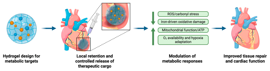
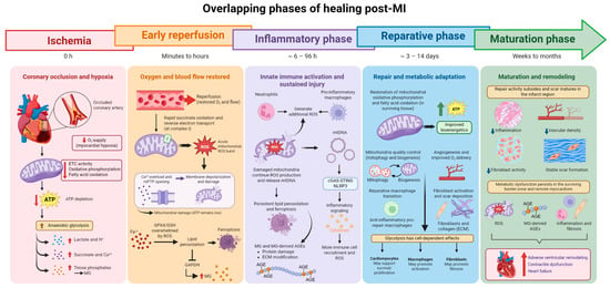
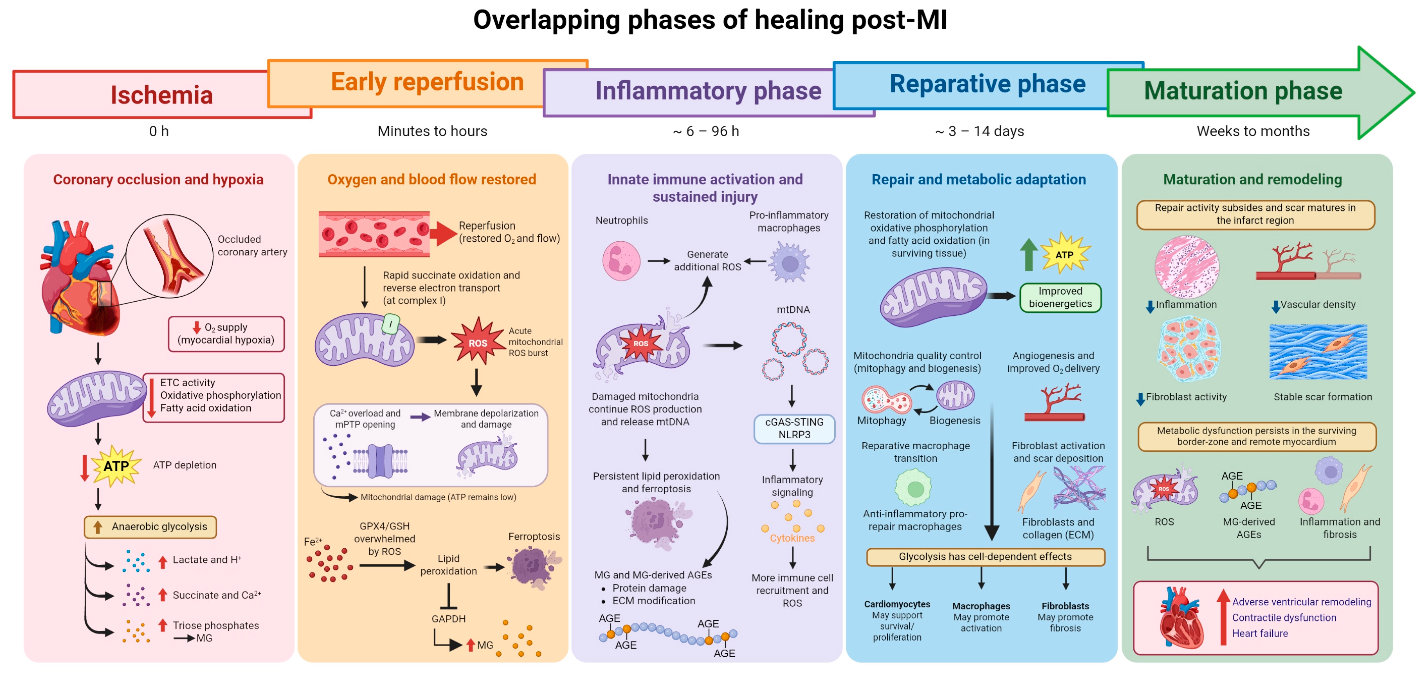
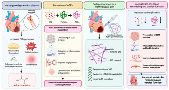
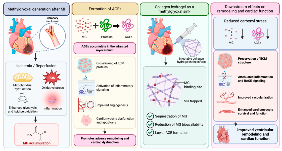

Disclaimer/Publisher’s Note: The statements, opinions and data contained in all publications are solely those of the individual author(s) and contributor(s) and not of MDPI and/or the editor(s). MDPI and/or the editor(s) disclaim responsibility for any injury to people or property resulting from any ideas, methods, instructions or products referred to in the content. |
© 2026 by the authors. Licensee MDPI, Basel, Switzerland. This article is an open access article distributed under the terms and conditions of the Creative Commons Attribution (CC BY) license.